Article

Protective Effects of a Combined Herbal Medicine against Amyotrophic Lateral Sclerosis-Associated Inflammation and Oxidative Stress

Department of KM Science Research, Korea Institute of Oriental Medicine, 1672 Yuseong-daero, Yuseong-gu, Daejeon 34054, Republic of Korea
Appl. Sci. 2024, 14(13), 5386; https://doi.org/10.3390/app14135386
Submission received: 16 May 2024 / Revised: 13 June 2024 / Accepted: 18 June 2024 / Published: 21 June 2024
(This article belongs to the Special Issue The Role of Food Nutrition on Health and Disease)

Abstract

:
Despite extensive studies to identify effective curative drugs for amyotrophic lateral sclerosis (ALS), only riluzole and edaravone have been approved by the Food and Drug Administration. However, these drugs only delay disease progression and exhibit adverse effects, necessitating the development of more effective drugs. Herbal medicines are effective against incurable diseases with various pathogenic factors owing to their low toxicity and presence of multiple components, which target multiple organs. Therefore, we aimed to investigate whether a combined herbal medicine (CHM), comprising Gastrodia elata, Cnidium officinale Makino, and Ostericum koreanum, affects muscle function and motor neuron death in an animal model of ALS. We treated 8-week-old hSOD1G93A mice with 1 mg/g CHM, administered orally once daily for 6 weeks. Muscle function was measured via a footprint test. Biochemical analyses, including immunoblotting, western blotting, and immunohistochemistry, of the muscles (tibialis anterior and gastrocnemius) and spinal cord of hSOD1G93A mice were performed. The CHM treatment improved movement and reduced motor neuron loss in the mouse spinal cord. It also enhanced anti-inflammatory and anti-oxidant activities and regulated autophagy in the mouse muscles and spinal cord. These findings suggest that CHM has multi-active components that effectively target muscles and the spinal cord, delaying disease progression.

1. Introduction

Amyotrophic lateral sclerosis (ALS) is a fatal neurodegenerative disease caused by motor neuron death in the motor cortex, brainstem, and spinal cord (SC) [1]. Clinically, ALS is characterized by muscle dysfunction, which progresses to muscle paralysis and respiratory failure, leading to death. The pathological mechanism of ALS involves excitotoxicity, oxidative damage, protein aggregate formation, autophagy dysfunction, and mitochondrial dysfunction [2]. Despite extensive drug development studies using animal models, only edaravone and riluzole have been approved by the Food and Drug Administration for ALS treatment. However, these drugs exert adverse effects, including gastrointestinal (GI) disturbances, nausea, headache, diarrhea, hepatotoxicity, and asthenia [3]. Moreover, these drugs do not cure the disease; they only preserve motor function and delay disease progression [4]. Therefore, effective treatments without adverse effects are urgently needed for patients with ALS.
Neuroinflammation is a critical pathological feature of neurodegenerative diseases, such as Alzheimer’s disease (AD), Parkinson’s disease (PD), and ALS. Neuroinflammation, which is caused by reactive astrocytes (astrogliosis) and activated microglial cells (microgliosis), induces the release of inflammatory cytokines, leading to neuron death [5]. In addition, hSOD1-mutant mice exhibit neuroinflammatory events and regulation of inflammatory responses involved in disease progression [6]. Antibiotics that reduce inflammation, such as minocycline, increase the survival rate of patients with ALS [7]. However, the inhibition of TNF-α activity blocked the proliferation of microglia, but did not improve the survival rate of mutant hSOD1 mice [8], suggesting that drugs inhibiting microglia proliferation do not sufficiently increase the survival rate or improve motor activity in patients with ALS.
Oxidative stress is also a principal factor that induces neuroinflammation, which in turn, contributes to the pathogenesis of ALS. Oxidative stress, including processes such as lipid peroxidation, oxidation, and DNA damage, is related to inflammation and mitochondrial dysfunction [9]. In addition, neuroinflammation induced by an increase in the number of reactive microglia is related to the induction of oxidative-stress-inducing radical generation, including reactive oxygen species (ROS) and reactive nitrogen species (RNS) [10]. Oxidative stress caused by an increase in the levels of free radicals induces neuroinflammation in neurodegenerative diseases [11]. The levels of 3-nitrotyrosine, a marker of peroxynitrite-mediated damage, and 8-hydroxy-2′-deoxyguanosine, a marker of oxidized DNA, are increased in the motor neurons of the SC of patients with ALS [12,13]. Furthermore, oxidative stress involves other pathological mechanisms such as glutamate excitotoxicity, cytoskeletal dysfunction, and activation of glial cells in ALS animal models and patients with ALS [14]. ROS-induced motor neuron damage causes glutamate transmission in astrocytes, leading to glutamate excitotoxicity [15]. Therefore, anti-oxidants, including selegiline, N-acetylcysteine, and coenzyme 10, have been used for ALS treatment, but an effective outcome has not been achieved in patients with ALS [16]. This finding suggests that treatment of ALS should involve multi-targeting drugs and not a single targeted therapy.
Herbal medicine has been used for treating various diseases, including gastrointestinal tract disorders; cancer; and neuronal disorders, such as AD, PD, and ALS, in East Asian (including Korea, China, and Japan), European, and Western countries [17]. In particular, herbal medicine has been used for incurable and chronic diseases, such as ALS [18] and cancer [19], owing to its minimal adverse effects, such as fever, nausea, vomiting, dizziness, and fatigue [20].
Gastrodia elata has been used for alleviating hand and foot problems, numbness of the limbs, and rheumatic arthralgia [21]. It exhibits anti-oxidant, anti-neuroinflammatory, and anti-aging activities [22,23,24]. Xie et al. reported that G. elata exerted anti-neuroinflammatory effects by reducing the expression of interleukin (IL)-6, IL-8, tumor necrosis factor-α (TNF-α), and IL-1β in a neuropathic pain model [25]. In addition, Zhou et al. have demonstrated that G. elata exerts a neuroprotective effect against corticosterone by inhibiting the endoplasmic reticulum stress-mediated pathway [26]. Cnidium officinale Makino (COM) reportedly demonstrated anti-inflammatory effects by downregulating the secretion of inflammatory cytokines, TNF-α, IL-1β, and IL-6, in the serum of rat endometrial inflammation models [27]. In addition, COM suppresses lipopolysaccharide (LPS)-induced NO, PGE2, IL-1β, IL-6, and TNFα production via AP-1 and NF-κB inactivation in RAW 264.7 macrophages [28] and augments the peripheral angiogenetic system [29]. Ostericum koreanum has been used for treating angina pectoris, headache, and neuralglia in traditional medicine. It exerts various pharmacological effects, including antimicrobial [30], anti-oxidant [31], and anti-inflammatory effects [32]. Specifically, bisabolangelone isolated from O. koreanum shows enhanced anti-inflammatory effects by inhibiting the secretion of proinflammatory cytokines (TNF-α, IL-1β, and IL-6) via the NF-κB and mitogen-activated protein kinase (MAPK) pathways under LPS-induced inflammation [33]. In addition, O. koreanum attenuates inflammation-mediated bone loss by suppressing osteoclast differentiation via regulation of the RANKL/OPG ratio [34]. Therefore, we combined G. elata, COM, and O. koreanum to obtain a combined herbal medicine (CHM) with enhanced anti-inflammatory and anti-oxidant activities and investigated its effects on the muscle function and survival of motor neurons in the muscles and SC of hSOD1G93A mice, a well-known ALS model, with an average lifespan of 129 days, 90-day disease onset, and gain-of-function mutations in SOD [35].

2. Materials and Methods

2.1. Animals

Hemizygous transgenic B6SJL-hSOD1G93A male mice (7 weeks old; weighing 20–23 g) expressing a mutant SOD1 allele with Gly93 → Ala (G93A) substitution were purchased from Jackson Laboratory (JAX:002726; Bar Harbor, ME, USA). The hSOD1G93A mice were maintained by crossing with B6SJL female mice. Genotyping of offspring was performed using polymerase chain reaction (PCR) assays with mouse tail DNA [35]. We used a total of 24 mice (non-transgenic [nTg] mice = 8, hSOD1G93A mice = 16) for the experiments. All mice were maintained in the animal facility under temperature- and light-controlled conditions (20 °C–23 °C, 12 h light/12 h dark cycle), with food and water provided ad libitum. All mice were acclimatized for 7 days prior to drug administration. The Institutional Animal Care Committee of the Korea Institute of Oriental Medicine (KIOM) approved the experimental protocol (#21-003), and all experiments were performed in accordance with the guidelines of KIOM.

2.2. Preparation of CHM Extracts

For preparing CHM extracts, G. elata, COM, and O. koreanum were purchased from Kwangmyungdang Medicinal Herbs Co. (Ulsan, Republic of Korea). These three herbal medicines were combined at a 1:1:1 ratio and extracted according to our previous report [36]. All mice were randomly divided into three groups: nTg (n = 8) and hSOD1G93A (Tg) mice (n = 8), administered distilled water; and Tg mice, treated with CHM (Tg-CHM, n = 8). Tg-CHM mice were treated with 1 mg/g CHM. All mice were orally administered the treatments once daily for 6 weeks.

2.3. Footprint Analysis

Footprint analysis for measuring movement was performed the day after the final treatment day with CHM as previously reported [36].

2.4. Western Blotting

After measuring motor activity with the footprint test, the mice were anesthetized with 20 mg/mL avertin in saline via intraperitoneal injection and tissues were collected. The tissues were stored at −80 °C until use. For western blotting, the gastrocnemius (GC) and tibialis anterior (TA) muscles and SC were homogenized in RIPA buffer (Biosesang, Yongin-si, Gyeonggi-do, Korea), protease inhibitor cocktail, and phosphatase inhibitor cocktail (Thermo Fisher Scientific, Waltham, MA, USA), and centrifuged (13,000× g for 15 min at 4 °C). The supernatants were collected and stored at −80 °C until protein analysis. Proteins were resolved using 8% or 12% SDS-polyacrylamide gel electrophoresis, transferred onto polyvinylidene difluoride (PVDF) membranes, and blocked with 5% milk in TBS (50 mM Tris, 150 mM NaCl) for 1 h. The membranes were then incubated with primary antibodies, namely, anti-CD11b, anti-Prox1, anti-SMAD, anti-oxygenase (HO)1, anti-ferritin, and anti-tubulin antibodies (all 1:1000; Abcam, Cambridge, UK); anti-transferrin and anti-actin antibodies (all 1:1000; Santa Cruz Biotechnology, Dallas, TX, USA); anti-p62, anti-GAPDH, and anti-microtubule-associated protein 1A/1B light chain (LC) 3B antibodies (all 1:1000; Cell Signaling Technology, Danvers, MA, USA); and anti-GFAP antibodies (1:5000; Agilent Technologies, Santa Clara, CA, USA). After washing three times with TBST (50 mM Tris, 150 mM NaCl, 0.1% Tween 20) buffer, the membranes were incubated with horseradish peroxidase-conjugated secondary antibodies (anti-mouse or anti-rabbit [Santa Cruz Biotechnology]). The protein band images were developed with a chemiluminescent horseradish peroxidase substrate (Thermo Fisher Scientific). Quantitative densitometric analysis of the immunoreactive bands was performed using the Image Lab software version 5.2 (Bio-Rad, Hercules, CA, USA), and the quantification of immunoblots was performed using the ImageJ software 1.54J (NIH, Bethesda, MD, USA).

2.5. Immunohistochemistry

The SC tissue was fixed with paraformaldehyde solution at 4 °C. Immunohistochemistry was performed as previously described [36]. Briefly, fixed SC tissues were embedded in paraffin and sectioned at 5 μm thickness using a microtome. The sections were then fixed on glass slides and incubated with the primary antibody anti-cholineacetyltransferase (ChAT) (Thermo Fisher Scientific) at 4 °C overnight. After washing three times with PBS, the slides were incubated with secondary antibodies (anti-mouse IgG or anti-rabbit IgG) for 2 h in the dark. After washing the slides with PBS, the antibody complex was detected using Vectastain ABC (Vector Laboratory Inc., Burlingame, CA, USA) and DAB kits (Vector Laboratory Inc). The immunostained sections were observed with an optical microscope (BX53; Olympus, Tokyo, Japan). Quantification of positive cells with primary antibodies was performed in a blinded manner.

2.6. Statistical Analysis

The results are presented as mean ± standard error of the mean (SEM). Comparisons among the nTg, Tg, and CHM-treated Tg groups were performed using one-way analysis of variance followed by Tukey’s test for multiple comparisons using Prism v.9.0 (GraphPad, La Jolla, CA, USA). Results with p < 0.05 were considered to be statistically significant.

3. Results

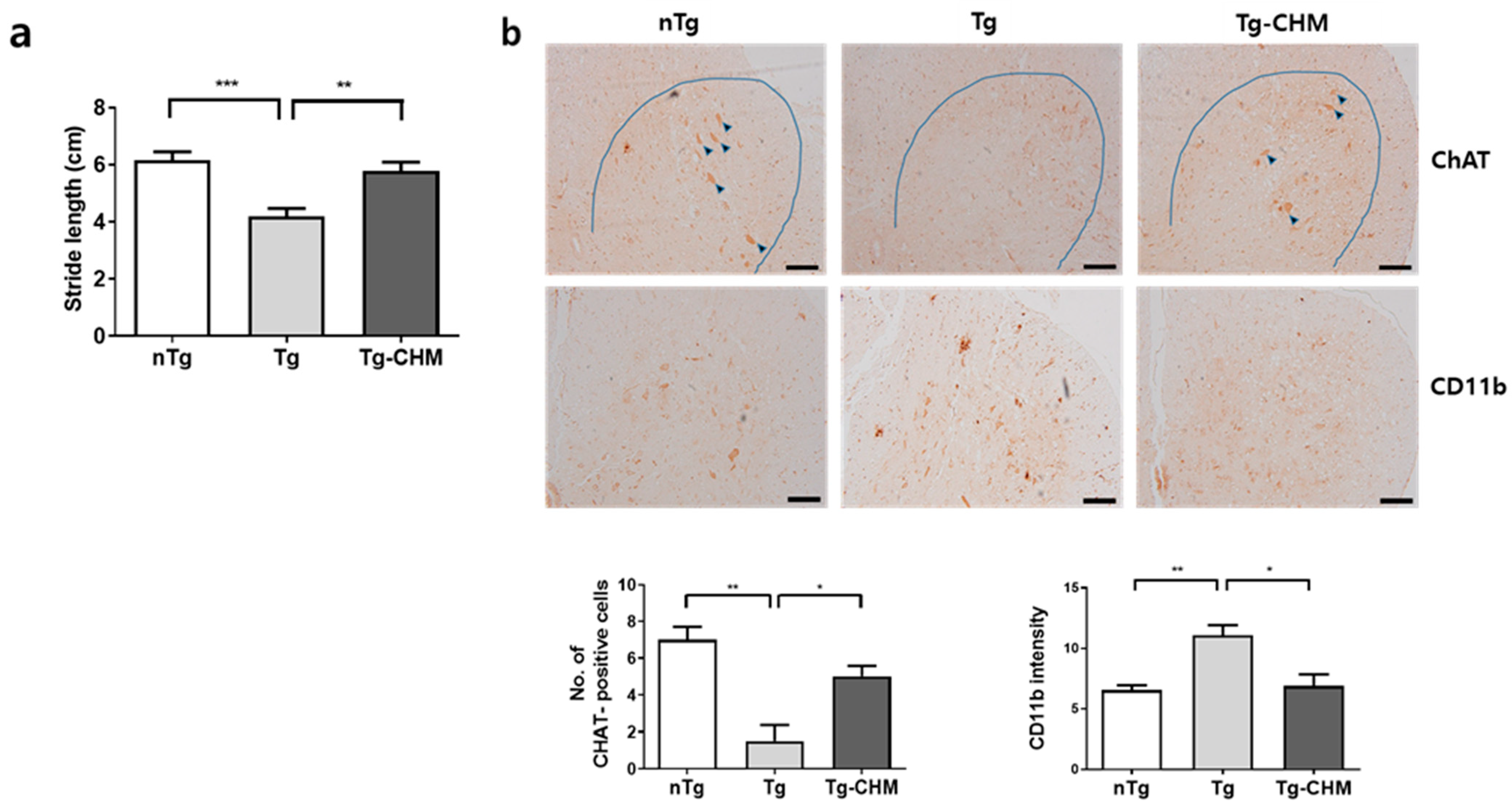
3.1. CHM Treatment Improves Movement in hSOD1G93A Mice

To investigate the effect of CHM on the movement of hSOD1G93A mice, we performed the footprint behavioral test to analyze movement. The stride length of movement of hSOD1G93A mice was reduced by 1.4-fold compared with that of nTg mice (Figure 1a). However, CHM administration to hSOD1G93A mice improved the stride length by 1.3-fold compared with that of the distilled water-treated hSOD1G93A mice, according to the footprint test. Furthermore, the motor neuron survival of CHM-treated hSOD1G93A mice increased compared with that of hSOD1G93A mice, as determined using immunohistochemistry analysis (Figure 1b). In addition, the intensity of positive-CD11b immunostaining was also significantly reduced by the CHM treatment in hSOD1G93A mice (Figure 1b).

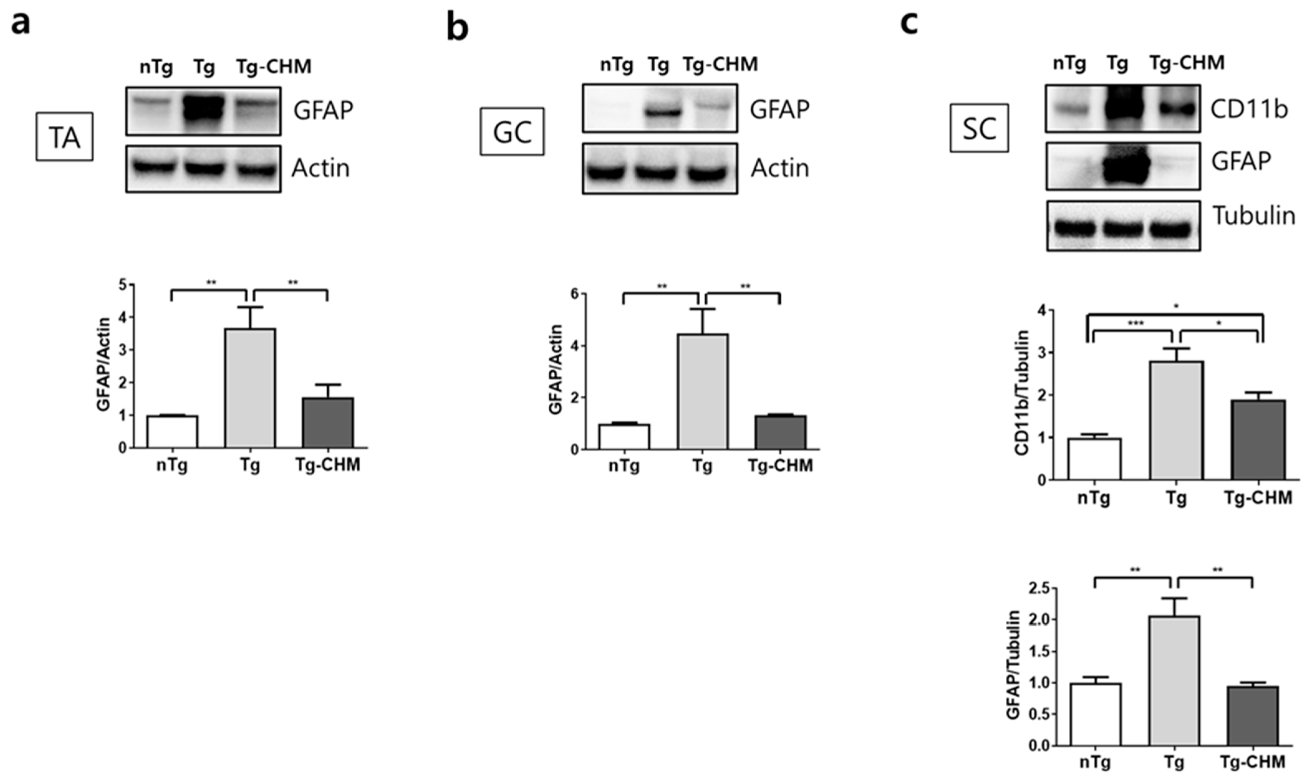
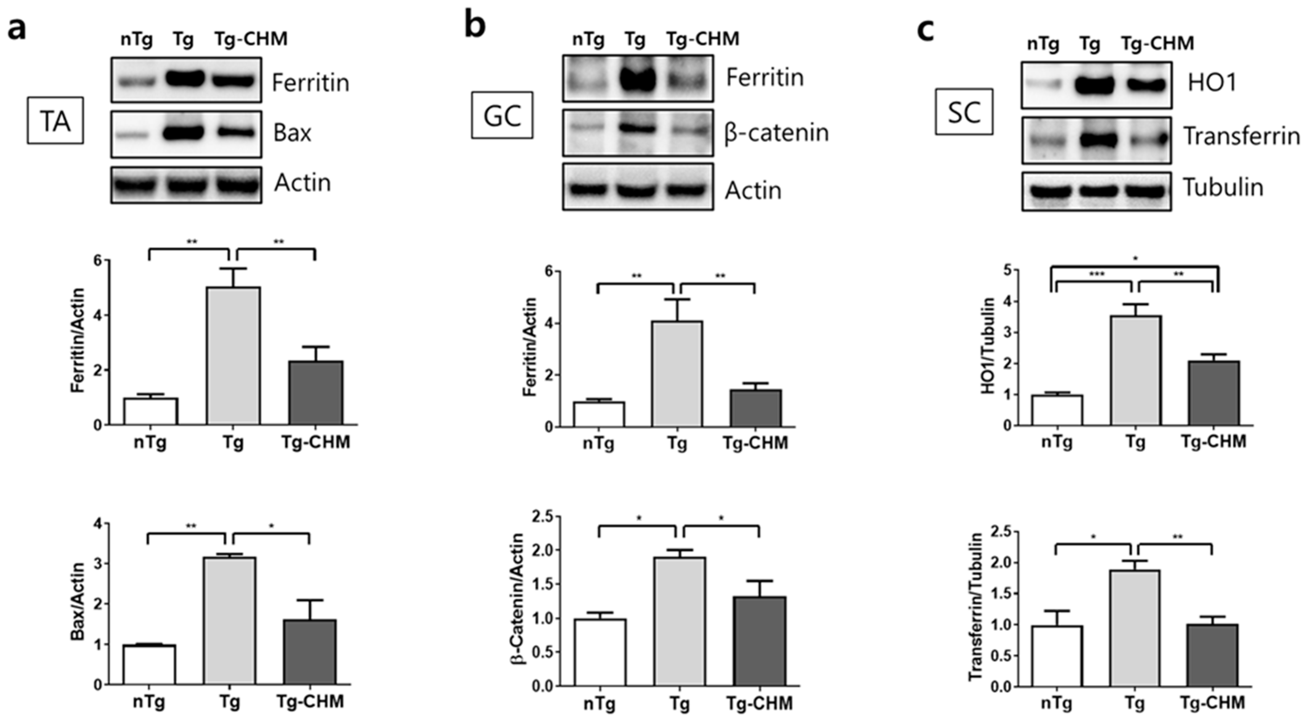
3.2. CHM Treatment Suppresses the Occurrence of Inflammation-Related Events in the Muscles and SC of hSOD1G93A Mice

To determine the molecular mechanism underlying the effects of CHM in hSOD1G93A mice, TA and GC muscles and SC tissues were analyzed using western blotting. Abnormal GFAP expression in muscle indicates the degeneration of neuromuscular junctions (NMJs) and motor neurons in ALS [37]. As shown in Figure 2a,b, the expression of GFAP protein increased by 3.7- and 4.5-fold in the TA and GC muscles of hSOD1G93A mice, respectively, compared with that in the TA and GC muscles of nTg mice. However, CHM treatment reduced the GFAP level by 2.4- and 3.4-fold in the TA and GC muscles of hSOD1G93A mice, respectively, compared with that in the TA and GC muscles of hSOD1G93A mice (Figure 2a,b). In the SC, the expression of the neuroinflammation-related proteins CD11b and GFAP was downregulated by 1.5- and 2.2-fold, respectively, in CHM-treated hSOD1G93A mice compared with that in hSOD1G93A mice.
Inflammation is involved in oxidative stress-related events. Therefore, we examined the effect of CHM on the expression of oxidative stress-related proteins in the muscles (TA and GC muscles) and SC of hSOD1G93A mice. The expression of ferritin was significantly reduced, by 2.2- and 2.8-fold, in the TA and GC muscles of CHM-treated hSOD1G93A mice, respectively, compared with that in the TA and GC muscles of hSOD1G93A mice (Figure 3a,b). In addition, the expression of Bax protein was reduced by 2-fold in the TA muscle and the level of β-catenin protein decreased by 1.4-fold in the GC muscles of CHM-treated hSOD1G93A mice compared with those in hSOD1G93A mice (Figure 3a,b). Furthermore, the expression of the oxidative stress-related proteins HO1 and transferrin was reduced by 1.7- and 1.9-fold, respectively, in the SC of CHM-treated hSOD1G93A mice compared with that in the SC of hSOD1G93A mice (Figure 3c). These results suggest that CHM augments anti-inflammatory and anti-oxidant activities for the protection of motor neurons in the muscles and SC of hSOD1G93A mice.

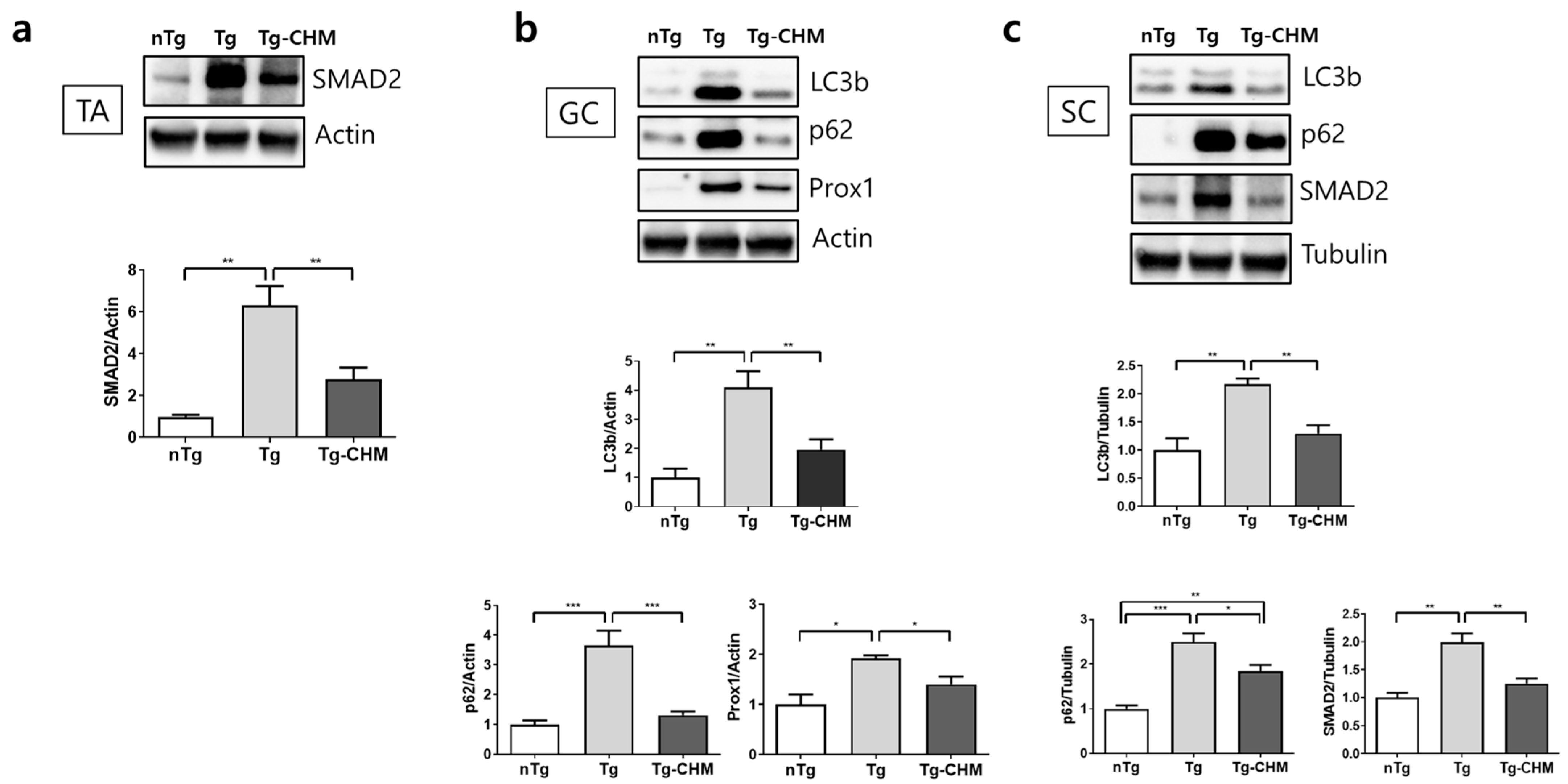
3.3. CHM Treatment Ameliorates Autophagy Dysfunction in the Muscle and SC of hSOD1G93A Mice

Autophagy dysfunction is associated with ALS pathophysiology, and ALS-inducing genes, namely, SQSTM1 (p62), SOD1, optineurin (OPTN), and valosin-containing protein (VCP), are involved in autophagy dysfunction [38]. In the TA muscle of hSOD1G93A mice, the expression of the autophagy dysfunction-related proteins LC3b and p62 significantly increased compared with those in nTg mice, but CHM treatment reduced their levels by 2.1- and 2.8-fold, respectively. In addition, CHM treatment reduced the expression of the muscle atrophy-related proteins Prox1 and SMAD2 by 1.4- and 2.3-fold, respectively, compared to that in hSOD1G93A mice (Figure 4a,b). Furthermore, the expression of LC3b and p62, related to autophagy function, was considerably decreased, by 1.7- and 1.4-fold, respectively, in CHM-treated hSOD1G93A mice compared with those in hSOD1G93A mice (Figure 4c). The increase in the level of SMAD2 was also reduced by 1.6-fold in the SC of CHM-treated hSOD1G93A mice.

4. Discussion

ALS is an incurable neurodegenerative disease that has no effective treatment. ALS has various neuropathological signatures, such as oxidative stress, mitochondrial dysfunction, defective axonal transport, glutamate excitotoxicity, and neuroinflammation. Defects occur that influence free radical accumulation in motor neurons, including the loss of anti-oxidant function of enzymes (e.g., SOD1) and alterations in DNA repair. Herbal medicines are composed of multiple compounds and have multiple effects, such as anti-inflammatory, anti-oxidant, and anticancer effects [39]. In this study, we investigated the effects of a CHM composed of G. elata, COM, and O. koreanum in hSOD1G93A mice to determine whether a multi-targeting herbal medicine can be effective for the treatment of ALS. To investigate whether CHM affects ALS progression, 8-week-old hSOD1G93A mice were treated with CHM for 6 weeks. CHM administration increased the stride length of movement in symptomatic hSOD1G93A mice. Furthermore, the loss of motor neurons was substantially attenuated by CHM treatment via anti-inflammatory effects as well as anti-oxidant and autophagy-regulating effects in the muscles and SC of hSOD1G93A mice.
Inflammation can be acute or chronic. Chronic inflammation induces cancer, autoimmune disorders, metabolic disorders, and neurological diseases, such as AD, PD, and ALS. In the brain, inflammation induces the reactions of glial cells (astrocyte and microglial activation) and an increase in the production of proinflammatory cytokines, such as IL-2 and IL-13 [40], by circulating immune cells, including monocytes, neutrophils, and lymphocytes. In addition, neurodegenerative diseases are characterized by the impairment of inflammatory events. In ALS, the loss of T regulatory cells is involved in neuroinflammation, and the transfer of T regulatory cells extends lifespan [41]. Specifically, activation of microglia and astrocytes induces toxic reactions and motor neuronal cell death in ALS. Inflammation can be caused by ROS produced via the mitochondrial respiratory chain [42]. In NMJs, ROS produced by impaired mitochondria activate intracellular Ca2+ to induce presynaptic decline in an ALS model [43]. In addition, neuroinflammation is accompanied by oxidative stress, which is a fundamental feature of the progression of neurodegenerative diseases. Anti-oxidants from plants or herbal medicines, as preventive medicines, include flavonoids and phenolic compounds, which have no adverse effects and are inexpensive [44].
In addition, flavonoids and phenolic compounds exhibit anti-inflammatory activities by inhibiting signaling pathways, such as NF-κB and MAPK, which induce the production of proinflammatory cytokines [45]. Furthermore, Krishnaiah et al. demonstrated that natural anti-oxidants obtained from consumption of natural foods prevent disease development [46]. In this study, we found that CHM treatment reduced the levels of oxidation-related or inflammatory proteins in the muscles (TA and GC muscles) and SC of hSOD1G93A mice. In addition, CHM administration improved motor function and attenuated motor neuron death in hSOD1G93A mice. These findings suggest that CHM can be useful for alleviating inflammatory diseases and neurodegenerative diseases by enhancing anti-oxidant activity and immune reactions.
Skeletal muscle is targeted to develop therapeutic drugs for ALS because muscle paralysis is a major symptom of ALS, and improved motor function or muscle metabolism enhances the quality of life of patients with ALS. A loss in muscle function is induced by type II fiber dysfunction in muscle diseases, including ALS. Therefore, some researchers have reported that the effects of troponin activators or neurite outgrowth stimulators depend on the type of ALS [47]. In addition, Chaves-Filho et al. showed that glucose metabolism in hSOD1G93A mice is related to fatty acid oxidation [48] and suggested mitochondria abnormality and lipid deposition in an ALS model. Consistently, ranolazine (an inhibitor of fatty acid oxidation) improves glycolysis, including enhancement of muscle strength and ATP level [49]. Patients with SOD1- and C90RF72-induced ALS show abnormal glucose metabolism and hypometabolism or hypermetabolism in different brain regions depending on pathological involvement [50]. Some researchers have reported alterations in the muscle metabolome, particularly abnormal metabolites related to carbohydrate metabolism and the amino acid pathway in patients with ALS [51,52]. This study demonstrated that mitochondria impairment is involved in muscle glycolysis, leading to ATP reduction. Da Cruz et al. demonstrated that improvement in muscle function by the regulation of mitochondrial function with PGC-1a enhanced the survival of hSOD1G93A mice [53]. In our study, we found that CHM administration improved the movement of hSOD1G93A mice. This finding suggests that CHM can regulate muscle metabolism and mitochondrial function by enhancing anti-inflammatory and anti-oxidant activity in hSOD1G93A mice. Therefore, future studies should investigate the effects of CHM on mitochondrial impairment and muscle glycolysis in hSOD1G93A mice.
Autophagy dysfunction is also a principal pathological marker of ALS. Sporadic ALS and familiar ALS are associated with autophagy impairment [54]. The levels of the autophagy-related proteins LC3b and p62 were increased by mTOR, and autophagic vacuoles were detected in motor neurons of hSOD1G93A mice [55,56]. In addition, Zhang et al. showed that mitophagy impairment induced metabolic dysfunction, neurotoxicity, and muscle denervation by SOD1 expression in ALS, and rapamycin attenuated these phenotypes [57]. This study showed that CHM treatment alleviated autophagy dysfunction in the muscle and SC of hSOD1G93A mice. This finding suggests that the regulation of autophagy by CHM could help reduce the amounts of protein inclusions in other neurodegenerative diseases, such as AD and PD. Therefore, the active compounds of CHM need to be identified to develop effective drugs to treat patients with ALS.

5. Conclusions

In the present study, CHM treatment enhanced the anti-inflammatory, anti-oxidant, and autophagy functions in not only the skeletal muscles, including the TA and GC muscles, but also the SC of hSOD1G93A mice. These findings suggest that CHM has multi-active components that effectively target the muscles and SC to delay disease progression. However, further clinical studies should investigate the effects of CHM in patients with ALS to facilitate its development into a therapeutic drug for ALS.

Funding

This research was supported by the Basic Science Research Program through the National Research Foundation of Korea funded by the Ministry of Science, ICT & Future Planning, South Korea, grant number NRF-2020R1A2C2006703, and Korea Institute of Oriental Medicine, grant number KSN2213010.

Institutional Review Board Statement

The animal study protocol was approved by the Institutional Animal Care Committee of the Korea Institute of Oriental Medicine (protocol code #21-003, 1 February 2021).

Informed Consent Statement

Not applicable.

Data Availability Statement

The original contributions presented in the study are included in the article, further inquiries can be directed to the corresponding author.

Conflicts of Interest

The authors declare no conflicts of interest.

References

  1. Swinnen, B.; Robberecht, W. The phenotypic variability of amyotrophic lateral sclerosis. Nat. Rev. Neurol. 2014, 10, 661–670. [Google Scholar] [CrossRef]
  2. Oakes, J.A.; Davies, M.C.; Collins, M.O. TBK1: A new player in ALS linking autophagy and neuroinflammation. Mol. Brain 2017, 10, 5. [Google Scholar] [CrossRef]
  3. Cho, H.; Shukla, S. Role of edaravone as a treatment option for patients with amyotrophic lateral sclerosis. Pharmaceuticals 2020, 14, 29. [Google Scholar] [CrossRef]
  4. Gupta, D.; Vagha, S.; Dhingra, H.; Shirsath, H. Advances in understanding and treating amyotrophic lateral sclerosis (ALS): A comprehensive review. Cureus 2023, 15, e48691. [Google Scholar] [CrossRef]
  5. Kwon, H.S.; Koh, S. Neuroinflammation in neurodegenerative disorders: The roles of microglia and astrocytes. Transl. Neurodegener. 2020, 9, 42. [Google Scholar] [CrossRef]
  6. Chiu, I.M.; Chen, A.; Zheng, Y.; Kosaras, B.; Tsiftsoglou, S.A.; Vartanian, T.K.; Brown, R.H.; Carroll, M.C. T lymphocytes po tentiate endogenous neuroprotective inflammation in a mouse model of ALS. Proc. Natl. Acad. Sci. USA 2008, 105, 17913–17918. [Google Scholar] [CrossRef]
  7. Xiao, Q.; Zhao, W.; Beers, D.R.; Yen, A.A.; Xie, W.; Henkel, J.S.; Appel, S.H. Mutant SOD1(G93A) microglia are more neurotoxic relative to wild-type microglia. J. Neurochem. 2007, 102, 2008–2019. [Google Scholar] [CrossRef]
  8. Gowing, G.; Philips, T.; Van Wijmeersch, B.; Audet, J.N.; Dewil, M.; Van Den Bosch, L.; Billiau, A.D.; Robberecht, W.; Julien, J.P. Ablation of proliferating microglia does not affect motor neuron degeneration in amyotrophic lateral sclerosis caused by mutant superoxide dismutase. J. Neurosci. 2008, 28, 10234–10244. [Google Scholar] [CrossRef]
  9. Olufunmilayo, E.O.; Gerke-Duncan, M.B.; Damian Holsinger, R.M. Oxidative Stress and Antioxidants in neurodegenerative Disorders. Antioxidants 2023, 12, 517. [Google Scholar] [CrossRef]
  10. Mendiola, A.S.; Ryu, J.K.; Bardehle, S.; Meyer-Franke, A.; Ang, K.K.; Wilson, C.; Baeten, K.M.; Hanspers, K.; Merlini, M.; Thomas, S.; et al. Transcriptional profiling and therapeutic targeting of oxidative stress in neuroinflammation. Nat. Immunol. 2020, 21, 513–524. [Google Scholar] [CrossRef]
  11. Ansari, M.A.; Scheff, S.W. Oxidative stress in the progression of Alzheimer disease in the frontal cortex. J. Neuropathol. Exp. Neurol. 2010, 69, 155–167. [Google Scholar] [CrossRef] [PubMed]
  12. Fitzmaurice, P.S.; Shaw, I.C.; Kleiner, H.E.; Miller, R.T.; Monks, T.J.; Lau, S.S.; Mitchell, J.D.; Lynch, P.G. Evidence for DNA damage in amyotrophic lateral sclerosis. Muscle Nerve 1996, 19, 797–798. [Google Scholar] [PubMed]
  13. Abe, K.; Pan, L.H.; Watanabe, M.; Kato, T.; Itoyama, Y. Induction of nitrotyrosine-like immunoreactivity in the lower motor neuron of amyotrophic lateral sclerosis. Neurosci. Lett. 1995, 199, 152–154. [Google Scholar] [CrossRef] [PubMed]
  14. Barber, S.C.; Shaw, P.J. Oxidative stress in ALS: Key role in motor neuron injury and therapeutic target. Free Radic. Biol. Med. 2010, 48, 62–641. [Google Scholar] [CrossRef] [PubMed]
  15. Rao, S.D.; Yin, H.Z.; Weiss, J.H. Disruption of glial glutamate transport by reactive oxygen species produced in motor neurons. J. Neurosci. 2003, 23, 2627–2633. [Google Scholar] [CrossRef]
  16. Orrell, R.W.; Lane, R.J.; Ross, M. Antioxidant treatment for amyotrophic lateral sclerosis/motor neuron disease. Cochrane Database Syst. Rev. 2007, 2007, CD002829. [Google Scholar] [CrossRef] [PubMed]
  17. Moreira, J.; Machado, M.; Dias-Teixeira, M.; Ferraz, R.; Delerue-Matos, C.; Grosso, C. The neuroprotective effect of traditional Chinese medicinal plants—A critical review. Acta Pharm. Sin. B 2023, 13, 3208–3237. [Google Scholar] [CrossRef] [PubMed]
  18. Song, Y.; Jia, Q.; Guan, X.; Kazuo, S.; Liu, J.; Duan, W.; Feng, L.; Zhang, C.; Gao, Y. Herbal medicine for amyotrophic lateral sclerosis: A systematic review and meta-analysis. Front. Pharmacol. 2022, 13, 946548. [Google Scholar] [CrossRef]
  19. Wang, S.; Long, S.; Deng, Z.; Wu, W. Positive role of Chinese herbal medicine in cancer immune regulation. Am. J. Chin. Med. 2020, 48, 1577–1592. [Google Scholar] [CrossRef]
  20. Cai, M.; Yang, E.J. Complementary and alternative medicine for treating amyotrophic lateral sclerosis: A narrative review. Integr. Med. Res. 2019, 8, 234–239. [Google Scholar] [CrossRef]
  21. Wu, Y.N.; Wen, S.H.; Zhang, W.; Yu, S.S.; Yang, K.; Liu, D.; Zhao, C.B.; Sun, J. Gastrodia elata BI.: A comprehensive review of its traditional use, botany, phytochemistry, pharmacology, and pharmacokinetics. Evid. Based Complement. Alternat. Med. 2023, 2023, 5606021. [Google Scholar] [CrossRef] [PubMed]
  22. Liu, J.; Mori, A. Antioxidant and free radical scavenging activities of Gastrodia elata Bl. and Uncaria rhynchophylla (Miq.) Jacks. Neuropharmacology 1992, 31, 1287–1298. [Google Scholar] [CrossRef] [PubMed]
  23. Kim, H.M.; Kwon, J.; Lee, K.; Lee, J.W.; Jang, D.S.; Kwon, H.C. Constituents of Gastrodia elata and their neuroprotective effects in HT22 hippocampal neuronal, R28 retinal cells, and BV2 microglial cells. Plants 2020, 9, 1051. [Google Scholar] [CrossRef] [PubMed]
  24. Glisic, S.; Ivanovic, J.; Ristic, M.; Skala, D. Extraction of sage (Salvia officinalis L.) by supercritical CO2: Kinetic data, chemical composition and selectivity of diterpenes. J. Supercrit. Fluid 2010, 52, 62–70. [Google Scholar] [CrossRef]
  25. Xie, H.; Chen, Y.; Wu, W.; Feng, X.; Du, K. Gastrodia elata blume polysaccharides attenuate vincristine-evoked neuropathic pain through the inhibition of neuroinflammation. Mediators Inflamm. 2021, 2021, 9965081. [Google Scholar] [CrossRef] [PubMed]
  26. Zhou, B.; Tan, J.; Zhang, C.; Wu, Y. Neuroprotective effect of polysaccharides from Gastrodia elata Blume against corticosterone-induced apoptosis in PC12 cells via inhibition of the endoplasmic reticulum stress-mediated pathway. Mol. Med. Rep. 2018, 17, 1182–1190. [Google Scholar] [CrossRef] [PubMed]
  27. Mikamo, H.; Kiwazoe, H.; Sato, Y.; Hayaski, Y.; Izumi, K.; Tamaya, T. Effects of crude herbal ingredients on serum levels of inflammatory cytokines in a rat uterine endometritis model. Curr. Ther. Res. 2000, 60, 105–110. [Google Scholar] [CrossRef]
  28. Lee, W.S.; Shin, J.S.; Jang, D.S.; Lee, K.T. Cnidilide, an alkylphthalide isolated from the roots of Cnidium officinale, suppresses LPS-induced NO, PGE2, IL-1β, IL-6 and TNF-α production by AP-1 and NF-κB inactivation in RAW 264.7 macrophages. Int. Immunopharmacol. 2016, 40, 146–155. [Google Scholar] [CrossRef] [PubMed]
  29. Yang, H.; Jung, D.H.; Lee, H.W. Therapeutic effect of Cnidium officinale Makino extract on ovariectomized hind-limb ischemic mice. Integr. Med. Res. 2019, 8, 107–115. [Google Scholar] [CrossRef] [PubMed]
  30. Shin, S. In vitro effects of essential oils from Ostericum koreanum against antibiotic-resistant Salmonella spp. Arch. Pharm. Res. 2005, 28, 765–769. [Google Scholar] [CrossRef]
  31. Park, Y.J.; Kim, H.J.; Lee, S.J.; Choi, H.Y.; Jin, C.; Lee, Y.S. A new chromone, 11-hydroxy-sec-O-glucosylhamaudol from Ostericum koreanum. Chem. Pharm. Bull. 2007, 55, 1065–1066. [Google Scholar] [CrossRef] [PubMed]
  32. Park, H.J.; Bae, G.S.; Kim, D.Y.; Seo, S.W.; Park, K.B.; Kim, B.J. Inhibitory effect of extract from Ostericum koreanum on LPS-induced proinflammatory cytokines production in RAW 264.7 cells. Korea J. Herbol. 2008, 23, 127–134. [Google Scholar]
  33. Jung, H.W.; Mahesh, R.; Park, J.H.; Boo, Y.C.; Park, K.M.; Park, Y.K. Bisabolangelone isolated from Ostericum koreanum inhibits the production of inflammatory mediators by down-regulation of NF-κB and ERK MAP kinase activity in LPS-stimulated RAW264.7 cells. Int. Immunopharmacol. 2010, 10, 155–162. [Google Scholar] [CrossRef] [PubMed]
  34. Kim, J.Y.; Ahn, S.J.; Baek, J.M.; Yoon, K.H.; Lee, M.S.; Oh, J. Ostericum koreanum reduces LPS-induced bone loss through inhibi tion of osteoclastogenesis. Am. J. Chin. Med. 2015, 43, 495–512. [Google Scholar] [CrossRef] [PubMed]
  35. Gurney, M.E.; Pu, H.; Chiu, A.Y.; Dal Canto, M.C.; Polchow, C.Y.; Alexander, D.D.; Caliendo, J.; Hentati, A.; Kwon, Y.W.; Deng, H.X. Motor neuron degeneration in mice that express a human Cu, Zn superoxide dismutase mutation. Science 1994, 264, 1772–1775. [Google Scholar] [CrossRef] [PubMed]
  36. Lee, S.H.; Cai, M.; Yang, E.J. Anti-inflammatory effects of a novel herbal extract in the muscle and spinal cord of an amyotrophic lateral sclerosis animal model. Front. Neurosci. 2021, 15, 743705. [Google Scholar] [CrossRef] [PubMed]
  37. Liu, J.X.; Brännström, T.; Andersen, P.M.; Pedrosa-Domellöf, F. Distinct changes in synaptic protein composition at neuromas cular junctions of extraocular muscles versus limb muscles of ALS donors. PLoS ONE 2013, 8, e57473. [Google Scholar]
  38. Lee, J.K.; Shin, J.H.; Lee, J.E.; Choi, E.J. Role of autophagy in the pathogenesis of amyotrophic lateral sclerosis. Biochim. Biophys. Acta 2015, 1852, 2517–2524. [Google Scholar] [CrossRef] [PubMed]
  39. Fabricant, D.S.; Farnsworth, N.R. The value of plants used in traditional medicine for drug discovery. Environ. Health Perspect. 2001, 109 (Suppl. S1), 69–75. [Google Scholar]
  40. Leńska-Mieciek, M.; Madetko-Alster, N.; Alster, P.; Królicki, L.; Fiszer, U.; Koziorowski, D. Inflammation in multiple system atrophy. Front. Immunol. 2023, 14, 1214677. [Google Scholar] [CrossRef]
  41. Beers, D.R.; Zhao, W.; Wang, J.; Zhang, X.; Wen, S.; Neal, D.; Thonhoff, J.R.; Alsuliman, A.S.; Shpall, E.J.; Rezvani, K.; et al. ALS patients’ regulatory T lymphocytes are dysfunctional, and correlate with disease progression rate and severity. JCI Insight 2017, 2, e89530. [Google Scholar] [CrossRef] [PubMed]
  42. Reuter, S.; Gupta, S.C.; Chaturvedi, M.M.; Aggarwal, B.B. Oxidative stress, inflammation, and cancer: How are they linked? Free Radic. Biol. Med. 2010, 49, 1603–1616. [Google Scholar] [CrossRef] [PubMed]
  43. Pollari, E.; Goldsteins, G.; Bart, G.; Koistinaho, J.; Giniatullin, R. The role of oxidative stress in degeneration of the neuromuscular junction in amyotrophic lateral sclerosis. Front. Cell. Neurosci. 2014, 8, 131. [Google Scholar] [CrossRef] [PubMed]
  44. Palanisamy, A.; Masoumeh, T.F.; Woan, S.T.; Si-vapragasam, G.; Sharida, F.; Mohd, M.N.; Suresh, K. Role of Antioxidants and Natural Products in Inflammation. Oxid. Med. Cell Longev. 2016, 2016, 5276130. [Google Scholar]
  45. Ravipati, A.S.; Zhang, L.; Koyyalamudi, S.R.; Jeong, S.C.; Reddy, N.; Bartlett, J.; Smith, P.T.; Shanmugam, K.; Münch, G.; Wu, M.J.; et al. Antioxidant and anti-inflammatory activities of selected Chinese medicinal plants and their relation with antioxidant content. BMC Complement. Altern. Med. 2012, 12, 173. [Google Scholar] [CrossRef]
  46. Krishnaiah, D.; Sarbatly, R.; Nithyanandam, R. A review of the antioxidant potential of medicinal plant species. Food Bioprod. Process. 2011, 89, 217–233. [Google Scholar] [CrossRef]
  47. Shefner, J.M.; Andrews, J.A.; Genge, A.; Jackson, C.; Lechtzin, N.; Miller, T.M.; Cockroft, B.M.; Meng, L.; Wei, J.; Wolff, A.A.; et al. A phase 2, double-blind, randomized, dose-ranging trial of reldesemtiv in patients with ALS. Amyotroph. Lateral Scler. Frontotemporal Degener. 2021, 22, 287–299. [Google Scholar] [CrossRef]
  48. Chaves-Filho, A.B.; Pinto, I.F.D.; Dantas, L.S.; Xavier, A.M.; Inague, A.; Faria, R.L.; Medeiros, M.H.G.; Glezer, I.; Yoshinaga, M.Y.; Miyamoto, S. Alterations in lipid metabolism of spinal cord linked to amyotrophic lateral sclerosis. Sci. Rep. 2019, 9, 11642. [Google Scholar] [CrossRef] [PubMed]
  49. Scaricamazza, S.; Salvatori, I.; Giacovazzo, G.; Loeffler, J.P.; Renè, F.; Rosina, M.; Quessada, C.; Proietti, D.; Heil, C.; Rossi, S.; et al. Skeletal-muscle metabolic reprogramming in ALS-SOD1G93A mice predates disease onset and is a promising therapeutic target. iScience 2020, 23, 101087. [Google Scholar] [CrossRef]
  50. De Vocht, J.; Van Weehaeghe, D.; Ombelet, F.; Masrori, P.; Lamaire, N.; Devrome, M.; Van Esch, H.; Moisse, M.; Koole, M.; Dupont, P.; et al. Differences in cerebral glucose metabolism in ALS patients with and without C9orf72 and SOD1 mutations. Cells 2023, 12, 933. [Google Scholar] [CrossRef]
  51. Lanznaster, D.; Bruno, C.; Bourgeais, J.; Emond, P.; Zemmoura, I.; Lefèvre, A.; Reynier, P.; Eymieux, S.; Blanchard, E.; Vourc’h, P.; et al. Metabolic profile and pathological alterations in the muscle of patients with early-stage amyotrophic lateral sclerosis. Biomedicines 2022, 10, 1307. [Google Scholar] [CrossRef] [PubMed]
  52. Goutman, S.A.; Boss, J.; Guo, K.; Alakwaa, F.M.; Patterson, A.; Kim, S.; Savelieff, M.G.; Hur, J.; Feldman, E.L. Untargeted metabolomics yields insight into ALS disease mechanisms. J. Neurol. Neurosurg. Psychiatry 2020, 91, 1329–1338. [Google Scholar] [CrossRef] [PubMed]
  53. Da Cruz, S.; Parone, P.A.; Lopes, V.S.; Lillo, C.; McAlonis-Downes, M.; Lee, S.K.; Vetto, A.P.; Petrosyan, S.; Marsala, M.; Murphy, A.N.; et al. Elevated PGC-1α activity sustains mitochondrial biogenesis and muscle function without extending survival in a mouse model of inherited ALS. Cell Metab. 2012, 1, 778–786. [Google Scholar] [CrossRef] [PubMed]
  54. Pasquali, L.; Lenzi, P.; Biagioni, F.; Siciliano, G.; Fornai, F. Cell to cell spreading of misfolded proteins as a therapeutic target in motor neuron disease. Curr. Med. Chem. 2014, 21, 3508–3534. [Google Scholar] [CrossRef] [PubMed]
  55. Morimoto, N.; Nagai, M.; Ohta, Y.; Miyazaki, K.; Kurata, T.; Morimoto, M.; Murakami, T.; Takehisa, Y.; Ikeda, Y.; Kamiya, T.; et al. Increased autophagy in transgenic mice with a G93A mutant SOD1 gene. Brain Res. 2007, 1167, 112–117. [Google Scholar] [CrossRef] [PubMed]
  56. Gal, J.; Ström, A.L.; Kilty, R.; Zhang, F.; Zhu, H. p62 accumulates and enhances aggregate formation in model systems of familial amyotrophic lateral sclerosis. J. Biol. Chem. 2007, 282, 11068–11077. [Google Scholar] [CrossRef]
  57. Zhang, X.; Li, L.; Chen, S.; Yang, D.; Wang, Y.; Zhang, X.; Wang, Z.; Le, W. Rapamycin treatment augments motor neuron degeneration in SOD1(G93A) mouse model of amyotrophic lateral sclerosis. Autophagy 2011, 7, 412–425. [Google Scholar] [CrossRef]
Figure 1. Treatment with combined herbal medicine (CHM) comprising Gastrodia elata, Cnidium officinale Makino (COM), and Ostericum koreanum improves motor function and prevents motor neuron death in hSOD1G93A mice. (a) Footprint test (n = 8). (b) Representative image of anti-ChAT-positive immunohistochemical analysis. nTg (n = 4), Tg (n = 4), and Tg-CHM (n = 3). Data are presented as mean ± SEM. * p < 0.05, ** p < 0.01, and *** p < 0.001 compared with the control group. Scale bar = 2 μm. DW-treated nTg mice (nTg), hSOD1G93A Tg mice (Tg), CHM-treated Tg mice (Tg-CHM).
Applsci 14 05386 g001
Figure 2. CHM treatment reduces the levels of inflammatory proteins in the tibialis anterior (TA) and gastrocnemius (GC) muscles and spinal cord (SC) of hSOD1G93A mice. (a,b) Western blots showing the changes in GFAP protein level in the (a) TA and (b) GC muscles of mice from the nTg, Tg, and Tg-CHM groups. (c) Representative immunoblots of CD11b and GFAP proteins in the SC of nTg, Tg, and Tg-CHM mice. Quantification of immunoblots showed the expression ratio of GFAP and CD11 in the TA and GC muscles and SC of each group compared with that in the nTg group. Actin or tubulin was used as a loading control to normalize the levels of the target proteins for the western blot analyses. nTg (n = 4), Tg (n = 4), and Tg-CHM (n = 3). Data are normalized relative to those of the control group and presented as mean ± SEM. * p < 0.05, ** p < 0.01, and *** p < 0.001 compared with the control group. DW-treated nTg mice (nTg), hSOD1G93A Tg mice (Tg), CHM-treated Tg mice (Tg-CHM).
Applsci 14 05386 g002
Figure 3. CHM treatment increases the levels of anti-oxidation-related proteins in the tibialis anterior (TA) and gastrocnemius (GC) muscles and spinal cord (SC) of hSOD1G93A mice. (a) The expression of ferritin and Bax was reduced following CHM treatment in the TA muscles (n = 3–4/group). (b) Immunoblot of ferritin and β-catenin in the GC muscles of nTg, Tg, and Tg-CHM mice. (c) The expression of oxidation-related proteins, HO1 and transferrin, was downregulated in the SC of nTg, Tg, and Tg-CHM mice. Quantification of protein bands in western blots was performed using ImageJ. Actin or tubulin was used as a loading control to normalize the levels of the target proteins for the western blot analyses. nTg (n = 4), Tg (n = 4), and Tg-CHM (n = 3). Data are normalized relative to those of the control group and presented as mean ± SEM. * p < 0.05, ** p < 0.01, and *** p < 0.001 compared with the control group. DW-treated nTg mice (nTg), hSOD1G93A Tg mice (Tg), CHM-treated Tg mice (Tg-CHM).
Applsci 14 05386 g003
Figure 4. CHM treatment regulates autophagy function in the tibialis anterior (TA) and gastrocnemius (GC) muscles and spinal cord (SC) of hSOD1G93A mice. (a) The expression of the muscle atrophy-related protein SMAD2 was decreased in the Tg-CHM group (n = 3–4/group). CHM treatment increased the expression of autophagy-related proteins (LC3b and p62) in the (b) GC muscles and (c) SC of hSOD1G93A mice. CHM treatment reduced the Prox1 and SMAD2 levels in the (b) GC muscles and (c) SC of hSOD1G93A mice. Actin or tubulin was used as a loading control to normalize the levels of the target proteins for the western blot analyses. nTg (n = 4), Tg (n = 4), and Tg-CHM (n = 3). Data are normalized relative to those of the control groups and presented as mean ± SEM. * p < 0.05, ** p < 0.01, and *** p < 0.001 compared with the control group. DW-treated nTg mice (nTg), hSOD1G93A Tg mice (Tg), CHM-treated Tg mice (Tg-CHM).
Applsci 14 05386 g004
Disclaimer/Publisher’s Note: The statements, opinions and data contained in all publications are solely those of the individual author(s) and contributor(s) and not of MDPI and/or the editor(s). MDPI and/or the editor(s) disclaim responsibility for any injury to people or property resulting from any ideas, methods, instructions or products referred to in the content.

