Article

Sport-Specific Abdominal Wall Muscle Differences: A Comparative Study of Soccer and Basketball Players Using Ultrasonography

by
Carlos Romero-Morales
1,
Jorge Hugo Villafañe
1,
Unai Torres
1,
Diego Miñambres-Martín
1,
Helios Pareja-Galeano
2,
Isabel Rodríguez-Costa
3 and
Sergio L. Jiménez-Sáiz
4,*
1
Faculty of Sport Sciences, Universidad Europea de Madrid, 28670 Madrid, Spain
2
Department of Physical Education, Sport and Human Movement, Universidad Autónoma de Madrid, 28049 Madrid, Spain
3
Department of Nursing and Physical Therapy, Faculty of Medicine and Health Sciences, Universidad de Alcalá, 28871 Madrid, Spain
4
Sport Sciences Research Centre, Universidad Rey Juan Carlos, 28942 Madrid, Spain
*
Author to whom correspondence should be addressed.
Appl. Sci. 2024, 14(13), 5742; https://doi.org/10.3390/app14135742
Submission received: 5 June 2024 / Revised: 26 June 2024 / Accepted: 27 June 2024 / Published: 1 July 2024
(This article belongs to the Special Issue Biomechanics and Sport Engineering: Latest Advances and Prospects)

Abstract

:
Aim: This study aims to compare the thickness of abdominal wall muscles—the external oblique (EO), internal oblique (IO), transversus abdominis (TrAb), rectus abdominis (RA), and inter-recti distance (IRD)—between amateur soccer and basketball players using ultrasonography. Methods: This cross-sectional study was conducted with 35 male amateur athletes, including 17 soccer players and 18 basketball players. Ultrasonographic measurements of the EO, IO, TrAb, RA muscles, and IRD were taken while the muscles were in a relaxed state for all the participants in both sides. Results: Significant differences were found in the RA muscle thickness, with basketball players showing a greater mean thickness compared to soccer players. No significant differences were observed in the TrAb, IO, and EO muscles between the two groups. The IRD showed a trend towards larger separation in basketball players, though this was not statistically significant. Conclusions: This study highlights sport-specific adaptations in the RA muscle, likely due to the distinct physical demands of basketball and soccer. The findings underscore the importance of tailored training and rehabilitation programs that consider these morphological differences to enhance performance and reduce injury risks.

1. Introduction

Athletic performance and susceptibility to sport-related injuries are deeply influenced by the specific physical demands and training regimens associated with different sports. This issue is particularly evident in team sports such as soccer and basketball, where players exhibit different physical and physiological features shaped by the unique requirements of each sport. Both types of players need to achieve high physical conditioning levels to provide specific training adaptations [1].
In this line, soccer and basketball impose different demands on athletes; for example, soccer involves continuous play with intermittent bursts of high-intensity activity, necessitating endurance, agility, and a robust core for effective ball and control kicking [2]. In contrast, basketball requires frequent, intense short bursts of activity during the game such as repeated jumping, sprinting, and quick changes in directions, thus demanding high levels of explosive strength and core stability to support these movements [3].
Abdominal wall muscle strength and morphology, especially the abdominal wall thickness, play a crucial role in not just the players’ performance but also in the prevention and management of injuries [4,5].
A strong and stable core reduces the injury risk by maintaining an appropriate alignment and function of the pelvis, hips, and lower spine during sports activities [6,7,8]. This is supported by studies showing that the cross-sectional area (CSA) and thickness of the multifidus muscle could be increased by activating this muscle, progressing from motor control to increased static and dynamic balance [9]. This protective feature of the abdominal wall muscles plays an important role given the high incidence of lower limb and low back injuries in sports like soccer and basketball [10]. Furthermore, differences in these muscles—such as the transversus abdominis (TrAb), internal oblique (IO), external oblique (EO), and rectus abdominis (RA)—may indicate variations in muscle function and injury risk between athletes from different disciplines [11,12,13].
Ultrasound imaging (USI) has emerged as an essential technique in evaluating the morphology of the abdominal wall muscles due to its reliability and validity, in static and dynamic modes [14,15]. This imaging modality allows for the precise measurement of muscle thickness or CSA and could detect changes in muscle size and structure with excellent intra- and inter-rater reliability [14,15].
USI studies that focus on the sports context have demonstrated its efficacy in assessing core muscles in athletes. For instance, research involving basketball players has shown significant differences in the thickness of the abdominal muscles when compared in elite and amateur athletes [16]. In this line, in soccer players, ultrasonography has been used to assess muscle adaptations and has proven effective in monitoring muscle changes over training periods, thus aiding in the development of targeted training programs [17].
The aim of the present study was to investigate the thickness differences in abdominal wall muscles—EO, IO, TrAb, RA and inter-recti distance (IRD)—between amateur soccer and basketball players by USI, providing a comparative analysis that could inform more tailored and injury prevention programs. This research not only adds to the existing literature by providing detailed morphological data on core muscles specific to these sports but also explores the implications of these differences for athletic training and performance.

2. Methods

2.1. Design

This cross-sectional study was conducted from Apr 2024 to May 2024 to compare the abdominal wall muscles thickness by USI between amateur soccer and basketball players. This study adhered to the Strengthening the Reporting of Observational Studies in Epidemiology (STROBE) guidelines to ensure the robustness and reproducibility of the findings [18].

2.2. Participants

A total of 35 male amateur athletes participated in the study, comprising 17 soccer players and 18 basketball players. Amateur athletes were defined as individuals who engage in regular, structured weekly training and participate in competitive events without receiving significant compensation or professional recognition. These athletes usually balance their sports activities with other commitments such as work or education. The classification of amateur athletes can vary, but they generally participate in local or regional competitions and maintain a high level of dedication and training similar to professionals, albeit on a non-professional basis [19]. Inclusion criteria required healthy participants with no history of musculoskeletal conditions affecting their performance. Exclusion criteria were as follows: fractures, foot and ankle disorders, abdominal surgeries, systemic or chronic diseases, skin alterations, or any condition which could influence the muscle morphology.

2.3. Ethical Considerations

The present study was approved by the Research Ethics Committee of Universidad Europea de Madrid. All participants were informed about the aims of the study, the procedures involved, and their rights to withdraw from the study at any time without any consequences and provided written informed consent form prior to their inclusion in the study, in accordance with the principles outlined in the Helsinki Declaration [20].

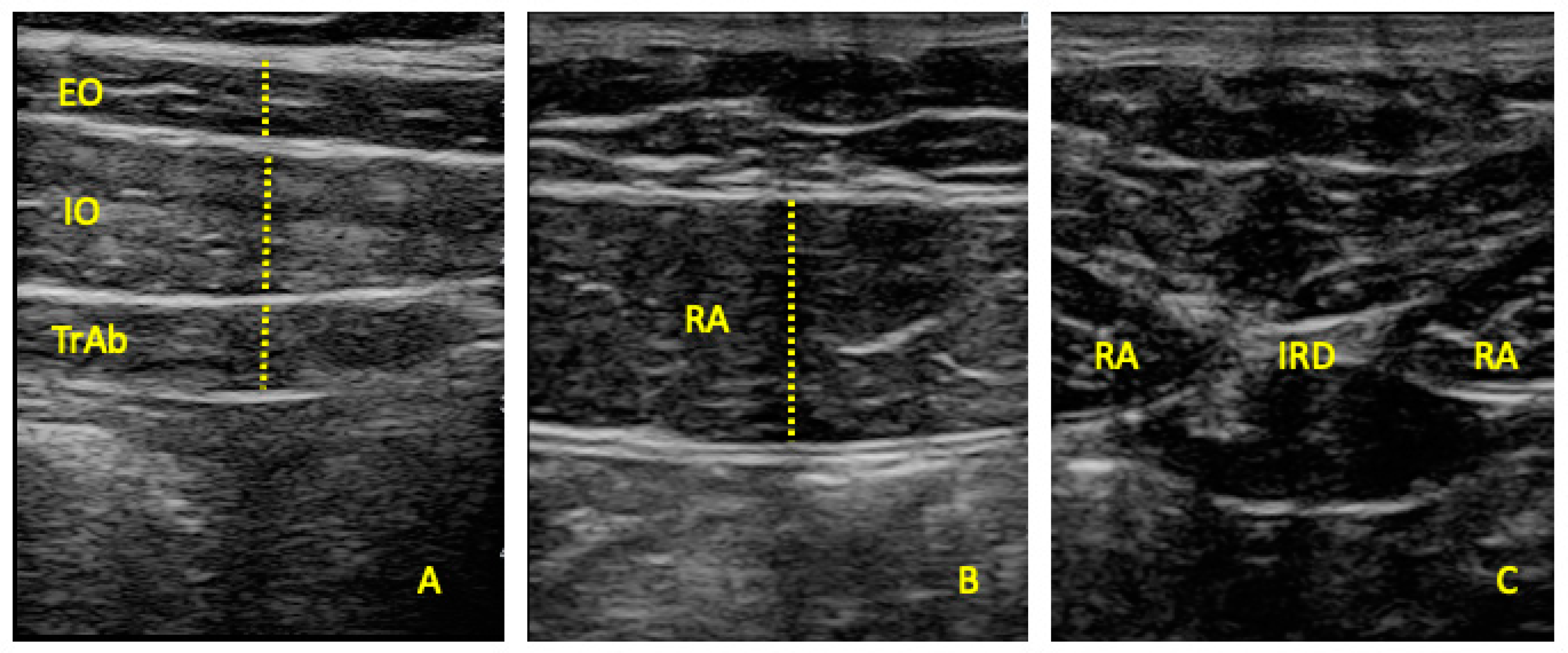
2.4. Outcome Measurements

Ultrasonography was performed using a SonoScape E2 ultrasound system (SonoScape, Madrid, Spain) with a linear transducer ranging from 8 to 13 MHz and having a 55 mm footprint in B-Mode. To ensure the reliability of the ultrasound data, the ultrasound equipment was calibrated before the start of the data collection following the manufacturer’s guidelines. Additionally, the operator (U.T.) conducting the ultrasound scans underwent extensive training prior to the commencement of the study. A comprehensive training session was conducted, which included both theoretical and practical components to familiarize the operator with the specific protocols of this study. Regular quality checks were performed throughout the study period to maintain high standards of data collection. All the measurements were performed by the same clinician (U.T.) who is experienced using USI assessment protocols. All the assessments were taken while the muscles were in a relaxed state to ensure accuracy and consistency across assessments. The imaging of the EO, IO, and TrAb muscles was performed in both sides by positioning the transducer along the mid-axillary line, between the subcostal line and the iliac crest (Figure 1A) [15]. For the rectus abdominis RA muscle examination, the transducer was aligned with the umbilicus, and for the IRD evaluation, it was placed just below the umbilicus (Figure 1B). Muscle thickness was defined as the distance between the superficial and deep borders of each muscle, while the IRD was measured as the distance between the medial borders of the RA muscles. (Figure 1C) [15,21].

2.5. Statistical Analysis

Data analysis was conducted using RStudio (version 1.4, RStudio, PBC, Boston, MA, USA) and Jamovi (version 1.6, The Jamovi Project). The Shapiro–Wilk test was utilized to assess the normality of the data (p > 0.05). Descriptive statistics were computed, presenting means and standard deviations (SDs) for parametric data and medians along with interquartile ranges (IRs) for non-parametric data. For comparing parametric data across groups, the independent samples Student’s t-test was employed. In contrast, the Mann–Whitney U test was applied for non-parametric data comparisons. Levene’s test was also employed to verify the homogeneity of variances across samples. To compare both sides in each sport discipline, a paired t-test was employed. Effect sizes were calculated using Cohen’s d, with values of 0.2 considered small, 0.5 medium, and 0.8 large.

3. Results

Table 1 shows that significant differences were found in height (p = 0.043), BMI (p = 0.001), shoe size (p = 0.018), and week training hours (p = 0.009). These results were expected due to the difference in morphotype between groups.
The present study employed ultrasonography to measure the abdominal wall muscles of elite soccer and basketball players, presenting notable results, albeit with minimal significant differences between groups (see Table 2). The thickness of the TrAb, IO, EO, and RA on both the right and left sides was measured. For the right RA, a statistically significant difference was observed, with basketball players showing a higher mean thickness (15.2 ± 2.48 mm) compared to soccer players (13.6 ± 1.79 mm), yielding a p-value of 0.039 and an effect size of 0.72. Despite this significant finding, other muscles such as the TrAb, IO, and EO did not show significant differences between the two groups, with p-values ranging from 0.220 to 0.641. The IRD also approached statistical significance (p = 0.087), suggesting a trend towards larger separation among basketball players compared to soccer players. These findings suggest that while there are specific differences in certain core muscles, the overall muscle morphology is similar across these sports, possibly reflecting the similar core demands of both soccer and basketball.
Considering Table 3, which reports the analysis between the right side and the left side independently of each sport, just the EO muscle reported differences (p = 0.029) between sides in basketball players. However, the rest of the variables showed that non-significant differences were observed for the right side with respect to the left side in soccer and basketball players.

4. Discussion

This novel research provides useful information regarding the abdominal wall muscle complex in soccer players with respect to basketball players. To date, this could be considered the first study to compare both sports’ populations with ultrasonography. However, non-significant differences were found in the thickness of the abdominal muscles; these findings provide valuable insights into sport-specific adaptations. Nevertheless, the assessment of the TrAb, EO, IO, and RA muscles’ morphology and structure plays an important role for injury prevention and rehabilitation management in different sports’ populations [22]. Recent research highlights the reliability of ultrasound measurements of lumbar multifidus and TrAb thickness during static and dynamic activities in subjects with and without muscle conditions related to the core muscles [23].
The significant difference in RA muscle thickness between basketball and soccer players can be attributed to the distinct physical demands of each sport. Basketball requires frequent jumping, sprinting, and rapid changes in direction, which place substantial stress on the RA muscle to maintain trunk stability and generate explosive power [16,24]. These results are coincident with studies that have shown that basketball players tend to develop greater muscle thickness in the core region due to the high-intensity nature of the movements [25].
Conversely, soccer players engage in activities that require endurance, agility, and continuous play with intermittent high-intensity actions which require a balanced development of the core muscles [2]. The lack of significant differences in the EO, IO, and TrAb muscles between soccer and basketball players suggests that both sports place similar demands on these muscle groups, leading to comparable adaptations [3]. In addition, according to D’Isanto et al., strength training is the most significant component of athlete performance [26]. In this context, core strength training improves the strength transmission from the lower to upper limbs, coordinated coordination, muscle control ability [27], and reduces injury risk in both sports [6].
We acknowledge the observed intra-subject differences between the left and right sides of the abdominal musculature. Similar discrepancies have been noted in other studies investigating asymmetries in muscle morphology and function. For instance, a study by Hides et al. observed asymmetries in the cross-sectional area of the lumbar multifidus muscle among elite cricketers, with significant differences between the left and right sides, which were attributed to the specific physical demands of their sport [14]. Similarly, Gildea et al. reported asymmetries in the thickness of the abdominal muscles in ballet dancers, which were linked to their training and performance activities [12]. These studies highlight that sport-specific demands can lead to unilateral adaptations in muscle morphology, supporting our findings of intra-subject differences in the abdominal musculature of soccer and basketball players.
Ultrasonography has proven to be a reliable and valid method for assessing muscle morphology, reporting precise measurements of muscle thickness and CSA [15]. The use of ultrasonography in this study allowed for accurate comparisons between the two groups, contributing valuable data to the sports science field. Previous studies utilizing ultrasonography in sports contexts have demonstrated its efficacy in monitoring muscle adaptations and guiding targeted training programs [28,29,30].
Soccer and basketball sports demand high loads to the spinal region, pelvic region, and lower limbs; therefore, proper core muscle complex conditioning is mandatory to prevent low back pain (LBP)—with a yearly prevalence of 64% (soccer) and 10.2%—or lower limb conditions [31,32]. Several methods have been used to assess the muscle behavior and muscle structure such as electromyography (EMG) being the “gold standard” or MRI [33,34]. Thus, ultrasonography is an excellent alternative due to its relatively low cost, validity, reliability, and portability which is highly advantageous in sports medicine given the frequent travel requirements. Its low cost and rapid use make it accessible for continuous monitoring and immediate assessments, crucial for maintaining athletes’ health during stages, away matches, or training camps. Additionally, the high reliability and accuracy for muscle thickness, CSA, or dynamic assessments ensure that it provides consistent and valuable insights into players’ physical conditions [35,36].

4.1. Limitations and Future Lines

This study has several limitations that should be acknowledged. The study design was cross-sectional, which precludes the ability to draw causal inferences about the relationship between sport-specific training and abdominal muscle adaptations. Another limitation of this study is the absence of data on participants’ hydration status, dietary intake, and recent physical activity levels. Variations in these factors can influence muscle morphology and ultrasound measurements. Hydration levels, in particular, can affect muscle thickness and echogenicity, while diet and recent physical activity can alter muscle tone and size. The present study only compared the left and right sides of the abdominal musculature independently, without averaging the measurements (R+L/2) for each player. Consequently, we did not evaluate whether basketball players still have a thicker average RA muscle mass when considering the average of both sides, which should be considered as a limitation. Future research should aim to address these limitations by including larger and more diverse samples, including both amateur and elite athletes from various sports disciplines. Longitudinal studies are needed to investigate the causal effects of sport-specific training on abdominal muscle morphology and to monitor changes over time. Further research should also consider dynamic assessments of muscle function during sport-specific tasks to provide a more comprehensive understanding of muscle performance. Additionally, exploring the impact of different training programs, including core stability and strength training, on abdominal muscle adaptations across different sports could provide valuable insights for developing targeted training and rehabilitation protocols. Integrating advanced imaging techniques such as MRI could also enhance the precision and detail of muscle morphology assessments.

4.2. Clinical Applications

The biomechanical implications of the present study highlighted that the specific core muscle adaptations in soccer and basketball players can inform the development of tailored training and rehabilitation programs. Basketball, with its frequent jumping, sprinting, and rapid changes in direction, places substantial stress on the RA muscle to maintain trunk stability and generate explosive power. This increased muscle thickness in basketball players suggests enhanced core strength and stability, which are crucial for the optimal performance and injury prevention in this sport. Therefore, in basketball players, emphasizing exercises that enhance RA strength and endurance could be beneficial, while soccer players might focus on maintaining balanced core strength to support their unique physical demands. This targeted approach could optimize performance and reduce the risk of injuries associated with core muscle imbalances.

5. Conclusions

The findings of the present study show a greater RA muscle thickness in basketball players with respect to soccer players assessed by ultrasonography. These findings suggest sport-specific adaptations in the core muscles, likely due to the distinct physical demands of each sport. While no significant differences were found in the rest of the abdominal wall muscles, the results underscore the importance of core muscle strength and stability in both sports using ultrasonography, which contributed valuable data to the understanding of core muscle morphology in athletes.

Author Contributions

Conceptualization, C.R.-M., I.R.-C. and S.L.J.-S.; Methodology, C.R.-M., J.H.V. and D.M.-M.; Software, C.R.-M.; Formal analysis, C.R.-M.; Investigation, U.T. and I.R.-C.; Resources, J.H.V.; Data curation, H.P.-G. and S.L.J.-S.; Writing—original draft, C.R.-M., J.H.V., D.M.-M., H.P.-G. and S.L.J.-S.; Writing—review & editing, C.R.-M., U.T., H.P.-G., I.R.-C. and S.L.J.-S.; Project administration, C.R.-M. All authors have read and agreed to the published version of the manuscript.

Funding

This research received no external funding.

Institutional Review Board Statement

The study was conducted in accordance with the Declaration of Helsinki and approved by the Institutional Review Board of the European University of Madrid (reference number: CIPI-19-003).

Informed Consent Statement

Informed consent was obtained from all subjects involved in the study.

Data Availability Statement

The original contributions presented in the study are included in the article, further inquiries can be directed to the corresponding author/s.

Conflicts of Interest

The authors declare no conflict of interest.

References

  1. Iaia, F.M.; Ermanno, R.; Bangsbo, J. High-Intensity Training in Football. Int. J. Sports Physiol. Perform. 2009, 4, 291–306. [Google Scholar] [CrossRef]
  2. Stølen, T.; Chamari, K.; Castagna, C.; Wisløff, U. Physiology of Soccer. Sports Med. 2005, 35, 501–536. [Google Scholar] [CrossRef]
  3. Luo, S.; Soh, K.G.; Soh, K.L.; Sun, H.; Nasiruddin, N.J.M.; Du, C.; Zhai, X. Effect of Core Training on Skill Performance Among Athletes: A Systematic Review. Front. Physiol. 2022, 13, 915259. [Google Scholar] [CrossRef]
  4. Bishop, D. Warm Up I. Sports Med. 2003, 33, 439–454. [Google Scholar] [CrossRef]
  5. Reilly, T.; Bangsbo, J.; Franks, A. Anthropometric and physiological predispositions for elite soccer. J. Sports Sci. 2000, 18, 669–683. [Google Scholar] [CrossRef] [PubMed]
  6. Kibler, W.B.; Press, J.; Sciascia, A. The Role of Core Stability in Athletic Function. Sports Med. 2006, 36, 189–198. [Google Scholar] [CrossRef]
  7. Maquirriain, J.; Ghisi, J.P.; Kokalj, A.M. Rectus abdominis muscle strains in tennis players. Br. J. Sports Med. 2007, 41, 842–848. [Google Scholar] [CrossRef]
  8. Behm, D.G.; Drinkwater, E.J.; Willardson, J.M.; Cowley, P.M. The use of instability to train the core musculature. Appl. Physiol. Nutr. Metab. 2010, 35, 91–108. [Google Scholar] [CrossRef] [PubMed]
  9. Pillastrini, P.; Ferrari, S.; Rattin, S.; Cupello, A.; Villafañe, J.H.; Vanti, C. Exercise and tropism of the multifidus muscle in low back pain: A short review. J. Phys. Ther. Sci. 2015, 27, 943–945. [Google Scholar] [CrossRef] [PubMed]
  10. Bahr, R.; Krosshaug, T. Understanding injury mechanisms: A key component of preventing injuries in sport. Br. J. Sports Med. 2005, 39, 324–329. [Google Scholar] [CrossRef]
  11. Hides, J.; Stanton, W. Muscle Imbalance Among Elite Australian Rules Football Players: A Longitudinal Study of Changes in Trunk Muscle Size. J. Athl. Train. 2012, 47, 314–319. [Google Scholar] [CrossRef]
  12. Gildea, J.E.; Hides, J.A.; Hodges, P.W. Morphology of the abdominal muscles in ballet dancers with and without low back pain: A magnetic resonance imaging study. J. Sci. Med. Sport 2014, 17, 452–456. [Google Scholar] [CrossRef] [PubMed]
  13. Vera-Garcia, F.J.; Brown, S.H.M.; Gray, J.R.; McGill, S.M. Effects of different levels of torso coactivation on trunk muscular and kinematic responses to posteriorly applied sudden loads. Clin. Biomech. 2006, 21, 443–455. [Google Scholar] [CrossRef]
  14. Hides, J.; Stanton, W.; Mcmahon, S.; Sims, K.; Richardson, C. Effect of Stabilization Training On Multifidus Muscle Cross-sectional Area Among Young Elite Cricketers With Low Back Pain. J. Orthop. Sports Phys. Ther. 2008, 38, 101–108. [Google Scholar] [CrossRef]
  15. Teyhen, D.S.; Gill, N.W.; Whittaker, J.L.; Henry, S.M.; Hides, J.A.; Hodges, P. Rehabilitative Ultrasound Imaging of the Abdominal Muscles. J. Orthop. Sports Phys. Ther. 2007, 37, 450–466. [Google Scholar] [CrossRef] [PubMed]
  16. Morales, C.R.; Polo, J.A.; Sanz, D.R.; López, D.L.; González, S.V.; Buría, J.L.A.; Lobo, C.C. Rehabilitative Ultrasound Imaging Features of the Abdominal Wall Muscles in Elite and Amateur Basketball Players. Appl. Sci. 2018, 8, 809. [Google Scholar] [CrossRef]
  17. Linek, P.; Noormohammadpour, P.; Mansournia, M.A.; Wolny, T.; Sikora, D. Morphological changes of the lateral abdominal muscles in adolescent soccer players with low back pain: A prospective cohort study. J. Sport Health Sci. 2020, 9, 614–619. [Google Scholar] [CrossRef] [PubMed]
  18. Von Elm, E.; Altman, D.G.; Egger, M.; Pocock, S.J.; Gøtzsche, P.C.; Vandenbroucke, J.P. The Strengthening the Reporting of Observational Studies in Epidemiology (STROBE) statement: Guidelines for reporting observational studies. J. Clin. Epidemiol. 2008, 61, 344–349. [Google Scholar] [CrossRef]
  19. Swann, C.; Moran, A.; Piggott, D. Defining elite athletes: Issues in the study of expert performance in sport psychology. Psychol. Sport Exerc. 2015, 16, 3–14. [Google Scholar] [CrossRef]
  20. Shrestha, B.; Dunn, L. The Declaration of Helsinki on Medical Research involving Human Subjects: A Review of Seventh Revision. J. Nepal Health Res. Counc. 2020, 17, 548–552. [Google Scholar] [CrossRef]
  21. Whittaker, J.L. Ultrasound imaging of the lateral abdominal wall muscles in individuals with lumbopelvic pain and signs of concurrent hypocapnia. Man. Ther. 2008, 13, 404–410. [Google Scholar] [CrossRef] [PubMed]
  22. Martin, C.; Olivier, B.; Benjamin, N. Asymmetrical abdominal muscle morphometry is present in injury free adolescent cricket pace bowlers: A prospective observational study. Phys. Ther. Sport 2017, 28, 34–42. [Google Scholar] [CrossRef] [PubMed]
  23. Sánchez Romero, E.A.; Alonso Pérez, J.L.; Muñoz Fernández, A.C.; Battaglino, A.; Castaldo, M.; Cleland, J.A.; Villafañe, J.H. Reliability of Sonography Measures of the Lumbar Multifidus and Transversus Abdominis during Static and Dynamic Activities in Subjects with Non-Specific Chronic Low Back Pain. Diagnostics 2021, 11, 632. [Google Scholar] [CrossRef] [PubMed]
  24. Morales, C.R.; Polo, J.A.; Sanz, D.R.; López, D.L.; González, S.V.; Buría, J.L.A.; Lobo, C.C. Ultrasonography features of abdominal perimuscular connective tissue in elite and amateur basketball players: An observational study. Rev. Assoc. Med. Bras. 2018, 64, 936–941. [Google Scholar] [CrossRef]
  25. Gong, J.; Gao, H.; Sui, J.; Qi, F. The effect of core stability training on the balance ability of young male basketball players. Front. Physiol. 2023, 14, 1305651. [Google Scholar] [CrossRef] [PubMed]
  26. D’Isanto, T.; D’Elia, F.; Raiola, G.; Altavilla, G. Assessment of Sport Performance: Theoretical Aspects and Practical Indications. Sport Mont 2019, 17, 79–82. [Google Scholar] [CrossRef]
  27. Suchomel, T.J.; Nimphius, S.; Bellon, C.R.; Stone, M.H. The Importance of Muscular Strength: Training Considerations. Sports Med. 2018, 48, 765–785. [Google Scholar] [CrossRef] [PubMed]
  28. Özçakar, L.; Çarl, A.B.; Tok, F.; Tekin, L.; Akkaya, N.; Kara, M. The Utility of Musculoskeletal Ultrasound in Rehabilitation Settings. Am. J. Phys. Med. Rehabil. 2013, 92, 805–817. [Google Scholar] [CrossRef] [PubMed]
  29. Haile, G.; Hailemariam, T.T.; Haile, T.G. Effectiveness of Ultrasound Therapy on the Management of Chronic Non-Specific Low Back Pain: A Systematic Review. J. Pain Res. 2021, 14, 1251–1257. [Google Scholar] [CrossRef]
  30. Valera-Calero, J.A.; Fernández-de-las-Peñas, C.; Varol, U.; Ortega-Santiago, R.; Gallego-Sendarrubias, G.M.; Arias-Buría, J.L. Ultrasound Imaging as a Visual Biofeedback Tool in Rehabilitation: An Updated Systematic Review. Int. J. Environ. Res. Public Health 2021, 18, 7554. [Google Scholar] [CrossRef]
  31. Bradley, J.; Gorijala, P.; Schindler, S.E.; Sung, Y.J.; Ances, B.; Alzheimer’s Disease Neuroimaging Initiative, the Human Connectome Project; Fernandez, M.V.; Cruchaga, C. Genetic architecture of plasma Alzheimer disease biomarkers. Hum. Mol. Genet. 2023, 32, 2532–2543. [Google Scholar] [CrossRef] [PubMed]
  32. Nandlall, N.; Rivaz, H.; Rizk, A.; Frenette, S.; Boily, M.; Fortin, M. The effect of low back pain and lower limb injury on lumbar multifidus muscle morphology and function in university soccer players. BMC Musculoskelet. Disord. 2020, 21, 96. [Google Scholar] [CrossRef] [PubMed]
  33. Taghipour, M.; Mohseni-Bandpei, M.A.; Behtash, H.; Abdollahi, I.; Rajabzadeh, F.; Pourahmadi, M.R.; Emami, M. Reliability of Real-time Ultrasound Imaging for the Assessment of Trunk Stabilizer Muscles: A Systematic Review of the Literature. J. Ultrasound Med. 2019, 38, 15–26. [Google Scholar] [CrossRef] [PubMed]
  34. Cervera-Cano, M.; Sáez-García, M.C.; Valcárcel-Linares, D.; Fernández-Carnero, S.; López-González, L.; Gallego-Izquierdo, T.; Pecos-Martin, D. Real-time ultrasound evaluation of CORE muscle activity in a simultaneous contraction in subjects with non-specific low back pain and without low-back pain. Protocol of an observational case-control study. PLoS ONE 2023, 18, e0285441. [Google Scholar] [CrossRef] [PubMed]
  35. May, S.; Locke, S.; Kingsley, M. Reliability of ultrasonographic measurement of muscle architecture of the gastrocnemius medialis and gastrocnemius lateralis. PLoS ONE 2021, 16, e0258014. [Google Scholar] [CrossRef]
  36. Johnson, G.G.R.J.; Jelic, T.; Derksen, A.; Unger, B.; Zeiler, F.A.; Ziesmann, M.T.; Gillman, L.M. Accuracy of Optic Nerve Sheath Diameter Measurements in Pocket-Sized Ultrasound Devices in a Simulation Model. Front. Med. 2022, 9, 831778. [Google Scholar] [CrossRef]
Figure 1. Ultrasonography of the external oblique, internal oblique, transversus abdominis (A), and rectus abdominis (B) muscles and inter-recti distance (C).
Applsci 14 05742 g001
Table 1. The sociodemographic data of the sample.
MeasurementSoccer (n = 17)
Mean ± SD
Basketball (n = 18)
Mean ± SD
p-Value
Age, (y)23.1 ± 3.5323.6± 2.450.136
Weight, (kg)81.8 ± 8.4478.2 ± 8.00.204
Height, (cm)1.81 ± 0.061.86 ± 0.080.043
BMI, (kg/cm2)25.1 ± 2.4222.6 ± 1.640.001
Shoe size (EU number)43.6 ± 1.9245.0 ± 1.500.018
Week training (hours)4.91 ± 1.686.83 ± 2.330.009
Table 2. The ultrasound imaging measurements of the abdominal wall muscles.
MeasurementSoccer (n = 17)
Mean ± SD
(95% CI)
Basketball (n = 18)
Mean ± SD
(95% CI)
p-Value (Effect Size)
TrAb_R5.00 ± 1.07 (2.9–7.2) *4.80 ± 0.89 (2.4–6.3) *0.550 (0.20) **
IO_R11.6 ± 2.61 (8.3–17.7) *11.1 ± 1.20 (9.6–13.8) *0.485 (0.23) **
EO_R7.94 ± 1.25 (5.3–10.4) *7.43 ± 1.17 (5.5–10.7) *0.220 (0.42) **
RA_R13.6 ± 1.79 (10.2–17.6) *15.2 ± 2.48 (10.4–19.1) *0.039 (0.72) **
TrAb_L4.87 ± 1.09 (2.5–7.3) *4.69 ± 1.08 (2.3–6.9) *0.641 (0.15) **
IO_L11.9 ± 3.00 (5.32–18.8) *10.9 ± 2.29 (7.5–15.3) *0.298 (0.35) **
EO_L7.77 ± 0.99 (5.2–9.8) *7.96 ± 1.09 (6.3–10.5) *0.605 (0.17) **
RA_L14.3 ± 1.83 (11.3–18) *15.2 ± 1.84 (11.7–19.0) *0.149 (0.50) **
IRD2.79 ± 0.90 (1.7–4.7) *3.38 ± 1.05 (1.6–5.5) *0.087 (0.59) **
Abbreviations: EO, external oblique; IO, internal oblique; IRD, inter-recti distance; RA, rectus abdominis; TrAb, transversus abdominis. * Mean (SD) was applied. ** Student’s t-test for independent samples was performed.
Table 3. The ultrasound imaging measurements of the abdominal wall muscles of the right vs. left side for soccer and basketball players.
MeasurementRight
Mean ± SD
(95% CI)
Left
Mean ± SD
(95% CI)
p-Value (Effect Size)
Soccer players
TrAb4.96 ± 1.01 (2.9–7.0) *4.85 ± 1.12 (2.5–7.1) *0.601 (0.12) **
IO11.30 ± 2.51 (8.3–17.7) *11.7 ± 3.02 (5.3–18.8) *0.529 (0.15) **
EO7.91 ± 1.28 (5.3–10.4) *7.80 ± 1.02 (5.2–9.1) *0.561 (0.14) **
RA13.57 ± 1.84 (10.1–17.6) *14.30 ± 1.83 (11.2–18.0) *0.145 (0.37) **
Basketball players
TrAb4.74 ± 0.89 (2.4–6.3) *4.62 ± 1.08 (2.3–6.9) *0.478 (0.16) **
IO11.09 ± 1.15 (9.6–13.7) *10.80 ± 2.28 (7.5–15.3) *0.379 (0.20) **
EO7.39 ± 1.28 (5.5–10.6) *7.80 ± 1.02 (6.3–10.4) *0.029 (0.54) **
RA15.1 ± 2.42 (10.4–19.0) *15.17 ± 1.69 (11.7–18.9) *0.856 (0.16) **
Abbreviations: EO, external oblique; IO, internal oblique; RA, rectus abdominis; TrAb, transversus abdominis. * Mean (SD) was applied. ** Paired t-test for independent samples was performed.
Disclaimer/Publisher’s Note: The statements, opinions and data contained in all publications are solely those of the individual author(s) and contributor(s) and not of MDPI and/or the editor(s). MDPI and/or the editor(s) disclaim responsibility for any injury to people or property resulting from any ideas, methods, instructions or products referred to in the content.

