Article

Digital Orofacial Identification Technologies in Real-World Scenarios

1
University of Coimbra, Forensic Dentistry Laboratory, Faculty of Medicine, 3000-354 Coimbra, Portugal
2
Psychology Research Center, Autonomous University of Lisbon, 1169-023 Lisbon, Portugal
*
Author to whom correspondence should be addressed.
Appl. Sci. 2024, 14(13), 5892; https://doi.org/10.3390/app14135892
Submission received: 15 January 2024 / Revised: 28 May 2024 / Accepted: 3 July 2024 / Published: 5 July 2024
(This article belongs to the Section Applied Dentistry and Oral Sciences)

Abstract

:
Three-dimensional technology using personal data records has been explored for human identification. The present study aimed to explore two methodologies, photography and orofacial scanning, for assessing orofacial records in forensic scenarios, highlighting their impact on human identification. A pilot and quasi-experimental study was performed using Canon 5D-Full Frame equipment (Tokyo, Japan) and an i700 scanner (Medit, Lusobionic, Portugal) (Seoul, Republic of Korea) with Medit Scan for Clinics (MSC) and Smile Design software (V3.3.2). The sample included living patients (n = 10) and individuals in forensic cases (n = 10). The study was divided into two complementary phases: (i) data collection using 2D and 3D technologies and (ii) visual comparison by superimposition procedures, 3D dental images with 3D facial records (3D–3D), and 2D photography with screen printing of 3D facial records (2D-3S). Statistical analyses were performed using descriptive procedures (Likert scale) and the Mann–Whitney U test. The Mann–Whitney U test comparing the data (n = 220 records) from living individuals and those in forensic cases identified statistically significant differences in the performance of the photographic methods for evaluating intraoral mineralisation (p = 0.004), intraoral soft tissues (p = 0.016), intraoral distortion (p = 0.005) and the scan methods for intraoral extra devices (p = 0.003) and extraoral soft tissues (p = 0.005). A visual comparison (n = 40) allowed 3D–3D superimposition. Additionally, 2D-3S superimposition qualitatively identified the middle third of the face as the corporal area within the anatomical features required for successful surgery. In conclusion, the present study presented evidence-based data suggesting that the IO scan method, as an emergent technology, should be explored as a valuable tool in forensic facial identification in real-world scenarios.

1. Introduction

The globally accepted standards for human identification (from INTERPOL and ABFO) ensure compliance with forensic quality controls, therefore facilitating information-sharing and exchange data [1,2].
Current methods of human identification involve primarily morphometric or morphological criteria [3,4,5,6,7,8,9,10] and, in extreme forensic scenarios, the genetic profile. The morphometric approach involves the measurement of anatomic structures and inter-population group mean dimension variability, and typically entails using different statistical methods to derive models or equations [10,11,12,13]. The morphological approach relies on the visual assessment of unique anatomic features and traits by experts [14].
Dental structures have been used for morphological analysis and genotype profile sampling [3,5,10,11], primarily via the use of geometric morphometric techniques [9,10]. Such techniques rely on the unique characteristics of dentition and dental restorations and the relative resistance of mineralised dental tissue and dental restorations [3,5,13,15]. In addition, the palatal rugae, specifically the second and third palatal rugae, are sufficiently unique and relatively stable [12,16]. Palatal rugae can even discriminate between identical monozygotic twins with identical DNA [11,12].
Antemortem data can be accessed from clinical dental records [1,2,5], which requires the preservation of dental records [3,8]. Clinical dental records contain valuable information, such as radiographs, photographs, dental charts, and dental models. These records include distinctive characteristics, such as missing teeth, the presence of dental implants, the use of prostheses, dental restorations (fillings), tooth crowding, or unusual arrangements of the teeth in the dental arch [3,8]. Clinical dental data can be recorded digitally, in handwritten form, or in two-dimensional dental charts and captured in dental cast models [3,5]. By comparing antemortem and postmortem dental records, forensic odontologists can positively identify the deceased or provide valuable information to assist in the identification process.
In contemporary dental clinical practice, intraoral scanner (IOS) use and cone bean computer tomography (CBCT) have become standard methods [6,9,11,12,17,18] since they offer highly accurate, three-dimensional records of the dental arches and surrounding structures [6,11,17]. Compared with traditional dental cast methods, IOSs reduce patient operation time, stress, and discomfort [4,12,18,19,20,21]. Furthermore, an IOS allows easy and quick access to patient information, eliminates impression material or gypsum deformation, reduces storage space, and facilitates communication among peers [4,5,6,17,18,19,20,22].
An intraoral scan can be conducted via two approaches: contact and laser [23]. A contact IOS explores the object’s surface through a probe with a hard-steel or sapphire tip, and a series of internal sensors determine the probe’s spatial positions. A laser IOS involves the projection of a light source, the return of which is detected to capture the object’s geometry by triangulation and visualisation of the dental arches more rapidly than a contact IOS [23]. Contact IOSs are often used in inspection and quality control systems, while laser IOSs are typically used for scanning dental arches in dental clinical practice. The scanning software processes the captured images and generates point clouds that reproduce the object’s geometry. These point clouds are then triangulated by the same software, creating a 3D surface model (mesh), which represents a virtual alternative to traditional plaster models [5,20,21,23,24]. A complete IOS 3D model can include stationary structures other than teeth, allowing the use of high-reliability methods, such as rugoscopy, for identification purposes [4,12]. In addition, biometric measurements, such as intercanine distance and geometric analysis, can be used to compare intraoral antemortem and postmortem digital scan data and assess their accuracy [9,11,20,23].
This study aimed to present evidence-based data on two methodologies, intraoral scanning and photography, in the context of orofacial records in forensic pathology. The development of the study is based on the application of digital technologies outside of the context of simulation or experimental study and extending them to the real scenario, filling this gap in the literature. The paper includes a detailed description of the criteria for selecting the sample; the design of the study, its development, and analysis; the presentation of the results; and their discussion.

2. Materials and Methods

2.1. Sample Selection

The study included data from a sample of oropathological exams conducted at the National Institute of Legal Medicine and Forensic Science (INMLCF, IP), Central Branch, Portugal. The exams were conducted between July 2022 and June 2023 and encompassed forensic cases involving individuals (i) older than 18 years of age, (ii) in possession of permanent dentition, and (iii) following the Portuguese protocol present in the Portuguese Record of Nondonors (RENNDA). The exclusion criteria were (i) fragmented and/or (ii) undocumented corpses, (iii) advanced stage of putrefaction, and (iv) edentulous status. Orofacial examinations of living individuals were performed at the Forensic Dentistry Laboratory (FDL), Faculty of Medicine, University of Coimbra, Portugal; the included participants were (i) older than 18 years of age and (ii) had permanent dentition. The examinations were completed between September 2022 and May 2023. The exclusion criteria were (i) edentulous status and (ii) use of removable dental prostheses. Those who satisfied the eligibility criteria were asked to provide written informed consent. The study was conducted according to the tenets of the Declaration of Helsinki and approved by the Ethics Committee CE12/2022 of the INMLCF, IP and CE-023/2027 of the FDL.

2.2. Study Design

A pilot and quasi-experimental study design was used to examine orofacial records acquired through two methods: (1) photography and (2) an IOS.
The study design was divided into two complementary phases: (P1) the first phase, or the data collection phase, involving the recording of two-dimensional (2D) and three-dimensional (3D) data; and (P2) the second phase, or the visual comparison phase, involving superimposition procedures (Figure 1).
Two-dimensional photographic data collection at P1 was conducted using a Canon EOS 5D Mark-II (Tokyo, Japan) and a 28–135 mm Macro 0.5 mm/1.6 ft lens. Mouth retractors (Optragate, Ivoclar), supplementary lighting (twin flash), and tripods (monkey tripod, Joby) were also selected. Photography was performed to collect 2D extraoral (EO) (frontal and lateral) and intraoral (IO) (maxillary and mandibular arches and interdental relation) data. In total, seven photographs were taken of each individual. The 2D photographic data collection followed the ABFO and Interpol guidelines [1,2].
An i700 wireless, intraoral, and facial laser scanner (Medit, Lusobionic, Portugal) (Seoul, Republic of Korea); MSC software (V3.2.1); and the Smile Design application running MacBook Air M2 with 24 GB of RAM were used to collect 3D data following the manufacturers’ guidelines. The equipment and application were used to record the intraoral (including the maxillary and mandibular arches and interdental relation) and facial 3D data (Supplementary Material Video S1). In total, four scans were taken for each individual.
This study evaluated faces divided into thirds according to the anatomic details described in Arnett’s study [25] (Figure 2).

2.3. Data Development and Analysis

A team of dentists supervised by forensic odontologists from the LFD performed the study. The research team, with up to 15 years of experience in the medicolegal field and anatomy education among them, analysed the consistency of the data records. Two researchers (TN and RR) assessed and scored each parameter separately, and in cases of doubt, a third person (ACR) was appointed as a decision maker. One researcher created superimposed images using MSC under the supervision of the team (PA).

2.3.1. Data Collection

Two-dimensional data were collected using the following settings: ISO 300, a shutter speed of 1/60, and an aperture of 5.6 for EO data; the aperture was adjusted to f/32 for IO data. The 2D records were saved as RAW and JPEG files with a resolution of 300 dpi. INTERPOL guidelines were used to select the views for recording (frontal and lateral views of the face) [1,2].
Three-dimensional data were collected in MSC following the i700 Medit technical guidelines for the recording procedure [1,2]. After the dental arches were recorded, the interdental relations were achieved based on the natural interarcade position in living individuals, manually aligned using reference points. The facial scan captured the anatomical details of the interlabial exposure zone, which was defined in the resting position and includes the incisor exposure, situated between the lower limit of the upper lip and the upper limit of the lower lip [22]. The scanning process continued in a circular motion, filling the lower third of the face. Anatomical details were captured from the subnasal cutaneous point to the glabella and following the external angle of the eye laterally for the middle third of the face.
The data were analysed in MSC software with the following parameters: (i) time elapsed (seconds), (ii) mineralisation details (Likert scale), (iii) soft tissue details (Likert scale), (iv) communication (Likert scale), (v) extra devices (Likert scale), and (vi) distortion (Likert scale). Parameter evaluations were based on a scoring system using a Likert scale (0 to 4), where higher scores indicated better performance.
The Likert scale for the analysis of mineralisation details included a score of: 4 points if teeth of both arches were visible, 3 points if teeth were visible in one arch, 2 points if teeth were partially visible in both arches, 1 point if teeth were partially visible in one arch and not visible in the other arch, and 0 points if no elements were visible in either arch. For soft details, a score of: 4 points indicated that soft anatomical details were visible, 3 points signified that all features were visible in the lateral portion (right or left), 2 points indicated that details were partially visible in the lateral portion, 1 point signified that details were partially visible in one lateral portion, and 0 points indicated that no elements were visible. The communication score was based on the type and format of the data included in the record. A score of: 4 points was assigned if the document had both 3D and 2D data, 3 points were awarded for only 3D data, 2 points were allocated for only 2D data with colour information, 1 point was awarded for only 2D data without colour information, and 0 points were given for unformatted data. The extra device score was based on the number and type of devices used: 4 points were assigned if no extra devices were used, 3 points were allocated if mouth retractors were used, 2 points were awarded if additional lighting was used, 1 point was assigned if both mouth retractors and additional lighting were used, and 0 points were given if mouth retractors, extra lighting, or a tripod were used. Distortion was scored as: 4 points if the records had no distortion (three circles of the ABFO-2 scale were respected), 3 points if the documents required a dimensional scale for some record details to be assessed (two circles of the ABFO-2 scale were respected), 2 points if a scale was required for some of the details to be evaluated (one circle of the ABFO-2 scale was respected), 1 point if the records showed partial distortion (no circles of the ABFO-2 scale were respected), and 0 points if the records were entirely distorted.
A statistical analysis was conducted using descriptive analyses and Mann–Whitney tests in the data recording phase.

2.3.2. Visual Comparison

The 3D–3D superimposition process was performed in MSC through alignment tools, including a resolution colour scale for accurate assessment (Figure 3). Coronary details of the anterior teeth achieved alignment for the superimposition between the intraoral and facial scans within a range of 2 to 3 mm of the occlusal limit described in Corte-Real and Reesu’s studies [26,27]. The tooth with the highest resolution was selected for analysis, and the highest and lowest scale values were identified for calculation of the mean value.
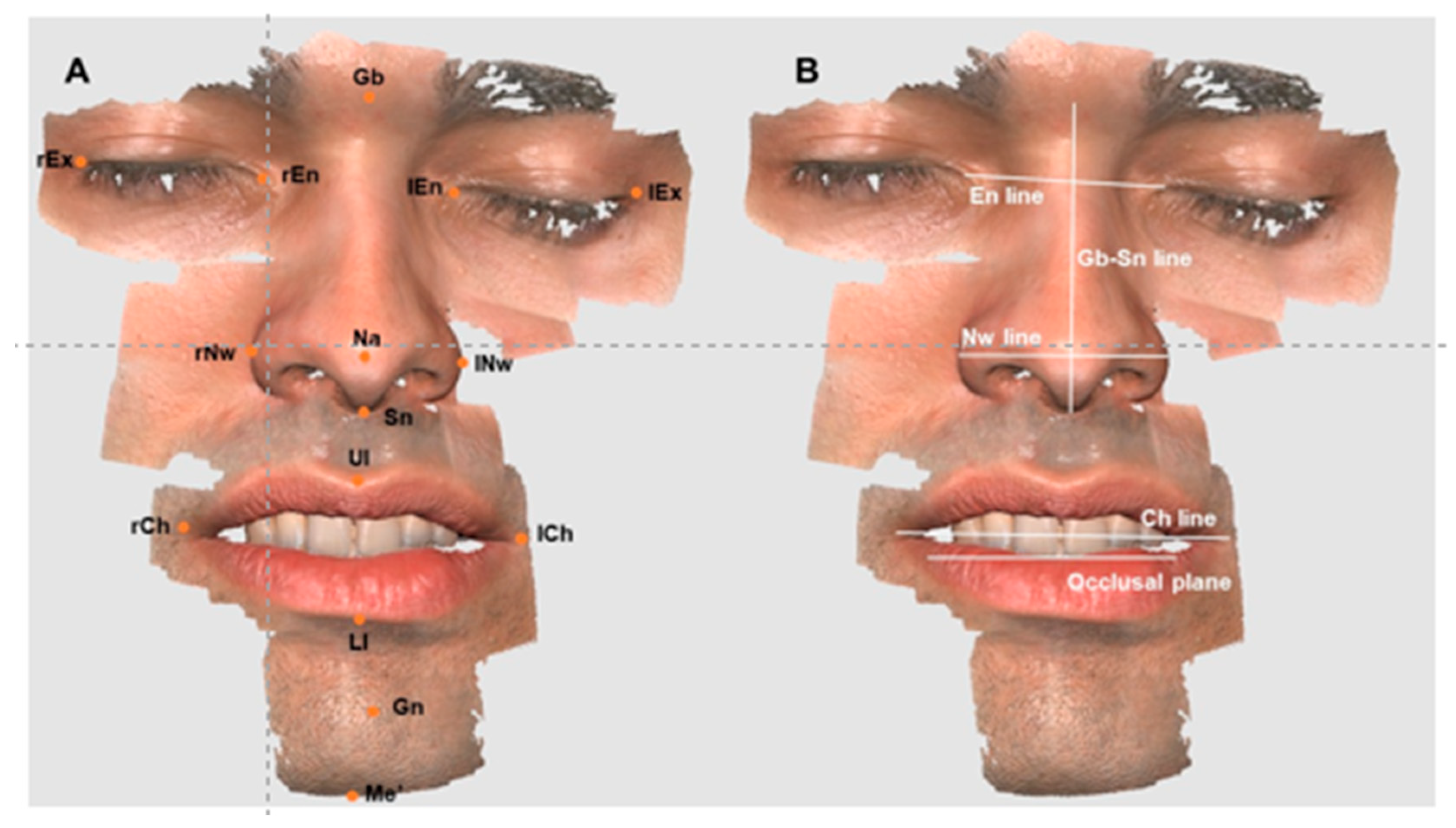
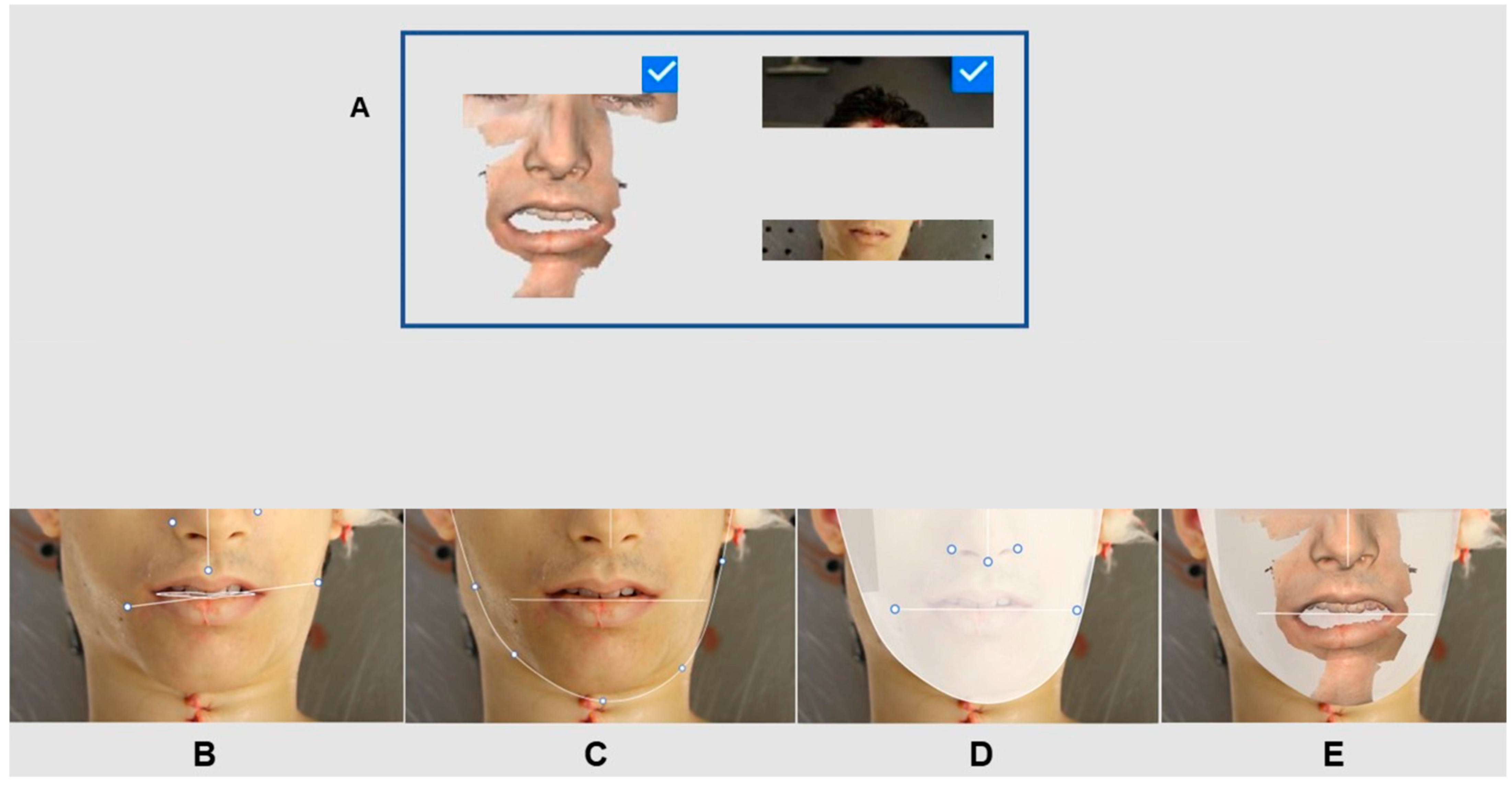
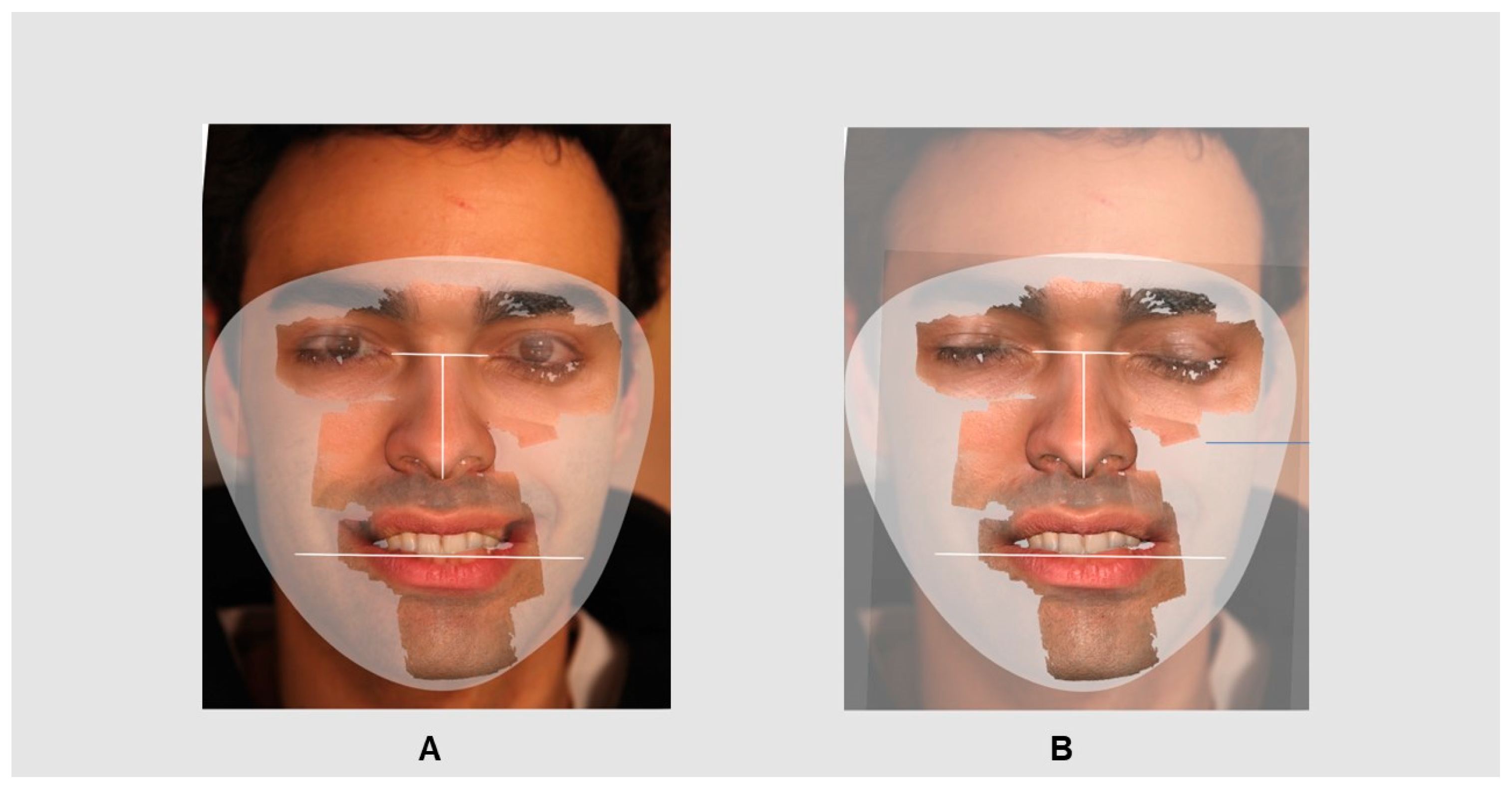
The 2D-3S superimposition process was performed using Smile Design software after uploading the 2D photographs and 3S records (Figure 4) and their alignment. The 3D face was reoriented to align anatomical facial details with the photos, achieving a 3D screenprint for accurate superimposition. The superimposition of 2D-3S was achieved through the manual identification of cutaneous biometric points, such as facial landmarks, as described in Gibelli’s study [28]. The cutaneous landmarks were the glabella, right and left endocanthions, right and left exocanthions, right and left nose wings, nasal and subnasal, right and left cheilions, upper and lower lips, and gnathion and mento (Figure 2).
The 2D-3S superimposition process is detailed in Figure 4, highlighting the assessment of the potential compatibility of the two overlaid records by Smile Design software.

3. Results

A total of 140 photos and 80 dental and facial scans were obtained for 10 living individuals (samples L1 to L10) and 10 individuals in forensic cases (samples D1 to D10) who met the previously described criteria. A total of 40 superimpositions were obtained and divided into 3D–3D volumes (n = 20) and 2D-3S datasets (n = 20).

3.1. Data Collection

The EO data were acquired by photography for all the samples (n = 20). All individual scores were summed and are presented in Table 1. Different devices, mouth retractors, and additional lighting approaches were used (1 score). The score of the recording of intraoral mineralisation data in individuals in forensic cases ranged from 2 to 3. Additionally, there was a notable decrease in intraoral distortion and soft data tissue, with scores ranging from 1 to 2.
The i700 wireless scanner recorded intraoral data for all samples (n = 20). All individual scores were summed and are presented in Table 2.
The scanner performed the worst in the EO view of living individuals. EO data demonstrated a minimum score of 3 and a maximum score of 4 for the soft tissue and distortion record parameters (Table 2). High scores were obtained with the scanner for all other parameters in both living individuals and those in forensic cases.
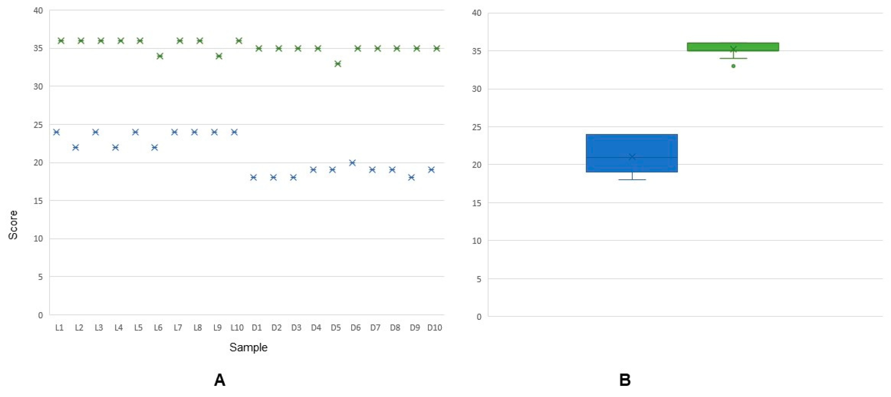
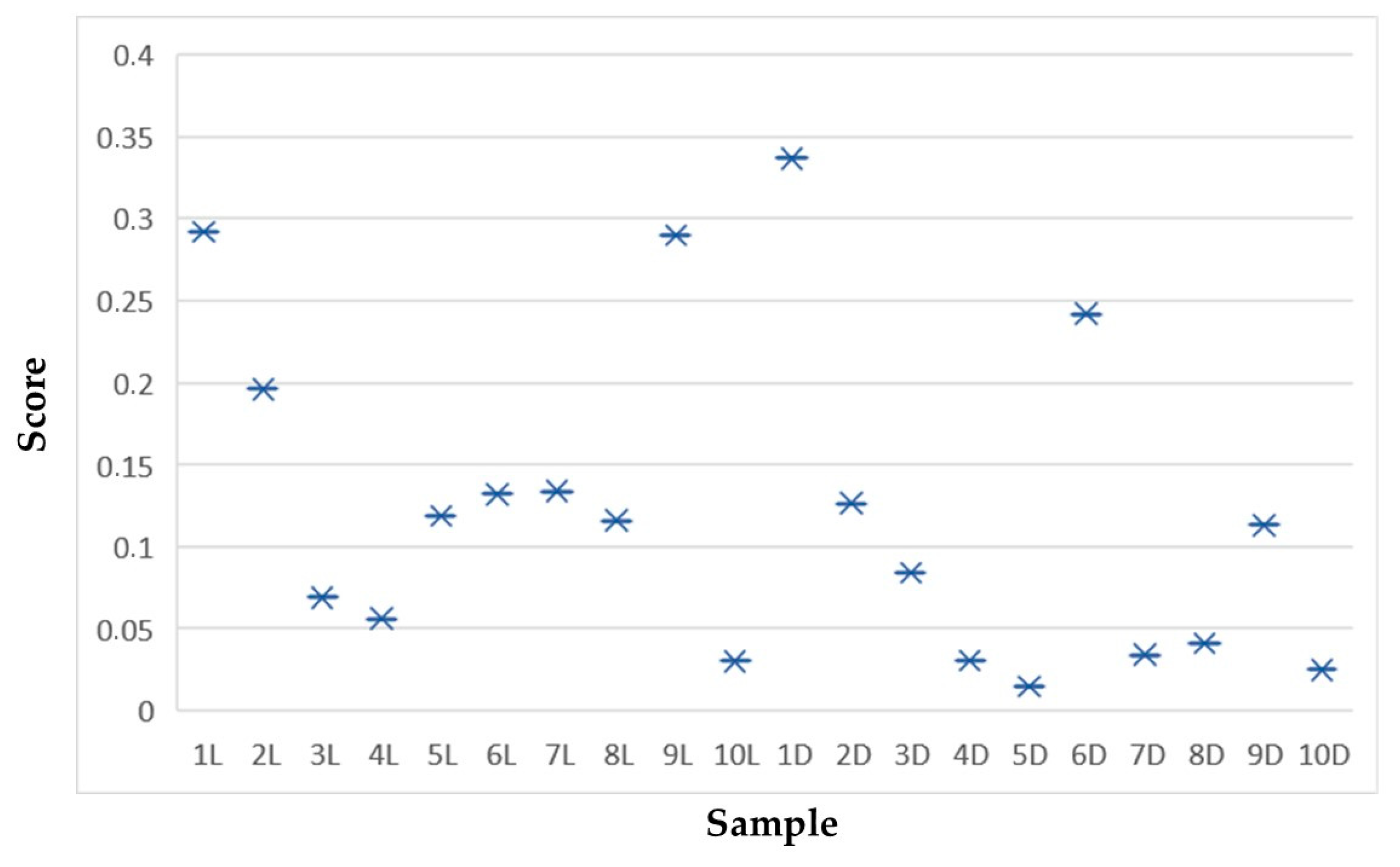
The descriptive analysis included the summed scores of each parameter for all samples (Figure 5).
The photographic methods had lower scores for individuals in forensic cases (score 18). All photographic scores were between 18 and 24, with living individuals having the highest scores.
Higher scores were observed with the IOS methods. The lowest score among the IOS methods was 34 for living individuals, and the highest was 36.
The Mann–Whitney test to evaluate data for living individuals and for those in forensic cases revealed statistically significant differences in IOS method performance for IO extra devices (p = 0.003) and EO soft tissues (p = 0.005) and statistically significant differences in comparison with the photographic methods for IO mineralisation (p = 0.004), IO soft tissues (p = 0.016), and IO distortion (p = 0.005).

3.2. Visual Comparison

3.2.1. The 3D–3D Superimposition

Medit software (V3.2.1) allowed 3D–3D superimposition for all samples (n = 20) (Figure 6, Figure 7 and Figure 8).
The findings after superimposition are summarised in Figure 9. The Mann–Whitney test performed to compare data between living individuals and those in forensic cases revealed a p value of 0.174, with no identified significant differences in 3D–3D superimposition (p > 0.050).

3.2.2. The 2D-3S Superimposition

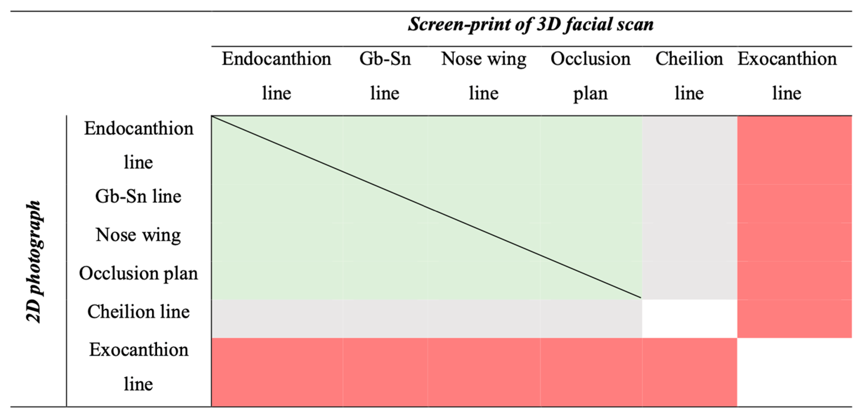
The descriptive analysis with the Smile Design application allowed the user to perform a 2D-3S superimposition process for all samples (n = 20) (Figure 8). Cutaneous biometric data were selected following the sequence of positive superimposition (match) versus negative superimposition (no match) for each individual (Figure 9).
The 2D-3S was successfully superimposed using the right and left endocanthion landmarks and their linear measures, right and left nose wing landmarks and their linear measures, glabella and subnasal landmarks and their linear measures, and the occlusal plane.
Only the exocanthion lines matched between the records; no other landmark alignment was observed. The cheilion line could ensure a match in living individuals but not in individuals in forensic cases.

4. Discussion

This ground-breaking study investigated the application of 3D scans for orofacial identification and for distinguishing dental and facial features (expressed as facial thirds). The research encompassed data from both living and dead individuals, thus considering an authentic forensic setting, aiming to address a significant gap in the literature [4,6,14,26].

4.1. Data Collection

IOS technology allows the capture of intraoral features (dental and soft tissues), which can be recorded in various file formats, namely, stereolithography (STL), then exported and integrated into a unified database referred to as the “virtual patient” [4,14,20,25]. The present study presents the potential of emerging technologies, namely Artificial Intelligence, integrated into clinical analysis and planning software as highlighted in Cortes’s study [24] for the forensic field, thus significantly expanding its functionality with the inclusion of recorded facial features.
Both clinical professionals and forensic experts have established the precision of 3D recording and analysis of anatomical features and morphology [6,19,21,29,30]. The reliability and advantages of using IOS technology for forensic purposes were discussed in Reesu’s study in a simulated scenario [13]. These authors studied photographic records from orthodontic exams, namely, EO and frontal views, and created models from scan data provided in STL format [13]. The challenges and limitations of the use of manual matching of dental features (using Adobe Photoshop) were noted, namely, the anatomical standards for superimposition, the anatomical distortion, and the proportions of 2D and 3D dental volumes [13].
Gibelli’s study focused on facial features, highlighting the challenge of methodologically comparing facial regions when divided into thirds [29]. The authors studied 3D facial models from stereophotogrammetric devices for aesthetic practice, and the root mean square (RMS) was calculated among the thirds of the face, allowing us to conclude that 3D–3D superimposition methods may be useful when analysing facial thirds [31]. Facial difference identification between monozygotic twins has been found to be effective when applying superimpositions involving 3D models [31].
Dental and facial 3D studies have focused on simulated forensic scenarios, highlighting the need for prospective analysis identifying individuals directly from human remains or using facial scanners in mass disasters [7]. Additionally, the deterioration of facial anatomy in forensic situations increases the accuracy of identification [7]. The present study intended to evaluate different methods of data recording for the same individual, including dental and facial anatomic features, analysing 2D and 3D features from human data records (MSC) with a unique software package, to overcome anatomical distortion and metric calibration among recorded data.
The i700 wireless scanner (Medit®) was selected for forensic purposes, based on its accuracy, in line with Bae’s study [5], because of its portability, wireless capability, and compatibility with software updates, addressing recent needs in the oral clinical rehabilitation field. These characteristics enable convenient usage and ensure adaptability to evolving technological advancements [9,19,20]. Furthermore, IOSs are distinguished by their ability to generate a 3D model, namely, in STL format, allowing the model’s exportation, visualisation, and comparison of data. The standardised recording of personal data in a universal format within a global space is significant for effective communication, seamless data sharing, comprehensive analysis, and reliable comparisons [32].
Trends in reaching a quantitative outcome in facial identification have emphasised the comprehensive examination of descriptive analyses of technical parameters to compare the performance of IOSs and photography. Unlike 3D images, photographic images are 2D projections of 3D objects, resulting in a lower recognition rate and increased difficulty in comparisons when the orientations are not identical [13,33]. Using photographic records, antemortem or postmortem data should reproduce the same perspective variations, which is a limitation for comparison proposals, as highlighted by Reesu and colleagues [13]. The conventional use of photographs as a standard complementary diagnostic examination in oropathological examinations in clinical practice in dentistry presents a challenge for its applicability in forensic situations. Three-dimensional data acquisition improves dynamic visualisation, allowing unlimited static or 2D profiles [26,34]. The functionalities of the 3D method were explored, and unlimited perspectives could be analysed to achieve the correct match data. The present findings elucidated the capabilities of these methods in forensic scenarios.
Focusing on data collection issues, for living individuals, photography performed well and demonstrated minimal distortion in 2D records, for both EO and IO images, including mineralised and soft tissues. In the photographic approach, geometric relationship management with the ABFO scale allowed us to overcome angular distortion, ensuring the specificity of the lens and perpendicular camera positioning [13,26]. However, achieving such precision often requires the use of additional devices [35]. The IOS procedure demonstrated excellent performance in recording EO and IO images, achieving high accuracy with the addition of mouth retractors. In forensic scenarios, photography performed better for EO data than for IO data (Table 1). The presence of rigor mortis and stiffening of the masticatory muscles posed challenges for accessing and fully capturing IO structures. In contrast, IOSs demonstrated faster and better performance, particularly for IO data, due to their smaller camera size and the absence of additional devices.

4.2. Superimposition Issues

Regarding superimposition issues, the present technique incorporates dental and facial morphoanatomical characteristics, such as shape, position, angulation, size, anomalies, alignment, and interrelations, into a score for comparison [13,26]. Reesu’s study empirically used antemortem data from the lower third of the face obtained via frontal photographs following conventional standards and 3D volumes to determine the anatomical relationships in superimposition procedures [13]. When comparing different facial scan procedures, it is essential to consider the potential for minor variations or slight modifications caused by involuntary facial mimicry resulting from facial muscular contractions. Such variations, which are easily detected by technology, can lead to false negatives when attempting to match 3D scans from the same individual [28,29]. Gibelli and colleagues suggested that acquiring data from a person while he or she is in a relaxed position reduces involuntary action, increasing the reliability of the 3D–3D superimposition procedure.
A 3D–3D superimposition through landmark-based or surface-based methods involves manually identifying references in both models, according to Stucki and Talaat’s studies [17,22]. Human errors during manual procedures affect the accuracy of the procedure, as does the expertise of the individual performing the procedures; the choice of reference areas and the number of landmarks used also significantly influence the outcome of the superimposition [17,22,34]. The MSC validated the manual procedure through a resolution scale and guided the matching process, presenting statistical data with increased precision, accuracy, and robustness, with minimal mismatch distances at the arch and tooth levels [8,10,26].
A 2D-3S superimposition through the Smile Design application was automatically performed on the dimensional adjustment of both records (Figure 6). The present study indicated that the advantages of using 3D models include the capability to compare images in the same orientation and of the same size, emphasising the findings of Gibelli’s study [28,29]. In a virtual environment, selecting the pattern for the chosen profile allowed the identification of the same anatomical structures via photography and screen printing. These records became digital replicas of real-world photographs, facilitating their geometric analysis, visualisation, and manipulation and overcoming bias in Reesu’s study [13]. Additionally, 3D data can be seamlessly overlaid with a wide range of AM data, encompassing diverse techniques, and providing an enhanced level of versatility and complexity [7,10,26,34].
In this overlapping stage, the importance of the anatomical details of the middle third of the face was verified in terms of the relationship between the two data types. It was possible to maintain the narrow anatomical overlap by combining the endocanthion, nose wing, glabella and subnasal line, and the occlusal plane (Figure 6), while associating the details of the face’s lower third compromised the success of the overlap, in line with Glibelli’s study.
These results aligned with works that focused on the need for adjustment in identifying individuals who underwent orthognathic surgery due to a class III diagnosis [36]. Furthermore, no match was found when using cheilion lines as a reference. This observation can be explained by the abnormal position of the mandible in individuals in forensic cases. Additionally, in living individuals, the lack of matching when using the exocanthion line can be explained by the involuntary movements of the orbicular muscles under the IOS light stimulus.
The use of emergent technologies and recent 2D data (namely, social records) is consistent with the need to update records in real scenarios. Nakamura, Reesu, and Mazur’s studies highlighted changes in the incisal margin, cusps, or tooth alignment over time due to traumatic facial injuries on the anterior dentition, including in the smile analysis [9,13,37]. The learning curve associated with using IOS technology is crucial when addressing potential bias. However, the reliability and precision of IOSs, particularly with newer generation models, such as the I700 wireless, make them a promising tool for forensic applications because they can detect sharp edges, such as the incisal edge of anterior teeth, with high precision [5,18,19,21]. IOSs also offer additional benefits, including minimising distortion and the ability to create a digital archive with proprietary and open-format files, which enables effortless sharing for legal purposes and eliminates the risk of losing essential data [4,5,20,21]. Moreover, the ability to present virtual expert witnesses in court and maintain a chain of evidence while enabling a seamless exchange of information between professionals and institutions further enhances the value of scanning technology in complex forensic scenarios [21].
Overall, a more comprehensive and balanced approach can be taken in forensic applications by considering the challenges and limitations of traditional visual identification and facial scanning methods and recognising the advantages and potential biases associated with using IOS technology.

5. Conclusions

The present study analysed IOS and traditional photographic methods via orofacial anatomical superimposition for medicolegal and forensic purposes. IOSs enable the creation of detailed 3D models of teeth and oral structures with exceptional reproducibility and unlimited perspectives of anatomical features following a standardised procedure.
The IOS was originally used to superimpose 3D screenprints with previously acquired 2D data. Subsequently, the IOS emerged as a potentially accurate and robust tool in forensics, thanks to the application of antemortem records, enabling efficient real-time communication.

Supplementary Materials

The following supporting information can be downloaded at: https://www.mdpi.com/article/10.3390/app14135892/s1, Video S1: Scanning procedure to capture face data.

Author Contributions

Conceptualization, A.C.-R. and T.N.; methodology and results analysis, R.R. and P.A.A.; writing—original draft preparation, A.C.-R. All authors have read and agreed to the published version of the manuscript.

Funding

This research received no external funding.

Institutional Review Board Statement

The study was conducted in accordance with the Declaration of Helsinki, and approved by the Institutional Review Board (or Ethics Committee) of INMLCF, IP (protocol code CE12/2022) and FMUC (protocol code CE-020/2017).

Informed Consent Statement

Written informed consent has been obtained from the patient(s) to publish this paper.

Data Availability Statement

Data is contained within the article and Supplementary Materials. The data presented in this study are available on request from the corresponding author.

Acknowledgments

The authors would like to thank the Laboratory of Forensic Dentistry and the National Institute of Legal Medicine and Forensic Science, namely, Gonçalo Carnim. They would also like to thank all those who helped conduct the research.

Conflicts of Interest

The authors declare no conflicts of interest.

References

  1. American Board of Forensic Odontology (ABFO). ABFO Standards and Guidelines for Dental Age Assessment. 2021. Available online: https://abfo.org/ (accessed on 1 May 2023).
  2. Interpol. Disaster Victim Identification Guide. Annexure 4. Phase 2 2023:48. Available online: https://www.interpol.int/en/content/download/589/file/18Y1344%20E%20DVI_Guide.pdf (accessed on 5 December 2023).
  3. Dostalova, T.; Eliasova, H.; Seydlova, M.; Broucek, J.; Vavrickova, L. The application of CamScan 2 in forensic dentistry. J. Forensic Leg. Med. 2012, 19, 373–380. [Google Scholar] [CrossRef] [PubMed]
  4. Putrino, A.; Bruti, V.; Enrico, M.; Costantino, C.; Ersilia, B.; Gabriella, G. Intraoral Scanners in Personal Identification of Corpses: Usefulness and Reliability of 3D Technologies in Modern Forensic Dentistry. Open Dent. J. 2020, 14, 255–266. [Google Scholar] [CrossRef]
  5. Bae, E.-J.; Woo, E.-J. Quantitative and qualitative evaluation on the accuracy of three intraoral scanners for human identification in forensic odontology. Anat. Cell Biol. 2022, 55, 72–78. [Google Scholar] [CrossRef] [PubMed]
  6. Santo, E.; Pinho, T.; Teixeira, A.; Perez-Mongiovi, D. Use of intraoral three-dimensional images for the identification of dental morphological traits related to ancestry estimation. J. Forensic Sci. Med. 2021, 7, 70. [Google Scholar] [CrossRef]
  7. Caplova, Z.; Obertova, Z.; Gibelli, D.M.; De Angelis, D.; Mazzarelli, D.; Sforza, C.; Cattaneo, C. Personal Identification of Deceased Persons: An Overview of the Current Methods Based on Physical Appearance. J. Forensic Sci. 2018, 63, 662–671. [Google Scholar] [CrossRef] [PubMed]
  8. Abduo, J.; Bennamoun, M. Three-dimensional image registration as a tool for forensic odontology: A preliminary investigation. Am. J. Forensic Med. Pathol. 2013, 34, 260–266. [Google Scholar] [CrossRef] [PubMed]
  9. Nakamura, Y.; Nakamura, M.; Kasahara, N.; Hashimoto, M. Personal Identification by Superimposition of Three-dimensional Intraoral Models. Bull. Tokyo Dent. Coll. 2020, 61, 169–178. [Google Scholar] [CrossRef] [PubMed]
  10. Mesejo, P.; Martos, R.; Ibáñez, Ó.; Novo, J.; Ortega, M. A Survey on Artificial Intelligence Techniques for Biomedical Image Analysis in Skeleton-Based Forensic Human Identification. Appl. Sci. 2020, 10, 4703. [Google Scholar] [CrossRef]
  11. Simon, B.; Aschheim, K.; Vág, J. The discriminative potential of palatal geometric analysis for sex discrimination and human identification. J. Forensic Sci. 2022, 67, 2334–2342. [Google Scholar] [CrossRef] [PubMed]
  12. Zhao, J.; Du, S.; Liu, Y.; Saif, B.S.; Hou, Y.; Guo, Y. Evaluation of the stability of the palatal rugae using the three-dimensional superimposition technique following orthodontic treatment. J. Dent. 2022, 119, 104055. [Google Scholar] [CrossRef]
  13. Reesu, G.V.; Mânica, S.; Revie, G.F.; Brown, N.L.; Mossey, P.A. Forensic dental identification using two-dimensional photographs of a smile and three-dimensional dental models: A 2D-3D superimposition method. Forensic Sci. Int. 2020, 313, 110361. [Google Scholar] [CrossRef]
  14. Roy, J.; Shahu, U.; Shirpure, P.; Soni, S.; Parekh, U.; Johnson, A. A literature review on dental autopsy an invaluable investigative technique in forensics. Autops. Case Rep. 2021, 11, e2021295. [Google Scholar] [CrossRef] [PubMed]
  15. Franco, A.; Willems, G.; Souza, P.H.C.; Bekkering, G.E.; Thevissen, P. The uniqueness of the human dentition as forensic evidence: A systematic review on the technological methodology. Int. J. Leg. Med. 2015, 129, 1277–1283. [Google Scholar] [CrossRef]
  16. Gibelli, D.; De Angelis, D.; Pucciarelli, V.; Riboli, F.; Ferrario, V.F.; Dolci, C.; Sforza, C.; Cattaneo, C. Application of 3D models of palatal rugae to personal identification: Hints at identification from 3D-3D superimposition techniques. Int. J. Leg. Med. 2018, 132, 1241–1245. [Google Scholar] [CrossRef] [PubMed]
  17. Stucki, S.; Gkantidis, N. Assessment of techniques used for superimposition of maxillary and mandibular 3D surface models to evaluate tooth movement: A systematic review. Eur. J. Orthod. 2020, 42, 559–570. [Google Scholar] [CrossRef] [PubMed]
  18. Róth, I.; Czigola, A.; Joós-Kovács, G.L.; Dalos, M.; Hermann, P.; Borbély, J. Learning curve of digital intraoral scanning—An in vivo study. BMC Oral Health 2020, 20, 287. [Google Scholar] [CrossRef] [PubMed]
  19. Róth, I.; Czigola, A.; Fehér, D.; Vitai, V.; Joós-Kovács, G.L.; Hermann, P.; Borbély, J.; Vecsei, B. Digital intraoral scanner devices: A validation study based on common evaluation criteria. BMC Oral Health 2022, 22, 140. [Google Scholar] [CrossRef] [PubMed]
  20. Mangano, F.; Gandolfi, A.; Luongo, G.; Logozzo, S. Intraoral scanners in dentistry: A review of the current literature. BMC Oral Health 2017, 17, 149. [Google Scholar] [CrossRef] [PubMed]
  21. Vilborn, P.; Bernitz, H. A systematic review of 3D scanners and computer assisted analyzes of bite marks: Searching for improved analysis methods during the Covid-19 pandemic. Int. J. Leg. Med. 2022, 136, 209–217. [Google Scholar] [CrossRef] [PubMed]
  22. Talaat, S.; Kaboudan, A.; Bourauel, C.; Ragy, N.; Kula, K.; Ghoneima, A. Validity and reliability of three-dimensional palatal superimposition of digital dental models. Eur. J. Orthod. 2017, 39, 365–370. [Google Scholar] [CrossRef] [PubMed]
  23. Molina, A.; Martin-de-las-Heras, S. Accuracy of 3D scanners in tooth mark analysis. J. Forensic Sci. 2015, 60 (Suppl. S1), S222–S226. [Google Scholar] [CrossRef] [PubMed]
  24. Cortes, A.R. Artificial Intelligence in Planning Oral Rehabilitations: The current status of the field is as follows. Appl. Sci. 2024, 14, 4093. [Google Scholar] [CrossRef]
  25. Arnett, G.W.; Jelic, J.S.; Kim, J.; Cummings, D.R.; Beress, A.; Worley, C.M., Jr.; Chung, B.; Bergman, R. Soft tissue cephalometric analysis: Diagnosis and treatment planning of dentofacial deformity. Am. J. Orthod. Dentofac. Orthop. 1999, 116, 239–253. [Google Scholar] [CrossRef] [PubMed]
  26. Reesu, G.V.; Brown, N.L. Application of 3D imaging and selfies in forensic dental identification. J. Forensic Leg. Med. 2022, 89, 102354. [Google Scholar] [CrossRef] [PubMed]
  27. Corte-Real, A.; Silva, D.N.; Corte-Real, F.; Anjos, M.J. Bitemarks in foodstuffs—An approach for genetic identification of the bitter. Forensic Sci. Int. Genet. Suppl. Ser. 2013, 4, e340-1. [Google Scholar] [CrossRef]
  28. Gibelli, D.; Palamenghi, A.; Poppa, P.; Sforza, C.; Cattaneo, C.; De Angelis, D. 3D-3D facial registration method applied to personal identification: Does it work with limited portions of faces? An experiment in ideal conditions. J. Forensic Sci. 2022, 67, 1708–1714. [Google Scholar] [CrossRef] [PubMed]
  29. Gibelli, D.; De Angelis, D.; Poppa, P.; Sforza, C.; Cattaneo, C. A View to the Future: A Novel Approach for 3D-3D Superimposition and Quantification of Differences for Identification from Next-Generation Video Surveillance Systems. J. Forensic Sci. 2017, 62, 457–461. [Google Scholar] [CrossRef] [PubMed]
  30. Soto-Álvarez, C.; Fonseca, G.M.; Viciano, J.; Alemán, I.; Rojas-Torres, J.; Zúñiga, M.H.; López-Lázaro, S. Reliability, reproducibility and validity of the conventional buccolingual and mesiodistal measurements on 3D dental digital models obtained from intra-oral 3D scanner. Arch. Oral Biol. 2020, 109, 104575. [Google Scholar] [CrossRef] [PubMed]
  31. Gibelli, D.M.; Cappella, A.; Dolci, C.; Rosati, R.; Bedoni, M.; Sforza, C. A Longitudinal 3D Investigation on Facial Similarity among Two Monozygotic Twins in Their First Childhood: An Application of the 3D-3D Facial Superimposition Technique. Children 2022, 9, 187. [Google Scholar] [CrossRef]
  32. Corte-Real, A.; Nunes, T.; Santos, C.; Rupino da Cunha, P. Blockchain technology and universal health coverage: Health data space in global migration. J. Forensic Leg. Med. 2022, 89, 102370. [Google Scholar] [CrossRef]
  33. Mou, Q.N.; Ji, L.L.; Liu, Y.; Zhou, P.R.; Han, M.Q.; Zhao, J.M.; Cui, W.T.; Chen, T.; Du, S.Y.; Hou, Y.X.; et al. Three-dimensional superimposition of digital models for individual identification. Forensic Sci. Int. 2021, 318, 110597. [Google Scholar] [CrossRef] [PubMed]
  34. Wilkinson, C.; Lofthouse, A. The use of craniofacial superimposition for disaster victim identification. Forensic Sci. Int. 2015, 252, 187.e1–187.e6. [Google Scholar] [CrossRef]
  35. Terry, D.A.; Snow, S.R.; McLaren, E.A. Contemporary dental photography: Selection and application. Compend. Contin. Educ. Dent. 2008, 29, 432–436, 438, 440–442 passim; quiz 450, 462. [Google Scholar] [PubMed]
  36. Corte-Real, A. Orofacial Anatomy Discrepancies and Human Identification—An Education Forensic Approach. Anatomia 2022, 1, 170–176. [Google Scholar] [CrossRef]
  37. Mazur, M.; Górka, K.; Aguilera, I.A. Smile photograph analysis and its connection with focal length as one of identification methods in forensic anthropology and odontology. For. Sci. Int. 2022, 35, 111285. [Google Scholar] [CrossRef] [PubMed]
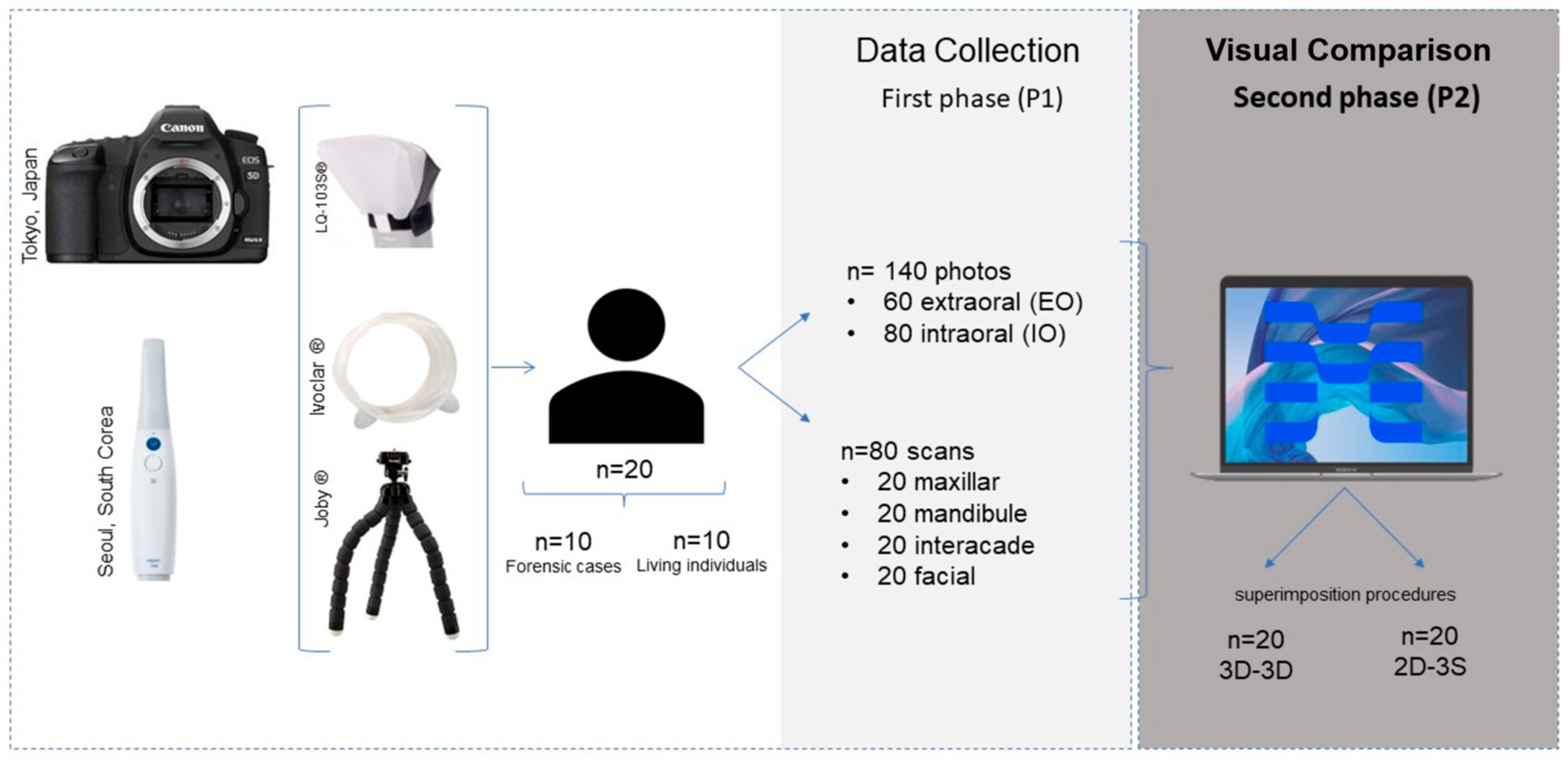
Figure 1. Summary and development of the study design.
Applsci 14 05892 g001
Figure 2. Facial scan of a living individual to illustrate the facial points (A) and lines collected (B). Legend: Gb (glabella); rEx (right exocanthion); lEx (left exocanthion); rEn (right endocanthion); lEn (left endocanthion); rNw (right nose wing); lNw (left nose wing); Na (nasion); Sn (subnasal); r Ch (right cheilion); lCh (left cheilion); Sl (superior lip); Il (inferior lip); Gn (gnathion); Me’ (cutaneous mento); En line (endocanthion line); Gb-Sn line (glabella and subnasal line); Nw line (nose wing line); Ch line (cheilion line); and occlusal plane
Applsci 14 05892 g002
Figure 3. A 3D–3D superimposition sequence. (A) Landmark of the upper right canine (cuspid) in 3D face; (B) landmark of the upper right canine (cuspid) in 3D intraoral space; and (C) analysis of the 3D–3D superimposition.
Applsci 14 05892 g003
Figure 4. The 2D-3S superimposition sequence (individual D7). (A). Selection and upload of records within the Smile Design software; (B) automatic profile of reference landmarks and their lines; (C) manual adjustment of landmarks and reference lines; (D) manual adjustment of study area; and (E) superimposition of volumes.
Applsci 14 05892 g004
Figure 5. Likert scale scores for the parameters. (A) The summed scores for living individuals (L1 to L10) and individuals in forensic cases (D1 to D10) (n = 20). (B) Boxplot graphic comparing scores between the photographic (blue) and IOS (green) approaches (n = 20).
Applsci 14 05892 g005
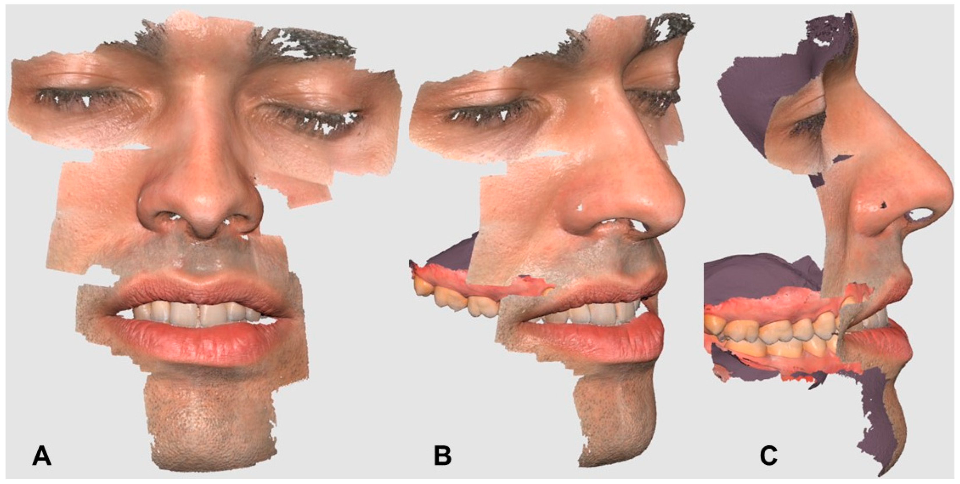
Figure 6. Personal data from living individual records. (A) Frontal view of extraoral scan, (B) 45 angle view of facial and maxillary scan, and (C) lateral view of 3D–3D superimposition.
Applsci 14 05892 g006
Figure 7. Descriptive analysis of 3D–3D superimposition results (n = 20) for living individuals (L1 to L10) and forensic cases (D1 to D10).
Applsci 14 05892 g007
Figure 8. Smile design representation of 2D-3S (case L3). (A) Landmark lines focusing on the 2D record and (B) landmark line superimposition in relation to the 3S record.
Applsci 14 05892 g008
Figure 9. Descriptive analysis of 2D-3D superimposition (n = 20). Legend: cohort match (green), no-match (red), and grey (depending on cohort definition).
Applsci 14 05892 g009
Table 1. Descriptive statistics for the photographs (n = 20). Intraoral (IO) and extraoral (EO) parameters (mineralisation details, soft tissue details, communication, extra devices, and distortion) were rated using a Likert scale ranging from 0 to 4, and the duration of the photography procedure was measured in seconds (s).
ParametersMeanStd. DeviationMinimunMaximum
LifeDeathLifeDeathLifeDeathLifeDeath
IntraoralMineralized3.702.400.460.493243
Soft2.701.700.460.462132
Communication2.002.000.000.002222
Extra-devices1.001.000.000.001111
Distortion3.001.400.000.493132
Duration300.90899.405.375.92298889309907
ExtraoralSoft4.004.000.000.004444
Communication2.002.000.000.002222
Extra-devices1.001.000.000.001111
Distortion4.003.200.000.404344
Duration89.30301.301.733.498629592308
Table 2. Descriptive statistics for the i700 wireless scan (n = 20). Intraoral (IO) and extraoral (EO) parameters (mineralisation details, soft tissue details, communication, extra devices, and distortion) were rated using a Likert scale ranging from 0 to 4, and the duration of the IOS procedure was measured in seconds (s).
ParametersMeanStd. DeviationMinimumMaximum
LifeDeathLifeDeathLifeDeathLifeDeath
IntraoralMineralized4.003.900.000.304344
Soft4.003.900.000.304344
Communication4.004.000.000.004444
Extra-devices4.003.000.000.004343
Distortion4.004.000.000.004444
Duration720.00788.1011.388.57709775750800
ExtraoralSoft3.804.000.400.003444
Communication4.004.000.000.004444
Extra-devices4.004.000.000.004444
Distortion3.804.000.400.003444
Duration123.7086.106.295.961157513593
Disclaimer/Publisher’s Note: The statements, opinions and data contained in all publications are solely those of the individual author(s) and contributor(s) and not of MDPI and/or the editor(s). MDPI and/or the editor(s) disclaim responsibility for any injury to people or property resulting from any ideas, methods, instructions or products referred to in the content.