Article

Effects of Age and Sex on the Kinematics of the Sprinting Technique in the Maximum Velocity Phase

by
Ioannis Chatzilazaridis
1,
Vassilios Panoutsakopoulos
2,
Eleni Bassa
3,*,
Mariana C. Kotzamanidou
1,4 and
Georgios I. Papaiakovou
4
1
Department of Coaching and Physical Education, School of Sport Sciences and Physical Education, Metropolitan College of Thessaloniki, 54624 Thessaloniki, Greece
2
Biomechanics Laboratory, School of Physical Education and Sport Science at Thessaloniki, Aristotle University of Thessaloniki, 54124 Thessaloniki, Greece
3
School of Physical Education and Sport Science at Serres, Aristotle University of Thessaloniki, Ag. Ioannis, 62110 Serres, Greece
4
Faculty of Health Sciences, Metropolitan College of Thessaloniki, 54624 Thessaloniki, Greece
*
Author to whom correspondence should be addressed.
Appl. Sci. 2024, 14(14), 6057; https://doi.org/10.3390/app14146057
Submission received: 10 June 2024 / Revised: 5 July 2024 / Accepted: 10 July 2024 / Published: 11 July 2024
(This article belongs to the Special Issue Advances in Sports Training and Biomechanics)

Abstract

:
The purpose of this study was to compare the step parameters, the Body Center of Mass (BCM) kinematics, as well as the angular and linear kinematics of the lower extremities’ joints of prepubescent and adult sprinters of both sexes. A total of forty-two athletes were examined, including adult men (AM) and women (AF) as well as preadolescent boys (PPB) and girls (PPG). A 2D-DLT analysis was conducted on video recordings (sampling frequency: 100 fps) of the participants’ maximum effort trial while in their maximum sprinting velocity phase. A 2 (age) × 2 (sex) ANOVA revealed significant (p < 0.05) effects of the factors age and sex, and an interaction of age × sex in the sprint running velocity. It was also found that the spatiotemporal structure of the step parameters was significantly (p < 0.05) different between AM and AF but not between PPB and PPG. Also, a significant (p < 0.05) main effect of age was evident mainly for the ankle joint of both legs, as well as for the angular kinematics of the swing leg’s knee joint. In conclusion, apart from the possible disparities in training experience, differences due to age and sex exist in the step parameters and the kinematics of the sprinting technique due to differences in the technical elements related to coordination, strength application capability, and stiffness.

1. Introduction

Sprint speed refers to the capacity of the human musculoskeletal system to facilitate forward body movement till reaching the point of maximum velocity [1]. Velocity assessments are frequently incorporated in talent identification tests [2,3] as they are recognized as a crucial factor for success in various adult [4,5] and youth sports [6,7].
The 100-m dash comprises the most representative and popular sprinting event in athletics. Data from both elite-level competitions and research studies have identified a typical running velocity (RV) development pattern during the 100-m sprint. This pattern consists of a rapid acceleration after the start (acceleration phase, 0–40 m), the RV regulation that leads to its maximization (maximum velocity phase, 40–80 m), and the decrease in RV as the finish line is approached (deceleration phase, 80–100 m) [8,9,10,11,12,13,14,15,16,17]. Each phase of sprinting exhibits variations in the neuro-muscular, kinetic, and kinematic demands of the movement [18].
In the maximum velocity phase, RV is a function of step length (SL) and step frequency (SF). The deterministic model of sprinting [19] suggests that SL comprises the stance distance and the flight distance. SF is defined by stride time, which is the sum of the duration of the support and flight phases within the stride. The body segment positioning at the instances of touchdown and take-off affects the duration of support and the stance distance, while the air resistance and the ground reaction forces at the stance phase affect the flight time and distance. It is suggested that the level of sprinting ability is associated with differences in key step kinematical parameters [20,21,22,23,24].
During development, RV is constantly increased [25,26,27,28,29,30,31]. However, the development of sprint speed follows a non-linear trajectory during childhood and adolescence [29,32] as a performance “spurt” in both preadolescence (between the ages of 5–9 years) and adolescence (between the ages of 12–14 years) has been reported [33]. In more detail, this cross-sectional study led to the conclusion that sprint RV was primarily increased in boys aged 7–11 years old and in girls between 6 and 9 years old [33]. Additionally, the most intensive improvement in RV, as noted in sprinting tests (30–50 m), has been reported to exist in children aged 9–11 years old [34,35,36]. However, comprehensive information regarding the biomechanical characteristics of running at speed in track-and-field athletes of this age is lacking.
During preadolescence, there is a development of sprinting ability without any apparent sex differences [32]. Sex differences in 30-m-sprint performance appear negligible until after 10 years of age [34] but after the age of 11, the rate of progression of speed development is significantly increased in boys compared to girls [37,38] and between early- and late-maturing girls [39]. In more detail, when examining sex differences in sprinting ability during puberty, there is a rapid development of sprinting ability in boys [33], whereas girls present a “plateau” in sprinting ability around 13 years of age [40]. These differences are suggested to be attributed to the larger increment in strength production by males [41]. The established relationship between the parameters composing the deterministic model of sprinting is decomposed at the age of 11–15 years old [26,31,42,43]. In general, the period of peak height velocity affects RV since SL increases and SF decreases [44]. The relative information for younger athletes—in order for developmental changes of running characteristics to be better understood—is not present in the respective literature.
The main difference between adult and young individuals in terms of sprint speed, apart from the lower maximum speed achieved by prepubescents, lies in the duration and total distance of the acceleration phase. Research revealed a difference regarding the secondary acceleration phase [45], which is very short in prepubescents and is completed between 20 and 30 m [46,47]. Furthermore, children achieve their maximum velocity around 30 m, they present a very short steady period [15,46], and a decrease afterward [46].
Although it is widely acknowledged that RV is determined by the combination of stride frequency and stride length [48], the leg joint angles are essential factors for the identification of the effectiveness of running gait [49,50]. Nevertheless, comprehensive research specifically addressing the developmental aspects of these characteristics during childhood is sparse. Studies in adults have indicated that faster runners achieve longer strides by increasing the application of ground reaction forces while reducing the duration of ground contact time [51,52], and enhancements in strength and power have been linked to improvements in both stride length and running speed [53]. Additionally, SF and SL values are not constant but vary across different phases of the sprint. In children, there is some controversy regarding stride frequency at maximum velocity. An earlier study reported that it remains consistent regardless of age [54] while another study indicated lower stride frequency for the less mature (pre-PHV) children [55]. Furthermore, boys’ speed developed at a low rate from 8.8 to 12.1 years of age and there was a decreased stride frequency in the older boys within this age span [40], whilst in age-matched girls, speed presented an accelerated improvement that was accompanied with a stride length enhancement at the phase of maximal speed—attributed to their probable ability to apply higher propulsive forces [56]. However, it seems that younger and less mature children rely more on stride frequency during maximum sprinting due to their lower musculotendinous stiffness and their lower ability to apply high rates of force development during stretch–shortening cycle (SSC) movements [57].
The comprehension of biomechanical factors in maximum sprint running is valuable due to their essential role in determining performance outcomes. Previous biomechanical analyses on young athletes’ sprint kinematics focused mainly on basic parameters such as SF, SL, duration of support, and flight time [26,31,42,43]. Dal Pupo et al. [9] provided some information about the knee joint angles at touchdown and take-off in 12-year-old boys and girls. Sex differences were not observed, and the same result was revealed in the comparison of the lower limb angular kinematics in a long (100 m) compared to a short (50 m) sprinting distance [9]. In general, the existence of this limited information in terms of understanding the mechanical determinants of youth’s sprint ability has been noted elsewhere [30]. Taking into consideration the previous conflicting results regarding sex differences in biomechanical parameters in untrained prepubescent children during sprinting, it would be of great interest to examine the possible age- and sex-related differences in the kinematic parameters in prepubescent and adult trained sprinters during the maximum sprinting phase.
The purpose of this study was to compare the step parameters, the Body Center of Mass (BCM) kinematics, as well as the linear and angular kinematics of the lower extremities’ joints of prepubescent and adult sprinters of both sexes. The hypotheses are as follows: (a) the spatiotemporal step parameters, i.e., SF, SL, duration of support, etc., will be influenced by age as they are strength-related; (b) the angular kinematics (angle and angular velocity of the lower limb joints) will not be altered between the age groups, since sprinting is a congenital ability; and (c) sex differences in RV and the biomechanical parameters will be present only in adults.

2. Materials and Methods

2.1. Participants

A total of forty-two athletes were examined (Table 1). Groups AM and AF comprised national- and international-level adult men and women sprinters, respectively. Groups PPB and PPG were formed by boys and girls, respectively, who were novice athletes but systematically trained in track sprints. The members of the PPB and PPG groups were classified as preadolescents based on the classification method proposed by Tanner [58]. A power analysis using G*Power software (version 3.1.9.6, Franz Faul, University Kiel, Kiel, Germany) was conducted for an effect size greater than 0.25, a probability error of 0.05, and a power of 0.95 for four groups; a sample size of 40 participants was considered sufficient.
The selection of PPB and PPG athletes, being about 11 years old, was conducted bearing in mind that they were at the age where mentionable changes in sprinting performance occur [26,31,37,38,39,42,43]. Regarding the inclusion criteria, all participants had to be in good physical condition, with no apparent or reported injury or disability, free from any musculoskeletal or neurological disorders that may affect sprinting performance, and with a recorded regular participation in their training program. Further inclusion criteria for the adult participants were that they should be national- and/or international-level men and women sprinters, whereas children should be systematically trained in track sprints for at least 2 years. Regarding the exclusion criteria, children who did not fall within the specified age range mentioned above, or whose maturation stage was higher than Tanner stage 2, were excluded. Participants with a history of lower limb injuries that may impact sprinting mechanics were excluded as well.
The investigation was conducted under the Institutional Research Committee guidelines for the use of human subjects, which require participants to be informed of the risks of contributing to the study and the acquisition of a signed informed consent document and/or parental approval prior to the experimental procedure. Thus, ethical approval was granted by the Institutional Reviewing Board (172/2023-16 November 2023).

2.2. Experimental Procedure

The experimental procedure was conducted at the beginning of the competitive season in the early spring. Every participant performed a maximal sprinting test on a level 100-m indoor track with a rubber surface, which was preceded by a self-selected warm-up. The test was a 50-m all-out sprinting task for groups AM and AF and a 40-m full-effort sprint for groups PPB and PPG. The shorter distance for PPB and PPG was selected since it has been found that secondary acceleration in kids concludes at 20–30 m [47,59]. Furthermore, previous findings suggest that children cannot maintain their maximum RV after the 40-m mark of a maximal sprinting test [60]. This was also established by the results of the recorded 10-m splits (according to Chatzilazaridis et al. [46]) during a familiarization session that was conducted three days before the actual testing session.
The participants started the test from the standing position 1 m behind the start line. The instruction was to “run as fast as possible from the beginning and to pass with maximum effort through the finish line”. A pair of photocells (Tag Heuer, La Chaux-de-Fonds, Switzerland) were positioned at both the start and finish lines to capture the performance of the sprinting test.
Three consecutive support phases of the sprinting strides were recorded at the 40 ± 4 m mark of the 50-m test and the 30 ± 4 m mark of the 40-m test with the digital video camera. A stationary video camera (JVC GR-DVL 9600EG, Victor Company of Japan Ltd., Yokohama, Japan) was attached to a 1.2 m height fixed tripod, which was positioned 12 m from the lane of the track where the sprinting tests were executed. The camera’s optical axis was perpendicular to the plane of motion. A 2.5 m × 2.5 m calibration frame with 12 control markers was placed, throughout the filming view, at the middle of the lane and perpendicular to the camera’s axis to execute a 2D-DLT kinematical analysis [61]. The X-axis represented the direction of running and the Y-axis was vertical to the X-axis.

2.3. Data Analysis

Eighteen anatomical points of the body (tip of the toe, ankle, knee, hip, shoulder, elbow, wrist, and fingers on both sides of the body, the neck, and the head) and selected points in the filming view were manually digitized in each field. To calculate the Body Center of Mass (BCM) and the segmental inertia properties for the link segment model, the proportional to subject-specific height and weight characteristic formulas recommended by Plagenhoef et al. [62] were used. Cut-off frequency for smoothing was set at 6 Hz [63]. Digitization, smoothing, and analyses were performed with the A.P.A.S. v.14.1.0.5 software (Ariel Dynamics Inc., Trabuco Canyon, CA, USA). The accuracy of the 2D reconstruction was determined by the root mean square error, after randomly re-digitizing 5% of the captured frames. An error of 1.1 cm and 0.7 cm was found for the X- and Y-axes, respectively.
Regarding the examined parameters, RV was calculated by dividing the test distance by the time recorded by the photocells, while SF was defined as the steps taken per second. The touchdown was defined as the first field where the foot had clearly contacted the ground. The take-off was defined as the first field where the foot had clearly left the ground. Thus, the time of the contact and the flight phases were assessed for each step.
Based on the XY coordinates of the digitized anatomical points, the experimental parameters were as follows:
  • SL: the horizontal distance between the touchdown points of the feet recorded for two consecutive supports;
  • Touchdown distance (STD): the horizontal distance between the toe of the support foot and the BCM projection at touchdown;
  • Take-off distance (STO): the horizontal distance between the toes of the support foot and the BCM projection at take-off;
  • Horizontal BCM take-off velocity (VxTO): the horizontal velocity of BCM at take-off;
  • Vertical BCM take-off velocity (VyTO): the vertical velocity of BCM at take-off;
  • Angle of take-off (AngPr): the Arc-tangent of the vertical/horizontal BCM take-off velocity;
  • Horizontal ankle touchdown velocity (VXankle): the horizontal velocity of the ankle at touchdown;
  • BCM height: the height of the BCM at touchdown (HTD), maximum lowering of the BCM (HAM), and take-off (HTO);
  • Knee joint angle: The angle formed between the thigh and the shank at touchdown (θkneeTD), maximum knee amortization (θkneeAM), and take-off (θkneeTO). The minimum swing leg’s knee angle at the support phase was also calculated (θkneeMIN);
  • Ankle joint angle: the angle formed between the shank and the foot at touchdown (θankleTD) and take-off (θkneeTO);
  • Knee joint angular velocity (ωKNEE): the angular velocity of the swing leg’s knee joint at take-off;
  • Ankle joint angular velocity (ωANKLE): the angular velocity of the support leg’s ankle joint at touchdown;
  • Thigh inclination (φTHIGH): the angle formed by the horizontal axis and the thigh of the swing leg at take-off;
  • Leg inclination: the angle formed by the horizontal axis and the line passing from the support leg’s ankle and hip at touchdown (φlegTD) and take-off (φlegTO).

2.4. Statistical Analysis

IBM SPSS Statistics v.28.0 software (International Business Machines Corp., Armonk, NY, USA) was used for the statistical analyses. For all analyses, an a = 0.05 level of significance was set. A 2 × 2 ANOVA with Bonferroni’s adjustment was run to examine the main effects of age and sex and the interaction effect between age and sex on the biomechanical parameters of the maximum RV sprint step. The effect sizes were checked with the partial eta-squared statistic (ηp2). Small, medium, and large effect sizes were defined by values of above 0.01, 0.06, and 0.14, respectively.

3. Results

The results regarding the RV are presented in Figure 1. A significant effect of age (F(1,41) = 281.38, p < 0.001, ηp2 = 0.89), a significant sex effect (F(1,41) = 7.93, p = 0.008, ηp2 = 0.19), and an interaction of age × sex (F(1,41) = 5.64, p < 0.001, ηp2 = 0.14) were found.
Table 2 depicts the results for the step parameters and the BCM kinematics, whereas Table 3 presents the results of the statistical analysis for the above-mentioned variables. Results indicated a significant (p < 0.05) main effect of sex in the step temporal parameters (SF and contact time); however, these parameters were significantly (p < 0.05) different between AM and AF but not between PPB and PPG.
This was also the case for the other parameters that were found to be statistically different between sexes in adulthood but were not different in prepubescence (horizontal BCM take-off velocity, angle of projection, touchdown distance). Also, a significant (p < 0.05) main effect of age was observed for the step parameters, the temporal structure of the step and the BCM kinetics (horizontal and vertical take-off velocity, its lowering during the support phase, and its distance from the point of support at the take-off). Finally, a significant (p < 0.05) interaction of age × sex was revealed for the horizontal BCM take-off velocity and the angle of projection.
The results for the linear and angular kinematics of the lower extremities’ joints and segments and the respective results of the statistical analysis are depicted in Table 4 and Table 5, respectively. Again, in the vast majority of the parameters that were different between AM and AF, the sex difference was absent in the comparison between PPB and PPG.
The only exception was the angular velocity of the ankle joint at touchdown, where significant (p < 0.05) sex differences were observed in both adults and prepubescents. A significant (p < 0.05) main effect of age was evident mainly for the ankle joint of both legs, as well as for the angular kinematics of the swing leg’s knee joint. A significant (p < 0.05) interaction of age × sex was found only for the swing leg’s knee angle at take-off.
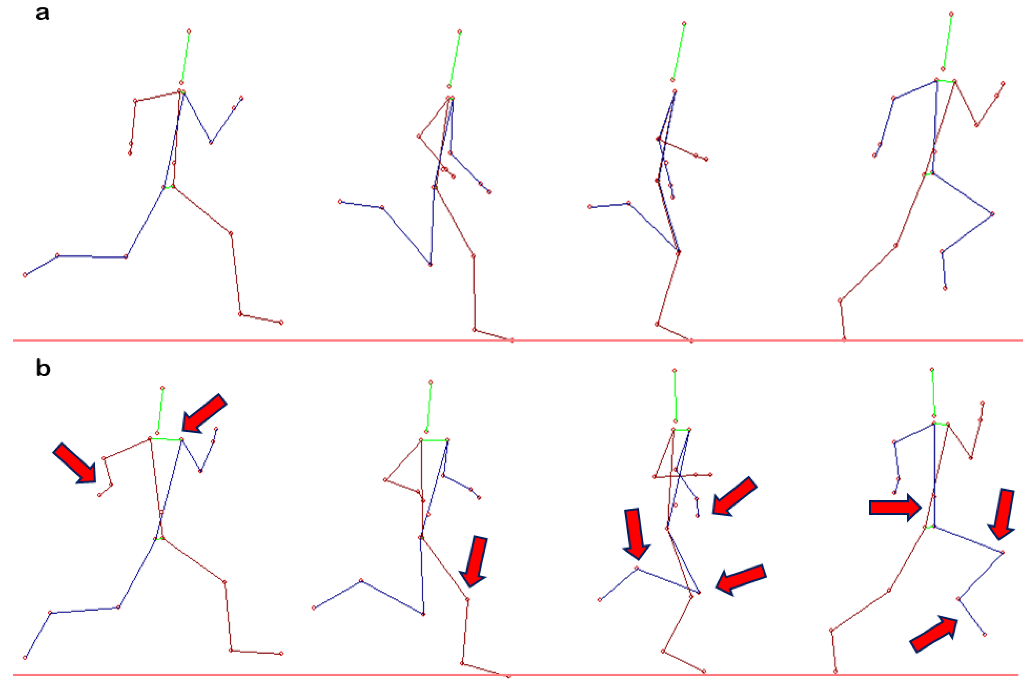
Figure 2 and Figure 3 depict representational stick figures of AF vs. PPG and AM vs. PPB, respectively. The qualitative information derived from Figure 2 and Figure 3 suggest that prepubescents performed the analyzed maximum velocity phase sprint step in a more upright torso position, with less dynamic movement of the arms and the swing leg, increased knee flexion during the braking phase, and less active ankle movement at both touchdown and take-off.

4. Discussion

The comparison of adult men and women sprinters with systematically trained prepubescent track-and-field athletes during maximum velocity sprinting revealed that the spatiotemporal structure of the step parameters was significantly (p < 0.05) different between AM and AF but not between PPB and PPG. This confirmed one of the hypotheses of this study. As for the other hypothesis, a significant (p < 0.05) main effect of age was evident mainly for the ankle joint of both legs, as well as for the angular kinematics of the swing leg’s knee joint. This did not support the hypothesis that the angular kinematics (angle and angular velocity of the lower limb joints) would not be altered between age groups as sprinting is considered a congenital ability.
Based on the results, it is observed that both boys and girls exhibit lower sprint velocity at maximal sprinting compared to adult men and women. Regarding the sprinting technique per se, it is suggested that the anthropometric parameters (body dimensions, musculature, and fat tissue) influence sprint performance [64,65]. The leg length and the body height were found to be related to step length [66]. As children are smaller in size, there is a possibility that this influenced our findings. Regarding the other step characteristic, namely the step frequency, it was found that the boys’ speed developed at a low rate from 8.8 to 12.1 years of age as a decreased stride frequency in the older boys within this age span was detected [40]. On the other hand, the speed of age-matched girls presented an accelerated improvement, which was accompanied with a stride length enhancement at the phase of maximal speed that was attributed to their probable ability to apply higher propulsive forces [56]. However, these sex-related differences in maximal sprinting were not confirmed in this study.
This difference in sprint performance may be attributed to several other factors than the size differences that affect step length. Firstly, one possible explanation is the higher propulsive force that adults can apply compared to children during stretch–shortening cycle (SSC) movements, as previously confirmed in drop-jump tasks [67,68]. Male and female adults can generate more force relative to their body weight, resulting in greater sprinting velocities compared to boys and girls. Secondly, the time required to achieve maximal propulsive force may also be a contributing factor. Adults reach their maximum force output more quickly than children in SSC actions [67,68], and this probably enables them to generate maximal force at a faster rate during sprinting as well.
The examined prepubescent athletes had a larger ratio of contact time within the duration of the step. This is related to the duration of the braking phase, which should be as short as possible [69]. To optimize the transition of the braking to the propulsive phase, foot placement plays a vital role [69]. Our findings regarding the BCM horizontal velocity of the ankle joint at touchdown suggest that the examined prepubescent athletes performed the foot placement with a blocking instead of a “pawing” action. This is a disadvantage for the fast transition from braking to propulsion, as well as for the application of optimum horizontal force. The application of horizontal force during sprinting is of utmost importance as power and horizontal force have been identified as the top predictors of maximal velocity within different ages and stages of maturity in females and males, respectively [55]. It is also suggested that less mature children differed significantly from the more mature in speed, step length, step frequency, vertical and horizontal force, and horizontal power. The lower sprinting performance of children is most likely caused by the lower mean propulsive forces and a shorter SL during maximal sprinting, with suggestions that the shorter SL is attributed to the lower vertical impulse and the lower height found in younger boys compared to older boys [40].
The more upright body position in prepubescent athletes should be examined together with the orientation of the pelvis. It is suggested that the orientation of the pelvis has an impact on the locomotion of the legs [70]. A backward orientation of the pelvis is not favorable for developing maximum-speed sprinting [70], and thus constitutes a technique element affected by age.
Another factor affecting sprinting performance may be adults’ lower antagonist activity, which was beyond the aims of this study. Initially, antagonist activity was proposed as a mechanism that reduces shear forces and enhances joint stiffness and stability [71]. Studies in single-joint movements have shown no differences in the coactivation level for the knee [72,73] and the elbow muscles [74] but higher level of coactivation in the plantar flexors in children [75]. However, in complex movements such as walking or running [76], children tend to demonstrate higher values of coactivation indicating that during dynamic and multi-joint movements, children rely more on simultaneous activation of agonist and antagonist muscles to achieve joint stabilization and control. This higher coactivation of children observed during both the braking and propulsive phases of jumps has been attributed and related to coordination challenges or the immaturity of the neuromuscular system [77] and may have a negative impact on their sprinting performance as well.
The time required to achieve maximal propulsive force may also be a contributing factor. Adults reach their maximum force output more quickly than children in SSC actions [67,68] and this probably enables them to generate maximal force at a faster rate during sprinting as well. Rate of force development is highly related with the muscle–tendon unit properties. The muscle–tendon unit should be optimally stiff to store elastic energy and to release it efficiently during the propulsive phase in SSC actions. Children present lower muscle tendon stiffness compared to adults [78], resulting in prolonged contact times in SSC actions, as also found in this study.

Limitations of This Study

This study is not free of limitations. The authors acknowledge that the sample size, especially for AF, is limited; however, the power analysis revealed that the sample size selected was sufficient to detect group differences. The chronic effects of training and experience in adults could not be comparable with those of children. Nevertheless, the aim of this study was to check the differences in the kinematics of the sprinting technique in a cross-sectional manner. In addition, no detailed anthropometric measures (body fat, segment length, muscularity, lean mass, etc.) were conducted to examine the differences using allometry. For example, lean body mass is considered a main contributor to rate of force development [79]. Thus, age and sex differences in body composition may have also accounted for the group differences in sprinting performance. Thus, further research is needed to explore the influence of maturation on the development of the neuromuscular parameters of maximum velocity sprint running from childhood until adulthood. Future research should be conducted on revealing how coactivation alterations during this period of human development may impact sprinting performance, as well as how the attained muscle mass after adolescence can contribute to higher power-production capability and, inductively, to greater sprinting velocity.

5. Conclusions and Practical Applications

In conclusion, the age difference between adults and prepubescent sprinters in the relative step length reflects the differences in strength and power-production capabilities. In addition, this difference influences the technique elements for an effective support phase, as less active touchdown action of the ankle joint leads to prolonged contact time and a non-optimal pattern of horizontal force application.
Training has the potential to modify running mechanics, and by adopting an efficient running technique, individuals can enhance their running economy, leading to improved performance. Therefore, according to the special biomechanical characteristics observed in children in this study, training should focus on the following: (1) differentiating the torso position for better forward movement; (2) improving foot placement in order to optimize horizontal force application; and (3) increasing children’s reactive strength to help them better control the braking phase and, therefore, be more capable to apply faster higher forces at the propulsive phase. Technique drills focusing on the active movement of the ankle joint at both touchdown and take-off while reducing contact time are suggested to be included in the training routine of prepubescent track-and-field athletes.

Author Contributions

Conceptualization, I.C., V.P. and G.I.P.; methodology, V.P. and G.I.P.; software, V.P.; validation, I.C., V.P. and G.I.P.; formal analysis, I.C. and V.P.; investigation, I.C., V.P. and G.I.P.; resources, G.I.P.; data curation, I.C.; writing—original draft preparation, I.C., V.P. and E.B.; writing—review and editing, V.P., E.B., M.C.K. and G.I.P.; visualization, V.P.; supervision, G.I.P.; project administration, G.I.P. All authors have read and agreed to the published version of the manuscript.

Funding

This research received no external funding.

Institutional Review Board Statement

This study was conducted in accordance with the Declaration of Helsinki and approved by the Institutional Review Board of the Aristotle University of Thessaloniki (172/2023-16 November 2023).

Informed Consent Statement

Informed consent or parental consent was obtained from all subjects involved in this study.

Data Availability Statement

The data presented in this study are available on reasonable request from the corresponding author due to ethical reasons.

Conflicts of Interest

The authors declare no conflicts of interest.

References

  1. Ross, A.; Leveritt, M.; Riek, S. Neural influences on sprint running: Training adaptations and acute responses. Sports Med. 2001, 31, 409–425. [Google Scholar] [CrossRef] [PubMed]
  2. Cardoso, P.; Tavares, F.; Loureiro, N.; Ferreira, R.; Araújo, J.P.; Reis, J.; Vaz, J.R. In-situ acceleration-speed profile of an elite soccer academy: A cross-sectional study. J. Sports Sci. 2023, 41, 1868–1874. [Google Scholar] [CrossRef] [PubMed]
  3. King, M.; Brown, M.; Cox, J.; McLellan, R.; Towlson, C.; Barrett, S. Talent identification in soccer: The influence of technical, physical and maturity-related characteristics on a national selection process. PLoS ONE 2024, 19, e0298359. [Google Scholar] [CrossRef] [PubMed]
  4. De Lacey, J.; Brughelli, M.E.; McGuigan, M.R.; Hansen, K.T. Strength, speed and power characteristics of elite rugby league players. J. Strength Cond. Res. 2014, 28, 2372–2375. [Google Scholar] [CrossRef] [PubMed]
  5. Djaoui, L.; Chamari, K.; Owen, A.L.; Dellal, A. Maximal Sprinting Speed of Elite Soccer Players During Training and Matches. J. Strength Cond. Res. 2017, 31, 1509–1517. [Google Scholar] [CrossRef] [PubMed]
  6. Joseph, J.; McIntyre, F.; Joyce, C.; Scanlan, A.; Cripps, A. A comparison of multidimensional qualities discriminant of selection in elite adolescent Australian basketball athletes. PLoS ONE 2021, 16, e0256032. [Google Scholar] [CrossRef]
  7. Tsoukos, A.; Drikos, S.; Brown, L.E.; Sotiropoulos, K.; Veligekas, P.; Bogdanis, G.C. Upper and Lower Body Power Are Strong Predictors for Selection of Male Junior National Volleyball Team Players. J. Strength Cond. Res. 2019, 33, 2760–2767. [Google Scholar] [CrossRef] [PubMed]
  8. Ae, M.; Ito, A.; Suzuki, M. The men’s 100 metres. N. Stud. Athl. 1992, 7, 47–52. [Google Scholar]
  9. Dal Pupo, J.; da Rocha, C.I., Jr.; Mota, C.B. Concentrações de lactato sangüíneo e o comportamento cinemático de corredores mirins em provas de 50 e 100m rasos. Rev. Bras. Cineantropom. Desempenho Hum. 2008, 10, 393–398. [Google Scholar] [CrossRef]
  10. Delecluse, C.; Van Coppenolle, H.; Willems, R.; Diels, M.; Goris, M.; Van Leempurte, M.; Vuylsteke, M. Analysis of 100 meter sprint performance as a multi-dimensional skill. J. Hum. Mov. Stud. 1995, 28, 87–101. [Google Scholar]
  11. Ferro, A.; Rivera, A.; Pagola, I.; Ferreruela, M.; Martin, A.; Rocandio, V. Biomechanical analysis of the 7th World Championships in Athletics Seville 1999. N. Stud. Athl. 2001, 16, 25–60. [Google Scholar]
  12. Gajer, B.; Thepaut-Mathieu, C.; Lehenaff, D. Evolution of stride and amplitude during course of the 100 m event in athletics. N. Stud. Athl. 1999, 14, 43–50. [Google Scholar]
  13. Graubner, R.; Nixdorf, E. Biomechanical analysis of the sprint and hurdles events at the 2009 IAAF World Championships in Athletics. N. Stud. Athl. 2011, 26, 17–53. [Google Scholar]
  14. Kersting, U.G. Biomechanical analysis of the sprinting events. In Biomechanical Research Project Athens 1997—Final Report; Brüggemann, G.P., Koszewski, D., Müller, H., Eds.; Meyer & Meyer Sport: Oxford, UK, 1999; pp. 12–61. [Google Scholar]
  15. Letzelter, S. The development of velocity and acceleration in sprints: A comparison of elite and juvenile female sprinters. N. Stud. Athl. 2006, 21, 15–22. [Google Scholar]
  16. Mackala, K. Optimisation of performance through kinematic analysis of the different phases of the 100 metres. N. Stud. Athl. 2007, 22, 7–16. [Google Scholar]
  17. Moravec, P.; Ruzicka, J.; Susanka, P.; Dostal, E.; Kodejs, M.; Nosek, M. The 1987 International Athletic Foundation/IAAF Scientific Project Report: Time analysis of Ihe 100 Metres events at the II World Championships in Athletics. N. Stud. Athl. 1988, 3, 61–96. [Google Scholar]
  18. Colyer, S.L.; Nagahara, R.; Salo, A.I.T. Kinetic demands of sprinting shift across the acceleration phase: Novel analysis of entire force waveforms. Scand. J. Med. Sci. Sports 2018, 28, 1784–1792. [Google Scholar] [CrossRef]
  19. Hay, J.G. The Biomechanics of Sports Techniques, 3rd ed.; Prentice-Hall: Englewood Cliffs, NJ, USA, 1985. [Google Scholar]
  20. Babic, V.; Coh, M.; Dizdar, D. Differences in kinematic parameters of athletes of different running quality. Biol. Sport 2011, 28, 115–121. [Google Scholar] [CrossRef]
  21. Mann, R.V.; Herman, J. Kinematic analysis of Olympic sprint performance: Men’s 200 meters. Int. J. Sports Biomech. 1985, 1, 151–162. [Google Scholar] [CrossRef]
  22. Mero, A.; Komi, P.V. Force-, EMG-, and elasticity-velocity relationships at submaximal, maximal and supramaximal running speeds in sprinters. Eur. J. Appl. Physiol. 1986, 55, 553–561. [Google Scholar] [CrossRef]
  23. Nummela, A.; Stray-Gundersen, J.; Rusko, H. Effects of fatigue on stride characteristics during a short-term maximal run. J. Appl. Biomech. 1996, 12, 151–160. [Google Scholar] [CrossRef]
  24. Vardaxis, V.; Hoshizaki, T.B. Power patterns of the leg during the recovery phase of the sprinting stride for advanced and intermediate sprinters. Int. J. Sport Biomech. 1989, 5, 332–349. [Google Scholar] [CrossRef]
  25. Coh, M.; Bracic, M.; Smajlovic, N. Methodical aspects of maximum speed development. Sport Sci. 2010, 3, 11–14. [Google Scholar]
  26. Kumar, H. Age changes in the speed of running during 30 meter sprint running. J. Exerc. Sci. Physiother. 2006, 2, 92–95. [Google Scholar]
  27. Loko, J.; Aule, R.; Sikkut, T.; Ereline, J.; Viru, A. Age differences in growth and physical abilities in trained and untrained girls 10–17 years of age. Am. J. Hum. Biol. 2003, 15, 72–77. [Google Scholar] [CrossRef] [PubMed]
  28. Milanese, C.; Bortolami, O.; Bertucco, M.; Verlato, G.; Zancanaro, C. Anthropometry and motor fitness in children aged 6–12 years. J. Hum. Sport Exerc. 2010, 5, 265–279. [Google Scholar] [CrossRef]
  29. Papaiakovou, G.; Giannakos, A.; Michailidis, C.; Patikas, D.; Bassa, E.; Kalopisis, V.; Anthrakidis, N.; Kotzamanidis, C. The effect of chronological age and gender on the development of sprint performance during childhood and puberty. J. Strength Cond. Res. 2009, 23, 2568–2573. [Google Scholar] [CrossRef]
  30. Rumpf, M.C.; Cronin, J.B.; Oliver, J.L.; Hughes, M. Assessing Youth Sprint Ability—Methodological Issues, Reliability and Performance Data. Ped. Exerc. Sci. 2011, 23, 442–467. [Google Scholar] [CrossRef] [PubMed]
  31. Vanderka, M.; Kampmiller, T. Ontogenetic development of kinematic parameters of the running stride. Sport Sci. Rev. 2011, 10, 5–24. [Google Scholar] [CrossRef]
  32. Malina, R.M.; Bouchard, C.; Bar-Or, O. Growth, Maturation, and Physical Activity; Human Kinetics: Champaign, IL, USA, 2004. [Google Scholar]
  33. Viru, A.; Loko, J.; Harro, M.; Volver, A.; Laaneots, L.; Viru, M. Critical periods in the development of performance capacity during childhood and adolescence. Eur. J. Phys. Educ. 1999, 4, 75–119. [Google Scholar] [CrossRef]
  34. Branta, C.; Haubenstricker, J.; Seefeldt, V. Age changes in motor skills during childhood and adolescence. Exerc. Sport Sci. Rev. 1984, 12, 467–520. [Google Scholar] [CrossRef]
  35. Shephard, R.J.; Lavallee, H. Changes of physical performance as indicators of the response to enhanced physical education. J. Sport Med. Phys. Fitness 1994, 34, 323–335. [Google Scholar]
  36. Loko, J.; Aule, R.; Sikkut, T. Dynamics of Estonian adolescents physical abilities. Acta Academ. Olymp. Est. 1994, 2, 102–114. [Google Scholar]
  37. Keller, B.A. Development of fitness in children: The influence of gender and physical activity. Am. J. Lifestyle Med. 2008, 2, 58–74. [Google Scholar] [CrossRef]
  38. Whithall, J. Development of locomotor co-ordination and control in children. In Development of Movement Coordination in Children: Applications in the Field of Ergonomics, Health Sciences and Sport; Savelsberg, G.P.J., Davids, K., Van der Kamp, J., Eds.; Routledge: London, UK, 2003; pp. 251–270. [Google Scholar]
  39. Sokołowski, B.; Chrzanowska, M. Development of selected motor skills in boys and girls in relation to their rate of maturation—A longitudinal study. Hum. Mov. 2012, 13, 132–138. [Google Scholar] [CrossRef]
  40. Nagahara, R.; Takai, Y.; Haramura, M.; Mizutani, M.; Matsuo, A.; Kanehisa, H.; Fukunaga, T. Age-Related Differences in Spatiotemporal Variables and Ground Reaction Forces During Sprinting in Boys. Pediatr. Exerc. Sci. 2018, 30, 335–344. [Google Scholar] [CrossRef] [PubMed]
  41. Coh, M.; Babic, V.; Mackała, K. Biomechanical, neuro-muscular and methodical aspects of running speed development. J. Hum. Kin. 2010, 26, 73–81. [Google Scholar] [CrossRef]
  42. Coh, M.; Skof, B.; Kugovnik, O.; Dolenec, A.; Kampmiller, T.; Laczo, E.; Šelinger, P. Kinematic and dynamic characteristics of maximal speed in young sprinters. Kinesiol. Slov. 1995, 2, 5–10. [Google Scholar]
  43. Kampmiller, T.; Vanderka, M.; Selinger, P.; Selingerova, M.; Cierna, D. Kinematic parameters of the running stride in 7- to 18-year-old youth. Kinesiol. Slov. 2011, 17, 63–75. [Google Scholar]
  44. Coh, M.; Babic, V. Biodynamic characteristics of maximum speed development. Facta Univers. Phys. Educ. Sport 2010, 8, 141–148. [Google Scholar]
  45. Meyers, R.W.; Oliver, J.L.; Hughes, M.G.; Cronin, J.B.; Lloyd, R.S. Maximal sprint speed in boys of increasing maturity. Pediatr. Exerc. Sci. 2015, 27, 85–94. [Google Scholar] [CrossRef] [PubMed]
  46. Chatzilazaridis, I.; Panoutsakopoulos, V.; Papaiakovou, G. Stride characteristics progress in a 40-m sprinting test executed by male preadolescent, adolescent and adult athletes. J. Biol. Exerc. 2012, 8, 58–77. [Google Scholar] [CrossRef]
  47. Kotzamanidis, C. The effect of sprint training on running performance and vertical jumping in pre-adolescent boys. J. Hum. Mov. Stud. 2003, 44, 225–240. [Google Scholar]
  48. Hunter, J.P.; Marshall, R.N.; McNair, P.J. Interaction of step length and step rate during sprint running. Med. Sci. Sport Exerc. 2004, 36, 261–271. [Google Scholar] [CrossRef] [PubMed]
  49. Xu, D.; Zhou, H.; Quan, W.; Jiang, X.; Liang, M.; Li, S.; Ugbolue, U.C.; Baker, J.S.; Gusztav, F.; Ma, X.; et al. A new method proposed for realizing human gait pattern recognition: Inspirations for the application of sports and clinical gait analysis. Gait Posture 2024, 107, 293–305. [Google Scholar] [CrossRef] [PubMed]
  50. Xu, D.; Quan, W.; Zhou, H.; Sun, D.; Baker, J.S.; Gu, Y. Explaining the differences of gait patterns between high and low-mileage runners with machine learning. Sci. Rep. 2022, 12, 2981. [Google Scholar] [CrossRef] [PubMed]
  51. Weyand, P.G.; Sternlight, D.B.; Bellizzi, M.J.; Wright, S. Faster top running speeds are achieved with greater ground forces not more rapid leg movements. J. Appl. Physiol. 2000, 89, 1991–1999. [Google Scholar] [CrossRef]
  52. Weyand, P.G.; Sandell, R.F.; Prime, D.N.; Bundle, M.W. The biological limits to running speed are imposed from the ground up. J. Appl. Physiol. 2010, 108, 950–961. [Google Scholar] [CrossRef]
  53. Wisløff, U.; Castagna, C.; Helgerud, J.; Jones, R.; Hoff, J. Strong correlation of maximal squat strength with sprint performance and vertical jump height in elite soccer players. Br. J. Sport Med. 2004, 38, 285–288. [Google Scholar] [CrossRef]
  54. Schepens, B.; Willems, P.A.; Cavagna, G.A. The mechanics of running in children. J. Physiol. 1998, 509 Pt 3, 927–940. [Google Scholar] [CrossRef]
  55. Rumpf, M.C.; Cronin, J.B.; Oliver, J.; Hughes, M. Kinematics and Kinetics of Maximum Running Speed in Youth Across Maturity. Pediatr. Exerc. Sci. 2015, 27, 277–284. [Google Scholar] [CrossRef]
  56. Nagahara, R.; Haramura, M.; Takai, Y.; Oliver, J.L.; Wichitaksorn, N.; Sommerfield, L.M.; Cronin, J.B. Age-related differences in kinematics and kinetics of sprinting in young female. Scand. J. Med. Sci. Sports 2019, 29, 800–807. [Google Scholar] [CrossRef] [PubMed]
  57. Rumpf, M.C.; Cronin, J.B.; Oliver, J.L.; Hughes, M. Vertical and leg stiffness and stretch-shortening cycle changes across maturation during maximal sprint running. Hum. Mov. Sci. 2013, 32, 668–676. [Google Scholar] [CrossRef] [PubMed]
  58. Tanner, J.M. Normal growth and techniques of growth assessment. Clin. Endocrinol. Metabol. 1986, 15, 411–451. [Google Scholar] [CrossRef] [PubMed]
  59. Kotzamandis, C. The acute and long-term effect of training on running velocity. Inq. Sport Phys. Educ. 2007, 5, 179–186. [Google Scholar]
  60. Babic, V.; Blazevic, I.; Vlasic, J. Characteristics of sprinting in pre-school and junior elementary school children. Hrvat. Športskomed. Vjesn. 2010, 25, 3–8. [Google Scholar]
  61. Kollias, I.A. Sources of error and their elimination in the use of DLT with the basic recording tools for the analysis of human body in motion. Exerc. Societ. J. Sport Sci. 1997, 18, 9–26. [Google Scholar]
  62. Plagenhoef, S.; Evans, F.G.; Abdelnour, T. Anatomical data for analyzing human motion. Res. Q. Exerc. Sport 1983, 54, 169–178. [Google Scholar] [CrossRef]
  63. Winter, D.A.; Sidwall, H.D.; Hobson, D.A. Measurement and reduction of noise in kinematics of locomotion. J. Biomech. 1974, 7, 157–159. [Google Scholar] [CrossRef] [PubMed]
  64. Babic, V.; Harasin, D.; Dizdar, D. Relations of the variables of power and morphological characteristics to the kinematic indicators of maximal speed running. Kinesiology 2007, 39, 28–39. [Google Scholar]
  65. Hoshikawa, Y.; Muramatsu, M.; Iida, T.; Uchiyama, A.; Nakajima, Y.; Kanehisa, H.; Fukunaga, T. Influence of the psoas major and thigh muscularity on 100-m times in junior sprinters. Med. Sci. Sports Exerc. 2006, 38, 2138–2143. [Google Scholar] [CrossRef]
  66. Chow, J.W. Maximum speed of female high school runners. Int. J. Sport Biomech. 1987, 3, 110–127. [Google Scholar] [CrossRef]
  67. Lazaridis, S.N.; Bassa, E.I.; Patikas, D.; Hatzikotoulas, K.; Lazaridis, F.K.; Kotzamanidis, C.M. Biomechanical comparison in different jumping tasks between untrained boys and men. Pediatr. Exerc. Sci. 2013, 25, 101–113. [Google Scholar] [CrossRef] [PubMed]
  68. Katsikari, K.; Bassa, E.; Skoufas, D.; Lazaridis, S.; Kotzamanidis, C.; Patikas, D.A. Kinetic and Kinematic Changes in Vertical Jump in Prepubescent Girls After 10 Weeks of Plyometric Training. Pediatr. Exerc. Sci. 2020, 32, 81–88. [Google Scholar] [CrossRef] [PubMed]
  69. Coh, M.; Jost, B.; Stuhec, S. Kinematic and dynamic structure of the sprinting stride of top female sprinters. Biol. Sport 1998, 15, 237–243. [Google Scholar]
  70. Vonstein, W. Some reflections on maximum speed sprinting technique. N. Stud. Athl. 1996, 11, 161–165. [Google Scholar]
  71. Kellis, E. Quantification of quadriceps and hamstring antagonist activity. Sports Med. 1998, 25, 37–62. [Google Scholar] [CrossRef]
  72. Bassa, E.; Patikas, D.; Kotzamanidis, C. Activation of Antagonist Knee Muscles during Isokinetic Efforts in Prepubertal and Adult Males. Pediatr. Exerc. Sci. 2005, 17, 171–181. [Google Scholar] [CrossRef]
  73. O’Brien, T.D.; Reeves, N.D.; Baltzopoulos, V.; Jones, D.A.; Maganaris, C.N. Strong relationships exist between muscle volume, joint power and whole-body external mechanical power in adults and children. Exp. Physiol. 2009, 94, 731–738. [Google Scholar] [CrossRef]
  74. Falk, B.; Usselman, C.; Dotan, R.; Brunton, L.; Klentrou, P.; Shaw, J.; Gabriel, D. Child-adult differences in muscle strength and activation pattern during isometric elbow flexion and extension. Appl. Physiol. Nutr. Metab. 2009, 34, 609–615. [Google Scholar] [CrossRef]
  75. Grosset, J.F.; Mora, I.; Lambertz, D.; Pérot, C. Voluntary activation of the triceps surae in prepubertal children. J. Electromyogr. Kinesiol. 2008, 18, 455–465. [Google Scholar] [CrossRef] [PubMed]
  76. Frost, G.; Dowling, J.; Dyson, K.; Bar-Or, O. Cocontraction in three age groups of children during treadmill locomotion. J. Electromyogr. Kinesiol. 1997, 7, 179–186. [Google Scholar] [CrossRef] [PubMed]
  77. Ford, K.R.; van den Bogert, J.; Myer, G.D.; Shapiro, R.; Hewett, T.E. The effects of age and skill level on knee musculature co-contraction during functional activities: A systematic review. Br. J. Sports Med. 2008, 42, 561–566. [Google Scholar] [CrossRef] [PubMed]
  78. Kubo, K.; Teshima, T.; Hirose, N.; Tsunoda, N. Growth changes in morphological and mechanical properties of human patellar tendon in vivo. J. Appl. Biomech. 2014, 30, 415–422. [Google Scholar] [CrossRef]
  79. Kavvoura, A.; Zaras, N.; Stasinaki, A.-N.; Arnaoutis, G.; Methenitis, S.; Terzis, G. The Importance of Lean Body Mass for the Rate of Force Development in Taekwondo Athletes and Track and Field Throwers. J. Funct. Morphol. Kinesiol. 2018, 3, 43. [Google Scholar] [CrossRef]
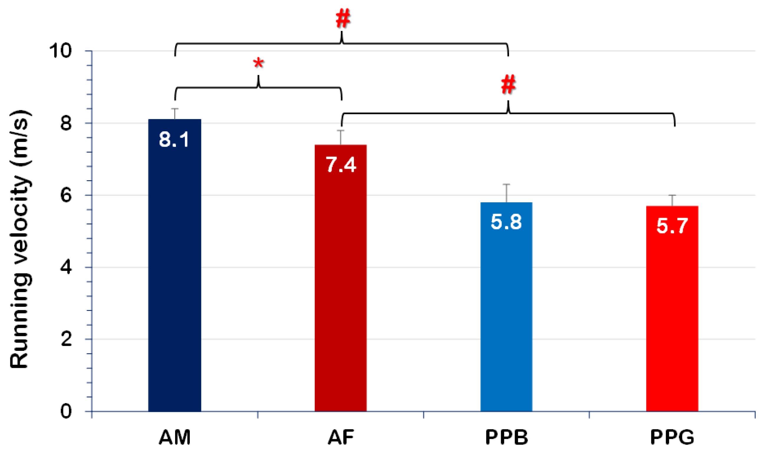
Figure 1. Results regarding the running velocity (AM: adult men; AF: adult women; PPB: prepubertal boys; PPG: prepubertal girls; *: significant sex difference; #: significant age difference).
Applsci 14 06057 g001
Figure 2. Representative stick figures of the sprinting technique: (a) adult woman; (b) prepubescent girl. The arrows indicate the differences between an adult woman and a prepubescent girl: before the touchdown, the girl had an extensive rotation of the shoulder axis, and the arm was not directed in the direction of the movement. During the support phase, an excessive knee flexion was observed in the girl. During the midstance, the woman had a higher heel rise and a more flexed knee joint of the swing leg. Finally, at take-off, the girl was in a more upright position, had a higher swing leg knee shift, and a more flexed ankle joint.
Applsci 14 06057 g002
Figure 3. Representative stick figures of the sprinting technique: (a) adult man; (b) prepubescent boy. The arrows indicate the differences between an adult man and a prepubescent boy: at take-off, the boy had a more extended swing leg knee joint and a less flexed ankle joint, as well as a less active arm swing of the swing leg. Before touchdown, the adult man had a more active ankle plantar flexion movement than the boy.
Applsci 14 06057 g003
Table 1. Mean ± standard deviation of the physical characteristics of the participants.
GroupnAge (yrs)Body Height (m)Body Mass (kg)Training Experience (yrs)Sprint Performance (s)
AM1421.4 ± 3.71.75 ± 0.0668.0 ± 8.411.1 ± 3.110.9 ± 0.3 a
AF821.1 ± 2.61.68 ± 0.0358.9 ± 4.710.6 ± 2.112.4 ± 0.3 a
PPB1010.8 ± 1.01.49 ± 0.0940.3 ± 8.32.8 ± 0.98.6 ± 0.7 b
PPG1011.2 ± 0.81.53 ± 0.0844.5 ± 8.53.1 ± 0.98.7 ± 0.4 b
a: indicates the season best for the 100-m dash; b: indicates the personal best for the 50-m dash.
Table 2. Mean ± standard deviation of the step parameters and the Body Center of Mass (BCM) kinematics.
ParameterAM (n = 14)AF (n = 8)PPB (n = 10)PPG (n = 10)
Step length (SL, m)2.08 ± 0.151.95 ± 0.121.56 ± 0.17 #1.55 ± 0.07 #
Step length (SL, %body height)118.3 ± 5.8115.7 ± 7.1104.4 ± 8.0 #101.6 ± 3.9 #
Step frequency (SF, Hz)4.54 ± 0.274.01 ± 0.23 *4.03 ± 0.41 #3.90 ± 0.34
Contact time (TC, s)0.10 ± 0.010.11 ± 0.02 *0.13 ± 0.02 #0.14 ± 0.02 #
Contact time (TC, % step time)43.0 ± 2.145.6 ± 6.153.2 ± 4.0 #54.2 ± 4.1 #
Flight time (Tf, s)0.13 ± 0.010.14 ± 0.020.12 ± 0.010.12 ± 0.01
Horizontal BCM take-off velocity (VxTO, m/s)9.57 ± 0.577.82 ± 0.65 *6.36 ± 0.66 #6.26 ± 0.49 #
Vertical BCM take-off velocity (VyTO, m/s)0.59 ± 0.140.62 ± 0.130.50 ± 0.100.46 ± 0.12 #
Angle of projection (AngPr, deg)3.5 ± 0.84.6 ± 1.2 *4.5 ± 0.9 #4.2 ± 1.2
Touchdown distance (STD, m)0.35 ± 0.030.39 ± 0.04 *0.35 ± 0.040.36 ± 0.04
Touchdown distance (STD, %body height)20.1 ± 1.622.8 ± 2.5 *23.8 ± 2.5 #23.9 ± 2.5
Take-off distance (STO, m)0.52 ± 0.050.49 ± 0.070.46 ± 0.050.45 ± 0.02
Take-off distance (STO, %body height)29.8 ± 2.630.7 ± 3.730.6 ± 2.4 #29.4 ± 1.8
BCM height at touchdown (HTD, %body height)54.7 ± 2.153.5 ± 1.453.9 ± 1.454.2 ± 1.3
Minimum BCM height (HAM, %body height)53.6 ± 2.252.5 ± 1.352.1 ± 1.3 #52.4 ± 1.3
Lowering of BCM during support phase (ΔHAM, m)−0.02 ± 0.01−0.02 ± 0.01−0.03 ± 0.01 #−0.03 ± 0.01 #
BCM height at take-off (HTO, %body height)56.2 ± 2.355.4 ± 0.855.8 ± 1.155.7 ± 0.9
AM: adult men; AF: adult women; PPB: prepubertal boys; PPG: prepubertal girls; *: significant sex difference; #: significant age difference.
Table 3. Results of the statistical analysis for the step parameters and the Body Center of Mass (BCM) kinematics.
SexAgeInteraction
ParameterFpηp2Fpηp2Fpηp2
Step length (SL, m)2.4040.1300.061111.041<0.001 #0.7551.8020.1880.046
Step length (SL, %body height)1.7190.1980.04645.637<0.001 #0.5590.0010.9710.000
Step frequency (SF, Hz)7.1300.011 *0.16511.7400.002 #0.2462.0300.1630.053
Contact time (TC, s)8.2040.007 *0.17851.074<0.001 #0.5731.7080.1990.043
Contact time (TC, % step time)2.0250.1630.05154.307<0.001 #0.5880.3660.5490.010
Flight time (Tf, s)0.0700.7920.0025.5830.024 #0.1340.0000.9950.000
Horizontal BCM take-off velocity (VxTO, m/s)23.789<0.001 *0.391158.841<0.001 #0.81118.822<0.001 0.337
Vertical BCM take-off velocity (VyTO, m/s)0.0060.9390.0009.4080.004 #0.2031.0430.3140.027
Angle of projection (AngPr, deg)1.6630.2050.0431.1160.2980.0294.9810.032 0.119
Touchdown distance (STD, m)4.2160.047 *0.1021.0730.3070.0281.4290.2400.037
Touchdown distance (STD, %body height)3.7590.0600.09510.7880.002 #0.2313.2610.0790.083
Take-off distance (STO, m)1.4010.2440.03611.9910.001 #0.2450.3930.5340.011
Take-off distance (STO, %body height)0.0250.8750.0010.0990.7540.0031.5980.2140.042
BCM height at touchdown (HTD, %body height)0.8020.3760.0220.0040.9500.0001.8410.1830.049
Minimum BCM height (HAM, %body height)0.5210.4750.0142.2300.1440.0581.6320.2100.043
Lowering of BCM during support phase (ΔHAM, m)0.2230.6390.0069.7330.003 #0.2080.2230.6390.006
BCM height at take-off (HTO, %body height)0.8280.3690.0220.0280.8680.0010.4620.5010.013
*: significant sex difference; #: significant age difference; : significant sex × age interaction.
Table 4. Mean ± standard deviation of the linear and angular kinematics of the lower extremities’ joints and segments.
ParameterAM (n = 14)AF (n = 8)PPB (n = 10)PPG (n = 10)
Knee angle at touchdown (θkneeTD, deg)148.9 ± 7.6148.7 ± 6.7149.0 ± 5.1144.0 ± 6.9
Minimum knee angle during support phase (θkneeAM, deg)145.4 ± 7.1139.1 ± 5.5143.9 ± 5.0134.3 ± 6.7
Knee flexion at the breaking phase (ΔkneeAM, deg)−3.5 ± 2.0−9.6 ± 6.2 *−5.2 ± 2.4−8.1 ± 3.8
Knee angle at take-off (θkneeTO, deg)153.7 ± 0.6154.2 ± 5.2157.5 ± 5.6158.0 ± 5.5
Ankle horizontal velocity at touchdown (Vxankle, m/s)1.95 ± 0.461.66 ± 0.402.45 ± 0.402.71 ± 1.20 #
Ankle horizontal velocity at touchdown (relative to VxTD, m/s)−7.40 ± 0.74−6.18 ± 0.43−3.69 ± 0.60 #−3.35 ± 1.08 #
Ankle angular velocity at touchdown (ωknee, rad/sec)−5.4 ± 1.4−6.1 ± 2.5 *−2.5 ± 2.8#−3.5 ± 2.1 *
Ankle angle at touchdown (θankleTD, deg)103.6 ± 5.4109.1 ± 6.0 *104.9 ± 4.4106.1 ± 7.8
Ankle angle at take-off (θankleTO, deg)123.2 ± 4.3131.4 ± 4.1 *125.9 ± 6.4128.8 ± 5.2
Ankle joint range of motion during support (ΔankleTO, deg)19.6 ± 5.322.3 ± 6.121.0 ± 8.022.7 ± 6.1
Minimum swing leg’s knee angle during support (θkneeMIN, deg)35.5 ± 5.933.9 ± 5.146.8 ± 14.639.9 ± 8.2
Maximum swing leg’s knee angular velocity (ωkneeS, rad/s)16.8 ± 2.516.7 ± 2.013.2 ± 1.512.8 ± 2.5
Swing leg’s knee angle at take-off (θkneeS, deg)69.0 ± 7.476.5 ± 12.2 *78.1 ± 14.471.3 ± 5.9
Swing leg’s ankle angle at take-off (θankleS, deg)109.7 ± 6.7118.7 ± 10.8 *108.5 ± 7.5107.0 ± 7.8
Swing leg’s thigh inclination at take-off (φthighTO, deg)−15.1 ± 7.2−18.5 ± 7.0 *−26.7 ± 5.6−23.4 ± 7.7
Support leg inclination at touchdown (φlegTD, deg)78.0 ± 4.075.7 ± 4.975.2 ± 3.974.2 ± 3.7
Support leg inclination at take-off (φlegTO, deg)51.9 ± 3.654.7 ± 2.351.9 ± 4.052.4 ± 3.8
AM: adult men; AF: adult women; PPB: prepubertal boys; PPG: prepubertal girls; *: significant sex difference; #: significant age difference.
Table 5. Results of the statistical analysis for the linear and angular kinematics of the lower extremities’ joints and segments.
SexAgeInteraction
ParameterFpηp2Fpηp2Fpηp2
Knee angle at touchdown (θkneeTD, deg)1.4760.2320.0381.0050.3230.0261.2610.2690.033
Minimum knee angle during support phase (θkneeAM, deg)15.051<0.001 *0.2952.4020.1300.0630.6750.4170.018
Knee flexion at the breaking phase (ΔkneeAM, deg)15.382<0.001 *0.2940.0190.8920.0011.9730.1680.051
Knee angle at take-off (θkneeTO, deg)0.0870.7700.0025.0510.031 #0.1200.0000.9980.000
Ankle horizontal velocity at touchdown (Vxankle, m/s)0.0050.9440.00011.9890.001 #0.2451.5370.2230.040
Ankle horizontal velocity at touchdown (relative to VxTD, m/s)9.7790.003 *0.209174.317<0.001 #0.8253.1630.0840.079
Ankle angular velocity at touchdown (ωknee, rad/s)1.7390.1950.04515.494<0.001 #0.2950.0490.8250.001
Ankle angle at touchdown (θankleTD, deg)2.9660.0930.0740.1960.6610.0051.2040.2800.032
Ankle angle at take-off (θankleTO, deg)11.6430.002 *0.2390.0000.9990.0002.6260.1140.066
Ankle joint range of motion during support (ΔankleTO, deg)1.2330.2740.0320.1730.6800.0050.0700.7920.002
Minimum swing leg’s knee angle during support (θkneeMIN, deg)2.0860.1570.0538.4350.006 #0.1860.8190.3710.022
Maximum swing leg’s knee angular velocity (ωkneeS, rad/s)0.1340.7160.00425.982<0.001 #0.4190.1650.8000.002
Swing leg’s knee angle at take-off (θkneeS, deg)0.0100.9210.0000.3470.5600.0094.8610.034 0.116
Swing leg’s ankle angle at take-off (θankleS, deg)2.0380.0160.0556.0380.019 #0.1474.0170.0530.103
Swing leg’s thigh inclination at take-off (φthighTO, deg)0.0000.9900.00012.972<0.001 #0.2652.1230.1540.056
Support leg inclination at touchdown (φlegTD, deg)1.9140.1750.0500.8640.3590.0230.9320.3410.025
Support leg inclination at take-off (φlegTO, deg)1.5670.2190.0422.5680.1180.0670.2510.6200.007
*: significant sex difference; #: significant age difference; : significant sex × age interaction.
Disclaimer/Publisher’s Note: The statements, opinions and data contained in all publications are solely those of the individual author(s) and contributor(s) and not of MDPI and/or the editor(s). MDPI and/or the editor(s) disclaim responsibility for any injury to people or property resulting from any ideas, methods, instructions or products referred to in the content.