Article

Investigation of the Complexation Activity of 2,4-Dithiouracil with Au(III) and Cu(II) and Biological Activity of the Newly Formed Complexes

1
Department of General and Inorganic Chemistry with Methodology of Chemistry Education, Faculty of Chemistry, “Tzar Assen” Str. 24, 4000 Plovdiv, Bulgaria
2
Department of Analytical Chemistry and Computer Chemistry, Faculty of Chemistry, University of Plovdiv, “Tzar Asen” Str. 24, 4000 Plovdiv, Bulgaria
3
Department of Organic Chemistry and Pharmacognosy, Faculty of Chemistry and Pharmacy, University of Sofia, 1, J. Bourchier Av., 1164 Sofia, Bulgaria
4
Department of Microbiology, University of Food Technologies, 26 Maritza Blvd., 4002 Plovdiv, Bulgaria
*
Author to whom correspondence should be addressed.
Appl. Sci. 2024, 14(15), 6601; https://doi.org/10.3390/app14156601 (registering DOI)
Submission received: 10 July 2024 / Revised: 24 July 2024 / Accepted: 26 July 2024 / Published: 28 July 2024
(This article belongs to the Section Chemical and Molecular Sciences)

Abstract

:
The goal of this study is to synthesize, determine the structure, and examine the antimicrobial properties of novel Cu(II) and Au(III) complexes of 2,4-dithiouracil and its derivatives. These complexes were obtained by mixing aqueous solutions of the corresponding metal salts with the ligand dissolved in DMSO and aqueous NaOH, using a metal-to-ligand ratio of 1:4:2. The structures of the new compounds were analyzed by melting point determination, microwave plasma atomic emission spectrometry (MP-AES) for Cu and Au, inductively coupled plasma optical emission spectrometry (ICP-OES) for S, attenuated total reflection (ATR), solution and solid-state NMR, and Raman spectroscopy. The data for 2,4-dithiouracil obtained from the 1H NMR, 13C NMR, distortionless enhancement by polarization transfer spectrum (DEPT-135), proton–proton homonuclear correlation spectrum (1H-1H COSY), long-range 1H-13C heteronuclear multiple bond correlation experiment (HMBC), and heteronuclear single quantum coherence spectra (HSQC) aided the interpretation of the NMR data for the gold and copper complexes. Furthermore, the antimicrobial effect of the free ligands and their complexes was assessed against Gram-positive and Gram-negative bacteria, as well as yeasts.

1. Introduction

Numerous sulfur-substituted pyrimidines and purines have been utilized as clinically effective drugs [1]. It has been observed that the specific position of sulfur substitution is crucial for their biological activity. For example, 2-thiouracil, but not 4-thiouracil, exhibits significant antithyroid activity [2]. Similarly, 6-thioguanine and 6-mercaptopurine, but not their corresponding 2-thio compounds, demonstrate antineoplastic effects [3].
Singh and Yadav performed DFT calculations of the vibrational spectra of 2,4-dithiouracil and its corresponding cation and anion forms [4]. M. Ruckenbauer and colleagues conducted a photoelectron study involving 2-thiouracil, 4-thiouracil, and 2,4-dithiouracil [5]. In the photoelectron spectra of all three molecules, distinct connections were observed, stemming from the ionization of electron pairs at the S- and O-atoms, as well as the pyrimidine π-system. The photodynamic properties of the DNA and RNA bases were examined from both experimental and computational perspectives by additionally exploring the coupled nuclear and electronic pathways responsible for the significantly different photochemistry in these nucleic acid base analogues [6]. The elucidation of the photophysical mechanisms in sulfur-substituted nucleobases (thiobases) is important for the development of drugs used in chemotherapeutic applications [7]. Time-Resolved Photoelectron Spectroscopy (TRPES) was applied to study the effect of double thionation on the photodynamics of 2,4-dithiouracil [8].
The structure of 2,4-dithiouracil was determined via X-ray diffraction as far back as 1967 [9]. Six tautomers of 2,4-dithiouracil were investigated with quantum chemical calculations [10]. The dithion tautomer was found to have the lowest energy. The energy difference with the second most stable tautomer (dithiol 2) was only 28 kJ mol−1. Replacement of oxygen by sulfur atoms in uracil can increase the probability of its biologically important process of spontaneous mutations by a factor of 103. Recently, a team of scientists conducted an investigation on the biomolecules 2-thiouracil, 4-thiouracil, and 2,4-dithiouracil using quantum chemical calculations, alongside an exploration of molecular packing and their impact on DNA:RNA microhelices [11]. They performed calculations to assess the potential effects of 2-thiouracil against three different pathogens: Bacillus subtilis, Escherichia coli, and Candida albicans. Additionally, the structure of 2,4-dithiouracil was analyzed in relation to various key proteins, including thyroid peroxidase, thyroid hormone receptors (TSHR), and others (Scheme 1).
Three Cu(II) complexes of 2-thiocytosine and 2,4-dithiouracil were obtained [12]. The authors suggest the formation of a polymer complex with paramagnetic properties. The reason for the stated hypothesis is the observed low solubility in almost all inorganic and organic solvents [12]. The study examined the structures and comparative stabilities of complexes formed between Cu2+ [13] and Cu+ [14] and uracil, 2-thiouracil, 4-thiouracil, and 2,4-dithiouracil by DFT calculations. Rastogi et al. described the synthesis and characterization of a Cu(II) complex with 5-carboxy-2-thiouracil [15], which was also used as a ligand for the preparation of other Mn(II), Co(II), Ni(II), Cu(II), Zn(II), and Cd(II) complexes [16]. In addition, a dinuclear complex of Cu(I), with the general formula [CuX(eitotH2)2]2 [17], where X = Cl, Br, I, was obtained by Papazoglou et al. Furthermore, Ni, Cu, and Mn complexes containing 2-thiouracil, 8-hydroxyquinoline, and 2-hydroxyquinoline as ligands were described in a previous report [18].
The complexation ability of 2-thiouracil and 2,4-dithiouracil with Cd(SeCN)2 and Hg(SeCN)2 was investigated [19]. Based on the obtained experimental data, the authors proposed a probable scheme for obtaining the Cd(II) and Hg(II) complexes with 2-thiouracil and 2,4-dithiouracil. This indicates that the ligand forms a bidentate chelation, involving the nitrogen atom in the third position and the sulfur atom in the second position. Tiekink et al. have shown that thiouracil-containing complexes are effective antitumor and arthritic compounds in vivo [20]. Initial findings from testing for antiarthritic activity in rats regarding the new complexes were also disclosed, demonstrating that certain complexes exhibit greater efficacy or lower toxicity compared to current clinically utilized gold(I) thiolates. Recently, Gimeno and Laguna presented a review about the gold complexes with N, S, P, C donor ligands and with oxygen-based ligands [21]. Yang and Hu studied the conformations, adsorption sites, and orientation of 2-thiouracil on gold substrates at different pH values using SERS (surface-enhanced Raman scattering) and DFT calculations [22]. The use of spherical gold nanoparticles, modified with biologically active molecules, shows considerable promise for diverse medical applications [23]. Specifically, considering the established uses of 2-thiouracil (2-TU) in the treatment of hyperthyroidism and skin cancer, the compound 2-TUAuNPs (gold nanoparticles) stands out as a potential drug candidate for these conditions, providing the benefit of markedly reduced side-effects [23]. Fernández-Moreira et al. presented the antitumor properties of gold complexes with biologically relevant ligands [24]. New anionic bis(thiolato)gold(I) complexes were obtained by Vicente et al. [25]. Recently, Seifert et al. published a review that explored the properties of molecular gold strings consisting of a theoretically infinite set of monomeric gold complexes, which are held together by aurophilic interactions, based on a direct gold–gold contact [26].
The synthesis of Rh(II), Ir(III), Pd(II), and Pt(II) complexes with dithiouracil was described [27], as their structures were determined by means of UV-Vis and IR spectroscopy and compared to the structures of some pyrimidine-containing complexes. The chelating ligands in the corresponding complexes bind to rhodium or iridium by forming octahedral structures, while their coordination with palladium and platinum leads to the formation of square planar complexes. In both cases, the ligand utilizes the N(3) and the C=S moieties to bind, although in the case of the divalent metals, the ligand is anionic. The synthesis of new complexes of Pt(IV) [28] and Pt(II) [29] with 2-thiouracil, 4-thiouracil, and 2,4-dithiouracil was also published.
Many authors have reported the in vitro antimicrobial activity of different complexes of metal ions with thiouracil derivatives against Gram-negative and Gram-positive bacteria, filamentous fungi, and yeasts [18,19,30,31,32,33,34,35]. Metal complexes of thiouracil derivatives have also exhibited cytotoxic effects against various tumor cell lines [17,30,36,37,38]. Marinova and Tamahkyarova (2024) published a review focused on the synthesis and the biological activities of 2-thiouracil and its derivatives [39].
The possible coordination of 2,4-dithiouracil resembles its non-sulfur derivatives, and the schematic representations can be found in our previous paper [32]. Some complexes exhibit a monodentate coordination binding through atoms, such as S (e.g., Cu(I), Au(I), Pt(IV) complexes) [17,20,28], and/or forming bidentate chelate through N3 and S2 (e.g., Cu(II), Cd(II), Hg(II), Pd(II), Pt(II), Pt(IV), Rh(III), Ir(III) complexes) [12,19,27,28].
This paper describes the synthesis and structure elucidation of novel metal complexes of 2,4-dithiouracil and its derivatives. Although a crystal growth system was employed for isolating a single crystal from each complex, the attempts were unsuccessful. The metal complexes were characterized using various methods, including melting point analysis, microwave plasma atomic emission spectrometry (MP-AES), inductively coupled plasma optical emission spectrometry (ICP-OES), attenuated total reflection (ATR), 1H NMR, heteronuclear single quantum coherence spectra HSQC, 13C NMR solid-state and Raman spectroscopy. The assignment of NMR signals for 2,4-dithiouracil was determined from 1H NMR, 13C NMR, distortionless enhancement by polarization transfer spectrum (DEPT-135), 1H-1H COSY, long-range 1H-13C heteronuclear multiple bond correlation experiment (HMBC), and HSQC spectra. Furthermore, their antimicrobial efficacy against both Gram-positive and Gram-negative bacteria, as well as yeasts, was evaluated.

2. Materials and Methods

2.1. Spectra Measurements

Reagents of high purity A.C.S. grade needed for the synthesis of the metal complexes, 2,4-dithiouracil, the metal salts (Cu(CH3COO)2.H2O and (NH4)[AuCl4].H2O), and the solvents, were purchased from Aldrich Chem. The Raman spectra (the stirred crystals placed in aluminum disc) of the compounds were measured in the range from 4000 to 100 cm−1 by using a RAM II (Bruker Optics) with a focused laser beam of Nd:YAG laser (1064 nm) at a resolution of 2 cm−1 with 25 scans. PIKE MIRacle Single Reflection technology was applied to measure the ATR spectra of the complexes. A Bruker Avance II NMR spectrometer working with frequencies of 600.130 and 150.903 MHz was utilized for the 1H and 13C NMR spectra measurements for 2,4-dithiouracil. The registration of the 1H and 13C NMR spectra for the metal complexes was performed on a Bruker Avance III HD spectrometer working at 125.76 MHz and 500.130 MHz, respectively. Standard Bruker software was used for both NMR instruments. A 2.5 mm Cross-Polarization Magic Angle Spinning (CP MAS) probehead was used for registering the solid-state NMR spectra on a Bruker Avance III HD 500 MHz spectrometer. A MAS speed of 15 kHz as well as α-glycine as external reference (α-glycine carbonyl C—176.03 ppm) were applied for measuring the CP MAS and Cross-Polarization with Polarization Inversion (CPPI) MAS spectra. All NMR experiments were performed at ambient temperature.

2.2. MP-AES Determination of Cu and Au and ICP-OES Determination of S in the Complexes

Sample digestion for determination of Cu, Au, and S: approximately 0.02 g of the complexes was weighed on an analytical balance and dissolved in concentrated HNO3 (p.a., Chem-Lab NV) for the Cu-containing complex and HNO3 and HCl (p.a., Fluka AG) for the Au-containing complex. Blank samples were prepared following the same digestion procedure. Determination of Cu, Au, and S concentration was performed with external calibration solutions prepared from monoelemental standards, respectively, 1000 mg L−1 Cu (Merck, Darmstadt, Germany), 1000 mg L−1 Au (High-purity Standards, Charleston, England), and 1000 mg L−1 S (CPAchem, Ltd., Bogomilovo, Bulgaria). Cu and Au were measured by microwave plasma–atomic emission spectrometry (MP-AES 4200, Agilent Technologies, Agilent Technologies, CA, USA) on analytical lines 324.754 nm, 327.395 nm, 510.554 nm for Cu and 242.795 nm, 267.595 nm, 312.278 nm for Au using standard measurement conditions. Sulfur was determined by inductively coupled plasma–optical emission spectrometry (ICP-OES iCap 6000, Thermo Scientific, Waltham, MA, USA) on analytical lines 180.731 nm, 182.034 nm, and 182.624 nm in axial mode.

2.3. Synthesis of Cu(II) and Au(III) Complexes—General Procedure

All metal complexes were synthesized by mixing aqueous solutions of the corresponding metal salts with the ligands dissolved in DMSO and aqueous NaOH, using a metal-to-ligand-to-base molar ratio of 1:4:2. Non-charged complexes precipitated out and were subsequently filtered, repeatedly washed with water, and dried over CaCl2 for two weeks.
An aqueous solution containing 159.7 mg (0.8 mmol) of Cu(CH3COO)2.H2O metal salt in 10 mL of water was added dropwise to a solution of 461.5 mg (3.2 mmol) of 2,4-DTu in 10 mL of DMSO. The ligand solution had previously been alkalized with an aqueous solution of sodium hydroxide, consisting of 64.0 mg (1.6 mmol) of NaOH in 5 mL of water. Similarly, the aqueous solution containing 71.3 mg (0.2 mmol) of NH4[AuCl4]·H2O in 5 mL of H2O was slowly added to a solution of 115.4 mg (0.8 mmol) of 2,4-dithiouracil in 5 mL of dimethyl sulfoxide. This solution had also been previously alkalized (16.0 mg (0.4 mmol) of sodium hydroxide dissolved in 5 mL of H2O).

2.4. Antimicrobial Assay

The antimicrobial activity of 2,4-dithiouracil and its complexes against Gram-positive bacteria—Enterococcus faecalis ATCC 19433, Staphylococcus aureus ATCC 25923, Listeria monocytogenes ATCC 8787, Bacillus subtilis ATCC 6633, Bacillus cereus ATCC 11778—and Gram-negative bacteria—Escherichia coli ATCC 8739, Salmonella enterica subsp. enterica ser. Enetritidis ATCC 13076, Pseudomonas aeruginosa ATCC 9027, Proteus vulgaris G, Klebsiella pneumoniae ATCC 13883—and the yeasts Candida albicans ATCC 10231 and Saccharomyces cerevisiae was tested using the agar diffusion method. A suspension of each test microorganism (106 cfu/cm3) was spread on the surface of PCA (Scharlau) nutrient medium for C. albicans and the bacteria and Wort agar (Sharlau) for S. cerevisiae. Wells of 7 mm diameter were made in the inoculated agar medium. A quantity of 50 μL of the tested substance solution (10 mg/cm3 in DMSO) was pipetted into the wells. The Petri dishes were incubated at 37 °C (for the bacteria and C. albicans) and 30 °C (for S. cerevisiae) for 24–48 h. The inhibition zones were measured. Zones with diameter more than 7 mm were considered as zones of inhibition. Each test was carried out in triplicate, and the accumulated data are presented as mean values.

3. Results and Discussion

3.1. Physical Characteristics of the Metal Complexes

All of the complexes are stable in air and moisture with limited solubility. We observed that the reaction of the ligand with the transition metal ions produced stable solid compounds with yields ranging from 52% to 56%. The resulting complexes exhibit yellow-green or beige coloration and have limited solubility in DMSO, DMF, and C6H12 for the Cu complex and DMSO and DMF for the Au complex, respectively. They are insoluble in water, THF, ethanol, and ethyl acetate. The analytical data, including the yield percentages of the complexes, are presented in Table 1.
The content of Au and Cu was determined by MP-AES, whereas ICP-OES was applied for S determination. Based on the results obtained from the NMR, ATR, Raman, and elemental analyses, possible tentative average compositions were suggested for the Au and Cu complexes (Table 2).

3.2. Structure Verification of 2,4-DTu by NMR

The 1H NMR spectrum of 2,4-dithiouracil showed two singlets at 12.90 ppm and 13.64 ppm for the protons in both NH groups (H-1 and H-3), respectively. The lack of HSQC correlations confirmed that these two singlets were for protons that are not bound to carbons. Additionally, there were two doublets at 6.50 ppm and 7.27 ppm in the 1H NMR spectrum that were assigned correspondingly to the H-5 and H-6 protons. In support of this assignment, there was one 1H-1H COSY correlation found between the signals at 6.50 ppm and 7.27 ppm. Also, there were two HMBC correlations for the H-5 proton with the C-4 and C-6 carbons, as well as three HMBC correlations for the H-6 proton with C-2, C-4 and C-5 carbons. The carbon assignments for 2,4-dithiouracil were additionally verified by using the option provided in the NMRShiftDB database for a 13C NMR chemical shift prediction based on hierarchically ordered spherical environment (HOSE) codes [40]. The complete NMR signal assignments for 2,4-dithiouracil are given in Table 3.

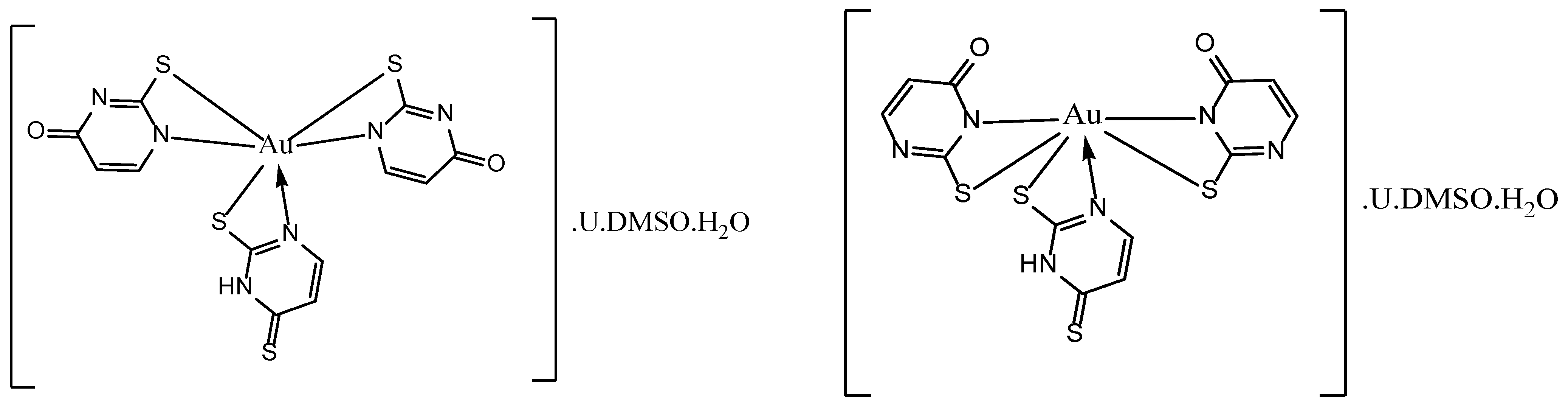
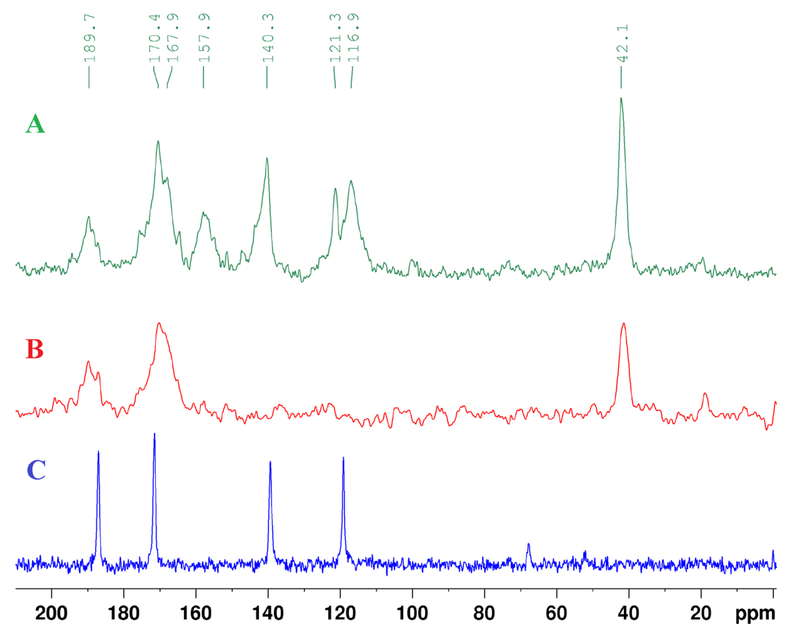
3.3. Structure Elucidation of the Au Complex by NMR

In comparison with the 1H NMR spectrum of 2,4-DTu, the 1H NMR spectrum of the gold complex showed 7 singlets at 10.81 ppm, 11.00 ppm, 12.27 ppm, 12.43 ppm, 12.88 ppm, 13.62 ppm, and 14.13 ppm. Additionally, there were 8 signals at 5.45 ppm, 5.81 ppm, 6.51 ppm, 7.10 ppm, 7.27 ppm, 7.39 ppm, 7.76 ppm, and 8.31 ppm. As can be seen, the 1H NMR spectrum of the gold complex contains more signals than the 1H NMR spectrum of the ligand only. Moreover, the HSQC spectrum showed the following 9 signal correlations: (5.44 ppm–99.94 ppm), (5.81 ppm–104.91 ppm), (6.51 ppm–116.83 ppm), (7.10 ppm–116.00 ppm), (7.26 ppm–137.92 ppm), (7.39 ppm–115.50 ppm), (7.39 ppm–142.04 ppm), (7.76 ppm–140.73 ppm), and (8.31 ppm–156.48 ppm). Consequently, it can be assumed that 2,4-dithiouracil could possibly undergo desulfurization [41] by the influence of the NaOH used during the synthesis of the Au complex causing the replacement of one of the sulfur atoms or both of them with oxygen, thus obtaining 2-thiouracil and uracil in the reaction mixture. The pairs of signals (5.44 ppm–99.94 ppm) and (5.81 ppm–104.91 ppm) were assigned correspondingly to the protons and carbons, H-5 and C-5, in the respective structures of uracil and 2-thiouracil. There was a multiplet at 7.39 ppm with an area of 3.48 in the 1H NMR spectrum where there were presumably four signals located closely to each other. Therefore, two of these signals at 7.39 ppm were assigned to the protons (H-6) in the structures of the obtained uracil and 2-thiouracil for which the corresponding HSQC correlations (7.39 ppm–142.04 ppm) were found. The signals at 10.81 ppm and 11.00 ppm as well as at 12.27 ppm and 12.43 ppm were assigned, respectively, to the NH-1 and NH-3 protons in uracil and 2-thiouracil. The 1H NMR spectral data for the Au complex are given in Table 4.
The signal assignments made for uracil and 2-thiouracil were in good agreement with the 1H and 13C NMR data provided in the Chemical Book spectral database (https://www.chemicalbook.com/SpectrumEN_66-22-8_1HNMR.htm (accessed on 22 July 2024)) for uracil, as well as with the signal assignments for 2-thiouracil presented in a previous paper [31] concerning the synthesis of new Au, Cu, and Pd complexes with 2-thiouracil.
In accordance with the assignments presented for the ligand in Table 3, the signals at 6.51 ppm, 7.26 ppm, 12.88 ppm, and 13.62 ppm in the 1H NMR spectra of the Au complex clearly showed the presence of uncoordinated 2,4-dithiouracil. However, the other HSQC correlations (7.10 ppm–116.00 ppm), (7.39 ppm–115.50 ppm), (7.76 ppm–140.73 ppm), and (8.31 ppm–156.48 ppm) were associated with the presence of some tautomeric forms of 2,4-dithiouracil and 2-thiouracil that could participate as ligands in the Au complex containing deprotonated nitrogen atoms. Thus, it can be hypothesized that not only under the alkaline conditions of the synthesis of the Au complex, a possible desulfurization of 2,4-dithiouracil could happen, but also the nitrogen atoms of 2,4-dithiouracil and 2-thiouracil could be deprotonated in the reaction mixture, thus stimulating the conversion of these ligands into some of their tautomeric forms during their complexation with Au as described in a previous study [28].
The signal at 14.13 ppm with an area of 1.00 showed that there is possibly a tautomeric form of 2,4-dithiouracil with one deprotonated nitrogen that could be coordinated with Au as the chemical shift 14.13 ppm most probably corresponded to the signal of the NH-3 proton. The 2,4-dithiouracil would additionally be coordinated to Au by the sulfur atom that is adjacent to the coordinated nitrogen (Figure 1). In support of this hypothesis, the chemical shifts of the carbon signals in the HSQC correlations (7.10 ppm–116.00 ppm) and (7.76 ppm–140.73 ppm) are close to those signals of the carbons (C-5 and C-6) in the structure of the uncoordinated 2,4-dithiouracil (see Table 3).
Therefore, the signals at 7.10 ppm, 7.76 ppm, 116.00 ppm, and 140.73 ppm could, respectively, be assigned to the protons (H-5 and H-6) and carbons (C-5 and C-6) of the coordinated 2,4-dithiouracil. The two signals left at each of the following chemical shifts (7.39 ppm and 8.31 ppm), in addition to the corresponding HSQC correlations (7.39 ppm–115.50 ppm) and (8.31–156.48 ppm), possibly indicated the presence of tautomeric forms of 2-thiouracil of the same kind in the Au complex containing deprotonated nitrogen atoms. In such case, these tautomeric forms could be coordinated to Au by the sulfur bound to the carbon at the second position, C-2, and its adjacent nitrogen atoms (see Figure 1).
The potential coordination binding sites for the coordinated 2-thiouracil and 2,4-dithiouracil in the Au complex are shown on Figure 1, where the free uncoordinated uracil that was probably produced by desulfurization of the 2,4-dithiouracil ligand could take a place in the outer sphere of the complex. Also, it is possible for the free uncoordinated 2-thiouracil and 2,4-dithiouracil ligands to participate in the outer sphere of the Au complex similarly to uracil (Table 4). This structure is similar to that described in our previous paper with 6-methyl-2-thiouracil [42], as this type of bidentate coordination mode is observed in other metal complexes of Cu(II), Cd(II), Hg(II), Rh(III), Ir(III), Pd(II), Pt(II), Pt(IV) [12,19,27,28].
Additionally, the proton solid-state NMR spectrum showed broad peaks at 5.37 ppm and 7.53 ppm, which could be an additional indication for the protons H-5 and H-6 in uracil, thus supporting the potential presence of uracil in the Au complex. The signals of the protons H-5 and H-6 in 2-thiouracil and 2,4-dithiouracil probably cannot be clearly observed in the proton solid-state NMR spectrum because they could also be a part of the broad peaks at 5.37 ppm and 7.53 ppm. The broad signal at 13.21 ppm could involve the signals of the NH protons in the uncoordinated uracil, 2-thiouracil, or 2,4-dithiouracil that could participate as ligands in the outer sphere of the Au complex (Table 2), as it could also contain the signal for the NH proton of the coordinated 2,4-dithiouracil (Figure 1). The broad peak at 3.05 ppm possibly indicated the presence of DMSO-h6 and H2O in the Au complex.
The solid-state CP MAS (Figure 2A), and CPPI MAS (Figure 2B) NMR spectra of the Au complex showed signals at ca 189.7 ppm and 170.4 ppm, thus confirming the presence of 2,4-dithiouracil in the inner coordination sphere of the Au. The signals at 116.9 ppm and 121.3 ppm would probably correspond to the carbons C-5 in the coordinated 2-thiouracil, whereas the signal at 157.9 ppm could be for the carbons C-6, respectively.
The presence of DMSO-h6 in the Au complex was confirmed by the signal at 42.1 ppm in the solid-state CP MAS (Figure 2A) and CPPI MAS (Figure 2B), probably in the outer sphere of the Au complex. Additionally, the 1H NMR solution spectrum showed a signal at 2.54 ppm, which was an indication of the presence of DMSO-h6 in the Au complex as there was also one HSQC correlation (2.54 ppm–40.11 ppm).

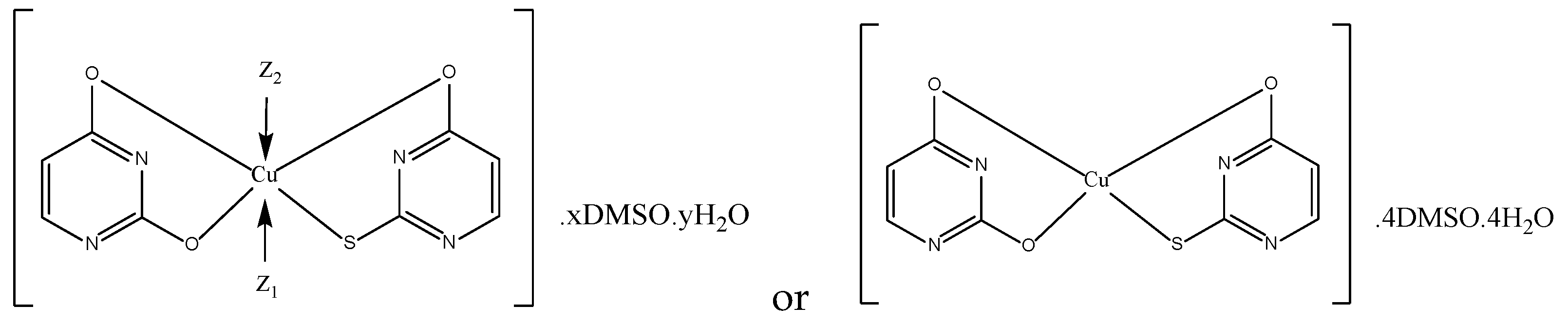
3.4. Structure Elucidation of the Cu Complex by NMR

In contrast with the 1H NMR spectrum of the Au complex, there were no signals for the NH-1 and NH-3 protons of 2,4-dithiouracil in the 1H NMR spectrum of the Cu complex (see Table 5). On the other hand, there were singlets at 10.80 ppm, 11.00 ppm, 12.26 ppm, and 12.43 ppm, similar to those found in the 1H NMR spectrum of the Au complex for 2-thiouracil and uracil, indicating again that 2,4-dithiouracil most probably underwent desulfurization [41] under the alkaline conditions of the synthesis of the Cu complex due to the used NaOH. Also, the HSQC correlations (5.45 ppm–99.78 ppm) and (5.81 ppm–105.03 ppm) were close to the ones observed in the HSQC spectrum of the Au complex for the protons and carbons (H-5 and C-5) in uracil and 2-thiouracil, respectively. In this case, there was a multiplet at 7.39 ppm with an area of 3.27, probably consisting of three proton signals.
Additionally, the HSQC spectrum showed the following correlations: (7.40 ppm–115.27 ppm) and (7.38 ppm–141.85 ppm), where the signals at 7.38 and 141.85 can be assigned to the protons and carbons, H-6 and C-6, in the structures of the uncoordinated ligands, i.e., uracil and 2-thiouracil. Thus, for the ligands coordinated with Cu, one signal was left at 7.40 ppm, one signal at 7.28 ppm, and two signals at 8.33 ppm in the 1H NMR spectrum. Based on the HSQC correlations (7.40 ppm–115.27 ppm) and (7.28 ppm–115.48 ppm), the pairs of the chemical shifts (7.28 ppm and 7.40 ppm) and (115.27 ppm and 115.48 ppm) were assigned to the signals of the protons and carbons (H-5 and C-5), whereas the signals at 8.33 ppm and 156.26 ppm were for the protons and carbons, H-6 and C-6, of the coordinated ligands. The HSQC correlations (7.40 ppm–115.27 ppm) and (8.33 ppm–156.26 ppm) were very close to the HSQC correlations (7.39 ppm–115.50 ppm) and (8.31 ppm–156.48 ppm) observed in the HSQC spectrum of the Au complex. Thus, it can be assumed there is one ligand that would be common for both complexes, i.e., a coordinated 2-thiouracil with the following chemical shifts for its protons and carbons (H-5, H-6, C-5 and C-6)—7.40 ppm, 8.33 ppm, 115.27 ppm, and 156.26 ppm, respectively. Thus, the other pairs of signals (7.28 ppm and 115.48 ppm) and (8.33 and 156.26 ppm) probably corresponded to the protons and carbons H-5, C-5, H-6, and C-6, respectively, in the coordinated uracil. Both ligands would probably be coordinated bidentately with Cu–uracil with both its oxygen atoms and the thiouracil ligand with its sulfur and oxygen atoms (Figure 3). As can be seen from the HSQC spectrum, there were no additional signals that could be observed for the protons and carbons of 2,4-dithiouracil.
The results obtained from the NMR, ATR, Raman, and elemental analyses suggested the possible composition of the Cu complex (Table 2) and its tentative structure (Figure 3). In addition, it is possible that Cu has a coordination number 6 or 4.
The proton solid-state NMR spectrum showed a broad peak at 7.6 ppm, which probably indicated the presence of H-5 and H-6 protons in the coordinated uracil and 2-thiouracil (Figure 3). There were no signals observed for NH protons in the proton solid-state NMR spectrum, confirming that the coordinated 2-thiouracil and uracil contain deprotonated nitrogen atoms. The broad peak at 2.9 ppm could indicate that DMSO-h6 and H2O participate in the Cu complex.
The solid-state NMR spectra also confirmed the absence of 2,4-DTu in the Cu complex. The signal at 172.8 ppm probably corresponded to the carbon, C-2, in the coordinated 2-Tu. The signals at 150.8 ppm (C-2) and 167.7 ppm (C-4) could indicate the carbons in the coordinated uracil. The signals for the carbons, C-6 and C-5, in the coordinated uracil can be found at 157.4 ppm (C-6) and 120.3 ppm (C-5), whereas for the coordinated 2-thiouracil, they are found at 157.4 ppm and 116.1 ppm. The solid-state CP MAS (Figure 4A) and CPPI MAS (Figure 4B) showed a signal at 40.5 ppm, confirming the presence of DMSO in the Cu complex. The 1H NMR solution spectrum showed a signal at 2.54 ppm, which is also an indication of the presence of DMSO-h6 in the Cu complex. In addition, there was one HSQC correlation (2.54 ppm–40.06 ppm).
Actually, the possible structure of the Cu complex proposed in the present study was similar to that reported by Ghosh et al. for Cu(II), Mn(II), Fe(II), Co(II), and Ni(II) complexes with uracil [43]. So, we suggested bidentate coordination through the S- and O-atoms of 2-Tu, as well as through both O-atoms of U. Another similarity with the proposed structure in the above cited article is the possible presence of H2O as a ligand in the inner coordination sphere of the Cu complex. In addition, the ligands were deprotonated at a nitrogen atom in the third position [43], whereas in our case, the deprotonation was in both the first and third positions. Various metal complexes (Cu(II), Cd(II), Hg(II), Pd(II), Pt(II), Pt(IV), Rh(III), and Ir(III)) exhibit a bidentate coordination mode with 2-thiouracil derivatives [12,19,27,28].
DFT calculations previously suggested a number of stable conformations of Cu(II) complexes with U or 2-Tu participating in monodentate or bidentate coordination with the metal [13]. In contrast, no DFT data exist in the literature for the stability of a Cu complex that contains both U and 2-Tu as bidentate ligands. Therefore, similar calculations should also be performed for such chelate Cu complexes with more than one ligand (e.g., U and/or 2-Tu) in order to assess their relative stability.

3.5. Spectral Data for 2,4-DTu and the Metal Complexes

The spectral data for 2,4-ditiouracil and metal complexes are given in Table 6. Due to the bidentate coordination of 2-Tu with Cu through its sulfur and oxygen atoms, the stretching band of the C=S bond was shifted in the Raman and ATR spectra of the Cu complex, as compared to our previous (1217 cm−1) [31].
In summary, as can be seen from the Raman and ATR spectra of 2,4-DTu, there is a band at 3080 cm−1 indicating ν(C-H). Also, there is a shoulder at 3096 cm−1 in the ATR spectrum and a second band in the Raman spectrum at 3097 cm−1. In the case of the Au complex, Raman and ATR bands for ν(C-H) were found at 3066 cm−1 and 3064 cm−1, while the Raman and ATR bands for the Cu complex were at 3018 cm−1 and 3078 cm−1. The Raman and ATR bands at 1547 cm−1 and 1565 cm−1 were for ν(C=C) stretching in 2,4-DTu. Similarly, the ν(C=C) bands at 1544 cm−1 and 1539 cm−1, as well as at 1546 cm−1, were found for the Au and Cu complexes, respectively. Indications for a C=S bond in 2,4-DTu and Au complex were the Raman and ATR bands in the range 1252–1254 cm−1.
The structures of all the compounds were verified by spectral data (see Supplementary Materials, Figures S1–S21).

3.6. Antimicrobial Activity

Table 7 depicts the results from the antimicrobial assay of 2,4-dithiouracil and its complexes. The addition of Cu(II) to the ligand slightly improved the antimicrobial effect on Staphylococcus aureus ATCC 25923, Escherichia coli ATCC 8739, and Salmonella enterica ssp. enterica ser. Enetritidis ATCC 13076, while Pseudomonas aeruginosa ATCC 9027 and Saccharomyces cerevisiae were more resistant to the activity of the Cu complex in comparison with the ligand. The rest of the test microorganisms did not exhibit differences in their sensitivity between the ligand and the Cu complex.
On the other hand, the 2,4-dithiouracil complex with Au(III) showed a stronger antimicrobial effect against all of the test microorganisms, except Escherichia coli ATCC 8739, where no change in activity was observed. The most significant increase in activity was determined against Staphylococcus aureus ATCC 25923, Eterococcus faecalis ATCC 19433, and Klebsiella pneumoniae ATCC 13883. The antimicrobial activity of Au(III) is well documented [44,45] and similarly to our previous study on the addition of Au(III) to 2-thiouracil [31], the complex exhibited increase antimicrobial activity in comparison with the ligand.

4. Conclusions

The complexation potential of 2,4-DTu and its derivatives was demonstrated for Au(III) and Cu(II), leading to the synthesis of new metal chelate complexes. A combination of various atomic and molecular spectroscopic techniques was used to determine the elemental compositions, as well as the possible structures of the proposed coordination compounds. The interpretation of the presented NMR data was the key factor for determining the possible type of ligands in the Au and Cu complexes. The antimicrobial activity of the newly formed complexes was studied in vitro against yeasts and Gram-positive and Gram-negative bacteria. The Cu(II) complex showed no significant improvement in the inhibition of most test-microorganisms, while the addition of Au(III) to 2,4-dithiouracil increased its antimicrobial effect, especially on Staphylococcus aureus ATCC 25923, Eterococcus faecalis ATCC 19433, and Klebsiella pneumoniae ATCC 13883.

Supplementary Materials

The following supporting information can be downloaded at: https://www.mdpi.com/article/10.3390/app14156601/s1, Figure S1: 1H NMR spectrum of 2,4-DTu; Figure S2: 13C NMR spectrum of 2,4-DTu; Figure S3: HSQC spectrum of 2,4-DTu; Figure S4: HMBC spectrum of 2,4-DTu;. Figure S5: 1H-1H COSY spectrum of 2,4-DTu; Figure S6: 1H NMR spectrum of the Au complex; Figure S7: 1H NMR spectrum of the Cu complex; Figure S8: HSQC spectrum of the Au complex; Figure S9: HSQC spectrum of the Cu complex; Figure S10: Raman spectrum of 2,4-DTu; Figure S11: ATR spectrum of 2,4-DTu; Figure S12: Raman spectrum of the Au complex; Figure S13: ATR spectrum of the Au complex; Figure S14: Raman spectrum of the Cu complex; Figure S15: ATR spectrum of the Cu complex; FigureS16: 1H solid state NMR of Au complex; Figure S17: 13C CP MAS NMR of Au complex; Figure S18: 13C CPPI MAS NMR of Au complex; Figure S19: 1H solid state NMR of Cu complex; Figure S20: 13C CP MAS NMR of Cu complex; Figure S21: 13C CPPI MAS NMR of Cu complex.

Author Contributions

Conceptualization, P.M. and P.P.; methodology, P.M.; formal analysis, P.P., N.B., D.S., S.T., E.V.; investigation, S.T., N.B., E.V.; resources, N.B.; data curation, P.M.; writing—original draft preparation, P.M., N.B., D.S., P.P., D.B., A.S.; writing—review and editing, P.M., D.S., P.P., N.B., D.B., A.S.; supervision, P.M.; project administration, S.T.; funding acquisition, P.P. All authors have read and agreed to the published version of the manuscript.

Funding

We acknowledge the financial support from the Fund for Scientific Research of the Plovdiv University, project CП 23-XФ-006.

Institutional Review Board Statement

Not applicable.

Informed Consent Statement

Not applicable.

Data Availability Statement

The original contributions presented in the study are included in the article/Supplementary Material, further inquiries can be directed to the corresponding author.

Acknowledgments

Research equipment of the Distributed Research Infrastructure INFRAMAT supported by the Bulgarian Ministry of Education and Science under contract D01-284/17.12.2019 was used.

Conflicts of Interest

The authors declare no conflicts of interest.

References

  1. Parker, W.B. Enzymology of Purine and Pyrimidine Antimetabolites Used in the Treatment of Cancer. Chem. Rev. 2009, 109, 2880–2893. [Google Scholar] [CrossRef] [PubMed]
  2. Astwood, E.B. Treatment of hyperthyroidism with thiourea and thiouracil. JAMA 1984, 251, 1743–1746. [Google Scholar] [CrossRef] [PubMed]
  3. Elgemeie, G.H.; Mohamed-Ezzat, R.A. Chapter 1—Medicinal chemistry of anticancer agents. In New Strategies Targeting Cancer Metabolism, Anticancer Drugs, Synthetic Analogs, and Antitumor Agents; Elsevier: Amsterdam, The Netherlands, 2022; pp. 1–33. ISBN 978-0-12-821783-2. [Google Scholar] [CrossRef]
  4. Singh, R.; Yadav, R.A. Raman and IR studies and DFT calculations of the vibrational spectra of 2,4-Dithiouracil and its cation and anion. Spectrochim. Acta Part A Mol. Biomol. Spectrosc. 2014, 130, 188–197. [Google Scholar] [CrossRef]
  5. Ruckenbauer, M.; Mai, S.; Marquetand, P.; González, L. Photoelectron spectra of 2-thiouracil, 4-thiouracil, and 2,4-dithiouracil. J. Chem. Phys. 2016, 144, 074303. [Google Scholar] [CrossRef] [PubMed]
  6. Pollum, M.; Martínez-Fernández, L.; Crespo-Hernández, C.E. Photochemistry of Nucleic Acid Bases and Their Thio- and Aza-Analogues in Solution. In Photoinduced Phenomena in Nucleic Acids I; Barbatti, M., Borin, A., Ullrich, S., Eds.; Topics in Current Chemistry; Springer: Cham, Switzerland, 2014; Volume 355. [Google Scholar] [CrossRef]
  7. Mai, S.; Pollum, M.; Martínez-Fernández, L.; Dunn, N.; Marquetand, P.; Corral, I.; Crespo-Hernández, C.E.; González, L. The origin of efficient triplet state population in sulfur-substituted nucleobases. Nat. Commun. 2016, 7, 13077. [Google Scholar] [CrossRef]
  8. Mohamadzade, A.; Ullrich, S. Internal conversion and intersystem crossing dynamics of uracil upon double thionation: A time-resolved photoelectron spectroscopy study in the gas phase. Phys. Chem. Chem. Phys. 2020, 22, 15608–15615. [Google Scholar] [CrossRef]
  9. Shefter, E.; Mautner, H.G. The Crystal and Molecular Structure of 2,4-Dithiouracil. J. Am. Chem. Soc. 1967, 89, 1249–1253. [Google Scholar] [CrossRef]
  10. Leszczyiiski, J.; Lammertsma, K. 2,4-Dithiouracil tautomers: Structures and energies. J. Phys. Chem. 1991, 95, 3128–3132. [Google Scholar] [CrossRef]
  11. Palafox, M.A.; Benial, A.M.F.; Rastogi, V.K. Biomolecules of 2-Thiouracil, 4-Thiouracil and 2,4-Dithiouracil: A DFT Study of the Hydration, Molecular Docking and Effect in DNA:RNA Microhelixes. Int. J. Mol. Sci. 2019, 20, 3477. [Google Scholar] [CrossRef]
  12. Nelson, H.C.; Villa, J.F. Copper(II) Complexes of 2-Thiocytosine and 2,4-Dithiouracil. Inorg. Chim. Acta 1979, 34, L235–L237. [Google Scholar] [CrossRef]
  13. Lamsabhi, A.M.; Alcamí, M.; Mó, O.; Yáñez, M.; Tortajada, J. Association of Cu2+ with Uracil and Its Thio Derivatives: A Theoretical Study. Chem. Phys. Chem. 2004, 5, 1871–1878. [Google Scholar] [CrossRef]
  14. Lamsabhi, A.M.; Alcamí, M.; Mó, O.; Yáñez, M. Gas-phase reactivity of uracil, 2-thiouracil, 4-thiouracil, and 2,4-dithiouracil towards the Cu+ cation: A DFT study. Chem. Phys. Chem. 2003, 4, 1011–1017. [Google Scholar] [CrossRef]
  15. Rastogi, V.K.; Alcolea Palafox, M.; Singh, C.; Gupta, S.L. Synthesis and characterisation of complex of Cu(II) with 5-carboxy-2-thiouracil. In Spectroscopy of Biological Molecules: New Directions; Greve, J., Puppels, G.J., Otto, C., Eds.; Springer: Dordrecht, The Netherlands, 1999. [Google Scholar]
  16. Singh, U.P.; Singh, S.; Singh, S.M. Synthesis, characterization and antitumour activity of metal complexes of 5-carboxy-2-thiouracil. Metal-Based Drugs 1998, 5, 35–39. [Google Scholar] [CrossRef] [PubMed]
  17. Papazoglou, I.; Cox, P.J.; Hatzidimitriou, A.G.; Kokotidou, C.; Choli-Papadopoulou, T.; Aslanidis, P. Copper(I) halide complexes of 5-carbethoxy-2-thiouracil: Synthesis, structure and in vitro cytotoxicity. Eur. J. Med. Chem. 2014, 78, 383–391. [Google Scholar] [CrossRef]
  18. Worachartcheewan, A.; Pingaew, R.; Lekcharoen, D.; Prachayasittikul, S.; Ruchirawat, S.; Prachayasittikul, V. Synthesis, Antioxidant and Antimicrobial Activities of Metal Complexes of 2-thiouracil-hydroxyquinoline Derivatives. Lett. Drug Des. Discov. 2018, 15, 602–611. [Google Scholar] [CrossRef]
  19. Shaikh, M.N.; Al-Maythalony, B.A.; Wazeer, M.I.M.; Isab, A.A. Complexations of 2-thiouracil and 2,4-dithiouracil with Cd(SeCN)2 and Hg(SeCN)2: NMR and anti-bacterial activity studies. Spectroscopy 2011, 25, 187–195. [Google Scholar] [CrossRef]
  20. Cookson, P.D.; Tiekink, E.R.T.; Whitehouse, M.W. Phosphinegold(I) complexes Containing the Purine-6-thiolate Anion, and their Antiart hritic Activity. Aust. J. Chem. 1994, 47, 577–586. [Google Scholar] [CrossRef]
  21. Gimeno, M.C.; Laguna, A. Some recent highlights in gold chemistry. Gold Bull. 2003, 36, 83–92. [Google Scholar] [CrossRef]
  22. Yang, W.; Hu, Y. Conformations of 2-thiouracil in the aqueous solution and its adsorption behavior on the gold substrates explored by DFT calculations and experimental methods. Spectrochim. Acta Part A Mol. Biomol. Spectrosc. 2015, 134, 399–405. [Google Scholar] [CrossRef]
  23. Lorenzana-Vázquez, G.; Pavel, I.; Meléndez, E. Gold Nanoparticles Functionalized with 2-Thiouracil for Antiproliferative and Photothermal Therapies in Breast Cancer Cells. Molecules 2023, 28, 4453. [Google Scholar] [CrossRef]
  24. Fernández-Moreira, V.; Herrera, R.P.; Gimeno, M.C. Anticancer properties of gold complexes with biologically relevant ligands. Pure Appl. Chem. 2018, 91, 247–269. [Google Scholar] [CrossRef]
  25. Vicente, J.; Chicote, S.M.-T.; Gonzalez-Herrero, P.; Jones, P.G. Complexes with S-Donor Ligands. Part 2. Synthesis of Anionic Bis(thiolato)gold(I) Complexes. Crystal Structure of [N(PPh3)2][Au(SR)2] (R = benzoxazol-2-yl)+. J. Chem. Soc. Dalton Trans. [CrossRef]
  26. Seifert, T.P.; Naina, V.R.; Feuerstein, T.J.; Knöfel, N.D.; Roesk, P.W. Molecular gold strings: Aurophilicity, luminescence and structure–property correlations. Nanoscale 2020, 12, 20065–20088. [Google Scholar] [CrossRef] [PubMed]
  27. Lusty, J.R.; Chan, H.S.O.; Peeling, J. The synthesis and characterisation of dithiouracil complexes of rodium(III), irdium(III), palladium(II) and platinum(II). Transition Met. Chem. 1983, 8, 343–345. [Google Scholar] [CrossRef]
  28. Vetter, C.; Kaluderovic, G.N.; Paschke, R.; Kluge, R.; Schmidt, J.; Steinborn, D. Synthesis, characterization and in vitro cytotoxicity studies of platinum(IV) complexes with thiouracil ligands. Inorg. Chim. Acta 2010, 363, 2452–2460. [Google Scholar] [CrossRef]
  29. Crestoni, M.E.; Corinti, D.; Chiavarino, B.; Fornarini, S.; Scuderi, D.; Salpin, J.-Y. Insights into Cisplatin Binding to Uracil and Thiouracils from IRMPD Spectroscopy and Tandem Mass Spectrometry. J. Am. Soc. Mass Spectrom. 2020, 31, 946–960. [Google Scholar]
  30. Kamalakannan, P.; Venkappayya, D.; Balasubramanian, T. A new antimetabolite, 5-morpholinomethyl-2-thiouracil—Spectral properties, thermal profiles, antibacterial, antifungal and antitumour studies of some of its metal chelates. J. Chem. Soc. Dalton Trans. 2002, Issue 17, 3381–3391. [Google Scholar] [CrossRef]
  31. Marinova, P.; Tsoneva, S.; Frenkeva, M.; Blazheva, D.; Slavchev, A.; Penchev, P. New Cu(II), Pd(II) and Au(III) complexes with 2-thiouracil: Synthesis, Characteration and Antibacterial Studies. Russ. J. Gen. Chem. 2022, 92, 1578–1584. [Google Scholar] [CrossRef]
  32. Marinova, P.; Hristov, M.; Tsoneva, S.; Burdzhiev, N.; Blazheva, D.; Slavchev, A.; Varbanova, E.; Penchev, P. Synthesis, Characterization and Antibacterial Studies of new Cu(II) and Pd(II) complexes with 6-methyl-2-thiouracil and 6-propyl-2-thiouracil. Appl. Sci. 2023, 13, 13150. [Google Scholar] [CrossRef]
  33. Abou-Melha, K.S. A Series of Nano-sized Metal ion-thiouracil Complexes, tem, Spectral, γ-irradiation, Molecular Modeling and Biological Studies. Orient. J. Chem. 2015, 31, 1897–1913. [Google Scholar] [CrossRef]
  34. Mohamed, M.S.; Youns, M.M.; Ahmed, N.M. Synthesis, antimicrobial, antioxidant activities of novel 6-aryl-5-cyano thiouracil derivatives. Eur. J. Med. Chem. 2013, 69, 591–600. [Google Scholar] [CrossRef]
  35. Masoud, M.S.; Soayed, A.A.; El-Husseiny, A.F. Coordination modes, spectral, thermal and biological evaluation of hetero-metal copper containing 2-thiouracil complexes. Spectrochim. Acta Part A Mol. Biomol. Spectrosc. 2012, 99, 365–372. [Google Scholar] [CrossRef]
  36. Tyagi, S.; Singh, S.M.; Gencaslan, S.; Sheldrick, W.S.; Singh, U.P. Metal-5-fluorouracil-histamine complexes: Solution, structural, and antitumor studies. Metal-Based Drugs 2002, 8, 337–345. [Google Scholar] [CrossRef]
  37. Kumar, B.; Suman, A. Synthesis, spectroscopic characterization and biological application of copper complex of 5-carbethoxy-2-thiouracil. J. Drug Deliv. Ther. 2020, 10, 145–148. [Google Scholar] [CrossRef]
  38. Illán-Cabeza, N.A.; García-García, A.R.; Moreno-Carretero, M.N.; Martínez-Martos, J.M.; Ramírez-Expósito, M.J. Synthesis, characterization and antiproliferative behavior of tricarbonyl complexes of rhenium(I) with some 6-amino-5-nitrosouracil derivatives: Crystal structure of fac-[ReCl(CO)3(DANU-N5,O4)] (DANU = 6-amino-1,3-dimethyl-5-nitrosouracil). J. Inorg. Biochem. 2005, 99, 1637–1645. [Google Scholar] [CrossRef]
  39. Marinova, P.E.; Tamahkyarova, K.D. Synthesis and Biological Activities of Some Metal Complexes of 2-Thiouracil and Its Derivatives: A Review. Compounds 2024, 4, 186–213. [Google Scholar] [CrossRef]
  40. Christoph Steinbeck, Stefan Kuhn, NMRShiftDB—Compound identification and structure elucidation support through a free community-built web database. Phytochemistry 2004, 65, 2711–2717. [CrossRef]
  41. Novakov, I.A.; Orlinson, B.S.; Navrotskii, M.B. Desulfurization of 2-Thioxo-1,2,3,4-tetrahydropyrimidin-4-ones with oxiranes and 2-Haloacetonitriles. Russ. J. Org. Chem. 2005, 41, 607–609. [Google Scholar] [CrossRef]
  42. Marinova, P.; Burdzhiev, N.; Blazheva, D.; Slavchev, A. Synthesis and Antibacterial Studies of a New Au(III) Complex with 6-Methyl-2-Thioxo-2,3-Dihydropyrimidin-4(1H)-One. Molbank 2024, 2024, M1827. [Google Scholar] [CrossRef]
  43. Ghosh, P.; Mukhopadhyay, T.K.; Sarkar, A.R. Interaction of Divalent Metal Ions with Uracil III. Complexes of MnII, FeII, CoII, NiII and CuII with Uracil Acting as Bidentate Ligand. Transit. Met. Chem. 1984, 9, 46–48. [Google Scholar] [CrossRef]
  44. Shareena Dasari, T.P.; Zhang, Y.; Yu, H. Antibacterial Activity and Cytotoxicity of Gold(I) and (III) Ions and Gold Nanoparticles. Biochem. Pharmacol. 2015, 4, 199–203. [Google Scholar] [CrossRef]
  45. Zhang, Y.; Dasari, T.; Deng, H.; Yu, H. Antimicrobial Activity of Gold Nanoparticles and Ionic Gold. J. Environ. Sci. Health C Environ. Carcinog. Ecotoxicol. Rev. 2015, 3, 286–327. [Google Scholar] [CrossRef] [PubMed]
Scheme 1. Different applications of 2-thiouracil and its derivatives and their metal complexes (antithyroid drugs [2]; antitumor [3]).
Applsci 14 06601 sch001
Figure 1. Possible structures of the Au complex with 2,4-DTu, 2-Tu, and U.
Applsci 14 06601 g001
Figure 2. 13C CP MAS NMR spectrum of Au complex (A); 13C CPPI MAS NMR spectrum of Au complex (B); 13C CP MAS NMR spectrum of the 2,4-DTu (C).
Applsci 14 06601 g002
Figure 3. A possible structure and coordination binding sites in the Cu complex. If Z1 = H2O or DMSO-h6, then Z2 = H2O or DMSO-h6.
Applsci 14 06601 g003
Figure 4. 13C CP MAS NMR spectrum of Cu complex (A); 13C CPPI MAS NMR spectrum of Cu complex (B); 13C CP MAS NMR spectrum of the 2,4-DTu (C).
Applsci 14 06601 g004
Table 1. Analytical and physical characteristics of metal complexes with 2,4-dithiouracil and its derivatives.
ComplexesColorYield (%)Melting Point (°C)Solubility
2,4-DTuyellow 279–281soluble in DMSO
Cu(II)yellow-green56>350 °Climited solubility in DMSO, C6H12, and DMF; and insoluble in EtOH, H2O, THF, and EtOAc.
Au(III)beige52>350 °Climited solubility in DMSO and DMF; insoluble in H2O, THF, EtOH, EtOAc, and C6H12.
Table 2. Elemental analyses data for the metal ions and sulfur of the complexes.
Metal ComplexComposition *FormulaMolecular Weight W(M)%
Calc./Exp.
W(S)% Calc./Exp.
Au(III)[2,4-DTu.(2-Tu)2.Au].U.5DMSO.5H2OC26H51N8O14S9Au1185.28 g/mol16.62/16.7 ± 1.524.35/25.2 ± 2.1
Au(III)[2,4-DTu.(2-Tu)2.Au].U.2-Tu.C28H37N12O10S9Au1187.22 g/mol16.59/16.7 ± 1.524.31/25.2 ± 2.1
2,4-DTu.2DMSO.3H2O
Cu(II)[2-Tu.U.Cu].4DMSO.4H2OC16H36N4O11S5Cu684.35 g/mol9.29/9.23 ± 0.6523.43/21.9 ± 1.8
* Tentative average composition of different complexes. 2,4-DTu = 2,4-dithiouracil; 2-Tu = 2-thiouracil; U = uracil.
Table 3. 1H and 13C NMR spectral data and 1H-1H COSY and HMBC correlations for 2,4-dithiouracil [600.13 MHz (1H) and 150.903 MHz (13C)] a.
Atomδ (13C) ppmDEPT-135 δ (1H) ppmMultiplicity (J, Hz)1H-1H COSY HMBC
1 (NH) 12.90s
2 (C=S)172.87C
3 (NH) 13.64s
4 (C=S)187.81C
5117.16CH6.50d (7.1)64, 6
6136.69CH7.27d (7.1)52, 4, 5
a In DMSO-d6 solution. All these assignments were in agreement with COSY, HMQC, and HMBC spectra.
Table 4. 1H NMR spectral data for the Au complex [500.13 MHz (1H) ] a.
Atom2,4-DTu b2-Tu bU b2,4-DTu.(2-Tu)2.Au c
112.88, s12.27, s10.81, s14.13, s (2,4-DTu)
2
313.62, s12.43, s11.00, s-
4
56.51, d(6.7 Hz)5.81, d(7.50 Hz)5.44, m7.10, d(6.8 Hz), (2,4-DTu)
7.39, m, (2-Tu)
7.39, m, (2-Tu)
67.26, t(6.2 Hz)7.39, m7.39, m7.76, d(6.7 Hz), (2,4-DTu)
8.31, d(4.7 Hz), (2-Tu)
a In DMSO-d6 solution. All these assignments were in agreement with the HSQC spectrum; b The corresponding assignments concern the uncoordinated ligands—2,4-dithiouracil (2,4-DTu), 2-thiouracil (2-Tu), uracil (U). c Spectral data suggest that 2,4-DTu and 2-Tu are ligands in our complex.
Table 5. 1H NMR spectral data for the Cu complex [500.13 MHz (1H)] a.
Atom2-Tu bU b2-Tu.U.Cu c
112.26, s10.80, s
2
312.43, s11.00, s
4
55.81, d (7.3 Hz)5.45, d (8.2 Hz)7.28, d (5.5 Hz) (U)
7.40 d, m (2-Tu)
67.38 d, m7.38 d, m8.33, m (U)
8.33, m (2-Tu)
a In DMSO-d6 solution. All these assignments were in agreement with the HSQC spectrum. b The corresponding assignments concern the uncoordinated ligands—2-thiouracil (2-Tu), uracil (U). c Spectral data suggest that 2-Tu and U are ligands in our complex. d This chemical shift was assigned from the HSQC spectrum.
Table 6. Vibrational spectroscopy data for 2,4-DTu and the metal complexes.
2,4-DTuAu ComplexCu Complex
Raman, cm−1ATR, cm−1Raman, cm−1ATR, cm−1Raman, cm−1ATR, cm−1
3097(ν(C-H))
3080 (ν(C-H))
3058
1605
1547 (ν(C=C))
1491
1425
1359
1357
1254 (ν(C=S))
1230
1189
1118
1076
984
965
858
683
611
461
444
399
387
229
3168 (ν(NH))
3096 ((ν(C-H))
3080 (ν(C-H)
2994
2923
2894
2720
1921
1695
1673
1610
1565 (ν(C=C))
1486
1411
1368
1358
1319
1252 (ν(C=S))
1230
1211
1123
1099
1076
984
965
858
820
792
695
680
614
3066 (ν(C-H))
2902
1604
1544 (ν(C=C))
1411
1363
1289
1253 (ν(C=S))
1219
1189
1147
1120
1099
978
967
816
715
683
611
542
461
444
399
387
269
246
230
3100
3064 (ν(C-H))
1603
1539 (ν(C=C))
1520
1486
1445
1407
1363
1326
1285
1252 (ν(C=S))
1226
1189
1122
1077
1013
978
966
949
932
857
803
789
761
735
706
680
3101
3018 (ν(C-H)) 2911
1546 (ν(C=C)) 1480
1412
1384
1305
1283
1193
1148
1096
1014
978
820
804
681
544
449
420
388
269
246
205
3386 (ν(O-H))
3078 (ν(C-H))
2996
2908
2362
1654
1600
1546 (ν(C=C))
1481
1433
1380
1366
1307
1282
1185
1159
1148
1095
1012
950
817
754
707
679
613
Table 7. Antimicrobial activity of 2,4-DTu and its Cu(II) and Au(III) complexes.
Test MicroorganismsDMSO2,4-DTuCu(II)Au(III)
Inhibition Zone, mm
Staphylococcus aureus ATCC 25923-14 ± 1.0015 ± 0.5819 * ± 1.00
Eterococcus faecalis ATCC 19433-12 ± 1.0013 ± 1.0016 ± 0.58
Listeria monocytogenes ATCC 8787-12 ± 1.0012 ± 0.5814 ± 0.58
Bacillus subtilis ATCC 6633-11 ± 0.5811 ± 0.5813 ± 0.58
Bacillus cereus ATCC 11778-11 ± 0.5811 ± 0.5814 * ± 0.58
Escherichia coli ATCC 8739-14 ± 0.5815 ± 0.5814 ± 1.00
Salmonella enterica ssp. enterica ser. Enetritidis ATCC 13076-14 ± 0.0015 ± 0.0015 ± 0.58
Pseudomonas aeruginosa ATCC 9027-13 ± 1.0012 ± 0.5815 ± 0.00
Proteus vulgaris G-12 * ± 0.5812 * ± 0.5814 * ± 1.00
Klebsiella pneumoniae ATCC 13883-12 * ± 0.5811 * ± 0.5815 ± 0.58
Candida albicans ATCC 10231-10 ± 0.5811 * ± 0.5812 ± 0.58
Saccharomyces cerevisiae-11 ± 1.0010 ± 0.0012 ± 1.00
Well diameter—7 mm; * Inhibition zone with single cell colonies.
Disclaimer/Publisher’s Note: The statements, opinions and data contained in all publications are solely those of the individual author(s) and contributor(s) and not of MDPI and/or the editor(s). MDPI and/or the editor(s) disclaim responsibility for any injury to people or property resulting from any ideas, methods, instructions or products referred to in the content.

