Article

Iron Removal from Low-Grade Pyrophyllite Ore by Microwave Irradiation and Dry Magnetic Separation

1
Mining Engineering Department, King Abdulaziz University, Jeddah P.O. Box 80204, Saudi Arabia
2
Environmental Studies Department, National Authority of Remote Sensing and Space Sciences, El-Nozha El-Gedida, Cairo P.O. Box 1564, Egypt
3
Saudi Mining Polytechnic, Arar P.O. Box 215, Saudi Arabia
4
Geological Research Authority of Sudan, Ministry of Minerals, Khartoum P.O. Box 410, Sudan
*
Authors to whom correspondence should be addressed.
Appl. Sci. 2024, 14(15), 6651; https://doi.org/10.3390/app14156651 (registering DOI)
Submission received: 17 June 2024 / Revised: 17 July 2024 / Accepted: 24 July 2024 / Published: 30 July 2024

Abstract

:
Owing to its unique properties, pyrophyllite is an economical alternative to many minerals in different applications. The presence of iron-bearing minerals in Saudi pyrophyllite hampers its industrial uses. The aim of this study was to examine the removal of iron from Saudi low-grade pyrophyllite ore using two approaches. The first approach involves dry high-intensity magnetic separation, whereas the second approach involves microwave pretreatment of the ore before dry magnetic separation. For the first approach, the studied operating parameters were roll speed; feed rate, field intensity, and feed particle size. For the alternative approach, microwave treatment followed by dry magnetic separation, the microwave irradiation time and the magnetic field intensity were studied. The results show that the combined microwave treatment and dry separation method could provide high-purity pyrophyllite for filler industries. Microwave irradiation for 30 min was optimal to change impurity phases (i.e., pyrite, hematite) into ferromagnetic phases in microwave-treated pyrophyllite samples. At a magnetic field intensity of 2000 Gauss, the 30 min microwave-irradiated pyrophyllite sample achieved an iron recovery of 11.2% in non-magnetic fractions, with a removal efficiency of 89% with an alumina recovery of 91.31%.

1. Introduction

Pyrophyllite (Al2Si4O10(OH)2) is a naturally existing hydrated silicate mineral with a high content of Al2O3. It is commonly associated with impurities such as rutile, quartz, mica, kaolin, epidote, talc, feldspar, muscovite, and siderite [1,2,3,4]. It is recognized as a multipurpose mineral owing to its unique chemical, physical, and thermal properties. It can be used in various sectors, including electronics, optical fibers, refractories, cardboard, rubber, plastics, and cosmetics [5,6,7,8]. It can also be utilized in the refractory and ceramic industries for its low thermal conductivity, high specific heat, low coefficient of expansion, and low deformation under hot loads [7,8], while also serving as multipurpose filler in industries such as paint, plastic, paper, pesticides, and pharmaceuticals. Its applications aim to reduce costs while enhancing the properties of the final product, such as hydrophobicity, chemical inertness, high reflectivity, softness, and brightness [9,10,11,12,13]. As an aluminosilicate, pyrophyllite has recently emerged as an advanced catalyst for diverse chemical applications [3,4,6]. With a hardness ranging from 1 to 2, pyrophyllite can be easily ground. If heated to 1450 and 1480 °C, pyrophyllite transforms into mullite and cristobalite, significantly increasing it is hardness to 7–8 and fracture strength to 25–50 MPa with only a 3–4% shrinkage. Pyrophyllite demonstrates good tolerance to strong acids and alkalis due to its electrical neutrality and chemical inertness [14]. Furthermore, it exhibits notable oil absorption ability and has a refractive index between 1.55 and 1.60 [15]. Pyrophyllite is mostly produced in Korea, Japan, India, Thailand, Saudi Arabia, Peru, and South Africa [16]. However, the primary producers of high-purity pyrophyllite, which is widely used in filler and fiberglass applications, are China, Japan, and Korea [16].
The quality and price of pyrophyllite depend mainly on its content of Al2O3 and impurities [17]. High-purity ore is desirable because of its high alumina content and acceptable levels of iron and titanium, whereas low-purity ore has higher levels of iron and titanium and a lower alumina content, making it less desirable for industrial applications [18,19]. Impurities such as titanium (Ti) and iron (Fe) adversely influence the final product [6,20], affecting its color and other characteristics. The presence of Fe lowers the melting point of refractory materials, reduces the mechanical strength of ceramics, and reduces transmission in optical fibers [21]. Therefore, the acceptable percentages of Fe and Ti in pyrophyllite ore are determined by industry. Industries such as refractory and paper have specific limits for Fe and Ti content < 1%, while pottery, tiles, and fillers this is <0.5% [6,22,23,24]. Since high-grade pyrophyllite ores are rarely found naturally, and the industry’s increasing demand has led to their rapid depletion, there has been a need to explore low-quality pyrophyllite ores. Low-quality pyrophyllite ores exist in large quantities with high Fe contents, which hinder their industrial use. Low-grade pyrophyllite ore treatment aims to raise the alumina content and remove impurities, particularly Fe- and Ti-bearing minerals, through economic and effective treatment techniques [4,6,25]. Upgrading processes for low-grade pyrophyllite ore, such as physical and chemical separation and combination techniques, are based on the gangue minerals and ore characteristics [26,27]. Magnetic separation methods are particularly effective for paramagnetic and ferromagnetic impurities. Dry high-intensity magnetic separation is used to remove iron-containing minerals from pyrophyllite for industrial use [6,28,29]. Parameters such as magnetic intensity, particle size, magnetic roll speed, feed rate, and solid content are crucial considerations in this process [4]. The combination of microwave roasting radiation and dry high-intensity magnetic separation is considered a promising and environmentally friendly technique for enhancing the iron removal efficiency from low-grade pyrophyllite [20,30]. Compared with conventional techniques, microwave heating has the advantage of allowing for volumetric heating because of its deep penetration into the sample. Moreover, certain iron minerals react with microwaves in a selective manner to form ferromagnetic phases, which improve their magnetic properties [27,31]. The microwave irradiation time and magnetic field intensity are the most important operating variables for this method [21].
Therefore, this study examined the removal of iron from Saudi low-grade pyrophyllite ore using two approaches. The first approach involves dry high-intensity magnetic separation, while the second involves microwave pretreatment of the ore samples before dry magnetic separation. For the dry high-intensity magnetic separation approach, the iron removal efficiency was investigated by changing different operating parameters such as roll speed, feed rate, field intensity, and feed particle size. Alternatively, for the combined method (microwave treatment followed by dry magnetic separation), the microwave irradiation time and magnetic field intensity were studied.

2. Materials and Methods

2.1. Sample Collection, and Preparation

Saudi low-grade pyrophyllite ore samples were obtained from the SAMIROCK pyrophyllite mine in Yanbu, Saudi Arabia. The samples were ground in a laboratory ball mill using a laboratory ball mill rotated by a roller machine (Sew-Eurodrive GmbH & Co. KG, Bruchsal, Germany) to a size of less than 80 µm. Representative samples were obtained using a mechanical riffle splitter (KHD Humboldt Wedag AG, Cologne, Germany).

2.2. Sample Characterization

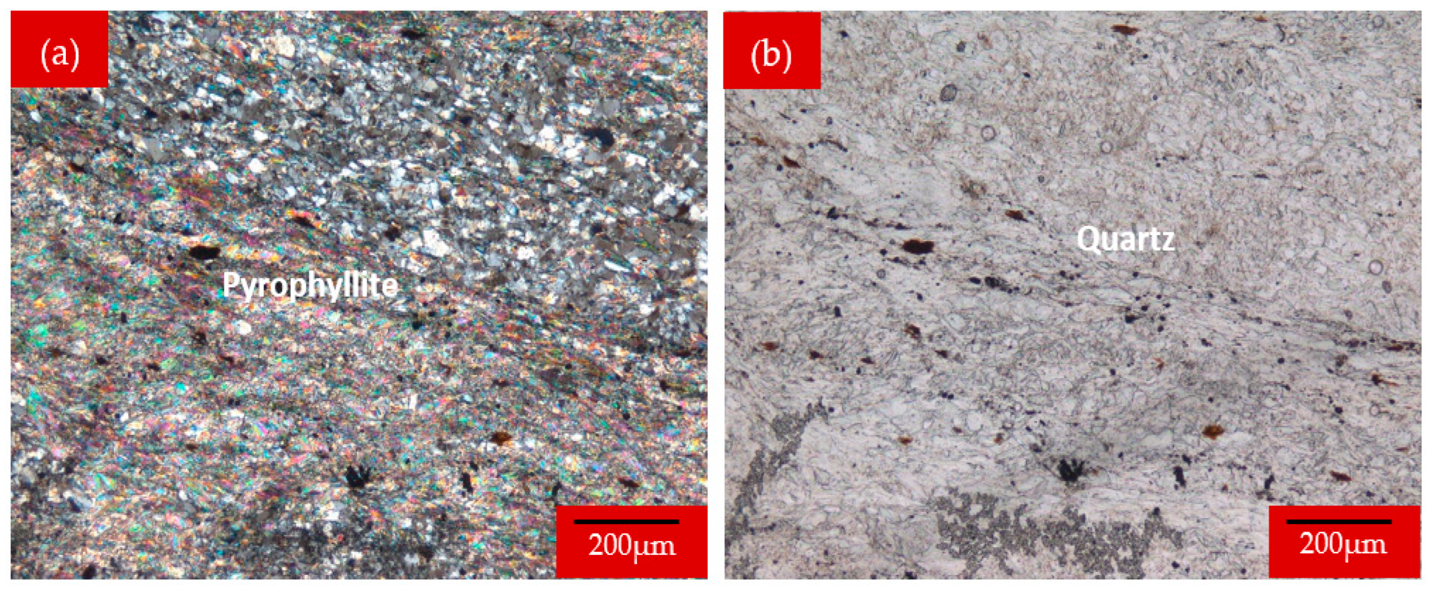
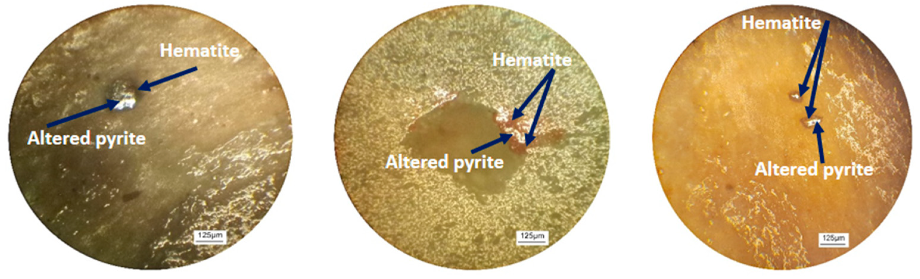
2.2.1. Microscopic Examination

The samples were characterized for their mineralogical contents using the transmitted and reflected light microscopy (ECLIPSE LV100DOL, Nikon, Melville, NY, USA) of both thin and polished sections.

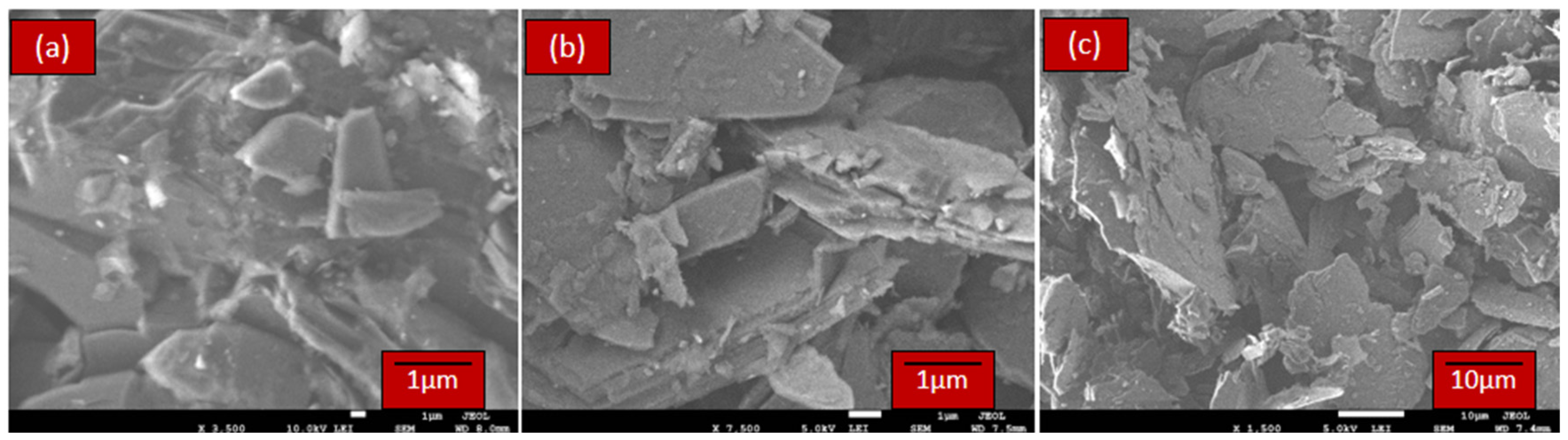
2.2.2. Scanning Electron Microscope (SEM)

The pyrophyllite samples were examined using a field emission scanning electron microscope FESEM Model (JSM-7600F, JEOL, Tokyo, Japan) combined with an energy dispersive X-ray (EDX) unit (Oxford Instruments, Abingdon, UK). SEM observations were conducted to detect changes in the phases before and after microwave treatment. The images were taken at a low voltage of 5 kV to avoid overcharging the sample. The intensity peaks of each element at specific energy levels were estimated by EDX.

2.2.3. X-ray Diffraction Analysis (XRD)

XRD was carried out for treated microwaved samples and untreated ones using an X-ray diffractometer (Regaku, Ultima 1V, Tokyo, Japan), with analytical conditions of Cu kα radiation (40 kV, 40 mA), a step of 0.05°, and scattering angle of 2θ in the range of 5° to 80°.

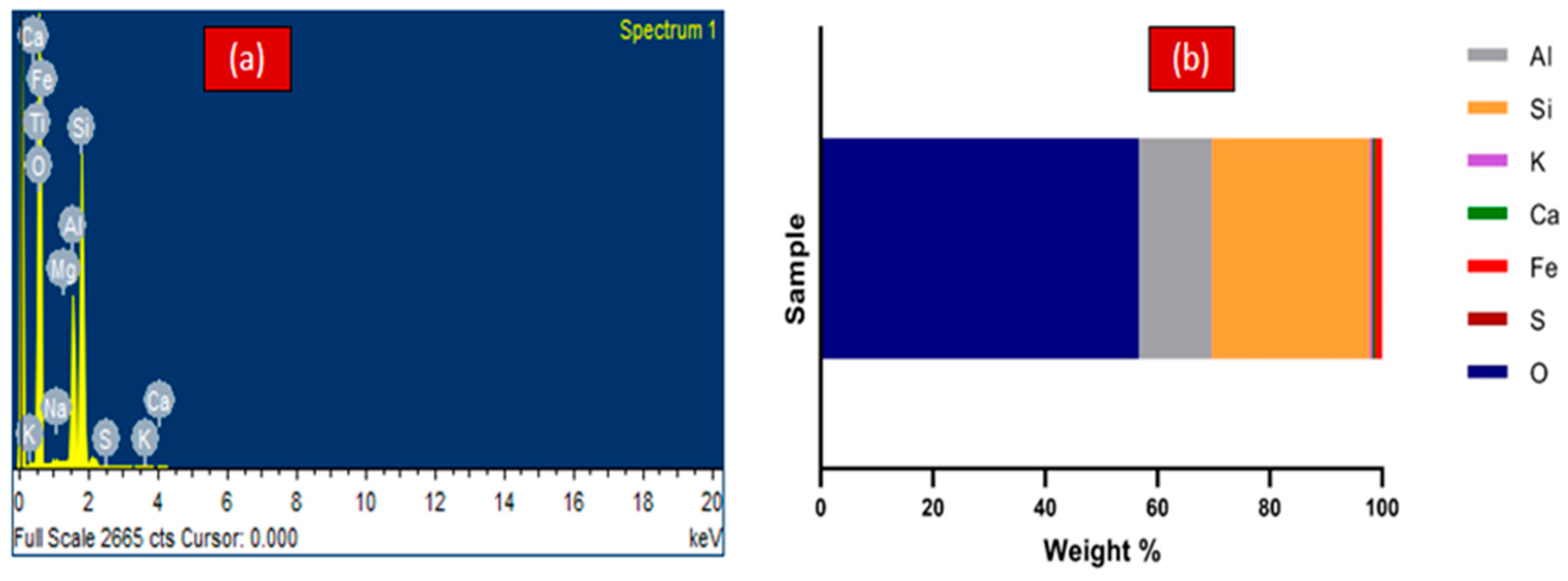
2.2.4. Chemical Analysis

Chemical analysis was conducted on low-grade Saudi pyrophyllite samples. A Rigaku RIX 2000 spectrometer, Tokyo, Japan was used to analyze the major oxides. The analysis included SiO2, CaO, Al2O3, MgO, Fe2O3 (total), K2O, Na2O, SO3, MnO, and P2O5. The samples were prepared by mixing 1.0 g of each sample with 6.0 g of lithium tetraborate beads to form glass beads. The glass beads were oxidized, melted, left to solidify in a casting mold, and then analyzed.

2.3. Approach 1: Dry Magnetic Separation

Upgrading of the Saudi low-grade pyrophyllite was carried out using an induced roll Outotec magnetic separator (Model MIH (13) 111-5, Outotec Inc., Espoo, Finland). Different operating parameters were examined, such as the magnetic field intensity, roll speed, feed rate, and feed size. The weights of both the non-magnetic and magnetic fractions were recorded in each experiment. Both fractions were chemically analyzed using XRF to determine their alumina and iron contents.
The recoveries of Al2O3, and Fe2O3 were calculated for the non-magnetic fraction according to Equation (1).
R e c o v e r y   ( % ) = C × c F × f × 100
where F is the weight of the considered feed, C is the weight of the produced concentrate, f is the assay of the considered component in the feed, and c is the assay of the same component in the concentrate.

2.4. Approach 2: Microwave Treatment Followed by Magnetic Separation

In this approach, microwave irradiation for pyrophyllite samples was performed prior to dry magnetic separation using a microwave oven (Amana RC17S2, 2.45 GHz, Whirlpool, MI, USA). The microwave irradiation times (10, 20, 30, 40, and 50 min) were investigated. The irradiated samples’ temperatures at the end of each irradiation time were 200, 390, 785, 800, and 805 °C, respectively. The treated and untreated samples were examined by XRD and SEM to detect any changes in the mineral phases resulting from microwave irradiation. The treated samples were then physically upgraded using the same dry magnetic separator used in the first pyrophyllite upgrading approach.

3. Results and Discussion

3.1. Characterization Results

3.1.1. Microscopic Analysis

Saudi low-grade pyrophyllite ore is generally metamorphic rock that is fine-grained, compact, and massive. Microscopic examination showed that pyrophyllite occurs as fine-grained anhedral flakes arranged in parallel with other constituents and exhibits a schistose texture (Figure 1a). Additionally, pyrophyllite was observed wrapping around quartz and feldspar crystals, creating an “Augen” texture. Associated minerals such as quartz and feldspars and rare amounts of muscovite, chlorite, and sericite were observed. Quartz was identified as finely grained, anhedral-strained crystals with wavy extinction and sutured edges. These crystals were often aligned in parallel and had a schistose texture (Figure 1b). Other associated minerals, such as feldspars and muscovite, are fine-grained anhedral crystals intercalated with rock constituents and arranged in parallel with other constituents to display the foliation texture. The observations showed that feldspars undergo partial alteration to form the minerals sericite, chlorite, and clay.
The results of reflected light microscopy shown in Figure 2 demonstrate that impurities are present in rare amounts and occur as very fine-grained anhedral crystals disseminated in the rocks following foliation. Moreover, these impurities are evident in altered pyrite and hematite forms [21,32,33]. The results also indicated that pyrite was completely and partially dissolved into iron oxide (hematite). Partial alteration occurs in the pyrite rim and within it. Figure 2 shows high pyrite reflectance with a white–gray color, dissolved at the rim and within it to hematite (brown).

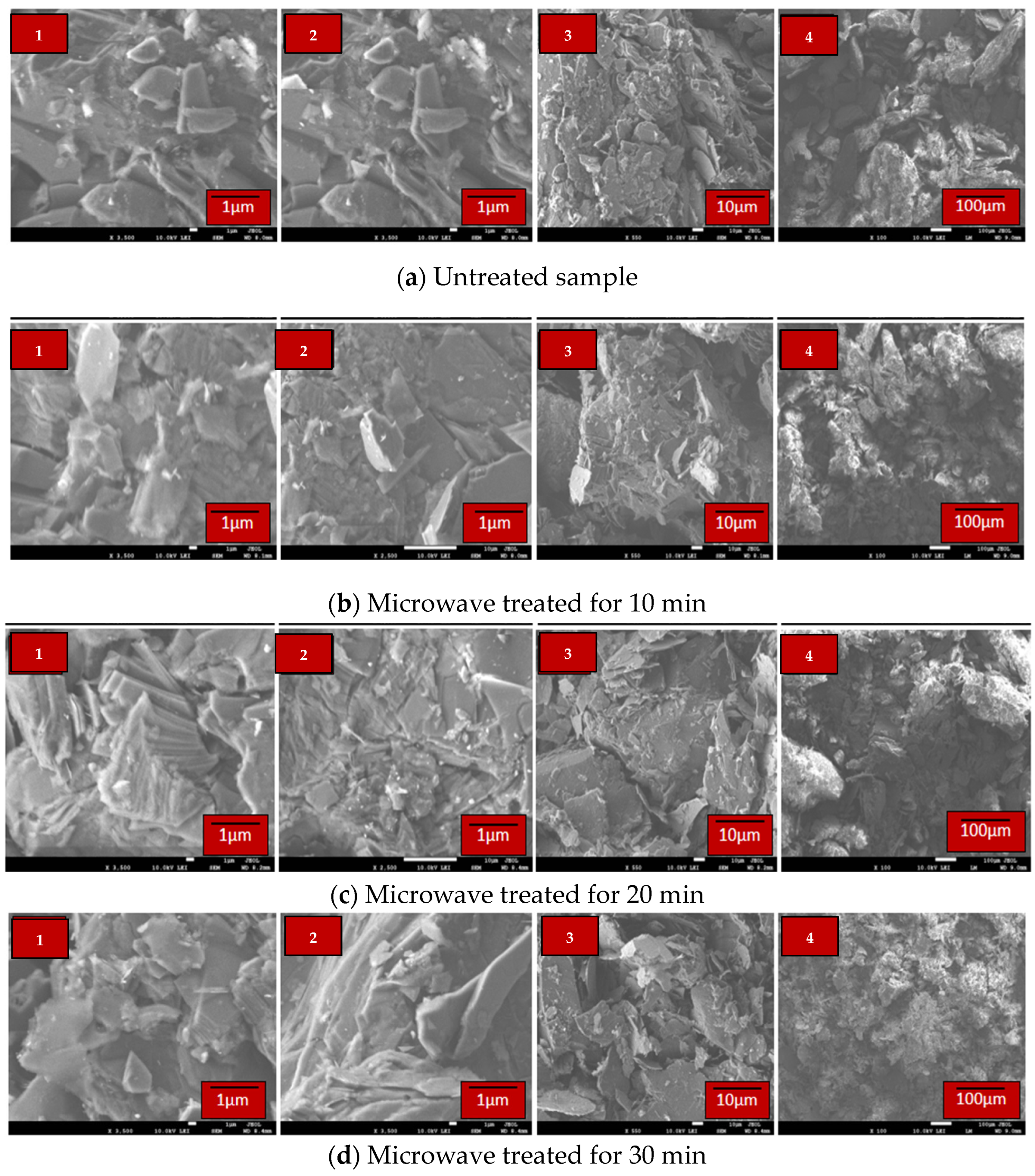
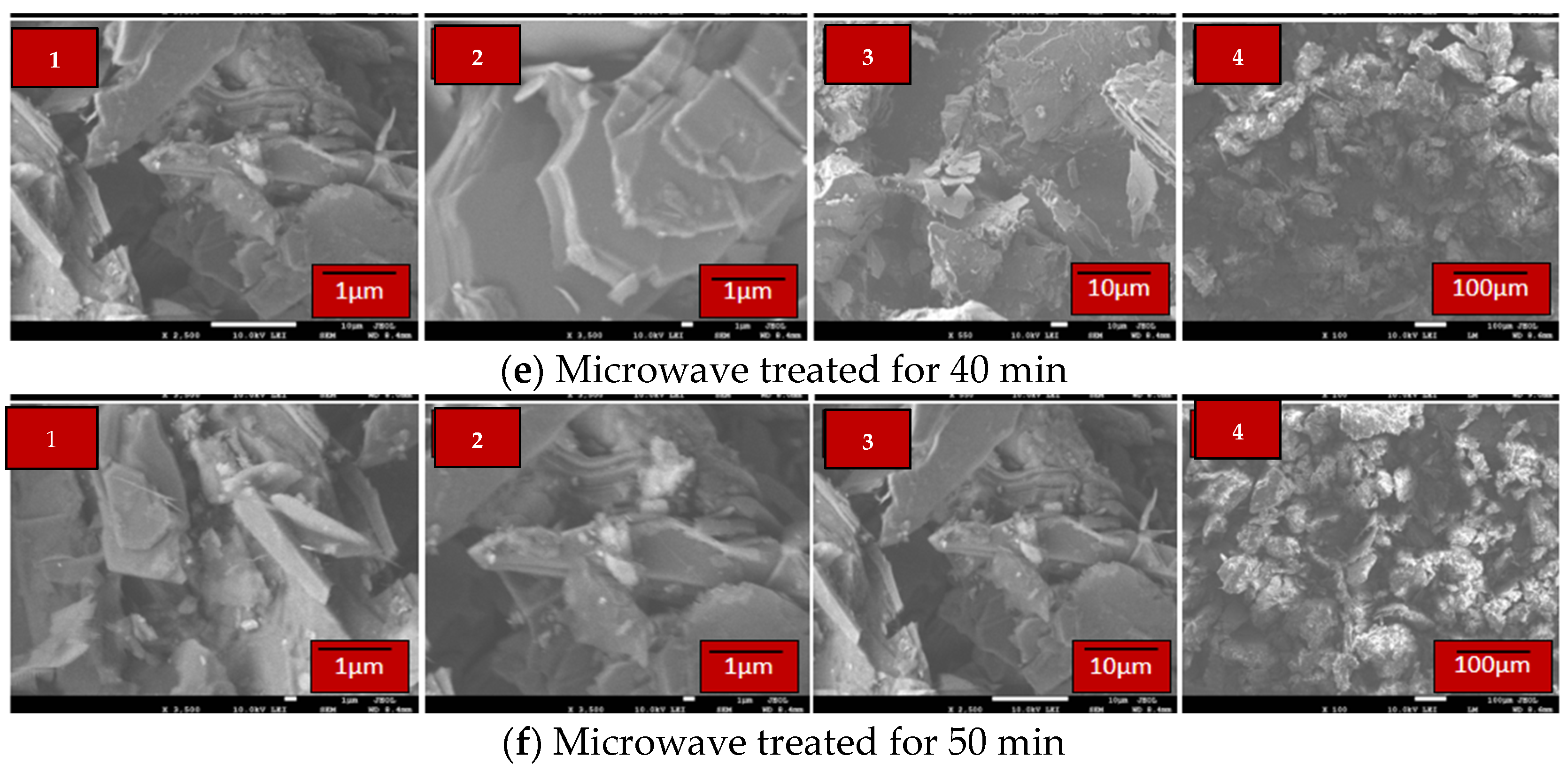
3.1.2. Scanning Electron Microscope (SEM)-EDX Characterization

Saudi low-grade pyrophyllite SEM images are shown in Figure 3. Pyrophyllite has a fine schistose texture, which agrees well with the results of the microscopic study. It can be seen that the pyrophyllite in the particles is highly foliated. Moreover, the particles were fine and had anhedral flakes arranged with sizes of about 2–4 μm in width and 0.1 μm in thickness.
The EDX pattern of pyrophyllite ore reveals peaks corresponding to aluminum and silicon, indicating the presence of alumina and silicates, which are the primary components of pyrophyllite, as shown in Figure 4a,b. The association of potassium (K) with aluminum (Al) and silicon (Si) suggests the presence of feldspar, muscovite, and sericite. The low occurrence of iron (Fe) indicates that Fe exists as an impurity.

3.1.3. Chemical Analysis
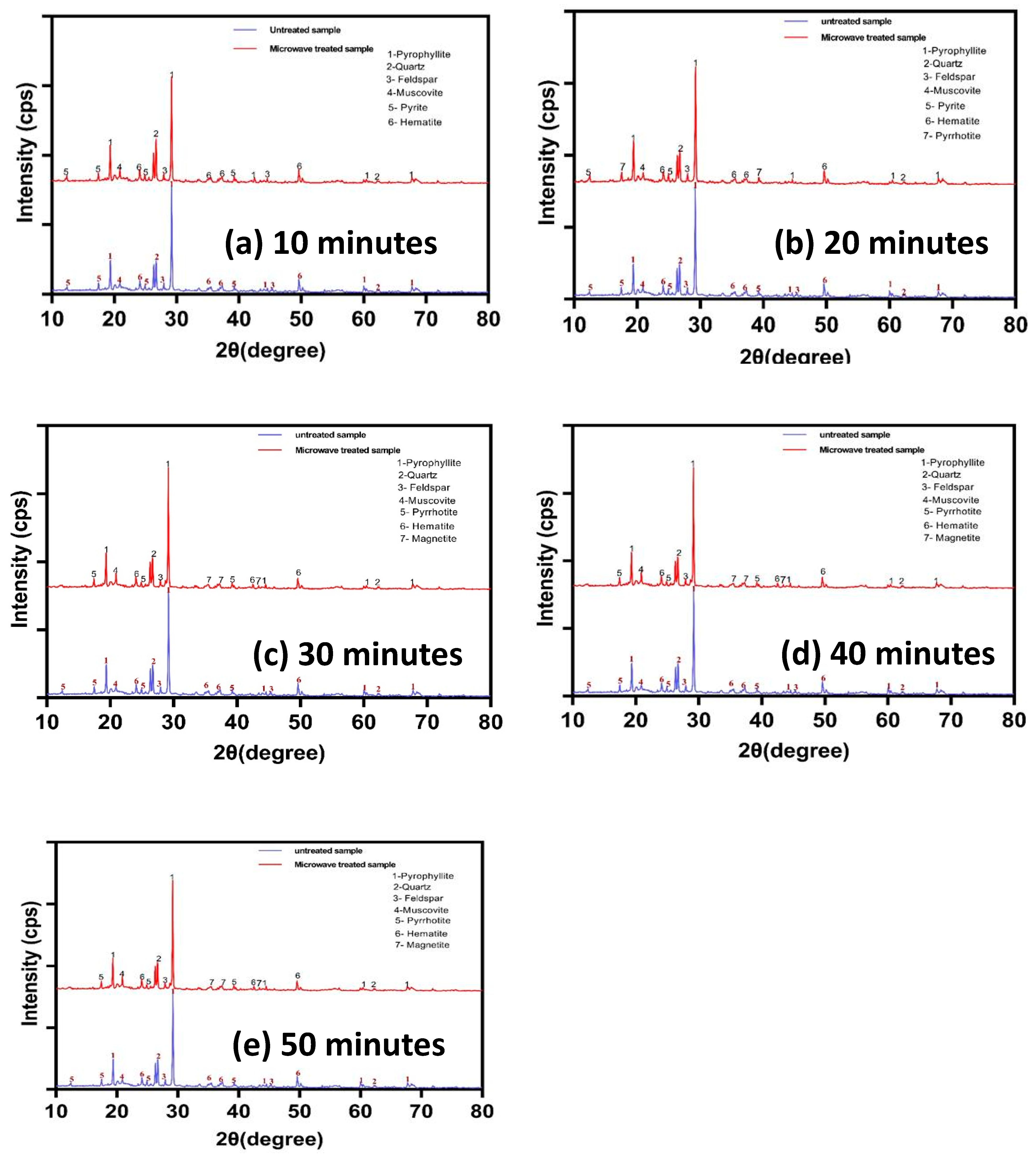
Table 1 shows the chemical analysis results for the low-grade samples. This shows that silicate and alumina minerals represented by silicate and aluminum oxides are the main constituents, accounting for 67.72% and 25.04% of the total constituents, respectively. The iron oxide content was 1.4%, indicating the low quality of the ore.

3.1.4. XRD

The analysis of the XRD spectra showed that the predominant phase was pyrophyllite (Al2Si4O10(OH)2), with quartz (SiO2) and feldspar (KAlSi3O8) as the main associated minerals, as shown in Figure 5. Meanwhile, the XRD showed a rare amount of muscovite (KF)2(Al2O3)3(SiO2)6(H2O), which agrees well with the results of EDX and chemical studies. Pyrite (FeS2) and hematite (Fe2O3) phases were observed. This result can be attributed to the hydrothermal alteration altering the formation of pyrite and hematite in the pyrophyllite.

3.2. Dry High-Intensity Magnetic Separation (DHIMS)

Dry magnetic separation experiments were carried out to determine the optimal conditions for achieving higher iron removal and alumina recovery in the non-magnetic concentrate fraction. Various parameters were investigated, including the magnetic roll speed, feed flow rate, magnetic field intensity, and feed particle size.

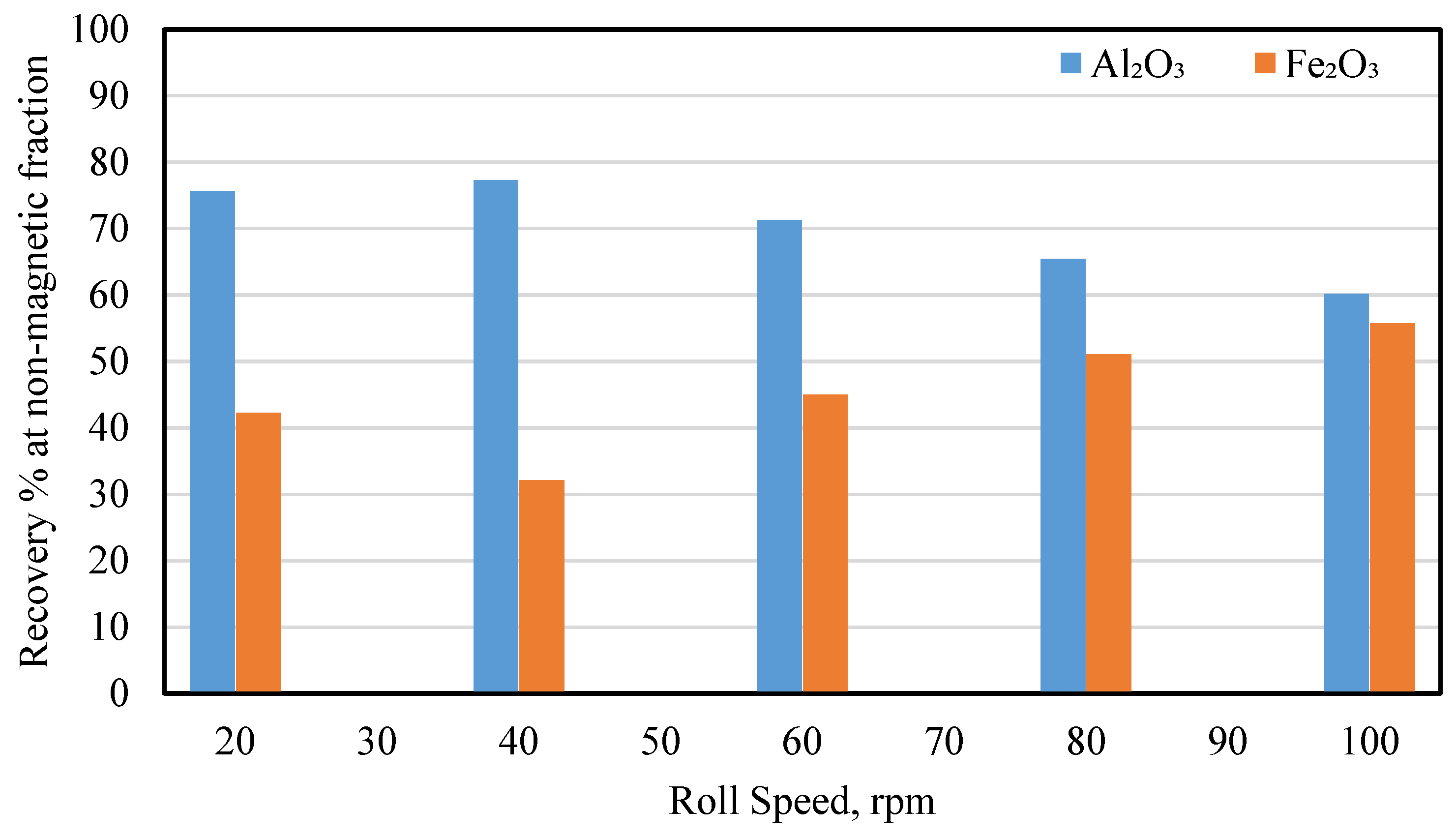
3.2.1. Effect of the Magnet Roll Speed

The effect of roll speed on the recovery of iron and alumina in the non-magnetic fraction was investigated by increasing the roll speed from 20 to 100 rpm (see Figure 6). The magnetic separation process was performed at a magnetic intensity of 8000 Gauss, feed size of 0.250 + 0.045 mm, and feed rate of 24 kg/h. The results showed that there was a decrease in Fe2O3 recovery to 32.14% in the non-magnetic fraction as the roll speed increased from 20 to 40 rpm, as shown in Figure 6. The recovery percentage began to rise as the speed increased from 40 to 100 rpm. On the contrary, by raising the roll speed to 40 rpm, the alumina recovery reached 77.28%, although a gradual decrease in the alumina recovery was observed as the roll speed increased to 100 rpm. The results revealed that increasing the roll speed increased the centrifugal force, which enhanced the selectivity of the magnetic and non-magnetic portions. However, the exceeding increase in roll speed limits the selectivity of magnetic and non-magnetic particles.

3.2.2. Effect of the Feed Flow Rate

A range of feed rates from 12 to 60 kg/h. were used to examine the impact of feed flow rate at 40 rpm, a feed size of −0.250 + 0.045 mm, and a magnetic intensity of 8000 Gauss. Figure 7 shows that a feed rate of 24 kg/h. was optimal as the lowest Fe2O3 recovery and highest Al2O3 recovery rates were 32.4% and 77.34%, respectively. The findings indicate that higher feed rates reduce the possibility of magnetic particle capture across the roll due to the large number of particles in the separation area.

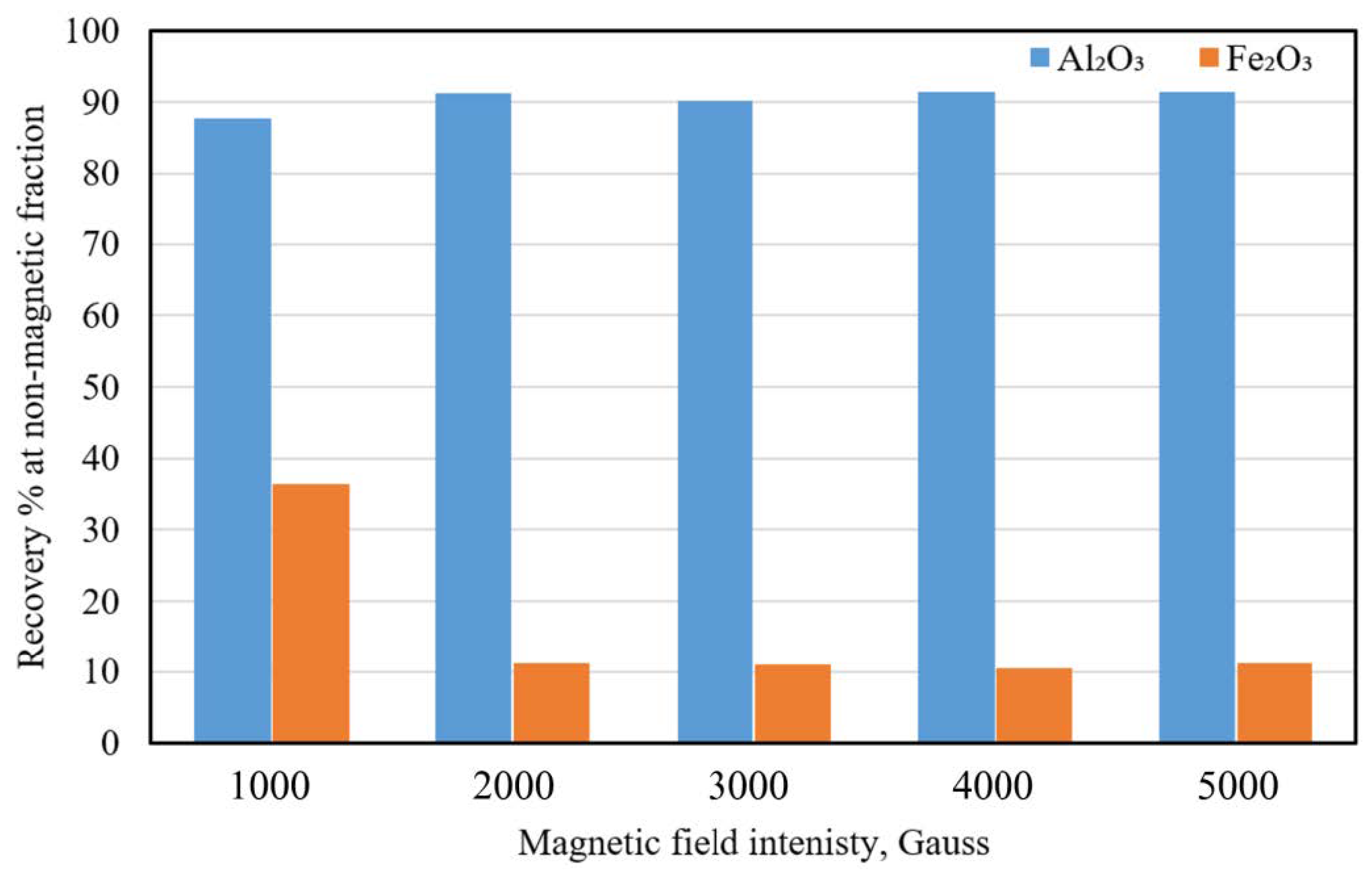
3.2.3. Effect of the Magnetic Field Intensity

Experiments were carried out to study the magnetic field intensity from 2000 to 12,000 Gauss at a roll speed of 40 rpm, feed rate of 24 kg/h, and feed size of −0.250 + 0.045 mm, as shown in Figure 8. Increasing the magnetic intensity to 12,000 Gauss decreased Fe2O3 recovery to 22.98% in the non-magnetic portion, as shown in Figure 8. Moreover, the Al2O3 recovery increased with increasing magnetic strength, reaching 79.49% at 12,000 Gauss.

3.2.4. Effect of the Particle Size

Table 2 investigates the efficiency of Fe2O3 and Al2O3 recovery for various fractions of non-magnetic concentrate using an induced roll separator with a magnetic intensity of 12,000 Gauss, roll speed of 40 rpm, and feed rate of 24 kg/h. The results show that the non-magnetic concentrate yield obtained for all fractions was not high enough but relatively acceptable. Lower Fe2O3 recovery in the non-magnetic portion was 21.91% at a fraction size of 0.125 + 0.045 mm. The highest value was 33.99% at a feed size of 0.045 mm. This result can be attributed to the non-capture of fine particles in the magnetic grooves. On the other hand, the alumina recovery showed different results, as a higher recovery of 80.47% was achieved at a feed size of 0.045 mm, and a lower recovery was achieved at 0.125 + 0.075 mm. However, the recovery percentage of Al2O3 at a size of 0.045 mm was very close to the recovery at a fraction size of 0.125 + 0.045 mm (optimum for Fe2O3 removal), which was 79.76%.

3.3. Microwave Treatment

3.3.1. Effect of Microwave Treatment on Impurities Phases

The effect of microwave treatment on the impurity phase was investigated using XRD, SEM, and EDX analyses (Figure 9 and Figure 10). The studied samples included untreated samples and microwave-traded samples with various irradiation times (10, 20, 30, 40, and 50 min) to identify phase changes during the microwave treatment. Figure 9a–e presents the XRD results of the untreated sample (at the bottom), and microwave-treated samples for different irradiation times (at the top). The results showed significantly higher X-ray intensities for samples treated for 10 min (Figure 9a), than for untreated samples. However, no phase change was detected after microwave treatment because the irradiation time and pyrophyllite sample temperature were not sufficient to cause a phase change. A phase change from pyrite (FeS2) to pyrrhotite (Fe7S8) was detected after 20 min of microwave treatment (Figure 9b). The sample exhibited higher intensities than the sample treated for only 10 min. These findings are consistent with the previous literature [21,30,34,35], suggesting that microwave treatment can indeed convert pyrite into ferromagnetic phases. Moreover, the X-ray intensity of the sample heated for 30 min was notably higher than that of the sample treated for 10 and 20 min (Figure 9c). The observed increase can be attributed to the prolonged irradiation time and higher temperature of the pyrophyllite samples. Additionally, the results indicated a phase change in the pyrite (FeS2), which was transformed into pyrrhotite (Fe7S8), while the hematite (Fe2O3) was converted into magnetite (Fe3O4). This finding aligns with previous research [21,30,35], which documented the phase change in pyrite and hematite into more ferromagnetic phases after 30 min of microwave treatment.
Similarly, the X-ray intensities after a microwave treatment of 40 min remained relatively similar to those obtained after 30 min, as shown in Figure 9d. The results indicate a slowing rate of increase in sample temperature. Consequently, the phase change persisted, with pyrite converting to pyrrhotite and hematite (Fe2O3) to magnetite (Fe3O4), confirming that microwave treatment after 40 min had the same effect as 30 min. Furthermore, the 50 minutes’ X-ray intensities of microwave treatment, Figure 9e, was relatively similar to those obtained for 30 and 40 min, suggesting a consistent rate of increase in sample temperature. The phase change that was shown after 50 min was similar to what was seen after 40 and 30 min, indicating that the effects of the microwave treatment were similar after 50 min.
Figure 10a–f shows the SEM micrographs of the untreated and microwave-treated samples at various durations (10, 20, 30, 40, and 50 min). The untreated sample displays a fine schistose texture, and the particles are highly foliated, with have anhedral flakes arranged, as shown in Figure 10a. For the 10 minute microwave treatment, cracks developed mostly within the grain boundary, as shown in Figure 10b. The observations agree with previous studies, which reported that microwave irradiation is more significant at the boundaries and the surfaces of the grains [21,30]. The 20 min treated samples display a very fine schistose texture and the particles are highly foliated, as in Figure 10c. Moreover, the phase transition of pyrite into pyrrhotite may potentially be attributed to the intense impact of microwave irradiation induction on the grain rims and surface. SEM micrograph of 30 min microwave-treated sample is shown in Figure 10d. The surface of the particles appears significantly induced by microwave irradiation compared to the samples treated for 10 and 20 min. Figure 10e illustrates that the notable impact of microwave irradiation induction for 40 min on the surface and rims of the grains is comparatively similar to that induced by 30 min of microwave treatment. The results of the 50 min microwave irradiation treatment showed similar results as the 40 and 30 min treatments, as shown in Figure 10f. The SEM findings are supported the XRD, suggesting that the microwave treatment’s effects were comparable for 30, 40, and 50 min.

3.3.2. Effect of Microwave Irradiation on Iron Removal

Effect of Microwave Irradiation Time on Iron Removal

Experiments were conducted for different microwave irradiation times (10 to 50 min) at a magnetic intensity of 5000 Gauss, roll speed of 40 rpm, and feed rate of 24 kg/h. The irradiation time results indicate that iron recovery in the non-magnetic concentrate decreased with increasing irradiation time (Figure 11). This decline can be attributed to the transformation of iron-impurity minerals into ferromagnetic minerals, leading to an increase in magnetic susceptibility and magnetic properties as the irradiation time increases. Conversely, the alumina recovery in the non-magnetic concentrate increased with increasing the irradiation time (Figure 11). However, the results indicate that irradiation for >30 min saturated the removal efficiency of iron minerals, reaching approximately 89%. This indicates that microwave treatment for 30 min is optimal for pre-treating Saudi low-grade pyrophyllite ore, a conclusion supported by the findings of [21].

Effect of Magnetic Field Intensity on the Iron Removal from Microwave Irradiated Pyrophyllite

In order to study the effect of field intensity on iron removal, samples that underwent 30 min of microwave irradiation were subjected to different magnetic field intensities ranging from 1000 to 5000 Gauss at a roll speed of 40 rpm and feed rate of 24 kg/h. The results show that as the magnetic field intensity increases, the alumina recovery percentage increases in the non-magnetic concentrate. The iron recovery decreased at higher intensities (Figure 12). However, magnetic field intensities exceeding 2000 Gauss saturated the iron removal efficiency, reaching approximately 89%. This may be attributed to the fact that a magnetic field intensity of 2000 Gauss was adequate for removing ferromagnetic minerals from microwave-treated pyrophyllite samples irradiated for 30 min.

4. Conclusions

This study aimed to investigate the removal of iron impurities from Saudi low-grade pyrophyllite ore. The study compared the use of a dry high-intensity magnetic separation approach with a combined one that involves microwave treatment prior to dry magnetic separation. The ore characteristics show that pyrite and hematite were the main iron impurities. Different parameters were studied, including roll speed, feed rate, field intensity, and feed particle size, for the dry high-intensity magnetic separation process. The microwave irradiation time and magnetic field intensity were studied for the combined method. The results indicated that the optimum conditions for dry magnetic separation were a feed rate of 24 kg/h, roll speed 40rpm, magnetic intensity to 12,000 Gauss, and feed size of 0.125 + 0.045. This achieved a lower iron recovery 21.92%, accompanied by an alumina recovery of 80.47%. On the other hand, 30 min of irradiation and a magnetic field intensity of 2000 Gauss were optimal for the microwave treatment process. This study shows that using dry high-intensity magnetic separation (DHIMS) at 12,000 Gauss achieved an iron recovery of 21.92%, accompanied by an alumina recovery of 80.47% in the non-magnetic portion. The utilization of a combined approach involving microwave treatment and magnetic separation proves to be a promising and effective method for enriching low-grade Saudi pyrophyllite, as it achieves an impressive lower iron recovery of 11.2% in the non-magnetic portion and a removal efficiency of 89%, in addition to the higher the alumina content of 91.31% in the non-magnetic yield. However, according to international market specifications for pyrophyllite, the concentrate obtained through the combined method could fetch USD 480/t [6,26,36]. On the economic side, the combined method utilizes a lower magnetic field intensity (2000 Gauss) compared to the high-intensity magnetic separation method (up to 12,000 Gauss), leading to reduced energy consumption and costs. Additionally, the entire process is conducted under dry conditions, eliminating the need for water usage and associated drying expenses, thus maximizing efficiency in workspace utilization.

Author Contributions

Conceptualization, H.A.M.A.; formal analysis, N.K., H.A.M.A. and M.A.A.; resources, M.A.A.; data curation, M.A.A. and H.A.M.A.; writing—original draft preparation, H.A.M.A., M.A.A. and N.K.; writing—review and editing, H.A.M.A.; visualization, H.A.M.A., M.A.A. and N.K.; supervision, H.A.M.A.; funding acquisition, H.A.M.A. All authors have read and agreed to the published version of the manuscript.

Funding

This work was supported by the Deanship of Scientific Research (DSR), King Abdulaziz University, Jeddah, under grant no. (D1434-135-002). The authors, therefore, gratefully acknowledge the DSR’s technical and financial support.

Institutional Review Board Statement

Not applicable.

Informed Consent Statement

Not applicable.

Data Availability Statement

All data are included in the article.

Conflicts of Interest

The authors declare no conflict of interest.

References

  1. Abdrakhimova, E.S. Physicochemical studies of pyrophyllite of the Nikol’sk deposit. Refract. Ind. Ceram. 2010, 51, 6. [Google Scholar] [CrossRef]
  2. Jeong, Y.; Cho, K.; Kwon, E.E.; Tsang, Y.F.; Rinklebe, J.; Park, C. Evaluating the feasibility of pyrophyllite-based ceramic membranes for treating domestic wastewater in anaerobic ceramic membrane bioreactors. Chem. Eng. J. 2017, 328, 567–573. [Google Scholar] [CrossRef]
  3. Kogel, J.E.; Trivedi, N.C.; Barker, J.M.; Krukowski, S.T. Industrial Minerals & Rocks: Commodities, Markets, and Uses; SME: Littleton, CO, USA, 2006; Volume 44. [Google Scholar]
  4. Ali, M.A.; Ahmed, H.A.M. “A Review of Low-Grade Pyrophyllite ore Upgrading Techniques” Researchers: Abstract. Arab. J. Sci. Publ. 2021, 37, 19–31. [Google Scholar]
  5. Bentayeb, A.; Amouric, M.; Olives, J.; Dekayir, A.; Nadiri, A. XRD and HRTEM characterization of pyrophyllite from Morocco and its possible applications. Appl. Clay Sci. 2003, 22, 211–221. [Google Scholar] [CrossRef]
  6. Ali, M.A.; Ahmed, H.A.M.; Ahmed, H.M.; Hefni, M. Pyrophyllite: An Economic Mineral for Different Industrial Applications. Appl. Sci. 2021, 11, 11357. [Google Scholar] [CrossRef]
  7. González-Miranda, F.d.M.; Garzón, E.; Reca, J.; Pérez-Villarejo, L.; Martínez-Martínez, S.; Sánchez-Soto, P.J. Thermal behaviour of sericite clays as precursors of mullite materials. J. Therm. Anal. Calorim. 2018, 132, 967–977. [Google Scholar] [CrossRef]
  8. Fitzgerald, J.J.; Hamza, A.I.; Dec, S.F.; Bronnimann, C.E. Solid-state 27Al and 29Si NMR and 1H CRAMPS studies of the thermal transformations of the 2:1 phyllosilicate pyrophyllite. J. Phys. Chem. 1996, 100, 17351–17360. [Google Scholar] [CrossRef]
  9. Zhao, X.; Ouyang, J.; Tan, Q.; Tan, X.; Yang, H. Interfacial characteristics between mineral fillers and phenolic resin in friction materials. Mater. Express 2020, 10, 70–80. [Google Scholar] [CrossRef]
  10. Song, S.; Wu, Z.; Nie, J. Paper fillers innovations: From design of particles to preparing filler composites. BioResources 2020, 15, 2117. [Google Scholar] [CrossRef]
  11. DeArmitt, C. Functional Fillers for Plastics. In Applied Plastics Engineering Handbook: Processing, Materials, and Applications; Kutz, M., Ed.; Elsevier Inc.: Amsterdam, The Netherlands, 2017. [Google Scholar]
  12. McGonigle, F.; Ciullo, P.A. Industrial Minerals and Their Uses: A Handbook and Formulary, 1st ed.; Ciullo, P.A., Ed.; Noyes Publications: Westwood, NJ, USA, 1996. [Google Scholar]
  13. Hasanbegović, E.; Huremović, J.; Žero, S. Adsorption capacity of nitrate from artificial fertilizers and soil on pyrophyllite. Int. J. Environ. Sci. Technol. 2021, 18, 3731–3738. [Google Scholar] [CrossRef]
  14. Qin, X.; Zhao, J.; Wang, J.; He, M. Atomic structure, electronic, and mechanical properties of pyrophyllite under pressure: A first-principles study. Minerals 2020, 10, 778. [Google Scholar] [CrossRef]
  15. Shayakhmetov, U.S.; Shayakhmetov, A.U.; Zakharov, A.V.; Khamidullin, A.R.; Gazizova, A.T. Refractory Composites Based on Pyrophyllite Raw Materials. Refract. Ind. Ceram. 2018, 59, 241–246. [Google Scholar] [CrossRef]
  16. Harvey, C.C.; Murray, H.H. Industrial clays in the 21st century: A perspective of exploration, technology and utilization. Appl. Clay Sci. 1997, 11, 285–310. [Google Scholar] [CrossRef]
  17. Decleer, J.G.M. Introduction to Industrial Minerals, 1st ed.; Chapman & Hall: London, UK, 1996; Volume 10. [Google Scholar]
  18. Sarikaya, M.; Depci, T.; Aydogmus, R.; Yucel, A.; Kizilkaya, N. Production of Nano Amorphous SiO2 from Malatya Pyrophyllite. IOP Conf. Ser. Earth Environ. Sci. 2016, 44, 052004. [Google Scholar] [CrossRef]
  19. Mansour, S.M. Use of natural pyrophyllite as cement substitution in ultra performance polypropylene fiber concrete. Int. J. Eng. Res. Africa 2021, 56, 123–135. [Google Scholar] [CrossRef]
  20. Ambikadevi, V.R.; Lalithambika, M. Effect of organic acids on ferric iron removal from iron-stained kaolinite. Appl. Clay Sci. 2000, 16, 133–145. [Google Scholar] [CrossRef]
  21. Kim, B.J.; Cho, K.H.; Chang, B.; Kim, H.S.; Lee, S.G.; Park, C.Y.; Lee, S.; Choi, N.C. Sequential microwave roasting and magnetic separation for removal of Fe and Ti impurities in low-grade pyrophyllite ore from Wando mine, South Korea. Miner. Eng. 2019, 140, 105881. [Google Scholar] [CrossRef]
  22. Mowla, D.; Karimi, G.; Ostadnezhad, K. Removal of hematite from silica sand ore by reverse flotation technique. Sep. Purif. Technol. 2008, 58, 419–423. [Google Scholar] [CrossRef]
  23. Mukhopadhyay, T.K.; Ghatak, S.; Maiti, H.S. Pyrophyllite as raw material for ceramic applications in the perspective of its pyro-chemical properties. Ceram. Int. 2010, 36, 909–916. [Google Scholar] [CrossRef]
  24. Chatterjee, K.K. Uses of Industrial Minerals, Rocks and Freshwater; Nova Science Publishers: New York, NY, USA, 2009. [Google Scholar]
  25. Perepelitsyn, V.A.; Proshkin, V.A.; Rytvin, V.M.; Ignatenko, V.G.; Yarosh, I.A.; Abyzov, A.N. Non-traditional domestic refractory materials for aluminum metallurgy. Refract. Ind. Ceram. 2008, 49, 257–260. [Google Scholar] [CrossRef]
  26. Jena, S.K.; Singh, S.; Rao, D.S.; Dhawan, N.; Misra, P.K.; Das, B. Characterization and removal of iron from pyrophyllite ore for industrial applications. Miner. Metall. Process. 2015, 32, 102–110. [Google Scholar] [CrossRef]
  27. Kim, B.J.; Cho, K.H.; Lee, S.G.; Park, C.Y.; Choi, N.C.; Lee, S. Effective gold recovery from near-surface oxide zone using reductive microwave roasting and magnetic separation. Metals 2018, 8, 957. [Google Scholar] [CrossRef]
  28. Andji, J.Y.Y.; Toure, A.A.; Kra, G.; Jumas, J.C.; Yvon, J.; Blanchart, P. Iron role on mechanical properties of ceramics with clays from Ivory Coast. Ceram. Int. 2009, 35, 571–577. [Google Scholar] [CrossRef]
  29. Ferreira, D.P. Structural Iron Removal from Clays Used in Ceramic Industries. Master’s Thesis, University of Coimbra, Combra, Portugal, 2017. [Google Scholar]
  30. Cho, K.-H.; Kim, B.-J.; Choi, N.-C.; Park, C.-Y. The Mineralogical and Chemical Characteristics of Fe Impurities and the Efficiency of their Removal Using Microwave Heating and Magnetic Separation in the Pyrophyllite Ore. J. Mineral. Soc. Korea 2016, 29, 47–58. [Google Scholar] [CrossRef]
  31. Omran, M.; Fabritius, T.; Elmahdy, A.M.; Abdel-Khalek, N.A.; El-Aref, M.; Elmanawi, A.E.H. Effect of microwave pre-treatment on the magnetic properties of iron ore and its implications on magnetic separation. Sep. Purif. Technol. 2014, 136, 223–232. [Google Scholar] [CrossRef]
  32. Wiewióra, A.; Sánchez-Soto, P.J.; Avilés, M.A.; Justo, A.; Pérez-Rodríguez, J.L. Effect of dry grinding and leaching on polytypic structure of pyrophyllite. Appl. Clay Sci. 1993, 8, 261–282. [Google Scholar] [CrossRef]
  33. Bozkaya, Ö.; Yalçin, H.; Başibüyük, Z.; Bozkaya, G. Metamorphic-hosted pyrophyllite and dickite occurrences from the hydrous Al-silicate deposits of the Malatya-Pütürge region, central eastern Anatolia, Turkey. Clays Clay Miner. 2007, 55, 423–442. [Google Scholar] [CrossRef]
  34. Waanders, F.B.; Mohamed, W.; Wagner, N.J. Changes of pyrite and pyrrhotite in coal upon microwave treatment. J. Phys. Conf. Ser. 2010, 217, 012051. [Google Scholar] [CrossRef]
  35. Kingman, S.W. Recent developments in microwave processing of minerals. Int. Mater. Rev. 2006, 51, 1–12. [Google Scholar] [CrossRef]
  36. Ali, M.A.; Ahmed, H.A.M. Chemical and Mineralogical Characterization of Saudi-Pyrophyllite ore and its potential applications. Arab Gulf J. Sci. Res. 2022, 39, 1–18. [Google Scholar] [CrossRef]
Figure 1. Photomicrographs of Saudi low-grade pyrophyllite ore.
Applsci 14 06651 g001
Figure 2. Photomicrographs of impurities in Saudi low-grade pyrophyllite ore: pyrite is gray in color and hematite is brown in color.
Applsci 14 06651 g002
Figure 3. SEM images of Saudi low-grade pyrophyllite ore grade: (a) ×3500, (b) ×7500, (c) ×1500, 5 kV.
Applsci 14 06651 g003
Figure 4. SEM-EDX analysis of the studied pyrophyllite ore grade: (a) EDX image; (b) elemental composition.
Applsci 14 06651 g004
Figure 5. XRD pattern of the Saudi low–grade pyrophyllite ore.
Applsci 14 06651 g005
Figure 6. Effect of roll speed on Fe2O3 and Al2O3 recovery% in non-magnetic fraction.
Applsci 14 06651 g006
Figure 7. Effect of feed rate on Fe2O3 and Al2O3 recovery in non-magnetic fraction.
Applsci 14 06651 g007
Figure 8. Effect of magnetic field intensity on Fe2O3 and Al2O3 recovery% in non-magnetic fraction.
Applsci 14 06651 g008
Figure 9. XRD patterns for the untreated pyrophyllite and the microwave-treated samples.
Applsci 14 06651 g009
Figure 10. SEM images of untreated and microwave-treated pyrophyllite samples.
Applsci 14 06651 g010aApplsci 14 06651 g010b
Figure 11. Effect of microwave irradiation time on Fe2O3 and Al2O3 recovery% in non-magnetic fraction.
Applsci 14 06651 g011
Figure 12. Effect of magnetic field intensity on Fe2O3 and Al2O3 recovery% in non-magnetic fraction treated microwave.
Applsci 14 06651 g012
Table 1. Chemical composition of Saudi low-grade pyrophyllite ore.
Constituent SiO2Al2O3Fe2O3TiO2Na2OK2OCaOMgP2O5LOI
wt. %67.7225.041.40.360.140.160.310.140.134.2
Table 2. Fe2O3 and Al2O3 recovery in non-magnetic concentrate for different pyrophyllite sizes.
Feed Size (mm)ProductsYield
wt. %
Assay %Recovery (%)
Fe2O3Al2O3Fe2O3Al2O3
−0.250 + 0.045Conc.76.50.4226.0222.9579.49
Tail.23.54.5921.8577.0520.51
Total 1001.4025.04100100
−0.125 + 0.045Conc.76.70.4026.0421.9179.76
Tail.23.34.6921.7578.0920.24
Total 1001.4025.04100100
−0.250 + 0.075Conc.75.040.5725.8730.5577.52
Tail.24.963.9022.5569.4522.48
Total 1001.4025.04100100
−0.125 + 0.075Conc.76.560.4625.9825.1679.43
Tail.23.444.4721.9774.8420.57
Total 1001.4025.04100100
−0.075 + 0.045Conc.770.5125.9328.0579.74
Tail.234.3822.0671.9520.26
Total 1001.4025.04100100
−0.045Conc.780.6125.8333.9980.47
Tail.224.222.2366.0119.53
Total 1001.4025.04100100
Disclaimer/Publisher’s Note: The statements, opinions and data contained in all publications are solely those of the individual author(s) and contributor(s) and not of MDPI and/or the editor(s). MDPI and/or the editor(s) disclaim responsibility for any injury to people or property resulting from any ideas, methods, instructions or products referred to in the content.

