Article

Mouthwash Containing Plant-Derived Biosurfactant and Chitosan Hydrochloride: Assessment of Antimicrobial Activity, Antibiofilm Activity, and Genotoxicity

by
Izabelle R. Souza
1,
Káren G. O. Bezerra
2,
Camila L. Oliveira
1,
Hugo M. Meira
2,
Thayza C. M. Stamford
3,
Attilio Converti
4,*,
Leonie A. Sarubbo
2,5,* and
Raquel D. Rufino
1
1
Escola de Saúde e Ciências da Vida, Universidade Católica de Pernambuco (UNICAP), Rua do Príncipe, n. 526, Boa Vista, Recife 50050-900, PE, Brazil
2
Instituto Avançado de Tecnologia e Inovação (IATI), Rua Potyra, n. 31, Prado, Recife 50070-280, PE, Brazil
3
Departamento de Medicina Tropical, Universidade Federal de Pernambuco (UFPE), Av. Prof. Moraes Rego, 1235—Cidade Universitária, Recife 50670-90, PE, Brazil
4
Department of Civil, Chemical and Environmental Engineering, University of Genoa (UNIGE), Pole of Chemical Engineering, Via Opera Pia, n. 15, 16145 Genova, Italy
5
Escola UNICAP Icam Tech, Universidade Católica de Pernambuco (UNICAP), Rua do Príncipe, n. 526, Boa Vista, Recife 50050-900, PE, Brazil
*
Authors to whom correspondence should be addressed.
Appl. Sci. 2024, 14(15), 6711; https://doi.org/10.3390/app14156711
Submission received: 10 July 2024 / Revised: 24 July 2024 / Accepted: 30 July 2024 / Published: 1 August 2024
(This article belongs to the Special Issue Bioenergy and Bioproducts from Biomass and Waste)

Abstract

:
Plant-derived biosurfactants are widely used due to their emulsifying and surface-active properties and can be applied in various products. The aim of this present study was to develop a mouthwash using chitosan hydrochloride and saponins extracted from the plants Chenopodium quinoa and Glycine max. After extraction of the biosurfactants using the Soxhlet method, they were characterized with the aid of infrared spectroscopy and subjected to determination of critical micelle concentration, which was found to be 4.0 and 3.5 g/L for C. quinoa and G. max, respectively. The stability of the emulsions was investigated in the presence of different oils and at different values of temperature, pH, and salinity, which showed an emulsification index greater than 40% under all conditions analyzed. After obtaining the mouthwash formulation, tests of foaming capacity, pH, and genotoxicity were performed in cells of onion (Allium cepa) roots. No presence of micronuclei was found in the roots exposed to the formulation, which indicates that there was no aggression to the cells. The results of antimicrobial susceptibility tests revealed bacteriostatic/bactericidal activity as well as antibiofilm activity of formulations against the microorganisms tested. In conclusion, the biosurfactants present in extracts from C. quinoa and G. max were found to be stable, non-toxic molecules with antimicrobial activity, with potential to replace toxic emulsifying agents commonly used in commercial products.

1. Introduction

Oral health, which is achieved mainly through the mechanical control of biofilm formation on the teeth, tongue, and mucous membranes, is important for maintaining the overall health of the human organism. The main hygiene practices for the oral cavity are toothbrushing and the use of mouthwash. However, many of the components of mouthwashes for the prevention of caries, periodontal diseases, and other oral infections, such as synthetic surfactants (poloxamer 407, sodium lauryl sulfate, etc.), are toxic to humans and the environment. Due to its reactivity, fluoride can have adverse bone and neurological effects, depending on the ingested dose, with ≥10 mg/day considered toxic [1]. Chlorhexidine is another compound used in the control of dental biofilm with a better antimicrobial effect and broad spectrum of action, being active against both anaerobic and aerobic bacteria. However, even this cationic detergent, if used for more than about a week, has side effects, such as stains on the teeth, changes in the sense of taste, and formation of supragingival calculus [2].
These drawbacks have motivated research into the development of alternative surface-active, bioactive, biocompatible substances with low toxicity to replace existing products [3]. Among the bioactive agents used in pharmaceutical and cosmetic formulations, the heteropolymer chitosan has received particular attention due to its biocidal and tissue regeneration properties, biocompatibility, low toxicity, adsorptive capacity, and biodegradability, making it an eco-sustainable substance. The solubility and stability of chitosan, which is obtained from the cell wall of fungi or by deacetylation of chitin extracted from the carapace of crustaceans [4,5], can be enhanced through chemical modification. Chitosan salts are among such modified structures. Chitosan hydrochloride, in particular, is highly hydrophilic, exhibits stability at physiological pH [6,7], and has enhanced antimicrobial activity [8].
Surfactants, which are among the most widely used ingredients in beauty and self-care products, are responsible for forming emulsions, with foaming, wetting, cleaning, and even antioxidant and antimicrobial activities, and play an essential role in the preparation of formulations [9]. However, most commercially available surfactants are synthesized from petroleum derivatives, which are a non-renewable and non-biodegradable sources, in addition to being toxic to humans and the environment [10]. Chemical surfactants are contaminants that represent a relevant problem regarding their presence in wastewater. For instance, perfluorinated compounds, due to their non-biodegradable and persistent content, are characterized as dangerous and harmful since they cause the destabilization of aquatic flora and fauna, in addition to being harmful to humans [11]. The presence of surfactants in water reduces water quality and causes taste and smell, causing short-term changes in the ecosystem [12]. An example of these chemical products is the anionic surfactant sodium dodecyl sulfate (SDS), which has a linear molecule with an alkyl tail linked to a sulfate group, which is synthesized by sulfonation of lauryl alcohols of petrochemical origin, while linear alkyl sulfonate, another surfactant, is produced solely from byproducts of petrochemicals [13]. Therefore, also considering the growing pressure from consumers towards natural, sustainable, eco-friendly products, it is necessary to use natural surfactants, denominated biosurfactants, as an alternative to their synthetic counterparts, although toxicity studies are also necessary for these compounds before they can be released into the environment [14].
Biosurfactants are amphipathic molecules found in some plants or produced by some microorganisms that offer advantages over chemical surfactants, such as high biodegradability, low toxicity, high surface activity, compatibility with the environment, thermal stability, and tolerance to extreme conditions of salinity and pH [15,16]. Among the procedures for extracting biosurfactants from plants, the Soxhlet method is the most widely used lixiviation technique to extract biologically active substances, having been the standard for more than a century and currently being the main reference for evaluating the performance of other lixiviation methods [9].
Saponins, widely distributed in the vegetal kingdom, are biosurfactants with potential for the formation and stabilization of systems dispersed in foods, cosmetics, and pharmaceutical products [17]. Emulsions containing saponins have an excellent performance, remain stable even when exposed to variations in temperature, pH, and salinity, and can easily replace synthetic surfactants in commercial products [18].
In this context, the development of strategies that enable the production and subsequent application of biosurfactants on an industrial scale is of fundamental importance. Therefore, the aims of this present study were to develop a low-toxicity biocompatible mouthwash, the active ingredients of which are plant-based biosurfactants and microbial chitosan hydrochloride, as well as to determine its toxicity and effectiveness in biofilm control for the future offering of a natural, eco-sustainable, safe, and effective commercial product.

2. Materials and Methods

2.1. Preparation and Characterization of Chitosan Hydrochloride

Chitosan hydrochloride, kindly donated by the research group of the Applied Microbiology Lab of the Centre for Medical Sciences of the Federal University of Pernambuco (UFPE), Brazil, was obtained from fungal chitosan (80% degree of deacetylation and molar mass of 3.2 × 10³ g/mol) extracted from the biomass of Cunninghamella elegans. After dilution in 10 g/L acetic acid, it was dialyzed using a cellophane membrane with an exclusion limit of 12,000 to 14,000 Da for a period of 36 h against an aqueous solution of 0.2 mol/L NaCl and then against deionized water for 36 h [19]. The final chitosan hydrochloride was freeze dried and stored for subsequent analyses.

2.2. Extraction and Characterization of Plant-Derived Biosurfactants

Two plants described in the literature as sources of saponins were used in the present study as sources of biosurfactants, namely Chenopodium quinoa (quinoa) and Glycine max (soy). The quinoa and soybean grains used to extract the biosurfactants were obtained from a commercial source, from sustainable family agricultural production, with the certification seal of “Produto Orgânico Brasil” (Brazil Organic Product), and from agroecology, with the participatory certification seal “Ecovida Produtos Ecológicos” (Ecovida Ecological Product). The grains were non-transgenic, allergen-free, and free from chemicals and pesticides. White quinoa grains were purchased from Ecobio Produto Orgânico® (Municipality of Coronel Bicaco, Brazil) and soybeans from Coopernatural Orgânicos® (Serra Gaúcha, Brazil). For this purpose, 200 g of each species were ground separately and submitted to extraction using the Soxhlet method with 96% ethanol as the extracting solvent. After this step, the obtained extracts were concentrated in an oven at 38 °C [20]. The samples were finally placed in different recipients for subsequent analysis of their surfactant properties.

2.3. Characterization of Biosurfactants in Plant Extracts Using Fourier Transform Infrared Spectroscopy

After the extraction of plant biosurfactants, a purification process was carried out. For this purpose, 5 g of plant extracts, obtained as described in the previous section, were dissolved in a solution containing 50 mL of ethanol and 50 mL of 1:1 (v/v) water. This mixture was placed in a decantation flask, and 50 mL of 2:1 (v/v) hexane was added, stirring vigorously for 1 min. After phase separation, two more extraction cycles were carried out on the ethanolic phase using hexane. The ethanolic phases were separated in a beaker, and the solvent was evaporated on a hot plate in a chemical fume hood. The extract obtained was resuspended in 100 mL of ethyl acetate and transferred to a decantation flask. After that, ethyl acetate was evaporated on a hot plate, and the extract obtained was used for Fourier transform infrared (FTIR) spectroscopy analysis. Infrared spectra were obtained by the transmission method, using KBr pellets in a FTIR spectrometer (Spectrum 400 model, Perkin Elmer, Waltham, MA, USA).

2.4. Determination of Surface Tension and Critical Micelle Concentration of Biosurfactants

Surface tension and critical micelle concentration (CMC) were determined using a Sigma 700 Tensiometer (KSV Instruments Ltd., Helsinki, Finland) at room temperature. The CMC was automatically determined by measurements of the surface tensions of successive dilutions of the solutions with extract until a constant value was reached.

2.5. Determination of Emulsification Activity

To determine the potential of biosurfactants in promoting the emulsification of different oils, emulsification activity tests were included in the working plan of the present study, along with the determination of their stability under extreme conditions of pH, salinity, and temperature. The oils were grape seed oil and sweet almond oil acquired commercially in the city of Recife, Brazil. Oil-in-water (O/W) emulsions were prepared by 1:1 (v/v) homogenization of the lipid phase and aqueous phase in tubes using a vortex mixer for 2 min for subsequent measurement of the height of the emulsified phase (cm). The aqueous phase consisted of solutions of the biosurfactant at concentrations of ½ CMC, CMC, and 2 CMC [18]. The emulsification index (E24) was determined as the ratio between the height, measured in cm with a caliper, of the emulsified phase and the total height of the tube content, as described by Cooper & Goldenberg [21].
To study the effect of temperature, biosurfactant solutions were placed in a water bath at different temperatures (40, 60, 80, and 100 °C) for one hour, followed by the formation of emulsions. After this time, the tubes were cooled to room temperature. As for the effect of pH, the biosurfactant solutions were placed in a set of beakers at pH values in the range of 2 to 12 by addition of 5 M HCl or NaOH solution. The solutions were then transferred to test tubes for the preparation of the O/W emulsions. Finally, the effect of salinity was investigated using NaCl solutions at concentrations ranging from 2 to 12% in test tubes and vortexed for 2 min, followed by the preparation of the O/W emulsions.

2.6. Preparation of Mouthwash Formulations

For the preparation of the mouthwash, the solid components were mixed and ground until obtaining a fine powder. The solution containing the biosurfactant, flavoring agent, chitosan hydrochloride, and distilled water to complete a volume of 100 mL were then added and vortexed for better homogenization of the components. Table 1 lists the composition of the mouthwash as well as the function and proportion of each component [22]. In particular, the soy-based formulations contained the G. max biosurfactant at concentrations of 1.75 g/L (½ CMC), 3.5 g/L (CMC), and 7.0 g/L (2 CMC), while the quinoa-based ones contained the C. quinoa biosurfactant at concentrations of 2.0 g/L (½ CMC), 4.0 g/L (CMC), and 8.0 g/L (2 × CMC).

2.7. Determination of pH

Two mL of the mouthwash and commercial formulations were placed separately in 250 mL beakers, followed by the addition of 80 mL of distilled water and vigorous agitation. After 30 min, the pH of the solutions was determined using a pH meter (Tecnal TEC-7, Piracicaba, Brazil). For comparative purposes, the pH of a commercial mouthwash was also determined.

2.8. Determination of Foaming Capacity

Foaming capacity was determined by adding distilled water to the mouthwash sample, followed by vortexing for 30 s at a rotational speed of 2000 rpm [22]. The nature and stability of the foam were studied, and the height of the foam was measured using a caliper and expressed in centimeters [21].

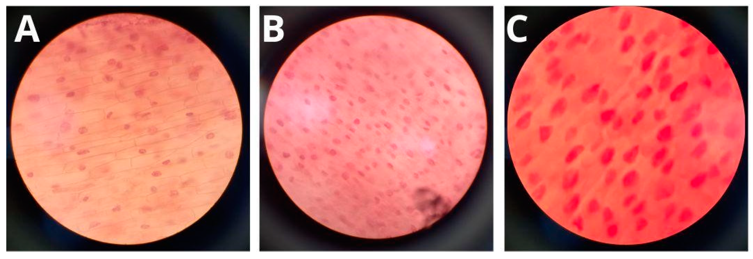
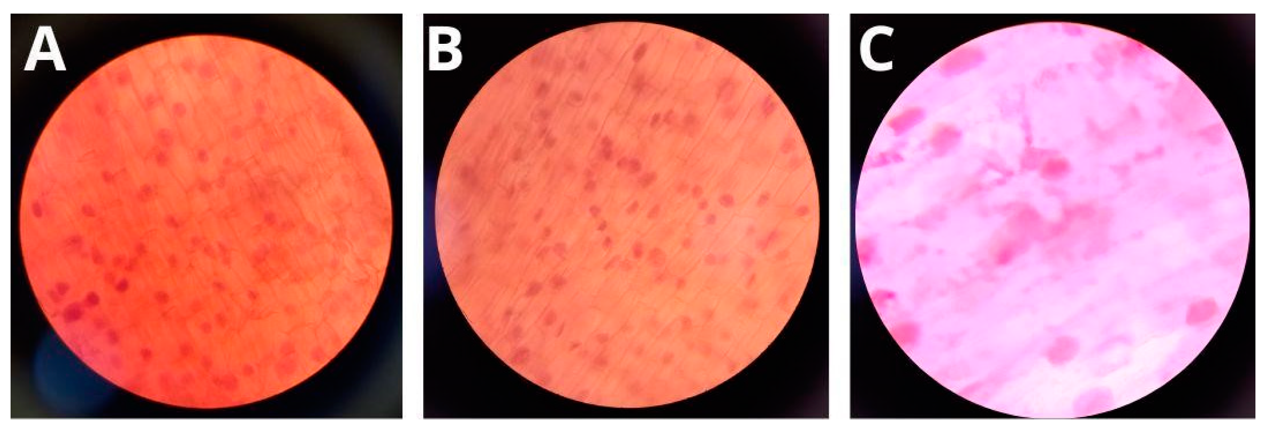
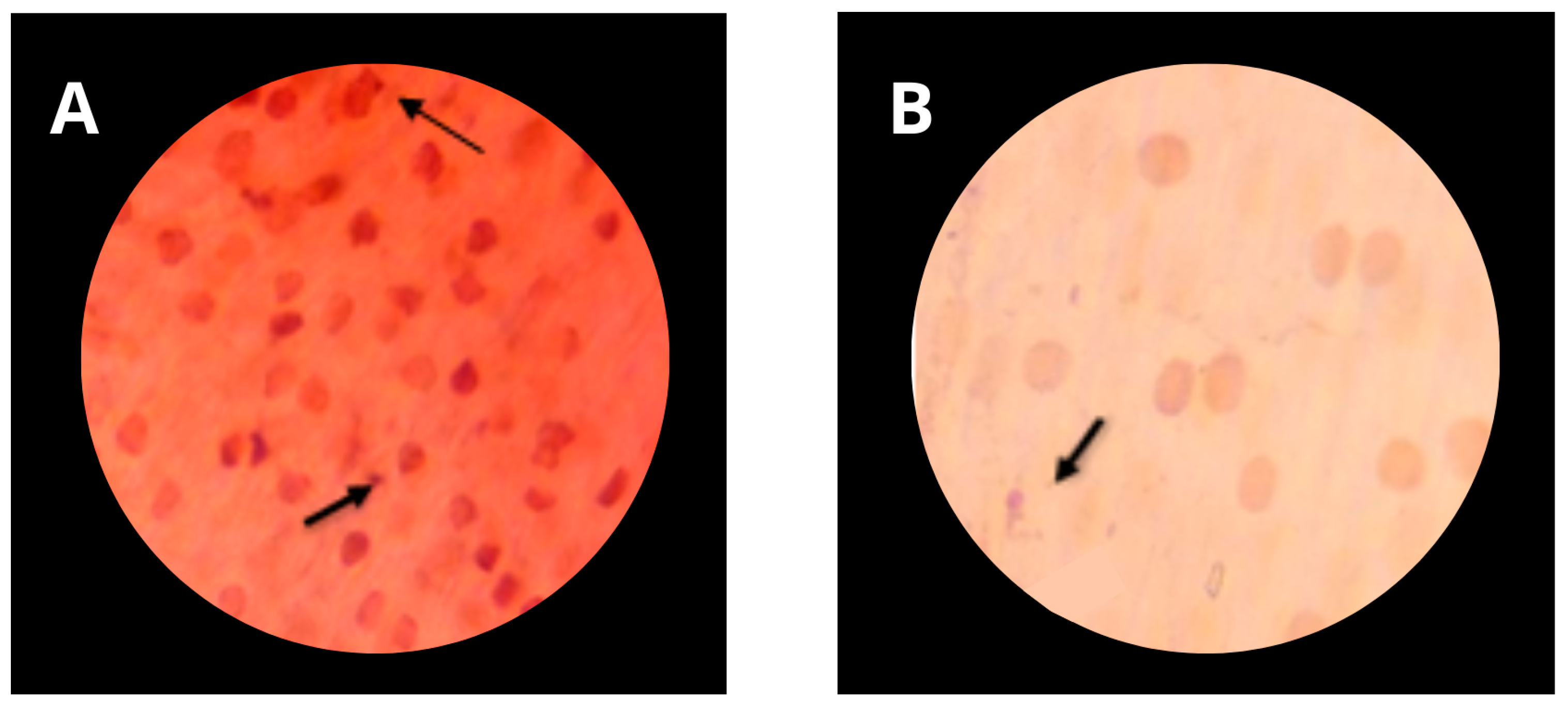
2.9. Determination of Genotoxicity of Formulations

Cells of onion roots (Allium cepa) were used for the genotoxicity tests, with the appearance of micronuclei indicating the toxicity of the molecules tested. To carry out the test, 18 onions were purchased commercially in the city of Recife, Brazil, and selected obeying size standards and the absence of visible contamination, as recommended by Parvan et al. [23]. After removal of dry roots with a scalpel, the bulbs were placed in contact with water for 48 h to allow the initial development of roots. The roots were exposed to the mouthwash formulations containing biosurfactants from C. quinoa and G. max for 48 h and then placed in Carnoy’s fixative for 12 h, followed by the removal of the root caps and placement on slides for microscopy. A drop of 2% acetic orcein stain was placed on each root cap, and the coverslip was placed. Mild pressure was exerted on the coverslip to expose the root cap cells for subsequent observation under an optical microscope with an objective lens of 40×. To analyze the mutagenic effect, the presence of micronuclei was taken into account. For negative control, the images provided in the literature were used as a parameter, as described by Parvan et al. [23]. On the other hand, as a positive control for micronuclei and cellular changes, the synthetic surfactants Tween 20 and SDS were used, at ½ CMC (0.03 and 1.0 g/L, respectively). The experiments were carried out in triplicates.

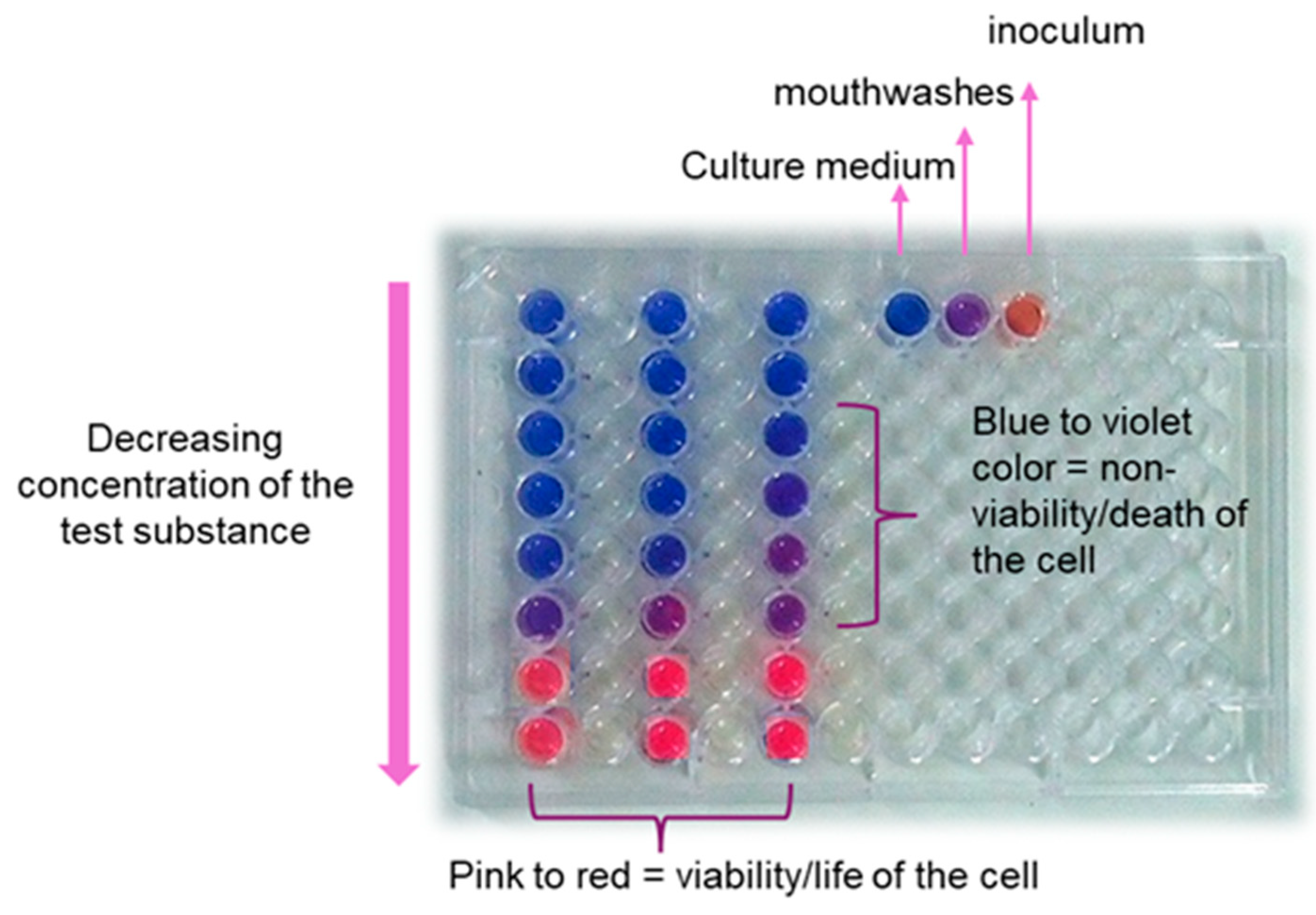
2.10. Determination of Antimicrobial Activity

Test substances and microorganisms were used for the determination of the minimum inhibitory concentration (MIC) and for biofilm inhibition tests. The tests were performed with the formulated products containing initial concentrations of chitosan hydrochloride of 10 g/L and of the biosurfactants from C. quinoa and G. max of 7 and 8 g/L, respectively. As positive controls, 3 commercial mouthwashes containing different active ingredients and surfactants were used: 1. cetylperidinium chloride (0.5 g/L) and surfactant polysorbate 80 (CMCC); 2. menthol (0.42 g/L) and surfactant poloxamer 407 (CMM); and 3. chlorhexidine digluconate (1.2 g/L) and surfactant polysorbate 20 (CMCD).
The antimicrobial activity was determined by the resazurin-based turbidometric assay. The buffered resazurin solution was prepared in a dark environment by dissolving 5 mg of powdered resazurin stain (Merck, Darmstadt, Germany), 375 mg of potassium dihydrogen phosphate, and 556 mg of dibasic sodium phosphate in 100 mL of sterile distilled water using a spatula, amber bottle, and magnetic stirrer, sterilized beforehand. For complete solubilization, the dye was stirred for 1 h. Buffered resazurin was used to minimize possible changes in color due to a reaction with any of the components of the mouthwash or due to color changes.
The MIC values of the isolated substances and formulation products were determined as described by Farias et al. [22]. Briefly, the 96-well flat-bottom microplates were filled with a final volume of 100 µL (mouthwash at different concentrations + culture medium + standardized inoculum). The concentrations of the mouthwashes were standardized with decreasing values following the letters marked on the microplate (A–H), with A being the highest and H being the lowest concentrations tested, respectively. Bacterial suspensions of Staphylococcus aureus (ATCC 6538), Enterococcus faecalis (ATCC 29212), Escherichia coli (ATCC 25922), Pseudomonas aeruginosa (ATCC 9027), and Streptococcus mutans (ATCC 25175) were obtained using brain heart infusion (BHI, Merck, Darmstadt, Germany). Suspension of the Candida albicans (URM 14053) yeast was obtained using Sabouraud dextrose broth (SDB, Merck). The pre-inocula of the target microorganisms were standardized on the 0.5 MacFarland scale, corresponding to 1.5 × 10⁸ colony-forming units (CFU)/mL. The microplates were incubated at 37 °C for 24 h for the bacteria and 37 °C for 48 h for the yeast. After the incubation period, 30 μL of resazurin stain was added to each well for the determination of cell viability, the microplate was incubated again at 37 °C for 1 h, and then the color was checked. Viable cells reduced resazurin and turned pink/reddish color, while the inactive ones remained blue/purple [24]. The lowest concentration of the mouthwash, at which there was no cell viability, was considered the MIC. The experiments were carried out in triplicate.

2.11. Determination of Antibiofilm Activity

To assess the antibiofilm action, the minimum adherence concentration of the substances was determined in the presence of the same microorganisms used for the antimicrobial activity tests, as described by Le et al. [25]. The pre-inocula of the microorganisms were prepared as described in the previous subsection. Twenty µL of the pre-inoculum of each microorganism in BHI broth for the bacterial strains and SDB for the yeast were inoculated in ELISA plates, followed by the addition of 80 µL of culture medium in each well. The plates containing the microorganisms were left in a laboratory oven for 24 h at 37 °C, as described by Amann et al. [26]. After that, 200 µL of the formulations were added, left for 1 min, and discarded. The microplates were then gently washed with saline three times. The residual microorganisms that remained adhered to the surface of the plate forming a biofilm were fixed with methanol for 15 min. After drying, 200 µL of 1% violet crystal stain was added and left to stand for 5 min. The plates were then washed with distilled water and left to dry in the microbiological hood. Re-suspension was performed with 200 µL of 33% acetic acid, followed by the reading of optical density at 630 nm. The formation of biofilm was quantified as the difference between the initial and final optical density multiplied by 100, according to Santos et al. [27]. Subtracting this value from 100 resulted in the inhibition rate expressed as a percentage.

2.12. Statistical Analyses

All experiments were conducted in triplicate. Means and standard deviations (mean ± SD) were calculated. Statistical analysis of variance (ANOVA) was applied to all values and tested for significance at p < 0.05.

3. Results

3.1. Characterization of Biosurfactants in Plant Extracts Using Fourier Transform Infrared Spectroscopy

The extraction of biosurfactants from G. max and C. quinoa with 96% ethanol yielded 55.5 and 60.0 mg per g of each plant, respectively. After that, the biosurfactant samples were purified and submitted to FTIR analysis. The images in Figure 1 show the presence in both samples of characteristic peaks of saponins, such as the hydroxyl group at 3402 cm−1, alkyl groups at 2940 cm−1, and glycoside bonds at 1634 and 1063 cm−1. Moreover, the spectrum of G. max extract (Figure 1A) shows two bands at 2925 and 2854 cm−1, which are characteristic of carboxylic acids, specifically to their OH group, respectively, as well as a characteristic band at 1745 cm−1 corresponding to the most important esters. On the other hand, no residual bands of esters, especially at the 1745 cm−1 wavenumber, appeared in the spectrum of C. quinoa (Figure 1B).

3.2. Reduction in Water Surface Tension and Critical Micelle Concentration of Biosurfactants

The surface tension of water was lowered by the C. quinoa and G. max biosurfactants from 72 mN/m to 37 and 38 mN/m, and their respective CMC values were 4.0 and 3.5 g/L, respectively.

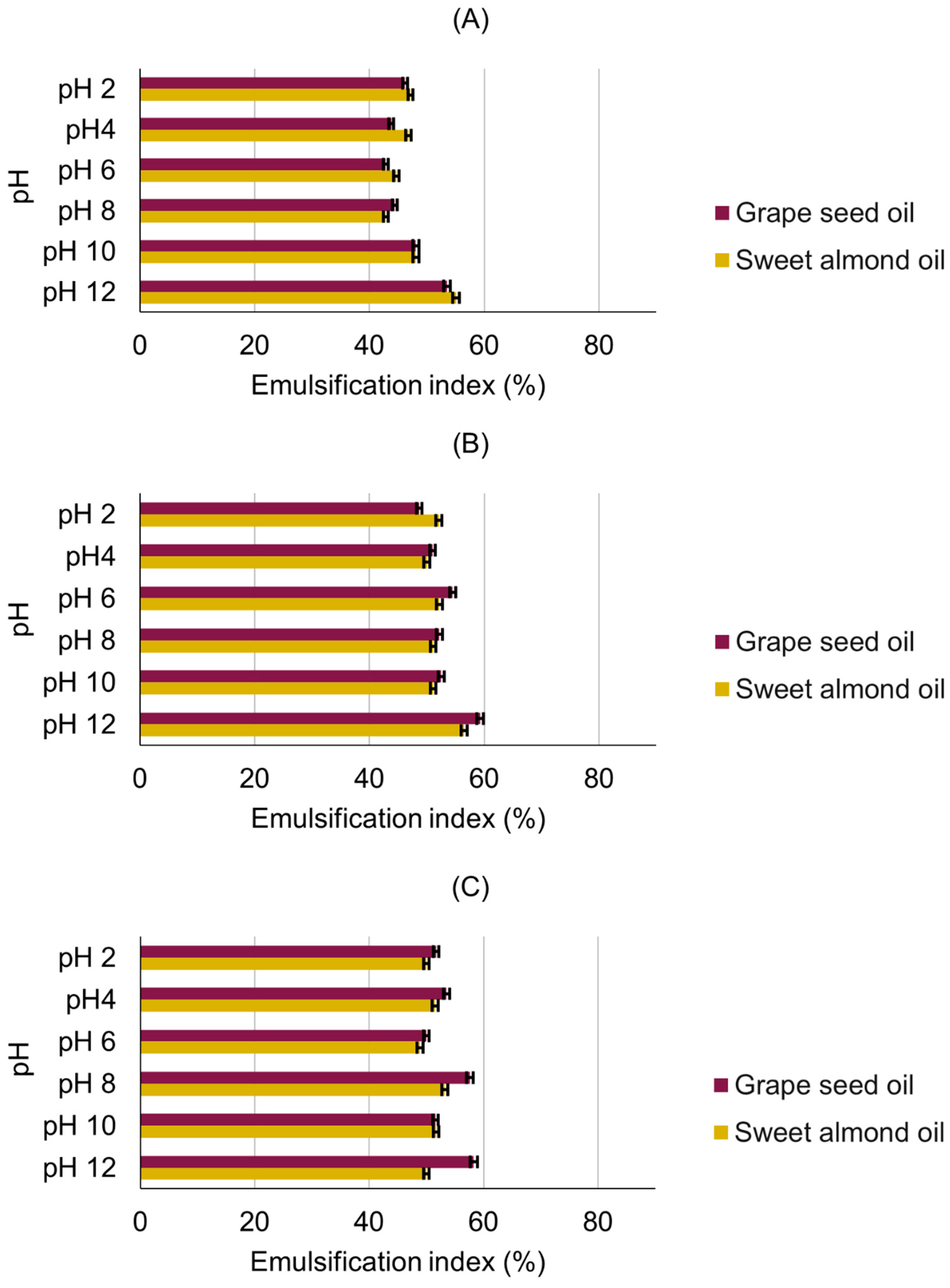
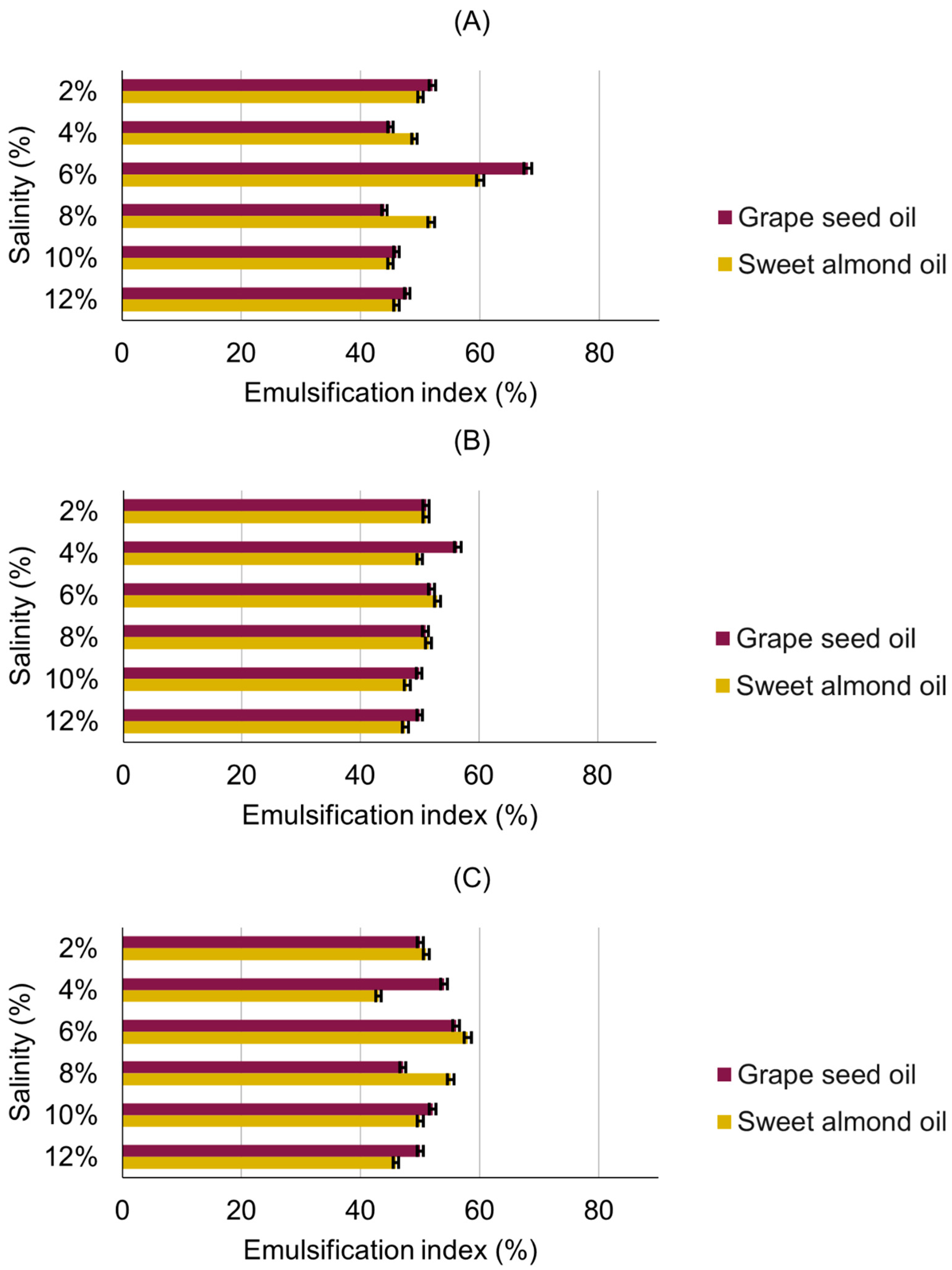
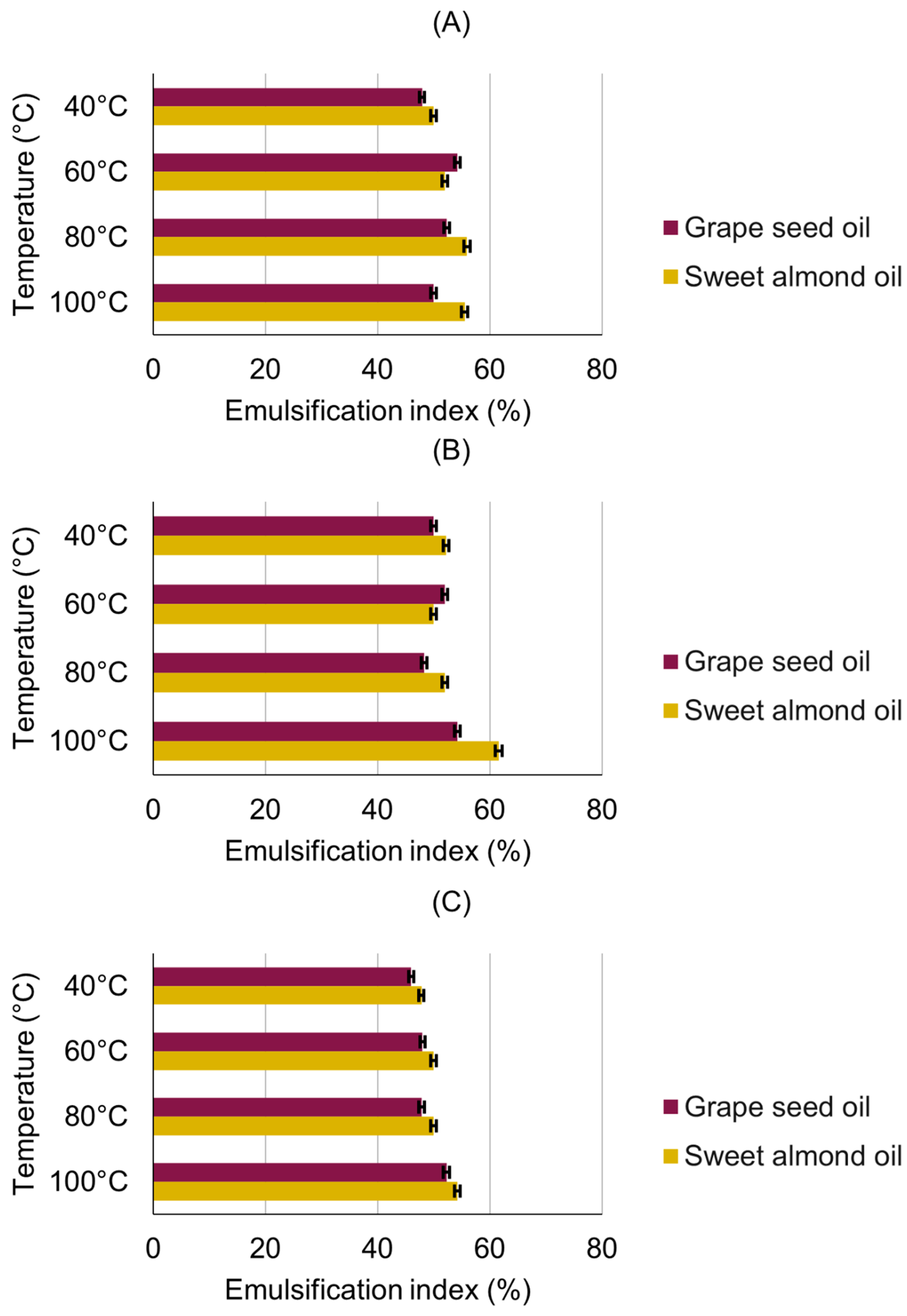
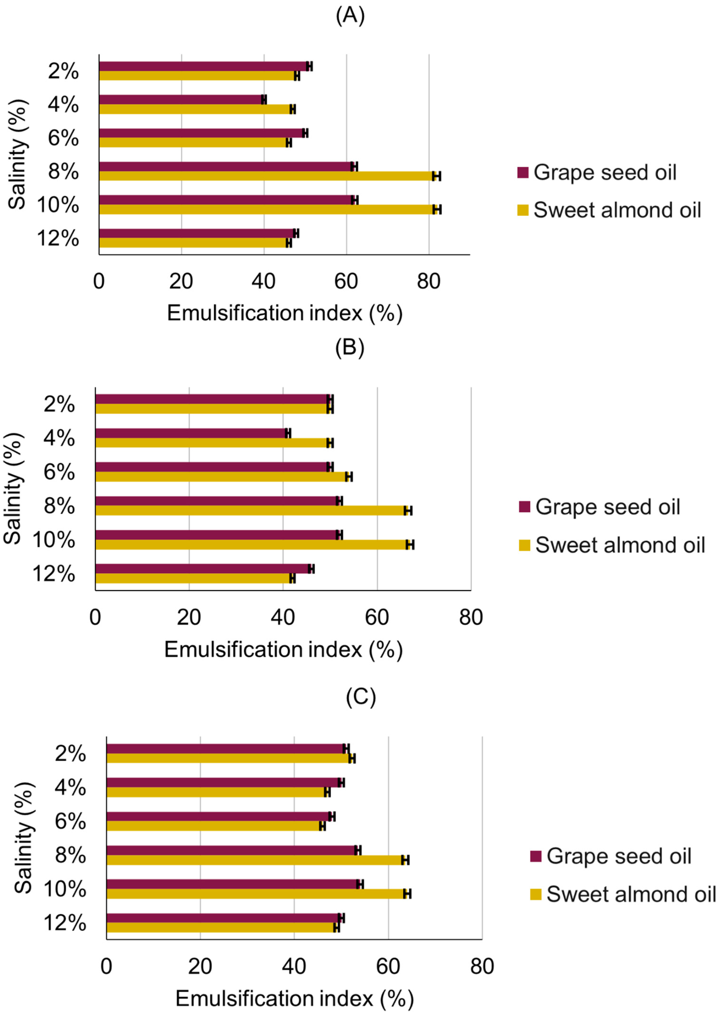
3.3. Emulsification Activity

The emulsification properties of the extracts from G. max and C. quinoa using grape seed and sweet almond oils were assessed to determine the stability of the solutions of the purified biosurfactants containing these oils when submitted to extreme variations in temperature, pH, and salinity. Figure 2 illustrates the activity of the emulsions of the G. max biosurfactant when exposed to changes in temperature for 24 h and in concentration (½ CMC, CMC, and 2 CMC). The biosurfactant exhibited satisfactory emulsification under all tested conditions, particularly at the highest temperature (100 °C), at which the emulsification index (E24) was always higher than 60% almost independent of the biosurfactant concentration and the type of oil. The only significant effect of the type of oil was detected with the CMC, where E24 was always higher for grape seed oil than for sweet almond oil.
The emulsions remained stable when the solutions of the purified biosurfactants were exposed to different pH values, with a margin of stability near 45–50% (Figure 3). Consistent with the results obtained by varying the temperature, the E24 achieved the highest values (≥50%) with the CMC.
The results of the emulsions when submitted to variations in salinity (Figure 4) were promising in terms of stability, as E24 was always ≥45%. With only one exception at ½ CMC using 6% NaCl, at which E24 was the highest (≥60%), no significant variation was observed in this parameter when varying either the concentration or the salinity level.
The results of the emulsification test using the biosurfactant from C. quinoa demonstrated that this biomolecule was stable and tolerant to variations in temperature, with maintenance of stability ≥50% at all temperatures on both oils tested (Figure 5).
The quinoa biosurfactant was stable at all pH values, with stability ≥50% in the oil-in-water emulsions, especially those composed of sweet almond oil, which was the system that achieved the best results as an average (Figure 6). Particularly, the highest E24 values (≥60%) were obtained with the CMC and pH 10 with both oils, confirming this biosurfactant concentration as the most effective in terms of emulsification capacity.
Finally, with E24 values always ≥40%, the biosurfactant from C. quinoa exhibited satisfactory stability when varying the medium salinity, even at the lowest biosurfactant concentration, which constitutes an economic advantage in its use. Sweet almond oil achieved the best emulsification results in the oil-in-water systems, with E24 values always ≥60% at NaCl concentrations in the 8–10% range at all biosurfactant concentrations (Figure 7).
Overall, the emulsification activity results demonstrated that both biosurfactants, even at the lowest concentration (½ CMC), showed adequate emulsion activity against the two oils tested when subjected to variations in temperature, pH, and salinity.

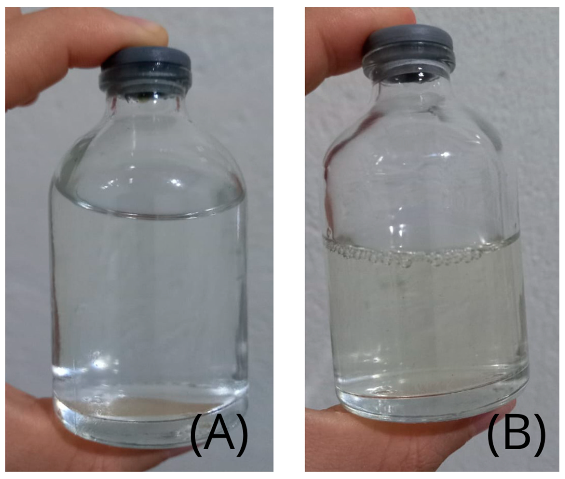
3.4. Main Features of Mouthwash Formulations

All formulations exhibited stability and homogeneity, with a limpid, transparent appearance and aroma characteristic of spearmint essential oil. Figure 8 shows the aforementioned visual aspects of the mouthwashes containing C. quinoa and G. max biosurfactants.
The pH of the formulations was around 4.19, which is close to that of a commercial mouthwash used for comparison purposes (4.25).
The foaming capacity of the formulations was tested after adjustment in pH to 5 and 7 using NaOH and HCl, followed by the determination of their stability. Foaming was around 60% in the pH 5 formulation, while it remained at 100% in the pH 7 one. Furthermore, by consulting the ingredients of a commercial mouthwash, we were able to identify the presence of petroleum-derived surfactants.

3.5. Genotoxicity of Formulations

Tests were performed to determine the preliminary genotoxicity profile of the formulations by examining the possible formation of micronuclei in cells of the onion roots (Allium cepa). Tests performed with this plant have scientific credibility for the initial examination of toxicity profiles. Micronuclei are small nuclei formed by acentric chromosomes or even whole nuclei that did not migrate properly to the poles of the cell during the anaphase.
Figure 9 and Figure 10 show the optical microscopy images. They were obtained by recording the images detected under an optical microscope, using a 40× magnification lens.
The onion roots came in contact with formulations containing the G. max and C. quinoa biosurfactants in different concentrations, respectively. In the images, plant cells can be seen, with preserved morphology and absence of micronuclei in the cells when onion roots were grown in contact with the formulations containing either biosurfactant.
Figure 11 shows optical microscopy images of roots exposed to synthetic surfactants for positive control of micronuclei. The roots were exposed to aqueous solutions containing chemical surfactants at a concentration of ½ CMC, corresponding to 0.03 g/L Tween 20 (Figure 11A) and 1.0 g/L SDS (Figure 11B). The micronuclei found in analyses of onion (Allium cepa) root cell slides are marked with arrows.

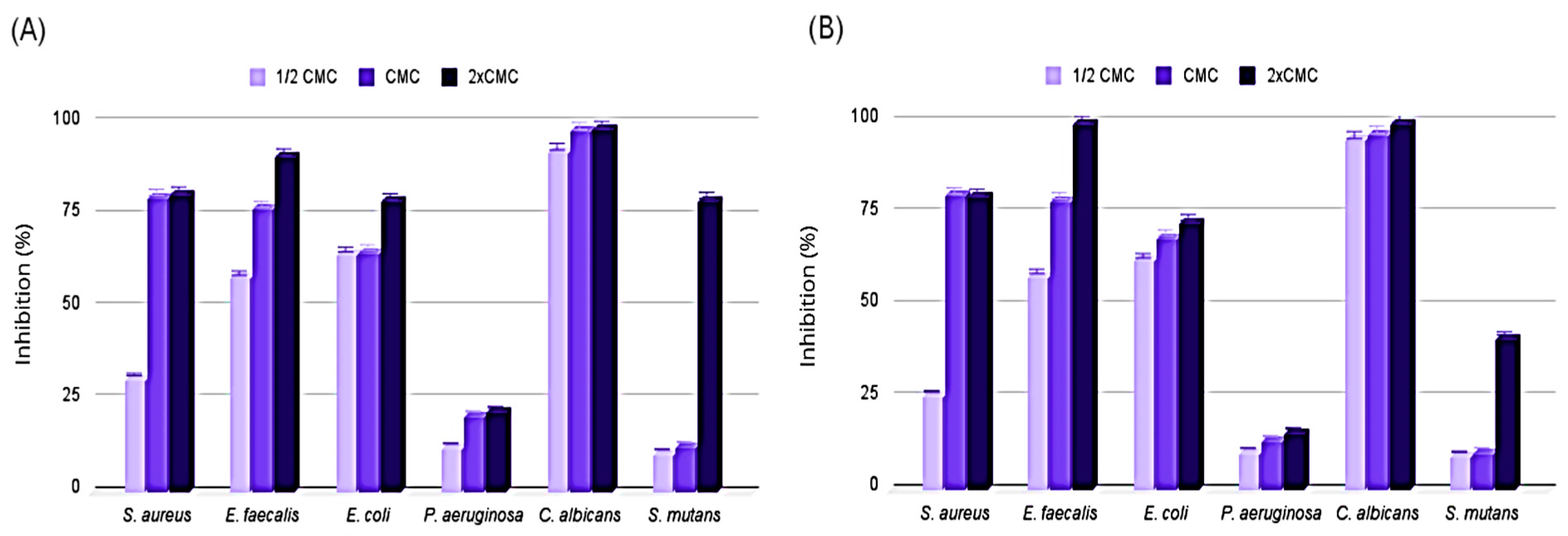
3.6. Antimicrobial Activity of Formulations

In Figure 12 it is possible to see how the minimum inhibitory concentration was determined against mouthwashes using the dye rezasurin as an indicator of cell viability. This figure corresponds to the antimicrobial activity test of the mouthwash with the active ingredient quinoa biosurfactant and chitosan hydrochloride, at the initial concentrations of 7.0 and 10.0 g/L, respectively, against S. mutans. Resazurin (7-hydroxy-3H-phenoxazine-3-one-10-oxide) is a blue oxidation–reduction indicator compound that, in the presence of viable cells, is oxidized to resofurin, a red substance. Therefore, the blue to violet color indicates the absence of bacterial growth, while variations from pink to red indicate the presence of viable cells, which have active metabolism [24].
Table 2 lists the values of minimum inhibitory concentrations (MICs) of the mouthwash formulations containing the biosurfactants from C. quinoa and G. max at concentrations of 8.0 and 7.0 g/L, respectively, against typical microorganisms belonging to the oral microbiota. In both formulations, chitosan hydrochloride had an initial concentration of 10.0 g/L. As a positive control, three types of mouthwash were tested with different active agents (cetylpyridinium chloride, essential oils, and chlorhexidine) and containing different surfactants in their formulations.
The antimicrobial activity of both formulations was effective against all microorganisms tested, with the best inhibitory results found for Enterococcus faecalis and Streptococcus mutans, the latter being the etiological agent of dental caries.

3.7. Antibiofilm Activity of Test Substances and Formulations

Due to the absence of antimicrobial activity of the pure plant extracts, further tests were only performed with the mouthwash formulations. According to the methodology described in the Section 2, it was possible to calculate the antibiofilm activity of the formulated products in terms of percentage inhibition of biofilm formation, the results of which are illustrated in Figure 13.
It can be observed that the formulations containing the C. quinoa biosurfactant (Figure 13A) showed inhibition of > 25% of biofilm development from most of the microorganisms tested, with emphasis against S. mutans when exposed to the formulation at a concentration of 2 CMC (inhibition > 75%). As for the formulations containing the biosurfactant from G. max (Figure 13B), almost 100% inhibition was found against E. faecalis and C. albicans and no less than 75% inhibition against S. aureus at the highest concentration.

4. Discussion

Saponins are glycosidic compounds found in a variety of vegetables, which consist of a hydrophilic glycan linked to a hydrophobic aglycone and which have several biological activities, such as cytotoxic, hemolytic and surfactant activities [28]. The structural diversity of saponins makes their analysis challenging, requiring spectroscopy techniques to study their complex structures and biological activities [29].
The results of the FTIR analysis are in agreement with those reported by Bezerra et al. [30], who demonstrated that the peaks found for isolated saponins from Chenopodium quinoa were similar to those found in the analysis of a commercial saponin used as the standard. It is important to highlight that this structural similarity corroborates the hypothesis that the biosurfactants tested are equivalent to commercial saponins, which we used as a reference. The determination of the CMC is essential for procedures involving surfactants, as the effectiveness of this component is greater when there is an important quantity of micelles in the medium [31]. Surfactants are components that cause the reduction in the surface tension of a liquid, which occurs until the formation of colloidal aggregates called micelles [32]. Surfactants have the capacity to reduce surface tension, which enables a better relationship among molecules with different polarities [33]. The plant-derived biosurfactants used in the mouthwash formulations were essential to maintain the stability of the compounds, since, due to their ability to reduce surface tension and cause emulsification, they were able to mix ingredients of different polarities and keep the formulation stable.
This behavior may be explained by the presence of fatty acid fractions that tolerate heating without inhibiting the action of the surfactant molecules. Despite singularities in terms of stability, biosurfactants generally remain stable even when submitted to variations in temperature and pH [30]. Studies have demonstrated that the stability of emulsions when exposed to different pH values depends on the composition of the extract, which may have positively or negatively charged components induced to aggregation and greater stability when attracted by charged saponins [34].
The best results in terms of emulsification activity were found in the current study for the sweet almond oil at a NaCl concentration of 10% for both the biosurfactants tested. This result is of great importance because chemical surfactants undergo inactivation at NaCl concentrations ≥2% [33].
One relevant characteristic is that C. quinoa has fatty acids similar to those found in soybean oil and is rich in essential fatty acids, such as linoleic and linolenic acids [35].
pH values are essential for determining the stability of a compound, as adequate organoleptic characteristics lead to the acceptance of a product by consumers [36]. Emulsions using oily components in aqueous solutions are found in numerous products, including personal hygiene products. This is a type of colloidal dispersion in which small droplets of oil are dispersed in the continuous aqueous phase [37]. Such oil-in-water systems must remain stable and could be submitted to reformulations if any sign of instability occurs. If approved, the emulsion can be exposed to other stability tests [38].
According to Saengsor and Jimtaisong [39], the stability of emulsions depends on the interaction of interfaces established by the emulsifying agent and the hydrophobic compound. Therefore, emulsifying agents must have affinity for the oil being tested in order to maintain its stability in the dispersing phase.
Many plant extracts rich in biosurfactants can serve as emulsifiers, which is an advantage for the preparation of “green” cosmetic products. The literature reports emulsifying action with excellent performance for surface-active plant-derived substances, such as tea saponins, glutelin of hydrolyzed rice, and saponins from the genus Quillaja [18].
Oil-in-water emulsions are important constituents of numerous commercial products, including foods, supplements, pharmaceutical products, cosmetics, personal care products, and pesticides [37]. Each plant extract has its particularity in terms of stability. However, biosurfactants are known to exhibit good stability when submitted to different temperatures and pH values [3]. The stability results of the extracts studied here confirm this trend, as the variation occurred in pH ranges not generally used in formulations for oral use, showing that the surface-active molecules in the extracts did not lose their function.
The pH of mouthwashes affects the metabolism of dental plaque. Thus, mouthwashes are used in combination with the mechanical cleaning of the teeth. These compounds with low pH are effective against the fermentation and production of extracellular polysaccharides by microorganisms [40]. However, a very low pH together with the absence of fluoride in the formulation could be harmful to dental tissues, which could cause the demineralization of tooth enamel [41]. The pH value of mouthwash formulations containing the vegetable biosurfactant (4.19) was close to that of commercial mouthwashes, which suggests that they would be effective against the proliferation of microorganisms.
In contrast, despite the low pH (4.19), the positively charged chitosan and chitosan hydrochloride have a buffering effect and adsorption action on negative surfaces, such as tooth enamel. Thus, even with the absence of fluoride, the present formulation is expected to maintain the balance of the demineralization–remineralization process of the enamel due to the interaction between chitosan hydrochloride and the phosphate group in hydroxyapatite. This expectation is based on the work of Stamford Arnaud et al. [42], who conducted an in vitro experiment to determine the influence of chitosan on the demineralization–remineralization process of tooth enamel. The authors found that chitosan interfered with the demineralization process by inhibiting the release of phosphorus in vitro and that demineralization was influenced by the concentration and exposure time of the biopolymer to the enamel. They also suggested that chitosan can serve as a barrier due to its adsorption to tooth enamel, impeding the penetration of acids from microbial fermentation or diet and contributing to the inhibition of the demineralization of the enamel.
Foaming capacity is an important factor in any formulation intended for oral use, as it enables the compound to widely reach in the oral cavity during hygiene activities [43]. According to Farias et al. [22], formulations with biosurfactants are effective as a good detergent for oral use. These observations corroborate the results obtained in this present work for the proposed formulations, which showed good emulsification activity and foaming capacity.
The occurrence of a large quantity of micronuclei in cells may be related to mutagenic events or be evidence of a carcinogenic process, which may be influenced by the contact between the plant and toxic compounds [23].
The macroscopic examination of the formulations prepared in this current study revealed no necrotic tissues or anomalies. The absence of micronuclei indicates that the molecules of the biosurfactants have no mutagenic or carcinogenic effects [23].
Bioassay with Allium cepa is a system accepted by international agencies as a tool for analyzing the cytotoxic effects of different products [44]. Plants have self-defense systems that become weakened when submitted to stress, which can be a triggering factor of disease [23]. Using the genotoxicity test, it was possible to test the safety of mouthwash formulations. Due to the absence of micronuclei in cells exposed to the formulations, we can conclude that the biosurfactants used in this study are reliable and harmless to human cells as they do not cause cellular changes. In contrast to natural biosurfactants, the synthetic surfactants tested, Tween 20 and SDS, induced alterations in onion cells, contributing to the formation of micronuclei, which are indicators of cellular toxicity. Other studies have highlighted the toxic potential of chemical surfactants, such as the presence of micronuclei in samples treated with Tween 80 and Tween 20, which are examples of synthetic surfactants widely used in cosmetic products [45].
The essential oil from spearmint (Mentha spicata) was used in our formulations, as the literature states that this essential oil has antimicrobial activity against S. aureus, Pseudomonas aeruginosa, Candida albicans, and other target microorganisms [46]. Moreover, spearmint essential oil gives the mouthwash a fresh taste and aroma characteristic of oral hygiene products; in mouthwash formulations with biosurfactants and chitosan hydrochloride, the essential oil (Mentha spicata) was in a sub-inhibitory concentration, thus being considered a flavoring agent, rather than an active antimicrobial agent.
Minimum inhibitory concentrations were obtained for all microorganisms tested, with the best antimicrobial action for Gram-positive bacteria and C. albicans. The antimicrobial action of plant-derived biosurfactants has previously been described in the literature [22]. This test is extremely important to find the exact amount of the test substance that causes the inhibition of a certain microorganism. In the case of this present study, the substances tested were mouthwash formulations containing vegetable biosurfactants, which were exposed to different microorganisms including Streptococcus mutans, the main etiological agent of dental cavitation. In this respect, the formulations showed excellent effectiveness against the microorganisms tested, especially S. mutans, whose growth was significantly inhibited even when using them in small amounts.
The microdilution method in 96-well plates proved to be useful for assessing the antimicrobial activity of the test substances, enabling a quantitative analysis of the inhibitory concentrations for the microorganisms [47]. The resazurin stain has a blue/purple color, which is reduced to resorufin when in contact with viable cells, which has a pink color [48]. The data show that the formulations exhibited bacteriostatic activity (MIC) against the microorganisms. Moreover, the formulations with the biosurfactant from C. quinoa exhibited bactericidal activity against S. aureus and E. faecalis at the highest concentrations tested.
The use of reagents that act as indicators of cell viability through colorimetric changes has been commonly described in the literature. Tetrazolium bromide (MTT) and resazurin present a colorimetric change, due to an oxidation–reduction reaction, which helps in reading and interpreting the results of in vitro susceptibility tests. Such compounds function as chromogenic substrates for dehydrogenases as oxidation–reduction indicators, related to the transport system in metabolically active cells [49,50]. The dye resazurin (7-hydroxy-3H-phenoxazine-3-one 10-oxide) is blue in color, non-fluorescent, and can be reduced to resorufin by oxidoreductase, which is a pink, fluorescent product. The microdilution method using resazurin has the advantage of its increasingly promising application for in vitro susceptibility tests to determine cell viability for bacteria and fungi (yeast and filamentous), as well as not requiring a spectrophotometer to interpret results, since the color change is observed visually [49,51].
However, among the limitations of this technique, the influence of the density of microbial cells and the composition of the culture medium can be cited as possible factors that can influence the results of colorimetric cell viability tests [50,51]. The characteristics of the test substance can also influence colorimetric tests, as many compounds can interact and disrupt the reading, giving a non-standard color or delaying/inhibiting the color change. However, the main disadvantage of the resazurin colorimetric test is that it results from the accumulation of a colored or fluorescent product (resorufin) that gradually increases fluorescence over time, making it impossible to detect a decrease in cell viability [51].
The use of biosurfactants as antibiofilm agents could be an effective method for reducing the adhesion of microorganisms, many of which are pathogenic, and combating their possible proliferation on various organic surfaces [52]. Based on the results shown in Figure 13, the formulations containing the highest concentration of the biosurfactant from C. quinoa had inhibition rates >70% against all microorganisms, except P. aeruginosa, and even >75% against S. mutans, the etiological agent of dental caries. For formulations containing G. max, inhibition rates were >25% against all microorganisms tested at CMC (3.5 g/L) and 2 CMC (7.0 g/L), once again with the exception of P. aeruginosa.

5. Conclusions

The pharmaceutical industry is actively working to develop sustainable technologies that have fewer harmful impacts on human health and the environment. They are incorporating aspects and principles of green chemistry at an industrial level. The cosmetics sector is particularly interested in searching out natural products, as many cosmetics are used daily, and a significant amount is thrown away directly into the environment. The formulation industry uses hundreds of chemicals and ingredients, many of which need to be replaced to fit the concept of sustainability. Consumers are increasingly interested in using natural products. In this respect, the results of the present study demonstrated that the biosurfactants contained in the extracts from the plants Glycine max and Chenopodium quinoa are stable, nontoxic, and promising biomolecules for use as emulsifiers in these products. The biosurfactants combined with chitosan hydrochloride in a mouthwash formulation exhibited antimicrobial action and promoted the inhibition of biofilm formation, thus proving to be biomolecules with potential for replacing chemical agents found in oral health care products. Compared to commercial rinses, which use petroleum-derived surfactants, the rinses developed in this study have non-toxic contents, as they are products that contain natural ingredients in their composition.

Author Contributions

Conceptualization, R.D.R., K.G.O.B. and L.A.S.; methodology, R.D.R., T.C.M.S., H.M.M., K.G.O.B. and L.A.S.; validation, L.A.S., R.D.R., K.G.O.B. and T.C.M.S.; formal analysis, R.D.R., H.M.M., K.G.O.B. and L.A.S.; investigation, I.R.S., C.L.O. and K.G.O.B.; resources, L.A.S. and R.D.R.; data curation, R.D.R., A.C. and T.C.M.S.; writing—original draft preparation, I.R.S., H.M.M., C.L.O. and K.G.O.B.; writing—review and editing, L.A.S. and A.C.; visualization, L.A.S. and A.C.; supervision, R.D.R. and L.A.S.; project administration, L.A.S. and R.D.R.; funding acquisition, L.A.S. and A.C. All authors have read and agreed to the published version of the manuscript.

Funding

This study was funded by the Brazilian fostering agencies Fundação de Apoio à Ciência e Tecnologia do Estado de Pernambuco (FACEPE), the Conselho Nacional de Desenvolvimento Científico e Tecnológico (CNPq), and the Coordenação de Aperfeiçoamento de Pessoal de Nível Superior (CAPES) (Finance Code 001).

Institutional Review Board Statement

Not applicable.

Informed Consent Statement

Not applicable.

Data Availability Statement

The original contributions presented in this study are included in the article; further inquiries can be directed to the corresponding authors.

Acknowledgments

The authors are grateful to the Laboratories of Universidade Católica de Pernambuco (UNICAP) and to the Instituto Avançado de Tecnologia e Inovação (IATI), Brazil.

Conflicts of Interest

The authors declare no conflicts of interest.

References

  1. Priya, A.; Nagaiah, H.P.; Malligarjunan, N.; Pandian, S.K. Oral biofilms: Architecture and control. In Understanding Microbial Biofilms., 1st ed.; Das, S., Kungwani, N.A., Eds.; Academic Press: Cambridge, MA, USA, 2023; Chapter 29; pp. 485–507. [Google Scholar] [CrossRef]
  2. Verma, T.; Grover, N.; Mlhotra, S. Various applications of chlorhexidine as an antimicrobial agente in dentistry: A review. Int. Dent. J. Stud. Res. 2023, 22, 50–53. [Google Scholar] [CrossRef]
  3. Johnson, P.; Trybala, A.; Starov, V.; Pinfield, V.J. Effect of synthetic surfactants on the environment and the potential for replacement by biosurfactants. Adv. Colloid Interface Sci. 2011, 288, 102–340. [Google Scholar] [CrossRef]
  4. Guzmán, E.; Ortega, F.; Rubio, R.G. Chitosan: A promising multifunctional cosmetic ingrediente for skin and hair care. Cosmetics 2022, 9, 99. [Google Scholar] [CrossRef]
  5. Narayanan, A.; Kartik, R.; Sangeetha, E.; Dhamodharan, R. Super water absorbing polymeric gel from chitosan, citric acid and urea: Synthesis and mechanism of water absorption. Carbohydr. Polym. 2018, 191, 152–160. [Google Scholar] [CrossRef] [PubMed]
  6. Visnjar, T.; Jerman, U.D.; Veranic, P.; Kreft, M.E. Chitosan hydrochloride has no detrimental effect on bladder urothelial cancer cells. Toxicol. Vitro 2017, 44, 403–413. [Google Scholar] [CrossRef] [PubMed]
  7. Li, X.M.; Xie, Q.T.; Zhu, J.; Pab, Y.; Meng, R.; Zhang, B.; Jin, Z.Y. Chitosan hydrochloride/carboxymethyl starch complex nanogels as novel pickering stabilizers: Physical stability and rheological properties. Food Hydrocoll. 2019, 93, 215–225. [Google Scholar] [CrossRef]
  8. Dias, K.B.; Silva, D.P.; Ferreira, L.A.; Fidelis, R.R.; Costa, J.L.; Silva, A.L.L.; Scheidt, G.N. Chitin and chitosan: Characteristics, uses and production current perspectives. J. Biotechnol. Biodivers. 2013, 4, 184–191. [Google Scholar] [CrossRef]
  9. Bezerra, K.G.O.; Silva, I.G.S.; Almeida, F.C.G.; Rufino, R.D.; Sarubbo, L.A. Plant-derived biosurfactants: Extraction, characteristics and properties for applications in cosmetics. Biocatal. Agric. Biotechnol. 2021, 34, 102036. [Google Scholar] [CrossRef]
  10. Zhou, T.; Ao, M.; Xu, G.; Liu, T.; Zhang, J. Interactions of bovine serum albumin with cationic imidazolium and quaternary ammonium gemini surfactants: Effects of surfactant architecture. J. Colloid Interface Sci. 2013, 389, 175–181. [Google Scholar] [CrossRef]
  11. Souza, A.C.; Silva, M.S.; Simões, L.A.; Fernandes, N.A.T.; Schwan, R.F.; Dias, D.R. Advantages of biosurfactants over petroleum-based surfactants. Indust. Appl. Biosurfactants Microorg. 2024, 1, 371–393. [Google Scholar] [CrossRef]
  12. Rebello, S.; Asok, A.K.; Mundayoor, S.; Jisha, M.S. Surfactants: Toxicity, remediation and green surfactants. Envirom. Chem. Lett. 2014, 12, 275–287. [Google Scholar] [CrossRef]
  13. Hartal, O.; Haddaji, C.; Anouzla, A.; Madinzi, A.; Souabi, S. Recent advances in physicochemical and biological techniques for the management of discharges loaded with surfactants. Chem. Biochem. Eng. 2023, 37, 1–16. [Google Scholar] [CrossRef]
  14. McClements, D.J.; Gumus, C.E. Natural emulsifiers—Biosurfactants, phospholipids, biopolymers, and colloidal particles: Molecular and physicochemical basis of functional performance. Adv. Colloid Interface Sci. 2016, 234, 3–26. [Google Scholar] [CrossRef] [PubMed]
  15. Jaysree, R.C.; Rajendran, N. A review on biosurfactants. Int. J. Res. Pharm. Sci. 2002, 23, 359–366. [Google Scholar] [CrossRef]
  16. Varvaresou, A.; Iakovou, K. Biosurfactants in cosmetics and biopharmaceuticals. Lett. Appl. Microbiol. 2015, 61, 214–223. [Google Scholar] [CrossRef] [PubMed]
  17. Bouzier, A.; Rojas, J.; Ibinga, S.K.K.; Lamarti, A.; Martin, P.; Morillo, M. The impact of saponins on health-review. Biointerface Res. Appl. Chem. 2023, 13, 362–382. [Google Scholar] [CrossRef]
  18. Zhu, Z.; Wen, Y.; Yi, J.; Cao, Y.; Liu, F.; McClements, D.J. Comparison of natural and synthetic surfactants at forming and stabilizing nanoemulsions: Tea saponin, Quillaja saponin, and Tween 80. J. Colloid. Interface Sci. 2019, 536, 80–87. [Google Scholar] [CrossRef]
  19. Ibrahim, M.A.; Alhalafi, M.H.; Eman, E.M.; Ibrahim, H.; Mosaad, R.M. A review of chitosan and chitosan nanofiber: Preparation, characterization and its potential applications. Polymers 2023, 25, 2820. [Google Scholar] [CrossRef] [PubMed]
  20. Costa, E.M.; Silva, S.; Madureira, A.R.; Cardelle-Cobas, A.; Tavaria, F.K.; Pintado, M.M. A comprehensive study into the impact of a chitosan mouthwash upon oral microorganism’s biofilm formation in vitro. Carbohydr. Polym. 2014, 101, 1081–1086. [Google Scholar] [CrossRef]
  21. Cooper, D.G.; Goldenberg, B.G. Surface active agents from two Bacillus species. Appl. Environ. Microbiol. 1987, 53, 224–229. [Google Scholar] [CrossRef]
  22. Farias, J.M.; Stamford, C.M.; Resende, A.H.M.; Aguiar, J.S.; Rufino, R.D.; Luna, J.M.; Sarubbo, L.A. Mouthwash containing biosurfactant and chitosan: An eco-sustainable option for controlling cariogenic microorganisms. Int. J. Biol. Macromol. 2019, 129, 853–860. [Google Scholar] [CrossRef] [PubMed]
  23. Parvan, L.G.; Leite, T.G.; Freitas, T.B.; Pedrosa, P.A.A.; Calixto, J.S.; Agostinho, L.A. Bioensaio con Allium cepa revela genotoxicidade de herbicida com flumioxazin. Rev. Pan-Amaz. Saude 2020, 11, e202000544. [Google Scholar] [CrossRef]
  24. Driessche, F.V.; Rigole, P.; Brackman, G.; Coenye, T. Optimization of resazurin-based viability staining for quantification of microbial biofilms. J. Microbiol. Methods 2014, 98, 31–34. [Google Scholar] [CrossRef] [PubMed]
  25. Amann, V.; Kissmann, A.; Kramer, M.; Krebs, I.; Perez-Erviti, J.A.; Otero-Gonzalez, A.J.; Vicente, F.M.; Rodríguez, A.; Standker, L.; Weil, T.; et al. Increased activities against biofilms of the pathogenic yeast Candida albicans of optimized Pom-1 derivatives. Polymers 2022, 14, 318. [Google Scholar] [CrossRef] [PubMed]
  26. Santos, R.; Callow, M.E.; Bott, T.R. The structure of Pseudomonas fluorescens biofilms in contact with flowing systems. Biofouling 1991, 4, 319–336. [Google Scholar] [CrossRef]
  27. Le, B.; Do, D.T.; Nguyen, H.M.; Do, B.H.; Le, H.T. Preparation, characterization and anti-adhesive activity of sulfate polysaccharide from Caulerpa lentillifera against Helicobacter pylori. Polymers 2022, 14, 4993. [Google Scholar] [CrossRef] [PubMed]
  28. Sharma, K.; Kaur, R.; Kumar, S.; Saini, R.K.; Sharma, S.; Pawde, S.V.; Kumar, V. Saponins: A concise review on food related aspects, applications and health implications. Food Chem. Adv. 2023, 2, 100191. [Google Scholar] [CrossRef]
  29. Savarino, P.; Demeyer, M.; Decroo, C.; Colson, E.; Gerbaux, P. Mass spectrometry analysis of saponin. Mass Spectrom. Rev. 2023, 42, 954–983. [Google Scholar] [CrossRef] [PubMed]
  30. Bezerra, K.G.O.; Meira, H.M.; Veras, B.O.; Stamford, T.C.M.; Fernandes, E.L.; Converti, A.; Rufino, R.D.; Sarubbo, L.A. Application of plants surfactants as cleaning agents in shampoo formulations. Processes 2023, 11, 879. [Google Scholar] [CrossRef]
  31. Wu, R.; Tian, M.; Shu, C.; Zhou, C.; Guan, W. Determination of the critical micelle concentration of surfactants using fluorescence strategies. Soft Matter 2022, 18, 8920–8930. [Google Scholar] [CrossRef]
  32. Unal, D.N.; Yildririm, S.; Kurbanoglu, S.; Uslu, B. Current trends and roles of surfactants for chromatographic and electrochemical sensing. Trend Anal. Chem. 2021, 144, 116418. [Google Scholar] [CrossRef]
  33. Sarubbo, L.A.; Maria da Gloria, C.S.; Durval, I.J.B.; Bezerra, K.G.O.; Ribeiro, B.G.; Silva, I.A.; Banat, I.M. Biosurfactants: Production, properties, applications, trends, and general perspectives. Biochem. Eng. J. 2022, 181, 108377. [Google Scholar] [CrossRef]
  34. Ralla, T.; Herz, E.; Salminen, H.; Edelmann, M.; Dawid, C.; Hofmann, T.; Weiss, J. Emulsifying properties of natural extracts from Panax ginseng L. Food Biophys. 2017, 12, 479–490. [Google Scholar] [CrossRef]
  35. Ferreira, D.S. Application of Infrared Spectroscopy and Multivariate Analysis to Predict Quality Parameters in Soybean and Quinoa. PhD Thesis, State University of Campinas, Campinas, Brazil, 2013. [Google Scholar] [CrossRef]
  36. Patil, A.; Bhide, S.; Bookwala, M.; Soneta, B.; Shankar, V.; Almotairy, A.; Almutairi, M.; Murthy, S.N. Stability of organoleptic agentes in pharmaceuticals and cosmetics. AAPS Pharm. SciTech. 2017, 19, 36–47. [Google Scholar] [CrossRef] [PubMed]
  37. McClements, D.J.; Jafari, S.M. Improving emulsion formation, stability and performance using mixed emulsifiers: A review. Adv. Colloid Interface Sci. 2018, 251, 55–79. [Google Scholar] [CrossRef] [PubMed]
  38. Xiao, F.; Li, K.; Wang, W.; Ge, Y.; Yu, Z.; Peng, Z.; Gong, J. Effect of oil-soluble/water-soluble surfactants on the stability of water-in-oil systems, an atomic force microscopy study. Langmuir 2023, 39, 3862–3870. [Google Scholar] [CrossRef] [PubMed]
  39. Saengsorn, K.; Jimtaisong, A. Determination of hydrophilic–lipophilic balance value and emulsion properties of sacha inchi oil. Asian Pac. J. Trop. Biomed. 2017, 7, 1092–1096. [Google Scholar] [CrossRef]
  40. Santos, D.S.F.; Peralta-Mamani, M.; Brandão, F.S.; Andrade, F.B.; Cruvinel, T.; Santos, P.S.S. Could polyhexanide and chlorine dioxide be used as an alternative to chlorhexidine? A systematic review. Sao Paulo Med. J. 2022, 140, 42–55. [Google Scholar] [CrossRef]
  41. Simmer, J.P.; Hardy, N.C.; Chinoy, A.F.; Bartlett, J.D.; Jan, C.C. How fluoride protects dental enamel from demineralization. J. Int. Soc. Prev. Community Dent. 2020, 10, 134–141. [Google Scholar] [CrossRef]
  42. Stamford Arnaud, T.M.S.; Barros Neto, B.; Diniz, F.B. Chitosan effect on dental enamel de-remineralization: An in vitro evaluation. J. Dent. 2010, 38, 848–852. [Google Scholar] [CrossRef] [PubMed]
  43. Bouassida, M.; Fourati, N.; Krichen, F.; Zouari, R.; Ellouz-Chaabouani, S.; Ghribi, D. Potential application of Bacillus subtilis SPB1 lipopeptides in toothpaste. J. Adv. Res. 2017, 8, 425–433. [Google Scholar] [CrossRef] [PubMed]
  44. Kuruppuarachchi, S.U.; Jayawardena, U.A.; Gunathilake, V.K. Use of the Allium cepa model to assess the cytogenotoxicity of Luffariella herdmani marine sponge extract. Altern. Lab. Anim. 2023, 51, 175–187. [Google Scholar] [CrossRef]
  45. Postay, L.F.; Cabral, D.S.; Heringer, O.A.; Vieira, L.V.; de Moraes, L.R.; Freitas, G.; Gomes, L.C. The effectiveness of surfactants applied with essential oil of Lippia alba in the anesthesia of Nile tilapia (Oreochromis niloticus) and their toxicity assessment for fish and mammals. Environ. Sci. Pollut. Res. Int. 2021, 28, 10224–10233. [Google Scholar] [CrossRef] [PubMed]
  46. Cmikova, N.; Galovicovva, L.; Schwarzova, M.; Kacaniova, M. use of Mentha spicata essential oil for prolonging postharvest life of fresh vegetables. Acta Hortic. Regiotect. 2023, 26, 35–42. [Google Scholar] [CrossRef]
  47. Fowler, P.W.; Wright, C.; Spiers, H.; Zhu, T.; Barten, E.M.L.; Hoosdally, S.W.; Gilbertoni-Cruz, A.L.; Roohi, A.; Kouchaki, S.; Walker, T.M.; et al. A crowd of BashTheBug volunteers reproducibly and accurately measure the minimum inhibitory concentrations of 13 antitubercular drugs from photographs of 96-well broth microdilution plates. Microbiol. Infect. Dis. 2023, 11, e75046. [Google Scholar] [CrossRef] [PubMed]
  48. Miranda, A.B.; Reinert, L.H.; Meneguello, J.E.; Ieque, A.L.; Siqueira, V.L.S.; Lima Scodro, R.B.; Caleffi-Ferracioli, K.R. Advantage of the use of oxyreductive dye and automated reading in the determination of pharmaceutical activity in bacteria of medical importance. Rev. Uningá 2023, 60, eUJ4398. [Google Scholar] [CrossRef]
  49. Monteiro, M.C.; De La Cruz, M.; Cantizani, J.; Moreno, C.; Tormo, J.R.; Mellado, E.; De Lucas, J.R.; Asensio, F.; Valiante, V.; Brakhage, A.A.; et al. A new approach to drug Discovery: High-throughput screening of microbial natural extracts against Aspergillus fumigatus using resazurin. J. Biomol. Screen. 2012, 17, 542–549. [Google Scholar] [CrossRef] [PubMed]
  50. Montoro, E.; Lemus, D.; Echemendia, M.; Martin, A.; Portaels, F.; Palomino, J.C. Comparative evaluation of the nitrate reduction assay, the MTT test, and the resazurin microlitre ssay for drug susceptibility testing of clinical isolates of Mycobacteruim turbeculosis. J. Antimicrob. Chemother. 2005, 55, 500–505. [Google Scholar] [CrossRef] [PubMed]
  51. Teh, C.H.; Nazni, W.A.; Nurulhusna, A.H.; Norazah, A.; Lee, H.L. Determination of antibacterial activity nd minimum inhibitory concentration of larval extract of fly via resazurin-based turbidometric assay. BMC Microbiol. 2017, 17, 1–8. [Google Scholar] [CrossRef]
  52. Rufino, R.D.; Luna, J.M.; Sarubbo, L.A.; Rodrigues, L.R.M.; Teixeira, J.A.C.; Campos-Takaki, G.M. Antimicrobial and anti-adhesive potential of a biosurfactant Rufisan produced by Candida lipolytica UCP 0988. Colloids Surf. B 2011, 84, 1–5. [Google Scholar] [CrossRef]
Figure 1. Fourier transform infrared spectroscopy analysis of plant extracts containing the biosurfactants from Glycine max (A) and Chenopodium quinoa (B).
Applsci 14 06711 g001
Figure 2. Emulsification index of the biosurfactant from Glycine max evaluated against two different oils (grape seed oil and sweet almond oil) at different temperatures (40 ≤ T ≤ 100 °C) and concentrations (½ CMC (A), CMC (B), and 2 CMC (C)). Results are expressed as means ± SD (n = 3), where means are significant at p < 0.05.
Applsci 14 06711 g002
Figure 3. Emulsification index of the biosurfactant from Glycine max evaluated against two different oils (grape seed oil and sweet almond oil) at different pH values (2 ≤ pH ≤ 12) and concentrations (½ CMC (A), CMC (B), and 2 CMC (C)). Results are expressed as means ± SD (n = 3), where means are significant at p < 0.05.
Applsci 14 06711 g003
Figure 4. Emulsification index of the biosurfactant from Glycine max evaluated against two different oils (grape seed oil and sweet almond oil) at different concentrations (½ CMC (A), CMC (B), and 2 CMC (C)) and under different salinity conditions (2 ≤ NaCl ≤ 12%). Results are expressed as means ± SD (n = 3), where means are significant at p < 0.05.
Applsci 14 06711 g004
Figure 5. Emulsification index of the biosurfactant from Chenopodium quinoa evaluated against two different oils (grape seed oil and sweet almond oil) at different temperatures (40 ≤ T ≤ 100 °C) and concentrations (½ CMC (A), CMC (B), and 2 CMC (C)). Results are expressed as means ± SD (n = 3), where means are significant at p < 0.05.
Applsci 14 06711 g005
Figure 6. Emulsification index of the biosurfactant from Chenopodium quinoa evaluated against two different oils (grape seed oil and sweet almond oil) at different pH values (2 ≤ pH ≤ 12) and concentrations (½ CMC (A), CMC (B), and 2 CMC (C)). Results are expressed as means ± SD (n = 3), where means are significant at p < 0.05.
Applsci 14 06711 g006
Figure 7. Emulsification index of the biosurfactant from Chenopodium quinoa evaluated against two different oils (grape seed oil and sweet almond oil) at different concentrations (½ CMC (A), CMC (B), and 2 CMC (C)) and under different salinity conditions (2 < NaCl < 12%). Results are expressed as means ± SD (n = 3), where means are significant at p < 0.05.
Applsci 14 06711 g007
Figure 8. Samples of the mouthwashes developed. (A) Mouthwash containing C. quinoa biosurfactant and (B) mouthwash containing G. max biosurfactant.
Applsci 14 06711 g008
Figure 9. Optical microscopy images (magnification: 40×) of slides of onion roots (Allium cepa) submitted to the action of the formulation containing the Glycine max biosurfactant at different concentrations: (A) ½ CMC (1.75 g/L); (B) CMC (3.5 g/L); (C) 2 CMC (7.0 g/L).
Applsci 14 06711 g009
Figure 10. Optical microscopy images (magnification: 40×) of slides of onion roots (Allium cepa) submitted to the action of the formulation containing the Chenopodium quinoa biosurfactant at different concentrations: (A) ½ CMC (2.0 g/L); (B) CMC (4.0 g/L); (C) 2 CMC (8.0 g/L).
Applsci 14 06711 g010
Figure 11. Optical microscopy image (magnification: 40×) of onion root blades (Allium cepa) subjected to the action of an aqueous solution containing the chemical surfactant. (A) Lamina of the roots subjected to the action of Tween 20 at a concentration of ½ CMC (0.03 g/L), and (B) lamina of the roots subjected to the action of SDS at a concentration of ½ CMC (1.0 g/L).
Applsci 14 06711 g011
Figure 12. Illustration of the determination of the minimum inhibitory concentration (MIC) of mouthwash with the active ingredients quinoa biosurfactant and chitosan hydrochloride (CHC) using the dye rezasurin as an indicator of cell viability for Streptococcus mutans (ATCC 25175).
Applsci 14 06711 g012
Figure 13. Antibiofilm activity of formulations containing the biosurfactant from (A) Chenopodium quinoa or (B) Glycine max at different concentrations against Staphylococcus aureus (ATCC 6538), Enterococcus faecalis (ATCC 29212), Escherichia coli (ATCC 25922), Pseudomonas aeruginosa (ATCC 9027), Candida albicans (URM 14053), and Streptococcus mutans (ATCC 25175). Results are expressed as means ± SD (n = 3), where means are significant at p < 0.05.
Applsci 14 06711 g013
Table 1. Components of mouthwash formulations and their respective functions and proportions [22].
ComponentFunctionProportion (%, w/w)
Chitosan hydrochlorideActive principle1.0
Mentha spicata essential oilRefreshment and flavor0.1
BiosurfactantActive ingredient and surfactant0.1
Sodium benzoatePreservative0.1
Sodium saccharinSweetener0.2
SorbitolSweetener3.0
Distilled waterSolventUp to 100 mL
Table 2. Minimum inhibitory concentrations (MIC), expressed in g/L, of mouthwash formulations containing the chitosan hydrochloride (CHC) and biosurfactant from Chenopodium quinoa (quinoa) or Glycine max (soy) against different target microorganisms. Positive controls: commercial mouthwashes containing cetylperidinium chloride and surfactant polysorbate 80 (CMCC); menthol and surfactant poloxamer 407 (CMM); and chlorhexidine digluconate and surfactant polysorbate 20 (CMCD).
MicroorganismMinimum Inhibitory Concentration of Mouthwash (g/L)
CMCCCMMCMCDChenopodium quinoaGlycine max
QuinoaCHCSoyCHC
Staphylococcus aureus ATCC 65380.20.1680.362.13.02.43.0
Enterococcus faecalis ATCC 292120.20.1260.241.051.51.62.0
Escherichia coli ATCC 259220.150.1260.241.42.01.62.0
Pseudomonas aeruginosa ATCC 90270.20.1680.362.13.01.62.0
Candida albicans URM 140530.250.1680.482.84.02.43.0
Streptococcus mutans ATCC 251750.150.1260.240.71.00.81.0
Disclaimer/Publisher’s Note: The statements, opinions and data contained in all publications are solely those of the individual author(s) and contributor(s) and not of MDPI and/or the editor(s). MDPI and/or the editor(s) disclaim responsibility for any injury to people or property resulting from any ideas, methods, instructions or products referred to in the content.

