Article

Comparative Study on the Responsiveness of Different Species to Coagulation and Complement for Hemocompatibility Evaluation

by
Jeonghwa Kim
1,
Geonyong Kim
1,
Jiyoung Lim
2,
Sekyung Kim
2,
Joonho Eom
2 and
Taewon Kim
1,*
1
Bio-Health Center (GLP), Korea Testing Certification Institute, Cheongju 28115, Republic of Korea
2
Department of Medical Device Research Division, National Institute of Food and Drug Safety Evaluation, Cheongju 28159, Republic of Korea
*
Author to whom correspondence should be addressed.
Appl. Sci. 2024, 14(15), 6721; https://doi.org/10.3390/app14156721
Submission received: 1 July 2024 / Revised: 25 July 2024 / Accepted: 30 July 2024 / Published: 1 August 2024

Abstract

:
Background: According to the International Standard (ISO10993-4), a test model using human blood must be used for blood compatibility evaluation of medical devices. However, various safety investigations have reported outcomes from animal models simulating clinical conditions. Additionally, the validation of animal blood in blood compatibility assessment models is limited. In this study, the differences in the reactivity of different species to coagulation and complement, as well as the main mechanisms governing blood clot formation upon contact with medical devices, were examined. Moreover, it aimed to acquire information required to design a hemocompatibility evaluation model and interpret the results. Results: Rabbits, porcine, rhesus monkeys, and cynomolgus monkeys were exposed to one negative and two positive control substances, and variations in the partial thromboplastin time (PTT) were observed. The activity of the complement system was observed in accordance with the test method in ISO 10993-4. Although the PTT absolute values varied across animal species, the normalized PTT values—61% for rabbits, 66% for pigs, 63% for rhesus monkeys, and 65% for cynomolgus monkeys—exhibited no statistically significant differences. After reacting human and porcine sera with the material, the test model using human serum distinguished the positive and negative control candidates, whereas the test model using porcine serum could not discriminate them. Conclusions: These results suggest similar reactivity in blood coagulation across species after substance contact. However, complement system activity indicated a significant difference in reactivity between human and porcine blood. This finding will be useful in the design of a blood compatibility evaluation model for medical devices and interpretation of the results.

Graphical Abstract

1. Introduction

Hemocompatibility testing provides information about whether a medical device or medical-device material can come in contact with human blood without adverse clinical effects. The standard for evaluating blood contact medical devices, ISO 10993-4 (2017) [1], is crucial in selecting potential tests. However, the tester must determine the specific testing methods and protocols. Two types of tests exist for evaluating thrombosis: (1) static tests to examine material-mediated thrombosis and (2) dynamic tests to evaluate flow-mediated thrombosis. Static tests are easier to perform and produce more consistent test results than dynamic tests. The ISO 10993-4 standard lists several potential static-test methods. However, the standard does not contain detailed testing protocols. Only two complete test methods have currently been standardized and approved by the FDA, namely (1) ASTM F2382-18: partial thromboplastin time (PTT) and (2) ASTM F2888-19 [2]: standard practice for platelet leukocyte count. The selection of an appropriate animal study design and blood parameters (species, anticoagulation, and collection method) in the design of tests to identify thrombosis is crucial for in vitro thrombotic studies. Considering the differences between species, human blood must be used, if possible; however, a large amount of blood is required and the use of human blood is limited [1].
However, safety studies involving artificial blood vessels (ISO 7198:2016) and blood gas exchangers (ISO 7199:2016) require the use of animal models that simulate clinical environments and animal blood in in vitro environments. A few studies on blood compatibility evaluation of medical devices using animal blood have been reported [3,4].
The standard (ISO 10993-4:2017) requires that the differences in the responsiveness of different species should be considered during blood contact when using animal blood and models [1]. However, only limited studies have been conducted on the validation of animal blood use in tests to confirm platelet activity, hematology, coagulation, and complement system activity, which are the main mechanisms inducing thrombogenesis due to contact between blood and substances [5,6].
According to previous studies that confirmed the interaction between animal blood and biomaterials, the platelet activity and hematology patterns for different species are similar [7,8,9,10,11,12,13,14].
This study was conducted to determine the information required to design and interpret the results of the hemocompatibility evaluation model for the safety evaluation of in vivo, in vitro, and implantable medical devices. The coagulation system (PTT) (ASTM F2382-18) and complement system activation (ISO 10993-4:2017) confirmed the differences in the responses of different animal species [1,15].

2. Materials and Methods

2.1. Material and Reagents

A high-density polyethylene (HDPE) film was purchased from Kemidas (Suwon-si, Republic of Korea). Black rubber and high-purity Tygon tubing were purchased from Saint Gobain (Courbevoie, France). Latex was purchased from Korea Ace Scientific (Hankook Latex Pvt. Ltd., Perumbavoor, India). The PTT reagent (rabbit brain cephalin without additional activators) was purchased from BioData (Horsham, PA, USA). Calcium chloride solution was acquired from Sigma-Aldrich (St. Louis, MO, USA). Cobra venom factor (CVF), normal human serum, SC5b-9, and a C3a ELISA kit were purchased from Quidel. Porcine SC5b-9 and C3a ELISA kits were purchased from MyBioSource (San Diego, CA, USA).

2.2. Blood Sampling and Preparation

Animal blood from healthy adult donors was obtained according to the protocols approved by the Institutional Review Board. Rabbit (Oryctolagus cuniculus) blood from healthy adult donors was obtained from the Korea Testing Certification Institute (Cheongju, Republic of Korea) in accordance with the protocol approved by their Institutional Review Board. Monkey (Rhesus macaque, Cynomolgus macaque) and porcine (Jeju native black pig, Sus scrofa) blood were purchased from the Korea Research Institute of Bioscience and Biotechnology (Daejeon, Republic of Korea) and Cronex (Hwaseong, Republic of Korea), respectively (Table 1). To separate plasma and serum, venous blood was drawn into polypropylene tubes with or without 3.2% sodium citrate (blood to sodium citrate solution volume ratio 9:1), respectively. Thereafter, plasma and serum were separated via centrifugation at 3000× g for 15 min for analysis. All the samples were processed within 2 h of the blood draw.

2.3. Traditional Coagulative Parameters

The PTT (seconds) was evaluated for plasma in accordance with ASTM F2382. The entire blood was centrifuged at 3000× g for 15 min to obtain platelet-poor plasma, which was used fresh or frozen at −80 °C for later use. To prepare the test samples for both assays, we incubated the test materials (4 cm2) in 1 mL of plasma at 37 °C for 15 min. Thereafter, the samples were chilled and kept on ice. The PTT clotting times were measured using the ACL ELITE coagulation analyzer (Werfen, Hartwell Road Bedford, MA, USA) following the typical test procedures provided by the reagent and instrument manufacturers.

2.4. Testing Methods: Enzyme-Linked Immunosorbent (ELISA) Assay

A serum that is functionally intact and retains the ability to activate its complement is used, and testing of devices or materials is always conducted using the direct contact method. A positive liquid control, such as the CVF, is inconsistent but can be used as a potent liquid complement activator to demonstrate that the test system is operating under specified conditions. Because complement activation is generally proportional to the blood contact surface area, it was tested according to ISO 10993-12 [16]. The MicroVue C3a Plus and MicroVue SC5b 9 Plus Enzyme Immunoassays measure the amounts of C3a and SC5b 9 present in human plasma and serum. The C3a and SC5b 9 Enzyme Immunoassays of MyBioSource measure the amounts of C3a and SC5b 9 present in porcine plasma and serum. The absorbance at 450 nm was determined using a spectrophotometer.

2.5. Statistics

Statistical analyses were performed using one-way ANOVA, followed by Dunnett’s multiple comparisons test where appropriate, and the obtained values were recorded as mean ± standard deviation (SD). Differences were considered significant when p < 0.05.

3. Results

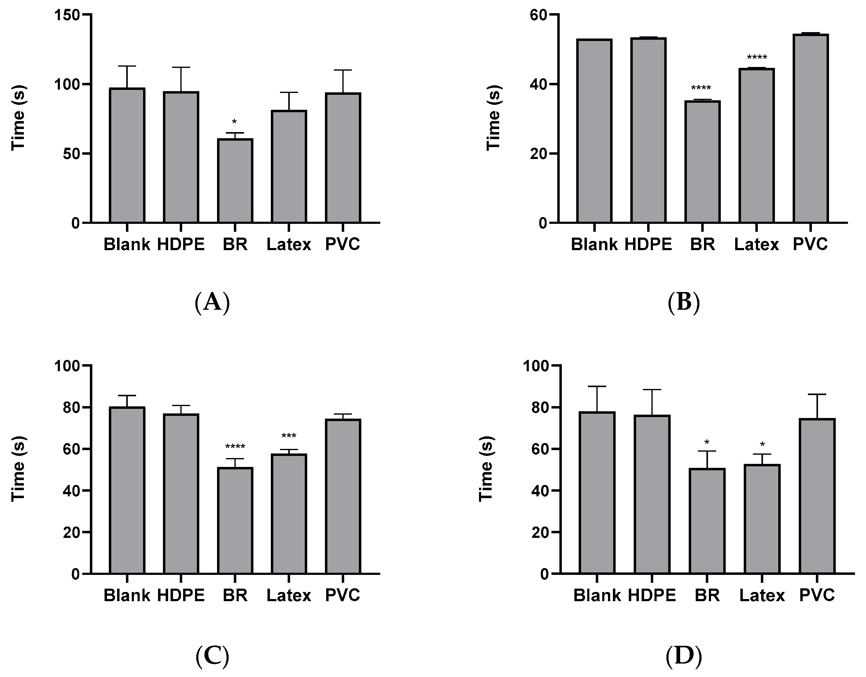
3.1. PTT of Polymeric Materials in Animal Plasma of Rabbit, Porcine, and Monkey

Plasma obtained from animal blood was reacted with the substance described in Table 1, and the PTT was subsequently determined. In rabbit plasma, the HDPE, Latex, and PVC test groups did not show any significant changes in PTT compared with the blank test group, whereas the black rubber test group showed a significant shortening of PTT (Figure 1A). In porcine and primate (Rhesus, Cynomolgus macaque) plasma, HDPE, which is a negative control material for hemocompatibility, and PVC, which is commercially available for medical use, did not show significant changes in PTT compared with the blank test group. In contrast, significant PTT shortening was observed in the positive control material candidates such as the black rubber and latex test groups (Figure 1B–D).

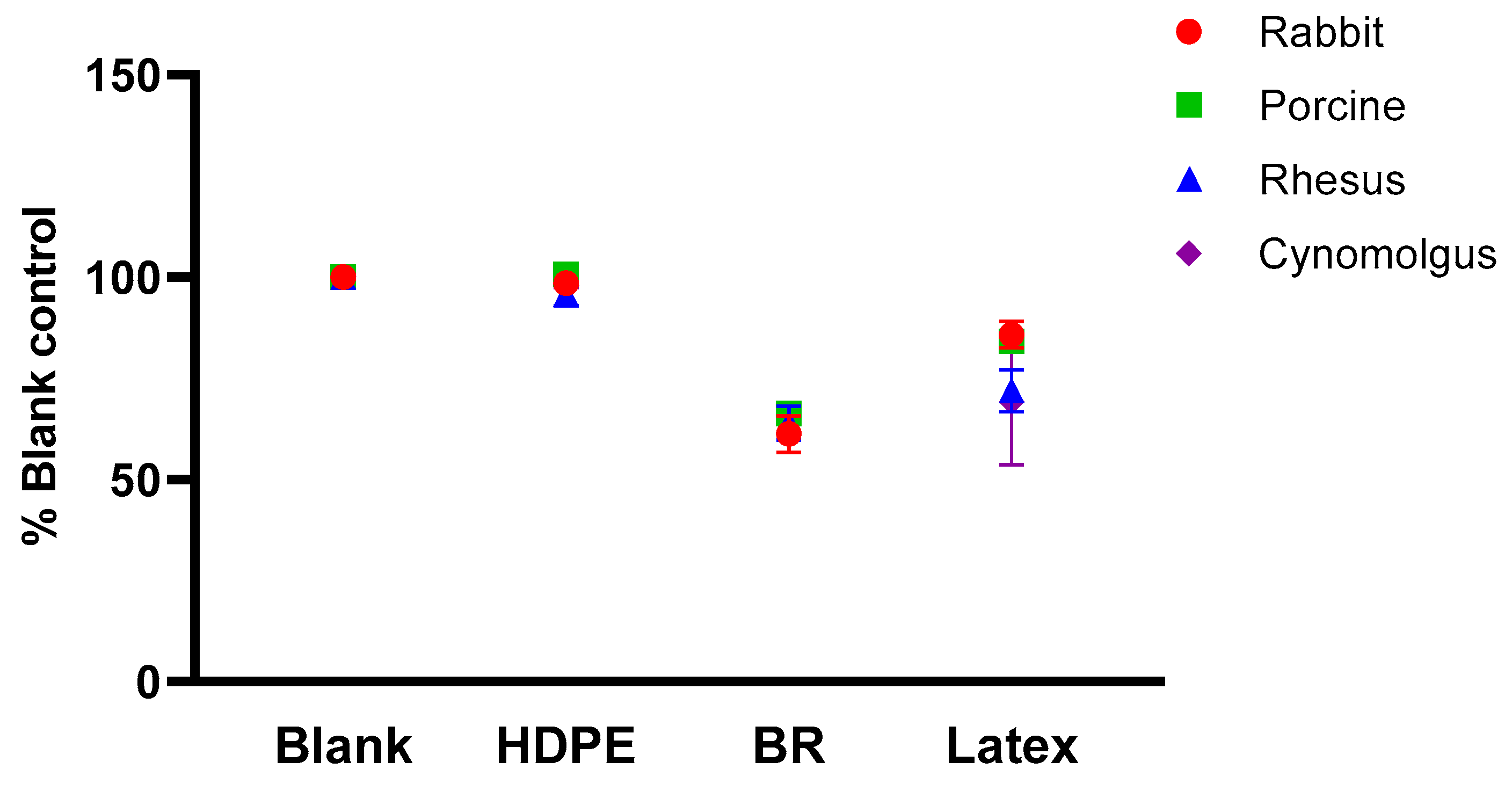
3.2. Comparison of Normalized PTT between Species

Figure 2 shows the result of reanalyzing Figure 1 with the normalized PTT. The normalized PTT was obtained by dividing the PTT measurement value of the test group by that of the blank control group, and the normalized PTT of each substance was compared for each plasma origin. The absolute value of the PTT of the blank control group was similar in primates regardless of species. Furthermore, the absolute PTT values of rabbits were higher than those of primates, and the porcine values were lower than those of primates. In contrast, no differences in the normalized PTT of the material were observed for different species (Figure 2).

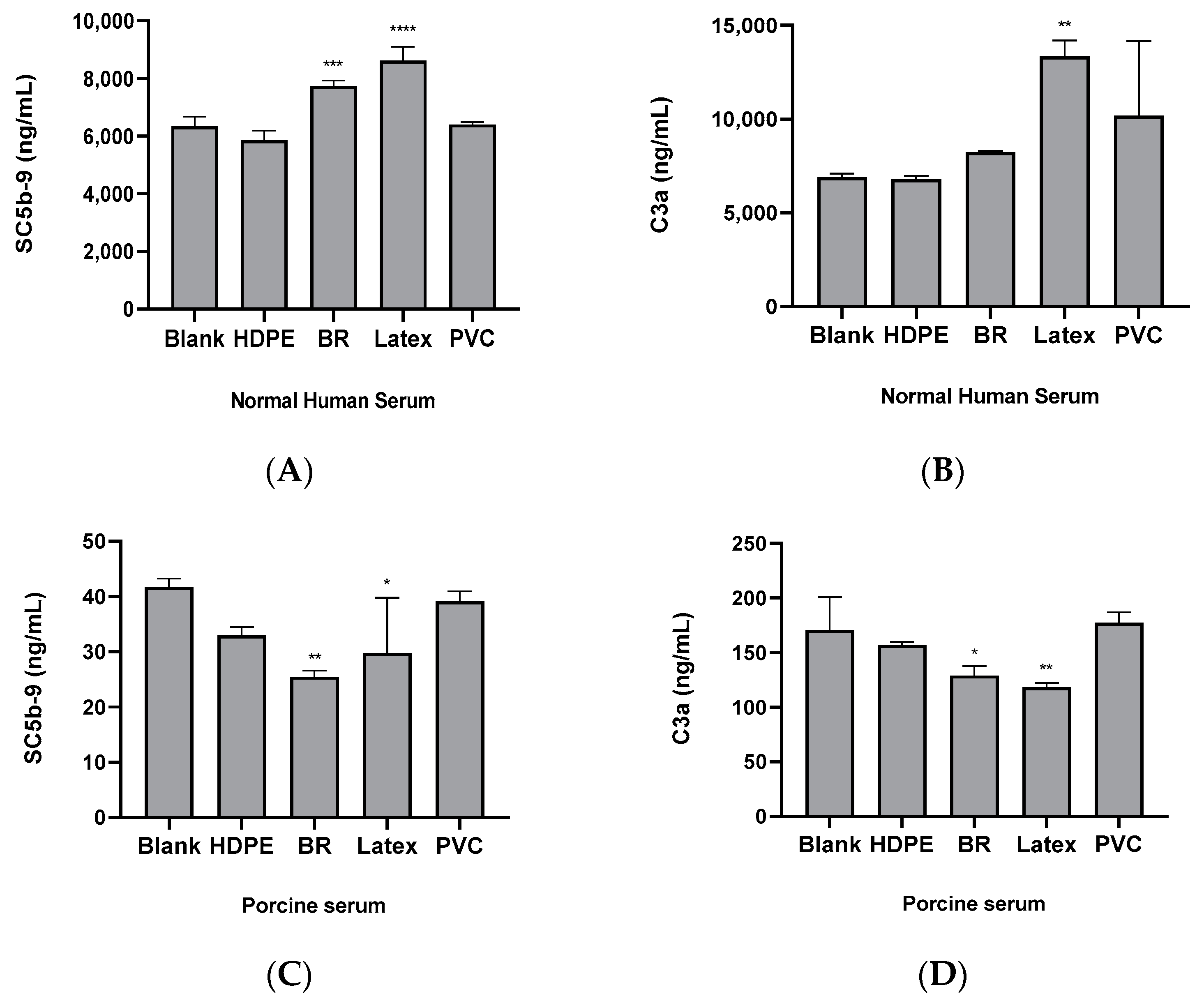
3.3. Comparison of the Complement System Activities by Substances in Human and Porcine Sera

In human serum, HDPE serves as a hemocompatibility negative control, and commercially available PVC for medical use did not show significant changes in complement system activity compared with the blank test group. In contrast, significant increases in complement system activity were observed in the positive control candidates of the black rubber and latex test groups, except for some test groups (black rubber, C3a) (Figure 3A,B). In contrast to the results of complement system activity in human serum, the substances in porcine serum showed no significant increase in complement system activity compared with the blank test group (Figure 3C,D).

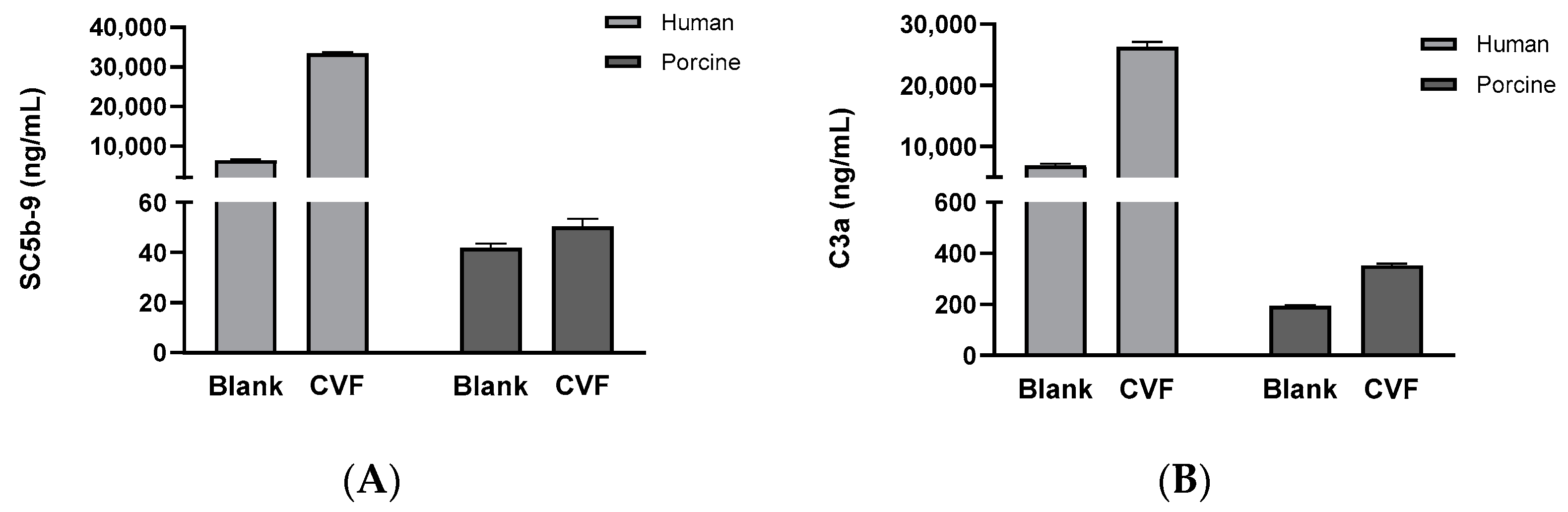
3.4. Comparison of Complement System Activity in Human and Porcine Sera Reacted with the CVF

To compare the levels of complement system activity of human and porcine sera, CVF, which is a strong complement system activator, was reacted with human and porcine sera, and the degree of complement system activity in each serum was observed. When porcine serum was reacted with CVF, SC5b-9 and C3a exhibited basal levels (50–354 ng/mL), whereas high levels of SC5b-9 and C3a (26,296–33,390 ng/mL) were observed in the case of human serum (Figure 4A,B).

4. Discussion

Static tests to evaluate thrombosis are relatively simple and yield more consistent results. However, device thrombogenicity related to blood flow and surface topography cannot be evaluated and limit the prediction of the long-term outcomes of implantable medical devices. In addition, distinguishing subtle differences in thrombosis between biomaterials may not be possible.
This study confirmed the blood coagulation potential in various animal species based on the ASTM F2382-18:PTT, a test method standardized and approved by the FDA, and identified the differences in reactivity of different species for complement. Furthermore, a hemocompatibility evaluation model was designed, and the information required to interpret the results was obtained.
The PTT assay is a modification of the activated partial thromboplastin time (APTT) assay. The APTT assay includes active agents such as kaolin, celite, or ellagic acid [17], which must be avoided when evaluating the effects of blood contact devices or device materials because it interrupts coagulation caused by the materials or device [18]. Unlike the APTT assay, the PTT assay evaluates whether the test material acts as an activator using reagents (rabbit brain cephalin) without activating substances such as kaolin, celite, or ellagic acid. The time it takes for citrate plasma exposed to the test material when exposed to phospholipid particles and calcium chloride suspension to form a clot is measured. Shortening the PTT after contact with a substance indicates activation of the intrinsic coagulation pathway of blood coagulation, whereas heparin and other anticoagulants induce prolonged PTTs [1,15]. Despite variations in coagulation factors, such as F-IX, F-X, and F-VII, across species, the coagulation pathway may essentially be similar among mammals [19,20,21]. Many studies have been conducted using different animal species for blood coagulation, and the degree of coagulation has been reported to differ depending on the animal species [5,6,22]. The applicability of using animal blood instead of human blood with the test blood of PTT assay was examined to determine the blood clotting potential in various animal species.
PTT changes were observed with one negative and two positive control candidate materials as the test material and plasma from rabbits, porcine, rhesus monkeys, and cynomolgus monkeys. The absolute value of the PTT reaction in the plasma of rabbits, porcine, and primates that reacted with the material showed slight differences among animals. However, the normalized values—61% for rabbits, 66% for pigs, 63% for rhesus monkeys, and 65% for cynomolgus monkeys—did not show any statistically significant differences. The evaluation of thrombosis using PTT demonstrated the possibility of replacing human blood if an appropriate animal model is selected.
The complement is activated by the surfaces of external materials, including blood-contacting medical devices, and usually occurs in the early stages [23,24]. The amount of complement proteins in the blood can be evaluated using ELISA assays, and the complement proteins that can be used in ELISA assays include, but are not limited to, C3a, C5a, and SC5b9; C3a is considered a general indicator of complement activation, and SC5b-9 is generally considered a marker representing the full extent of complement activation [25,26,27]. An increase in complement components indicates that the complement system is activated. Only complement components in plasma can be measured, and measuring complement components that attach to the material surface is difficult. Commercially available ELISA kits are species-specific, and a suitable control group needs to be included for comparison. The typical CH-50 assay is useful for human, bovine, porcine, and rabbit sera; however, the sensitivity is too low to detect complement activation after contact with the material. Another functional method is complementing production of C3- or C5-converting enzymes as measured via substrate conversion assays.
The ISO 10993-4 standard (2017) provides information about complement testing of medical materials and devices. No standard blood or blood product is currently used for complement testing, and when serum and plasma are used, they must be carefully selected so as not to reduce or increase complement activation [1]. In this study, to select standard blood and blood products used for complement testing, human and porcine sera were reacted with the material using the direct contact method, and then complement activation was assayed. By observing complement activation with human serum and one negative and two positive control candidate materials as the test materials, negative and positive control candidate materials could be effectively distinguished. In contrast, they were difficult to distinguish in porcine serum. Tests with strong activators, such as CVF, confirmed that it works under specific conditions. Complement activation was measured in CVF, a potent activator, and human and porcine sera. Complement activation was significantly increased in human serum but showed a basal level of activation in porcine serum. This indicates significant differences between humans and porcine in their potential to activate the complement system [28,29]. Porcine ex vivo and implantation models are the preferred preclinical models for hemocompatibility evaluation of blood-contacting medical devices owing to their anatomical similarity to human organs [30,31,32].
Despite porcine blood exhibiting high sensitivity in the coagulation reaction after contact with biomaterials, the result showing a basal reaction in the activity of the complement system must be considered in the design of the preclinical test model and interpretation of the results.
The results revealed that the normalized values of PTT in substance-reacted plasma exhibited similarities regardless of the animal species, and the blood coagulation potential was confirmed in various animal species. In addition, the results of complement activation for a material indicate that the reactivity differs between species and provide important clues in the interpretation of the results of preclinical testing of blood-contacting medical devices and in the design of experiments. In addition, the selection of an appropriate animal model for thrombosis evaluation indicates the potential to substitute human blood and may correlate with in vivo and clinical thrombosis data for thrombosis evaluation.

5. Conclusions

Under the conditions of this study, the complement system activity induced in the blood in contact with the substance differed for different species, whereas the normalized PTT response values for the cases of animal species in the plasma induced after reaction with the substance were similar. These results demonstrate the feasibility of evaluating thrombosis using PTT if an appropriate animal model is selected. In addition, this information is crucial in the design of in vitro and in vivo hemocompatibility evaluation models and interpreting the results.

Author Contributions

J.K. performed most of the in vitro experiments and wrote the original draft. G.K., J.L., S.K. and J.E. advised the experiment. J.K. and T.K. analyzed the final data and edited the final manuscript. All authors have read and agreed to the published version of the manuscript.

Funding

This research was supported by a grant (21174MFDS242) from the Ministry of Food and Drug Safety in 2022.

Institutional Review Board Statement

The animal study protocol was approved by the Institutional Review Board of Futuristic Animal Resource and Research Center (FARRC-220401 and date of approval 1 April 2022), the Korea Research Institute of Bioscience and Biotechnology (KRIBB) Institutional Animal Care and Use Committee (NPRC-220401 and date of approval 1 April 2022), and the Korea Testing Certification Institutional Animal Care and Use Committee (Protocol-ETCS-02 and date of approval 14 May 2021).

Informed Consent Statement

Not applicable.

Data Availability Statement

The original contributions presented in the study are included in the article, further inquiries can be directed to the corresponding author.

Acknowledgments

We thank everyone who helped.

Conflicts of Interest

The authors declare no conflicts of interest.

References

  1. ISO 10993-4; Biological Evaluation of Medical Devices—Part 4: Selection of Tests for Interactions with Blood. The International Organization for Standardization (ISO): Arlington, TA, USA, 2017.
  2. ASTM F2888-19; Standard Practice for Platelet Leukocyte Count-An In-Vitro Measure for Hemocompatibility Assessment of Cardiovascular Materials. ASTM International: West Conshohocken, PA, USA, 2019.
  3. ISO 7198:2016; Cardiovascular Implants and Extracorporeal Systems—Vascular Prostheses—Tubular Vascular Grafts and Vascular Patches. International Organization for Standardization: Geneva, Switzerland, 2016.
  4. ISO 7199:2016; Cardiovascular Implants and Artificial Organs—Blood-Gas Exchangers (Oxygenators). International Organization for Standardization: Geneva, Switzerland, 2016.
  5. Mason, R.G.; Read, M.S. Some species differences in fibrinolysis and blood coagulation. J. Biomed. Mater. Res. 1971, 5, 121–128. [Google Scholar] [CrossRef] [PubMed]
  6. Todd, M.E.; McDevitt, E.; Goldsmith, E.I. Blood-clotting mechanisms of nonhuman primates. Choice of the baboon model to simulate man. J. Med. Primatol. 1972, 1, 132–141. [Google Scholar] [CrossRef] [PubMed]
  7. Lechner, R.; Helm, M.; Müller, M.; Wille, T.; Riesner, H.J.; Friemert, B. In-vitro study of species-specific coagulation differences in animals and humans using rotational thromboelastometry (ROTEM). J. R. Army Med. Corps. 2019, 165, 356–359. [Google Scholar] [CrossRef] [PubMed]
  8. Lu, Q.; Hofferbert, B.V.; Koo, G.; Malinauskas, R.A. In vitro shear stress-induced platelet activation: Sensitivity of human and bovine blood. Artif. Organs. 2013, 37, 894–903. [Google Scholar] [CrossRef] [PubMed]
  9. Chan, C.H.H.; Pieper, I.L.; Robinson, C.R.; Friedmann, Y.; Kanamarlapudi, V.; Thornton, C.A. Shear stress-induced total blood trauma in multiple species. Artif. Organs. 2017, 41, 934–947. [Google Scholar] [CrossRef] [PubMed]
  10. Goodman, S.L. Sheep, pig, and human platelet-material interactions with model cardiovascular biomaterials. J. Biomed. Mater. Res. 1999, 45, 240–250. [Google Scholar] [CrossRef]
  11. Pelagalli, A.; Belisario, M.A.; Tafuri, S.; Lombardi, P.; D’Angelo, D.; Avallone, L.; Staiano, N. Adhesive properties of platelets from different animal species. J. Comp. Pathol. 2003, 128, 127–131. [Google Scholar] [CrossRef] [PubMed]
  12. Siller-Matula, J.M.; Plasenzotti, R.; Spiel, A.; Quehenberger, P.; Jilma, B. Interspecies differences in coagulation profile. Thromb. Haemost. 2008, 100, 397–404. [Google Scholar] [CrossRef] [PubMed]
  13. Pelagalli, A.; Lombardi, P.; D’Angelo, D.; Della Morte, R.; Avallone, L.; Staiano, N. Species variability in platelet aggregation response to different agonists. J. Comp. Pathol. 2002, 127, 126–132. [Google Scholar] [CrossRef]
  14. Kessler, U.; Grau, T.; Gronchi, F.; Berger, S.; Brandt, S.; Bracht, H.; Marcucci, C.; Zachariou, Z.; Jakob, S.M. Comparison of porcine and human coagulation by thrombelastometry. Thromb. Res. 2011, 128, 477–482. [Google Scholar] [CrossRef]
  15. ASTM F2382-18; Standard Test Method for Assessment of Circulating Blood-Contacting Medical Device Materials on Partial Thromboplastin Time (PTT). ASTM International: West Conshohocken, PA, USA, 2018.
  16. ISO 10993-12; Biological Evaluation of Medical Devices—Part 12: Sample Preparation and Reference Materials. The International Organization for Standardization (ISO): Arlington, VA, USA, 2021.
  17. Roshal, M.; Gil, M.R. Activated Partial Thromboplastin Time. In Transfusion Medicine and Hemostasis, 3rd ed.; Elsevier: Amsterdam, The Netherlands, 2019; pp. 779–781. [Google Scholar] [CrossRef]
  18. Groth, T.; Synowitz, J.; Malsch, G.; Richau, K.; Albrecht, W.; Lange, K.P.; Paul, D. Contact activation of plasmatic coagulation on polymeric membranes measured by the activity of kallikrein in heparinized plasma. J. Biomater. Sci. Polym. Ed. 1997, 8, 797–807. [Google Scholar] [CrossRef] [PubMed]
  19. Gando, S.; Nanzaki, S.; Sasaki, S.; Kemmotsu, O. Significant correlations between tissue factor and thrombin markers in trauma and septic patients with disseminated intravascular coagulation. Thromb. Haemost. 1998, 79, 1111–1115. [Google Scholar] [CrossRef] [PubMed]
  20. Pliszczak-Król, A.; Rząsa, A.; Gemra, M.; Król, J.; Łuczak, G.; Zyzak, A.; Zalewski, D.; Iwaszko-Simonik, A.; Graczyk, S. Age-related changes of platelet and plasma coagulation parameters in young pigs. J. Vet. Diagn. Investig. 2016, 28, 561–567. [Google Scholar] [CrossRef] [PubMed]
  21. Gentry, P.A. Comparative aspects of blood coagulation. Vet. J. 2004, 168, 238–251. [Google Scholar] [CrossRef] [PubMed]
  22. Counts, R.B.; Haisch, C.; Simon, T.L.; Maxwell, N.G.; Heimbach, D.M.; Carrico, C.J. Hemostasis in massively transfused trauma patients. Ann. Surg. 1979, 190, 91–99. [Google Scholar] [CrossRef] [PubMed]
  23. Walport, M.J. Complement. First of two parts. N. Engl. J. Med. 2001, 344, 1058–1066. [Google Scholar] [CrossRef] [PubMed]
  24. Walport, M.J. Complement. Second of two parts. N. Engl. J. Med. 2001, 344, 1140–1144. [Google Scholar] [CrossRef] [PubMed]
  25. Harboe, M.; Thorgersen, E.B.; Mollnes, T.E. Advances in assay of complement function and activation. Adv. Drug Deliv. Rev. 2011, 63, 976–987. [Google Scholar] [CrossRef] [PubMed]
  26. Zwirner, J.; Felber, E.; Reiter, C.; Riethmüller, G.; Feucht, H.E. Deposition of complement activation products on plastic-adsorbed immunoglobulins. A simple ELISA technique for the detection of defined complement deficiencies. J. Immunol. Methods 1989, 124, 121–129. [Google Scholar] [CrossRef]
  27. Noris, M.; Remuzzi, G. Overview of complement activation and regulation. Semin. Nephrol. 2013, 33, 479–492. [Google Scholar] [CrossRef]
  28. Schaapherder, A.F.M.; Gooszen, H.G.; te Bulte, M.T.J.W.; Daha, M.R. Human complement activation via the alternative pathway on porcine endothelium initiated by IgA antibodies. Transplantation 1995, 60, 287–291. [Google Scholar] [CrossRef] [PubMed]
  29. Sakai, R.; Kitano, E.; Hatanaka, M.; Lo, P.; Matsuura, R.; Deguchi, K.; Eguchi, H.; Maeda, A.; Watanabe, M.; Matsunari, H.; et al. Studies of Pig Complement: Measurement of Pig CH50, ACH50, and Components. Transplant. Proc. 2016, 48, 1282–1284. [Google Scholar] [CrossRef] [PubMed]
  30. Szebeni, J.; Bawa, R. Human clinical relevance of the porcine model of pseudoallergic infusion reactions. Biomedicines 2020, 8, 82. [Google Scholar] [CrossRef] [PubMed]
  31. Dewanjee, M.K.; Kapadvanjwala, M.; Sanchez, A.; Elson, R.; Serafini, A.N.; Zilleruelo, G.E.; Sfakianakis, G.N. Quantitation of comparative thrombogenicity of dog, pig, and human platelets in a hemodialyzer. ASAIO J. 1992, 38, 88–90. [Google Scholar] [CrossRef] [PubMed]
  32. Didisheim, P.; Dewanjee, M.K.; Frisk, C.S.; Kaye, M.P.; Fass, D.N. Animal models for predicting clinical performance of biomaterials for cardiovascular use. In Contemporary Biomaterials: Material and Host Response, Clinical Applications, New Technology, and Legal Aspects; Noyes Publications: Saddle River, NJ, USA, 1984; pp. 132–179. [Google Scholar]
Figure 1. PTT in animal plasma such as rabbit, porcine, and monkey. The PTT measured with the plasma reacted with the materials. (A) Plasma obtained from rabbits, (B) plasma obtained from porcine, (C) plasma obtained from Rhesus macaque, and (D) plasma obtained from Cynomolgus macaque. * Statistical significance (* p < 0.05; *** p < 0.0005; **** p < 0.0001, one-way ANOVA) compared with the blank control.
Applsci 14 06721 g001
Figure 2. Comparison of the normalized PTT. The normalized PTT of each material was compared for the animal plasma of rabbit, porcine, and monkey.
Applsci 14 06721 g002
Figure 3. Comparison of complement activity in human and porcine sera. (A) Effects of test substances on SC5b-9 production in normal human serum. (B) Effects of test substances on C3a production in normal human serum. (C) Effects of test substances on SC5b-9 production in porcine serum. (D) Effects of test substances on C3a production in porcine serum. The mean ± SD values of the three independent experiments are shown. * Statistical significance (* p < 0.05; ** p < 0.005; *** p < 0.0005; **** p < 0.0001, one-way ANOVA) compared with the blank control.
Applsci 14 06721 g003
Figure 4. Comparison of complement activity in human and porcine sera reacted with the CVF. (A) Effects of test substances on SC5b-9 production. (B) Effects of test substances on C3a production. The mean ± SD values of the three independent experiments are shown.
Applsci 14 06721 g004
Table 1. Summary of experimental animals.
ParametersConditions
Species
(strain, sex, age)
Rabbit (Oryctolagus cuniculus, Female, 16 weeks),
Monkey (Rhesus macaque and Cynomolgus macaque, Male, 5 months)
Porcine (Sus scrofa, Female, 3 months)
Anticoagulant
(final ratio with blood)
3.2% sodium citrate
(blood: sodium citrate solution volume ratio = 9:1)
Test materialsHDPE, Black rubber, Latex rubber, PVC
Disclaimer/Publisher’s Note: The statements, opinions and data contained in all publications are solely those of the individual author(s) and contributor(s) and not of MDPI and/or the editor(s). MDPI and/or the editor(s) disclaim responsibility for any injury to people or property resulting from any ideas, methods, instructions or products referred to in the content.

