Article

Study of the Effects of Plasma Pretreatment on the Microstructure of Peanuts

1
School of Food and Strategic Reserves, Henan University of Technology, Zhengzhou 450001, China
2
Academy of National Food and Strategic Reserves Administration, Beijing 100037, China
*
Authors to whom correspondence should be addressed.
Appl. Sci. 2024, 14(17), 7752; https://doi.org/10.3390/app14177752
Submission received: 25 July 2024 / Revised: 28 August 2024 / Accepted: 31 August 2024 / Published: 2 September 2024

Abstract

:
In this study, cold plasma treatments are employed to modify peanuts. This study systematically investigates the effects of various plasma treatment conditions, including power, duration, and gas type, on the microstructure of peanut seed coats and embryos. Observations under a scanning electron microscope (SEM) reveal that as plasma treatment power increases from 100 W to 500 W, the etching level of peanut seed coats significantly intensifies, surface roughness deepens, and concavities become more pronounced. Additionally, micro-pores on the seed coat gradually enlarge and form cracks. Specifically, when the plasma treatment is set at 200 W for 60 s, the oxygen (O2) treatment group shows interconnected cracks on the peanut seed coat surface, with lipid particles exuding and protein particles and polymers decomposing. In contrast, the helium (He) treatment group displays clear cell structures and deep grooves, with no noticeable lipid particles exuding around surface cracks. The argon (Ar) treatment group exhibits a distinct rectangular cell structure with clear boundaries, and although surface cracks form, only a few protein particles escape from the cracks. The embryo surface structure becomes looser after plasma treatment, leading to the disintegration of lipid particles, protein particles, and polymers, affecting the fusion and migration of large and small lipid bodies within the peanut’s internal structure. Increasing treatment duration intensifies the etching phenomenon, resulting in more lipid particles exuding, which indicates a positive correlation between lipid particles exuding and treatment duration. This study sheds light on the mechanisms underlying changes in peanut microstructure due to cold plasma treatment, providing scientific evidence for improving peanut quality, enhancing oil extraction efficiency, and optimizing food processing techniques.

1. Introduction

Plasma pretreatment technology is a non-thermal processing technique known for generating plasma containing a multitude of high-energy particles. These particles include excited-state molecules, atoms, free radicals, electrons, ions, and ultraviolet light. Their reactivity far surpasses that of particles produced in conventional chemical reactions. This unique property allows plasma to yield a greater variety of highly reactive particles and makes it more conducive to surface interactions with materials [1,2,3]. Consequently, plasma is employed for modifying food surfaces to enhance specific functionalities or extend shelf life [4,5]. Wang et al. [6] found that cold plasma can improve the functional properties of proteins, delay the quality deterioration of peanuts after storage, and improve the shelf life of peanuts, which provides a theoretical basis for peanut storage as well as production and processing. In recent years, cold plasma technology has been increasingly used in the food industry and its potential and advantages have been widely recognized. Notably, in the field of fruit and vegetable sterilization, cold plasma demonstrates remarkable antimicrobial efficacy, effectively reducing microbial contamination and ensuring food safety [7,8,9,10]. Furthermore, this technology contributes to enhancing the biological activity of seeds. By altering seed surface properties, it promotes germination rates and overall growth, offering novel solutions for agricultural production [11,12]. Prakash et al. found that the application of cold plasma technology can inactivate pathogenic microorganisms on the surface of grains and produce active species, thereby extending their shelf life with minimal impact on food quality [13]. Cold plasma treatment also influences the activity and distribution of enzymes in food. It plays a crucial role in enzyme activity regulation and controlling enzymatic reactions during food processing [14]. Additionally, research on the dispersibility of natural antioxidants, such as phenolic compounds, reveals that cold plasma treatment can enhance the bioavailability of such compounds, thereby augmenting the nutritional value and functionality of food products [15,16]. For instance, Gebremical et al. [17] found that cold plasma technology can significantly increase the content of phenolic compounds such as total phenols, flavonoids, and proanthocyanidins in the red seed coat of peanuts. Li et al. [18] showed that the plasma etching depth was 328 nm at a plasma treatment power of 500 W and a treatment time of 30 s. However, the nutrient content of the maize kernels was essentially unaffected, which contributed to the activity of the maize seeds during the treatment of the seeds.
Peanuts are a high-yield crop used for both food and oil production. They have high nutritional value and are often referred to as the “longevity fruit” [19]. However, peanuts can produce harmful aflatoxins that pose a serious health risk to humans. As a result, recent research on plasma technology for peanuts has primarily focused on reducing aflatoxin levels and improving peanut quality. Specifically, plasma technology applications in peanut processing have centered around two main directions. The first is to significantly reduce aflatoxin content. For instance, studies by Devi et al. [20] and Wu et al. [21] have confirmed that plasma treatment at 60 W for 12 min can reduce aflatoxin B1 concentration by over 90%. Prakash et al.’s study pointed out that cold plasma treatment caused protein denaturation, oxidative stress, and cytotoxic compounds, causing DNA mutation, damage, catabolism, and ultimately cell death; however, the potential toxicological aspects and impact on overall food quality are not known [13]. The second is to enhance lipid quality and protein functionality in peanuts. Wang et al. [6] found that plasma treatment can significantly reduce the peroxide value and acid value of peanut crude fat while maintaining the basic composition of peanuts. Liu et al. [22] found that plasma treatment did not significantly change peanut oil quality, acid value reduction was more conducive for peanut storage, and the molecular structure of peanut oil was no damaged. Mollakhalili-Meybodi et al. [23] discovered that peanut protein’s water absorption, emulsification properties, and solubility were significantly enhanced. Wang et al. [24] observed that plasma treatment significantly altered the molecular structure and particle size distribution of peanut protein, thereby significantly improving peanut protein functionality.
Despite the remarkable achievements of plasma technology in detoxifying peanuts and enhancing their quality, there have been few reports exploring its impact on peanut microstructure. Moreover, plasma ionization is influenced by numerous factors, including power level, relative humidity, gas composition, and treatment duration [25]. Therefore, this study aims to fill this gap by investigating the effects of cold plasma treatment on the microstructure of peanut seed coats and embryo surfaces under a scanning electron microscope (SEM). The findings could provide essential theoretical support and practical guidance for peanut cultivation, efficient oil extraction, and deep processing, contributing to the sustainable development of the peanut industry.

2. Materials and Methods

2.1. Experimental Materials

Peanuts from the same batch, harvested in 2023, with a moisture content of 5.43% (w.b.) and a blade were used.

2.2. Instruments and Equipment

The following instruments and equipment were used: SY-DT02S low-temperature plasma treatment equipment (Ops Technology Co., Ltd., Suzhou, China); solid-state transistor plasma emitter at 40 kHz, plasma grain modification equipment, freeze dryer, JEC-3000FC ion sputtering instrument, JSM-IT700HR scanning electron microscope (JEOL, Tokyo, Japan).

2.3. Experimental Procedure

2.3.1. Plasma Pretreatment of Peanuts

Spread an appropriate amount of peanuts on a tray and place them inside the sealed chamber of the plasma treatment equipment. Evacuate the vacuum chamber to 110 Pa using a vacuum pump. Set the plasma source frequency to 40 kHz and treatment durations to 30 s, 60 s, 90 s, 120 s, and 150 s. Adjust the cold plasma treatment power to 100 W, 200 W, 300 W, 400 W, and 500 W. Introduce helium gas (He), oxygen gas (O2), and argon gas (Ar) [26,27,28]. After treatment, temporarily store each group of peanuts in a 4 °C refrigerator. Untreated peanuts from the same batch serve as the control group.

2.3.2. Observation of Peanut Surface Microstructure under a Scanning Electron Microscope

The surface microstructures of peanuts from both experimental and control groups are observed under a scanning electron microscope (SEM) at various magnifications. Representative images under identical conditions are selected for comparative analysis. From each group, three randomly selected peanuts out of ten with similar sizes and shapes are used for analysis, with the procedure repeated three times. SEM observations [27] are conducted at 4 kV.
Observation of seed coat structure: The peanuts are sliced into 1 cm × 0.5 cm × 0.3 cm samples using a blade. The samples from both the control and experimental groups are positioned with the seed coat facing upwards, affixed to the stage of an ion sputter with conductive adhesive, and subjected to gold sputtering for 130 s. Subsequently, SEM observations are conducted.
Observation of embryo structure: With slight modifications to the method used by Wang et al. [29] for observing the microstructure of wheat grain endosperm, the peanuts are split down the middle along the embryo axis and the embryonic shoots are removed. The peanuts are then cross-sectioned with a blade to remove 0.5 cm from both ends and the remaining central sections, approximately 5 mm thick, are used as observation samples. One section of the observation sample is adhered to a conductive adhesive and then subjected to gold sputtering for 160 s on the stage of an ion sputterer. Subsequently, SEM observations are conducted.

3. Results and Discussion

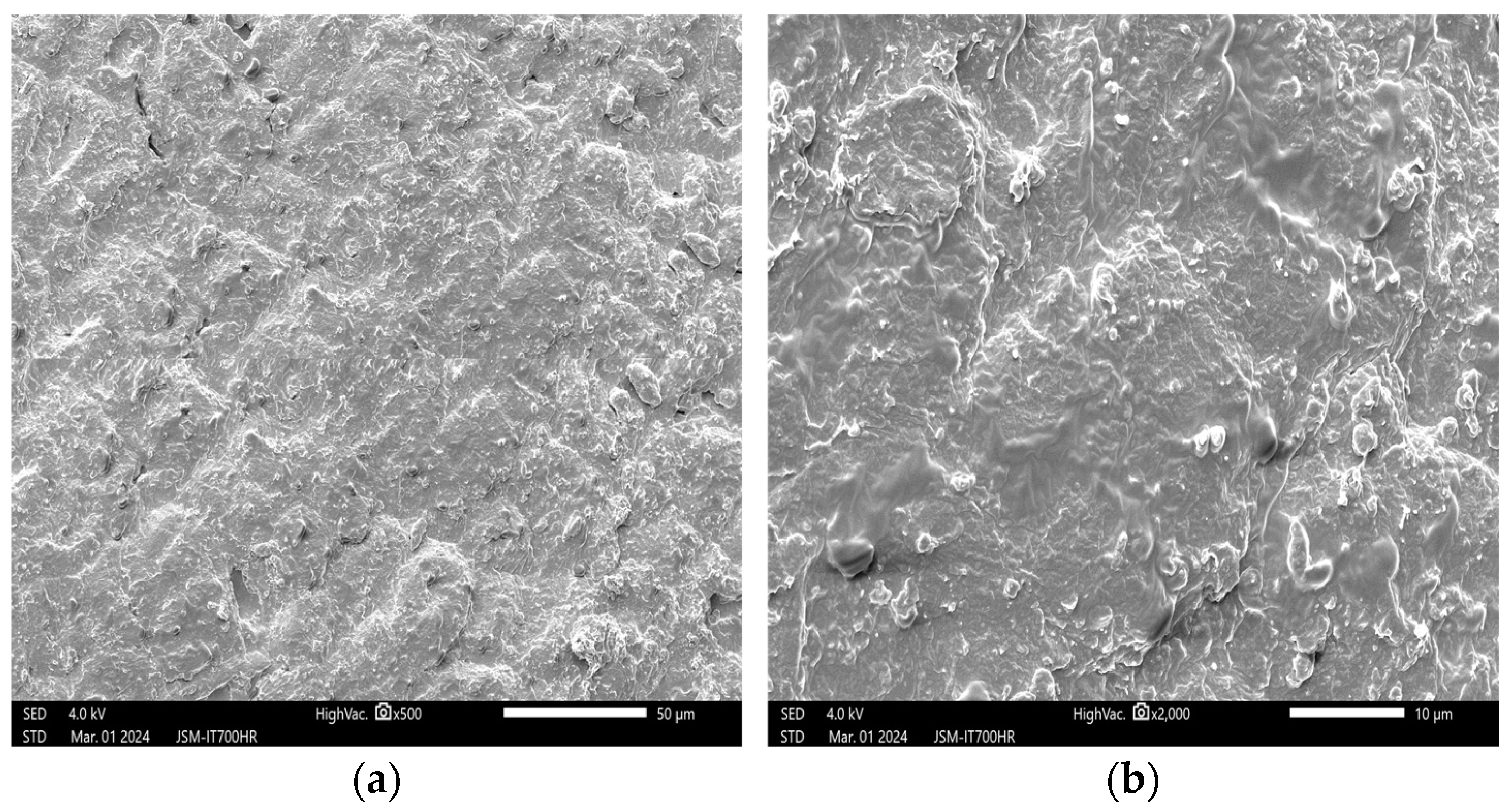
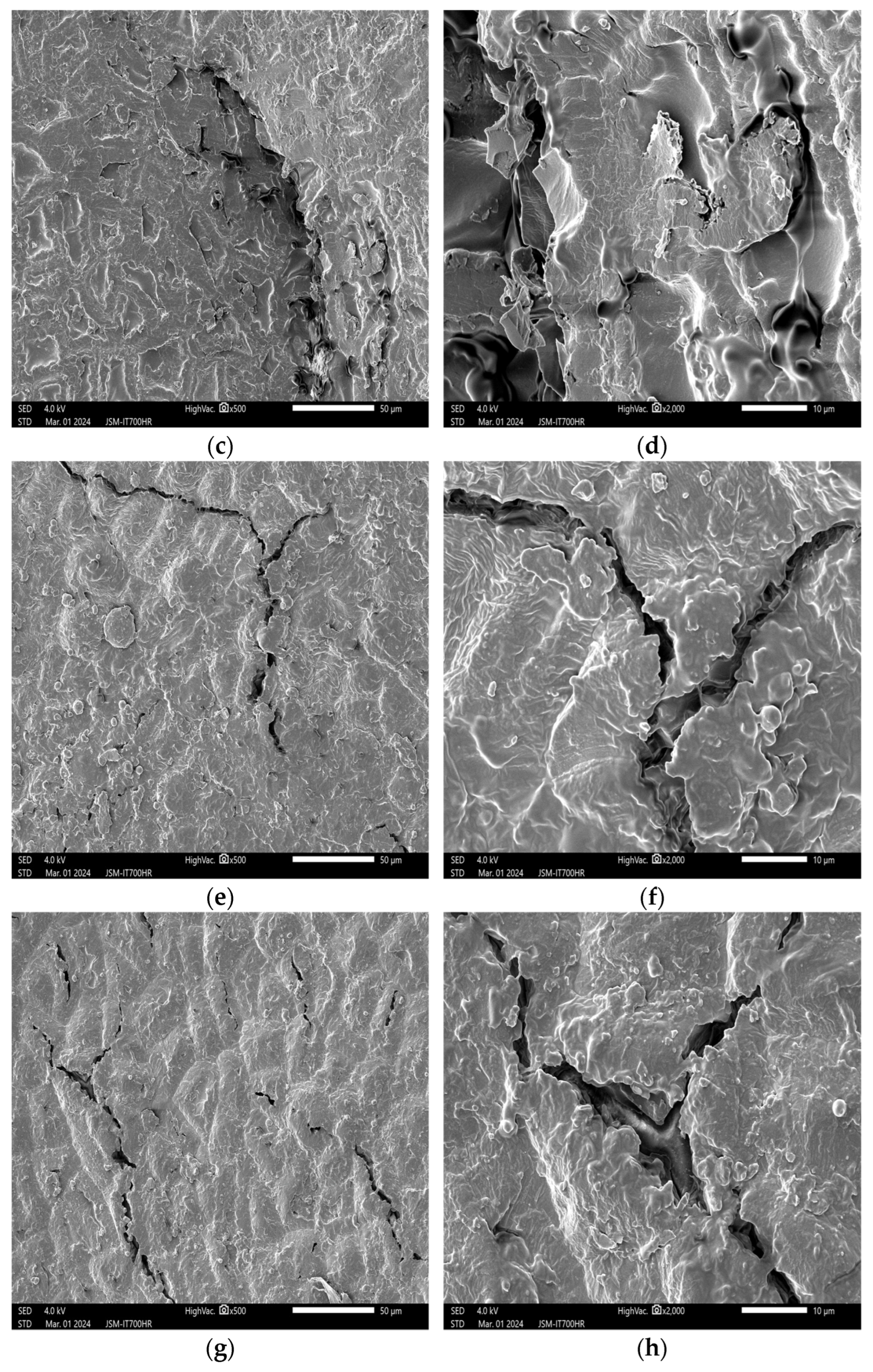
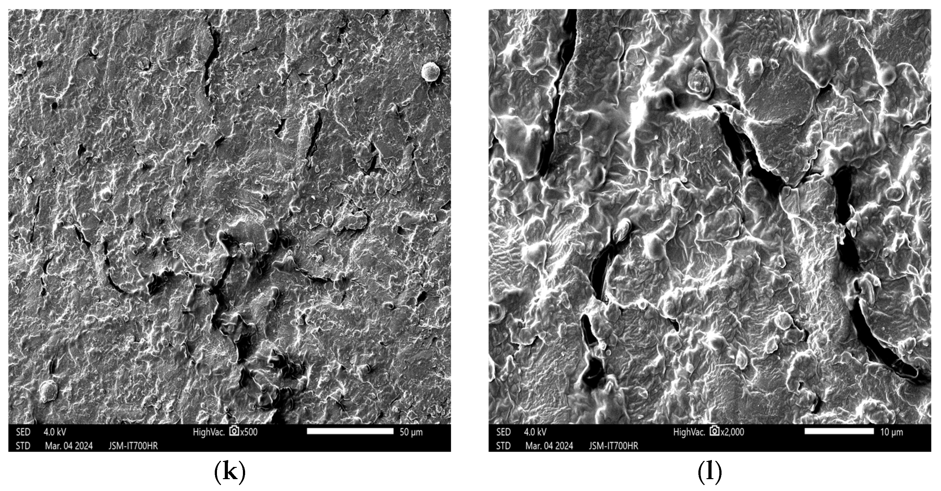
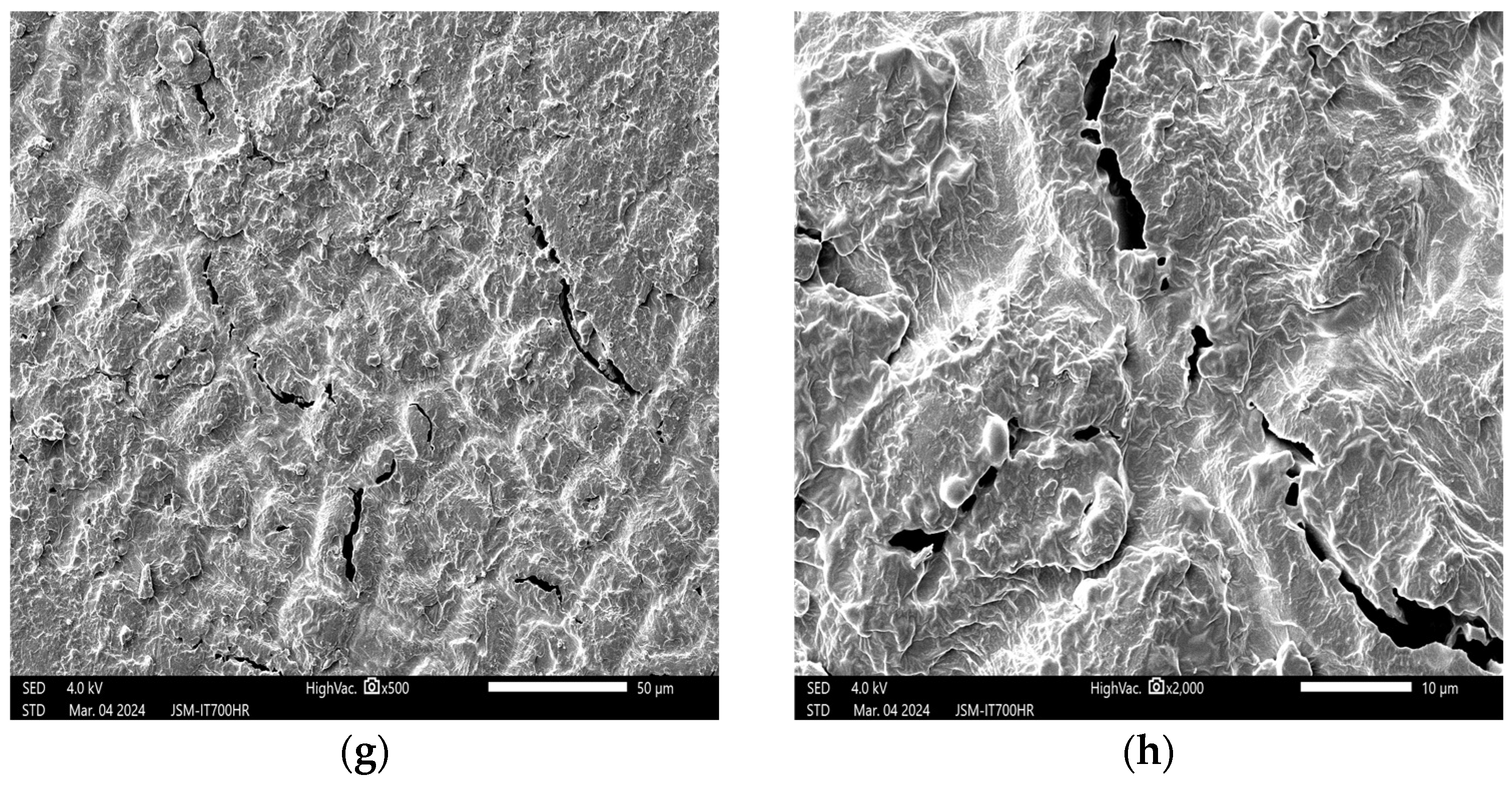
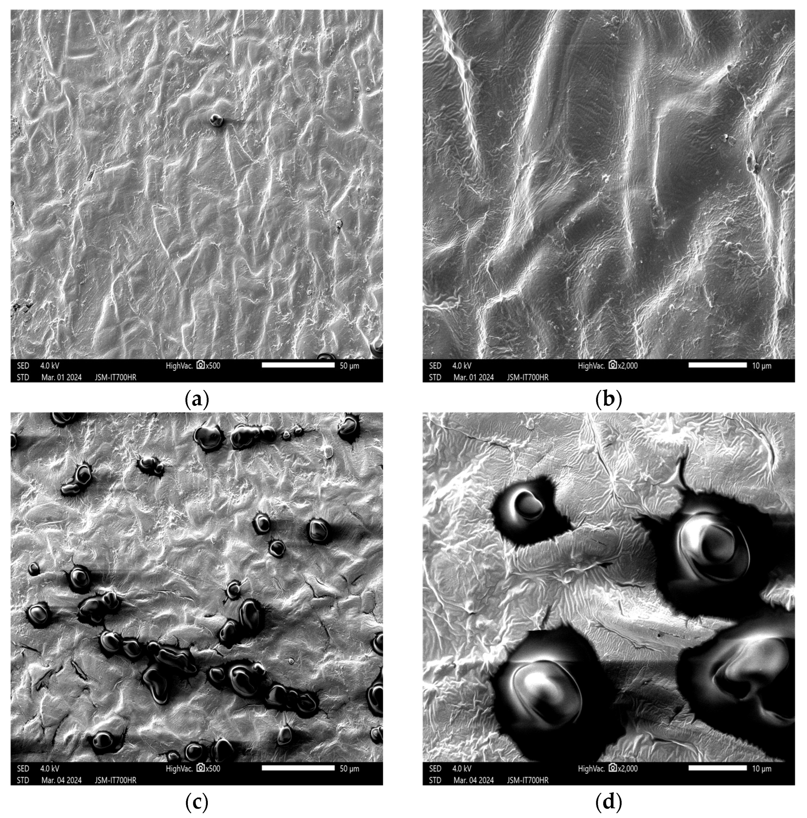
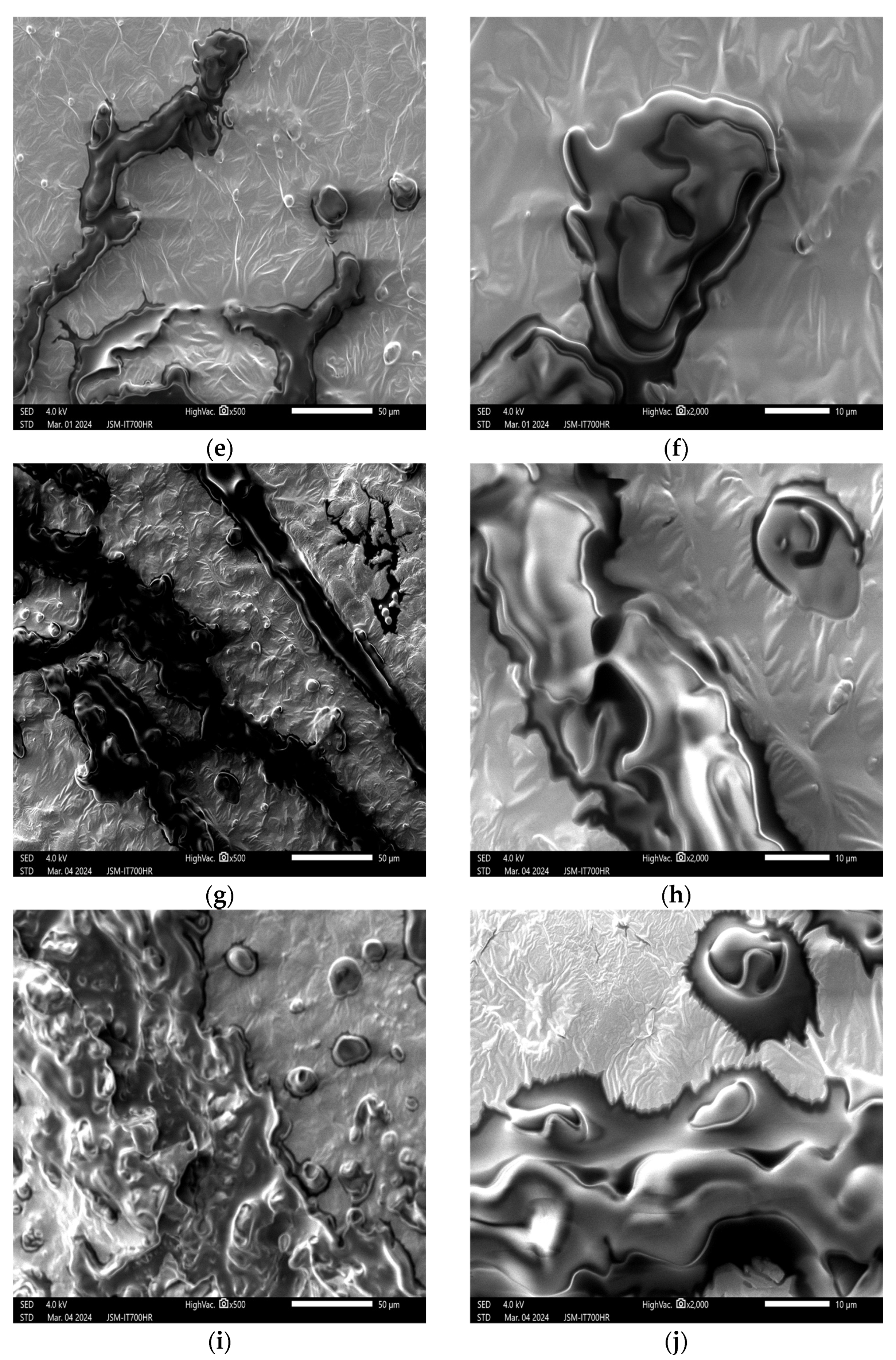
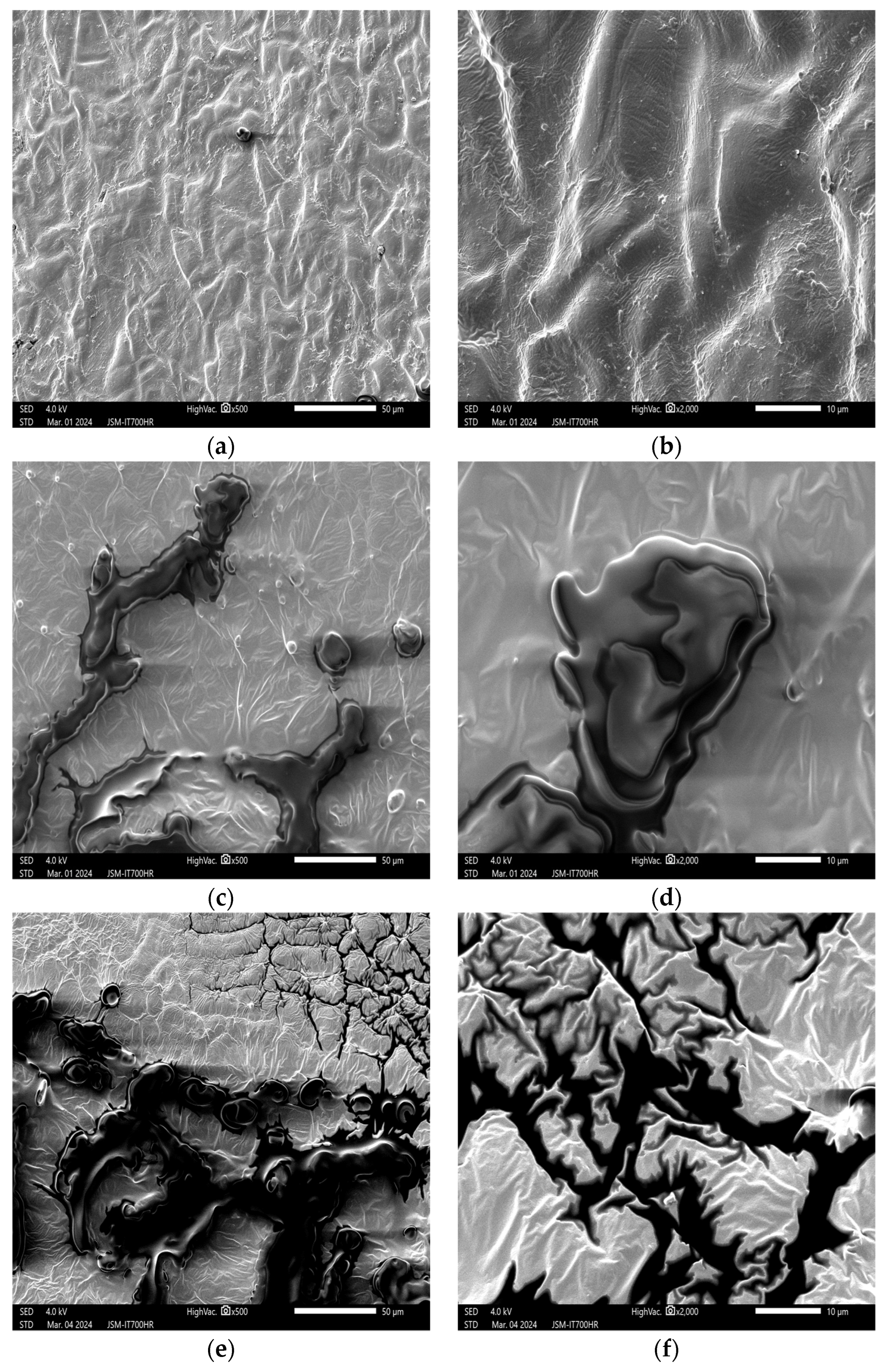
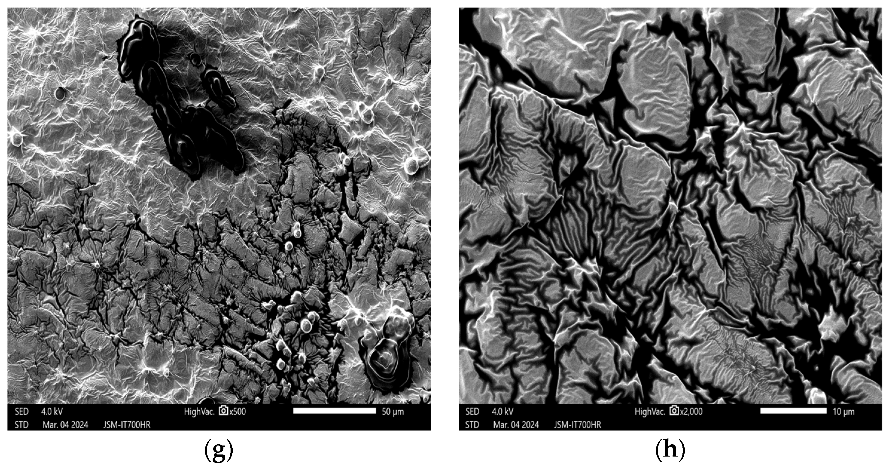
3.1. Effects of Plasma on the Structure of Peanut Seed Coat

3.1.1. Impact of Plasma Treatment Power on the Structure of Peanut Seed Coat

As shown in Figure 1, Figure 1a,b are the SEM images of the untreated peanut seed coat at 500× and 2000× magnifications, respectively. According to the figures, the surface of the peanut seed coat appears smooth, with slightly wide but shallow protrusions, clear and irregularly shaped textures, indistinct cell boundaries, and no cracks or obvious concavities. However, small micropores are present, likely serving as channels for material exchange within peanut cells. The changes were highly significant when comparing the control and treated groups. Figure 1c depicts the peanut seed coat treated with a plasma device at 100 W for 60 s and observed at 500× magnification. Compared to Figure 1a, fine cracks appear along the micropores on the seed coat.
As the power increases from 100 W to 300 W in Figure 1c,e,g, the surface cracks gradually widen along with the micropores, evolving from small cracks to small holes and eventually clustering into longer cracks. The surface textures of the seed coat transform from irregular shapes to quadrilaterals or trapezoids, becoming deeper and denser with clear cell boundaries arranged in a tight and orderly manner. Nevertheless, the cellular structure and shape distribution remain largely intact. Cross-linking of these boundaries results in grooves, creating a tough protective layer that shields the interior of the seed from external environmental factors, consistent with findings reported by Gebremariam et al. [24]. As the power gradually increases from 300 W to 500 W, Figure 1i,k reveal that the cracks on the seed coat gradually transform into holes. At 2000× magnification under SEM (Figure 1j,i compared to Figure 1d,f,h), lipid particles exude and cover the peanut seed coat, causing the polygonal structure of the cells to become shallower and looser until the morphological changes in the seed coat become unobservable.
This is attributed to the increasing number of high-energy reactive particles released per unit time as the plasma treatment power increases. These high-energy particles penetrate the peanut seed coat through the formed holes, reaching the embryo layer and reacting vigorously with the macromolecular compounds on the surface of the embryo. This leads to the disintegration of these macromolecules, damaging the cellular structure of the embryo layer and causing the cytoplasm to leak out. The higher the treatment power, the more lipid particles exude around the holes, eventually covering the entire seed coat, consistent with the findings of Ahmed et al. [30]. Low-power cold plasma treatment creates cracks on the surface of the seed coat, enhancing water absorption and stimulating seed germination. Conversely, high-power cold plasma treatment damages the surface of the seed coat, breaking down the barrier for material exchange and disrupting the structural morphology of peanuts, potentially having adverse effects on peanut processing, storage, and breeding. Wang et al. [31] analyzed the breakage of the seed coats of mechanically shelled peanuts with different degrees of drying and found that peanut seed coats with low water content and prolonged storage time are extremely easy to break. Additionally, etching after cold plasma pretreatment increases the pore-like structures and material exchange channels; however, the quality and characteristics of the subsequent peanut should be analyzed further to provide guidance.

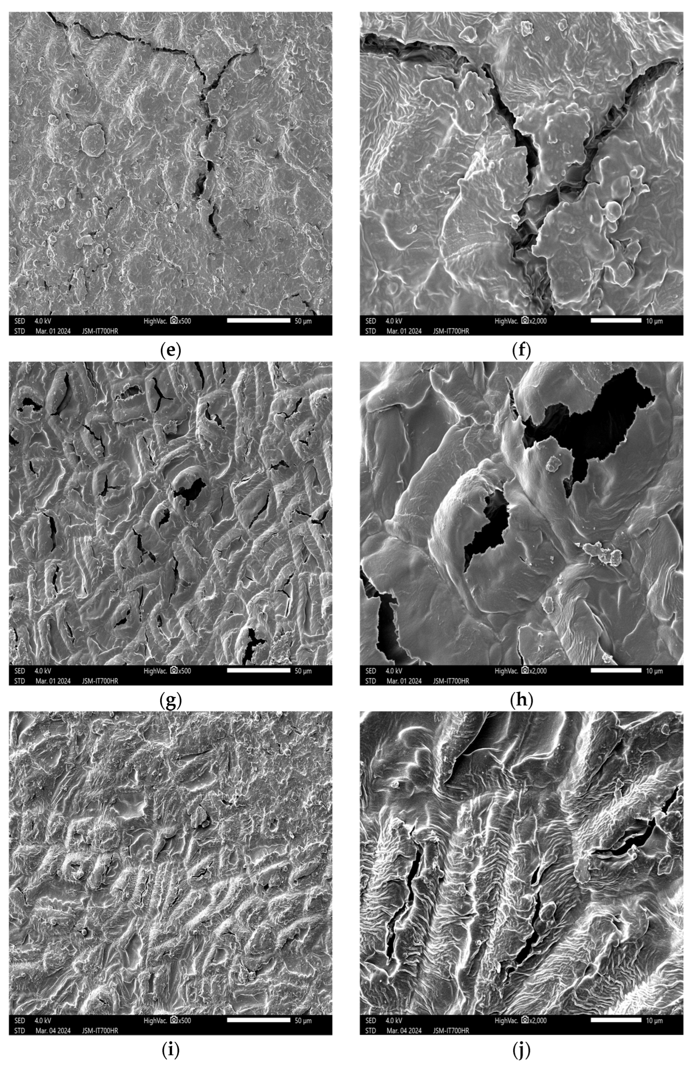
3.1.2. Impact of Plasma Treatment Duration on the Structure of Peanut Seed Coat

The results are presented in Figure 2. Figure 2c,d show the microstructures at 500× and 2000× magnifications, respectively, under SEM after treatment with helium gas (He) at 200 W for 30 s. Compared to the control group (Figure 2a), etching begins to appear on the seed coat surface with increased surface roughness, and the number of pores facilitating material exchange starts to increase. As the treatment duration increases, as shown in Figure 2c,e,g, cell boundaries become gradually clearer, arranged in tight order, with deeper and denser textures. The seed coat surface exhibits irregular concavities and protrusions. When magnified at 2000× under SEM (Figure 2d,f,h), the pores for material exchange gradually transform into large cracks and holes with prolonged treatment, and the etching phenomenon intensifies significantly. The impact resembles that observed with increased plasma treatment power. With the continuous extension of plasma treatment time, the plasma continuously releases more reactive particles like reactive oxygen species (ROS), reactive nitrogen species (RNS), and high-energy electrons. These particles act on the seed coat surface for an extended period, gradually exposing and even breaking the surface fiber layer macromolecules, as seen in Figure 2j. The number of wrinkles and irregular fine scaly protrusions on the seed coat increases, with more pronounced white cracks indicating breakage. When treatment time is extended to 150 s and the peanuts are viewed at 2000× magnification under SEM (Figure 2l), small molecular protein bodies are observed to escape from the seed coat surface and to adhere freely above the seed coat.
The reason for the above phenomenon may be that the peanut seed coat serves as a protective tissue, with its outermost layer being a cuticle composed primarily of fibrous compounds rich in flavonoids, polyphenols, and complex polysaccharides such as cellulose and hemicellulose. These substances play a crucial role in maintaining cell morphology and function [31,32]. This may be attributed to the plasma treatment device releasing more high-energy electrons, ions, photons, and other particles. These particles undergo cross-linking reactions with macromolecules on the peanut seed coat surface, such as fibrous protein in the fiber structure, ultimately leading to this phenomenon. Improved hydrophilicity of peanuts, as observed by SEM, folds, and porosity on the surface increase water uptake and provide a simple transfer pathway for nutrient transfer to provide the energy required for germination and increased conductivity. Surface oxidizability is also associated with the degree of seed coat breakage, as found by Ahmed N et al. [33]. Li B et al. [34] found that the relationship between the intensity of cold plasma treatment and protein function was not linear and that physical modifications via surface etching can control the treatment conditions and the number of proteins to obtain the desired modification results.

3.1.3. Impact of Plasma Treatment Gas Type on the Structure of Peanut Seed Coat

The results are presented in Figure 3. Figure 3c,e,g are SEM images at 500× magnification, corresponding to plasma treatments with helium (He), oxygen (O2), and argon (Ar), respectively, at 200 W for 60 s. Interestingly, the structure in Figure 3c,d closely resembles the peanut seed coat morphology observed after plasma treatment at 200 W. In contrast, in Figure 3e, interconnected pores gradually form cracks on the seed coat surface. Lipid particles exude from these cracks and are distributed evenly around the cracks. Decomposed protein particles and polymers are covered by lipid particles. The surface exhibits increased irregular wrinkles, and cell structures are less distinct. As depicted in Figure 3g, numerous quadrilateral cell structures with clear boundaries are evident. Cross-linking results in deep grooves, distinct textures, and wrinkle superpositions. Although cracks form on the cell surface, there is no significant lipid particle exudation around the cracks or within the pores, and only a small amount of protein body particles and polymers escape the cells.
Comparisons between treatment groups show large differences in the apparent phenomena produced by the different gas species. The different ions and free radicals produced during the ionization of He, O2, and Ar gases result in varying etching effects on the peanut seed coat surface. It is speculated that O2, as an oxidative reactant, generates reactive plasma ions upon ionization, which readily interact with the surface, forming new functional groups and generating novel substances. Due to the oxidation susceptibility of lipid bodies, the ionization of O2 generates highly reactive species such as oxygen atoms (O), negative oxygen ions (O), and ozone (O3). These species interact with the seed coat surface, leading to an etching effect that accelerates lipid oxidation. Consequently, cell walls rupture, causing lipid particles to escape from the cracks and altering cell membrane permeability and porosity, which aligns with research findings by Surowsky et al. [35] and Meinlschmidt et al. [36]. In contrast, He and Ar are inert gases, and their ionized ions are non-reactive plasma that only excite the material surface. Consequently, the ionization of different gases exerts distinct apparent phenomena on the peanut seed coat.

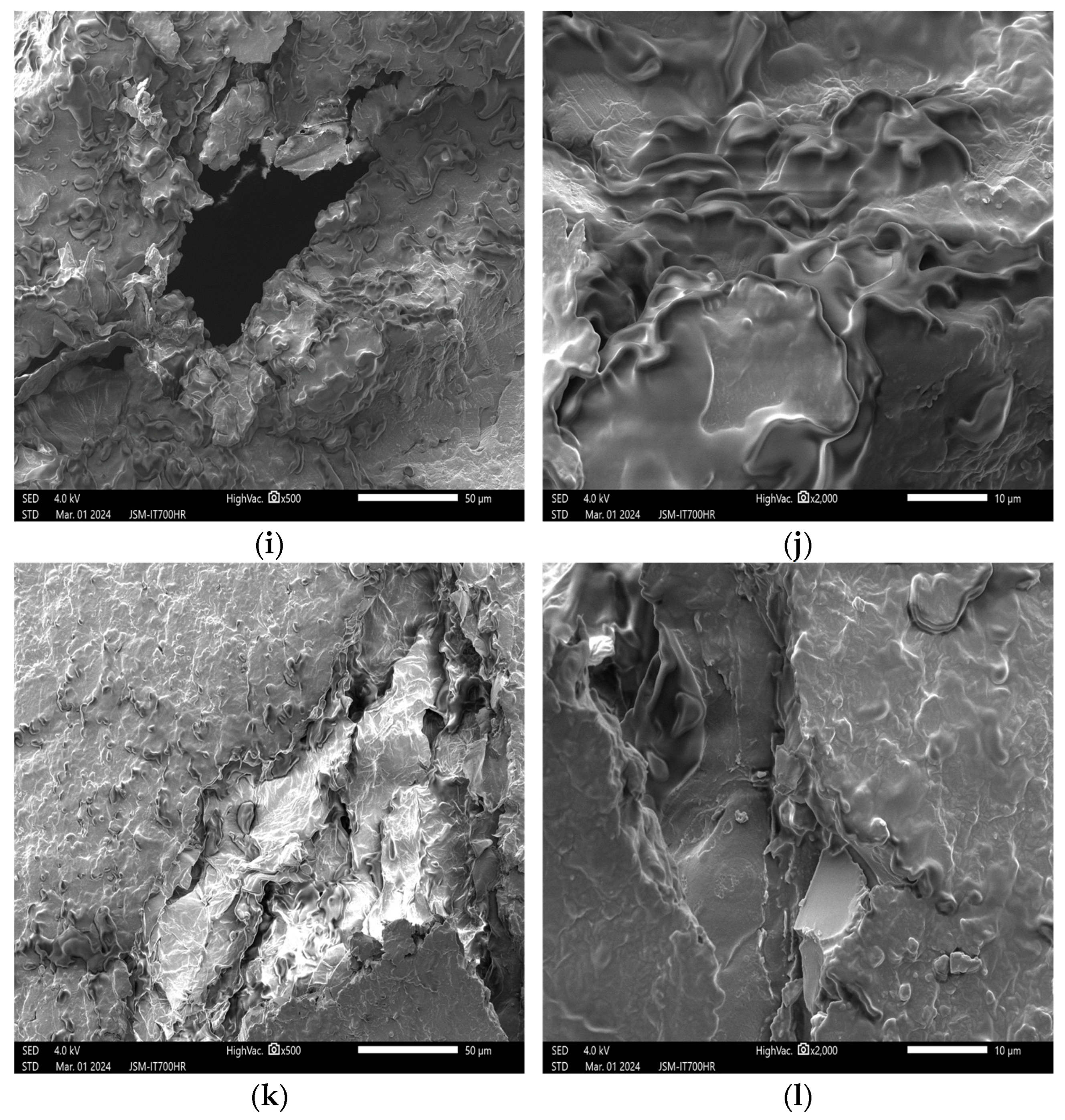
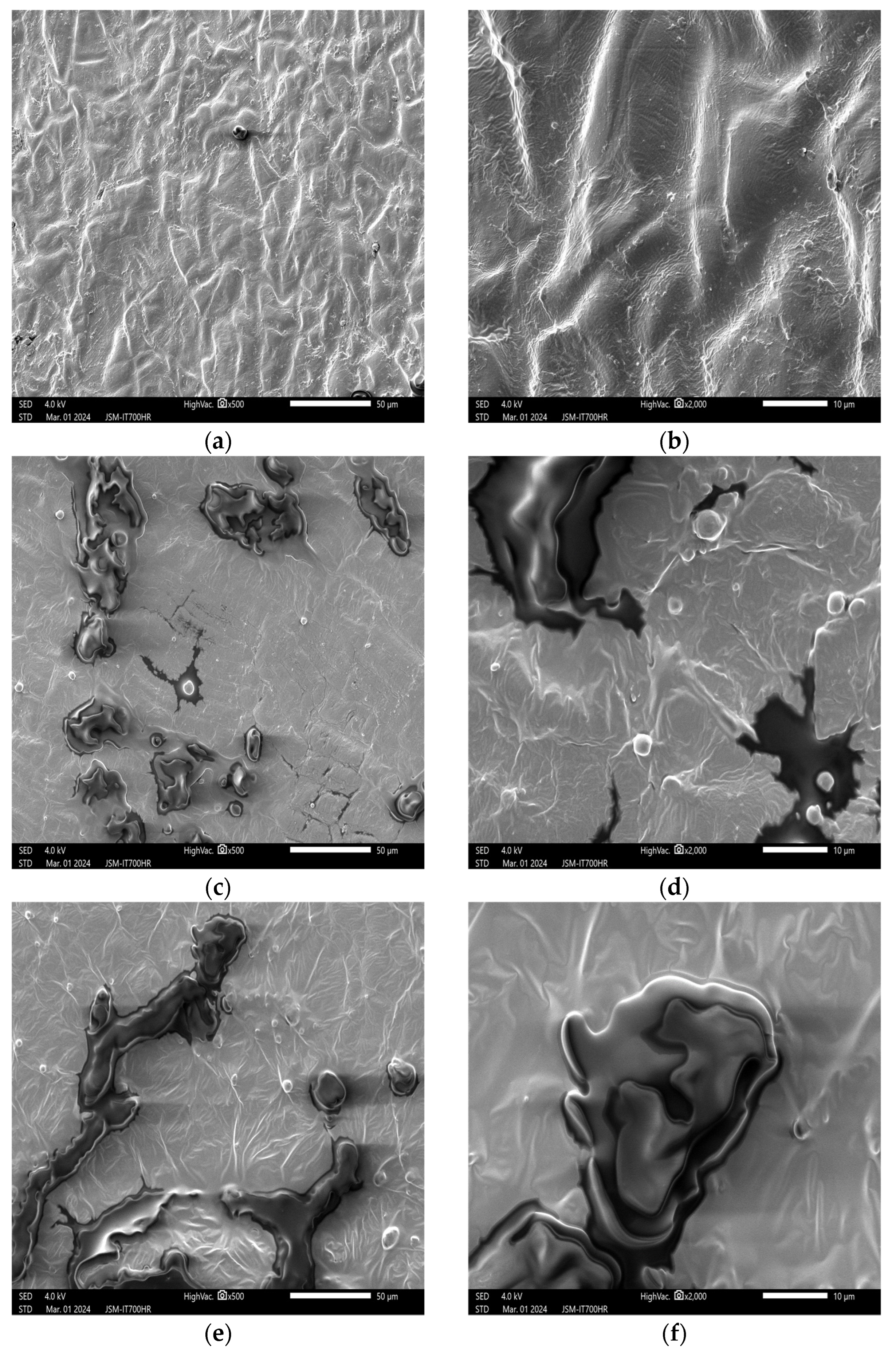
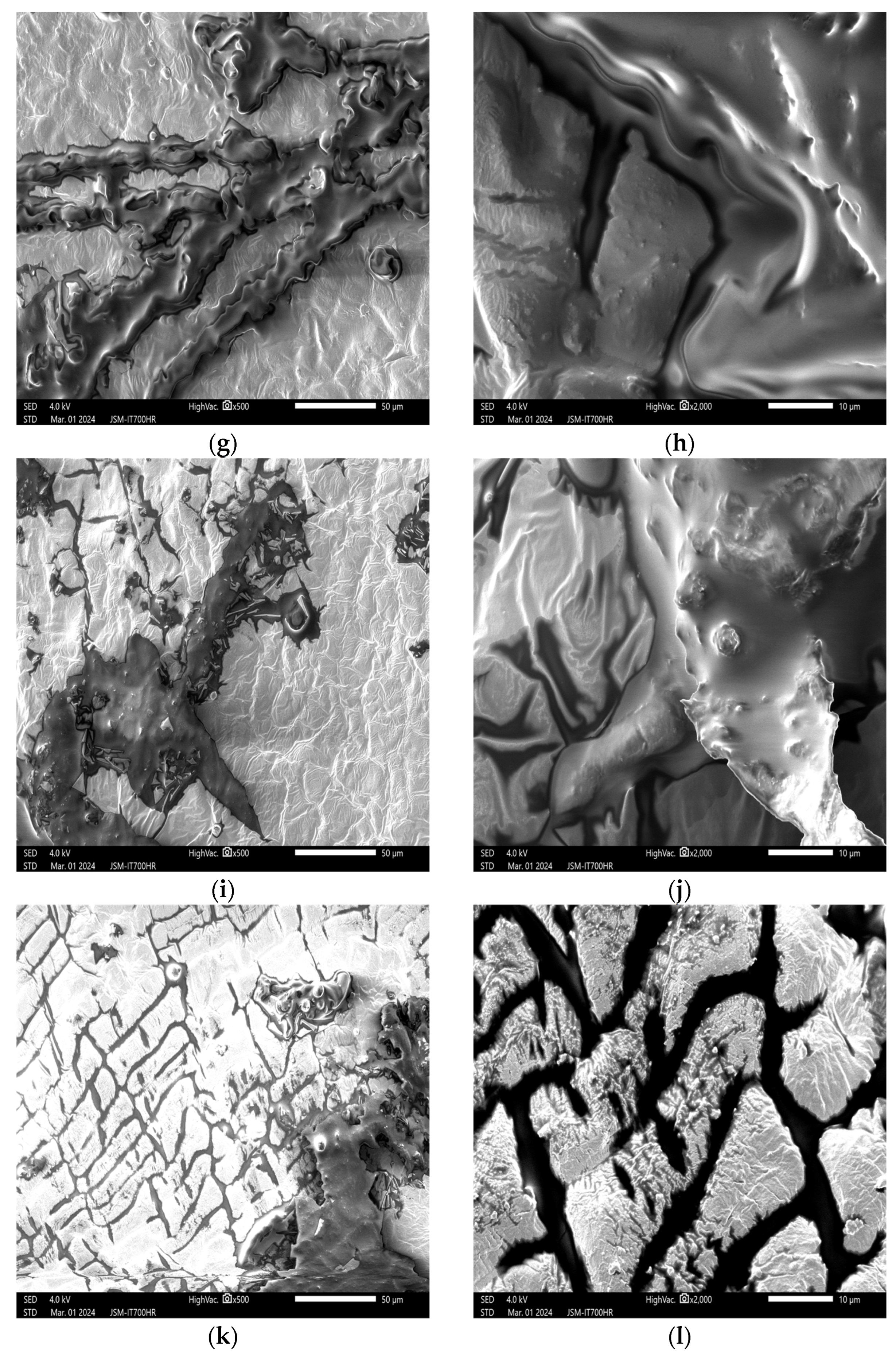
3.2. Impact of Plasma Treatment on the Appearance and Structure of Peanut Embryos

3.2.1. Impact of Plasma Treatment Power on the Appearance and Structure of Embryos

The results are presented in Figure 4. Figure 4a,b are, respectively, the surface appearance images of peanut embryos in the control group at 500× and 2000× magnifications. At different magnifications, the peanut embryo surface exhibits a grid-like structure with slight undulations. Despite the irregularities, the surface appears clean and well-organized, with clear textures and micropores. Figure 4c,e,g show SEM images at 500× magnification, corresponding to plasma treatment powers of 100 W, 200 W, and 300 W, respectively. Overall, the embryo surface remains smooth and structurally intact. However, cracks gradually increase, and lipid particles exude from the fissures, accumulating on the surface. The wrinkles become more pronounced and overlap and exposed protein particles are enveloped by the lipid bodies. This phenomenon is likely due to plasma etching. Figure 4i,k show SEM images at 500× magnification, corresponding to plasma powers of 400 W and 500 W, respectively. Lipid bodies stack up, cracks increase and widen, and some cell structures rupture from cross-linked concavities. It is clearly observable that the higher the power, the greater the degree of destruction compared to the treatment group. Figure 4l shows the SEM image at 2000× magnification. It reveals lipid particles migrating from the surface to the cracks, forming a mesh-like pattern. Protein aggregates disintegrate into smaller particles, possibly due to intense reactions between the released active species from ionization and surface macromolecules.
The apparent phenomena observed in peanut seed embryos after plasma treatment could indicate that the peanut seed coat plays a certain protective role. The cell structure remains intact, with a single layer of epidermal cells and thin-walled cells forming the outer layer. Within the thin-walled cells, particles include lipid bodies, proteins, and starch granules. Peanut cell fat exists in the form of lipid bodies, and proteins are evenly dispersed within the fat [37]. As plasma treatment power increases, more high-energy active ions are released, enhancing penetration and exacerbating surface etching, leading to thin-walled cell wall rupture, allowing lipid particles to flow out through channels, covering exposed protein particles and starch granules. This phenomenon is evident in Figure 4g,i,k.

3.2.2. Impact of Plasma Treatment Duration on the Appearance and Structure of Embryos

The results are presented in Figure 5. Figure 5c shows the embryo treated with helium gas plasma for 30 s at 200 W. SEM images at 500× magnification reveal an increased number of pores on the embryo surface compared to the control group (Figure 2a). Some lipid particles are also observed exuding. In Figure 5e, after 60 s of plasma treatment, SEM images at 500× magnification show an increase in surface cracks. The peanut surface becomes more uneven, and cell boundaries gradually become clearer. Cells are sparsely arranged, and the overlapping wrinkles intensify. Lipid particles continue to exude and gradually aggregate, accompanied by a small number of protein particles and polymers. Figure 5g,i,k are SEM images at 500× magnification, corresponding to treatment durations of 90 s, 120 s, and 150 s, respectively. As treatment time extends, surface etching becomes more pronounced, cracks increase, and lipid particles gradually migrate toward the cracks. Small protein particles and polymers also accumulate. The grooves and cell structures at the convex–concave intersections on the surface are increasingly covered by escaping lipid particles. These particles tend to accumulate in cell concavities and folded regions. Wang Yun et al. [38] found that larger lipid bodies are formed by the fusion of smaller ones, and surface proteins envelop the lipid particles, resulting in continuous plasma exposure, which leads to the breakdown of starch granules, lipid bodies, protein particles, and macromolecular polymers and the exudation of lipid particles. Longer treatment duration results in greater decomposition of lipid bodies and protein particles, leading to increased lipid particle exudation. In summary, the surface etching phenomenon and oil exudation in peanut embryos are positively correlated with plasma treatment duration.

3.2.3. Impact of Plasma Treatment Gas Type on the Appearance and Structure of Embryos

The results are presented in Figure 6. Figure 6c,e,g are SEM images at 500× magnification, corresponding to plasma treatments with helium (He), argon (Ar), and oxygen (O2), respectively, at 200 W for 60 s. The structural changes observed in Figure 6c,d are consistent with the effects on peanut embryos at the same power level. As depicted in Figure 6e, the embryo surface exhibits tight arrangement, intensified etching, and significant overlapping wrinkles. Exuding lipid particles primarily accumulate in cell concavities and cracks, while relatively fewer protein particles and polymers escape to the surface or may be covered by lipids. In Figure 6g, the embryo surface texture appears chaotic, but cell structures remain clear. The depth of cross-linked concavities increases, and wrinkle severity intensifies. Micromolecule lipid particles stack up, resulting in the exudation of more protein particles and starch granules compared to Figure 6e,g, along with finer and more dense cracks.
This is attributed to the interaction between abundant active species generated by the plasma and compounds on the peanut embryo surface. Three mechanisms—depolymerization, cross-linking, and etching—likely contribute to the surface modifications [39]. Helium (He), being a stable inert gas, experiences minimal ionization at this voltage, resulting in minor etching on peanut embryos. In contrast, argon (Ar) ionizes into high-energy Ar+ ions and electrons, leading to depolymerization of macromolecule protein particles on the embryo surface and structural changes. The micromolecule protein particles and lipid particles formed by the decomposition of polymers are exposed on the embryo surface. Unlike He and Ar, oxygen (O2) ionization generates abundant reactive oxygen species (such as O2+, O, O+, O3, and H2O2) [16]. Even at low voltage, O2 treatment induces significant oxidation, causing bond cleavage and chemical modifications on side chains, ultimately altering protein structures [35]. Therefore, O2 treatment can also lead to etching and oxidation on peanut embryo surfaces.

3.3. Effects of Plasma Treatment on the Internal Structure of Peanuts

The results are presented in Figure 7. Figure 7a,b represent the control group observed under SEM at 200× and 500× magnification, respectively. The images reveal that the internal fiber morphology of peanuts remains intact, resembling irregular leaf shapes. Protein particles, starch granules, and macromolecule polymer structures inside the peanuts are tightly and orderly arranged, uniformly congregating around the fiber structures, with some covered by lipid bodies. Figure 7c,d are SEM images at 200× and 500× magnification, corresponding to plasma treatments with helium (He) for 150 s. The images reveal that the internal fiber morphology of peanuts nearly disappears. Starch granules, protein particles, and polymers undergo depolymerization, while lipid particles increase and stack, resulting in larger gaps. The surface exhibits a concave and piled-up appearance. Figure 7e is an SEM image at 200× magnification, corresponding to plasma treatment with helium (He) at 500 w. It shows similar structural changes to Figure 7c, with more escaping protein particles, starch granules, and micromolecule polymers. This may be attributed to the rise in temperature within the plasma chamber as treatment duration extends and power increases. At 500 W, the vacuum chamber temperature measures approximately 45–50 °C. Under these conditions, protein cell walls are disrupted, causing proteins and lipids to flow out during plasma treatment and interact [40], resulting in the observed phenomenon.
Figure 7h is an SEM image at 500× magnification, corresponding to plasma treatment with O2 at 200 w for 60 s. This image reveals increased internal pores. Lipid particles become smaller and denser, and the fiber structure breaks into plate-like protrusions. No protein particles or polymers escape, but lipid bodies are distributed around the small pores. Figure 7j is an SEM image at 500× magnification, corresponding to plasma treatment with argon (Ar) at 200 w for 60 s. This image reveals that the structural changes resemble those in Figure 7h, but with slightly lighter etching. The internal structure of peanuts remains mostly intact, although the fiber structure breaks. Fewer lipid particles escape, while starch granules exude more. The reason lies in Ar being an inert gas, relatively stable, with its ionization producing only excitation effects; in contrast, O2 ionization, even at a lower voltage, generates abundant reactive oxygen species, exhibiting strong oxidative properties. This leads to lipid cell wall rupture, promoting the fusion of large and small lipid particles and exposing protein particles previously enveloped by lipid bodies [41]. Cao et al. [37] found that after high-temperature frying of peanuts, the cell wall undergoes significant rupture, and the loss of water leads to the emergence of more porous structures inside the peanut, which provides a channel for the oil body to enter the cell. Additionally, the etching phenomenon resulting from plasma treatment increases the number of internal pore-like structures, accelerating oil leakage and water loss within peanuts. It also provides suitable plasma treatment conditions for seed screening for high oil content [41] and oil extraction.

4. Conclusions

This study investigates the impact of cold plasma treatment on peanut microstructures under a scanning electron microscope (SEM). It reveals significant effects based on treatment power, duration, and gas type on both peanut seed coat and embryo structure. As plasma treatment power increases from 100 W to 500 W, the etching intensity on the peanut seed coat gradually strengthens. Surface roughness and concavity become more pronounced, and lipid particles begin to exude and cover the seed coat. At 500 W, distinct pores appear on the seed coat surface, and cell structures become blurred due to lipid particle exudation, resulting in complete coverage by lipids. Prolonged treatment duration, especially at 150 s of plasma treatment, further enhances these changes, with protein particles becoming more liberated and cracks more evident. Regarding gas types, oxygen (O2) has the most significant impact, followed by argon (Ar), while helium (He) exhibits the weakest influence. Oxygen-treated peanuts show increased internal pores and denser lipid particles, whereas the Ar-treated group experiences fewer lipid particle escapes and increased starch granule exudation. These varying effects highlight the importance of gas selection on peanut structural modifications.
The study demonstrates that plasma treatment significantly alters the microstructure of peanut seed coats and embryos. Beyond affecting seed health and integrity, it may also impact physical properties such as permeability and water absorption. These modifications have far-reaching implications for peanut breeding, oil extraction, and processing. Additionally, careful consideration of treatment parameters to regulate changes in peanut microstructure, such as power, duration, and gas type, is crucial for optimizing peanut quality and processing performance. This study provides a theoretical basis and practical guidance for directional selection for subsequent storage, breeding, oil extraction, food processing, etc. However, the changes in the quantitative relationships occurring in pretreated peanuts and their application in the processing of actual products are yet to be investigated in depth. Future research should explore the long-term effects of plasma treatment on peanut quality and how this approach can enhance nutritional value and food safety.

Author Contributions

Y.W.: Writing—original draft; Q.Y.: Project administration, Methodology, Formal analysis; X.L.: Writing—review and editing; J.Y.: Validation; Z.Z.: Funding acquisition, Resources; X.Z.: Supervision. All authors have read and agreed to the published version of the manuscript.

Funding

This work was financially supported by a Special Project Funded by Surplus Funds at the Academy of the National Food and Strategic Reserves Administration (No: JY2304).

Institutional Review Board Statement

Not applicable.

Informed Consent Statement

Not applicable.

Data Availability Statement

The data presented in this paper are available for verification by readers.

Acknowledgments

We express our gratitude to the reviewers for their invaluable guidance on this article.

Conflicts of Interest

The authors declare no conflicts of interest.

References

  1. Muhammad, A.I.; Xiang, Q.; Liao, X.; Liu, D.H.; Ding, T. Understanding the impact of nonthermal plasma on food constituents and microstructure—A review. Food Bioprocess Technol. 2018, 11, 463–486. [Google Scholar] [CrossRef]
  2. Han, Y.; Cheng, J.H.; Sun, D.W. Activities and conformation changes of food enzymes induced by cold plasma: A review. Crit. Rev. Food Sci. 2019, 59, 794–811. [Google Scholar] [CrossRef]
  3. Zhang, M.; Ye, Z.; Xing, C.; Chen, H.J.; Zhang, J.H.; Yan, W.J. Degradation of deoxynivalenol in wheat by double dielectric barrier discharge cold plasma: Identification and pathway of degradation products. J. Sci. Food Agr. 2023, 103, 2347–2356. [Google Scholar] [CrossRef]
  4. Chaple, S.; Sarangapani, C.; Jones, J.; Carey, E.; Causeret, L.; Genson, A.; Duffy, B.; Bourke, P. Effect of atmospheric cold plasma on the functional properties of whole wheat (Triticum aestivum L.) grain and wheat flour. Innov. Food Sci. Emerg. 2020, 66, 102529. [Google Scholar] [CrossRef]
  5. Sainz-García, E.; Alba-Elías, F. Advances in the Application of Cold Plasma Technology in Foods. Foods 2023, 12, 1388. [Google Scholar] [CrossRef] [PubMed]
  6. Wang, X.Y. Research on the Effect of Cold Plasma Treatment on Peanut Quality Enhancement. Master’s Thesis, Henan University of Technology, Zhengzhou, China, 2022. [Google Scholar] [CrossRef]
  7. Gavahian, M.; Sarangapani, C.; Misra, N.N. Cold plasma for mitigating agrochemical and pesticide residue in food and water: Similarities with ozone and ultraviolet technologies. Food Res. Int. 2021, 141, 110138. [Google Scholar] [CrossRef] [PubMed]
  8. Anbarasan, R.; Jaspin, S.; Bhavadharini, B.; Pare, A.; Pandiselvam, R.; Mahendran, R. Chlorpyrifos pesticide reduction in soybean using cold plasma and ozone treatments. Lwt 2022, 159, 113193. [Google Scholar] [CrossRef]
  9. Cong, L.; Huang, M.; Zhang, J.; Yan, W.J. Effect of dielectric barrier discharge plasma on the degradation of malathion and chlorpyrifos on lettuce. J. Sci. Food Agr. 2021, 101, 424–432. [Google Scholar] [CrossRef]
  10. Mir, S.A.; Siddiqui, M.W.; Dar, B.N.; Shah, M.A.; Wani, M.H.; Roohinejad, S.; Annor, G.A.; Mallikarjunan, K.; Chin, C.F.; Ali, A. Promising applications of cold plasma for microbial safety, chemical decontamination and quality enhancement in fruits. J. Appl. Microbiol. 2020, 129, 474–485. [Google Scholar] [CrossRef]
  11. Li, L.; Li, J.; Shen, M.; Hou, J.F.; Shao, H.L.; Dong, Y.H.; Jiang, J.F. Improving seed germination and peanut yields by cold plasma treatment. Plasma Sci. Technol. 2016, 18, 1027. [Google Scholar] [CrossRef]
  12. Terebun, P.; Kwiatkowski, M.; Starek, A.; Reuter, S.; Mok, Y.; Pawłat, J. Impact of short time atmospheric plasma treatment on onion seeds. Plasma Chem. Plasma Process. 2021, 41, 559–571. [Google Scholar] [CrossRef]
  13. Prakash, S.D.; Siliveru, K.; Zheng, Y. Emerging applications of cold plasma technology in cereal grains and products. Trends Food Sci. Technol. 2023, 141, 104177. [Google Scholar] [CrossRef]
  14. de Araújo Bezerra, J.; Lamarão, C.V.; Sanches, E.A.; Rodrigues, S.; Fernandes, F.; Ramos, G.; Esmerino, E.A.; Cruz, A.; Campelo, P. Cold plasma as a pre-treatment for processing improvement in food: A review. Food Res. Int. 2023, 167, 112663. [Google Scholar] [CrossRef]
  15. Loureiro, A.C.; Souza, F.C.A.; Sanches, E.A.; Bezerra, J.A.; Lamarão, C.V.; Rodrigues, S.; Fernandes, F.A.N.; Campelo, P.H. Cold plasma technique as a pretreatment for drying fruits: Evaluation of the excitation frequency on drying process and bioactive compounds. Food Res. Int. 2021, 147, 110462. [Google Scholar] [CrossRef] [PubMed]
  16. Zhang, B.; Tan, C.; Zou, F.; Sun, Y.; Shang, N.; Wu, W. Impacts of cold plasma technology on sensory, nutritional and safety quality of food: A review. Foods 2022, 11, 2818. [Google Scholar] [CrossRef] [PubMed]
  17. Gebremariam, G.; Admassu, S.; Berhanu, T.; Tučeková, Z.; Krumpolec, R.; Černák, M. Optimization and influence of multi-hollow surface dielectric barrier discharge plasma operating conditions on the physical quality of peanut. Eur. Phys. J. D 2019, 73, 1–16. [Google Scholar] [CrossRef]
  18. Li, S.; Chen, S.; Liang, Q.; Ma, Z.; Han, F.; Xu, Y.; Jin, Y.; Wu, W. Low temperature plasma pretreatment enhances hot-air drying kinetics of corn kernels. J. Food Process. Eng. 2019, 42, e13195. [Google Scholar] [CrossRef]
  19. Sun, H.X.; Ren, L.; Wang, H.X.; Yu, G.Q.; Shi, P.X. Comprehensive evaluation of appearance and nutritional quality of edible peanuts. Chin. J. Oil Crop Sci. 2023, 45, 907–915. [Google Scholar] [CrossRef]
  20. Devi, Y.; Thirumdas, R.; Sarangapani, C.; Deshmukh, R.R.; Annapure, U.S. Influence of cold plasma on fungal growth and aflatoxins production on groundnuts. Food Control. 2017, 77, 187–191. [Google Scholar] [CrossRef]
  21. Wu, Y.; Cheng, J.H.; Sun, D.W. Blocking and degradation of aflatoxins by cold plasma treatments: Applications and mechanisms. Trends Food Sci. Technol. 2021, 109, 647–661. [Google Scholar] [CrossRef]
  22. Liu, Z.; Wang, S.; Xiao, J.; Xiong, X.; Zhang, Y.; Jiang, W. Effect of plasma treatment on the quality of peanut oil. Morn. Food Sci. Technol. 2016, 32, 203–208. [Google Scholar] [CrossRef]
  23. Mollakhalili-Meybodi, N.; Yousefi, M.; Nematollahi, A.; Khorshidian, N. Effect of atmospheric cold plasma treatment on technological and nutrition functionality of protein in foods. Eur. Food Res. Technol. 2021, 247, 1579–1594. [Google Scholar] [CrossRef]
  24. Wang, W.T.; Zhang, H.; Liu, X.L.; Xiang, Q.S.; Chen, Y.F.; Zhang, Y.Y. Effects of Plasma Treatment on Functional Properties and Structure of Peanut Protein. J. Chin. Cere Oils 2021, 36, 54–59+66. [Google Scholar]
  25. Iqdiam, B.M.; Abuagela, M.O.; Boz, Z.; Marshall, S.M.; Goodrich-Schneider, R.; Sims, C.A.; Marshall, M.R.; MacIntosh, A.J.; Welt, B.A. Effects of atmospheric pressure plasma jet treatment on aflatoxin level, physiochemical quality, and sensory attributes of peanuts. J. Food Process. Preserv. 2019, 44, e14305. [Google Scholar] [CrossRef]
  26. Chen, Q. Research on the Changing Law of Drying Quality of Indica Rice Based on Cold Plasma Pretreatment. Master’s Thesis, Central South University of Forestry and Technology, Changsha, China, 2022. [Google Scholar] [CrossRef]
  27. Li, R.M.; Zhang, Z.J.; Yao, Q.; Wang, S.H.; Yin, J. Effects of cold plasma treatment on the appearance and structure of corn kernels. J. Chin. Cere Oils 2021, 36, 29–34. [Google Scholar]
  28. Li, S. Effects of low-temperature plasma pretreatment on drying kinetics and storage characteristics of corn kernels. Ph.D. Thesis, Jilin University, Changchun, China, 2020. [Google Scholar] [CrossRef]
  29. Wang, L.H.; Wei, Y.M.; Li, Z.X. Studies on the relationship between endosperm microstructure and quality of wheat grain. Acta Bot. Bori-Occid Sin. 1993, 150–153, 174–175. [Google Scholar]
  30. Ahmed, N.; Masood, A.; Siow, K.S.; Wee, M.M.R.; Auliya, R.Z.; Ho, W.K. Effect of H2O-based low-pressure plasma (LPP) treatment on the germination of bambara groundnut seeds. Agronomy 2021, 11, 338. [Google Scholar] [CrossRef]
  31. Wang, T.; Yu, S.H.L.; Xie, H.F.; Chen, M.N.; Wang, M.; Chen, N.; Pan, L.J.; Xu, J.; Feng, H.; Chi, X.Y. Effects of peanut seed coat changes on seed resistance to breakage. J. Peanut Sci. 2020, 49, 69–72. [Google Scholar] [CrossRef]
  32. Liu, Y.L.; Liu, W.; Chen, F.S. Effect of ultrasonic treatment of peanut red coat on the extraction of peanut oil by water enzyme method and its quality. China Oils Fats 2024, 1–16. [Google Scholar] [CrossRef]
  33. Ahmed, N.; Siow, K.S.; Wee, M.F.M.R.; Patra, A. A study to examine the ageing behaviour of cold plasma-treated agricultural seeds. Sci. Rep. 2023, 13, 1675. [Google Scholar] [CrossRef]
  34. Li, B.; Peng, L.; Cao, Y.; Liu, S.; Zhu, Y.; Dou, J.; Yang, Z.; Zhou, C. Insights into Cold Plasma Treatment on the Cereal and Legume Proteins Modification: Principle, Mechanism, and Application. Foods 2024, 13, 1522. [Google Scholar] [CrossRef] [PubMed]
  35. Surowsky, B.; Fischer, A.; Schlueter, O.; Knorr, D. Cold plasma effects on enzyme activity in a model food system. Innov. Food Sci. Emerg. 2013, 19, 146–152. [Google Scholar] [CrossRef]
  36. Meinlschmidt, P.; Ueberham, E.; Lehmann, J.; Reineke, K.; Schlüter, O.; Schweiggert-Weisz, U.; Eisner, P. The effects of pulsed ultraviolet light, cold atmospheric pressure plasma, and gamma-irradiation on the immunoreactivity of soy protein isolate. Innov. Food Sci. Emerg. 2016, 38, 374–383. [Google Scholar] [CrossRef]
  37. Cao, X.Y.; Zhao, G.J.; Zhang, H.L.; Liu, X.G.; Wang, Q.; Liu, H.Z. Effect of frying process on quality and microstructure of peanuts with high oleic acid. J. Chin. Inst. Food Sci. Technol. 2022, 22, 200–206. [Google Scholar] [CrossRef]
  38. Wang, Y.; Liu, T.; He, X.Y.; Zhang, X.G.; Ma, X.L.; Yin, D.M. Microstructural analysis of oil bodies of peanut seeds at different developmental periods. J. Henan Agric. Univ. 2017, 51, 775–780. [Google Scholar] [CrossRef]
  39. Thirumdas, R.; Kadam, D.; Annapure, U.S. Cold plasma: An alternative technology for the starch modification. Food Biophys. 2017, 12, 129–139. [Google Scholar] [CrossRef]
  40. Liu, Y.; Liu, K.; Zhao, Y. Effect of storage conditions on the protein composition and structure of peanuts. ACS Omega 2022, 7, 21694–21700. [Google Scholar] [CrossRef]
  41. Yin, D.M.; Song, J.J.; Zhang, X.G.; Li, H.M.; Wang, Y.; Cui, D.Q. Microstructural analysis of peanut seeds at different developmental periods. J. Nuc. Agric. Sci. 2013, 27, 344–349. [Google Scholar]
Figure 1. Appearance of peanuts under scanning electron microscope after treatment at different powers. Note: The left column shows SEM images at 500× magnification. (a) Corresponds to the control group peanuts. (c,e,g,i,k) Correspond to peanuts treated with cold plasma at 100 W, 200 W, 300 W, 400 W, and 500 W for 60 s, respectively. The right column shows SEM images at 2000× magnification. (b) Corresponds to the control group peanuts. (d,f,h,j,l) Correspond to peanuts treated with cold plasma at 100 W, 200 W, 300 W, 400 W, and 500 W for 60 s, respectively.
Applsci 14 07752 g001aApplsci 14 07752 g001bApplsci 14 07752 g001c
Figure 2. Appearance of peanuts under scanning electron microscope after different treatment durations. Note: The left column shows SEM images at 500× magnification. (a) Corresponds to the control group peanuts. (c,e,g,i,k) Correspond to peanuts treated with cold plasma for 30 s, 60 s, 90 s, 120 s, and 150 s, respectively. The right column shows SEM images at 2000× magnification. (b) Corresponds to the control group peanuts. (d,f,h,j,l) Correspond to peanuts treated with cold plasma for 30 s, 60 s, 90 s, 120 s, and 150 s, respectively.
Applsci 14 07752 g002aApplsci 14 07752 g002bApplsci 14 07752 g002c
Figure 3. Appearance of peanuts subjected to different treatment gases under a scanning electron microscope. Note: The left column shows SEM images at 500× magnification. (a) Corresponds to the control group peanuts. (c,e,g) Correspond to peanuts treated with cold He, O2, and Ar plasma, respectively, at 200 w for 60 s. The right column shows SEM images at 2000× magnification. (b) Corresponds to the control group peanuts. (d,f,h) Correspond to peanuts treated with cold He, O2, and Ar plasma, respectively, at 200 w for 60 s.
Applsci 14 07752 g003aApplsci 14 07752 g003b
Figure 4. Appearance of peanut embryos under scanning electron microscope after treatment at different powers. Note: The left column shows SEM images at 500× magnification. (a) Corresponds to the control group peanuts. (c,e,g,i,k) Correspond to peanuts treated with cold plasma at 100 W, 200 W, 300 W, 400 W, and 500 W for 60 s, respectively. The right column shows SEM images at 2000× magnification. (b) Corresponds to the control group peanuts. (d,f,h,j,l) Correspond to peanuts treated with cold plasma at 100 W, 200 W, 300 W, 400 W, and 500 W for 60 s, respectively.
Applsci 14 07752 g004aApplsci 14 07752 g004b
Figure 5. Appearance of peanut embryos under scanning electron microscope after different treatment durations. Note: The left column shows SEM images at 500× magnification. (a) Corresponds to the control group peanuts. (c,e,g,i,k) Correspond to peanuts treated with cold plasma for 30 s, 60 s, 90 s, 120 s, and 150 s, respectively. The right column shows SEM images at 2000× magnification. (b) Corresponds to the control group peanuts. (d,f,h,j,l) Correspond to peanuts treated with cold plasma for 30 s, 60 s, 90 s, 120 s, and 150 s, respectively.
Applsci 14 07752 g005aApplsci 14 07752 g005bApplsci 14 07752 g005c
Figure 6. Appearance of peanut embryos subjected to different treatment gases observed under a scanning electron microscope. Note: The left column shows SEM images at 500× magnification. (a) Corresponds to the control group peanuts. (c,e,g) Correspond to peanuts treated with cold plasma of He, Ar, and O2, respectively, at 200 w for 60 s. The right column shows SEM images at 2000× magnification. (b) Corresponds to the control group peanuts. (d,f,h) Correspond to peanuts treated with cold plasma of He, Ar, and O2, respectively, at 200 w for 60 s.
Applsci 14 07752 g006aApplsci 14 07752 g006b
Figure 7. Internal structure of peanuts under scanning electron microscope after different treatment durations. Note: The left column shows SEM images at 200× magnification. (a) Corresponds to the control group peanuts. (c,e,g,i) Correspond to peanuts subjected to cold plasma treatment with He at 200 w for 150 s, with He at 500 w for 60 s, with O2 at 200 w for 60 s, and with Ar at 200 w for 60 s, respectively. The right column shows SEM images at 500× magnification. (b) Corresponds to the control group peanuts. (d,f,h,j) Correspond to peanuts subjected to cold plasma treatment with He at 200 w for 150 s, with He at 500 w for 60 s, with O2 at 200 w for 60 s, AND with Ar at 200 w for 60 s, respectively.
Applsci 14 07752 g007aApplsci 14 07752 g007b
Disclaimer/Publisher’s Note: The statements, opinions and data contained in all publications are solely those of the individual author(s) and contributor(s) and not of MDPI and/or the editor(s). MDPI and/or the editor(s) disclaim responsibility for any injury to people or property resulting from any ideas, methods, instructions or products referred to in the content.

