Article

Degradation of a Sauce-Glazed Ware from the Song Dynasty Salvaged Out of Water at the Dalian Island Wharf: Part II—The Effect of Surface-Attached Marine Organism Remains

1
Shanghai Institute of Ceramics, Chinese Academy of Sciences, Shanghai 201899, China
2
Key Scientific Research Base of Ancient Ceramics, State Administration for Cultural Heritage, Shanghai 201899, China
3
Key Laboratory of the Comprehensive Analysis Technology for Ancient Ceramics and Its Applications, Ministry of Culture and Tourism, Shanghai 201899, China
4
University of Chinese Academy of Sciences, Beijing 100049, China
5
Institute of Cultural Relics and Archeology, Fujian Museum, Fuzhou 350025, China
*
Author to whom correspondence should be addressed.
Appl. Sci. 2024, 14(19), 8596; https://doi.org/10.3390/app14198596
Submission received: 26 July 2024 / Revised: 5 September 2024 / Accepted: 20 September 2024 / Published: 24 September 2024
(This article belongs to the Special Issue Archaeological Analysis and Characterization of Ceramics Materials)

Abstract

:
Dalian Island, located in the northwest of Pingtan County, Fujian Province, China, has been an important junction on the Maritime Silk Road since the Tang dynasty. This study focuses on sauce-glazed ceramic ware from the Song dynasty salvaged from the waters near Dalian Island Wharf. The composition, phase attributes, and microstructures of the marine organism remains attached to the ceramic ware were analyzed using an optical microscope, scanning electron microscope, and micro-Raman spectrometer to investigate the influence of marine organisms on the degradation of the ceramic ware. Long-term abrasion by sea wave-borne debris led to the increased surface roughness and wettability of the ceramic ware, facilitating the attachment of marine organisms. Differences in surface roughness between the inner and outer walls led to varying levels of biomass. Coralline algae secreted inducers to attract the larvae of macrofoulers. The attachment of different types of marine organisms had varying effects on the degradation of the ceramic ware. Firmly attached unitary organisms could alleviate the scouring of sea wave-borne debris and hinder the intrusion of foreign pollutants, thereby playing a ‘bio-protective’ role. In contrast, the group skeletons of modular organisms could reinforce the mechanically damaged surface but failed to block the intrusion of iron rust and other pollutants, resulting in chemical alterations of the glaze. Therefore, the specific species of the attached marine organisms should be considered in subsequent conservation efforts.

1. Introduction

Since the establishment of the Convention on the Protection of Underwater Cultural Heritage by the United Nations Educational, Scientific, and Cultural Organization (UNESCO) in 2001 [1], the preservation and restoration of salvaged underwater relics have garnered significant attention. Due to the prosperous trade along the ancient Maritime Silk Road, ceramics were a dominant group among the relics salvaged from China’s coast. Many factors in the marine environment contribute to the degradation of these underwater relics. Therefore, gaining in-depth scientific knowledge of the degradation process of ceramic relics is crucial for effective conservation efforts.
Biofouling marine organisms can cover the surface or even fully encase ceramic ware, further bonding with adjacent relics to form large complex coagula. Researchers have primarily focused on removing the calcareous remains of marine organisms from the surface of salvaged ceramic relics to restore their original appearance [2,3,4,5,6]. However, the effect of bio-attachment on the corrosion and degradation of ceramics has been poorly investigated. High-fired Chinese glaze contains a lower content of fluxing agents compared to European soda–lime glass, which has a more open network structure. Ion exchange and Si–O–Si network hydrolysis are important factors leading to the corrosion of soda–lime glass in marine environments [7,8,9]. The fouling of marine organisms on glass relics has also received significant attention. For instance, the degree of corrosion observed in glass relics from the Mazarrón Port, Italy (dating between the 3rd and 6th centuries), and those found in Navidad, was related to the burial time and chemical composition. Glass relics from Mazarrón Port, with an Na2O content as high as 18–23 wt%, exhibited a corrosion layer after long-term de-alkalization. In contrast, glass relics from Navidad showed no obvious corrosion due to the attached organism remains that disrupted direct contact between the glass surface and seawater [10]. Researchers have investigated the effects of marine organisms on different types of cultural relics salvaged from the British Royal Marines HMS Swift shipwrecks. They found that glass with a rougher surface showed higher bio-attachment abundance and greater biodiversity. Biomass on the wooden body of the ship was higher than on glass and ironware, but the diversity was lower. Hazardous substances, such as copper, and bio-fouling, were distributed throughout the shipwreck remains exposed on the seabed [11]. Rough ceramic surfaces with open pores showed stronger larvae attachment. The analysis of attached organisms on four types of building materials from the underwater ruins of Baia revealed that calcareous remains, such as Bryozoans and Serpulids, attached to tuff and sintered bricks. Marbles and limestones, primarily composed of calcium carbonate, were vulnerable to the mechanical action of biological anchoring structures and chemical corrosion from acidic secretions, resulting in intense bio-erosion phenomena such as pitting and perforation [12]. Desulfovibrio can facilitate the reduction of sulfate to hydrogen sulfide under anaerobic conditions. Hydrogen sulfide reacts with lead ions in lead glaze to generate black lead sulfide, which is insoluble in water [2]. Researchers generally mechanically remove these marine remains or occasionally use chemical methods, such as mild acid or alkali, for local elimination [3].
This paper presents “Part II” of the degradation mechanism study of a salvaged sauce-glazed stoneware, focusing on the surface-attached marine organism remains. “Part I” investigated the complex coagula composed of coralline algae, iron rusts, and sea mud [13]. The examined shard belongs to a stoneware sauce-glazed pot produced during the Song dynasty (960–1279 CE) (Figure 1). Sauce glaze is so named because it has a color similar to sesame sauce. Based on the analyses in Part I [13], the residual glaze is a high-temperature glaze with Fe2O3 as a coloring agent.

2. Methods and Samples

2.1. Methods

The morphology of the natural surface and polished cross-sections of the samples was observed using an optical microscope (OM, Keyence VHX-2000, Osaka, Japan). The microstructures and micro-area compositions of the samples were analyzed using a field-emission scanning electron microscope equipped with an energy-dispersive spectrometer (SEM-EDS, FEI Magellan 400, Hillsboro, OR, USA). The semi-quantitative chemical compositions of the samples were analyzed using an energy-dispersive X-ray fluorescence spectrometer (EDXRF, Bruker M4 TORNADO, Billerica, MA, USA). During the observation, the power of the X-ray light tube was 30 W (with a voltage of 50 V and a current of 600 μA), and the diameter of the X-ray focus was 25 μm. The coagula were analyzed using a microscopic Raman spectrometer (µ-Raman, Horiba XploRA one, Kyoto, Japan), with the Raman spectral signal ranging from 100 cm−1 to 3500 cm−1 and an excitation wavelength of 532 nm.

2.2. Samples

In this study, a sauce-glazed stoneware shard, numbered DL [13], was salvaged from the Dalian Island Wharf, Pingtan County, Fujian, China. The glaze layer on the outer wall had significantly peeled off, exposing large areas of the body; the inner wall was not glazed. Various complex coagula were distributed across the surface. Shard DL was sectioned at characteristic positions for further analysis (Figure 1a,b), revealing different types of attached marine organism remains. Specifically, for sample DL-2, red coral remains were attached to the outer wall, while a few white Serpulid remains were found on both the outer and inner walls. Sample DL-3 displayed a mixture of red coral remains and dark coagula. A solitary coral was attached to the outer wall of sample DL-4, and Bryozoans were attached to both the inner and outer walls of sample DL-5. After coarse and fine grinding, the cross-sections of samples DL-2 to DL-5 were polished with 1500-mesh boron carbide powder. Subsequently, each sample was ultrasonically cleaned twice with deionized water and once with absolute ethyl alcohol, for 15 min. Finally, the cleaned samples were dried at 110 °C for 3 h.

3. Experimental Results

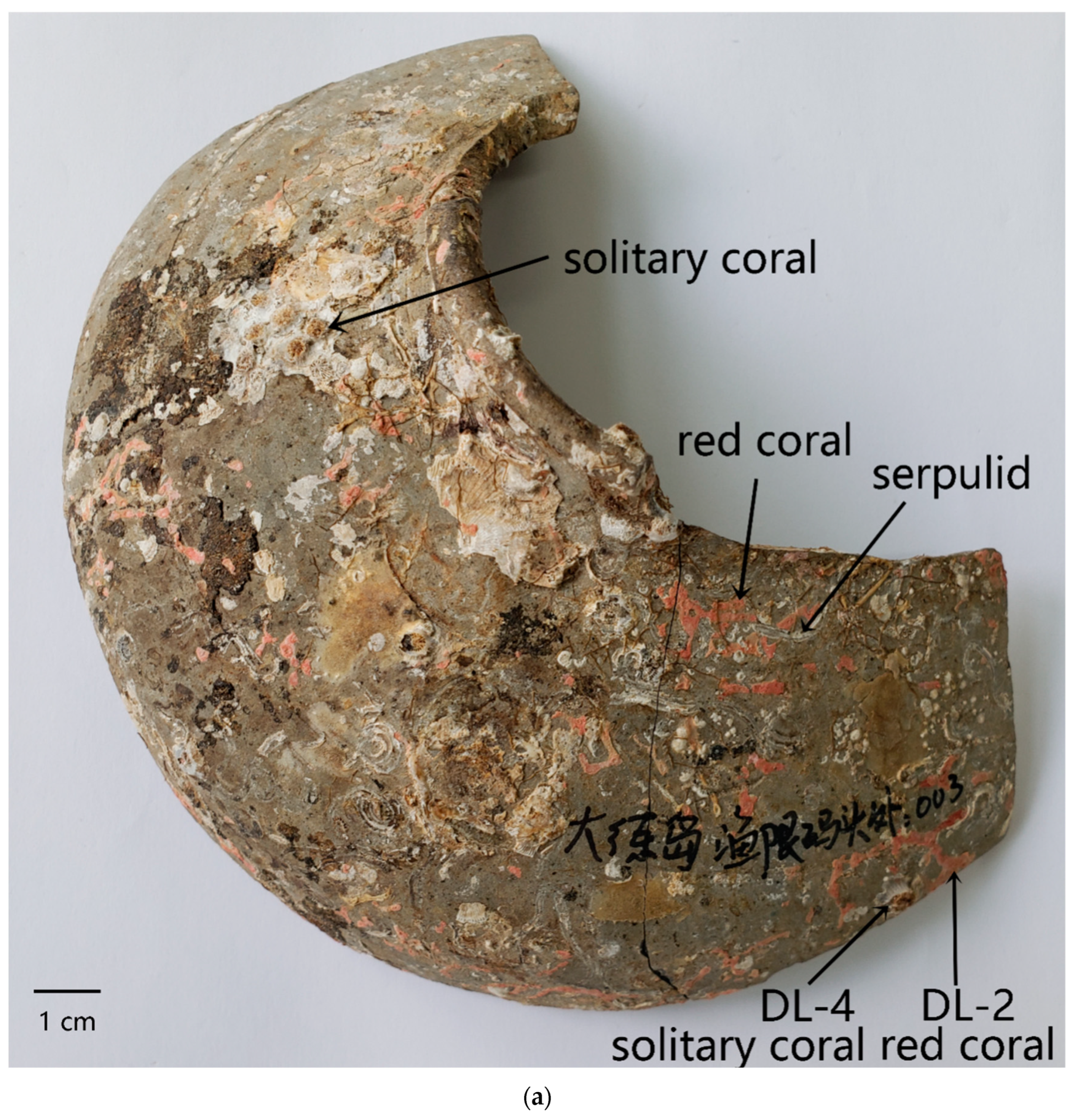
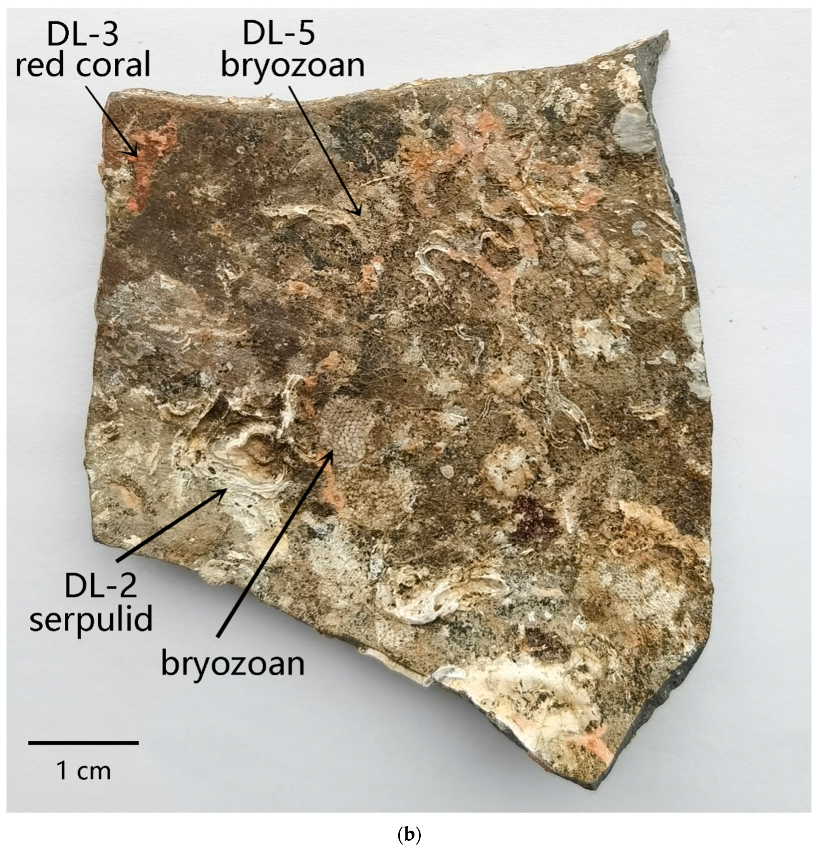
3.1. Morphology of the Attached Marine Organism Remains

The influence of bio-attachment on the appearance of Shard DL was evident, with more than 90% of the surface area covered with stacked marine organism remains. Notably, more organisms were attached to the unglazed inner wall than to the glazed outer wall. Optical microscope (OM) images were taken of marine organism remains with different morphologies. The attached organisms were classified into the following two types: (1) unitary organisms including Serpulids and solitary coral (i.e., self-maintaining units derived from a single zygote [14]); and (2) modular organisms including red coral and Bryozoans (i.e., structural individuals consisting of more than one iterated basic unit [15]). These marine organisms were not only directly attached to the surface of the stoneware but were also found atop marine plant coralline algae, forming biological stacking and attachment.
The solitary coral attached to the outer wall of sample DL-4 was a type of polypoid coral belonging to the class Anthozoa of the phylum Coelenterata. The top diameter of the epitheca, showing white cylinders, was approximately 3 mm, and the covering diameter of the bottom wall was up to 8–10 mm. The septum, with an overall radial symmetry, resembled wheels (Figure 2a). There were gaps of more than 100 microns between the walls, with numerous jagged protuberances on the surface. Some epitheca were intact, with a vertical height of 3–5 mm. Some corals were in the early growth stage, with almost no cylindrical epitheca observed. During the lifetime of Anthozoans, the septum supported the polypoid on the top floor of the skeleton, with sunken soft tissues existing in the interspace between septa.
Numerous Serpulid tubes were observed on the surface of sample DL-2. Serpulids are a type of annelid belonging to the class Polychaeta of the phylum Annelida, characterized by yellow-white hollow tubes. The tubes varied in length (10–20 mm) and thickness (approximately 0.5 mm). As shown in Figure 2b, two parallel ridges were observed on the tube top. Polypides originally lived inside the tubes, and it was not easy to peel off the calcareous tubes left after the death and degradation of the polypides from the surface [16,17].
Both the red coral and solitary coral belonged to the class Anthozoa of the phylum Coelenterata. Some striped or dotted red coral remains were found on both the inner and outer walls of sample DL-2, with varying lengths (from 2 to 20 mm) and shades of red. The surface was loose and porous. As shown in Figure 2c, the remains were dendritic group skeletons formed by biomineralization when a large number of red Anthozoans (polypoids) gathered. The red color of the skeleton was sourced from polyenes when the Anthozoan preyed on plankton [18].
The Bryozoan attached to the inner wall of sample DL-5 belonged to the phylum Bryozoa. They were fixed in a benthic pattern and grew in a covering pattern [19]. Many yellow-white fan-shaped or flaky group skeletons were found to spread on the surface. As shown in Figure 2d, a single Bryozoan was approximately 0.5 mm in length. While the Bryozoans constantly bred new individuals in a sprouting pattern, the formed group skeletons reached a considerably large coverage area. Figure 2d showed individual zooecium structures with a clear boundary. The large circular hole, located on the top, was the orifice of the zooecium that served as the access channel for Bryozoans. The small circular hole was the pore, a fine structure of the Bryozoan skeleton.

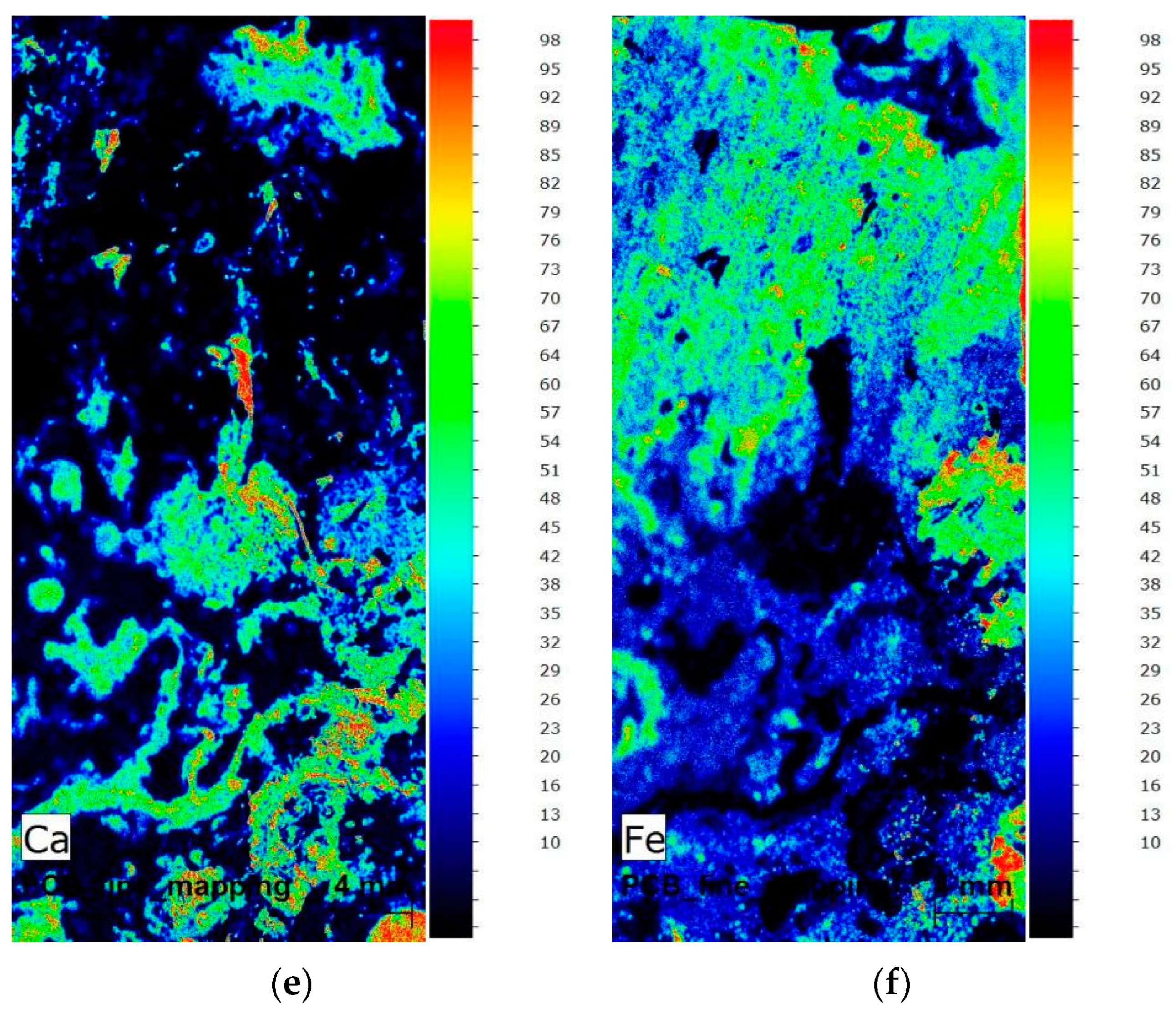
3.2. EDXRF Analysis Results of the Attached Marine Organism Remains

Table 1 lists the energy-dispersive X-ray fluorescence (EDXRF) analysis of various types of marine organism remains. The results showed that the Ca contents in the remains of several organisms were higher than 70 wt%, originating from the calcareous skeletons formed after biomineralization. Among all known biominerals, approximately half are calciferous, highlighting the key functional role of Ca in cellular metabolism. Calcium carbonate is the most abundant biomineral [19]. Comparing the chemical compositions of several types of organism remains, it was found that the Mg contents in red coral and Serpulid remains were higher than those in Bryozoan and solitary coral remains. This might be attributed to different crystal phases of calcium carbonates in organic skeletons and varying substitution degrees of Mg in calcium carbonate lattices, further discussed in Section 3.4. The Bryozoans and tube worms attached to the surface of the samples might have been contaminated by marine sediments such as quartz and feldspar, resulting in higher Al and Si contents. Additionally, 3–4 wt% of Fe in the chemical composition of several marine organism remains, except for solitary coral, might come from contamination by iron rust [13]. The element S was found in several types of organism remains, primarily sourced from two aspects. During the biomineralization process, organisms require highly negatively charged sulfated organic macromolecules to attract and bond bivalent cations such as Ca2⁺ and Mg2⁺. Tanur et al. also found sulfated polysaccharides in the lining of Serpulids [20,21]. Another source is sulfate in the sea or sulfides in other forms. The Desulfovibrio spp. in the seafloor sediments are active, and the sulfate might be reduced to sulfide or exist in other low-valency sulfur forms [2,22]. The elements Na and Cl, as two major elements in seawater, except H and O, were slightly detected in several types of organism remains.
Figure 3 displays the element mapping images of an area on the inner wall of DL with a significant amount of organism remains; Table 2 lists the chemical composition analysis results. As shown in Figure 3c,d, elements Al and Si are mainly concentrated in the top left, where fewer foreign pollutants were found, and the body is partly exposed. The chemical composition at position P1 also revealed high contents of Si and Al in the body. The Fe content was also as high as 15 wt%, suggesting the deposition of foreign pollutants with rusts. Figure 3e shows the element distribution in the top right and lower part where red corals were attached, indicating a high concentration of Ca. According to the chemical compositions at positions P2, P5, and P7, the Ca content was the highest (95 wt%). This indicated that the marine organism remains attached to the inner wall of DL were calcareous. The dark coagulum in the selected area was mainly distributed in the middle part. The element mapping images demonstrated the enrichment of Al, Si, and Fe (Figure 3c,d,f). The chemical composition at positions P3 and P4 suggested that the Fe and Si contents in the dark coagula area were 35–47 wt% and 21–31 wt%, respectively. This could be attributed to the deposition of foreign rusts, sea sand, and sea mud.
In addition, the P, S, and Cl contents at positions P5 and P6 were obviously higher than those at other positions. The S and Cl were presumed to have originated from sulfate and chloride ions in seawater, while P may have originated from phosphate minerals in marine sediments, which were preserved in the form of inorganic phosphorus in foreign pollutants on the surface of Shard DL [23].

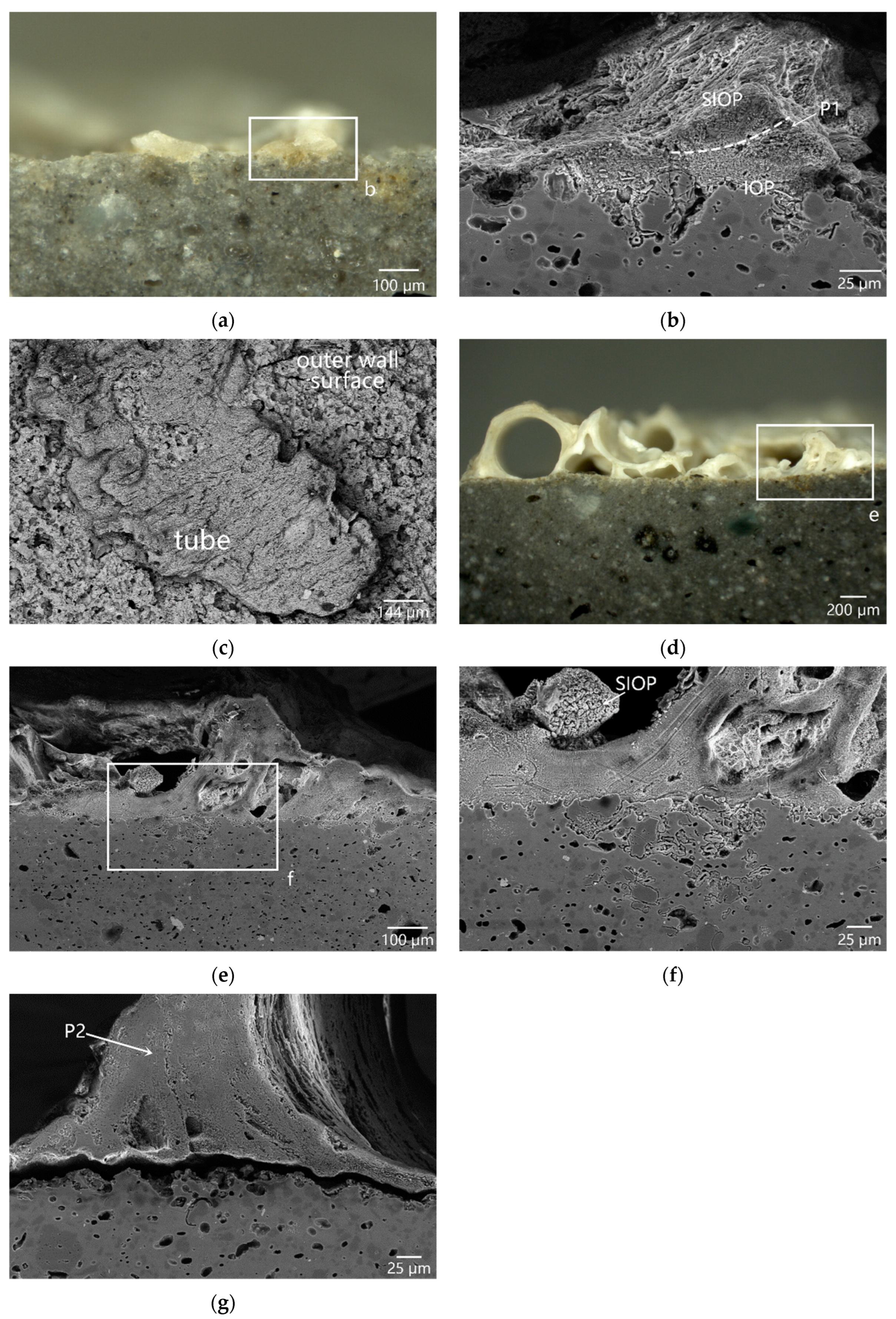
3.3. Microstructure and Micro-Area Composition of the Attached Marine Organism Remains

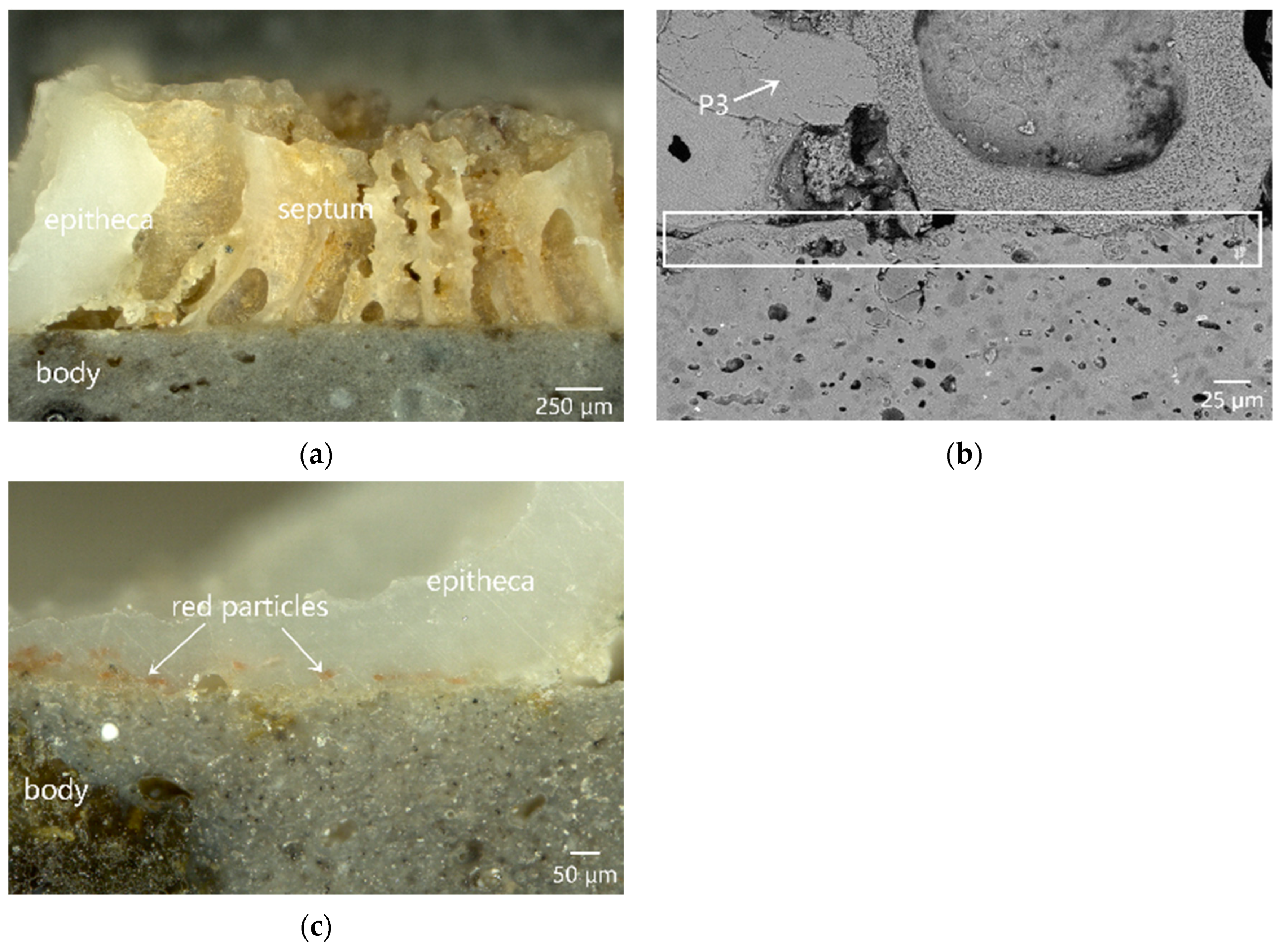
3.3.1. Unitary Organism: Serpulid and Solitary Coral

During the attachment process of the Serpulids, larvae first secreted biogum to attach themselves to the matrix surface and then completed metamorphosis. Serpulid tube formation involved the secretion of calcareous granules and organic components from the calcium-secreting glands [24]. Although the life cycle of the Serpulid was only 1–2 years, the remains of the tube could still be preserved after the death and degradation of the worm (Figure 4a). Figure 4b shows the SEM images of the region in the box of Figure 4a. A few tubes attached to the outer wall of sample DL-2, and the bottom of the tube penetrated the surface cracks and bonded tightly with the surface, without an interspace. The fibers constituting Serpulid tubes were secreted in the form of liquid and then gradually permeated into micron-level cracks and holes on the surface. Finally, the tubes hardened and formed hard calcareous skeletons in the presence of seawater and organics to achieve permanent attachment [25]. The residual tube wall on the outer wall of sample DL-2 was composed of two layers (see both sides of the dashed line in Figure 4b). Each layer had a specific crystal ultrastructure [26], in which the spherulitic irregularly oriented prismatic (SIOP) structures were on the upper side and the irregularly oriented prismatic (IOP) structures were on the lower side. The formation of these complex ultrastructures in Serpulid tube skeletons was tightly correlated with the organics involved in the bio-mineralization process. The organic lining on the inner walls of the Serpulid tubes provided the template for inorganic skeletons and mediated the calcification, which could control both the morphology and the polymorphism of crystals to a certain degree [20]. Figure 4c shows that a significant number of pores and cracks were distributed on the outer wall where the normal glaze was no longer in existence, which greatly attracted the larvae of marine fouling organisms to settle down.
Figure 4d–g shows the residual Serpulid tubes on the inner wall of sample DL-2. The unglazed inner wall, with great roughness, attracted many larva clusters. Therefore, many accumulated tubes were left, as shown in Figure 4d. The SEM images clearly show that the bottom wall of the Serpulid was completely fitted with the body surface, without an interspace (Figure 4e). The SIOP structure in Figure 4f can be clearly observed. As shown in Figure 4g, the tube bottom and sample surface show good agreement on the contour, indicating that the 20 µm gap between the tube bottom and sample surface was caused by external mechanical actions. According to the EDS analysis, the calcareous perch tube contained about 24 wt% of Mg, which indicated that it was calcite calcium carbonate with a large amount of Mg in the crystal lattice (see the EDS data at positions P1 and P2 in Table 3).
Figure 5a shows the cross-section of sample DL-4, where a yellow-white solitary coral is attached to the outer wall, with no remaining glaze (see Figure 5b). The bottom of the solitary coral fits perfectly into the pits on the exposed surface of the body, creating a fixation interface akin to a key and lock (refer to the boxed region in Figure 5b), resulting in a compact structure. Compared to the Serpulids, the calcareous skeleton of the coral contains almost no magnesium (see P3 in Table 3). The coral skeleton is composed of calcium carbonate in the aragonite phase. Red substances were also observed at the interface between the outer wall of the coral and the body, which were presumed to be red coral debris. It is speculated that the outer wall was abraded by sea wave-borne debris, causing the glaze to peel off before the coral’s attachment. Consequently, the coral larva selected the rough surface as a foothold and began to grow, establishing a firm attachment to the rough surface.

3.3.2. Modular Organisms: Bryozoans and Red Coral

As illustrated in Figure 6a, the skeletons of the Bryozoans attached to the outer wall of the sample were predominantly white, indicating that they were uncontaminated. The morphology of the Bryozoans suggested they belonged to the Schizoporellidae family (see Figure 6b,c). Each zooecium was approximately 400 µm in length and 150 µm in width, featuring a slim-lined structure. Numerous spines were observed on the top wall of the zooecium. An orifice at one end of the zooecium likely served as an entry point for living Bryozoans (Figure 6b). The cross-sectional view revealed that the zooecium was roughly circular, with a diameter of 75 µm (Figure 6c). Many zooecia were stacked to form a three-dimensional skeleton. The cross-section showed that both the bottom and interior walls of the Bryozoan were 25 µm thick. An interspace was visible at the bonding interface between the bottom wall and the sample surface.
The Bryozoans attached to the inner wall appear to be yellow white (Figure 6d), likely due to foreign pollutants deposited on the inner wall of the sample. Figure 6e shows that the aperture of the Bryozoan zooecium is 70 µm in diameter; a calcareous ring structure can be observed on the fringe (referred to as Gymnolaemata in biology). Figure 6f reveals the presence of perioral spines on the peristome. The maximum diameter of the zooecium is up to 300 µm, with the bottom wall thickness approximately 15 µm and the thickness of the interior wall similar to that of the frontal wall (about 50 µm). The Bryozoans are more tightly bound to the unglazed surface on the inner wall compared to the outer wall. However, noticeable interspaces remain at the bonding surface (Figure 6f).
Three structural layers were observed from the top to the bottom wall: the cuticle, the mineralized layer, and the epithelia lining the body cavity. The cuticle was the outermost layer, interfacing between the Bryozoans and the external environment. The epithelia were the innermost layer, in contact with the polypides. The epithelia were distributed on both sides of the interior wall, lacking a cuticle. During the attachment process, undifferentiated epithelial cells on the bottom wall first secreted a cuticle; the adhesive for attachment was a simple acid mucopolysaccharide secretion that did not etch the sample surface [27] As epithelial cells differentiated, ionized calcium accumulated within the cells and was gradually released outside. Calcium salts were then deposited in the framework of the cuticle to form mineralized skeletons. Throughout this process, the outermost cuticle became at least partially mineralized, aiding in the fusion between the Bryozoans and the sample surface [28]. EDS analysis confirmed that both Bryozoans and other organisms had calcareous skeletons composed of aragonite calcium carbonate, with low magnesium content (see positions P4 and P5 in Table 3).
The red coral was not firmly attached to the surface of sample DL. During the experimental process, some red coral skeletons entirely detached. Figure 7a shows that no residual glaze layer is present on the outer wall of sample DL-2. However, scanning electron microscopy (SEM) revealed that the surface of the outer wall beneath the red coral skeletons was densely pitted. This condition might be related to the shedding of anorthite crystal clusters from the interlayer between the glaze and the body. Unlike the bottom wall of the solitary coral, where the pits were completely filled by the coral skeletons, the red coral skeletons retained their original structure, with noticeable interspaces between the skeletons and the sample surface. The mode of attachment of red coral to the stoneware surface might be associated with the bio-mineralization process of red coral. Energy-dispersive spectroscopy (EDS) analysis further revealed that the magnesium (Mg) content in the red coral skeletons was significantly higher compared to that of other marine animal remains, as the skeletons formed under bio-mineralization were composed of calcite (see position P6 in Table 3).
The red coral skeletons in Figure 7a exhibit a three-dimensional interconnected structure with mesh diameters ranging from 15 to 35 µm (Figure 7c). Protrusions are visible on the red coral skeletons. Figure 7d shows that textures formed during bio-mineralization are clearly observable on the protruded sections. These textures resulted from the stacking of columnar calcite crystals in a helical pattern around a central axis. The red coral skeletons are composites of organic and inorganic materials, with organic matter distributed among calcite particles, forming a multi-level stockwork. The arrangement of the crystals followed specific patterns regulated by organic matter [29].

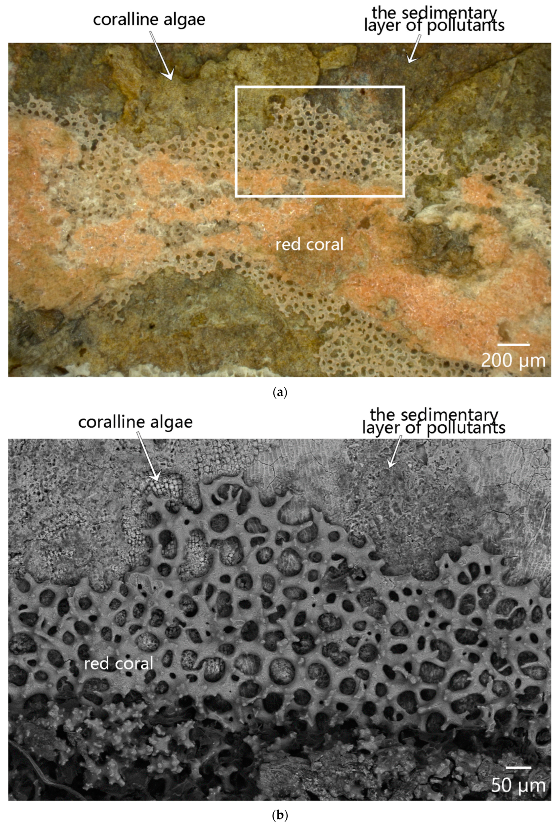
3.3.3. Biological Overlapping Phenomenon between Coralline Algae and Red Coral

The crusted surface of the coralline algae provided an ideal substrate for the attachment of red coral larvae, resulting in a biological overlapping phenomenon between the two organisms, as depicted in Figure 8. Figure 8a illustrates the distribution of pink-white, red coral skeletons, yellow coralline algae skeletons, and brick-red sediments from top to bottom. Figure 8b provides a close-up of the boxed region in Figure 8a. The group skeletons of red coral reached a thickness of up to 100 μm and exhibited noticeable interspaces with the coralline algae. The coralline algae skeletons were approximately 50 μm thick, with their lower edges tightly bound to the body surface of sample DL-3. Foreign pollutants were deposited within the pore channels of the coralline algae skeletons and the surface layer cracks of the body. Based on the contrast in the scanning electron microscopy (SEM) images and energy-dispersive spectroscopy (EDS) data (see positions P7 and P8 in Table 3), these foreign pollutants were primarily iron rusts, which were deposited without reacting chemically with the body, resulting in the brick-red region shown in Figure 8a.
The cross-sectional element mapping images, as shown in Figure 9, reveal a distinct layered structure. Specifically, the uppermost layer of red coral group skeletons was predominantly composed of calcium (Ca), magnesium (Mg), and oxygen (O), indicative of calcite skeletons formed through bio-mineralization. The foreign pollutants filling the cracks in the coralline algae skeletons and the body mainly consisted of iron (Fe), magnesium (Mg), and silicon (Si), which may also include iron rust, sea sand, sea mud, and sediments derived from Mg2⁺ in seawater.
The surface observation reveals a biological overlap between the red coral and coralline algae. Figure 10a shows that the inner wall of sample DL-3 is entirely covered with foreign pollutants and remains of organisms. Figure 10b highlights the remains of coralline algae skeletons on the surface of the pollutant sedimentary layer. It is speculated that coralline algae initially attached to the sedimentary layer of pollutants, continuously grew, and mineralized, thus covering a significant portion of the sediment. Subsequently, red coral accumulated and adhered to the crusted surface of the coralline algae, growing and forming its skeletons. Pollutants transported by seawater were further deposited on the coralline algae skeletons, filling the pore channels. External mechanical damage led to partial fragmentation and detachment of the coralline algae group skeletons, exposing the underlying pollutant sedimentary layer.

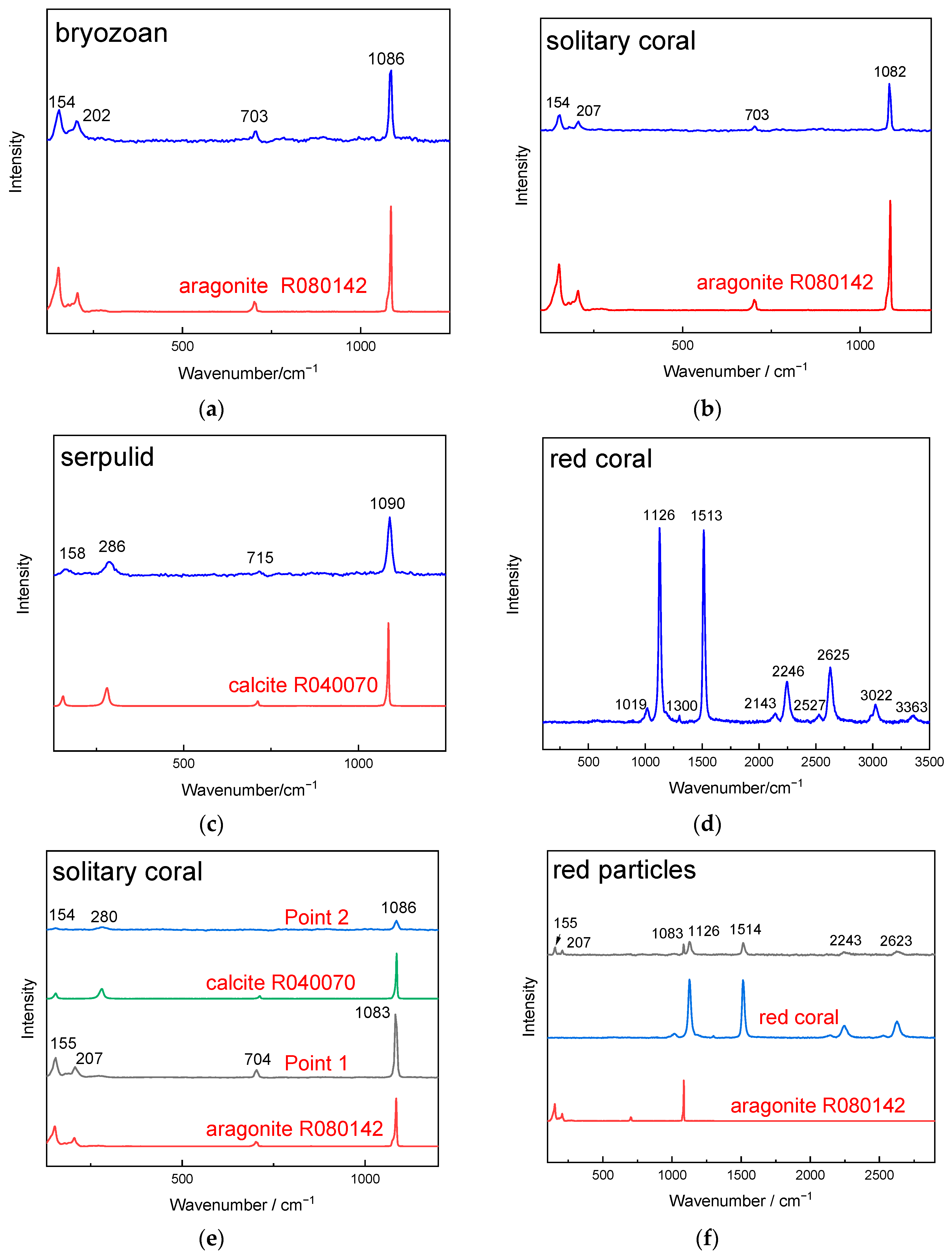
3.4. μ-Raman Analysis Results of Attached Marine Organism Remains

Figure 11 presents the μ-Raman analysis results of various marine organism remains. Phase analysis indicated that the Bryozoan and solitary coral remains were predominantly composed of aragonite, with characteristic peaks at 154, 202, and 1086 cm−1 (Figure 11a,b). The Serpulid remains were primarily composed of calcite, showing peaks at 154, 280, and 1086 cm−1 (Figure 11c). The red coral remains exhibited strong peaks for C-C and C=C bonds at 1126 and 1513 cm−1, respectively, indicating the presence of organic olefin pigments [18,30]. The calcite peak at 1086 cm−1 merged with the strong organic pigment peak at 1126 cm−1.
μ-Raman analysis of the cross-section of solitary coral showed the presence of calcite (Figure 11e). This is likely due to the incorporation of Mg2⁺ from seawater into the calcite lattice, potentially caused by a phase transformation from aragonite to calcite [31]. μ-Raman analysis of the red substances shown in Figure 5c revealed peaks for both aragonite and organic pigments in the red coral (Figure 11f), confirming that the red coral adhered to the outer wall before the solitary coral and was subsequently covered by the solitary coral, becoming encapsulated within its epitheca.

4. Discussion

4.1. Effect of Surface Roughness on the Attachment of Marine Organisms

The biofouling process typically involves four stages, as follows: conditioning film formation; microbial biofilm development; protozoan attachment; and marine organism settlement [32]. Despite changes in the surface properties due to biofilm formation, macrofouler larvae often directly penetrate the conditioning and microbial biofilms to contact the substrate [33,34]. Thus, the increase in marine organism attachment on DL can ultimately be attributed to changes in the surface properties.
Sample DL was originally glazed only on its outer wall, making it flat and smooth, while the unglazed inner wall was rough. Previous research [13] indicated that DL experienced prolonged mechanical abrasion from sea wave-borne debris in an isolated, dynamic marine environment, leading to its degradation. Consequently, the surface of DL became increasingly rough. The glaze on DL, a calcium glaze belonging to alumina silicate glass with unfused quartz particles and precipitated anorthite crystals [13], underwent two primary corrosion processes: ion exchange and hydrolysis. When the pH was <9, Cl or hydronium ions in the water exchanged with alkali metal ions in the glass, forming a de-alkalinized hydrated layer on the glass surface [35]. When the pH was >9, OH ions disrupted Si–O–Si bonds in the silicate network, leading to gradual dissolution [36]. The seawater in Fujian, China is a weakly alkaline environment, with a pH of about 8.2. However, the low K and Na content and high Ca content in the DL glaze contributed to the stability of the Si–O–Si network, making ion exchange slow [10]. Nevertheless, the high pH in the localized microenvironment of iron rusts and marine organism remains accelerated the hydrolysis of the Si–O–Si network [13].
Only trace amounts of S and Cl were detected on the inner wall surface of DL, originating from sulfate and chloride salts in the marine environment. These factors are significant for concrete corrosion. Sulfate reacts with Ca2⁺ in concrete to produce ettringite, gypsum, and calcium silicate, leading to expansive corrosion and structural damage [37,38]. Chloride ions can destroy passive layers on rebar surfaces, accelerating electrochemical corrosion [39]. The low Ca content in DL was insufficient to react with the sulfate, and the sintered body was not affected by chlorate.
Marine fouling organisms typically adhere using wet adhesion [40]. They synthesize and secrete a viscous adhesive that wets and disperses on the substrate surface, forming a firm attachment through chemical bonding, electrostatic interactions, mechanical interlocking, and spreading [41]. The increased surface roughness of DL, resulting in numerous micron-level pores and cracks, raised its specific surface area and wettability. The adhesive filled defects or permeated into the sample surface, forming a strong bond. When marine organisms completed their life cycles or were displaced by seawater, significant detachment of the biofouling occurred. The adhesive layer, however, often took away some of the weak glaze, further roughening the surface [11]. This cyclic process led to continuous biological colonization, increasing the biomass. Coralline algae contributed to this increase by providing a preferred matrix for larval attachment. The polysaccharide sulfides secreted by coralline algae induced larval attachment and growth [31,42,43].
The difference in marine organism attachment between the inner and outer walls is related to their surface properties. The unglazed inner wall, being rougher, more readily accommodated macrofouler larvae compared to the glazed outer wall. Marine fouling organisms more effectively adhered to the rough surface of the inner wall, which directly faced the marine environment, accelerating biofouling formation. Consequently, the inner wall accumulated more marine organism remains than the outer wall.

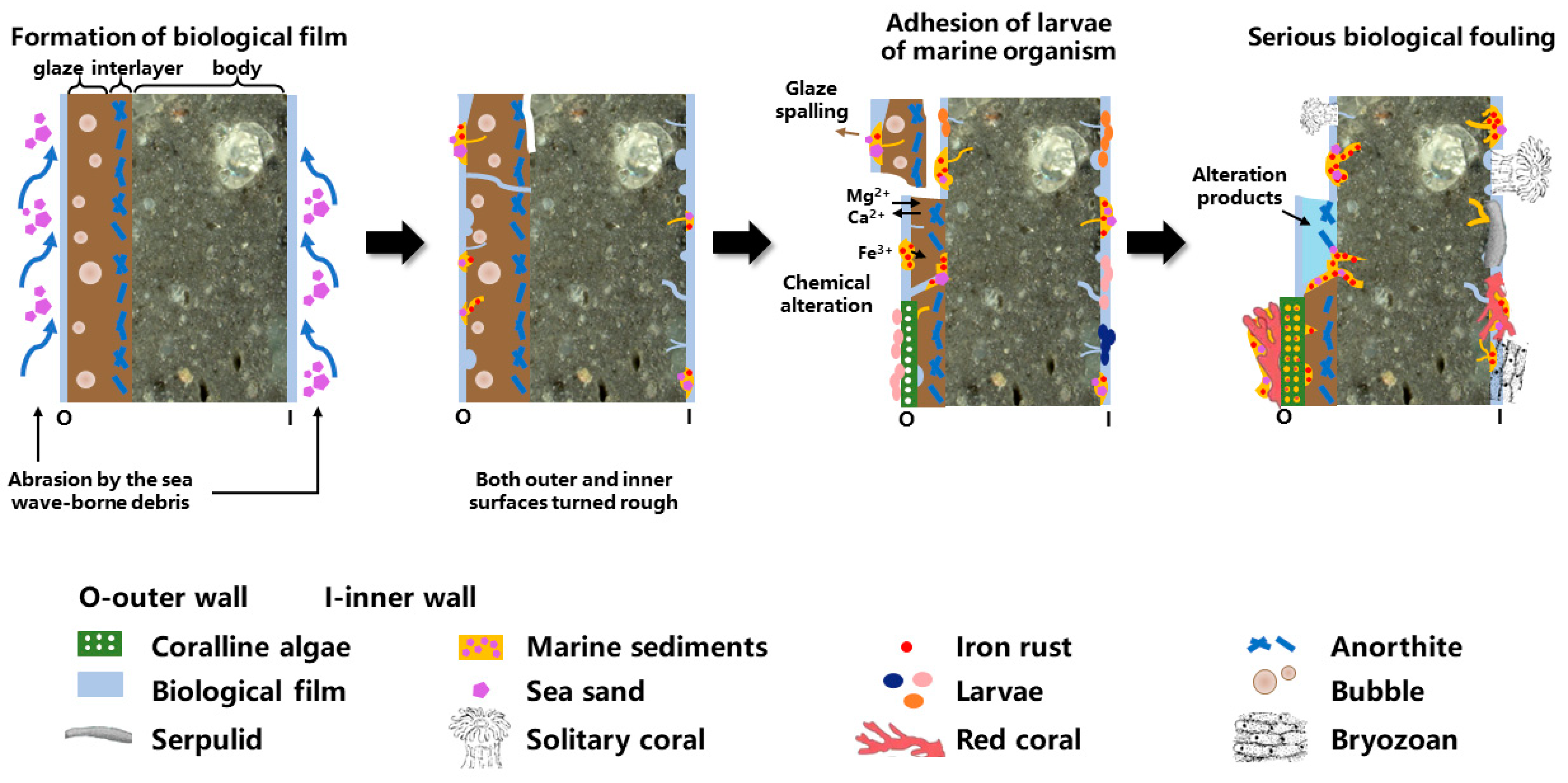
4.2. Effect of Bio-Attachment on the Degradation of Shard DL

The attachment of marine organisms is a significant factor influencing the degradation of ceramics salvaged from water. The marine organisms attached to the surface of DL mainly included unitary organisms (Serpulids and solitary coral) and modular organisms (coralline algae, red coral, and Bryozoans). The unitary organisms were firmly bound to the defective surface of the sample, likely due to the bio-calcification and bio-mineralization processes. When the Serpulids built their attached tubes, the larvae would push the tube material away from their bodies, thus ensuring maximum attachment to the sample surface [44,45]. The epidermal cells of the coral larval ectoderm first attached to the sample surface, which served as the bio-mineralization site for secreting calcareous skeletons. The formation of skeletons began with the generation of the bottom; the firm bond between the bottom and the pits on the matrix surface facilitates the solid, long-term attachment of the single coral [46]. After the bottom walls of unitary organisms and the defective surface formed a mechanical lock–key interlocking structure, the hard calcareous skeletons firmly attached to the surface could alleviate the abrasion caused by sea wave-borne debris and the impact of water flow fluctuations on the local surface of the sample [47]. Unitary organisms’ compact skeletons could seal open pores and cracks, reducing the deposition of insoluble pollutants and decreasing the permeability of soluble salts like chlorates and sulfates [48,49]. If mechanical methods alone, such as scalpels, were used to remove these organisms, the underlying surface could suffer irreversible damage. Hence, it is recommended to first soak the sample in dilute acids, such as oxalic acid or EDTA disodium salt solutions, to weaken the bond before mechanical removal [5].
Modular organisms’ group skeletons covered extensive areas and reinforced surfaces damaged by mechanical action, thereby withstanding ongoing scouring by sea debris. However, interspace suggests weaker bonding between modular organisms and the stoneware surface compared to unitary organisms, due to the variability in spatial growth among individuals and genetic variations affecting the group skeleton arrangement [15]. The open cellular structure of coralline algae and the net-like structure of red coral did not effectively block inorganic pollutants, which could thus infiltrate and deposit in the stoneware body and pore channels, possibly contributing to chemical alterations of the glaze [13]. The remains of modular organisms could generally be removed by simple mechanical methods.
Previous studies have shown that the adhesives and acidic secretions of macrofoulers can erode various materials, including concrete [38,50], metals [34,51], and stone relics [12,43]. Acidic carboxyls generated during metabolism can complex with metal ions, such as Fe, Ti, and Ca, causing bio-erosion phenomena such as pitting and micro-boring. However, no significant bio-erosion was observed on DL, which could be attributed to the material properties of the shard itself.
Figure 12 illustrates the degradation process of Shard DL. Initially, the outer wall of DL was glazed and smooth, while the unglazed inner wall was rough with defects. In the marine environment, a biological film less than 1000 nm thick formed on the surface. Mechanical abrasion by sea debris roughened the outer wall glaze, creating pores and cracks. Coralline algae attached to the roughened surface, forming mineralized skeletons and secreting substances that attracted macrofouler larvae such as red coral larvae. As a result, the remaining glaze fell off, exposing the body. The unglazed inner wall attracted macrofouler larvae earlier, leading to their firm attachment and growth. Foreign pollutants deposited on both the inner and outer wall surfaces, filling the skeletons of red coral and coralline algae. The biomass level of the inner wall increased significantly, resulting in near-complete coverage with marine fouling organisms. Pollutants in the defects associated with solitary corals and Serpulids did not increase further, whereas pollutants in areas associated with red coral and Bryozoans continued to accumulate.
For salvaged ceramic wares subjected to mechanical damage, chemical alteration, and biological fouling, protection measures should be implemented. Most marine organism remains are calcareous; thus, salvaged ceramics should first be soaked in a mild dilute acid solution to weaken the binding force between the remains and the surface. After rinsing with clean water, softened remains can be removed with tools such as brushes and scalpels. Iron pollutants on the ceramic surface can be removed using localized applications of oxalic acid or EDTA, followed by rinsing and mechanical removal. Additional protection measures, such as desalination and reinforcement, should be considered after removing the coagula [5,33]. The decision to remove attached marine organism remains should be case-specific, given that chemical treatments may cause irreversible damage to the ceramics.

5. Conclusions

The surface of the sauce-glazed stoneware DL was covered by various marine organisms, including marine plant coralline algae, unitary organisms, such as Serpulids and solitary coral, and modular organisms such as Bryozoans and red coral. These marine organism remains were predominantly calcareous, with calcite and aragonite as the main phases.
The surface roughness of DL increased significantly after prolonged abrasion from sea wave-borne debris, creating a favorable condition for marine organism attachment. The unglazed inner wall exhibited higher roughness compared to the outer wall, resulting in greater biological colonization on the inner wall. The inducer secreted by coralline algae covered a substantial area of the DL surface, attracting larvae and further enhancing biological colonization.
The effects of different types of marine organisms on the degradation of DL varied. Unitary organisms formed a mechanical interlock with the surface during attachment, mitigating abrasion from sea wave-borne debris, sealing open pores and cracks, and blocking the intrusion of foreign pollutants. This process provided a ‘bio-protective’ effect on the local surface. In contrast, the group skeletons of modular organisms covered a larger area, reinforcing the mechanically damaged surface. However, the spatial arrangement of modular organisms was less predictable, and their bonding with the stoneware surface was weaker. Interspaces between the calcareous skeletons and the stoneware surface allowed pollutants to intrude, leading to chemical alterations of the glaze.
For the effective removal of the attached skeletons of unitary organisms, the sample should be initially soaked in dilute acid followed by mechanical removal. The remains of attached modular organisms can be directly removed using mechanical methods. Subsequent conservation efforts for the salvaged ceramic wares should implement targeted protection measures based on the specific types of marine organisms attached to the surface.
This study contributes towards an understanding of what ancient ceramic wares experience during prolonged exposure to the marine environment and can provide a scientific basis for further targeted conservation work.

Author Contributions

Conceptualization, W.L.; Methodology, W.L., C.X. and X.L.; Investigation, R.D.; Resources, Z.Y.; Writing—original draft, R.D.; Writing—review & editing, W.L. All authors have read and agreed to the published version of the manuscript.

Funding

This work was supported by National Key R&D Program of China [2023YFF0906401].

Institutional Review Board Statement

Not applicable.

Informed Consent Statement

Not applicable.

Data Availability Statement

The original contributions presented in the study are included in the article, further inquiries can be directed to the corresponding author.

Conflicts of Interest

The authors declare no conflict of interest.

References

  1. Dromgoole, S. UNESCO Convention on the Protection of the Underwater Cultural Heritage. Int. J. Mar. Coast. Law 2001, 18, 59–108. [Google Scholar]
  2. Pearson, C. Conservation of Marine Archaeological Objects; Butterworth & Co. Ltd.: London, UK, 1987; pp. 16–22, 107–109, 261–264. [Google Scholar]
  3. Buys, S.; Oakley, V. Conservation and Restoration of Ceramics; Routledge: New York, NY, USA, 2014. [Google Scholar]
  4. Luka, B.; Martina, C.; Anita, J.; Antonija, J.; Ivo, M.; Mladen, M.; Tanja, P.; Mladen, P. Conservation of Underwater Archaeological Finds Manual; Luka, B., Ed.; International Centre for Underwater Archaeology in Zadar: Zadar, Croatia, 2014. [Google Scholar]
  5. Li, N.S. Research on the Protection of Porcelain Excavated from the Ocean; Science Press: Beijing, China, 2016. [Google Scholar]
  6. Wang, Y.R.; Zhu, T.Q.; Yang, G.C.; Tan, X.; Ye, D.; Chen, H. The method to soften the concretions of ceramics in the “Nanhai I” Shipwreck of China Southern Song Dynasty (1127–1279AD). Herit. Sci. 2018, 6, 4. [Google Scholar] [CrossRef]
  7. Carmona, N.; García-Heras, M.; Gil, C.; Villegas, M.A. Chemical degradation of glasses under simulated marine medium. Mater. Chem. Phys. 2005, 94, 92–102. [Google Scholar] [CrossRef]
  8. Tournie, A.; Ricciardi, P.; Colomban, P. Glass corrosion mechanisms: A multiscale analysis. Solid State Ion. 2008, 179, 2142–2154. [Google Scholar] [CrossRef]
  9. Palomar, T.; Llorente, I. Decay processes of silicate glasses in river and marine aquatic environments. J. Non-Cryst. Solids 2016, 449, 20–28. [Google Scholar] [CrossRef]
  10. Palomar, T. Characterization of the alteration processes of historical glasses on the seabed. Mater. Chem. Phys. 2018, 214, 391–401. [Google Scholar] [CrossRef]
  11. Elkin, D.; Argüeso, A.; Grosso, M.; Murray, C.; Vainstub, D.; Bastida, R.; Dellino-Musgrave, V. Archaeological research on HMS Swift: A British Sloop-of-War lost off Patagonia, Southern Argentina, in 1770. Int. J. Naut. Archaeol. 2007, 36, 32–58. [Google Scholar] [CrossRef]
  12. Aloise, P.; Ricca, M.; Russa, M.F.; Ruffolo, S.A.; Belfiore, C.M.; Padeletti, G.; Crisci, G.M. Diagnostic analysis of stone materials from underwater excavations: The case study of the Roman archaeological site of Baia (Naples, Italy). Appl. Phys. A 2013, 114, 655–662. [Google Scholar] [CrossRef]
  13. Ding, R.; Li, W.D.; Yang, Z.L.; Xu, C.; Lu, X. Degradation Mechanism of a Sauce-GlazedWare of the Song Dynasty Salvaged out of theWater at Dalian Island Wharf: Part I—The Effect of the Surface-Attached Composite Coagula. Materials 2023, 16, 1176. [Google Scholar] [CrossRef]
  14. Vuorisalo, T.; Tuomi, J. Unitary and Modular Organisms: Criteria for Ecological Division. Oikos 1986, 47, 382–385. [Google Scholar] [CrossRef]
  15. Pineda-Krch, M.; Poore, A.G. Spatial interactions within modular organisms: Genetic heterogeneity and organism fitness. Theor. Popul. Biol. 2004, 66, 25–36. [Google Scholar] [CrossRef] [PubMed]
  16. Bastida-Zavala, J.R. Serpulids (Annelida: Polychaeta) from the Eastern Pacific, including a brief mention of Hawaiian serpulids. Zootaxa 2008, 1722, 1–61. [Google Scholar] [CrossRef]
  17. Ten Hove, H.A.; Kupriyanova, E.K. Taxonomy of Serpulidae (Annelida, Polychaeta): The state of affairs. Zootaxa 2009, 2036, 1–126. [Google Scholar]
  18. Yang, Y.; Guo, Y.; Zhang, Y.; Zou, Y.; Wei, J.; Liang, L. Contribution of coral composition to color red in the uniform color space CIE 1976L*a*b*. Desalin. Water Treat. 2019, 168, 384–393. [Google Scholar] [CrossRef]
  19. Taylor, P.D.; Kudryavtsev, A.B.; Schopf, J.W. Calcite and aragonite distributions in the skeletons of bimineralic bryozoans as revealed by Raman spectroscopy. Invertebr. Biol. 2008, 127, 87–97. [Google Scholar] [CrossRef]
  20. Addadi, L.; Berman, A.; Oldak, J.M.; Weiner, S. Structural and stereochemical relations between acidic macromolecules of organic matrices and crystals. Connect. Tissue Res. 1989, 21, 127–135. [Google Scholar] [CrossRef]
  21. Tanur, A.E.; Gunari, N.; Sullan, R.M.; Kavanagh, C.J.; Walker, G.C. Insights into the composition, morphology, and formation of the calcareous shell of the serpulid Hydroides dianthus. J. Struct. Biol. 2010, 169, 145–160. [Google Scholar] [CrossRef]
  22. Rodgers, B.A. The Archaeologist’s Manual For Conservation; Kluwer Academic/Plenum Publishers: New York, NY, USA, 2004. [Google Scholar]
  23. Feng, S.; Li, F.; Li, S. Introduction to Marine Science; Higher Education Press: Beijing, China, 1999. [Google Scholar]
  24. Huggett, M.J.; Carpizo-Ituarte, E.J.; Nedved, B.T.; Hadfield, M.G. Formation and Function of the Primary Tube During Settlement and Metamorphosis of the Marine Polychaete Hydroides elegans (Haswell, 1883) (Serpulidae). Biol. Bull. 2021, 240, 82–94. [Google Scholar] [CrossRef]
  25. Vinn, O.; Zatoń, M.; Tovar-Hernández, M.A. Tube microstructure and formation in some feather duster worms (Polychaeta, Sabellidae). Mar. Biol. 2018, 165, 98. [Google Scholar] [CrossRef]
  26. Vinn, O.; Ten Hove, H.A.; Mutvei, H.; KirsimÄE, K. Ultrastructure and mineral composition of serpulid tubes (Polychaeta, Annelida). Zool. J. Linn. Soc. 2008, 154, 633–650. [Google Scholar] [CrossRef]
  27. Bromley, R.G.; Heinberg, C. Attachment strategies of organisms on hard substrates: A palaeontological view. Palaeogeogr. Palaeoclimatol. Palaeoecol. 2006, 232, 429–453. [Google Scholar] [CrossRef]
  28. Taylor, P.D.; Lombardi, C.; Cocito, S. Biomineralization in bryozoans: Present, past and future. Biol. Rev. Camb. Philos. Soc. 2015, 90, 1118–1150. [Google Scholar] [CrossRef] [PubMed]
  29. Fan, L.W.; Yang, M.X.; Wang, F.Z. Observations of Surface Microstructures of Corallium Elatius by SEM. In Proceedings of the 2009 China Gems & Jewelry Academic Conference, Beijing, China, 12–16 May 2009; pp. 115–117. [Google Scholar]
  30. Bergamonti, L.; Bersani, D.; Csermely, D.; Lottici, P.P. The Nature of the Pigments in Corals and Pearls: A Contribution from Raman Spectroscopy. Spectrosc. Lett. 2011, 44, 453–458. [Google Scholar] [CrossRef]
  31. Wray, J.L. Calcareous Algae; Elsevier Scientific Pub.: Rio de Janeiro, Brazil, 1998. [Google Scholar]
  32. Yebra, D.M.; Kiil, S.; Dam-Johansen, K. Antifouling technology—Past, present and future steps towards efficient and environmentally friendly antifouling coatings. Prog. Org. Coat. 2004, 50, 75–104. [Google Scholar] [CrossRef]
  33. Abarzua, S.; Jakubowski, S. Biotechnological investigation for the prevention of biofouling. I. Biological and biochemical principles for the prevention of biofouling. Mar. Ecol. Prog. Ser. 1995, 123, 301–312. [Google Scholar] [CrossRef]
  34. Raman, S.; Kumar, R. Interfacial morphology and nanomechanics of cement of the barnacle, Amphibalanus reticulatus on metallic and non-metallic substrata. Biofouling 2011, 27, 569–577. [Google Scholar] [CrossRef]
  35. Conradt, R. Chemical Durability of Oxide Glasses in Aqueous Solutions: A Review. J. Am. Ceram. Soc. 2008, 91, 728–735. [Google Scholar] [CrossRef]
  36. Lombardo, T.; Gentaz, L.; Verney-Carron, A.; Chabas, A.; Loisel, C.; Neff, D.; Leroy, E. Characterisation of complex alteration layers in medieval glasses. Corros. Sci. 2013, 72, 10–19. [Google Scholar] [CrossRef]
  37. Gowers, K.R.; Millard, S.G. On-site linear polarization resistance mapping of reinforced concrete structures. Corros. Sci. 1993, 35, 1593–1600. [Google Scholar] [CrossRef]
  38. Rodríguez, P.; Ramírez, E.; González, J.A. Methods for studying corrosion in reinforced concrete. Mag. Concr. Res. 1994, 46, 81–90. [Google Scholar] [CrossRef]
  39. Li, Q.; Ye, X. Surface deterioration analysis for probabilistic durability design of RC structures in marine environment. Struct. Saf. 2018, 75, 13–23. [Google Scholar] [CrossRef]
  40. Gorb, S.N. Biological attachment devices: Exploring nature’s diversity for biomimetics. Philos. Trans. R. Soc. A 2008, 366, 1557–1574. [Google Scholar] [CrossRef] [PubMed]
  41. Brady, R.F. A fracture mechanical analysis of fouling release from nontoxic antifouling coatings. Prog. Org. Coat. 2001, 43, 188–192. [Google Scholar] [CrossRef]
  42. Morse, D.E.; Morse, A.N.C. Enzymatic Characterization of the Morphogen Recognized by Agaricia humilis (Scleractinian Coral) Larvae. Biol. Bull. 1991, 181, 104–122. [Google Scholar] [CrossRef]
  43. La Russa, M.F.; Ricca, M.; Belfiore, C.M.; Ruffolo, S.A.; De Buergo Ballester, M.A.; Crisci, G.M. The Contribution of Earth Sciences to the Preservation of Underwater Archaeological Stone Materials: An Analytical Approach. Int. J. Conserv. Sci. 2015, 6, 335–348. [Google Scholar]
  44. Carpizo-Ituarte, E.; Hadfield, M.G. Stimulation of Metamorphosis in the Polychaete Hydroides elegans Haswell (Serpulidae). Biol. Bull. 1998, 194, 14–24. [Google Scholar] [CrossRef]
  45. Hadfield, M.G. The D P Wilson Lecture. Research on settlement and metamorphosis of marine invertebrate larvae: Past, present and future. Biofouling 1998, 12, 9–29. [Google Scholar] [CrossRef]
  46. Allemand, D.; Ferrier-Pagès, C.; Furla, P.; Houlbrèque, F.; Puverel, S.; Reynaud, S.; Tambutté, É.; Tambutté, S.; Zoccola, D. Biomineralisation in reef-building corals: From molecular mechanisms to environmental control. Comptes Rendus Palevol 2004, 3, 453–467. [Google Scholar] [CrossRef]
  47. Gowell, M.R.; Coombes, M.A.; Viles, H.A. Rock-protecting seaweed? Experimental evidence of bioprotection in the intertidal zone. Earth Surf. Process. Landf. 2015, 40, 1364–1370. [Google Scholar] [CrossRef]
  48. Kawabata, Y.; Kato, E.; Iwanami, M. Enhanced Long-Term Resistance of Concrete with Marine Sessile Organisms to Chloride Ion Penetration. J. Adv. Concr. Technol. 2012, 10, 151–159. [Google Scholar] [CrossRef]
  49. Lv, J.; Mao, J.; Ba, H. Influence of Crassostrea gigas on the permeability and microstructure of the surface layer of concrete exposed to the tidal zone of the Yellow Sea. Biofouling 2015, 31, 61–70. [Google Scholar] [CrossRef] [PubMed]
  50. Cragg, S.M.; Pitman, A.J.; Henderson, S.M. Developments in the understanding of the biology of marine wood boring crustaceans and in methods of controlling them. Int. Biodeterior. Biodegrad. 1999, 43, 197–205. [Google Scholar] [CrossRef]
  51. Sangeetha, R.; Kumar, R.; Doble, M.; Venkatesan, R. Barnacle cement: An etchant for stainless steel 316L? Colloids Surf. B Biointerfaces 2010, 79, 524–530. [Google Scholar] [CrossRef] [PubMed]
Figure 1. A sauce-glazed shard from the Song dynasty (DL) salvaged from the Dalian Island Wharf: (a) outer wall; the samples DL-2 and DL-4 were cut at the characteristic positions where Serpulids, red coral, and solitary coral were attached to the surface; and (b) part of DL; the samples DL-2, DL-3, and DL-5 were cut at the characteristic positions where Serpulids, red coral, and Bryozoans were attached to the surface.
Applsci 14 08596 g001aApplsci 14 08596 g001b
Figure 2. OM images of the marine organism remains attached to sample DL: (a) Solitary organism: solitary coral remains on the outer wall of sample DL-4; (b) Modular organism: Serpulid remains on the outer wall of sample DL-2; (c) Red coral remains with light red color on the outer wall of sample DL-2; and (d) Bryozoan remains on the inner wall of sample DL-5.
Applsci 14 08596 g002
Figure 3. EDXRF element mapping images of an area on the inner wall of DL with a significant amount of organism remains: (a) surface scanning area; (b) selected points; and (cf) element mapping images. The examined positions are indicated by white box in (a).
Applsci 14 08596 g003aApplsci 14 08596 g003b
Figure 4. Morphology of Serpulids attached to the surface of sample DL-2: (a) optical microscopy (OM) image of the fracture surface showing residual tubes on the outer wall; (b) scanning electron microscopy (SEM) image of the close-up region indicated in (a), revealing the layered structure (SIOP and IOP) in the cross-section of the tube; (c) SEM image of the top view of (a), showing the rough surface of the outer wall; (d) OM image of the fracture surface with residual tubes on the inner wall; (e) SEM image of the close-up region indicated in (d); (f) SEM image of the close-up region indicated in (e); and (g) SEM image of the fractured surface of the tube/body, showing the bottom of the tube with a loose attachment to the body under external mechanical force.
Applsci 14 08596 g004
Figure 5. Cross-sectional morphology of the solitary coral attached to the outer wall of sample DL-4: (a) optical microscopy (OM) image showing the cross-section of the sample; (b) scanning electron microscopy (SEM) image providing a detailed view of the coral attachment; and (c) OM image highlighting additional features of the coral attachment.
Applsci 14 08596 g005
Figure 6. Morphology of the Bryozoans attached to sample DL-5: (a) optical microscopy (OM) image of the cross-section of Bryozoans attached to the outer wall; (b) scanning electron microscopy (SEM) image showing the surface of Bryozoans on the outer wall; (c) SEM image providing a close-up view of the region indicated in (a); (d) OM image of the surface of Bryozoans attached to the inner wall; (e) SEM image showing a close-up of the region indicated in (d); and (f) SEM image of the cross-section of Bryozoans attached to the inner wall.
Applsci 14 08596 g006
Figure 7. Morphology of the red coral attached to the outer wall of sample DL-2: (a) optical microscopy (OM) image showing the cross-section of the red coral attached to the outer wall; (b) scanning electron microscopy (SEM) image of the close-up region indicated in (a), highlighting the section where red coral and residual glaze are present; (c) SEM image displaying the 3D network structure of the red coral skeletons; and (d) SEM image providing a close-up view of the region indicated in (c).
Applsci 14 08596 g007
Figure 8. Morphology of the cross-section of sample DL-3 where coralline algae and red coral are attached: (a) optical microscopy (OM) image showing three distinct layers of different colors; and (b) scanning electron microscopy (SEM) image of the boxed region in (a), highlighting the clear boundary between red coral and coralline algae, large interspaces, tight bonding between coralline algae and the body, and pollutants deposited in the microcracks of the coralline algae skeletons and the body.
Applsci 14 08596 g008
Figure 9. Cross-sectional element mapping images corresponding to the position shown in Figure 8b.
Applsci 14 08596 g009
Figure 10. Surface morphology of coralline algae and red coral attached to the inner wall of sample DL-3: (a) layer-by-layer arrangement showing pollutants, coralline algae, and red coral; and (b) scanning electron microscopy (SEM) image of the boxed region in (a), highlighting the exposed surface area where sedimentary pollutants, coralline algae, and red coral intersect. Both coralline algae and the sedimentary pollutant layer, including coralline algae skeleton remains, are visible beneath the red coral skeletons.
Applsci 14 08596 g010
Figure 11. μ-Raman spectra of marine organism remains: (a) Bryozoan remains; (b) solitary coral remains; (c) Serpulid remains; (d) red coral remains; (e) two calcium carbonate phases in solitary coral remains; and (f) red particles at the interface between the epitheca of solitary coral and the body of Shard DL.
Applsci 14 08596 g011
Figure 12. Illustration of the degradation process of Shard DL.
Applsci 14 08596 g012
Table 1. EDXRF analysis results of attached organism remains on the surface of DL (wt%).
Organism RemainsNaMgAlSiSClKCaFe
Serpulid 3.433.466.060.540.260.7079.993.90
Solitary coral1.070.380.961.511.792.470.1287.020.48
Red coral 4.650.561.310.660.230.3688.043.07
Bryozoan0.151.517.5713.260.420.451.1670.643.47
Table 2. EDXRF analysis results at several points within the surface scanning area on the inner wall of DL (wt/%).
NaMgAlSiPSClKCaTiMnFe
P10.591.1624.4749.93 0.281.393.501.501.670.2215.03
P21.655.070.671.750.240.521.010.0787.030.070.490.49
P32.272.6613.1931.140.042.083.224.003.421.171.6434.97
P4 7.544.9720.820.120.352.631.0411.780.083.1047.22
P51.803.291.161.823.666.713.520.7263.030.120.652.59
P63.003.220.762.6817.244.4911.172.3832.020.941.2520.28
P7 0.480.77 0.370.21 95.360.010.050.29
Table 3. EDS analysis results of the samples (wt%).
NaMgAlSiCaFePS
Tube of Serpulid on the surface of outer wall (P1)3.0324.231.131.7066.92 1.431.56
Tube of Serpulid on the surface of inner wall (P2) 23.564.3512.6658.35 1.08
Epitheca of solitary coral (P3)1.550.59 97.35 0.52
Bryozoans skeletons on the surface of outer wall (P4)3.955.65 86.41 3.99
Bryozoans skeletons on the surface of inner wall (P5)4.891.03 92.94 1.14
Red coral skeletons (P6)2.0722.83 73.35 1.75
Foreign pollutants (P7) 5.905.1114.701.5870.622.09
Quartz in body (P8) 1.6694.29 4.05
Disclaimer/Publisher’s Note: The statements, opinions and data contained in all publications are solely those of the individual author(s) and contributor(s) and not of MDPI and/or the editor(s). MDPI and/or the editor(s) disclaim responsibility for any injury to people or property resulting from any ideas, methods, instructions or products referred to in the content.

