Review

Plant-Derived Flavonoids as AMPK Activators: Unveiling Their Potential in Type 2 Diabetes Management through Mechanistic Insights, Docking Studies, and Pharmacokinetics

Department of Biology Education, Daegu University, 201, Daegudae-ro, Gyeongsan-si 38453, Gyeongsangbuk-do, Republic of Korea
Appl. Sci. 2024, 14(19), 8607; https://doi.org/10.3390/app14198607
Submission received: 29 August 2024 / Revised: 18 September 2024 / Accepted: 23 September 2024 / Published: 24 September 2024
(This article belongs to the Section Chemical and Molecular Sciences)

Abstract

:
Type 2 diabetes mellitus (T2DM) remains a significant global health issue, marked by insulin resistance and disrupted glucose metabolism. AMP-activated protein kinase (AMPK) serves as a key regulator of cellular energy balance, playing a crucial role in enhancing insulin sensitivity, promoting glucose uptake, and reducing glucose production in the liver. Recently, there has been growing interest in plant-derived flavonoids as natural activators of AMPK, offering a promising complementary approach to conventional diabetes treatments. This review delves into ten flavonoids identified as AMPK activators, including baicalein, dihydromyricetin, bavachin, 7-O-MA, derrone, and alpinumisoflavone. Their activation mechanisms are explored, which include both direct binding to the AMPK complex and indirect pathways involving upstream signaling. Through molecular docking studies, the binding affinities and interaction profiles of these flavonoids with AMPK are assessed, revealing varying levels of activation potential. Notably, baicalein and dihydromyricetin showed strong binding to the α1 subunit of AMPK, indicating high potential for robust activation. Additionally, this review provides a thorough analysis of the pharmacokinetic properties and drug-likeness of these flavonoids using the SwissADME tool, focusing on aspects such as ADME (Absorption, Distribution, Metabolism, and Excretion). While the overall profiles of these compounds are promising, issues like solubility and possible drug–drug interactions are areas that need further refinement. In summary, plant-derived flavonoids emerge as a promising avenue for developing new natural therapies for T2DM. Moving forward, research should aim at optimizing these compounds for clinical application, elucidating their specific mechanisms of AMPK activation, and confirming their efficacy in T2DM treatment. This review highlights the potential of flavonoids as safer and more holistic alternatives or adjuncts to current diabetes therapies.

1. Introduction

T2DM is a chronic metabolic disorder characterized by insulin resistance and impaired insulin secretion, leading to elevated blood glucose levels [1,2]. It is one of the most prevalent chronic diseases worldwide, contributing to significant morbidity and mortality due to its complications, including cardiovascular disease, neuropathy, retinopathy, and nephropathy. The management of T2DM often involves lifestyle modifications, such as diet and exercise, as well as pharmacological interventions. Several classes of drugs have been approved by the U.S. Food and Drug Administration (FDA) for the treatment of T2DM [3,4], each targeting different aspects of glucose regulation. These include alpha-glucosidase inhibitors, which slow down carbohydrate absorption in the intestines; sulfonylureas, which stimulate insulin secretion from pancreatic β cells; GLP-1 receptor agonists, which enhance insulin secretion and suppress glucagon release; dipeptidyl peptidase-4 (DPP4) inhibitors, which prolong the action of incretin hormones; AMPK activators, which improve insulin sensitivity; PPAR-γ activators, which enhance glucose uptake in the adipose tissue; and SGLT2 inhibitors, which promote glucose excretion through urine.
Among these, AMPK plays a critical role in maintaining cellular energy homeostasis. AMPK is activated in response to low-energy states, such as during exercise or caloric restriction, and functions to restore energy balance by promoting ATP-generating processes while inhibiting ATP-consuming pathways. In the context of T2DM, AMPK activation improves insulin sensitivity, enhances glucose uptake in muscle and adipose tissue, and reduces hepatic glucose production. Metformin, one of the most widely prescribed drugs for T2DM, exerts its glucose-lowering effects primarily through the activation of AMPK, highlighting the therapeutic potential of targeting this pathway [5,6]. The advantages of AMPK activators include their ability to target multiple aspects of glucose metabolism and their potential benefits in reducing inflammation and oxidative stress, both of which are associated with T2DM.
AMPK not only regulates energy homeostasis but also plays a critical role in modulating inflammation and oxidative stress, both of which are implicated in the pathogenesis of T2DM. AMPK activation inhibits the activity of nuclear factor-κB (NF-κB), a key transcription factor involved in the inflammatory response, thereby reducing the production of pro-inflammatory cytokines such as TNF-α and IL-6 [7,8]. In addition, AMPK plays a critical role in regulating oxidative stress, particularly in the context of fatty acid metabolism. Studies have shown that chronic exposure to fatty acids, such as palmitate, can inhibit AMPK activity in tissues like the liver and skeletal muscle, independent of AMP levels [9,10]. This inhibition is mediated through pathways involving PP2A, which is activated by the ceramide synthesis induced by fatty acids. Reduced AMPK activity under these conditions contributes to a decrease in fat oxidative capacity, which has been observed in T2DM and in rodent models fed a high-fat diet [11]. These anti-inflammatory and antioxidant effects of AMPK suggest that its activation could have significant therapeutic benefits in mitigating the chronic low-grade inflammation and oxidative stress associated with T2DM.
To understand the potential role of natural compounds, particularly flavonoids, in T2DM management better, it is essential to explore their ability to modulate AMPK activation. Flavonoids, as natural AMPK activators, have been shown to influence both glucose metabolism and oxidative stress regulation. This transition highlights the growing interest in these compounds as complementary therapies for T2DM, providing a bridge between traditional pharmacotherapy and emerging natural treatments.
Flavonoids are a diverse group of phytonutrients (plant chemicals) found in almost all fruits and vegetables, and they have garnered attention for their potential health benefits, including their anti-inflammatory, antioxidant, and anti-diabetic effects [12,13]. Flavonoids have been used for centuries in traditional herbal therapies, particularly in Asia and Europe, where their medicinal properties are valued for managing inflammation, oxidative stress, and diabetes. Commonly found in medicinal plants like green tea, citrus fruits, and various herbs, these compounds play a crucial role in traditional Chinese and Ayurvedic medicine, where flavonoid-rich plant extracts have long been employed to treat chronic conditions such as diabetes, cardiovascular diseases, and inflammation [14,15]. This historical use underscores the therapeutic potential of flavonoids as natural treatments for metabolic diseases, including T2DM.
Several plant-derived flavonoids have been identified as natural AMPK activators, offering a complementary approach to traditional pharmacotherapy in T2DM management [16]. These compounds not only activate AMPK but also possess a broad spectrum of biological activities, including cardiovascular protection and cancer risk reduction. The use of flavonoids as AMPK activators could provide a safer, more holistic alternative for managing T2DM, with fewer side effects compared to synthetic drugs. For example, metformin, an oral anti-diabetic drug derived from French lilac, is well known for its safety and efficacy in diabetes treatment [17]. A comparative study found that anthocyanins extracted from blueberries (595 mg/g total anthocyanins) led to a 33% to 51% reduction in blood glucose, compared to a 27% reduction with metformin [18]. Additionally, quercetin has been shown to stimulate an insulin-dependent AMPK pathway, similar to the activity of metformin [19].
Recent animal studies and preliminary clinical trials have demonstrated the potential of flavonoids for managing T2DM. For example, quercetin has been shown to improve glucose tolerance and reduce oxidative stress in animal models of T2DM by activating the AMPK pathway [20]. In another study, genistein, a soy-derived flavonoid, was found to enhance insulin sensitivity and reduce blood glucose levels in diabetic mice, again through AMPK activation [21]. Preliminary clinical trials have also suggested that supplementation with flavonoid-rich extracts can improve insulin resistance and reduce markers of inflammation in individuals with T2DM, though more extensive research is needed to confirm these findings [22].
The purpose of this review is to explore the role of plant-derived flavonoids as AMPK activators in the management of T2DM. This review examines the mechanisms by which these compounds activate AMPK, their potential therapeutic benefits, and how they compare to other conventional therapies. The aim is to provide a comprehensive overview of the current understanding of flavonoids in T2DM therapy and to highlight the potential of these natural compounds as an alternative or complementary treatment strategy.

2. The Structure and Activation of AMPK

AMPK is a critical regulator of cellular energy homeostasis activated under conditions that deplete energy, such as nutrient deprivation, hypoxia, or exposure to mitochondrial toxins [23,24]. AMPK is composed of a catalytic α subunit (α1 and α2), a scaffolding β subunit (β1 and β2), and a regulatory γ subunit (γ1, γ2, and γ3) [25]. The widely expressed α1β1γ1 complex is often used as a model for identifying AMPK activators, but relying solely on this complex may not fully represent the diverse physiological roles of AMPK given the distinct functions and tissue-specific distributions of its isoforms [25,26].
The AMPKα subunit is crucial for the kinase activity of the complex. It contains a kinase domain (KD), which includes the Thr-172 residue, essential for activation by upstream kinases. This subunit also has an autoinhibitory domain (AID) that interacts with the KD to keep the enzyme in an inactive state. Structural studies have shown that the AID interacts with both the small and large lobes of the KD [27]. The α subunits also feature two α-RIMs, which are regulatory subunit-interacting motifs. These motifs trigger conformational changes in response to AMP binding to the AMPKγ subunit. Additionally, the α subunit has a C-terminal domain (α-CTD) that binds to the β subunit, facilitating the stability and regulation of the AMPK complex.
The AMPKβ subunit serves as a scaffold that connects the α and γ subunits, ensuring the structural integrity of the AMPK complex. It contains a carbohydrate-binding module (CBM), where the Ser108 residue plays a key role in the action of direct AMPK activators, such as thienopyridone (A-769662) and salicylate. The β subunit also features a C-terminal domain (β-CTD), which interacts with both the α and γ subunits, further stabilizing the complex and enabling the regulation of its activity.
The AMPKγ subunit is critical for the enzyme’s response to cellular energy levels, as it contains cystathione-β-synthase (CBS) domains that bind adenosine nucleotides (AMP, ADP, and ATP). This subunit has four CBS domains that form potential adenine nucleotide-binding sites, labeled sites 1–4. Typically, site 2 remains unoccupied, while site 4 often binds AMP tightly. Sites 1 and 3 are regulatory sites that can bind AMP, ADP, or ATP [27]. Binding of AMP to site 1 initiates the allosteric activation of AMPK, while binding of AMP or ADP to site 3 modulates the phosphorylation state of Thr-172 on the AMPKα subunit [24,28]. Although the levels of ADP are generally higher than those of AMP, recent studies have demonstrated that AMP is a key activator, significantly enhancing the LKB1-dependent phosphorylation of Thr-172 in vivo [29].
Physiological activation of AMPK is primarily regulated by the phosphorylation of Thr-172 within the KD’s activation loop in the AMPKα subunit. This phosphorylation is mediated by two upstream kinases, LKB1 and CaMKKβ (Ca2+/calmodulin-dependent protein kinase β) [30,31]. AMP binding to the AMPKγ subunit significantly enhances the LKB1-dependent phosphorylation of Thr-172, while also inhibiting its dephosphorylation by protein phosphatases such as PP2A and PP2C [32,33]. Interestingly, the effect of AMP on Thr-172 phosphorylation is influenced by N-terminal myristoylation of the β subunit, although the exact mechanism is not yet fully understood [34]. Unlike the LKB1 complex, CaMKKβ can activate AMPK in response to increased intracellular Ca2+ levels, independent of changes in ATP/ADP/AMP ratios. Treatments that deplete cellular ATP do not effectively activate AMPK in LKB1-deficient tumors because the basal activity of CaMKKβ is too low to affect Thr-172 phosphorylation, despite increased AMP levels. However, these treatments can activate AMPK when intracellular Ca2+ levels rise. These findings suggest that the phosphorylation and dephosphorylation balance at Thr-172 on the AMPKα subunit is regulated by both AMP binding to the AMPKγ subunit and N-terminal modification of the AMPKβ subunit, adding further complexity to AMPK activation. The structure and activation mechanism of AMPK are illustrated in Figure 1.

3. The Role of AMPK in T2DM

3.1. The Role of AMPK in Insulin Secretion from Pancreatic Beta Cells

AMPK plays a critical role in the regulation of energy homeostasis within pancreatic β cells, particularly influencing the process of insulin secretion. Insulin, a key hormone responsible for glucose regulation, is released from β cells in response to increased blood glucose levels. The secretion process is tightly coupled with the energy status of these cells, where glucose acts as the primary stimulant for insulin release. As glucose is metabolized within the β cells, ATP production increases, leading to the closure of ATP-sensitive potassium channels, membrane depolarization, and the subsequent influx of calcium ions through voltage-dependent calcium channels. This rise in intracellular calcium triggers the exocytosis of insulin-containing granules [35,36].
AMPK is activated when cellular energy levels are low, such as during glucose deprivation or increased physical activity. Upon activation, AMPK typically promotes ATP-generating processes while inhibiting ATP-consuming pathways. However, in pancreatic β cells, high glucose levels inhibit AMPK activity, which is essential for the cells to fully engage in the insulin secretion process. This inhibition allows for the full activation of the energy-demanding processes necessary for insulin exocytosis [37,38].
Pharmacological studies have demonstrated that activating AMPK, for instance, using AICAR, can enhance insulin secretion in the presence of glucose. This effect is primarily due to the increase in intracellular calcium levels, which are critical for insulin release. However, the potentiation of insulin secretion by AMPK activation only occurs when glucose or other insulin-stimulating agents are present, indicating that AMPK activation alone is not sufficient to trigger insulin release without concurrent glucose metabolism [39,40].
In pathological conditions, such as T2DM, the regulation of AMPK and its impact on insulin secretion become more complex. In individuals with T2DM, pancreatic islets often show downregulated AMPK activity, which is associated with impaired insulin secretion, reduced glucose oxidation, and increased oxidative stress. Furthermore, in animal models, such as those fed a high-fat diet or genetically predisposed to obesity, the typical glucose-mediated inhibition of AMPK is less pronounced, leading to dysregulated insulin secretion [41,42].
Additionally, AMPK has been implicated in protecting β cells under conditions of glucolipotoxicity, where high levels of glucose and fatty acids lead to β-cell dysfunction. Pharmacological activation of AMPK under these conditions has been shown to restore normal insulin secretion and improve β-cell function, suggesting a potential therapeutic role of AMPK activators in preserving β-cell functionality in T2DM [43].
While AMPK is generally an inhibitor of ATP-consuming processes, in pancreatic β cells, its activity is tightly regulated in response to glucose. The inhibition of AMPK under high-glucose conditions is crucial for proper insulin secretion. However, under pathological conditions, such as in T2DM, the dysregulation of AMPK contributes to β-cell dysfunction. Understanding the precise role of AMPK in β cells could lead to new therapeutic strategies aimed at modulating its activity to improve insulin secretion and β-cell function in diabetes [38,44].

3.2. The Role of AMPK in Skeletal Muscle

AMPK plays a pivotal role in the regulation of glucose metabolism within skeletal muscle, particularly in the context of T2DM. In skeletal muscle, which is a major site for glucose disposal, AMPK activation enhances glucose uptake and improves insulin sensitivity, making it a crucial target for T2DM management.
AMPK is activated in response to low-energy states, such as during exercise or caloric restriction, which leads to an increase in the AMP/ATP ratio. Once activated, AMPK promotes the translocation of glucose transporter 4 (GLUT4) to the cell membrane, facilitating glucose uptake into muscle cells [45]. This mechanism is particularly important in overcoming insulin resistance, a common issue in DM where muscle cells fail to absorb glucose effectively due to impaired insulin signaling.
Studies have shown that certain compounds can activate AMPK in skeletal muscle, thereby enhancing glucose uptake. For instance, rosemary extract has been demonstrated to induce AMPK phosphorylation, which increases GLUT4 levels and promotes glucose uptake, even in insulin-resistant muscle cells [46]. Similarly, asprosin, a hormone, has been found to activate AMPK, leading to the upregulation of GLUT4 and increased glucose uptake, highlighting its potential therapeutic effects in T2DM [47].
Moreover, AMPK activation in skeletal muscle not only increases glucose uptake but also improves glucose metabolism by enhancing mitochondrial biogenesis and fatty acid oxidation. This dual effect helps reduce hyperglycemia and improves overall metabolic health. For example, in experiments with L6 myotubes, the compound promalabaricone B was shown to activate AMPK, leading to increased GLUT4 expression and glucose uptake, offering a protective effect against T2DM [48].
In addition to GLUT4, AMPK also regulates GLUT1 expression in skeletal muscle, which is another glucose transporter involved in maintaining basal glucose uptake. Compounds like isonymphaeol B and 3′-geranyl naringenin have been reported to enhance GLUT1 levels through AMPK activation, suggesting their potential as anti-diabetic agents [49]. These findings emphasize the importance of the AMPK/GLUT axis in regulating glucose homeostasis and suggest that targeting this pathway could be beneficial in managing T2DM.
Overall, AMPK acts as a critical energy sensor in skeletal muscle, orchestrating a series of responses that enhance glucose uptake and metabolism, thereby improving insulin sensitivity. This makes AMPK a valuable target for therapeutic strategies aimed at treating or managing T2DM, particularly in overcoming insulin resistance and enhancing glucose utilization in skeletal muscle. The therapeutic potential of AMPK activators, including natural compounds and pharmacological agents, continues to be a significant area of research for developing effective treatments for T2DM [50]. The role of AMPK in T2DM is illustrated in Figure 2.

4. Plant-Derived Flavonoids That Induce AMPK Activation

Flavonoids, a diverse group of over 6000 phenolic compounds, are abundantly found in various plant-based foods and beverages, including fruits, vegetables, tea, and red wine. Structurally, flavonoids consist of two aromatic rings connected by a three-carbon chain, forming an oxygenated heterocyclic ring. These compounds are categorized into several classes, such as flavan-3-ols, flavanones, flavonols, anthocyanidins, flavones, and isoflavones, based on their structural variations [51,52].
Numerous studies, including epidemiological research and meta-analyses, suggest that diets rich in flavonoids are associated with a reduced risk of cardiovascular disease and diabetes [53,54]. Moreover, in vitro and animal studies have demonstrated that flavonoids positively influence glucose metabolism by affecting processes such as carbohydrate digestion, insulin secretion, and glucose uptake in insulin-sensitive tissues [55].
One notable flavonoid, dihydromyricetin, has been shown to enhance insulin sensitivity in skeletal muscle by promoting autophagy through the AMPK signaling pathway. Treatment with dihydromyricetin (50 mg/kg/day for 3 months) significantly improved insulin sensitivity while increasing the levels of p-AMPK, PGC-1α, and SIRT3 in skeletal muscle. SIRT3, a critical member of the sirtuin family, plays a key role in regulating insulin resistance in skeletal muscle, where its expression is notably diminished in diabetic mice. Additionally, PGC-1α has been identified as an endogenous regulator of SIRT3, further linking the AMPK-PGC-1α pathway to improved insulin sensitivity. These findings underscore the potential of dihydromyricetin as a therapeutic agent for managing T2DM [56].
Hyejin Lee and her team explored the effects of bavachin, a flavonoid derived from Psoralea corylifolia, on glucose uptake in 3T3-L1 adipocytes [57]. Their research revealed that bavachin significantly enhances insulin-stimulated glucose uptake by activating both the Akt and AMPK pathways. This effect is achieved through increased GLUT4 translocation and elevated GLUT4 mRNA and protein expression. Notably, bavachin was shown to boost AMPK phosphorylation up to fourfold in insulin-stimulated adipocytes at a concentration of 10 μM, indicating its strong potential as a therapeutic agent for T2DM through the enhancement of insulin sensitivity.
Wei Yun Zhang and colleagues investigated the effects of 7-O-methylaromadendrin (7-O-MA), a flavonoid, on glucose uptake and insulin resistance in vitro [58]. Their study demonstrated that 7-O-MA enhances glucose uptake in HepG2 cells and 3T3-L1 adipocytes by activating the AMPK and PI3K pathways, suggesting its potential as a therapeutic agent for T2DM.
Ying Ma and her team focused on the activation of AMPK by baicalin in LKB1-deficient cell lines such as HeLa and A549 cells [59]. Their study demonstrated that baicalin activates AMPK via a CaMKKβ-dependent pathway, leading to the phosphorylation of AMPK and its downstream targets. This suggests that CaMKKβ acts as an upstream kinase for AMPK activation in response to baicalin. Similarly, Le-Le Yang and colleagues investigated the differential effects of baicalin on glucose disposal, focusing on its interactions with the AMPK and Akt pathways in adipocytes [60]. Their study revealed that baicalin selectively activates AMPK, leading to increased phosphorylation of AS160 and enhanced glucose uptake, while scutellarin primarily activates Akt. These findings suggest that baicalin’s beneficial effects on glucose metabolism are largely mediated through AMPK activation.
Peng Pu and colleagues explored the effects of baicalein on high-fat-diet-induced metabolic syndrome in mice [61]. Their study found that baicalein significantly improved conditions such as obesity, dyslipidemia, fatty liver, diabetes, and insulin resistance. These beneficial effects were primarily mediated through the activation of AMPKα2, which influenced various signaling pathways, including the inhibition of the MAPK pathway and activation of the IRS1/PI3K/Akt pathway.
In another study, R. Dhanya and colleagues investigated the effects of quercetin, a citrus flavonoid, on glucose uptake in skeletal muscle cells (L6 myotubes) [62]. They discovered that quercetin enhances glucose uptake via the AMPK pathway and its downstream target p38 MAPK, independent of the insulin signaling pathway. This mechanism, which also involves an increase in intracellular calcium and a transient change in mitochondrial membrane potential, highlights quercetin’s potential as a promising compound for managing T2DM by improving insulin resistance through an AMPK-mediated pathway.
Jin-Taek Hwang and colleagues explored the effects of the soy isoflavone genistein on adipocyte differentiation and its relation to AMPK activation in 3T3-L1 cells [63]. They found that genistein significantly inhibited adipocyte differentiation and induced apoptosis in mature adipocytes through the rapid activation of AMPK, suggesting that AMPK is a critical target in the anti-obesity effects of genistein.
Derrone, an isoflavone isolated from Tetracera scandens, was found to significantly enhance glucose uptake in L6 myotubes [64]. This effect is associated with the activation of AMPK and the overexpression of the glucose transport proteins GLUT4 and GLUT1. In addition to promoting glucose uptake, derrone also inhibited protein tyrosine phosphatase 1B (PTP1B) activity, a known negative regulator of insulin signaling, with no reported toxicity to muscle cells. These findings suggest that derrone holds potential therapeutic value for the treatment of T2DM by improving glucose metabolism.
Alpinumisoflavone, another isoflavone from Tetracera scandens, demonstrated significant glucose uptake activity in L6 myotubes under both basal and insulin-stimulated conditions [64]. This compound also activated AMPK and increased the expression of the glucose transport proteins GLUT4 and GLUT1. Additionally, alpinumisoflavone inhibited PTP1B activity, which plays a role in regulating insulin signaling, without exhibiting toxicity to muscle cells. These properties make alpinumisoflavone a promising candidate for the treatment of T2DM by enhancing glucose metabolism and insulin sensitivity.
Myoung Jin Son and colleagues investigated the anti-diabetic effects of aspalathin, a primary polyphenol found in rooibos extract, with a focus on its molecular mechanisms in glucose metabolism and oxidative stress reduction [65]. Aspalathin was shown to improve glucose uptake by activating AMPK and promoting GLUT4 translocation in L6 myotubes while reducing the ROS levels in pancreatic β cells. In vivo studies using ob/ob mice demonstrated that aspalathin significantly lowered their fasting blood glucose levels, improved glucose intolerance, and downregulated hepatic genes involved in gluconeogenesis and lipogenesis, highlighting its potential as an anti-diabetic agent. The research findings on flavonoids regulating AMPK activation in T2DM are summarized in Table 1.

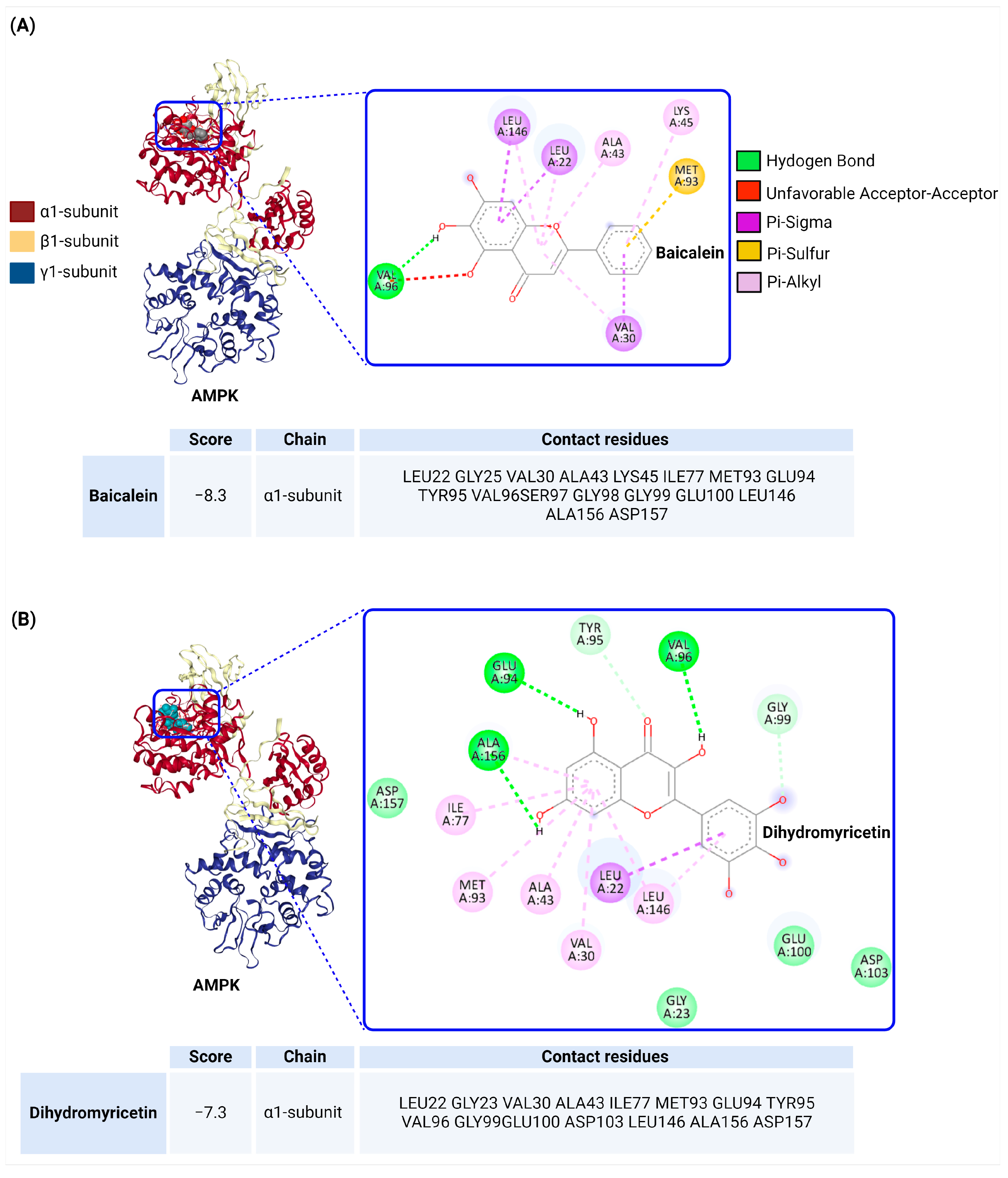
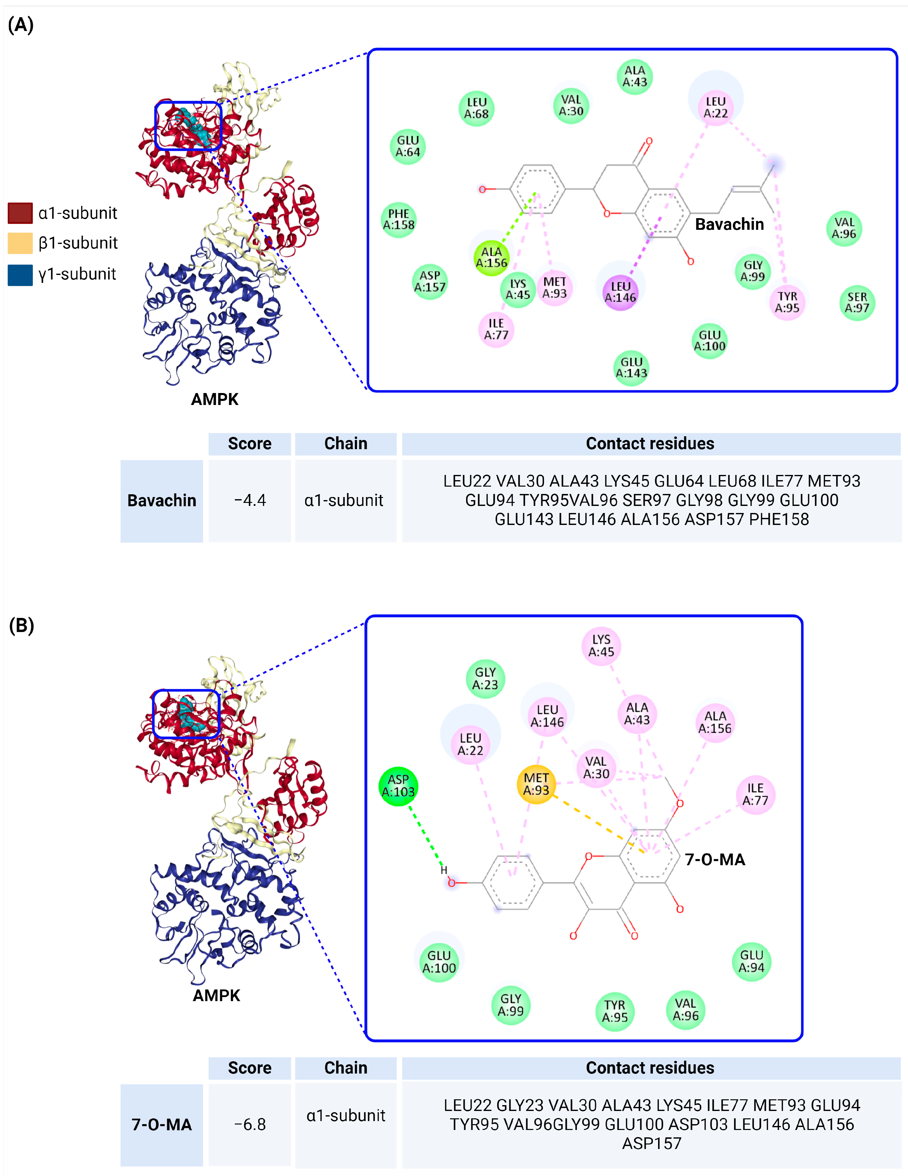
5. Docking Analysis of AMPK with Flavonoids

AMPK activators are broadly categorized into two types: indirect AMPK activators and direct AMPK activators. Indirect activators, such as biguanides and thiazolidinediones (TZDs), activate AMPK by altering the cellular energy status, typically by increasing the AMP/ATP ratio. Metformin, a widely used anti-diabetic drug, exemplifies this mechanism by inhibiting mitochondrial complex I, which results in elevated AMP levels and subsequent AMPK activation, although some effects of metformin may be independent of AMPK [66]. Similarly, TZDs, including rosiglitazone, activate AMPK by inhibiting mitochondrial function and inducing adiponectin release, which further activates AMPK in other tissues [67].
In contrast, direct AMPK activators work by directly binding to and activating AMPK without significantly altering the cellular levels of ATP, ADP, or AMP. For example, AICAR, the first known direct activator, mimics AMP and binds to the γ subunit of AMPK, effectively activating it [68,69]. Thienopyridones, such as A-769662, allosterically activate AMPK, particularly in β1-containing complexes, without the need for Thr172 phosphorylation [70,71]. Salicylate, a pro-drug of aspirin, directly activates AMPK by binding to a site overlapping with that of A-769662, especially in β1-containing complexes [72]. Compound-2 (C-2) acts as a potent allosteric activator of AMPK by mimicking the effects of AMP. It binds to the AMPKγ subunit, competing with AMP, but does not affect other AMP-regulated enzymes [73,74]. The activation of AMPK through direct activators involves specific interactions with the α and β subunits. Thr172 phosphorylation on the α subunit is crucial for AMPK activation, and while AMP typically promotes this phosphorylation by binding to the γ subunit, direct activators like A-769662 can enhance Thr172 phosphorylation without AMP. The β subunit, particularly the carbohydrate-binding module (CBM), plays a critical role in AMPK’s stability and activation. Direct activators like A-769662 bind to the CBM, inducing structural changes that facilitate necessary phosphorylation events for AMPK activation. This allosteric activation mechanism allows AMPK to be activated even in the absence of AMP, highlighting the diverse pathways through which AMPK can be modulated for therapeutic benefits.
In this review, 10 flavonoids known to activate AMPK were investigated. Among them, quercetin, genistein, and aspalathin have been shown to indirectly activate AMPK by modulating the cellular ADP/AMP ratio, which, in turn, adjusts energy homeostasis [62,75,76]. These compounds do not directly bind to AMPK but instead initiate its activation through changes in cellular energy status. Baicalin, however, activates AMPK via a different pathway. It induces AMPK activation through a CaMKKβ-dependent mechanism without altering the ADP/AMP ratio [46]. This indicates that AMPK activation can occur via calcium/CaMKKβ, independent of direct energy shifts. For the remaining six flavonoids, the mechanism of AMPK activation remains uncertain. Whether these flavonoids activate AMPK directly or indirectly has not yet been clarified in the existing research. To address this gap, docking studies were performed on these six flavonoids to assess whether they act as direct AMPK activators. Molecular docking analysis was used to evaluate their binding affinity and interactions with the α1, β1, and γ1 subunits of AMPK. The goal was to determine whether these flavonoids directly bind to AMPK, thus providing deeper insights into their potential role as direct AMPK activators and their therapeutic applications in metabolic diseases.
Ligand-protein docking studies offer significant advantages in elucidating how small molecules, such as flavonoids, interact with their protein targets, like AMPK. These studies not only predict the binding affinity and orientation of the ligands within the protein’s active site but also provide valuable insights into the molecular basis of enzyme activation or inhibition. Moreover, docking studies can help identify potential binding sites and guide the design of more potent and selective AMPK activators, thereby accelerating the drug discovery process. By simulating ligand–protein interactions at the atomic level, docking studies contribute to a deeper understanding of the mechanisms underlying both direct and indirect AMPK activation, which is essential for developing targeted therapies for metabolic diseases.
Molecular docking analysis was conducted using CB-Dock2 to assess the interactions between AMPK (comprising the α1, β1, and γ1 subunits; PDB ID 4QFG) and various flavonoids. The docking scores, binding chains, and contact residues were analyzed using Biovia Discovery Studio Visualizer (version 21.1.0.20298), providing a detailed understanding of the molecular interactions involved.
Baicalein (Figure 3A) showed a strong binding affinity, with a docking score of −8.3, primarily interacting with the α1 subunit of AMPK. The key contact residues included LEU22, GLY25, VAL30, ALA43, LYS45, ILE77, MET93, GLU94, TYR95, VAL96, SER97, GLY98, GLU100, LEU146, ALA156, and ASP157. These interactions were mediated through a combination of hydrogen bonds and pi–sigma, pi–alkyl, and pi–sulfur interactions, suggesting that baicalein could potentially activate AMPK through a robust and multifaceted binding mode.
Dihydromyricetin (Figure 3B) displayed a slightly lower docking score of −7.3 but still showed significant binding affinity to the α1 subunit of AMPK. The binding involved residues such as LEU22, GLY23, VAL30, ALA43, ILE77, MET93, GLU94, TYR95, VAL96, GLY99, GLU100, ASP103, LEU146, ALA156, and ASP157, indicating stable interactions through hydrogen bonds and pi–alkyl forces that could facilitate AMPK activation.
Bavachin (Figure 4A) exhibited a moderate docking score of −4.4, indicating a weaker interaction with the α1 subunit of AMPK. The binding involved residues such as LEU22, VAL30, ALA43, LYS45, GLU64, LEU68, ILE77, MET93, GLU94, TYR95, and others. The interactions included hydrogen bonds and pi–alkyl contacts, suggesting that bavachin might have a less potent effect on AMPK activation.
In contrast, 7-O-MA (Figure 4B) demonstrated a more robust interaction with a docking score of −6.8. This flavonoid also targeted the α1 subunit, interacting with key residues like LEU22, GLY23, VAL30, ALA43, LYS45, ILE77, and MET93, among others. The binding involved a stable network of hydrogen bonds and pi–alkyl interactions, indicating that 7-O-MA could be a stronger candidate for AMPK activation than bavachin.
Derrone (Figure 5A) showed a strong docking score of −7.4, with significant interactions involving the α1 subunit of AMPK. The binding included residues such as LEU22, GLY23, VAL24, THR26, VAL30, and others, suggesting that derrone could effectively stabilize and activate the AMPK complex through a combination of hydrogen bonding and non-covalent interactions.
Alpinumisoflavone (Figure 5B), however, displayed a lower docking score of −3.4, indicating a weaker binding affinity to the α1 subunit of AMPK. The interaction profile involved residues like LEU22, GLY23, VAL24, THR26, and GLU100, suggesting that while alpinumisoflavone might interact with AMPK, its potential to modulate AMPK activity is likely less pronounced compared to derrone.
All six flavonoids interact with the α1 subunit of AMPK, indicating a common site of action that could be crucial to their role as AMPK activators. However, the strength of these interactions varies significantly, as evidenced by the differing docking scores, suggesting that not all flavonoids are equally effective in activating AMPK. The binding interactions predominantly involve hydrogen bonds and pi–alkyl interactions, which are common across the flavonoids, but the specific contact residues differ, leading to variations in binding affinity and possibly activation potency.
The docking results suggest that these flavonoids could activate AMPK through direct binding to the α1 subunit, thereby inducing the necessary conformational changes for activation. While some flavonoids, like baicalein and dihydromyricetin, exhibit strong binding and potentially more effective activation, others like alpinumisoflavone show weaker interactions, which might translate to less potent AMPK activation. Further experimental validation is required to determine whether these flavonoids activate AMPK through the same or different mechanisms compared to known direct activators like A-769662. Compared to established direct AMPK activators such as A-769662, which have well-characterized mechanisms of action, these flavonoids offer a diverse range of interactions and potential activation pathways. While A-769662 primarily interacts with the β1 subunit, most of the flavonoids in this study target the α1 subunit, suggesting an alternative activation route. This difference could lead to varying therapeutic outcomes, making these flavonoids interesting candidates for further research in the development of AMPK-targeted therapies.
Molecular docking studies, while useful for predicting how small molecules interact with proteins, have certain limitations. They are typically conducted in simplified environments and do not fully reflect the complexity of the human body. Factors like protein flexibility, the presence of cofactors, and the surrounding cellular environment are often not considered, which can lead to inaccurate predictions of how a molecule will behave in vivo. Additionally, docking studies provide static snapshots of interactions, missing out on the dynamic conformational changes that occur in real biological systems. Therefore, experimental validation through in vitro and in vivo studies is necessary to confirm docking predictions.

6. Comprehensive ADME and Drug-Likeness Evaluation of Derrone Using SwissADME

Flavonoids show great potential as therapeutic agents for diabetes management due to their ability to regulate glucose metabolism and reduce oxidative stress. However, challenges remain in translating these benefits to clinical applications. Bioavailability is a key issue, as many flavonoids are poorly absorbed and rapidly metabolized and have limited systemic availability [77]. Additionally, drug interactions are a concern, particularly because flavonoids can affect drug-metabolizing enzymes like cytochrome P450, potentially interfering with conventional anti-diabetic treatments [78]. Lastly, their poor water solubility limits their absorption [79]. Therefore, examining the ADME properties of these flavonoids is crucial for optimizing their clinical use.
ADME analysis is crucial in drug development, as it provides essential insights into how a compound behaves in the body, helping to predict its efficacy, safety, and potential interactions [80,81]. Early assessment of these properties can significantly reduce the risk of late-stage failures in drug development.
SwissADME (available at http://www.swissadme.ch, accessed on 15 July 2024) is a freely accessible online tool that provides robust predictions for various ADME parameters, physicochemical properties, drug-likeness, and medicinal chemistry friendliness [82,83]. The tool is widely used by researchers to rapidly evaluate the drug-like potential of small molecules and make informed decisions during the drug discovery process.
The six flavonoids (baicalein, dihydromyricetin, derrone, 7-O-MA, bavachin, and alpinumisoflavone) analyzed by the SwissADME tool exhibit varying physicochemical properties, pharmacokinetic profiles, and drug-likeness characteristics, each contributing to their potential as drug candidates.
Baicalein has a molecular weight of 270.24 g/mol and a Topological Polar Surface Area (TPSA) of 90.90 Å2, both of which are within the acceptable ranges for drug-like compounds, suggesting its favorable bioavailability. It shows moderate lipophilicity, with a consensus Log p value of 2.24, and moderate solubility in water. Baicalein is predicted to have high gastrointestinal absorption but is unlikely to permeate the blood–brain barrier (BBB). It inhibits CYP1A2 and CYP3A4, indicating potential drug–drug interaction risks. Despite minor concerns related to its catechol structure, baicalein passes all the drug-likeness rules and has a moderate synthetic accessibility score, making it a viable candidate for further development.
Dihydromyricetin (DMY) presents a molecular weight of 320.25 g/mol and a TPSA of 147.68 Å2, which, although high, is typical for molecules with strong hydrogen bonding potential. The compound is highly hydrophilic, as indicated by its consensus Log p value of 0.17, and is classified as soluble in water. However, DMY shows low gastrointestinal absorption and is unlikely to cross the BBB. It does not inhibit major cytochrome P450 enzymes, which reduces the risk of drug–drug interactions. Despite several violations in drug-likeness rules, its synthetic accessibility score suggests that DMY can be relatively easily synthesized, though further optimization may be required to improve its bioavailability.
Derrone, with a molecular weight of 336.34 g/mol and a TPSA of 79.90 Å2, demonstrates moderate lipophilicity (consensus Log P of 3.30) and moderate solubility. It is predicted to have high gastrointestinal absorption but does not cross the BBB. Derrone inhibits multiple cytochrome P450 enzymes, which could lead to potential drug–drug interactions. Despite this, it passes all drug-likeness filters and has a good synthetic accessibility score, indicating its strong potential as a drug candidate with further optimization.
7-O-MA, weighing 302.28 g/mol with a TPSA of 96.22 Å2, exhibits moderate lipophilicity (consensus Log P of 1.40) and is classified as soluble. It shows high gastrointestinal absorption but is not expected to cross the BBB, limiting its central nervous system effects. This compound does not inhibit major cytochrome P450 enzymes, reducing the risk of interactions. 7-O-MA passes all drug-likeness filters and has a moderate synthetic accessibility score, making it a strong candidate for oral drug development.
Bavachin has a molecular weight of 324.37 g/mol and a TPSA of 66.76 Å2, which suggests its good membrane permeability and bioavailability. It shows moderate lipophilicity (consensus Log P of 3.53) and is moderately soluble. Bavachin has high gastrointestinal absorption and is predicted to cross the BBB, indicating potential CNS activity. However, it inhibits several cytochrome P450 enzymes, necessitating careful management of drug–drug interactions. Bavachin passes all drug-likeness rules and has moderate synthetic accessibility, indicating that it is a feasible candidate for further development with potential CNS applications.
Alpinumisoflavone, with a molecular weight of 336.34 g/mol and a TPSA of 79.90 Å2, demonstrates moderate lipophilicity (consensus Log P of 3.18) and is moderately soluble in water. It is predicted to have high gastrointestinal absorption but does not cross the BBB. Alpinumisoflavone inhibits several cytochrome P450 enzymes, suggesting a need for careful consideration of drug–drug interactions. It passes all drug-likeness rules and has a synthetic accessibility score of 3.68, making it a viable candidate for further development, particularly for non-CNS applications.
Figure 6 provides a comparative analysis of the drug-likeness properties of six different flavonoids through radar charts that highlight their physicochemical characteristics. The parameters considered include lipophilicity (LIPO), molecular size (SIZE), polarity (POLAR), solubility (INSOLU), degree of saturation (INSATU), and molecular flexibility (FLEX). These properties are crucial in determining the compounds’ potential as orally active drugs.
Baicalein, dihydromyricetin, and 7-O-MA generally fit well within the optimal drug-likeness space, particularly in terms of size and lipophilicity, which are critical for effective membrane permeability and bioavailability. However, dihydromyricetin shows some limitations in its solubility and polarity, potentially impacting its gastrointestinal absorption. Derrone, bavachin, and alpinumisoflavone also demonstrate good alignment with the desired drug-likeness profile, with derrone and bavachin particularly excelling in size and lipophilicity. However, bavachin’s profile suggests a potential concern in terms of its flexibility, which might influence its binding interactions with target proteins. Alpinumisoflavone shows moderate performance across most of the parameters, with some concerns in terms of its solubility that may require attention during formulation development.
These radar charts underscore the balance of properties required for effective drug development and highlight areas where each flavonoid may need further optimization. The analysis suggests that while all six compounds have strong potential as drug candidates, considerations such as solubility and molecular flexibility will be critical in guiding their further development as AMPK activators or other therapeutic agents.
Table 2 provides a detailed ADME analysis of baicalein and dihydromyricetin, the compounds with the lowest docking scores.

7. Conclusions

The comprehensive review presented here highlights the significant potential of plant-derived flavonoids as AMPK activators in the management of T2DM. AMPK, a key regulator of cellular energy homeostasis, plays a crucial role in enhancing insulin sensitivity, promoting glucose uptake, and reducing hepatic glucose production, central mechanisms in combating T2DM. While current pharmacological interventions, such as metformin, target the AMPK pathway, the incorporation of flavonoids offers a promising complementary or alternative strategy.
The flavonoids reviewed, baicalein, dihydromyricetin, bavachin, 7-O-MA, derrone, and alpinumisoflavone, exhibit diverse mechanisms of AMPK activation, either directly by binding to the AMPK complex or indirectly by influencing upstream pathways or cellular energy states. Notably, baicalein and dihydromyricetin demonstrated strong binding affinities in molecular docking studies, suggesting robust interactions with the α1 subunit of AMPK, which may translate to potent AMPK activation in vivo. However, the exact mechanisms of action for many of these flavonoids remain to be fully elucidated, necessitating further research using advanced techniques like mutational analyses and in vivo studies.
In addition to their potential as AMPK activators, these flavonoids offer broad therapeutic benefits, including anti-inflammatory and antioxidant effects, which may further enhance their utility in managing T2DM and its complications. The ADME and drug-likeness profiles of these compounds, as analyzed using the SwissADME tool, indicate their favorable pharmacokinetic properties, although certain challenges, such as solubility and potential drug–drug interactions, need to be addressed in the drug development process.
Flavonoids regulate carbohydrate digestion, insulin secretion, insulin signaling, glucose uptake, and adipose deposition [14], targeting multiple molecules involved in β-cell proliferation, promoting insulin secretion, reducing apoptosis, and improving hyperglycemia by regulating glucose metabolism in the liver [53]. In support of their potential, a large-scale study in the US involving 200,000 men and women confirmed that higher consumption of anthocyanins from sources like blueberries, apples, and pears was associated with a reduced risk of diabetes. It is hypothesized that much of the bioactivity of flavonoids is due to their hydroxyl groups, as well as α and β ketones [84]. However, to fully confirm the promising effects of flavonoids on glucose metabolism, insulin sensitivity, and AMPK activation observed in preclinical studies, large-scale human clinical trials are essential. These trials should focus on determining their optimal dosages, long-term safety, and overall efficacy in managing diabetes, facilitating the integration of flavonoids into clinical practice. Moreover, exploring the combined use of flavonoids with conventional diabetes therapies, such as metformin or insulin, could reveal potential synergies or interactions, ensuring safer and more effective treatment options. Enhancing the bioavailability of flavonoids through advanced delivery methods, like nanoformulations or bio-enhancers, is another critical area of research to maximize their therapeutic potential. Finally, personalized medicine approaches should be considered, as genetic and environmental factors significantly influence flavonoid metabolism, paving the way for more tailored diabetes treatments that cater to individual patient profiles.
However, despite their many health benefits, the long-term use of flavonoids may also lead to side effects. Studies have indicated that flavonoids can potentially induce carcinogenic effects, such as promoting tumor cell proliferation under certain conditions and doses. For instance, phytoestrogens like genistein and daidzein have shown biphasic effects, with low doses promoting cell growth and higher doses inhibiting it [85,86]. Additionally, flavonoids have been linked to hepatotoxicity and nephrotoxicity, particularly at high doses, as seen in animal studies where liver and kidney damage was observed [87,88]. They can also interfere with thyroid function, especially isoflavones, which may disrupt thyroid hormone metabolism and contribute to conditions like goiter. Moreover, flavonoids have been shown to alter gut microbiota composition [89], with emerging evidence suggesting a link between these changes and neurobehavioral disorders. Therefore, while flavonoids hold therapeutic potential, careful attention to their dosage and long-term effects is crucial, especially when used in high concentrations or as supplements.

Funding

The author declares that no funds, grants, or other support were received during the preparation of this manuscript.

Data Availability Statement

The data presented in this study are available on request from the author.

Conflicts of Interest

The author declares no conflicts of interest.

References

  1. Artasensi, A.; Pedretti, A.; Vistoli, G.; Fumagalli, L. Type 2 Diabetes Mellitus: A Review of Multi-Target Drugs. Molecules 2020, 25, 1987. [Google Scholar] [CrossRef] [PubMed]
  2. Zheng, Y.; Ley, S.H.; Hu, F.B. Global aetiology and epidemiology of type 2 diabetes mellitus and its complications. Nat. Rev. Endocrinol. 2018, 14, 88–98. [Google Scholar] [CrossRef] [PubMed]
  3. Gourgari, E.; Wilhelm, E.E.; Hassanzadeh, H.; Aroda, V.R.; Shoulson, I. A comprehensive review of the FDA-approved labels of diabetes drugs: Indications, safety, and emerging cardiovascular safety data. J. Diabetes Complicat. 2017, 31, 1719–1727. [Google Scholar] [CrossRef] [PubMed]
  4. Dahlén, A.D.; Dashi, G.; Maslov, I.; Attwood, M.M.; Jonsson, J.; Trukhan, V.; Schiöth, H.B. Trends in Antidiabetic Drug Discovery: FDA Approved Drugs, New Drugs in Clinical Trials and Global Sales. Front. Pharmacol. 2021, 12, 807548. [Google Scholar] [CrossRef]
  5. Szymczak-Pajor, I.; Wenclewska, S.; Śliwińska, A. Metabolic Action of Metformin. Pharmaceuticals 2022, 15, 810. [Google Scholar] [CrossRef]
  6. Goel, S.; Singh, R.; Singh, V.; Singh, H.; Kumari, P.; Chopra, H.; Sharma, R.; Nepovimova, E.; Valis, M.; Kuca, K.; et al. Metformin: Activation of 5′ AMP-activated protein kinase and its emerging potential beyond anti-hyperglycemic action. Front. Genet. 2022, 13, 1022739. [Google Scholar] [CrossRef]
  7. Salminen, A.; Hyttinen, J.M.; Kaarniranta, K. AMP-activated protein kinase inhibits NF-κB signaling and inflammation: Impact on healthspan and lifespan. J. Mol. Med. 2011, 89, 667–676. [Google Scholar] [CrossRef]
  8. Chen, X.; Li, X.; Zhang, W.; He, J.; Xu, B.; Lei, B.; Wang, Z.; Cates, C.; Rousselle, T.; Li, J. Activation of AMPK inhibits inflammatory response during hypoxia and reoxygenation through modulating JNK-mediated NF-κB pathway. Metabolism 2018, 83, 256–270. [Google Scholar] [CrossRef]
  9. Clark, H.; Carling, D.; Saggerson, D. Covalent activation of heart AMP-activated protein kinase in response to physiological concentrations of long-chain fatty acids. Eur. J. Biochem. 2004, 271, 2215–2224. [Google Scholar] [CrossRef]
  10. Suchankova, G.; Tekle, M.; Saha, A.K.; Ruderman, N.B.; Clarke, S.D.; Gettys, T.W. Dietary polyunsaturated fatty acids enhance hepatic AMP-activated protein kinase activity in rats. Biochem. Biophys. Res. Commun. 2005, 326, 851–858. [Google Scholar] [CrossRef]
  11. Kelley, D.E.; Simoneau, J.A. Impaired free fatty acid utilization by skeletal muscle in non-insulin-dependent diabetes mellitus. J. Clin. Investig. 1994, 94, 2349–2356. [Google Scholar] [CrossRef] [PubMed]
  12. Yi, X.; Dong, M.; Guo, N.; Tian, J.; Lei, P.; Wang, S.; Yang, Y.; Shi, Y. Flavonoids improve type 2 diabetes mellitus and its complications: A review. Front. Nutr. 2023, 10, 1192131. [Google Scholar] [CrossRef] [PubMed]
  13. Hussain, T.; Tan, B.; Murtaza, G.; Liu, G.; Rahu, N.; Saleem Kalhoro, M.; Hussain Kalhoro, D.; Adebowale, T.O.; Usman Mazhar, M.; Rehman, Z.U.; et al. Flavonoids and type 2 diabetes: Evidence of efficacy in clinical and animal studies and delivery strategies to enhance their therapeutic efficacy. Pharmacol. Res. 2020, 152, 104629. [Google Scholar] [CrossRef] [PubMed]
  14. Vinayagam, R.; Xu, B. Antidiabetic properties of dietary flavonoids: A cellular mechanism review. Nutr. Metab. 2015, 12, 60. [Google Scholar] [CrossRef] [PubMed]
  15. Mutha, R.E.; Tatiya, A.U.; Surana, S.J. Flavonoids as natural phenolic compounds and their role in therapeutics: An overview. Future J. Pharm. Sci. 2021, 7, 25. [Google Scholar] [CrossRef]
  16. Francini, F.; Schinella, G.R.; Ríos, J.L. Activation of AMPK by Medicinal Plants and Natural Products: Its Role in Type 2 Diabetes Mellitus. Mini Rev. Med. Chem. 2019, 19, 880–901. [Google Scholar] [CrossRef]
  17. Thomas, I.; Gregg, B. Metformin; a review of its history and future: From lilac to longevity. Pediatr. Diabetes 2017, 18, 10–16. [Google Scholar] [CrossRef]
  18. Grace, M.H.; Ribnicky, D.M.; Kuhn, P.; Poulev, A.; Logendra, S.; Yousef, G.G.; Raskin, I.; Lila, M.A. Hypoglycemic activity of a novel anthocyanin-rich formulation from lowbush blueberry, Vaccinium angustifolium Aiton. Phytomedicine 2009, 16, 406–415. [Google Scholar] [CrossRef]
  19. Babu, P.V.; Liu, D.; Gilbert, E.R. Recent advances in understanding the anti-diabetic actions of dietary flavonoids. J. Nutr. Biochem. 2013, 24, 1777–1789. [Google Scholar] [CrossRef]
  20. Zhang, F.; Feng, J.; Zhang, J.; Kang, X.; Qian, D. Quercetin modulates AMPK/SIRT1/NF-κB signaling to inhibit inflammatory/oxidative stress responses in diabetic high fat diet-induced atherosclerosis in the rat carotid artery. Exp. Ther. Med. 2020, 20, 280. [Google Scholar] [CrossRef]
  21. Zhang, N.; Zhang, W.; Guo, X.; Liu, J.; Li, S.; Zhang, H.; Fan, B. Genistein protects against hyperglycemia and fatty liver disease in diet-induced prediabetes mice via activating hepatic insulin signaling pathway. Front. Nutr. 2022, 9, 1072044. [Google Scholar] [CrossRef] [PubMed]
  22. Liu, C.; Ma, J.; Sun, J.; Cheng, C.; Feng, Z.; Jiang, H.; Yang, W. Flavonoid-Rich Extract of Paulownia fortunei Flowers Attenuates Diet-Induced Hyperlipidemia, Hepatic Steatosis and Insulin Resistance in Obesity Mice by AMPK Pathway. Nutrients 2017, 9, 959. [Google Scholar] [CrossRef] [PubMed]
  23. Herzig, S.; Shaw, R.J. AMPK: Guardian of metabolism and mitochondrial homeostasis. Nat. Rev. Mol. Cell Biol. 2018, 19, 121–135. [Google Scholar] [CrossRef] [PubMed]
  24. Steinberg, G.R.; Hardie, D.G. New insights into activation and function of the AMPK. Nat. Rev. Mol. Cell Biol. 2023, 24, 255–272. [Google Scholar] [CrossRef]
  25. Yan, Y.; Zhou, X.E.; Xu, H.E.; Melcher, K. Structure and Physiological Regulation of AMPK. Int. J. Mol. Sci. 2018, 19, 3534. [Google Scholar] [CrossRef] [PubMed]
  26. Birk, J.B.; Wojtaszewski, J.F. Predominant alpha2/beta2/gamma3 AMPK activation during exercise in human skeletal muscle. J. Physiol. 2006, 577 Pt 3, 1021–1032. [Google Scholar] [CrossRef] [PubMed]
  27. Kim, J.; Yang, G.; Kim, Y.; Kim, J.; Ha, J. AMPK activators: Mechanisms of action and physiological activities. Exp. Mol. Med. 2016, 48, e224. [Google Scholar] [CrossRef]
  28. Xiao, B.; Sanders, M.J.; Underwood, E.; Heath, R.; Mayer, F.V.; Carmena, D.; Jing, C.; Walker, P.A.; Eccleston, J.F.; Haire, L.F.; et al. Structure of mammalian AMPK and its regulation by ADP. Nature 2011, 472, 230–233. [Google Scholar] [CrossRef]
  29. Gowans, G.J.; Hawley, S.A.; Ross, F.A.; Hardie, D.G. AMP is a true physiological regulator of AMP-activated protein kinase by both allosteric activation and enhancing net phosphorylation. Cell Metab. 2013, 18, 556–566. [Google Scholar] [CrossRef]
  30. Woods, A.; Dickerson, K.; Heath, R.; Hong, S.P.; Momcilovic, M.; Johnstone, S.R.; Carlson, M.; Carling, D. Ca2+/calmodulin-dependent protein kinase kinase-beta acts upstream of AMP-activated protein kinase in mammalian cells. Cell Metab. 2005, 2, 21–33. [Google Scholar] [CrossRef]
  31. Ciccarese, F.; Zulato, E.; Indraccolo, S. LKB1/AMPK Pathway and Drug Response in Cancer: A Therapeutic Perspective. Oxid. Med. Cell. Longev. 2019, 2019, 8730816. [Google Scholar] [CrossRef] [PubMed]
  32. Hawley, S.A.; Selbert, M.A.; Goldstein, E.G.; Edelman, A.M.; Carling, D.; Hardie, D.G. 5′-AMP activates the AMP-activated protein kinase cascade, and Ca2+/calmodulin activates the calmodulin-dependent protein kinase I cascade, via three independent mechanisms. J. Biol. Chem. 1995, 270, 27186–27191. [Google Scholar] [CrossRef]
  33. Davies, S.P.; Helps, N.R.; Cohen, P.T.; Hardie, D.G. 5′-AMP inhibits dephosphorylation, as well as promoting phosphorylation, of the AMP-activated protein kinase. Studies using bacterially expressed human protein phosphatase-2C alpha and native bovine protein phosphatase-2AC. FEBS Lett. 1995, 377, 421–425. [Google Scholar] [PubMed]
  34. Oakhill, J.S.; Steel, R.; Chen, Z.P.; Scott, J.W.; Ling, N.; Tam, S.; Kemp, B.E. AMPK is a direct adenylate charge-regulated protein kinase. Science 2011, 332, 1433–1435. [Google Scholar] [CrossRef]
  35. Leclerc, I.; Woltersdorf, W.W.; da Silva Xavier, G.; Rowe, R.L.; Cross, S.E.; Korbutt, G.S.; Rajotte, R.V.; Smith, R.; Rutter, G.A. Metformin, but not leptin, regulates AMP-activated protein kinase in pancreatic islets: Impact on glucose-stimulated insulin secretion. Am. J. Physiol. Endocrinol. Metab. 2004, 286, E1023–E1031. [Google Scholar] [CrossRef]
  36. Sun, Y.; Ren, M.; Gao, G.Q.; Gong, B.; Xin, W.; Guo, H.; Zhang, X.J.; Gao, L.; Zhao, J.J. Chronic palmitate exposure inhibits AMPKalpha and decreases glucose-stimulated insulin secretion from beta-cells: Modulation by fenofibrate. Acta Pharmacol. Sin. 2008, 29, 443–450. [Google Scholar] [CrossRef] [PubMed]
  37. Henquin, J.C. The dual control of insulin secretion by glucose involves triggering and amplifying pathways in β-cells. Diabetes Res. Clin. Pract. 2011, 93 (Suppl. 1), S27–S31. [Google Scholar] [CrossRef]
  38. Prentki, M.; Matschinsky, F.M.; Madiraju, S.R. Metabolic signaling in fuel-induced insulin secretion. Cell Metab. 2013, 18, 162–185. [Google Scholar] [CrossRef] [PubMed]
  39. Akkan, A.G.; Malaisse, W.J. Insulinotropic action of AICA riboside. I. Insulin release by isolated islets and the perfused pancreas. Diabetes Res. 1994, 25, 13–23. [Google Scholar]
  40. Malaisse, W.J.; Conget, I.; Sener, A.; Rorsman, P. Insulinotropic action of AICA riboside. II. Secretory, metabolic and cationic aspects. Diabetes Res. 1994, 25, 25–37. [Google Scholar]
  41. Pepin, É.; Al-Mass, A.; Attané, C.; Zhang, K.; Lamontagne, J.; Lussier, R.; Madiraju, S.R.; Joly, E.; Ruderman, N.B.; Sladek, R.; et al. Pancreatic β-Cell Dysfunction in Diet-Induced Obese Mice: Roles of AMP-Kinase, Protein Kinase Cε, Mitochondrial and Cholesterol Metabolism, and Alterations in Gene Expression. PLoS ONE 2016, 11, e0153017. [Google Scholar] [CrossRef] [PubMed]
  42. Wang, X.; Zhou, L.; Li, G.; Luo, T.; Gu, Y.; Qian, L.; Fu, X.; Li, F.; Li, J.; Luo, M. Palmitate activates AMP-activated protein kinase and regulates insulin secretion from beta cells. Biochem. Biophys. Res. Commun. 2007, 352, 463–468. [Google Scholar] [CrossRef] [PubMed]
  43. Kim, J.W.; You, Y.H.; Ham, D.S.; Yang, H.K.; Yoon, K.H. The Paradoxical Effects of AMPK on Insulin Gene Expression and Glucose-Induced Insulin Secretion. J. Cell Biochem. 2016, 117, 239–246. [Google Scholar] [CrossRef] [PubMed]
  44. Prentki, M.; Joly, E.; El-Assaad, W.; Roduit, R. Malonyl-CoA signaling, lipid partitioning, and glucolipotoxicity: Role in beta-cell adaptation and failure in the etiology of diabetes. Diabetes 2002, 51 (Suppl. 3), S405–S413. [Google Scholar] [CrossRef] [PubMed]
  45. Herman, R.; Kravos, N.A.; Jensterle, M.; Janež, A.; Dolžan, V. Metformin and Insulin Resistance: A Review of the Underlying Mechanisms behind Changes in GLUT4-Mediated Glucose Transport. Int. J. Mol. Sci. 2022, 23, 1264. [Google Scholar] [CrossRef]
  46. Shamshoum, H.; Vlavcheski, F.; MacPherson, R.E.K.; Tsiani, E. Rosemary extract activates AMPK, inhibits mTOR and attenuates the high glucose and high insulin-induced muscle cell insulin resistance. Appl. Physiol. Nutr. Metab. 2021, 46, 819–827. [Google Scholar] [CrossRef]
  47. Zhang, Y.; Zhu, Z.; Zhai, W.; Bi, Y.; Yin, Y.; Zhang, W. Expression and purification of asprosin in Pichia pastoris and investigation of its increase glucose uptake activity in skeletal muscle through activation of AMPK. Enzyme Microb. Technol. 2021, 144, 109737. [Google Scholar] [CrossRef]
  48. Prabha, B.; Sini, S.; Sherin, D.R.; Neethu, S.; Rameshkumar, K.B.; Manojkumar, T.K.; Jayamurthy, P.; Radhakrishnan, K.V. Promalabaricone B from Myristica fatua Houtt. seeds demonstrate antidiabetic potential by modulating glucose uptake via the upregulation of AMPK in L6 myotubes. Nat. Prod. Res. 2021, 35, 867–872. [Google Scholar] [CrossRef]
  49. Natsume, N.; Yonezawa, T.; Saito, Y.; Woo, J.T.; Teruya, T. Prenylflavonoids from fruit of Macaranga tanarius promote glucose uptake via AMPK activation in L6 myotubes. J. Nat. Med. 2021, 75, 813–823. [Google Scholar] [CrossRef]
  50. Lee, H.A.; Cho, J.H.; Afinanisa, Q.; An, G.H.; Han, J.G.; Kang, H.J.; Choi, S.H.; Seong, H.A. Ganoderma lucidum Extract Reduces Insulin Resistance by Enhancing AMPK Activation in High-Fat Diet-Induced Obese Mice. Nutrients 2020, 12, 3338. [Google Scholar] [CrossRef]
  51. Manach, C.; Scalbert, A.; Morand, C.; Rémésy, C.; Jiménez, L. Polyphenols: Food sources and bioavailability. Am. J. Clin. Nutr. 2004, 79, 727–747. [Google Scholar] [CrossRef] [PubMed]
  52. Beecher, G.R. Overview of dietary flavonoids: Nomenclature, occurrence and intake. J. Nutr. 2003, 133, 3248s–3254s. [Google Scholar] [CrossRef] [PubMed]
  53. Graf, B.A.; Milbury, P.E.; Blumberg, J.B. Flavonols, flavones, flavanones, and human health: Epidemiological evidence. J. Med. Food 2005, 8, 281–290. [Google Scholar] [CrossRef]
  54. Arts, I.C.; Hollman, P.C. Polyphenols and disease risk in epidemiologic studies. Am. J. Clin. Nutr. 2005, 81 (Suppl. 1), 317s–325s. [Google Scholar] [CrossRef]
  55. Hanhineva, K.; Törrönen, R.; Bondia-Pons, I.; Pekkinen, J.; Kolehmainen, M.; Mykkänen, H.; Poutanen, K. Impact of dietary polyphenols on carbohydrate metabolism. Int. J. Mol. Sci. 2010, 11, 1365–1402. [Google Scholar] [CrossRef] [PubMed]
  56. Shi, L.; Zhang, T.; Zhou, Y.; Zeng, X.; Ran, L.; Zhang, Q.; Zhu, J.; Mi, M. Dihydromyricetin improves skeletal muscle insulin sensitivity by inducing autophagy via the AMPK-PGC-1α-Sirt3 signaling pathway. Endocrine 2015, 50, 378–389. [Google Scholar] [CrossRef]
  57. Lee, H.; Li, H.; Noh, M.; Ryu, J.H. Bavachin from Psoralea corylifolia Improves Insulin-Dependent Glucose Uptake through Insulin Signaling and AMPK Activation in 3T3-L1 Adipocytes. Int. J. Mol. Sci. 2016, 17, 527. [Google Scholar] [CrossRef]
  58. Zhang, W.Y.; Lee, J.J.; Kim, I.S.; Kim, Y.; Park, J.S.; Myung, C.S. 7-O-methylaromadendrin stimulates glucose uptake and improves insulin resistance in vitro. Biol. Pharm. Bull. 2010, 33, 1494–1499. [Google Scholar] [CrossRef]
  59. Ma, Y.; Yang, F.; Wang, Y.; Du, Z.; Liu, D.; Guo, H.; Shen, J.; Peng, H. CaMKKβ is involved in AMP-activated protein kinase activation by baicalin in LKB1 deficient cell lines. PLoS ONE 2012, 7, e47900. [Google Scholar] [CrossRef]
  60. Yang, L.L.; Xiao, N.; Liu, J.; Liu, K.; Liu, B.; Li, P.; Qi, L.W. Differential regulation of baicalin and scutellarin on AMPK and Akt in promoting adipose cell glucose disposal. Biochim. Biophys. Acta Mol. Basis Dis. 2017, 1863, 598–606. [Google Scholar] [CrossRef]
  61. Pu, P.; Wang, X.A.; Salim, M.; Zhu, L.H.; Wang, L.; Chen, K.J.; Xiao, J.F.; Deng, W.; Shi, H.W.; Jiang, H.; et al. Baicalein, a natural product, selectively activating AMPKα(2) and ameliorates metabolic disorder in diet-induced mice. Mol. Cell Endocrinol. 2012, 362, 128–138. [Google Scholar] [CrossRef] [PubMed]
  62. Dhanya, R.; Arya, A.D.; Nisha, P.; Jayamurthy, P. Quercetin, a Lead Compound against Type 2 Diabetes Ameliorates Glucose Uptake via AMPK Pathway in Skeletal Muscle Cell Line. Front. Pharmacol. 2017, 8, 336. [Google Scholar] [CrossRef] [PubMed]
  63. Hwang, J.T.; Park, I.J.; Shin, J.I.; Lee, Y.K.; Lee, S.K.; Baik, H.W.; Ha, J.; Park, O.J. Genistein, EGCG, and capsaicin inhibit adipocyte differentiation process via activating AMP-activated protein kinase. Biochem. Biophys. Res. Commun. 2005, 338, 694–699. [Google Scholar] [CrossRef]
  64. Lee, M.S.; Kim, C.H.; Hoang, D.M.; Kim, B.Y.; Sohn, C.B.; Kim, M.R.; Ahn, J.S. Genistein-derivatives from Tetracera scandens stimulate glucose-uptake in L6 myotubes. Biol. Pharm. Bull. 2009, 32, 504–508. [Google Scholar] [CrossRef]
  65. Son, M.J.; Minakawa, M.; Miura, Y.; Yagasaki, K. Aspalathin improves hyperglycemia and glucose intolerance in obese diabetic ob/ob mice. Eur. J. Nutr. 2013, 52, 1607–1619. [Google Scholar] [CrossRef]
  66. Owen, M.R.; Doran, E.; Halestrap, A.P. Evidence that metformin exerts its anti-diabetic effects through inhibition of complex 1 of the mitochondrial respiratory chain. Biochem. J. 2000, 348 Pt 3, 607–614. [Google Scholar] [CrossRef] [PubMed]
  67. Fryer, L.G.; Parbu-Patel, A.; Carling, D. The Anti-diabetic drugs rosiglitazone and metformin stimulate AMP-activated protein kinase through distinct signaling pathways. J. Biol. Chem. 2002, 277, 25226–25232. [Google Scholar] [CrossRef]
  68. Corton, J.M.; Gillespie, J.G.; Hawley, S.A.; Hardie, D.G. 5-aminoimidazole-4-carboxamide ribonucleoside. A specific method for activating AMP-activated protein kinase in intact cells? Eur. J. Biochem. 1995, 229, 558–565. [Google Scholar] [CrossRef]
  69. Sullivan, J.E.; Brocklehurst, K.J.; Marley, A.E.; Carey, F.; Carling, D.; Beri, R.K. Inhibition of lipolysis and lipogenesis in isolated rat adipocytes with AICAR, a cell-permeable activator of AMP-activated protein kinase. FEBS Lett. 1994, 353, 33–36. [Google Scholar] [CrossRef]
  70. Scott, J.W.; van Denderen, B.J.; Jorgensen, S.B.; Honeyman, J.E.; Steinberg, G.R.; Oakhill, J.S.; Iseli, T.J.; Koay, A.; Gooley, P.R.; Stapleton, D.; et al. Thienopyridone drugs are selective activators of AMP-activated protein kinase beta1-containing complexes. Chem. Biol. 2008, 15, 1220–1230. [Google Scholar] [CrossRef]
  71. Moreno, D.; Knecht, E.; Viollet, B.; Sanz, P. A769662, a novel activator of AMP-activated protein kinase, inhibits non-proteolytic components of the 26S proteasome by an AMPK-independent mechanism. FEBS Lett. 2008, 582, 2650–2654. [Google Scholar] [CrossRef] [PubMed]
  72. Hawley, S.A.; Fullerton, M.D.; Ross, F.A.; Schertzer, J.D.; Chevtzoff, C.; Walker, K.J.; Peggie, M.W.; Zibrova, D.; Green, K.A.; Mustard, K.J.; et al. The ancient drug salicylate directly activates AMP-activated protein kinase. Science 2012, 336, 918–922. [Google Scholar] [CrossRef] [PubMed]
  73. Hunter, R.W.; Foretz, M.; Bultot, L.; Fullerton, M.D.; Deak, M.; Ross, F.A.; Hawley, S.A.; Shpiro, N.; Viollet, B.; Barron, D.; et al. Mechanism of action of compound-13: An α1-selective small molecule activator of AMPK. Chem. Biol. 2014, 21, 866–879. [Google Scholar] [CrossRef]
  74. Xiao, B.; Sanders, M.J.; Carmena, D.; Bright, N.J.; Haire, L.F.; Underwood, E.; Patel, B.R.; Heath, R.B.; Walker, P.A.; Hallen, S.; et al. Structural basis of AMPK regulation by small molecule activators. Nat. Commun. 2013, 4, 3017. [Google Scholar] [CrossRef]
  75. Arunkumar, E.; Anuradha, C.V. Genistein promotes insulin action through adenosine monophosphate-activated protein kinase activation and p70 ribosomal protein S6 kinase 1 inhibition in the skeletal muscle of mice fed a high energy diet. Nutr. Res. 2012, 32, 617–625. [Google Scholar] [CrossRef]
  76. Akinfenwa, A.O.; Abdul, N.S.; Marnewick, J.L.; Hussein, A.A. Protective Effects of Linearthin and Other Chalcone Derivatives from Aspalathus linearis (Rooibos) against UVB Induced Oxidative Stress and Toxicity in Human Skin Cells. Plants 2021, 10, 1936. [Google Scholar] [CrossRef]
  77. Thilakarathna, S.H.; Rupasinghe, H.P. Flavonoid bioavailability and attempts for bioavailability enhancement. Nutrients 2013, 5, 3367–3387. [Google Scholar] [CrossRef] [PubMed]
  78. Křížková, J.; Burdová, K.; Stiborová, M.; Křen, V.; Hodek, P. The effects of selected flavonoids on cytochromes P450 in rat liver and small intestine. Interdiscip. Toxicol. 2009, 2, 201–204. [Google Scholar] [CrossRef]
  79. Zhao, J.; Yang, J.; Xie, Y. Improvement strategies for the oral bioavailability of poorly water-soluble flavonoids: An overview. Int. J. Pharm. 2019, 570, 118642. [Google Scholar] [CrossRef]
  80. Wang, Y.; Xing, J.; Xu, Y.; Zhou, N.; Peng, J.; Xiong, Z.; Liu, X.; Luo, X.; Luo, C.; Chen, K.; et al. In silico ADME/T modelling for rational drug design. Q. Rev. Biophys. 2015, 48, 488–515. [Google Scholar] [CrossRef]
  81. Grossman, I. ADME pharmacogenetics: Current practices and future outlook. Expert Opin. Drug Metab. Toxicol. 2009, 5, 449–462. [Google Scholar] [CrossRef] [PubMed]
  82. Daina, A.; Michielin, O.; Zoete, V. SwissADME: A free web tool to evaluate pharmacokinetics, drug-likeness and medicinal chemistry friendliness of small molecules. Sci. Rep. 2017, 7, 42717. [Google Scholar] [CrossRef] [PubMed]
  83. Kumar, A.; Sharma, S.; Mishra, S.; Ojha, S.; Upadhyay, P. ADME Prediction, Structure-activity Relationship of Boswellic Acid Scaffold for the Aspect of Anticancer & Anti-inflammatory Potency. Anticancer. Agents Med. Chem. 2023, 23, 1499–1505. [Google Scholar] [PubMed]
  84. Al-Ishaq, R.K.; Abotaleb, M.; Kubatka, P.; Kajo, K.; Büsselberg, D. Flavonoids and Their Anti-Diabetic Effects: Cellular Mechanisms and Effects to Improve Blood Sugar Levels. Biomolecules 2019, 9, 430. [Google Scholar] [CrossRef]
  85. Kabała-Dzik, A.; Rzepecka-Stojko, A.; Kubina, R.; Iriti, M.; Wojtyczka, R.D.; Buszman, E.; Stojko, J. Flavonoids, bioactive components of propolis, exhibit cytotoxic activity and induce cell cycle arrest and apoptosis in human breast cancer cells MDA-MB-231 and MCF-7—A comparative study. Cell Mol. Biol. 2018, 64, 1–10. [Google Scholar] [CrossRef]
  86. Murata, M.; Midorikawa, K.; Koh, M.; Umezawa, K.; Kawanishi, S. Genistein and daidzein induce cell proliferation and their metabolites cause oxidative DNA damage in relation to isoflavone-induced cancer of estrogen-sensitive organs. Biochemistry 2004, 43, 2569–2577. [Google Scholar] [CrossRef] [PubMed]
  87. James, K.D.; Forester, S.C.; Lambert, J.D. Dietary pretreatment with green tea polyphenol, (-)-epigallocatechin-3-gallate reduces the bioavailability and hepatotoxicity of subsequent oral bolus doses of (-)-epigallocatechin-3-gallate. Food Chem. Toxicol. 2015, 76, 103–108. [Google Scholar]
  88. Lambert, J.D.; Kennett, M.J.; Sang, S.; Reuhl, K.R.; Ju, J.; Yang, C.S. Hepatotoxicity of high oral dose (-)-epigallocatechin-3-gallate in mice. Food Chem. Toxicol. 2010, 48, 409–416. [Google Scholar] [CrossRef]
  89. Matthies, A.; Loh, G.; Blaut, M.; Braune, A. Daidzein and genistein are converted to equol and 5-hydroxy-equol by human intestinal Slackia isoflavoniconvertens in gnotobiotic rats. J. Nutr. 2012, 142, 40–46. [Google Scholar] [CrossRef]
Figure 1. Structural domains and interactions of AMPK subunits. This figure shows the key structural domains of AMPK’s α1, β1, and γ1 subunits and their interactions within the AMPK complex, with the central quaternary structure derived from PDB ID 4QFG. The α1 subunit (red) includes the kinase domain (KD) containing the Thr-172 phosphorylation site, crucial for activation by upstream kinases LKB1 and CaMKKβ; the autoinhibitory domain (AID), which maintains AMPK in an inactive state; and the C-terminal domain (CTD) for β subunit binding. The β1 subunit (magenta) features a carbohydrate-binding module (CBM) involved in glycogen binding and activation by direct activators and a C-terminal domain (CTD) that connects with both α and γ subunits, acting as a scaffold for the complex. The γ1 subunit (yellow) contains four cystathionine-β-synthase (CBS) domains forming nucleotide-binding sites (sites 1–4), where AMP binding at sites 1 and 3 modulates the phosphorylation of Thr-172 and site 4 is tightly bound to AMP. Dotted arrows indicate the functional interactions and conformational changes within the complex that are essential for AMPK activation. Figure 1 was created with https://BioRender.com and accessed on 5 April 2024.
Applsci 14 08607 g001
Figure 2. The role of AMPK in pancreatic beta cells and skeletal muscle. (A) This panel illustrates the role of AMPK in the regulation of insulin secretion within pancreatic beta cells. Under normal conditions, glucose enters the beta cells through glucose transporter 2 (GLUT2), where it undergoes metabolism, leading to an increase in the ATP/ADP ratio. This rise in ATP closes ATP-sensitive potassium channels (K_ATPs), resulting in membrane depolarization and the opening of voltage-dependent calcium channels (CaVGICs). The influx of Ca2+ triggers the exocytosis of insulin-containing vesicles. AMPK activity is inhibited in high-glucose conditions, allowing the full engagement of energy-demanding processes necessary for insulin secretion. However, when AMPK activators are present, AMPK may be activated, which can impact the insulin secretion process. (B) This panel demonstrates the role of AMPK in skeletal muscle cells, focusing on glucose uptake. Insulin binds to its receptor on the muscle cell surface, initiating a signal cascade that leads to the translocation of glucose transporter 4 (GLUT4) to the cell membrane. Concurrently, AMPK activation, which may occur during conditions of low energy or through specific activators, enhances the expression of glucose transporter 1 (GLUT1) and promotes the exocytosis of GLUT4 to the membrane. This results in increased glucose uptake into the muscle cells, which is critical for maintaining glucose homeostasis, especially under insulin-resistant conditions. Figure 2 was created with https://BioRender.com and accessed on 7 April 2024.
Applsci 14 08607 g002
Figure 3. Molecular docking analysis of baicalein and dihydromyricetin with AMPK’s α1, β1, and γ1 subunits (PDB ID 4QFG). This figure illustrates the molecular docking interactions between AMPK (composed of α1, β1, and γ1 subunits; PDB ID 4QFG) and two flavonoids, baicalein (A) and dihydromyricetin (B), analyzed using CB-Dock2 and visualized with Biovia Discovery Studio Visualizer (version 21.1.0.20298). (A) Baicalein demonstrated a docking score of −8.3, indicating strong binding affinity primarily to the α1 subunit of AMPK. Key interacting residues include LEU22, GLY25, VAL30, ALA43, LYS45, ILE77, MET93, GLU94, TYR95, VAL96, SER97, GLY98, GLU100, LEU146, ALA156, and ASP157. The interaction features hydrogen bonds and pi–sigma, pi–alkyl, and pi–sulfur interactions, suggesting a robust binding configuration that may contribute to AMPK activation. (B) Dihydromyricetin exhibited a docking score of −7.3, signifying a significant, though slightly lower, binding affinity to the α1 subunit of AMPK. The primary contact residues include LEU22, GLY23, VAL30, ALA43, ILE77, MET93, GLU94, TYR95, VAL96, GLY99, GLU100, ASP103, LEU146, ALA156, and ASP157. The interaction primarily involved hydrogen bonds and pi–alkyl interactions, indicating stable binding that could also facilitate AMPK activation.
Applsci 14 08607 g003
Figure 4. Molecular docking analysis of bavachin and 7-O-methylaromadendrin with AMPK’s α1, β1, and γ1 subunits (PDB ID 4QFG). This figure presents the molecular docking results for AMPK (α1, β1, and γ1 subunits; PDB ID: 4QFG) with the flavonoids bavachin (A) and 7-O-methylaromadendrin (7-O-MA) (B). (A) Bavachin showed a docking score of −4.4, indicating moderate binding affinity with the α1 subunit. The key contact residues involved in this interaction include LEU22, VAL30, ALA43, LYS45, GLU64, LEU68, ILE77, MET93, GLU94, and others, involving hydrogen bonds and pi–alkyl and pi–sulfur interactions. (B) 7-O-MA demonstrated a stronger binding affinity with a docking score of −6.8, also targeting the α1 subunit. The interaction was characterized by several contact residues, such as LEU22, GLY23, VAL30, ALA43, LYS45, ILE77, MET93, GLU94, and others, forming a stable interaction network primarily through hydrogen bonds and pi–alkyl interactions.
Applsci 14 08607 g004
Figure 5. Molecular docking analysis of derrone and alpinumisoflavone with AMPK’s α1, β1, and γ1 subunits (PDB ID 4QFG). This figure presents the results of molecular docking studies involving the interaction of AMPK (α1, β1, and γ1 subunits; PDB ID 4QFG) with the flavonoids derrone (A) and alpinumisoflavone (B). (A) Derrone exhibited a docking score of −7.4, indicating strong binding affinity to the α1 subunit of AMPK. The key residues involved in this interaction include LEU22, GLY23, VAL24, THR26, VAL30, and several others, forming a stable network of hydrogen bonds and non-covalent interactions. (B) Alpinumisoflavone demonstrated a docking score of −3.4, suggesting a weaker interaction with the α1 subunit of AMPK. The binding interaction of Alpinumisoflavone with AMPK primarily involved hydrogen bonds with VAL96, GLY25, GLY99, and GLU143, an unfavorable acceptor-acceptor interaction with GLU100, a pi-sigma interaction with LEU146, and pi-alkyl interactions with LEU22, TYR95, and VAL96.
Applsci 14 08607 g005
Figure 6. Analysis of drug-likeness properties of six flavonoids. The radar charts, generated using http://www.swissadme.ch, accessed on 15 July 2024, represent the drug-likeness properties of six flavonoids: baicalein, dihydromyricetin, 7-O-MA, derrone, bavachin, and alpinumisoflavone. Each radar chart evaluates the compounds based on six key parameters: lipophilicity (LIPO), size, polarity (POLAR), insolubility (INSOLU), insaturation (INSATU), and flexibility (FLEX). The shaded red areas illustrate how well each compound aligns with the optimal drug-likeness space according to these parameters.
Applsci 14 08607 g006
Table 1. Summary of flavonoid-induced AMPK activation in T2DM models. Table 1 provides a detailed summary of the effects of various flavonoids on AMPK activation in different experimental models related to T2DM. “N.T” denotes cases where the change in the ADP/AMP ratio was not tested.
FlavonoidsTreatment ConcentrationModel
(Cell or Animal)
Change in ADP/AMP
Ratio
ResultsRef.
Dihydromyricetin50 mg/kg/dayIn vivo (skeletal muscle of mice)N.TIncreased insulin sensitivity, glucose uptake, and autophagy in skeletal muscle via the activation of the AMPK-PGC-1α-Sirt3 signaling pathway.[56]
BavachinUp to 10 µM3T3-L1 adipocytesN.TIncreased glucose uptake via GLUT4 translocation, activated the Akt and AMPK pathways, and enhanced insulin signaling.[57]
7-O-MAUp to 10 µMHepG2 cells, 3T3-L1 adipocytesN.TEnhanced glucose uptake, activated the AMPK and PI3K pathways, and improved insulin resistance.[58]
BaicalinUp to 5 µMHeLa cells, A549 cells, HepG2 cellsNoActivated AMPK via a CaMKKβ-dependent pathway, increased intracellular calcium, and reduced lipid accumulation.[59]
Baicalein400 mg/kg/dayHigh-fat-diet-induced mice, primary hepatocytesN.TImproved metabolic syndrome via the activation of AMPKα2, inhibited inflammation and insulin resistance, and promoted lipid oxidation through various pathways.[61]
QuercetinUp to 100 µML6 myotubes (skeletal muscle cells)Yes
(Increase)
[49]
Enhanced glucose uptake via the AMPK pathway and p38 MAPK, independent of insulin signaling; involved changes in mitochondrial potential and intracellular calcium.[62]
Genistein20–200 μM3T3-L1 adipocytes (cells)Yes
(Increase) [53]
Inhibited adipocyte differentiation and induced apoptosis in mature adipocytes via the rapid activation of AMPK.[63]
DerroneUp to 25 µML6 myotubes (cells)N.TEnhanced glucose uptake, activated AMPK, increased GLUT4 and GLUT1 expression, and inhibited PTP1B activity without muscle cell toxicity.[64]
AlpinumisoflavoneUp to 25 µML6 myotubes (cells)N.TEnhanced glucose uptake, activated AMPK, upregulated GLUT4 and GLUT1 expression, and inhibited PTP1B activity.[64]
Aspalathin0.1% (in diet)ob/ob mice,
L6 myotubes
Yes
(Increase)
[54]
Increased glucose uptake, promoted AMPK phosphorylation, enhanced GLUT4 translocation, decreased fasting blood glucose levels, and improved glucose intolerance.[65]
Table 2. Physiochemical parameters of baicalein and dihydromyricetin.
DescriptionValue/Range (If Applicable)BaicaleinDihydromyricetin
Formula C15H10O5C15H12O8
Molecular weightHelps determine drug absorption and bioavailability.Typically <500 g/mol (Lipinski rule)270.24 g/mol320.25 g/mol
Num. H-bond acceptorsImportant for solubility and interaction with biological molecules.0–10 (Lipinski rule)58
Num. H-bond donorsImportant for solubility and interaction with biological molecules.0–5 (Lipinski rule)36
TPSAInfluences drug absorption, permeability, and ability to cross biological membranes.<140 Å2 (Lipinski rule)90.90 Å2147.68 Å2
Consensus Log Po/wReflects the balance between water and fat solubility, crucial for absorption and distribution.Optimal range: 0–5 (Lipinski rule)2.240.17
GI absorptionDetermines how well the compound is absorbed in the gastrointestinal tract.High, Medium, or Low (qualitative)HighLow
BBB-permeantCritical for evaluating whether the compound can cross the blood–brain barrier.Yes/No (qualitative)NoNo
CYP3A4 inhibitorKey enzyme involved in drug metabolism, which can affect drug–drug interactions.Yes/No (qualitative)YesNo
LipinskiWidely used rule of thumb for predicting oral bioavailability.Follows four of Lipinski’s rules: MW < 500, H-bond acceptors < 10, H-bond donors < 5, Log Po/w < 5Yes; 0 violationYes; 1 violation: NHorOH > 5
Bioavailability ScoreAssesses the compound’s potential to be absorbed and used by the body.0.55–1 (high bioavailability)0.550.55
Disclaimer/Publisher’s Note: The statements, opinions and data contained in all publications are solely those of the individual author(s) and contributor(s) and not of MDPI and/or the editor(s). MDPI and/or the editor(s) disclaim responsibility for any injury to people or property resulting from any ideas, methods, instructions or products referred to in the content.

