Article

Lymphatic Vessel Remodeling in the Hearts of Ang II-Treated Obese db/db Mice as an Integral Component of Cardiac Remodeling

by
Aleksandra Flaht-Zabost
1,†,
Elżbieta Czarnowska
1,
Ewa Jankowska-Steifer
2,
Justyna Niderla-Bielińska
2,
Tymoteusz Żera
3,
Aneta Moskalik
4,†,
Mateusz Bartkowiak
5,
Krzysztof Bartkowiak
2,†,
Mateusz Tomczyk
6,7,
Barbara Majchrzak
1,
Daria Kłosińska
8,
Hanna Kozłowska
9,
Bogdan Ciszek
10,
Magdalena Gewartowska
11,
Agnieszka Cudnoch-Jędrzejewska
3 and
Anna Ratajska
1,*
1
Department of Pathology, Medical University of Warsaw, 02-091 Warsaw, Poland
2
Department of Histology and Embryology, Center for Biostructure, Medical University of Warsaw, 02-004 Warsaw, Poland
3
Department of Experimental and Clinical Physiology, Medical University of Warsaw, 02-091 Warsaw, Poland
4
Postgraduate School of Molecular Medicine, Medical University of Warsaw, 02-091 Warsaw, Poland
5
Department of General, Transplant and Liver Surgery, Medical University of Warsaw, 02-091 Warsaw, Poland
6
Department of Medical Biotechnology, Faculty of Biochemistry, Biophysics and Biotechnology, Jagiellonian University, 30-387 Krakow, Poland
7
School of Cardiovascular and Metabolic Medicine & Sciences, King’s College London, London SE5 8AF, UK
8
Division of Histology and Embryology, Department of Morphological Sciences, Faculty of Veterinary Medicine, Warsaw University of Life Sciences, 02-776 Warsaw, Poland
9
Laboratory of Advanced Microscopy Techniques, Mossakowski Medical Research Institute, Polish Academy of Sciences, 02-106 Warsaw, Poland
10
Department of Clinical and Descriptive Anatomy, Center for Biostructure, Medical University of Warsaw, 02-004 Warsaw, Poland
11
Electron Microscopy Research Unit, Mossakowski Medical Research Institute, Polish Academy of Sciences, 02-106 Warsaw, Poland
*
Author to whom correspondence should be addressed.
Current Address: Independent Researcher, 02-091 Warsaw, Poland.
Appl. Sci. 2024, 14(19), 8675; https://doi.org/10.3390/app14198675
Submission received: 23 August 2024 / Revised: 17 September 2024 / Accepted: 17 September 2024 / Published: 26 September 2024

Abstract

:
Cardiac lymphatic vessels (LyVs) are suggested to be important players in cardiovascular disease-associated myocardial remodeling. However, there is a gap in the knowledge of whether LyV remodeling is an integral component of cardiac remodeling, especially in obesity associated with other comorbidities, including increased levels of circulating angiotensin II (Ang II). We studied the structural alterations in the myocardium and LyVs in Ang II-treated db/db mice compared with db/db mice and Ang II-treated wild-type mice with histopathological imaging methods, confocal microscopy, ultrastructural morphology, and morphometric analysis. We demonstrated that Ang II-treated db/db mice exhibited significantly increased fibrosis, cardiomyocyte hypertrophy, and local edema compared with untreated db/db mice; however, the cardiomyocyte hypertrophy was similar to that in Ang II-treated control mice. The decreased density of the LyVs and their wall shape alterations, with disorganized anchoring filaments, widened junctional gaps, decreased numbers of cytoplasmic vesicles indicative of a leaky phenotype, and increased basement membrane (BM) thickness, were observed in Ang II-treated db/db mice compared with Ang II-treated controls. Our findings revealed a structural basis for intensive LyV remodeling in association with cardiac remodeling in obesity.

1. Introduction

Although the cardiac lymphatic system has received increasing attention during the past decade, little is known about its structural remodeling in the disease states, especially in obesity, which nowadays has become a pandemic. In addition, obese individuals are often affected by hypertension, hyperglycemia/type 2 diabetes (T2DM) and dyslipidemia, which together define metabolic syndrome (MetS) [1,2,3,4]. Each of these factors, acting individually or together, may contribute to cardiac LyV dysfunction and rarefaction. The effects of these factors have been examined extensively on extracardiac lymphatic vessels, as summarized before [5]. Disruption of LyV integrity and impaired lymphangiogenesis have been reported in mice models of obesity and T2DM [6,7,8,9], and in obesity alone [10,11,12]. Ang II, a component of the renal and tissue renin–angiotensin system, upon infusion to animals, leads to an increased arterial blood pressure, cardiac remodeling [13,14], and enhanced lymphangiogenesis, with LyV hyperpermeability in the heart tissue of individuals without metabolic disorders [15,16]. However, it remains unclear what effect Ang II exposure has on cardiac LyVs in obese individuals.
The architecture of the cardiac lymphatic system and the basis of its functioning are described in several review articles [17,18,19,20,21]. Shortly, interstitial fluid with solutes and inflammatory cells in the heart is absorbed by blind-ended initial capillaries where lymph is formed [22]. These capillaries are located within the subepicardial area and in the outer half of the myocardial wall in the mouse heart (as shown in schematic Figure 1A,B) and successively propel the lymph into precollectors, which are situated in the subepicardial area, and further into collectors located outside the heart [23,24]. After passing through the mediastinal lymph nodes, the lymph is drained into the venous system via the thoracic duct and the right lymphatic duct [24,25].
The initial capillaries are composed of a single layer of oak leaf-shaped lymphatic endothelial cells (LECs), which form the primary valve system created by loosely overlapping flaps of adjacent LECs interconnected by button-like junctions to ensure the structural integrity of the LyV wall. The primary valve system regulates the entry of fluid with solutes and immune cells between the LECs (paracellular route of transport) and prevents backflow movement of the lymph to the interstitium [26,27,28]. Alternatively, fluid and solute transport from the extracellular space to the lumen of the LyV occurs via cytoplasmic vesicles (transcellular transport) in the LECs [29,30]. Dysregulated transport via the LyV wall at the level of initials is described as lymph leakage. The morphological features of leakage from initials in the myocardium remain unknown, although leaky LyVs in extracardiac locations have been reported in obesity [31,32,33], T2DM [6] and hypercholesterolemia [34]. The abluminal cytoplasmic membrane of the LECs is connected with the extracellular matrix (ECM) via anchoring filaments (AnchFs). It has been reported that degradation of AnchFs impedes lymphatic drainage [35,36,37]. A recent experimental study on LECs in vitro demonstrates that acute or chronic inflammation has many different effects on AnchFs [37]. Therefore, the effect of obesity with accompanying metabolic anomalies and the effect of Ang II in vivo on the AnchFs is unknown. The abluminal side of the LECs within the initial capillary compartment is either naked or covered with a discontinuous BM. The BM forms a barrier for fluid and solute diffusion and cell trafficking. However, no studies have focused on the BM of the initial LyVs and therefore, it is not clear whether it changes in the pathological environment. Precollectors are endowed with bi-leaflet secondary valves preventing backflow of lymph into initials and zipper-like interendothelial junctions that prevent lymph leakage [18,19].
In general, little is known about the structural changes of cardiac lymphatic initials in vivo under an inflammatory and abnormal metabolic state and hypertension. Only one study has previously examined the ultrastructure of lymphatic initials in the myocardium of hearts in Dahl S rats, demonstrating that cardiac fibrosis may have a strong effect on changes in the structure and function of cardiac lymphatics [38]. In contrast, the specificity of remodeling of the cardiac LyV system, including initials in obese individuals, has not been characterized. This is especially important since LyVs can actively adapt to changes within the tissue environment and its biomechanical properties, as has recently been revealed [39].
In addition, other studies demonstrate that cardiac LyV insufficiency leads to myocardial edema, chronic inflammation, and subsequently fibrosis, and it is usually associated with cardiac hypertrophy [40], which may finally result in cardiac diastolic dysfunction [41]. Therefore, our studies aimed to visualize the structural remodeling of the cardiac LyV system in obese db/db mice treated with Ang II. In reference to the literature data on the biological properties of lymphatic vessels in various environments, we hypothesized that Ang II in obese individuals may have an increased adverse effect on cardiac lymphatic initials. Accordingly, in the present study, we analyzed the cardiac LyV density and their structure in relation to myocardial tissue remodeling. Here, we used histopathological, fluorescence, confocal, ultrastructural, and morphometric methods to assess the structural differences in cardiac lymphatics and the myocardial tissue in obese Ang II-treated db/db mice, Ang II-treated control mice, db/db mice, and healthy control mice. We showed that Ang II treatment of db/db mice causes a decrease in the density of LyVs compared with that in Ang II-treated control mice, in addition to the pathological effect of Ang II on the myocardium in both groups. At the same time, there were specific changes in the structure of the LyV walls and their abnormal connection with the ECM. It appears that the environment of db/db mice itself has a limited effect on the myocardium and cardiac LyVs.

2. Materials and Methods

2.1. Animals

BKS.Cg-Dock7m+/+Leprdb/J male mice (db/db, n = 25) and C57BL/6J mice (control mice, n = 27) were used for the experiments. All the animal experiments were approved by the First Local Committee for Ethics in Animal Experiments in Warsaw (at the University of Warsaw, Poland, no. of license 140/2016). Nine-week-old male mice were purchased from Charles River (Italy) and kept under specific pathogen-free conditions, 12/12 h dark/light cycle, at 20–24 °C, with access to water and LabDiet® 5K52 (6% fat) chow (Charles River Laboratory, Sant’Angelo Lodigiano, Italy) ad libitum. The mice were divided into four groups: (1) control (designated as contr) (n = 14) (2) control treated with angiotensin II (designated as contr + AngII) (n = 13), (3) db/db (n = 13), and (4) db/db treated with angiotensin II (designated as db/db + AngII) (n = 12). Ang II was delivered at a rate of 500 ng/kg/min by means of osmotic minipumps (Alzet, model 1004, Cupertino, CA, USA) for 4 weeks. The minipumps were implanted subcutaneously under ketamine–xylazine anesthesia (87.5 mg/kg and 12.5 mg/kg, i.p.) at 14–15 weeks of age. The Db/db mice destined for pump implantation were kept in separate cages from the non-treated db/db and control animals.

2.2. Assessment of Obesity and Other Metabolic Anomalies

The mice were weighed at one-week intervals. The blood pressure (BP) was measured by the tail cuff method during the morning hours (9.00–12.00) with a CODA apparatus (Kent Scientific, Torrington, CT, USA) in animals allowed to adapt to the apparatus for several days according to the American Heart Association recommendations [42]. The glucose levels in the blood were assessed weekly without starvation in the morning hours with a OneTouch Select Plus® blood glucose meter from a blood droplet collected by tail puncture (LifeScan, Milpitas, CA, USA). The triglyceride (TG) serum concentration at the time of animal death was measured with an EnzyChromTM Triglyceride Assay Kit (BioAssay Systems, Hayward, CA, USA).

2.3. Tissue Collection

At an age of 20 weeks, the animals were killed according to a licensed procedure. The hearts were weighed and the tibial length was measured to assess the heart hypertrophy, according to Yin et al. [43]. Blood was collected by heart puncture, allowed to clot, centrifuged at 300× g, and the serum was frozen for further biochemical analyses.
The collected tissues were snap frozen in liquid nitrogen for further frozen tissue processing, or immersion-fixed in 4% buffered paraformaldehyde for paraffin block embedding and routine histology analyses. Small tissue samples were processed for transmission electron microscopy (TEM) after immediate immersion in ½ strength Karnovsky fixative [44].

2.4. Confocal Microscopic Evaluation of Cardiac Lymphatics

Myocardial 10 μm thick cryosections were fixed in buffered 4% paraformaldehyde solution and stained with primary antibodies in various combinations, followed by incubation with secondary antibodies and/or with Alexa-fluor-conjugated lectin (reagents are listed in the Table 1), according to published protocols [45]. Sections from at least three hearts from each group were cut at the midlevel between the base and the apex. The stained sections (tile scans) were viewed under a Cell Observer SD fluorescent microscope (Zeiss, Oberkochen, Germany) for morphometric and quantitative purposes and under a laser confocal microscope LSM780/Elyra PS.1 (Zeiss, Oberkochen, Germany) for imaging, and the most representative images were selected for this paper.
The density of the LyVs, i.e., those bearing the Lyve-1+/CD31+ phenotype, was calculated in two ways:
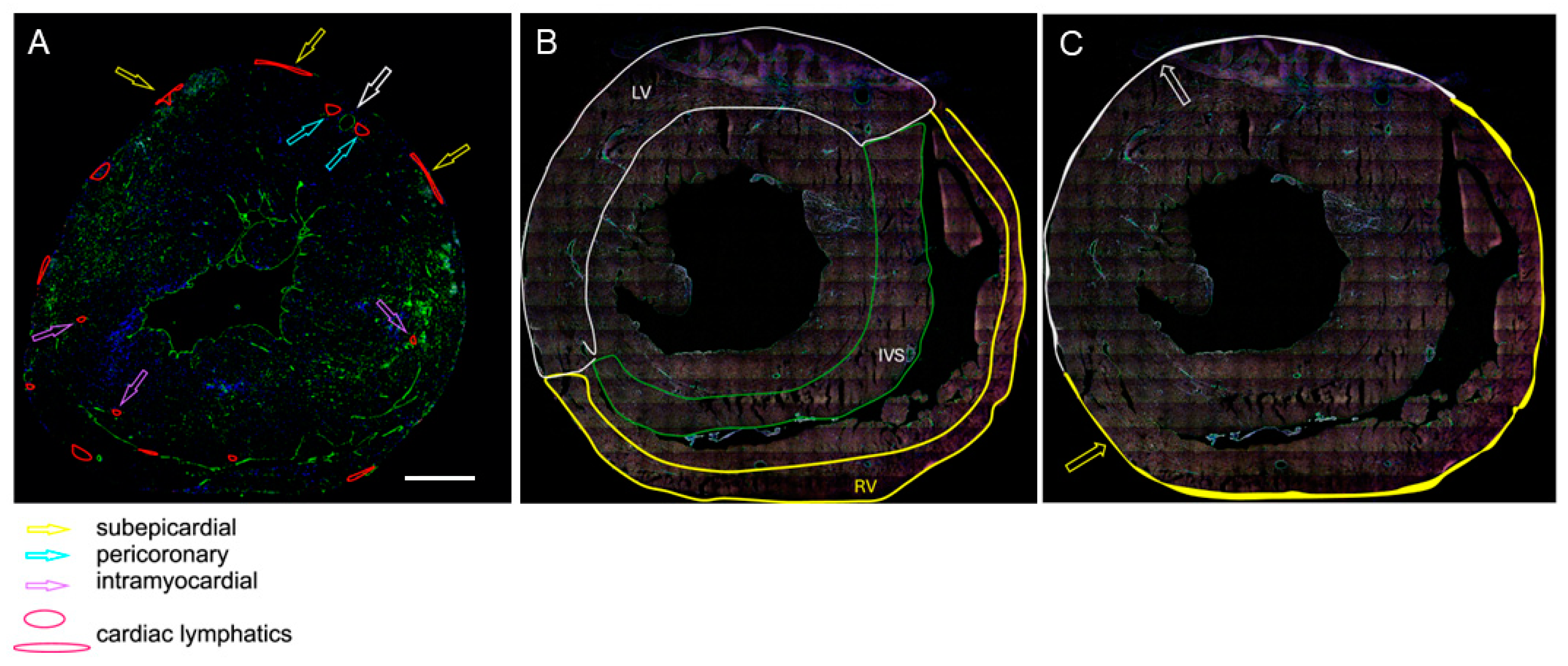
  • As the ratio of the number of LyVs to the number of cardiomyocytes within the same area of tissue. The assessment was carried out at three different locations: the outer half of the left ventricular wall, the outer half of the right ventricular wall, and the half of the interventricular septum (IVS) facing the right ventricle (RV). These areas were selected because cardiac LyV in mice are only located in certain regions of the heart, as shown in the schematic drawings in Figure 1A,B.
  • As the density of the subepicardial and intramyocardial LyVs, which was calculated as the number of LyVs per area of tissue (expressed in mm2) and the density of pericoronary LyVs, which was calculated as the ratio of the number of pericoronary LyVs to the number of all cross-sectioned coronary arteries in a given tissue section (a coronary artery is marked with the white-edged arrow in Figure 1A). The terms subepicardial, pericoronary, and intramyocardial were understood as follows: subepicardial LyVs (enclosed with white and yellow lines and marked with white- and yellow-edged arrows in Figure 1A,C) were located within the subepicardial area (defined as the distance between the epicardial mesothelium and the myocardial border of mesenchymal tissue); pericoronary LyVs (marked with blue-edged arrows in Figure 1A) were immediately adjacent to coronary vessels; and intramyocardial LyVs (marked with violet-edged arrows in Figure 1A) were in neither of these locations, instead being scattered in the myocardial wall.

2.5. Histology and Morphometric Analyses

Paraffin sections were stained with hematoxylin–eosin (H&E) and Picrosirius red for collagen (Coll) according to routine procedures [46,47]. Myocardial fibrosis was assessed on digital images taken under objective magnification of ×40 across the whole section of the left ventricular wall stained with Picrosirius red. Measurements were performed separately for the outer half (epicardial) and inner half of the heart muscle (endocardial) without scars. The red-stained areas in the tissue sections were assessed morphometrically with ZEN 3 v.6 software (Zeiss, Oberkochen, Germany).
Cardiomyocyte hypertrophy was calculated on the basis of their diameter measured at the level of the cell nucleus in the H&E-stained sections. For longitudinally cut cells, it was one measurement, and for transversely cut cells, the diameter value was calculated from two measurements with axes perpendicular to each other and the mean diameter of each cardiomyocyte was taken for further statistical analysis.

2.6. Inflammatory Cell Infiltration

The numbers of inflammatory cells (CD45+/CD68) and macrophages (CD45+/CD68+) were counted per a given area of the myocardium of the LV, RV, and IVS (in mm2) on frozen cross-sections immunolabelled with anti-CD45, anti-CD68 antibodies and WGA lectin with ZEN (Zeiss) software. Areas of wound healing/tissue scars and those located adjacent to coronary vessels were omitted from this calculation.

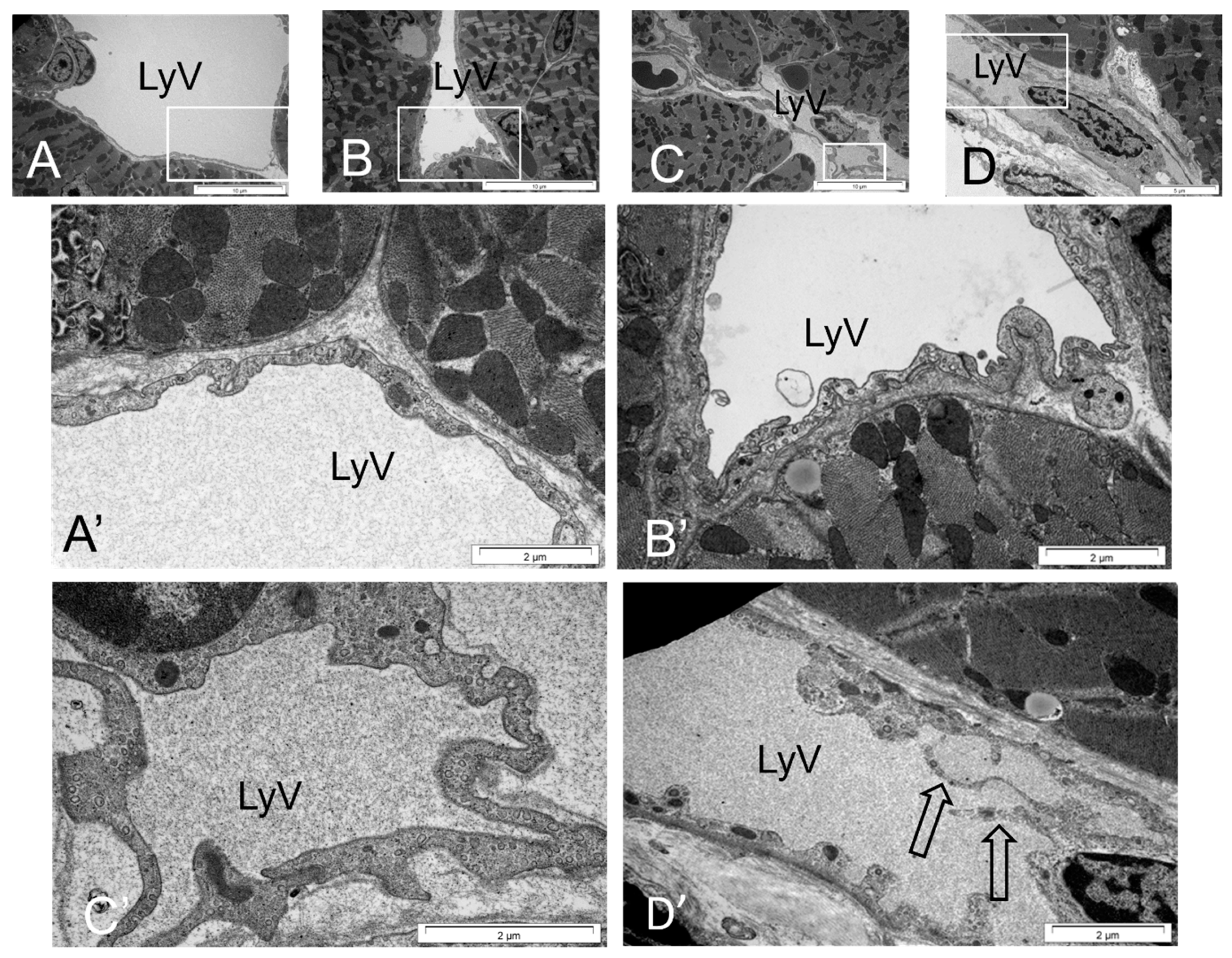
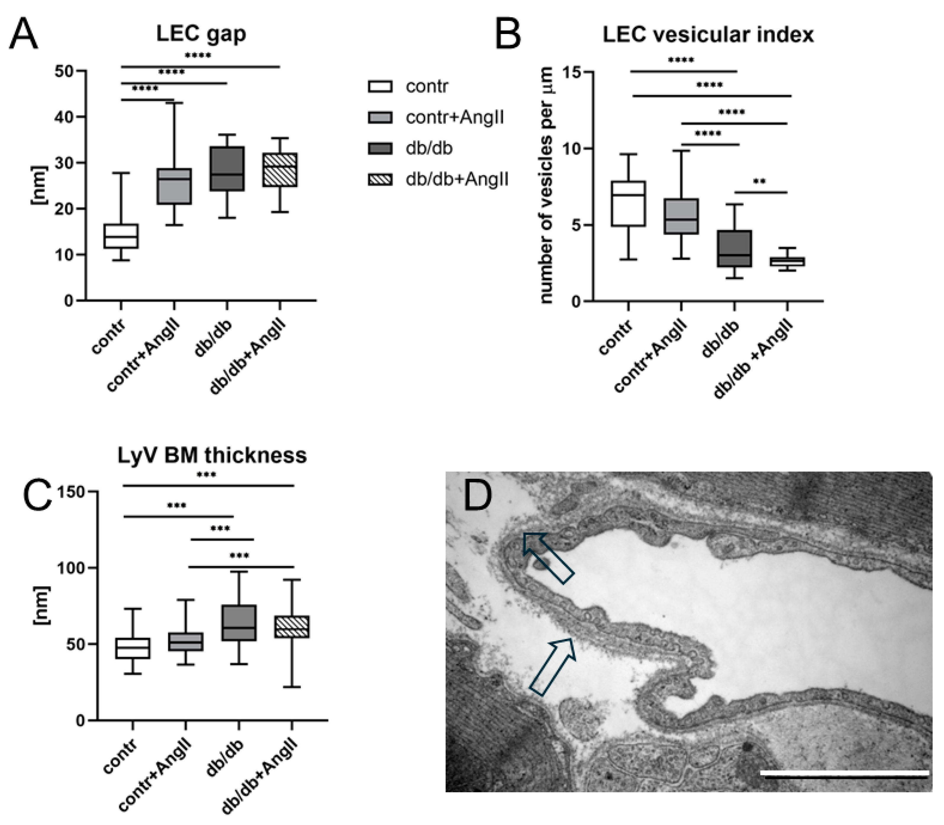
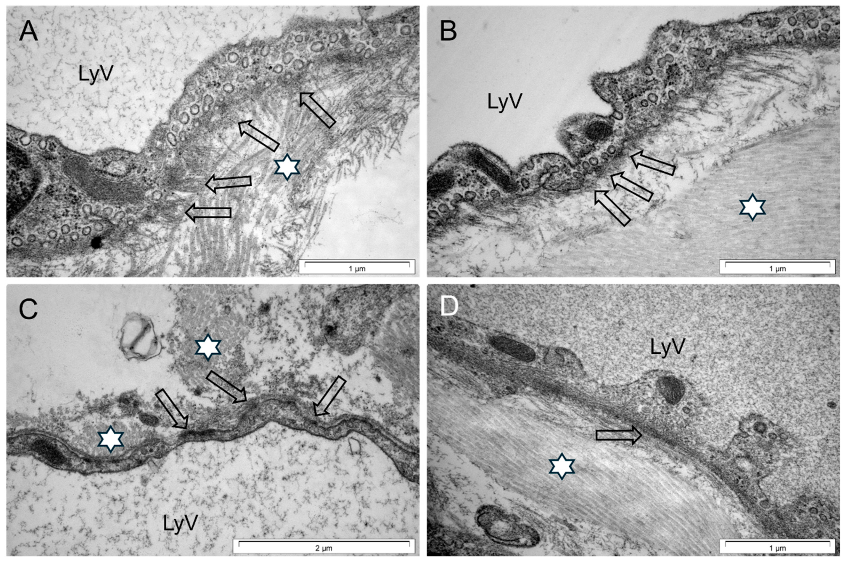
2.7. Ultrastructure and Morphometric Analysis

Karnovsky-fixed small tissue specimens from mouse hearts were postfixed in 1% buffered osmium tetroxide and processed for Epon embedding (Polysciences, Inc., Eppelheim, Germany), according to a published protocol [48]. Ultrathin sections were cut from selected areas potentially containing LyVs, contrasted with uranyl acetate and lead citrate, and examined by TEM (Jeol JEM 1011, Tokyo, Japan). Ultrastructural details of the LyVs were collected on images at 6000×, 10,000×, 20,000×, 40,000× and 60,000× magnifications. Next, the following measurements were performed with the iTEM (Olympus) morphometric program: dimensions of gaps in areas of intercellular junctions between the LECs of cardiac lymphatic microvessels, the thickness of the BM around the LyVs, the number of cytoplasmic vesicles per 1 µm of the cellular membrane length of the LEC (specified here as vesicular indexes), and the distance between the cell membrane edges in areas of the most closely spaced walls of blood capillaries and cardiomyocytes in the left ventricular wall (as presented in Supplementary Figure S1).
The measurements of the latter parameters were only performed in areas with transverse sections of cell membranes, which made it possible to assess the local tissue edema, according to published protocols [49,50]. Ultrastructural analyses were performed in precisely defined areas of the myocardium (as shown in Figure 1A,B).
Ultimately, for each tested parameter, at least 21 morphometric readings or measurements were performed for the parameter assessed in each study group. The number of readings limited the availability of cross-sections for the examined elements.

2.8. Statistics

Statistical analyses to establish the significance of the differences were performed with GraphPad Prism software version 8 (GraphPad Software). An independent t-test with Welch’s correction was used to analyze the differences between two groups when the data were normally distributed, and the Mann–Whitney test was used for nonnormally distributed data.
One- or two-way ANOVA was used to analyze the differences among multiple groups. The Kruskal–Wallis test, followed by Dunn’s multiple-comparison post-test was used when one or more groups did not show a Gaussian/normal distribution. Data are expressed as the mean ± SE. Statistical significance was set at <0.05. Details are described in the figure legends.

3. Results

3.1. Metabolic Anomalies in Obese Mice

Weekly weighing of the animals revealed a gradual increase in the body weight of the db/db and Ang II-treated db/db mice. The body weight of these animals was significantly higher during the development of the disease compared to the control ones but not to Ang II-treated control mice (Figure 2, where for clear presentation, only the last measurements are shown; details of the weekly measurements are presented in Supplementary Figure S2A,B). A markedly increased amount of subcutaneous and visceral adipose tissue was noted in all the db/db and Ang II-treated mice on the day of tissue collection.
Increased blood glucose levels were observed in the 11–14-week-old db/db mice compared to glucose levels in healthy individuals (Supplementary Figure S2C), and at 17 weeks in the Ang II-treated db/db mice compared to the controls and compared with the contr + Ang II-treated mice (Supplementary Figure S2D). A week before tissue collection, the blood glucose levels were higher in the db/db and in the Ang II-treated db/db mice compared with the control and higher in the db/db mice compared with the control + Ang II (Figure 2B). Of note, the blood glucose levels varied markedly between individuals from the db/db and Ang II-treated db/db mice (Supplementary Figure S2C,D).
Moreover, the levels of serum triglycerides (TGs) were increased in the db/db mice and further increased in the Ang II-treated db/db animals compared to the control individuals. Interestingly, the serum TG levels in the db/db + Ang II were not higher than the TG levels in the control + Ang II (Figure 2C).
Thus, comorbidities of obesity, such as hyperglycemia and dyslipidemia, were observed in the db/db and in Ang II-treated db/db animals, albeit hyperglycemia was not prominent at and after 15 weeks of age in the Ang II-treated db/db in comparison with the db/db mice.
What is more, the blood pressure (BP) did not change in the db/db mice over their 19 weeks of life, remaining significantly lower compared to the control and to Ang II-treated control mice (Supplementary Figure S2E,F). Nonetheless, the Ang II-treated control mice had markedly elevated systolic BP compared to the db/db when assessed at 19 weeks (Figure 2D and Supplementary Figure S2F). Astonishingly, by the end of the experiment, the systolic BP in the Ang II-treated db/db mice was lower compared with the BP in the contr + Ang II mice (Supplementary Figure S2F).

3.2. Heart Hypertrophy and Myocardial Remodeling

Cardiac hypertrophy was observed in the Ang II-treated control and Ang II-treated db/db mice compared to the control and db/db animals, as revealed by evaluation of the heart weight to tibial length ratio (Figure 3A). In the same groups, the cardiomyocyte diameters in the LV significantly increased, as revealed by morphometric measurements (Figure 3B). Moreover, the distances between the cardiomyocyte borders and the external blood endothelial cells (BEC) of the capillary walls were increased in the myocardium of the hearts of mice from all three experimental groups compared to the control, but especially in both groups of mice treated with Ang II (Figure 3C).
Of note, histopathological analysis indicated signs of cardiomyocyte injury visible as scattered clumps of eosinophilic material within the cytoplasm (Figure 4B) and the focal infiltration of inflammatory cells in the Ang II-treated animals (Figure 4B,C,G,H compared to Figure 4A,E). Remarkably, in areas devoid of granulation tissue and/or scars, macrophages of CD45+/CD68+ phenotype were less numerous in the LV of the db/db mice compared to the macrophage density in the Ang II-treated db/db mice (Figure 5), whereas the total number of CD45+ inflammatory cells (of CD45+/CD68+ and CD45+/CD68 phenotypes) was diminished in the db/db compared to the number in the Ang II-treated control mice (Figure 5). The density of macrophages (CD45+/CD68+) and other inflammatory cells (CD45+) was measured in areas devoid of scars and areas adjacent to coronary arteries.
Abundant collagen deposits around the coronary vessels (perivascular fibrosis) (Figure 4F,J) and in fibrotic foci/fibrous scars of various sizes (Figure 4H,L) were features of the Ang II-treated mice, both the control and db/db mice. Moreover, reactive interstitial fibrosis was additionally observed in the db/db, Ang II-treated control, and Ang II-treated db/db mice (Figure 4K,L) and confirmed in the Picrosirius red-stained sections when measured in the inner half (endocardial) and outer half (epicardial) of the left ventricular wall (Figure 6A,F). Moreover, there were regional differences in the collagen deposits, with more significant collagen deposits in the inner half compared to the outer half of the myocardial wall (Figure 6A–J). There was no difference in the interstitial collagen amount between the control and db/db animals in the outer half (epicardial) of the heart wall (Figure 6F).

3.3. Cardiac LyV Density and Structural Remodeling

The density of LyVs expressed as the ratio of the LyV number to the number of cardiomyocytes in the left ventricular and IVS myocardium was diminished in the db/db and Ang II-treated db/db mic, compared to this ratio in the wild-type animals (Figure 7A,C). Of note, the density of LyVs in the control mice receiving Ang II was higher than that in the Ang II-treated db/db mice and almost equal to the density in the control animals in the RV (Figure 7B).
Furthermore, the density of LyVs in subepicardial and pericoronary areas was almost the same among the four groups (Figure 7D,E), in contrast to the diminished density of LyVs in intramyocardial regions in the db/db and Ang II-treated db/db mice compared to the control (Figure 7F). Occasionally, LECs with morphological signs of injury were observed in the db/db and db/db mice treated with Ang II (Supplementary Figure S3).
The confocal microscopic images did not present marked differences in the LyV shapes and structures among the groups. The structural abnormalities in the cardiac LyVs in the experimental mice were rather subtle. In the db/db mice, odd-shaped vessels in myocardial locations were seen compared to the shapes of the LyVs in the control animals. Importantly, in the Ang II-treated control and db/db mice, LyVs positioned in pericoronary locations were partially embedded in a thickened fibrous tissue of adventitia and were mostly of oval shapes (Figure 8B,D).
TEM analyses revealed details of the altered LyV wall shapes in the Ang II-treated control (Figure 9B,B′) and db/db mice (Figure 9C,C′) compared with the controls and with the contr + Ang II mice (Figure 9A,A′). The vessel walls in the myocardium of the db/db animals and in both Ang II-treated groups were wavy and undulating, in contrast to those observed in the control mice (Figure 9C vs. Figure 9A). Moreover, in the Ang II-treated db/db mice, long cytoplasmic processes protruding toward the vessel lumina were often visible (Figure 9D,D′). In addition, the overlapping flaps of the LECs were altered in the experimental groups vs. control, i.e., the flaps were often long and wavy, especially those projecting into the vessel lumen (Supplementary Figure S4B–D, vs. Figure S4A). The intercellular junctions of these flaps had larger gaps compared to those in the control mice (Supplementary Figure S4A vs. Figure S4B–D and Figure 10A).
Moreover, collagen fibers were visible in the immediate vicinity of the flaps in the db/db mice and in both groups of Ang II-treated mice (Supplementary Figure S4B–D).
These changes were accompanied by the altered distribution and structure of tufts with AnchFs that connect the LyV walls with the ECM. The AnchFs were shorter and formed abnormally oriented tufts in the Ang II-treated control and db/db mice compared to those visible in the control, and tufts were almost absent in the Ang II-treated db/db mice (Figure 11B–D, vs. Figure 11A). At the same time, there was also an increase in collagen deposits directly adjacent to the LyV walls in the experimental groups compared to the control, with the most dense masses of collagen in the Ang II-treated db/db mice (Figure 11D). The thickness of the BM surrounding the initial lymphatics was significantly increased in the db/db and Ang II-treated db/db mice compared to the control (Figure 10C). A significantly thicker BM surrounded the LyVs in the Ang II-treated db/db mice than in the Ang II-treated control mice (Figure 10C). Occasionally, the BM around the LyVs in the Ang II-treated db/db mice was multilayered (Figure 10D).
In addition, transcellular transport, as assessed by the vesicular index, was significantly decreased in the db/db, Ang II-treated control, and Ang II-treated db/db mice compared to the control (Figure 10B), with the smallest number of cytoplasmic vesicles detected in the Ang II-treated db/db mice.

4. Discussion

Our data provide a new insight into the morphological features of cardiac LyV remodeling associated with myocardial tissue remodeling in obese db/db mice and after Ang II treatment. Our main findings are that cardiac LyV remodeling is an integral component of myocardial remodeling. Collective image analysis indicates that ultrastructural changes in lymphatic initials are closely related to ECM alterations. In addition, Ang II treatment of db/db mice intensifies the unfavorable remodeling of the myocardium and its lymphatic vessels compared with Ang II treatment of control mice, indicating the importance of obesity and the associated metabolic environment for these processes.

4.1. Metabolic Anomalies in Obese Mice Create Specific Environment

Obesity was a symptom present in the db/db and Ang II-treated db/db mice, but a higher blood pressure was observed in only the Ang II-treated control compared to the control mice in our studies. Thus, the morphological cardiac changes reported in our study were not dependent on Ang II-induced and obesity-related hypertension, which is consistent with other reports [51]. The Ang II-treated control mice exhibited high variabilities in blood pressure among individuals within the period of measurements, the highest at 19 weeks and almost equal to the level of blood pressure level in the Ang II-treated db/db mice at 15–17 weeks.
Our data on the blood glucose concentrations in the db/db and in Ang II-treated db/db varied among individual mice and showed a transient glucose level increase in the db/db mice compared to the control in the early phases of the disease (10 to 14 weeks of age), but these data generally do not confirm a diabetic phenotype in obese mice in the later stages (15 to 19 weeks, db/db and Ang II-treated db/db) (Supplementary Figure S2D), contrary to what has been reported by others in similar animal models [52] and in db/db mice [53]. This discrepancy might be explained by the blood sample collection during the progression of disease in these mice: blood samples collected from the tail might result in different results from those detected in blood collected from large blood vessels, such as the saphenous vein or heart chambers [52].
The high concentration of blood TGs in db/db and especially in Ang II-treated db/db mice is considered a marker of dyslipidemia and one of the MetS comorbidities [54]. In our experiment, the levels of serum TG were increased in the db/db mice and further increased in the Ang II-treated db/db animals compared to the control individuals. Earlier studies reported a 1.5- to 2-fold increase in the TG amount in db/db and leptin-deficient ob/ob mice compared to control mice [55]. Considering the above, it should be explained that db/db mice themselves create a tissue environment for a serum TG level increase due to TG production in the liver and adipose tissue [56]. Moreover, a continuous infusion of Ang II to Wistar rats for 14 days stimulates hepatic TG production in a dose-dependent manner, increasing the TG concentration in the blood [57]. TG production is downregulated after Ang II receptor blockage and correlates with insulin resistance in various animal models [58,59]. Therefore, Ang II enhances TG production in db/db mice.
The Ang II-treated db/db mice demonstrated increased systolic BP compared to the non-treated db/db ones at 19 weeks; however, it was less pronounced compared to what was observed by van Bilsen in Ang II-treated db/db animals, possibly due to the use of higher doses of Ang II (1 µg/min/kg) [52] than in our experiments (500 ng/min/kg). Our observation of low BP in the db/db mice compared with that in the controls is consistent with that reported by van Bilsen et al. [52], but in contrast to earlier studies by Gonclaves et al., who showed that the BP measured in db/db mice by radiotelemetry is higher compared to the BP in control mice. Moreover, the authors did not observe circadian variations in db/db mice (so-called “non-dipping” arterial pressure), contrary to the controls, which exhibited lower arterial pressure during daytime [60].
These MetS comorbidities are reported to be followed by diastolic dysfunction and HF with preserved ejection fraction (HFpEF) [61,62,63].

4.2. Heart Remodeling Is Related to Ang II

The observed heart and cardiomyocyte hypertrophy in the Ang II-treated control and Ang II-treated db/db mice seem to be dependent on Ang II signaling. A similar effect of Ang II on cardiac hypertrophy was observed by van Bilsen et al. [52]. Our results indicate that the hypertrophic heart phenotype in the Ang II-treated db/db mice was associated with cardiomyocyte hypertrophy, increased fibrosis and myocardial tissue edema. Such features of cardiac remodeling are consistent with the Ang II influence described in the literature, as reviewed by Forrester et al. [14]. Ang II induces an inflammatory response and oxidative stress [64,65], which further triggers fibrosis and cardiomyocyte hypertrophy [66,67,68]. In fact, inflammatory signaling in obesity and diabetes, which is considered a state of chronic subclinical sterile inflammation, may stimulate cardiac hypertrophy [69,70] and Ang II could further intensify this process [66,67,68]. Of note, fewer macrophages infiltrated the myocardial interstitium of the db/db mice compared to the db/db mice treated with Ang II and fewer inflammatory cells were found in the myocardial interstitium of the db/db compared to the Ang II-treated control mice. In other words, the number of interstitially distributed inflammatory cells (without scars and granulation tissue) increased significantly under Ang II treatment in the control versus db/db mice. Similarly, van Bilsen et al. [52] reported significantly increased numbers of inflammatory cells (CD45+) in control mice treated with Ang II, whereas the levels of these cells remained unchanged in our study in the db/db and Ang II-treated db/db vs. control mice. These results are in slight contrast to ours, probably due to the counting of inflammatory cells in areas of the myocardium including granulation tissue/scars and presumably due to the age of animals and doses of Ang II. The intensity of the inflammatory cell infiltration and inflammatory cell profile may change during the progression of disease. For example, Papinska et al. [71] reported an enormous increase in inflammatory cells (polymorphonuclear cells) in db/db mice vs. control at 11 weeks. Moreover, van Bilsen et al. did not observe altered expression of mRNA for the inflammatory marker nuclear factor-kappa B inhibitor-alpha in the myocardium and found a significant decline in the IL-6 mRNA levels in db/db mice vs. control mice. In addition, van Bilsen’s group reported that the level of cardiac phosphorylated AMP-activated protein kinase (pAMPK) was lower in Ang II-treated db/db mice compared to controls, which did not confirm cardiac metabolic remodeling [52]. It is known that Ang II action involves TGF signaling in fibroblasts and cardiomyocytes, thus inducing both cardiac fibrosis and cardiomyocyte hypertrophy, and is involved in the inflammatory response (reviewed in [72]). Moreover, cardiomyocyte hypertrophy in Ang II-treated db/db mice might depend on impaired leptin signaling [73,74], which is a characteristic symptom in Leprdb/db mice [53,75]. Leptin has been reported to prevent cardiomyocyte hypertrophy by a pathway involving stimulation of cardiac nitric oxide signaling [76,77]. In our study, this pathway did not seem to influence Ang II-triggered cardiomyocyte hypertrophy since we also detected hypertrophy in the Ang II-treated control animals with the functional leptin receptor.
Myocardial fibrosis in the diffuse interstitial and perivascular forms, as well as within the microscopic scars, was only present in the Ang II-treated control and Ang II-treated db/db mice, albeit in the db/db animals (vs. control) a significantly increased diffuse interstitial fibrosis was also found in the inner half (endocardial) of the myocardial wall (Figure 6A). It is known that these types of fibrosis are associated with pathological cardiac remodeling in hypertension [78]. The increased myocardial fibrosis and edema in Ang II-treated db/db mice potentiating myocardial stiffness provide a specific mechanical “shear stress”. This stress influences fibroblast activity through a mechanism involving Ang II/AT1 receptor signaling [79]. Furthermore, the cross-linking of collagen fibers [80] and increased ratio of type I collagen (thick fibers resistant to tearing) to type III collagen (thin fibers with elastic properties) lead to an increased LV stiffness, slowing the rate of relaxation [81]. Since cardiac lymphatic initials drain extracellular fluids with each cardiac contraction–relaxation cycle, propelling the lymph from the endocardial to the epicardial portion of the heart and from the apex to the base [82,83], the impairment in heart diastole due to the stiffness of the myocardial tissue may affect the cardiac lymph flow velocity by retarding it [23].

4.3. Cardiac LyV Density Is Related to Obesity

The decrease in the LyV density in the Ang II-treated db/db mice compared to the controls but similar to the density in the db/db mice observed in our study suggests the significance of the environment associated with obesity and metabolic anomalies for vessel regression and/or impaired lymphangiogenesis. LyV regression may occur in this environment, as indicated by signs of injury observed in the LEC of the Ang II-treated db/db mice (Supplementary Figure S3A). In the literature, LyVs rarefaction in MetS has been shown in skin tissue in patients [84] and in obese mice, both with genetic and diet-induced obesity [85], suggesting the significance of obesity for LyV rarefaction. However, the dose and duration of Ang II administration, and other associated factors such as elevated plasma VEGF levels, are crucial for lymphangiogenesis. It has also been reported that Ang II infusion in mice leads not only to increased lymphangiogenesis and increased LyV permeability but also to myocardial edema, and these effects are dose- and time-dependent [16]. Similarly, Song et al. [86] observed that Ang II treatment for 6 weeks causes increased lymphangiogenesis, high VEGF expression and impaired lymph transport within lymphatics. Moreover, Lin et al. have reported that Ang II, acting through the AT1 receptor, promotes the expression of lymphatic genes such as Lyve-1, Prox1, VEGF-C, and VEGFR3 and stimulates the proliferation and migration of LECs in vitro [15]. This type of LyV remodeling may also involve an altered balance between proliferation and death of LECs under the influence of Ang II particularly, because obesity and other MetS comorbidities can accelerate apoptosis, including apoptosis of endothelial cells [87].
The relationship between the amount and remodeling of fibrotic tissue into more stiff collagen fibers and LyV rarefaction should be also taken into account, since the ECM provides a physical and biological environment for the formation and stabilization of the LyV network. It is known that the ECM mediates external mechanical signals into intracellular pathways via the cytoskeleton of the lymphatic endothelial cells, regulating LEC proliferation, vessel wall barrier tightness and other functions [39,88]. Although we did not examine the ECM composition in detail, the TEM imaging allowed us to confirm the presence of numerous thick collagen I fibers, in particular in the Ang II-treated db/db mice, whose presence is associated with an increased myocardial stiffness [89]. In addition, the increased tension of the ECM and cardiac wall may contribute to damage or apoptosis of the LECs [90].
Moreover, the BM, a component of the ECM, becomes significantly thickened in cardiac LyVs of db/db and Ang II-treated db/db mice compared to controls (Figure 10C). Since the major molecules of BM are collagen IV (Coll IV) and laminin [91,92], we presume that the BM thickness also relates to the increase in the structural stiffness of LyVs. This is especially true in relation of reports on Coll IV being considered an “inelastic” molecule [92,93,94]. Our results concerning the thickened BM in the Ang II-treated db/db mice are in line with the study by van Bilsen, who demonstrated a significantly increased level of mRNA for Coll IV in the myocardium of Ang II-treated db/db mice [52]. In addition, the BM determines the distribution of AnchFs, since AnchFs are visible only on the “bare” cell membrane and play a role in the recruitment of growth factors and interactions with cell receptors; the BM components also regulate cell proliferation [95]. The abnormal structure and disruption of the BM may be caused by pathological MAPK activation [96]. MAPK activation triggered by Ang II binding to the AT1 receptor stimulates multiple downstream pathways important for the progression of vascular remodeling, hypertension and other cardiovascular diseases (e.g., atherosclerosis) [97]. Our findings indicate that Ang II exposure enhances the thickness of the BM; moreover, it stimulates the formation of the multilayered and thickened BM discontinuously adjoining the LyV walls (Figure 10D), which is often also visible in db/db mice. This may be an effect of a prolonged exposure to abnormal stimuli found in diabetic nephropathy and has been discussed as an adaptive or maladaptive feature [98], since a normal BM is necessary for the proper function of a cellular barrier.
The unaltered density of lymphatics located adjacent to the coronary arteries (pericoronary location) in the db/db and Ang II-treated db/db mice compared to the controls indicates that no lymphatic vessel regression takes place in this area. This would suggest that the proximity of coronary arteries provides a protective effect on the survival of adjacent lymphatics in normal and in obese environments; however, this occurs by an unknown mechanism. Presumably, pulsation of the coronary arteries themselves and/or stimulation of LyV activity by myocardial contraction–relaxation cycles provides a supporting mechanism for continuous lymph flow and therefore contributes to LEC survival.

4.4. LyV Remodeling Is Part of Cardiac Remodeling

Several processes are associated with the structural remodeling of LyVs: rarefaction of lymphatic capillaries and changes in their wall shape and wall structure.
In our studies, the shape of the vessels was clearly different in the db/db and in Ang II-treated db/db mice (vs. the control), presumably depending on the altered myocardium, including the interstitial tissue, due to specific environmental conditions. We observed folding of the LyV walls in the control mice treated with Ang II, which also exhibited increased myocardial fibrosis, variable irregular shapes in the db/db mice with a moderate increase in the collagen amount in the interstitial area, and oval shapes in the tissue with intensive collagen deposition adjacent to LyVs in the Ang II-treated db/db mice. These differences in the LyV shape may relate to the different activities of MMPs and collagen composition, as indicated by the results of other researchers, including differences in the mRNA expression for matrix metalloproteinase 2 (MMP2), Coll I and Coll III [52]. The proximity of collagen deposits may be the reason for the preserved oval shape of the initials. This may make these vessels resistant to elevated interstitial fluid pressure. Similar observations have been reported in reference to lymphatic vessels in the hearts of hypertensive Dahl S rats [38].
The main structural anomalies in the LyVs were expressed by abnormal organization of AnchFs into bundles and almost a complete absence of AnchFs in the Ang II-treated db/db mice; the marked expression of structural features related to impaired transport of fluids, molecules, and cells through the wall, i.e., widened gaps at intercellular junctions, in the obese and Ang II-treated mice; and a significant decrease in the number of cytoplasmic vesicles in both groups of obese mice.
The functional significance of AnchFs for primary valve function is still discussed [99,100]. It has been hypothesized that high interstitial fluid pressure acts on AnchFs and thus opens pull-up flaps without the need for LEC junction dissolution to facilitate the transport of fluid, solutes, and cells via the paracellular route [26,27,28,99,101,102]. However, there are also other suggestions for the role of AnchFs, i.e., maintenance of the LyV shape or the transduction of signals from local forces [100].
Previous observations by Danussi and Pivetta [35,103] demonstrated a relationship between the absence of anchoring filament protein Emilin-1 and dysfunctional LyVs, as well as an improvement of LyV function after restoring anchoring filament protein expression. Another protein that constitutes AnchFs is fibrillin, which is involved in mechanotransduction and sensing of lymph pulsatile movement, and its presence is crucial for normal LyV function [104,105].
Therefore, the alterations in or disappearance of AnchFs may reflect the impaired absorbing function of LyVs, as reported by others in transgenic animal models, in cancer, and in inflammation [27,106,107,108,109].
Moreover, the observed alterations in the AnchFs in our study are accompanied by anomalies in the LEC flaps in both the Ang II-treated controls and Ang II-treated db/db mice. A normal ECM generates passive tension, which protects against excessive myocardial stretching as well as allows the opening of the LEC flaps for fluid and solute drainage from the ECM during increased interstitial pressure due to fluid accumulating in the extracellular space [110]. In contrast, a stiff ECM impairs force transmission during cardiac systole and diastole [111] and affects the actin filament alignment in the LECs, leading to rupture of their anchoring points [112,113]. We do not know whether such a process occurs in mice treated with Ang II. In fact, the dense masses of collagen located in the vicinity of the flaps in the Ang II-treated db/db mice certainly induced stiffness in this area and impeded the movement of the flaps, already limited by the AnchF alterations. In addition to that, edema was observed in these hearts.
Therefore, structural anomalies in the anchoring system together with altered morphology of LEC flaps may contribute to LyV dysfunction and may result from fibrosis.
The flexibility of the ECM, resulting primarily from its composition around the vessel wall, is known to influence the tightness of intercellular junctions. The widening of intercellular junctional gaps in the db/db and Ang II-treated db/db mice compared to the controls may reflect the status of “leaky vessels” (increased paracellular permeability). It may also indicate the loosening of “buttons” important for sealing the hinge regions of primary lymphatic valves (LEC flaps). Recent studies [114] on tissue-engineered LyVs indicate that ECM flexibility and pulling forces regulate junctional contacts. The culture of engineered lymphatics on Coll I (rigid matrix) induced a “leaking” phenotype compared to culture on a fibronectin/Coll I matrix (more flexible), on which the LECs are connected by more tightened junctions. Thus, ECM alterations accompanying obesity could contribute to the loosening of connections between LECs.
A “leaking” phenotype of LyVs is also associated with an anomaly in transcytosis reflected by the activity of the cytoplasmic vesicular transcellular route [29,115]. Thus, the decreased vesicular indices in the LECs of both db/db groups in our study indicate impaired transcytosis compared with that in the Ang II-treated control mice and healthy controls. Importantly, vesicular transport in LECs spectacularly decreases in Ang II-treated mice compared with db/db mice, as visualized by morphological signs of a lower number of cytoplasmic vesicles (Figure 10B). According to the mechanism described by Miteva et al., this diminished vesicular transport could result from an insufficiency or lack of mechanical stimuli for transmural flow [116] and might be correlated with paracellular leakage like in blood capillaries, especially because transcellular transport is performed by cytoplasmic vesicles containing caveolin-1 and clathrin [29,117], and LECs derived from caveolin-1 knockout (Cav1 KO) mice demonstrate increased paracellular permeability in vitro and decreased intralymphatic transport velocity of labelled molecules to sentinel lymph nodes [118].
The long protrusions of LECs into the lumina of LyVs in the Ang II-treated db/db mice observed in our study could be additional morphological evidence of the functional maintenance of the wall tension, crucial for lymph propulsion and preventing lymph backflow. Similarly, protrusions into LyV lumina, although less numerous, were observed in hypertensive Dahl salt-sensitive rats and were regarded as an adaptive response to hypertension [38]. The long protrusions in the LECs together with regression of lymphatics and leakage of their walls must lead to impairment in the drainage of fluid in the cardiac tissue. Moreover, the oval shape of these initials may be a result of reduced lymph flow due to the stiffness of the myocardial wall. Thus, the morphological defects in the LyVs in obese mice shown in our study are associated with the pathophysiological basis of myocardial edema and fibrosis [110].

5. Concluding Remarks

Our observations suggest that LyVs are highly plastic structures that became altered in obesity associated with metabolic anomalies and increased levels of Ang II, which is presumably followed by their impaired function. Moreover, we observed a significant increase in the adverse effects of Ang II on LyVs in obese individuals. These effects included structural features, indicating LyV leakage and abnormal AnchFs. Leaking LyVs can contribute to edema and fibrosis, adversely interplaying with microvascular rarefaction and permeability. Thus, structural changes associated with metabolic anomalies and functional alterations of the heart may be involved in the development of cardiac dysfunction described by others [119,120,121]. In addition, the morphological remodeling of cardiac LyVs during advanced obesity is an important element when we consider the etiology and progression of metabolism-associated cardiac and systemic diseases.
Consequently, LyV remodeling should be considered an important symptom of obesity apart from other structural abnormalities of the heart, such as blood capillary rarefaction, cardiomyocyte remodeling and death, fibroblast activation and others. Revealing these structural alterations can be the cornerstone for further studies in this field, with the aim of elucidating the pathophysiology and mechanism(s) triggering these adverse changes. We believe that mitigating these alterations tested in various animal models should become objectives of novel therapies of HF in obesity and other MetS symptoms; however, this warrants further investigation.

6. Limitations of the Study

We investigated cardiac lymphatic system remodeling under the influence of Ang II in db/db metabolic environments using histopathological imaging techniques (light microscopy, confocal microscopy and electron microscopy) in combination with morphometric methods. Although these techniques allowed us to assess the similarities of the morphological changes associated with cardiac hypertrophy, this was not sufficient to explain what mechanisms are behind the differences in the response to Ang II in the control and db/db mice due to marked individual variabilities in the blood glucose and BP levels within all the study subgroups.
Furthermore, there is no explanation for the lack of increase in the BP after Ang II administration in the db/db mice despite this treatment being followed by extremely severe interstitial fibrosis with fibrous scar formation. The inter-individual BP variability and decreased BP in the db/db mice compared with those in the control and in the Ang II-treated control mice seem to be related to the tail-cuff measurement technique and not to the telemetric method [122].
Our morphological analyses suggest that quantitative and qualitative changes in cardiac fibrotic tissue profoundly affect cardiac LyVs. However, the extent to which LyV anomalies reflect the consequences of the many pathophysiologic factors associated with obesity remains unclear. However, db/db mice may be particularly susceptible to developing increased fibrosis due to impaired leptin signaling and high blood glucose levels, which are implicated as factors inducing TGF-β synthesis [123]. We assume that advanced glycation end products (AGEs) increase the ECM stiffness in obesity and metabolic dysfunction, as demonstrated in numerous studies (reviewed by Cavalera M et al. [124] and Hartog et al. [125]). However, we did not measure the AGE levels. These products mediate the crosslinking of interstitial proteins, such as collagen and laminin, and activate interstitial fibroblasts. However, van Bilsen (2014) found neither increased AGE levels nor increased fibrosis in Ang II-treated db/db mice.
In addition, our study should be expanded in the future by the use of the mRNA NGS technique to examine isolated myocardial LECs to find out to what extent LEC abnormalities are related to fibrotic remodeling, the inflammatory response, or other factors.

7. Future Directions

It is important to consider that the observed changes in the LyV wall shapes in the experimental versus control groups might be related strictly to AnchF abnormalities. AnchFs are important structural elements of LyVs, since they bind the LECs of the initials to the ECM molecules [100,126,127,128,129,130,131].
Considering the above observations of AnchF alterations in the ECM, it can be stated that the initial LyVs in all organs, including the heart, are more dependent on the ECM than blood microvessels [88]. Therefore, exploring in detail the composition of the ECM located adjacent to LyVs via modern techniques, such as in situ single-cell mRNA sequencing [132], would help identify the composition of novel proteins or other molecules and be a milestone for further research in this field. Finally, a cyto-map in situ [133,134] or a multiplex imaging technique [135] performed in a confocal microscope on tissue sections would help elucidate the triggering molecular signals triggered in the “obese” or “MetS” tissue environment compared with those in controls. Finally, the heretofore neglected functional and structural LyV remodeling should be taken into consideration as contributing factors in altered tissue fluid and solutes homeostasis, edema, and fibrosis during progression of obesity/MetS, which lead to heart failure.
Future functional experiments based on tracer injection in 3D reconstructed lymphatic capillaries, as reported by Gibot et al. [136], cultured in a MetS milieu (high TG, high blood glucose, and high AGE levels) or in situ with fluorescent tracer injection into and its detection in the myocardium compared to its retention by LyVs in obese versus control animals [22] would shed a new light on the AnchF and LEC activity in this tissue environment.
The increased thickness of the BM surrounding LyVs in experimental groups versus controls is also of great relevance in terms of the molecular interaction of LECs with the ECM. Of note, no studies were performed on the functional importance of the BM thickness and molecular ECM changes as factors contributing to myocardial wall stiffness, apart from increased collagen deposition. Both BM thickening and collagen deposits lead to diastolic dysfunction. Moreover, it would be interesting to explore the role of BM thickening in the decreased permeability of vessels in obesity and other MetS components.

Supplementary Materials

The following supporting information can be downloaded at https://www.mdpi.com/article/10.3390/app14198675/s1.

Author Contributions

Conceptualization: E.C., E.J.-S., J.N.-B., T.Ż. and A.R.; data curation: A.F.-Z., E.C., E.J.-S., J.N.-B., T.Ż., A.M., K.B., M.T. and A.R.; formal analysis: A.F.-Z., E.C., E.J.-S., J.N.-B., T.Ż., A.M., M.B., K.B., M.T., B.M. and A.R.; funding acquisition: A.F.-Z., K.B. and A.R.; investigation: E.J.-S.; methodology: A.F.-Z., E.C., E.J.-S., J.N.-B., T.Ż., A.M., M.B., K.B., M.T., B.M., D.K., H.K., B.C., M.G. and A.R.; project administration: A.R.; resources: T.Ż. and B.C.; software: A.F.-Z., J.N.-B., A.M., M.B., K.B. and B.M.; supervision: E.C. and A.R.; validation: E.C., E.J.-S., J.N.-B., T.Ż. and A.R.; visualization: A.F.-Z., J.N.-B., M.B., H.K., B.C. and M.G.; writing—original draft: A.F.-Z., E.C., E.J.-S., J.N.-B., T.Ż., M.T., D.K., A.C.-J. and A.R.; writing—review and editing: E.C., E.J.-S., J.N.-B., T.Ż., B.M., A.C.-J. and A.R. All authors have read and agreed to the published version of the manuscript.

Funding

This research has received funding from the ERA-CVD Research Program, which is a translational R&D program jointly funded by national funding organizations within the framework of the ERA-NET ERA-CVD, funding agency: NCBR, Poland, grant number: ERA-CVD/LyMitDis/1/2017 (to AR); the National Science Center grant no UMO-2016/21/N/NZ5/01919 (to—AF-Z); and the Ministry of Science and Higher Education, grant no: D/2018020048, “Diamond” grant (to—KB).

Institutional Review Board Statement

The animal study protocol was approved by the First Local Committee for Ethics in Animal Experiments in Warsaw (at the University of Warsaw, Poland), protocol code 140/2016, 16 November 2016.

Informed Consent Statement

Not applicable.

Data Availability Statement

The original contributions presented in the study are included in the article and Supplementary Materials, further inquiries can be directed to the corresponding author.

Acknowledgments

The authors thank Józef Dulak, Jagiellonian University, Kraków, for granting us access to his lab and lab equipment. The excellent technical support of Barbara Tomaszyńska (Department of Histology and Embryology, Medical University of Warsaw), Emilia Kruk (Department of Pathology, Medical University of Warsaw), and Grzegorz Pałka (Laboratory of Advanced Microscopy Techniques Mossakowski Medical Research Institute) is appreciated.

Conflicts of Interest

The authors declare no conflict of interest.

References

  1. Reilly, M.P.; Rader, D.J. The metabolic syndrome: More than the sum of its parts? Circulation 2003, 108, 1546–1551. [Google Scholar] [CrossRef]
  2. Alberti, K.G.; Eckel, R.H.; Grundy, S.M.; Zimmet, P.Z.; Cleeman, J.I.; Donato, K.A.; Fruchart, J.C.; James, W.P.; Loria, C.M.; Smith, S.C., Jr. Harmonizing the metabolic syndrome: A joint interim statement of the International Diabetes Federation Task Force on Epidemiology and Prevention; National Heart, Lung, and Blood Institute; American Heart Association; World Heart Federation; International Atherosclerosis Society; and International Association for the Study of Obesity. Circulation 2009, 120, 1640–1645. [Google Scholar] [CrossRef] [PubMed]
  3. Huang, P.L. A comprehensive definition for metabolic syndrome. Dis. Model. Mech. 2009, 2, 231–237. [Google Scholar] [CrossRef] [PubMed]
  4. Grundy, S.M. Metabolic syndrome update. Trends Cardiovasc. Med. 2016, 26, 364–373. [Google Scholar] [CrossRef] [PubMed]
  5. Balasubbramanian, D.; Mitchell, B.M. Lymphatics in Cardiovascular Physiology. Cold Spring Harb. Perspect. Med. 2022, 12, a041173. [Google Scholar] [CrossRef]
  6. Scallan, J.P.; Hill, M.A.; Davis, M.J. Lymphatic vascular integrity is disrupted in type 2 diabetes due to impaired nitric oxide signalling. Cardiovasc. Res. 2015, 107, 89–97. [Google Scholar] [CrossRef]
  7. Wu, H.; Rahman, H.N.A.; Dong, Y.; Liu, X.; Lee, Y.; Wen, A.; To, K.H.; Xiao, L.; Birsner, A.E.; Bazinet, L.; et al. Epsin deficiency promotes lymphangiogenesis through regulation of VEGFR3 degradation in diabetes. J. Clin. Invest. 2018, 128, 4025–4043. [Google Scholar] [CrossRef]
  8. Cifarelli, V.; Appak-Baskoy, S.; Peche, V.S.; Kluzak, A.; Shew, T.; Narendran, R.; Pietka, K.M.; Cella, M.; Walls, C.W.; Czepielewski, R.; et al. Visceral obesity and insulin resistance associate with CD36 deletion in lymphatic endothelial cells. Nat. Commun. 2021, 12, 3350. [Google Scholar] [CrossRef]
  9. Zawieja, S.D.; Gasheva, O.; Zawieja, D.C.; Muthuchamy, M. Blunted flow-mediated responses and diminished nitric oxide synthase expression in lymphatic thoracic ducts of a rat model of metabolic syndrome. Am. J. Physiol. Heart Circ. Physiol. 2016, 310, H385–H393. [Google Scholar] [CrossRef]
  10. Nitti, M.D.; Hespe, G.E.; Kataru, R.P.; Garcia Nores, G.D.; Savetsky, I.L.; Torrisi, J.S.; Gardenier, J.C.; Dannenberg, A.J.; Mehrara, B.J. Obesity-induced lymphatic dysfunction is reversible with weight loss. J. Physiol. 2016, 594, 7073–7087. [Google Scholar] [CrossRef]
  11. Chakraborty, A.; Barajas, S.; Lammoglia, G.M.; Reyna, A.J.; Morley, T.S.; Johnson, J.A.; Scherer, P.E.; Rutkowski, J.M. Vascular Endothelial Growth Factor-D (VEGF-D) Overexpression and Lymphatic Expansion in Murine Adipose Tissue Improves Metabolism in Obesity. Am. J. Pathol. 2019, 189, 924–939. [Google Scholar] [CrossRef]
  12. Chakraborty, A.; Scogin, C.K.; Rizwan, K.; Morley, T.S.; Rutkowski, J.M. Characterizing Lymphangiogenesis and Concurrent Inflammation in Adipose Tissue in Response to VEGF-D. Front. Physiol. 2020, 11, 363. [Google Scholar] [CrossRef] [PubMed]
  13. Wang, L.; Zhang, Y.L.; Lin, Q.Y.; Liu, Y.; Guan, X.M.; Ma, X.L.; Cao, H.J.; Liu, Y.; Bai, J.; Xia, Y.L.; et al. CXCL1-CXCR2 axis mediates angiotensin II-induced cardiac hypertrophy and remodelling through regulation of monocyte infiltration. Eur. Heart J. 2018, 39, 1818–1831. [Google Scholar] [CrossRef] [PubMed]
  14. Forrester, S.J.; Booz, G.W.; Sigmund, C.D.; Coffman, T.M.; Kawai, T.; Rizzo, V.; Scalia, R.; Eguchi, S. Angiotensin II Signal Transduction: An Update on Mechanisms of Physiology and Pathophysiology. Physiol. Rev. 2018, 98, 1627–1738. [Google Scholar] [CrossRef] [PubMed]
  15. Lin, Q.Y.; Bai, J.; Liu, J.Q.; Li, H.H. Angiotensin II Stimulates the Proliferation and Migration of Lymphatic Endothelial Cells Through Angiotensin Type 1 Receptors. Front. Physiol. 2020, 11, 560170. [Google Scholar] [CrossRef]
  16. Bai, J.; Yin, L.; Yu, W.J.; Zhang, Y.L.; Lin, Q.Y.; Li, H.H. Angiotensin II Induces Cardiac Edema and Hypertrophic Remodeling through Lymphatic-Dependent Mechanisms. Oxid. Med. Cell Longev. 2022, 2022, 5044046. [Google Scholar] [CrossRef]
  17. Cui, Y. The role of lymphatic vessels in the heart. Pathophysiol. Off. J. Int. Soc. Pathophysiol. 2010, 17, 307–314. [Google Scholar] [CrossRef]
  18. Brakenhielm, E.; Alitalo, K. Cardiac lymphatics in health and disease. Nat. Rev. Cardiol. 2019, 16, 56–68. [Google Scholar] [CrossRef]
  19. Klaourakis, K.; Vieira, J.M.; Riley, P.R. The evolving cardiac lymphatic vasculature in development, repair and regeneration. Nat. Rev. Cardiol. 2021, 18, 368–379. [Google Scholar] [CrossRef]
  20. Bai, L.; Wang, Y.; Du, S.; Si, Y.; Chen, L.; Li, L.; Li, Y. Lymphangiogenesis: A new strategy for heart disease treatment (Review). Int. J. Mol. Med. 2024, 53, 35. [Google Scholar] [CrossRef]
  21. Brakenhielm, E.; Sultan, I.; Alitalo, K. Cardiac Lymphangiogenesis in CVDs. Arterioscler. Thromb. Vasc. Biol. 2024, 44, 1016–1020. [Google Scholar] [CrossRef] [PubMed]
  22. Trzewik, J.; Mallipattu, S.K.; Artmann, G.M.; Delano, F.A.; Schmid-Schonbein, G.W. Evidence for a second valve system in lymphatics: Endothelial microvalves. FASEB J. 2001, 15, 1711–1717. [Google Scholar] [CrossRef] [PubMed]
  23. Breslin, J.W.; Yang, Y.; Scallan, J.P.; Sweat, R.S.; Adderley, S.P.; Murfee, W.L. Lymphatic Vessel Network Structure and Physiology. Compr. Physiol. 2018, 9, 207–299. [Google Scholar] [CrossRef]
  24. Zhou, Y.; Huang, C.; Hu, Y.; Xu, Q.; Hu, X. Lymphatics in Cardiovascular Disease. Arterioscler. Thromb. Vasc. Biol. 2020, 40, e275–e283. [Google Scholar] [CrossRef]
  25. Null, M.; Arbor, T.C.; Agarwal, M. Anatomy, Lymphatic System; StatPearls Publishing: St. Petersburg, FL, USA, 2023. [Google Scholar]
  26. Mendoza, E.; Schmid-Schönbein, G.W. A model for mechanics of primary lymphatic valves. J. Biomech. Eng. 2003, 125, 407–414. [Google Scholar] [CrossRef] [PubMed]
  27. Baluk, P.; Fuxe, J.; Hashizume, H.; Romano, T.; Lashnits, E.; Butz, S.; Vestweber, D.; Corada, M.; Molendini, C.; Dejana, E.; et al. Functionally specialized junctions between endothelial cells of lymphatic vessels. J. Exp. Med. 2007, 204, 2349–2362. [Google Scholar] [CrossRef] [PubMed]
  28. Bazigou, E.; Wilson, J.T.; Moore, J.E., Jr. Primary and secondary lymphatic valve development: Molecular, functional and mechanical insights. Microvasc. Res. 2014, 96, 38–45. [Google Scholar] [CrossRef] [PubMed]
  29. Triacca, V.; Guc, E.; Kilarski, W.W.; Pisano, M.; Swartz, M.A. Transcellular Pathways in Lymphatic Endothelial Cells Regulate Changes in Solute Transport by Fluid Stress. Circ. Res. 2017, 120, 1440–1452. [Google Scholar] [CrossRef]
  30. Jannaway, M.; Scallan, J.P. VE-Cadherin and Vesicles Differentially Regulate Lymphatic Vascular Permeability to Solutes of Various Sizes. Front. Physiol. 2021, 12, 687563. [Google Scholar] [CrossRef]
  31. Sawane, M.; Kajiya, K.; Kidoya, H.; Takagi, M.; Muramatsu, F.; Takakura, N. Apelin inhibits diet-induced obesity by enhancing lymphatic and blood vessel integrity. Diabetes 2013, 62, 1970–1980. [Google Scholar] [CrossRef]
  32. Savetsky, I.L.; Torrisi, J.S.; Cuzzone, D.A.; Ghanta, S.; Albano, N.J.; Gardenier, J.C.; Joseph, W.J.; Mehrara, B.J. Obesity increases inflammation and impairs lymphatic function in a mouse model of lymphedema. Am. J. Physiol. Heart Circ. Physiol. 2014, 307, H165–H172. [Google Scholar] [CrossRef] [PubMed]
  33. Westcott, G.P.; Rosen, E.D. Crosstalk between Adipose and Lymphatics in Health and Disease. Endocrinology 2022, 163, bqab224. [Google Scholar] [CrossRef] [PubMed]
  34. Lim, H.Y.; Rutkowski, J.M.; Helft, J.; Reddy, S.T.; Swartz, M.A.; Randolph, G.J.; Angeli, V. Hypercholesterolemic mice exhibit lymphatic vessel dysfunction and degeneration. Am. J. Pathol. 2009, 175, 1328–1337. [Google Scholar] [CrossRef]
  35. Danussi, C.; Spessotto, P.; Petrucco, A.; Wassermann, B.; Sabatelli, P.; Montesi, M.; Doliana, R.; Bressan, G.M.; Colombatti, A. Emilin1 deficiency causes structural and functional defects of lymphatic vasculature. Mol. Cell Biol. 2008, 28, 4026–4039. [Google Scholar] [CrossRef]
  36. Danussi, C.; Del Bel Belluz, L.; Pivetta, E.; Modica, T.M.; Muro, A.; Wassermann, B.; Doliana, R.; Sabatelli, P.; Colombatti, A.; Spessotto, P. EMILIN1/alpha9beta1 integrin interaction is crucial in lymphatic valve formation and maintenance. Mol. Cell Biol. 2013, 33, 4381–4394. [Google Scholar] [CrossRef]
  37. Kraus, S.; Lee, E. A human initial lymphatic chip reveals distinct mechanisms of primary lymphatic valve dysfunction in acute and chronic inflammation. Lab. Chip. 2023, 23, 5180–5194. [Google Scholar] [CrossRef]
  38. Li, X.; Shimada, T.; Zhang, Y.; Zhou, X.; Zhao, L. Ultrastructure changes of cardiac lymphatics during cardiac fibrosis in hypertensive rats. Anat. Rec. 2009, 292, 1612–1618. [Google Scholar] [CrossRef]
  39. Angeli, V.; Lim, H.Y. Biomechanical control of lymphatic vessel physiology and functions. Cell. Mol. Immunol. 2023, 20, 1051–1062. [Google Scholar] [CrossRef] [PubMed]
  40. Brakenhielm, E.; González, A.; Díez, J. Role of Cardiac Lymphatics in Myocardial Edema and Fibrosis: JACC Review Topic of the Week. J. Am. Coll. Cardiol. 2020, 76, 735–744. [Google Scholar] [CrossRef]
  41. Cuijpers, I.; Simmonds, S.J.; van Bilsen, M.; Czarnowska, E.; González Miqueo, A.; Heymans, S.; Kuhn, A.R.; Mulder, P.; Ratajska, A.; Jones, E.A.V.; et al. Microvascular and lymphatic dysfunction in HFpEF and its associated comorbidities. Basic. Res. Cardiol. 2020, 115, 39. [Google Scholar] [CrossRef]
  42. Kurtz, T.W.; Griffin, K.A.; Bidani, A.K.; Davisson, R.L.; Hall, J.E. Recommendations for blood pressure measurement in humans and experimental animals. Part 2: Blood pressure measurement in experimental animals: A statement for professionals from the subcommittee of professional and public education of the American Heart Association council on high blood pressure research. Hypertension 2005, 45, 299–310. [Google Scholar] [CrossRef] [PubMed]
  43. Yin, F.C.; Spurgeon, H.A.; Rakusan, K.; Weisfeldt, M.L.; Lakatta, E.G. Use of tibial length to quantify cardiac hypertrophy: Application in the aging rat. Am. J. Physiol. 1982, 243, H941–H947. [Google Scholar] [CrossRef]
  44. Karnovsky, M.L. A formaldehyde-glutaraldehyde fixative of high osmolality for use in electron microscopy. J. Cell Biol. 1965, 27, 1A–149A. [Google Scholar]
  45. Flaht-Zabost, A.; Gula, G.; Ciszek, B.; Czarnowska, E.; Jankowska-Steifer, E.; Madej, M.; Niderla-Bielinska, J.; Radomska-Lesniewska, D.; Ratajska, A. Cardiac mouse lymphatics: Developmental and anatomical update. Anat. Rec. 2014, 297, 1115–1130. [Google Scholar] [CrossRef] [PubMed]
  46. Dolber, P.C.; Spach, M.S. Picrosirius red staining of cardiac muscle following phosphomolybdic acid treatment. Stain. Technol. 1987, 62, 23–26. [Google Scholar] [CrossRef]
  47. Percival, K.R.; Radi, Z.A. A modified Verhoeff’s elastin histochemical stain to enable pulmonary arterial hypertension model characterization. Eur. J. Histochem. EJH 2016, 60, 2588. [Google Scholar] [CrossRef]
  48. Jankowska-Steifer, E.; Madej, M.; Niderla-Bielinska, J.; Ruminski, S.; Flaht-Zabost, A.; Czarnowska, E.; Gula, G.; Radomska-Lesniewska, D.M.; Ratajska, A. Vasculogenic and hematopoietic cellular progenitors are scattered within the prenatal mouse heart. Histochem. Cell Biol. 2015, 143, 153–169. [Google Scholar] [CrossRef]
  49. Johansson, B.; Mörner, S.; Waldenström, A.; Stål, P. Myocardial capillary supply is limited in hypertrophic cardiomyopathy: A morphological analysis. Int. J. Cardiol. 2008, 126, 252–257. [Google Scholar] [CrossRef] [PubMed]
  50. Harris, N.R.; Balint, L.; Dy, D.M.; Nielsen, N.R.; Méndez, H.G.; Aghajanian, A.; Caron, K.M. The Ebb and Flow of Cardiac Lymphatics: A Tidal Wave of New Discoveries. Physiol. Rev. 2022, 103, 391–432. [Google Scholar] [CrossRef]
  51. Shariq, O.A.; McKenzie, T.J. Obesity-related hypertension: A review of pathophysiology, management, and the role of metabolic surgery. Gland. Surg. 2020, 9, 80–93. [Google Scholar] [CrossRef]
  52. van Bilsen, M.; Daniels, A.; Brouwers, O.; Janssen, B.J.; Derks, W.J.; Brouns, A.E.; Munts, C.; Schalkwijk, C.G.; van der Vusse, G.J.; van Nieuwenhoven, F.A. Hypertension is a conditional factor for the development of cardiac hypertrophy in type 2 diabetic mice. PLoS ONE 2014, 9, e85078. [Google Scholar] [CrossRef] [PubMed]
  53. Alex, L.; Russo, I.; Holoborodko, V.; Frangogiannis, N.G. Characterization of a mouse model of obesity-related fibrotic cardiomyopathy that recapitulates features of human heart failure with preserved ejection fraction. Am. J. Physiol. Heart Circ. Physiol. 2018, 315, H934–H949. [Google Scholar] [CrossRef] [PubMed]
  54. Ginsberg, H.N.; Zhang, Y.L.; Hernandez-Ono, A. Metabolic syndrome: Focus on dyslipidemia. Obesity 2006, 14 (Suppl. S1), 41s–49s. [Google Scholar] [CrossRef] [PubMed]
  55. Nishina, P.M.; Lowe, S.; Wang, J.; Paigen, B. Characterization of plasma lipids in genetically obese mice: The mutants obese, diabetes, fat, tubby, and lethal yellow. Metabolism 1994, 43, 549–553. [Google Scholar] [CrossRef] [PubMed]
  56. Alves-Bezerra, M.; Cohen, D.E. Triglyceride Metabolism in the Liver. Compr. Physiol. 2017, 8, 1–8. [Google Scholar] [CrossRef]
  57. Ran, J.; Hirano, T.; Adachi, M. Chronic ANG II infusion increases plasma triglyceride level by stimulating hepatic triglyceride production in rats. Am. J. Physiol. Endocrinol. Metab. 2004, 287, E955–E961. [Google Scholar] [CrossRef]
  58. Okada, K.; Hirano, T.; Ran, J.; Adachi, M. Olmesartan medoxomil, an angiotensin II receptor blocker ameliorates insulin resistance and decreases triglyceride production in fructose-fed rats. Hypertens. Res. 2004, 27, 293–299. [Google Scholar] [CrossRef]
  59. Ran, J.; Hirano, T.; Adachi, M. Angiotensin II type 1 receptor blocker ameliorates overproduction and accumulation of triglyceride in the liver of Zucker fatty rats. Am. J. Physiol. Endocrinol. Metab. 2004, 287, E227–E232. [Google Scholar] [CrossRef]
  60. Goncalves, A.C.; Tank, J.; Diedrich, A.; Hilzendeger, A.; Plehm, R.; Bader, M.; Luft, F.C.; Jordan, J.; Gross, V. Diabetic hypertensive leptin receptor-deficient db/db mice develop cardioregulatory autonomic dysfunction. Hypertension 2009, 53, 387–392. [Google Scholar] [CrossRef]
  61. Alpert, M.A.; Lavie, C.J.; Agrawal, H.; Aggarwal, K.B.; Kumar, S.A. Obesity and heart failure: Epidemiology, pathophysiology, clinical manifestations, and management. Transl. Res. J. Lab. Clin. Med. 2014, 164, 345–356. [Google Scholar] [CrossRef]
  62. Alpert, M.A.; Omran, J.; Mehra, A.; Ardhanari, S. Impact of obesity and weight loss on cardiac performance and morphology in adults. Prog. Cardiovasc. Dis. 2014, 56, 391–400. [Google Scholar] [CrossRef] [PubMed]
  63. Carbone, S.; Lavie, C.J.; Arena, R. Obesity and Heart Failure: Focus on the Obesity Paradox. Mayo Clin. Proc. 2017, 92, 266–279. [Google Scholar] [CrossRef] [PubMed]
  64. Jia, L.; Li, Y.; Xiao, C.; Du, J. Angiotensin II induces inflammation leading to cardiac remodeling. Front. Biosci. 2012, 17, 221–231. [Google Scholar] [CrossRef] [PubMed]
  65. Dandona, P.; Dhindsa, S.; Ghanim, H.; Chaudhuri, A. Angiotensin II and inflammation: The effect of angiotensin-converting enzyme inhibition and angiotensin II receptor blockade. J. Hum. Hypertens. 2007, 21, 20–27. [Google Scholar] [CrossRef]
  66. González, A.; López, B.; Querejeta, R.; Díez, J. Regulation of myocardial fibrillar collagen by angiotensin II. A role in hypertensive heart disease? J. Mol. Cell Cardiol. 2002, 34, 1585–1593. [Google Scholar] [CrossRef]
  67. Watkins, S.J.; Borthwick, G.M.; Oakenfull, R.; Robson, A.; Arthur, H.M. Angiotensin II-induced cardiomyocyte hypertrophy in vitro is TAK1-dependent and Smad2/3-independent. Hypertens. Res. 2012, 35, 393–398. [Google Scholar] [CrossRef]
  68. Thomas, T.P.; Grisanti, L.A. The Dynamic Interplay Between Cardiac Inflammation and Fibrosis. Front. Physiol. 2020, 11, 529075. [Google Scholar] [CrossRef]
  69. Nguyen, D.V.; Shaw, L.C.; Grant, M.B. Inflammation in the pathogenesis of microvascular complications in diabetes. Front. Endocrinol. 2012, 3, 170. [Google Scholar] [CrossRef]
  70. Romeo, G.R.; Lee, J.; Shoelson, S.E. Metabolic syndrome, insulin resistance, and roles of inflammation--mechanisms and therapeutic targets. Arterioscler. Thromb. Vasc. Biol. 2012, 32, 1771–1776. [Google Scholar] [CrossRef]
  71. Papinska, A.M.; Mordwinkin, N.M.; Meeks, C.J.; Jadhav, S.S.; Rodgers, K.E. Angiotensin-(1-7) administration benefits cardiac, renal and progenitor cell function in db/db mice. Br. J. Pharmacol. 2015, 172, 4443–4453. [Google Scholar] [CrossRef]
  72. Jiang, X.; Cui, J.; Yang, C.; Song, Y.; Yuan, J.; Liu, S.; Hu, F.; Yang, W.; Qiao, S. Elevated lymphatic vessel density measured by Lyve-1 expression in areas of replacement fibrosis in the ventricular septum of patients with hypertrophic obstructive cardiomyopathy (HOCM). Heart Vessel. 2020, 35, 78–85. [Google Scholar] [CrossRef]
  73. Barouch, L.A.; Berkowitz, D.E.; Harrison, R.W.; O’Donnell, C.P.; Hare, J.M. Disruption of leptin signaling contributes to cardiac hypertrophy independently of body weight in mice. Circulation 2003, 108, 754–759. [Google Scholar] [CrossRef] [PubMed]
  74. Yang, R.; Barouch, L.A. Leptin signaling and obesity: Cardiovascular consequences. Circ. Res. 2007, 101, 545–559. [Google Scholar] [CrossRef]
  75. Chen, H.; Charlat, O.; Tartaglia, L.A.; Woolf, E.A.; Weng, X.; Ellis, S.J.; Lakey, N.D.; Culpepper, J.; Moore, K.J.; Breitbart, R.E.; et al. Evidence that the diabetes gene encodes the leptin receptor: Identification of a mutation in the leptin receptor gene in db/db mice. Cell 1996, 84, 491–495. [Google Scholar] [CrossRef] [PubMed]
  76. Kimura, K.; Tsuda, K.; Baba, A.; Kawabe, T.; Boh-oka, S.; Ibata, M.; Moriwaki, C.; Hano, T.; Nishio, I. Involvement of nitric oxide in endothelium-dependent arterial relaxation by leptin. Biochem. Biophys. Res. Commun. 2000, 273, 745–749. [Google Scholar] [CrossRef]
  77. Winters, B.; Mo, Z.; Brooks-Asplund, E.; Kim, S.; Shoukas, A.; Li, D.; Nyhan, D.; Berkowitz, D.E. Reduction of obesity, as induced by leptin, reverses endothelial dysfunction in obese (Lep(ob)) mice. J. Appl. Physiol. 2000, 89, 2382–2390. [Google Scholar] [CrossRef]
  78. Diez, J. Mechanisms of cardiac fibrosis in hypertension. J. Clin. Hypertens 2007, 9, 546–550. [Google Scholar] [CrossRef] [PubMed]
  79. Galie, P.A.; Russell, M.W.; Westfall, M.V.; Stegemann, J.P. Interstitial fluid flow and cyclic strain differentially regulate cardiac fibroblast activation via AT1R and TGF-β1. Exp. Cell Res. 2012, 318, 75–84. [Google Scholar] [CrossRef]
  80. Brower, G.L.; Gardner, J.D.; Forman, M.F.; Murray, D.B.; Voloshenyuk, T.; Levick, S.P.; Janicki, J.S. The relationship between myocardial extracellular matrix remodeling and ventricular function. Eur. J. Cardio-Thorac. Surg. Off. J. Eur. Assoc. Cardio-Thorac. Surg. 2006, 30, 604–610. [Google Scholar] [CrossRef]
  81. Yamamoto, K.; Masuyama, T.; Sakata, Y.; Nishikawa, N.; Mano, T.; Yoshida, J.; Miwa, T.; Sugawara, M.; Yamaguchi, Y.; Ookawara, T.; et al. Myocardial stiffness is determined by ventricular fibrosis, but not by compensatory or excessive hypertrophy in hypertensive heart. Cardiovasc. Res. 2002, 55, 76–82. [Google Scholar] [CrossRef]
  82. Bradham, R.R.; Parker, E.F.; Barrington, B.A., Jr.; Webb, C.M.; Stallworth, J.M. The cardiac lymphatics. Ann. Surg. 1970, 171, 899–902. [Google Scholar] [CrossRef] [PubMed]
  83. Bradham, R.R.; Parker, E.F. The cardiac lymphatics. Ann. Thorac. Surg. 1973, 15, 526–535. [Google Scholar] [CrossRef] [PubMed]
  84. Rossitto, G.; Mary, S.; McAllister, C.; Neves, K.B.; Haddow, L.; Rocchiccioli, J.P.; Lang, N.N.; Murphy, C.L.; Touyz, R.M.; Petrie, M.C.; et al. Reduced Lymphatic Reserve in Heart Failure with Preserved Ejection Fraction. J. Am. Coll. Cardiol. 2020, 76, 2817–2829. [Google Scholar] [CrossRef] [PubMed]
  85. Garcia Nores, G.D.; Cuzzone, D.A.; Albano, N.J.; Hespe, G.E.; Kataru, R.P.; Torrisi, J.S.; Gardenier, J.C.; Savetsky, I.L.; Aschen, S.Z.; Nitti, M.D.; et al. Obesity but not high-fat diet impairs lymphatic function. Int. J. Obes. 2016, 40, 1582–1590. [Google Scholar] [CrossRef]
  86. Song, L.; Chen, X.; Swanson, T.A.; LaViolette, B.; Pang, J.; Cunio, T.; Nagle, M.W.; Asano, S.; Hales, K.; Shipstone, A.; et al. Lymphangiogenic therapy prevents cardiac dysfunction by ameliorating inflammation and hypertension. eLife 2020, 9, e58376. [Google Scholar] [CrossRef]
  87. Summer, G.; Kuhn, A.R.; Munts, C.; Miranda-Silva, D.; Leite-Moreira, A.F.; Lourenço, A.P.; Heymans, S.; Falcão-Pires, I.; van Bilsen, M. A directed network analysis of the cardiome identifies molecular pathways contributing to the development of HFpEF. J. Mol. Cell Cardiol. 2020, 144, 66–75. [Google Scholar] [CrossRef]
  88. Czarnowska, E.; Ratajska, A.; Jankowska-Steifer, E.; Flaht-Zabost, A.; Niderla-Bielińska, J. Extracellular matrix molecules associated with lymphatic vessels in health and disease. Histol. Histopathol. 2023, 39, 13–34. [Google Scholar] [CrossRef]
  89. Biernacka, A.; Dobaczewski, M.; Frangogiannis, N.G. TGF-β signaling in fibrosis. Growth Factors 2011, 29, 196–202. [Google Scholar] [CrossRef]
  90. Stritt, S.; Koltowska, K.; Mäkinen, T. Homeostatic maintenance of the lymphatic vasculature. Trends Mol. Med. 2021, 27, 955–970. [Google Scholar] [CrossRef] [PubMed]
  91. Kefalides, N.A. Structure and biosynthesis of basement membranes. Int. Rev. Connect. Tissue Res. 1973, 6, 63–104. [Google Scholar] [CrossRef]
  92. Gatseva, A.; Sin, Y.Y.; Brezzo, G.; Van Agtmael, T. Basement membrane collagens and disease mechanisms. Essays Biochem. 2019, 63, 297–312. [Google Scholar] [CrossRef] [PubMed]
  93. Murphy, M.E.; Johnson, P.C. Possible contribution of basement membrane to the structural rigidity of blood capillaries. Microvasc. Res. 1975, 9, 242–245. [Google Scholar] [CrossRef] [PubMed]
  94. Pöschl, E.; Schlötzer-Schrehardt, U.; Brachvogel, B.; Saito, K.; Ninomiya, Y.; Mayer, U. Collagen IV is essential for basement membrane stability but dispensable for initiation of its assembly during early development. Development 2004, 131, 1619–1628. [Google Scholar] [CrossRef]
  95. Nikolova, G.; Strilic, B.; Lammert, E. The vascular niche and its basement membrane. Trends Cell Biol. 2007, 17, 19–25. [Google Scholar] [CrossRef] [PubMed]
  96. Janardhan, H.P.; Dresser, K.; Hutchinson, L.; Trivedi, C.M. Pathological MAPK activation-mediated lymphatic basement membrane disruption causes lymphangiectasia that is treatable with ravoxertinib. JCI Insight 2022, 7, e153033. [Google Scholar] [CrossRef]
  97. Ma, J.; Li, Y.; Yang, X.; Liu, K.; Zhang, X.; Zuo, X.; Ye, R.; Wang, Z.; Shi, R.; Meng, Q.; et al. Signaling pathways in vascular function and hypertension: Molecular mechanisms and therapeutic interventions. Signal Transduct. Target. Ther. 2023, 8, 168. [Google Scholar] [CrossRef]
  98. Marshall, C.B. Rethinking glomerular basement membrane thickening in diabetic nephropathy: Adaptive or pathogenic? Am. J. Physiol. Renal Physiol. 2016, 311, F831–F843. [Google Scholar] [CrossRef]
  99. Lynch, P.M.; Delano, F.A.; Schmid-Schonbein, G.W. The primary valves in the initial lymphatics during inflammation. Lymphat. Res. Biol. 2007, 5, 3–10. [Google Scholar] [CrossRef]
  100. Gerli, R.; Solito, R.; Weber, E.; Agliano, M. Specific adhesion molecules bind anchoring filaments and endothelial cells in human skin initial lymphatics. Lymphology 2000, 33, 148–157. [Google Scholar]
  101. Johnson, L.A.; Banerji, S.; Lawrance, W.; Gileadi, U.; Prota, G.; Holder, K.A.; Roshorm, Y.M.; Hanke, T.; Cerundolo, V.; Gale, N.W.; et al. Dendritic cells enter lymph vessels by hyaluronan-mediated docking to the endothelial receptor LYVE-1. Nat. Immunol. 2017, 18, 762–770. [Google Scholar] [CrossRef]
  102. Pflicke, H.; Sixt, M. Preformed portals facilitate dendritic cell entry into afferent lymphatic vessels. J. Exp. Med. 2009, 206, 2925–2935. [Google Scholar] [CrossRef] [PubMed]
  103. Pivetta, E.; Wassermann, B.; Del Bel Belluz, L.; Danussi, C.; Modica, T.M.; Maiorani, O.; Bosisio, G.; Boccardo, F.; Canzonieri, V.; Colombatti, A.; et al. Local inhibition of elastase reduces EMILIN1 cleavage reactivating lymphatic vessel function in a mouse lymphoedema model. Clin. Sci. 2016, 130, 1221–1236. [Google Scholar] [CrossRef] [PubMed]
  104. Rossi, A.; Weber, E.; Sacchi, G.; Maestrini, D.; Di Cintio, F.; Gerli, R. Mechanotransduction in lymphatic endothelial cells. Lymphology 2007, 40, 102–113. [Google Scholar] [PubMed]
  105. Rossi, A.; Pasqui, D.; Barbucci, R.; Gerli, R.; Weber, E. The topography of microstructured surfaces differently affects fibrillin deposition by blood and lymphatic endothelial cells in culture. Tissue Eng. Part A 2009, 15, 525–533. [Google Scholar] [CrossRef]
  106. Jackson, D.G. Leucocyte Trafficking via the Lymphatic Vasculature—Mechanisms and Consequences. Front. Immunol. 2019, 10, 471. [Google Scholar] [CrossRef]
  107. Dejana, E.; Orsenigo, F.; Molendini, C.; Baluk, P.; McDonald, D.M. Organization and signaling of endothelial cell-to-cell junctions in various regions of the blood and lymphatic vascular trees. Cell Tissue Res. 2009, 335, 17–25. [Google Scholar] [CrossRef]
  108. Yao, L.C.; Baluk, P.; Srinivasan, R.S.; Oliver, G.; McDonald, D.M. Plasticity of button-like junctions in the endothelium of airway lymphatics in development and inflammation. Am. J. Pathol. 2012, 180, 2561–2575. [Google Scholar] [CrossRef]
  109. Wiig, H.; Swartz, M.A. Interstitial fluid and lymph formation and transport: Physiological regulation and roles in inflammation and cancer. Physiol. Rev. 2012, 92, 1005–1060. [Google Scholar] [CrossRef]
  110. Vasques-Nóvoa, F.; Angélico-Gonçalves, A.; Alvarenga, J.M.G.; Nobrega, J.; Cerqueira, R.J.; Mancio, J.; Leite-Moreira, A.F.; Roncon-Albuquerque, R., Jr. Myocardial oedema: Pathophysiological basis and implications for the failing heart. ESC Heart Fail. 2022, 9, 958–976. [Google Scholar] [CrossRef]
  111. Münch, J.; Abdelilah-Seyfried, S. Sensing and Responding of Cardiomyocytes to Changes of Tissue Stiffness in the Diseased Heart. Front. Cell Dev. Biol. 2021, 9, 642840. [Google Scholar] [CrossRef]
  112. Gupta, M.; Doss, B.L.; Kocgozlu, L.; Pan, M.; Mège, R.M.; Callan-Jones, A.; Voituriez, R.; Ladoux, B. Cell shape and substrate stiffness drive actin-based cell polarity. Phys. Rev. E 2019, 99, 012412. [Google Scholar] [CrossRef] [PubMed]
  113. Doss, B.L.; Pan, M.; Gupta, M.; Grenci, G.; Mège, R.M.; Lim, C.T.; Sheetz, M.P.; Voituriez, R.; Ladoux, B. Cell response to substrate rigidity is regulated by active and passive cytoskeletal stress. Proc. Natl. Acad. Sci. USA 2020, 117, 12817–12825. [Google Scholar] [CrossRef] [PubMed]
  114. Henderson, A.R.; Ilan, I.S.; Lee, E. A bioengineered lymphatic vessel model for studying lymphatic endothelial cell-cell junction and barrier function. Microcirculation 2021, 28, e12730. [Google Scholar] [CrossRef] [PubMed]
  115. Kalucka, J.; Teuwen, L.A.; Geldhof, V.; Carmeliet, P. How to Cross the Lymphatic Fence: Lessons From Solute Transport. Circ. Res. 2017, 120, 1376–1378. [Google Scholar] [CrossRef] [PubMed]
  116. Miteva, D.O.; Rutkowski, J.M.; Dixon, J.B.; Kilarski, W.; Shields, J.D.; Swartz, M.A. Transmural flow modulates cell and fluid transport functions of lymphatic endothelium. Circ. Res. 2010, 106, 920–931. [Google Scholar] [CrossRef] [PubMed]
  117. Podgrabinska, S.; Braun, P.; Velasco, P.; Kloos, B.; Pepper, M.S.; Skobe, M. Molecular characterization of lymphatic endothelial cells. Proc. Natl. Acad. Sci. USA 2002, 99, 16069–16074. [Google Scholar] [CrossRef] [PubMed]
  118. Baranwal, G.; Creed, H.A.; Cromer, W.E.; Wang, W.; Upchurch, B.D.; Smithhart, M.C.; Vadlamani, S.S.; Clark, M.C.; Busbuso, N.C.; Blais, S.N.; et al. Dichotomous effects on lymphatic transport with loss of caveolae in mice. Acta Physiol. 2021, 103, e13656. [Google Scholar] [CrossRef] [PubMed]
  119. Paulus, W.J.; Tschope, C. A novel paradigm for heart failure with preserved ejection fraction: Comorbidities drive myocardial dysfunction and remodeling through coronary microvascular endothelial inflammation. J. Am. Coll. Cardiol. 2013, 62, 263–271. [Google Scholar] [CrossRef]
  120. Paulus, W.J. Unfolding Discoveries in Heart Failure. N. Engl. J. Med. 2020, 382, 679–682. [Google Scholar] [CrossRef]
  121. Simmonds, S.J.; Cuijpers, I.; Heymans, S.; Jones, E.A.V. Cellular and Molecular Differences between HFpEF and HFrEF: A Step Ahead in an Improved Pathological Understanding. Cells 2020, 9, 242. [Google Scholar] [CrossRef]
  122. Su, W.; Guo, Z.; Randall, D.C.; Cassis, L.; Brown, D.R.; Gong, M.C. Hypertension and disrupted blood pressure circadian rhythm in type 2 diabetic db/db mice. Am. J. Physiol. Heart Circ. Physiol. 2008, 295, H1634–H1641. [Google Scholar] [CrossRef] [PubMed]
  123. Singh, V.P.; Baker, K.M.; Kumar, R. Activation of the intracellular renin-angiotensin system in cardiac fibroblasts by high glucose: Role in extracellular matrix production. Am. J. Physiol. Heart Circ. Physiol. 2008, 294, H1675–H1684. [Google Scholar] [CrossRef] [PubMed]
  124. Cavalera, M.; Wang, J.; Frangogiannis, N.G. Obesity, metabolic dysfunction, and cardiac fibrosis: Pathophysiological pathways, molecular mechanisms, and therapeutic opportunities. Transl. Res. J. Lab. Clin. Med. 2014, 164, 323–335. [Google Scholar] [CrossRef] [PubMed]
  125. Hartog, J.W.; Voors, A.A.; Bakker, S.J.; Smit, A.J.; van Veldhuisen, D.J. Advanced glycation end-products (AGEs) and heart failure: Pathophysiology and clinical implications. Eur. J. Heart Fail. 2007, 9, 1146–1155. [Google Scholar] [CrossRef] [PubMed]
  126. Leak, L.V.; Burke, J.F. Ultrastructural studies on the lymphatic anchoring filaments. J. Cell Biol. 1968, 36, 129–149. [Google Scholar] [CrossRef]
  127. Gerli, R.; Ibba, L.; Fruschelli, C. Ultrastructural cytochemistry of anchoring filaments of human lymphatic capillaries and their relation to elastic fibers. Lymphology 1991, 24, 105–112. [Google Scholar]
  128. Solito, R.; Alessandrini, C.; Fruschelli, M.; Pucci, A.M.; Gerli, R. An immunological correlation between the anchoring filaments of initial lymph vessels and the neighboring elastic fibers: A unified morphofunctional concept. Lymphology 1997, 30, 194–202. [Google Scholar]
  129. Bishop, M.A.; Malhotra, M. An investigation of lymphatic vessels in the feline dental pulp. Am. J. Anat. 1990, 187, 247–253. [Google Scholar] [CrossRef]
  130. Leak, L.V.; Schannahan, A.; Scully, H.; Daggett, W.M. Lymphatic vessels of the mammalian heart. Anat. Rec. 1978, 191, 183–201. [Google Scholar] [CrossRef]
  131. Berens von Rautenfeld, D.; Lubach, D.; Wenzel-Hora, B.; Klanke, J.; Hunneshagen, C. New techniques of demonstrating lymph vessels in skin biopsy specimens and intact skin with the scanning electron microscope. Arch. Dermatol. Res. 1987, 279, 327–334. [Google Scholar] [CrossRef]
  132. Calcagno, D.M.; Taghdiri, N.; Ninh, V.K.; Mesfin, J.M.; Toomu, A.; Sehgal, R.; Lee, J.; Liang, Y.; Duran, J.M.; Adler, E.; et al. Single-cell and spatial transcriptomics of the infarcted heart define the dynamic onset of the border zone in response to mechanical destabilization. Nat. Cardiovasc. Res. 2022, 1, 1039–1055. [Google Scholar] [CrossRef] [PubMed]
  133. Gerner, M.Y.; Kastenmuller, W.; Ifrim, I.; Kabat, J.; Germain, R.N. Histo-cytometry: A method for highly multiplex quantitative tissue imaging analysis applied to dendritic cell subset microanatomy in lymph nodes. Immunity 2012, 37, 364–376. [Google Scholar] [CrossRef] [PubMed]
  134. Stoltzfus, C.R.; Filipek, J.; Gern, B.H.; Olin, B.E.; Leal, J.M.; Wu, Y.; Lyons-Cohen, M.R.; Huang, J.Y.; Paz-Stoltzfus, C.L.; Plumlee, C.R.; et al. CytoMAP: A Spatial Analysis Toolbox Reveals Features of Myeloid Cell Organization in Lymphoid Tissues. Cell. Rep. 2020, 31, 107523. [Google Scholar] [CrossRef] [PubMed]
  135. Radtke, A.J.; Kandov, E.; Lowekamp, B.; Speranza, E.; Chu, C.J.; Gola, A.; Thakur, N.; Shih, R.; Yao, L.; Yaniv, Z.R.; et al. IBEX: A versatile multiplex optical imaging approach for deep phenotyping and spatial analysis of cells in complex tissues. Proc. Natl. Acad. Sci. USA 2020, 117, 33455–33465. [Google Scholar] [CrossRef]
  136. Gibot, L.; Galbraith, T.; Kloos, B.; Das, S.; Lacroix, D.A.; Auger, F.A.; Skobe, M. Cell-based approach for 3D reconstruction of lymphatic capillaries in vitro reveals distinct functions of HGF and VEGF-C in lymphangiogenesis. Biomaterials 2016, 78, 129–139. [Google Scholar] [CrossRef]
Figure 1. (A) Schematic presentation of the LyV location within cardiac compartments in mice; the subepicardial LyVs are marked with yellow-edged arrows, pericoronary LyVs are marked with blue-edged arrows, and intramyocardial LyVs are marked with violet-edged arrows, LyVs are marked with red lines (ovals/circles), coronary artery is marked with the white-edged arrow. (B) Areas selected for calculations of the cardiac LyV number per number of cardiomyocytes; the left ventricular area containing LyVs is encircled with the white line, the IVS area is encircled with the green line, the right ventricular area is encircled with the yellow line. (C) Areas selected for counting the subepicardially located LyVs: encircled with the white line—the subepicardium of the LV, encircled with the yellow line—the subepicardium of the RV. Scale bar—0.9 mm.
Applsci 14 08675 g001
Figure 2. Box-plot graphs presenting the obesity-associated symptoms in the experimental groups vs. control at 19 weeks: (A) body weight; (B) blood glucose levels, (C) triglyceride serum levels, (D) blood pressure. Statistical analyses were performed with an unpaired Student’s t-test followed by Mann–Whitney correction (A), one-way ANOVA test (BD); * p < 0.05, ** p < 0.002, *** p < 0.0002; number of animals in (A,B,D), as in the Materials and Methods section; number of samples in (C): control n = 14, control + Ang II n = 12, db/db n = 13, db/db + Ang II n = 10.
Applsci 14 08675 g002
Figure 3. Myocardial remodeling: comparison of the heart weight (A), cardiomyocyte diameter (B), and myocardial edema (C) presented in box-plot graphs. Cardiac hypertrophy as assessed by the heart weight to tibia length ratio (A); cardiomyocyte hypertrophy evaluated by the cardiac cell diameters on cross-sectioned paraffin sections stained with H&E (B); myocardial edema measured as the distances between microcapillary BEC borders and adjacent cardiomyocyte borders on electron micrographs with cross-sectioned microcapillaries (C). Statistical analyses for panels (AC) were performed with a two-tailed Mann–Whitney test; p values—* p < 0.05, ** p < 0.002, **** p < 0.0001; the number of animals in graph (A) was the same as the number of measurements, as in Figure 2; number of samples in (AC) = 3; number of measurements—at least 21 on each sample.
Applsci 14 08675 g003
Figure 4. Morphological symptoms of myocardial remodeling in Ang II-treated control (B,F,J), db/db (C,G,K), and Ang II-treated db/db mice (D,H,L) compared with control mice (A,E,I). Hematoxylin–eosin-stained histological paraffin sections (AH) in small (AD) and high (EH) magnifications and Picrosirius red-stained sections (IL) showing details of myocardial remodeling: cardiomyocyte injury (panels: (B), insert and (G), black-edged arrows), local accumulation of inflammatory cells ((panels D,G,H), black-edged arrows); (panel G) shows the boxed area in (C). Areas of collagen deposits shown in Picrosirius red-stained sections: (panels J,L)—located perivascularly, marked with black-edged arrows; (panel K)—interstitial collagen deposits, marked with blue-edged arrows; (panel L)—located in scar tissue, marked with yellow-edged arrows. Scale bars: (AD)—250 µm; (EL)—100 µm. Number of samples—tissue sections (n = 3) for each H&E and Picrosirius red staining from each study group.
Applsci 14 08675 g004
Figure 5. Inflammatory cell density in the LV of the control, Ang II-treated control, db/db, and Ang II-treated db/db mice assessed by the number of CD45+/CD68+ and CD68-positive cells per mm2 of cardiac tissue in at least three cardiac tissue sections from all the experimental groups. Measurements were taken in areas devoid of scars and of pericoronary locations on tissue cross-sectioned in the middle part of the heart at the midlevel between the base and the apex; statistical analysis was performed between each of two groups by Student’s t-test with Welch’s correction; * p < 0.05, ** p < 0.002; number of tile scan sections from each study group = 3; n = 20–40 number of measurements taken on every entire tile scan section.
Applsci 14 08675 g005
Figure 6. Interstitial fibrosis presented in the outer half (AE), and inner half of the left ventricular wall (FJ). (A,F)—graphs showing the percentage of collagen deposits per total measured myocardial areas in all the experimental groups in the epicardial half (epi-) and the endocardial half (endo-) of the myocardial wall, respectively. Measurements were performed in areas without scars and without pericoronary fibrosis. Statistics—* p < 0.05, ** p < 0.02, *** p < 0.0002, **** p < 0.0001; number of tissue sections from each study group = 3; number of measurements: on average, 20 from every tissue section. (BE,GJ)—panels showing interstitial collagen deposits in Picrosirius-stained histological sections; (B), (G)—control; (C,H)—contr + Ang II; (D,I)—db/db; (F,J)—db/db + Ang II; scale bar—100 µm.
Applsci 14 08675 g006
Figure 7. Density of cardiac lymphatic vessels presented as the ratio between the number of LyVs and the number of cardiomyocytes within a given area of the myocardium. (A)—the LV, (B)—the RV, and (C)—the IVS. (D,F)—presented as the number of LyVs within various cardiac locations: subepicardial (D), pericoronary (E), and intramyocardial (F) areas; pericoronary LyVs are calculated as the number of pericoronary LyVs to the number of all the cross-sectioned coronary arteries in a given tissue section (showed as percentage). The number of intramyocardial LyVs decreased markedly in only the db/db and Ang II-treated db/db mice compared to the controls and to the Ang II-treated control mice. Statistics—* p < 0.05, ** p < 0.02, *** p < 0.001; number of samples = 3—tile scan sections from each study group.
Applsci 14 08675 g007
Figure 8. Confocal microscopy images of cardiac LyVs in the control, Ang II-treated control, db/db, and Ang II-treated db/db mice. (AH)—confocal microscopic images of myocardial sections stained with anti-LYVE (red), anti-CD31 (green), Alexa Fluor 388-conjugated WGA lectin (white), Hoechst 33,342 for nuclei (blue); (A,E)—control; (B,F)—control + Ang II; (C,G)—db/db; (D,H)—db/db + Ang II; (AD)—pericoronary LyVs; (EH)—intramyocardial LyVs; scale bar—50 µm; abnormally shaped LyVs in db/db mice (C,G, arrows), pericoronary LyVs are embedded in the enlarged/hypertrophied adventitial tissue of coronary arteries (B,D arrows); LyVs in intramyocardial locations (F, white-edged arrows); pericoronary LyVs in Ang II-treated mice usually present open lumina (D, the white-edged arrow); number of tissue sections: 3–5 from each study group; scale bar—50 μm.
Applsci 14 08675 g008
Figure 9. Ultrastructure of myocardial initial LyVs. Details boxed in (panels AD) are presented at higher magnifications in (panels A′D′). (A,A′)—control, (B,B′)—contr + Ang II, (C,C′)—db/db, and (D,D′)—db/db + Ang II. LyV—lymphatic vessel lumina. Arrows in (D′) point to cytoplasmic protrusions extending into the LyV lumen. Scale bars: (AC)—10 µm, (D)—5 µm, (A′D′)—2 µm; number of samples from each study group = 6.
Applsci 14 08675 g009
Figure 10. Ultrastructural characteristics of LyVs indicative of vessel leakage: widened gaps at intercellular junctions between the LECs—(A); decreased transcellular transport—(B); increased thickness of BM—(C) in experimental groups versus control; TEM image showing a multilayered BM adjacent to LyV wall in db/db mouse treated with Ang II—(D, arrows); scale bar—2 μm; number of samples from each study group, for (AD) = 3, number of measurements—at least 20 from each sample. Statistics—** p < 0.02. *** p < 0.001, **** p < 0.0002.
Applsci 14 08675 g010
Figure 11. Contact of a LyV with the ECM via anchoring filaments. (A)—a LyV of a control mouse contacting with the ECM via clusters of short filaments (tufts) regularly distributed on the outer membrane of the LEC (arrow); (B)—control treated with Ang II with shorter filaments and irregularly oriented abnormal tufts, and a vessel wall surrounded by thick collagen deposits (asterisks) (B) arrows; (C)—db/db with odd-shaped AnchFs (arrows) separated by BM fragments (black arrows); (D)—db/db treated with Ang II with almost absent AnchFs (arrow) and thick collagen deposits (asterisk). Scale bars: (A,B,D)—1 µm; (C)—2 µm; number of samples—as in Figure 9.
Applsci 14 08675 g011
Table 1. Antibodies used for immunostaining.
AntibodyHostDilutionSource, Cat. No/Clone No.
Primary antibodies
Lyve-1Rabbit1:300AngioBio, cat. no. 11-034; San Diego, CA, USA
CD31Rat1:100BD Biosciences, cat. no. 550274; clone no. Mec 13.3; Franklin Lakes, NJ, USA
CD45Rat1:50BD Biosciences, cat. no. 550539
CD68Rabbit1:100Abcam cat no. 125212
Secondary antibodies/lectin
Cy™3-conjugated anti-rabbit IgGDonkey1:800Jackson ImmunoResearch, cat. no 711-165-152; Baltimore, MD, USA
Alexa Fluor® 647-conjugated AffiniPure Anti-Mouse IgG (H + L)Donkey1:200Jackson ImmunoResearch, cat. no 715-605-151; Baltimore, MD, USA
AlexaFluor® 647-conjugated anti-rat IgG (H + L)Donkey1:500Jackson ImmunoResearch, cat. no 712-605-153; Baltimore, MD, USA
Fluorescein (FITC)-conjugated anti-Goat IgG-(H + L) Donkey1:250Jackson ImmunoResearch, cat. no 705-095-147; Baltimore, MD, USA
Alexa Fluor 488 Conjugated-Wheat Germ Agglutin (WGA)lectin1:1800Thermo Fisher, cat. no. W11261; Waltham, MA, USA
Disclaimer/Publisher’s Note: The statements, opinions and data contained in all publications are solely those of the individual author(s) and contributor(s) and not of MDPI and/or the editor(s). MDPI and/or the editor(s) disclaim responsibility for any injury to people or property resulting from any ideas, methods, instructions or products referred to in the content.

