Article

Evaluation of Biodentine Tricalcium Silicate-Based Cement after Chlorhexidine Irrigation

by
Katarzyna Dąbrowska
1,
Aleksandra Palatyńska-Ulatowska
1 and
Leszek Klimek
2,*
1
Department of Endodontics, Chair of Conservative Dentistry and Endodontics, Medical University of Lodz, 251 Pomorska Street, 92-217 Lodz, Poland
2
Institute of Materials Science and Technology, Technical University of Lodz, 1/15 Stefanowskiego Street, 90-924 Lodz, Poland
*
Author to whom correspondence should be addressed.
Appl. Sci. 2024, 14(19), 8702; https://doi.org/10.3390/app14198702
Submission received: 13 August 2024 / Revised: 17 September 2024 / Accepted: 24 September 2024 / Published: 26 September 2024

Abstract

:
The effectiveness of biocements applied in specialistic endodontic procedures can be influenced by multiple factors, including the postplacement chemical action of the irrigating solution. This in vitro study aimed to assess the impact of 2% chlorhexidine digluconate on the surface structure and chemical composition of Biodentine as a perforation repair cement. A total of 54 Biodentine specimens were prepared with strict adherence to the manufacturer’s instructions and irrigated with 2% chlorhexidine with or without ultrasonic activation. The material specimens were divided into three setting-time-based groups: group A—rinsed after 12 min of setting, group B—after 45 min, and group C—after 24 h. The control group was not subjected to any irrigation protocol. The evaluation of the microappearance of biocement surface was performed with the aid of a scanning electron microscope (SEM). The chemical composition of Biodentine was analyzed with the energy dispersive spectroscopy (EDS) method. The SEM images of the specimens in group B and C revealed a heterogeneous and layered surface morphology. The EDS results are comparable between pairs of cement specimens in both groups: after 5 min and 20 min CHX irrigation as well as after 5 min and 20 min ultrasonically activated CHX irrigation. To conclude, the 12 min Biodentine setting time is not recommended when used in perforation closure. Irrigation protocol involving 2% chlorhexidine visibly affected the tested material surface. The EDS results did not confirm any significant changes in Biodentine chemical composition. Further research is required to analyze the influence of the observed changes on the outcome of the endodontic treatment.

1. Introduction

According to the European Society of Endodontology, calcium silicate-based cements are currently the most recommended group of bioactive materials for specialistic endodontic regenerative treatment, such as for the management of deep caries and the exposed pulp [1]. The main advantage of these materials is their ability to stimulate the ameloblast activity to create a mineralized barrier between the cavity and the pulp [2]. Their curative properties, biocompatibility, and antimicrobial activity facilitate biological therapy of pulpal and periodontal tissues [3,4]. Research has also shown a lack of toxic, mutagenic, or carcinogenic effects on the pulp and periapical tissues [5]. Calcium silicate-based cements are hydraulic. This means, that they can be applied in a humid environment without a significant risk of losing their properties, as they set in a process of hydration [6]. The key product of the setting reaction is calcium hydroxide, which is responsible for, among others, the antibacterial activity of biocements [7].
In 2009, Septodont (Saint-Maur-des-Fossés, France) presented a promising product named Biodentine, defining it as a bioactive dentin substitute [8]. It is a synthetic tricalcium silicate-based cement of high biocompatibility [7,9]. It has been proven that in contact with the exposed pulp, Biodentine creates optimal conditions for maintaining its vitality. It releases calcium ions much faster than other cements from this group, inducing the formation of a dentin bridge [10]. Thanks to its properties, Biodentine is a material with a wide range of applications. These include: the replacement of the lost dentin structure with immediate or nonimmediate composite enamel reconstruction [11], pulp coverage—direct or indirect [12], pulpotomy of primary or permanent teeth in cases of reversible and irreversible pulp inflammation [13], treatment of teeth with incomplete apical development or open apex—apexogenesis and apexification [14,15], repair of perforations or perforating internal resorptions [16], and root canal filling during conventional endodontic treatment or retrograde filling during endodontic microsurgery procedures [17].
Root or chamber wall perforation was a clinical scenario taken into account by the authors when designing this research study. It is a communication between the internal root canal system and the external surface of the tooth caused by pathological (resorption, caries) or iatrogenic mechanisms [18,19,20]. This nonphysiological connection can lead to the transfer of bacterial infection from the tooth cavity towards the periodontium and vice versa, which, in the absence of appropriate treatment, may result in tooth extraction. Therapeutic success depends largely on early diagnosis and immediate implementation of appropriate treatment [21]. Therefore, any perforation should be treated as soon as discovered to prevent further complications. This may pose a technical problem in the chemo-mechanical preparation of the root canal system, where the cleaning and shaping has to be performed after closing the perforation. When the perforation is closed prior to completing the root canal preparation procedures, the material has to withstand the forces and factors arising from the continuation of treatment [22]. In this paper, the authors researched the influence of irrigation with 2% chlorhexidine digluconate on the Biodentine dicalcium silicate-based cement surface.
Chlorhexidine (CHX) is a strong base, currently used in endodontics as an irrigant in the form of 2% chlorhexidine digluconate solution. It is bacteriostatic and bactericidal, with a strong effect on Gram-positive bacteria and a slightly weaker effect on Gram-negative bacteria [23]. This effect is prolonged due to the ability to bind to the hydroxyapatite of enamel and dentin [24]. It also has some fungicidal and virucidal properties. Chlorhexidine is particularly useful during revised endodontic treatment, as it is active against pathogens responsible for the failure of primary endodontic treatment—Enterococcus faecalis and Candida albicans [24]. When accidentally pushed beyond the apical foramen of the tooth, it has no toxic effect [23]. It cannot be used as the main irrigant during root canal treatment due to the inability to dissolve organic tissues (e.g., remnants of dead pulp) and insufficient activity against Gram-negative bacteria [23]. However, in some clinical protocols it is recommended as an adjuvant rinsing agent [25].
Efficacy of irrigants can be improved via the ultrasonic activation of the liquid known as passive ultrasonic irrigation (PUI). For a long time it has been the most popular activation method [26]. Its major advantage is the ability to dislodge debris from the canal walls and disrupt bacterial aggregations via the acoustic streaming generated at the oscillating device’s tip [27]. Ultrasonic waves agitate the irrigant within the main canal, helping to transport it deeper into the more remote regions of the root canal system [26]. The process of mechanical canal wall cleaning is further enhanced by an increase in the irrigating solution’s temperature and microcavitation [28]. The sonochemical effects can be triggered, under certain conditions, by the rapid fluctuations in irrigant pressure that causes the production of the shockwaves, the shear stress applied to the walls, and the localized increases in pressure and temperature [26].
The objective of this in vitro study was the assessment of the impact of chlorhexidine on the structure and chemical composition of Biodentine with the use of ultrasounds. The null hypothesis was adopted that chlorhexidine, either ultrasonically activated or not, should not alter the structure of the tested biocement, because it targets organic matter and does not affect inorganic substances.

2. Materials and Methods

Fifty-four Biodentine specimens were prepared with strict adherence to the manufacturer’s instructions; five drops of dedicated liquid were added to the powder inside the capsule that was immediately closed and mixed for thirty seconds at a speed of 4000 rpm in a mixing device (Dental Mixer SYG200, Septodont, Saint-Maur-des-Fosées, France). Subsequently, the cement was transferred to cylindrical polyvinyl molds and compacted with a fitted plugger on a smooth glass surface. The size of specimens was 8 mm in diameter and 3 mm in height. Three setting times, measured from the end of mixing process, were adopted: 12 min [9] (group A), 45 min [5] (group B) and 24 h [29] (group C). The specimens from each group, after being removed from the PVC mold, were immediately polished with rotating sandpapers for 20 s (successive grits of 600, 800, 1000, and 1200) and immersed in 10 mL of 2% chlorhexidine digluconate (Gluko-Chex 2%, Cerkamed, Stalowa Wola, Poland) for 5 or 20 min. Half of the containers with irrigation liquid and specimens were additionally placed in ultrasonic device Sonic-0.5 (50 Hz frequency, Polsonic, Warsaw, Poland) in order to enhance the action of chlorhexidine. All of the specimens were then immersed in demineralized water for 30 s and dried with compressed air, in order to eliminate any unnecessary precipitates from cement surface. The control group of 6 specimens did not undergo any irrigation protocol.
Figure 1 illustrates the distribution of cement specimens as well as adapted irrigation protocols.
The influence of chlorhexidine on the cement surface was evaluated using two techniques: visual microscopic assessment and chemical analysis. The surface of cement specimens was investigated with a scanning electron microscope (SEM, S–3000N, HITACHI, Tokyo, Japan). The analysis was performed from the area of 2 mm × 3 mm with magnification of 1.0 k, 15 kV accelerating voltage, and 15 mm working distance. The cement chemical composition was evaluated via SEM using the X-ray microanalysis with energy dispersive spectroscopy method (EDS) with the aid of Vantage software (Version 4.1, Thermo Fisher Scientific, Waltham, MA, USA) and AZtec software (Version 1.0A, Oxford Instruments, TubneyWoods, Abingdon, UK). The detector dead time was set at 25–30%.

3. Results

The cement specimens left to set for 12 min (group A) were unstable, so that even the preparation phase (i.e., polishing) was not possible to perform. Despite this obstacle, the unpolished specimens were subjected to planned research protocol. During the procedure they became significantly damaged. Therefore, all specimens from group A were excluded from further investigation. Also, the consistency of the two control specimens made the correct course of SEM analysis impossible.
Specimens from group B and C, with longer setting times, proved to be sufficiently set for further investigation utilizing all intended methodologies.

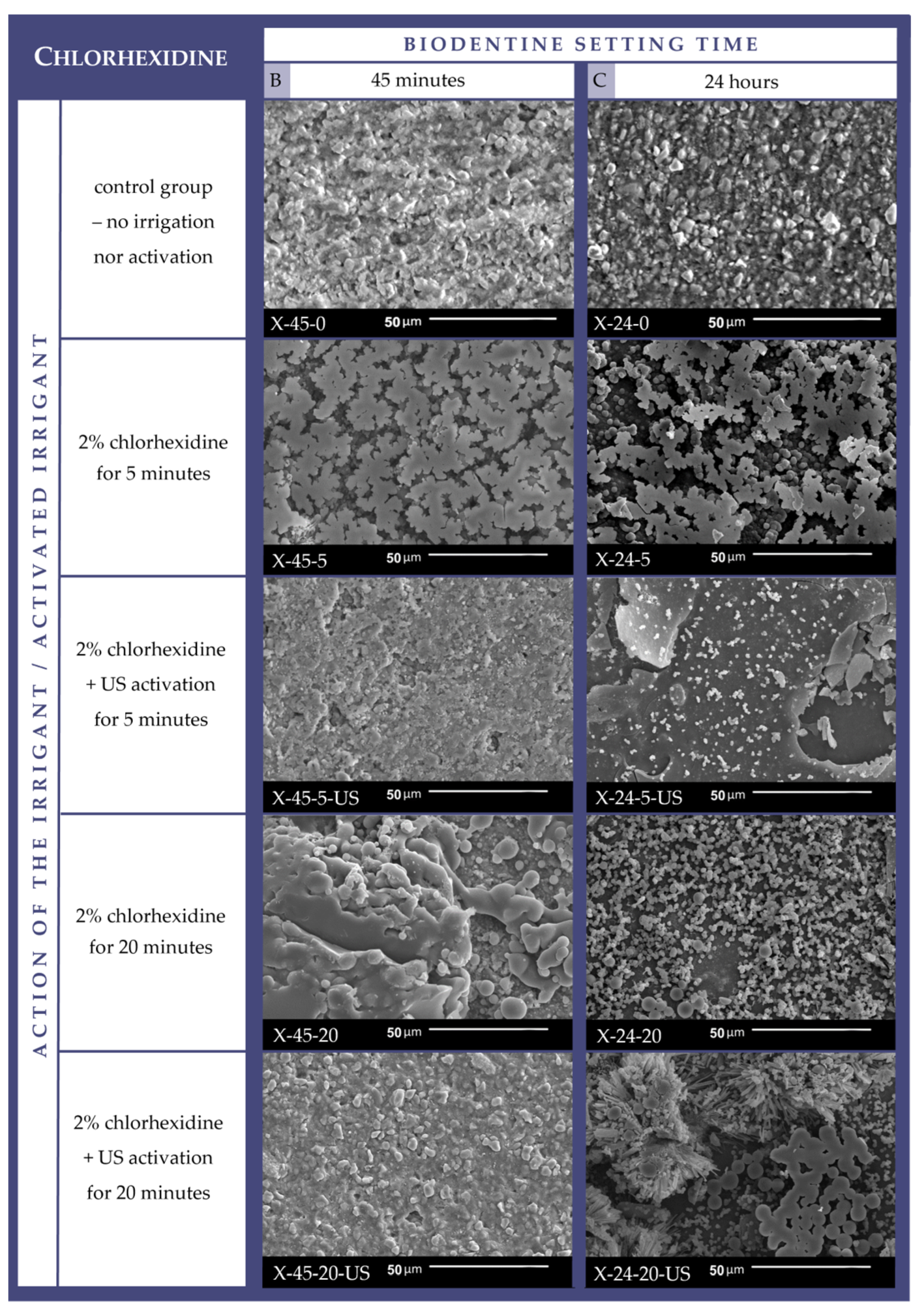
3.1. Cement Surface Microappearance

Images of cement specimens obtained via scanning electron microscope were compared. The results indicate that 2% chlorhexidine solution, both alone and ultrasonically activated, influenced the surface appearance of the investigated material in comparison to the control group (Figure 2).
Coarser grains are observable within the intragranular matrix of the control specimens. The overall appearance of the nonirrigated specimens was “clean”. The only result comparable with the control specimens was observed on the 45 min setting material after ultrasonically enhanced irrigation with 2% CHX for 20 min (X-45-20-US). The SEM images of the specimens that underwent other irrigation protocols revealed a heterogeneous surface morphology that incorporated varying granular, spike-like, and flake-like structures. The images are characterized by a complex layered microstructural composition.

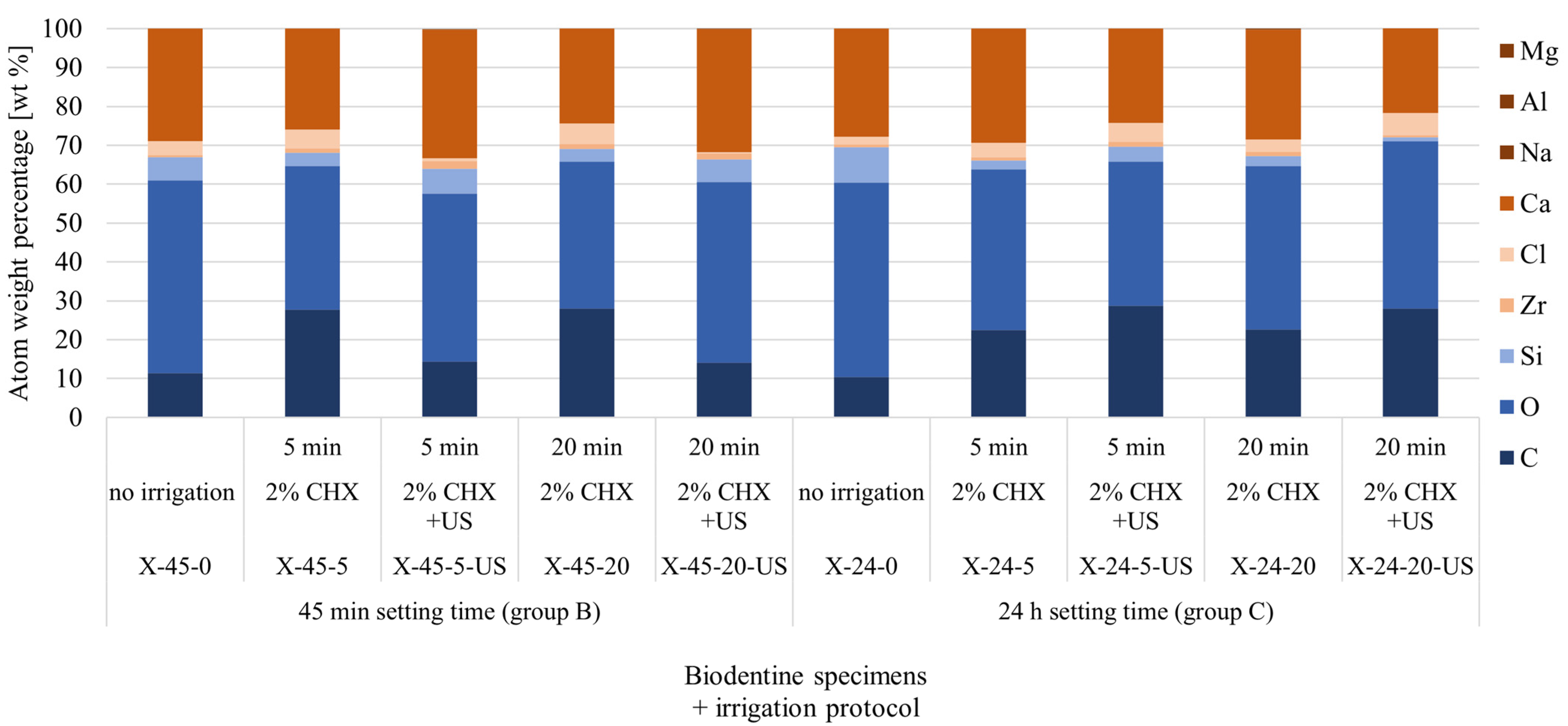
3.2. Cement Surface Chemical Composition

The results of the EDS spectroscopic examination of the tested Biodentine specimens’ surface are presented in the form of spectrograms on Figure 3 and atom weight percentages are displayed in Table 1 and Figure 4.
A certain regularity can be observed in the atom weight percentage of the tested specimens. Results are comparable between pairs of cement specimens such as in group B after 5 min and 20 min CHX irrigation (X-45-5 and X-45-20) as well as after 5 min and 20 min CHX irrigation protocol involving ultrasonic activation (X-45-5-US and X-45-20-US). Analogous tendency was recorded between specimens from group C (X-24-5 and X-24-20; X-24-5-US and X-24-20-US). Also, the atom percentage of the cement surface after 45 min of setting and nonactivated CHX irrigation was quasi-alike to the cement surface after 24 h of setting followed by ultrasonically activated CHX irrigation (X-45-5, X-45-20, X-24-5-US and X-24-20-US).
When compared to the control groups, the observed changes revealed the percentage increase in carbon (C) after all investigated irrigation protocols from 11.35 wt% (X-45-0) to maximum 28.0 wt% (X-45-20) in group B and from 10.41 wt% (X-24-0) to maximum 28.7 wt% (X-24-5-US) in group C. Similarly, the decrease in oxygen (O) was observed in every specimen from 49.61 wt% (X-45-0) to minimum 36.9 wt% (X-45-5) in group B and from 49.93 wt% (X-24-0) to minimum 37.1 wt% (X-24-5) in group C. A tendency was identified concerning the percentage of calcium (Ca). In group B, it was increased after CHX irrigation protocol involving ultrasonic activation from 28.87 wt% (X-45-0) to maximum 33.2 wt% (X-45-5-US), and slightly decreased after nonactivated rinsing procedure from 28.87 wt% (X-45-0) to minimum 24.4 wt% (X-45-20). The opposite scenario was discovered in group C where the percentage of Ca slightly rose after the CHX irrigation protocol without US activation from 27.74 wt% (X-24-0) to maximum 29.4 wt% (X-24-5), and decreased after ultrasonically enhanced irrigation from 27.74 wt% (X-24-0) to minimum 21.8 wt% (X-24-20-US). A decrease in the silicon (Si) level was noticeable in all of the material specimens from group C from 9.09 wt% (X-24-0) to minimum 1.0 wt% (X-24-20-US), and nonultrasonically irrigated material from group B from 5.96 wt% (X-45-0) to minimum 3.3 wt% (X-45-20). The specimens from group B that underwent the ultrasonically activated rinsing protocol revealed quasi-equal Si levels to the control group: 5.96 wt% (X-45-0), 6.3 wt% (X-45-5-US) and 5.8 wt% (X-45-20-US). On the contrary, an increase in chlorine (Cl) level was noticeable in all of the material specimens from group C from 2.09 wt% (X-24-0) to maximum 5.7 wt% (X-24-20-US), and nonultrasonically irrigated material from group B from 3.64 wt% (X-45-0) to maximum 5.3 wt% (X-45-20). The specimens from group B that underwent the ultrasonically activated rinsing protocol revealed a decreased Cl level when compared to the control group: 3.64 wt% (X-45-0), 0.7 wt% (X-45-5-US) and 0.2 wt% (X-45-20-US). The zirconium (Zr) percentage was augmented in material from both groups after every irrigation protocol, except the 24 h setting specimen after 20 min ultrasonically enhanced rinsing with CHX (X-24-20-US).

4. Discussion

Perforation closure requires specialistic endodontic skills, choice of the adequate material, and its proper management. The knowledge of how this material will behave in chosen clinical situations can impact therapeutic decisions and further treatment success. This research considered specific biocement being used under certain circumstances, i.e., Biodentine used for perforation closure before the finalization of endodontic treatment, thus being exposed to various mechanical and chemical factors, such as irrigation with 2% chlorhexidine digluconate. The study was part of a wider project researching the influence of different irrigating solutions used in endodontics on Biodentine tricalcium silicate-based cement consisting of 5.25% and 2% sodium hypochlorite [19], 40% citric acid [22], and 17% ethylenediaminetetraacetic acid [30].
The study plan included material specimen division into three setting time-based groups. While 45 min and 24 h proved to be sufficiently long for Biodentine to achieve the stability needed to withstand the researched irrigation protocol, the 12 min setting time, as recommended by the manufacturer was not enough. The results of the study showed that the irrigation with 2% chlorhexidine solution degraded the three-dimensional form of the cement specimen and thus reduced the stability of the material. The effect was aggravated when ultrasonic activation was applied. This means that the biocement used for perforation closure can be completely removed in a corresponding clinical situation. In the light of the results of this study, the 12 min setting time of Biodentine used in perforation closure is not enough to proceed with further cleaning and shaping of the canal space. Nevertheless, it should be noted that biocement specimens from group A retained their shape and dimensions after being removed from the polyvinyl molds. This suggests that 12 min Biodentine setting time may be sufficient for use, under special circumstances, in conservative dentistry for procedures such as dentin structure reconstruction and indirect or direct pulp capping [31,32], as well as in endodontic microsurgery as retrograde root canal filling [17]. This aspect has been confirmed in previous studies that show Biodentine as a weak material for hard tissues restoration in its early setting phase [33]. It was concluded that composite restoration covering biocement should be delayed for two weeks in order to allow sufficient Biodentine maturation to withstand contraction forces from the overlaying material [33].
Biodentine surface microappearance visualized under SEM revealed various forms of deposits and no clear signs of the main structure degradation of the material. Previous studies have shown that 2% chlorhexidine digluconate solution alone is not able to properly clean the root canal surface and remove the smear layer deriving from root canal chemo-mechanical preparation [34,35]. This study shows that chlorhexidine can also induce the formation of unwanted deposits on the surface of the tested material.
The EDS results should be interpreted with the awareness that the software equalizes the obtained data up to 100%. Clear peaks of carbon, calcium, silica, and zirconium in the EDS analysis may be treated as a confirmation of the chemical composition of the tested material: tricalcium silicate, calcium carbonate, zirconium oxide, and calcium hydroxide [36]. Rinsing with 2% CHX slightly modified the atom percentage of each element. This may be the result of creating a CHX-derived coating on the specimen’s surface. C and Cl, whose atom percentage rose in most of the specimens, are the constituents of CHX with the chemical formula [–(CH2)3NHC(=NH) NHC(=NH)NHC6H4Cl]2 [37].
The observed trends in the atom weight percentage of individual elements in the surface composition of Biodentine specimens, visualized in the form of tables and graphs, are only a preliminary indicator of the changes taking place. In order to obtain more accurate results, it would be necessary to increase the number of measurement points and the number of tested specimens, which may be a direction for further research.
It has been reported that chlorhexidine can enhance the antimicrobial activity of mineral trioxide aggregate (MTA), a golden-standard biocement, while concurrently increasing its cytotoxicity [38,39]. No similar studies were found regarding Biodentine. In the light of the results of this research, it could be advisable to investigate the toxicity of the residues found on the tested material surface.
All of the proposed research methods were used to evaluate and analyze the surface of the tested material. While the obtained data can provide a reliable representative image of the modifications that occur in the biocement specimens, it would be of scientific interest to investigate the phenomena occurring in the deeper layers of the set cement. Another limitation of the study is its in vitro form. Any future research could involve the observation and analysis of Biodentine clinical behavior after perforation closure performed prior to completing endodontic procedures, thus exposed to chemical action of chlorhexidine.
In the era of biocompatible bioceramic materials, it is essential to understand their specific indications, proper handling, and modes of application. This knowledge is particularly important for ensuring long-term success in complex cases, such as chamber or root perforations, that arise just prior to cleaning, shaping, and irrigation protocols.
The null hypothesis of this study was confirmed—chlorhexidine, either ultrasonically activated or not, does not alter the structure of the tested biocement as it targets only the organic matter. The surface morphology and chemical composition is altered by the sediments resulting from material irrigation with chlorhexidine.

5. Conclusions

In the light of the results of this study, the 12 min setting time of Biodentine is not recommended when used in perforation closure just before cleaning and shaping procedures. As the material needs more time to mature and properly set, it is advisable to continue the endodontic procedures on the following appointment.
Irrigation protocol involving 2% chlorhexidine digluconate solution visibly affects the tested material surface. Biodentine surface visualized under SEM revealed various forms of deposits, but no clear signs of cement main structure degradation. It can be stated that CHX is able to induce the formation of unwanted deposits on the material’s surface. While a complex microstructure was observed in SEM, the EDS results did not confirm any significant changes in Biodentine chemical composition. Further research is needed to analyze the influence of the observed changes on the outcome of the endodontic treatment.

Author Contributions

Conceptualization, K.D. and A.P.-U.; methodology, L.K.; software, K.D. and L.K.; validation, K.D., A.P.-U. and L.K.; investigation, K.D. and L.K.; resources, K.D., A.P.-U. and L.K.; data curation, K.D.; writing—original draft preparation, K.D.; writing—review and editing, K.D., L.K. and A.P.-U.; visualization, K.D.; supervision, A.P.-U. and L.K.; project administration, K.D.; funding acquisition, K.D. and A.P.-U. All authors have read and agreed to the published version of the manuscript.

Funding

This work was supported by grant No. 503/2-044-02/503-21-001-18 from the Medical University of Lodz. It was carried out as part of doctoral research supported by the project: “InterChemMed-Interdisciplinary Doctoral Studies of Łódź Public Universities”, cofinanced by the European Social Fund under Measure 3.2. Doctoral Studies of Priority Axis III Higher Education for the Economy and Development of the Operational Programme Knowledge Education Development 2014-2020 (POWR.03.02.00-00-I029/16).

Institutional Review Board Statement

Not applicable.

Informed Consent Statement

Not applicable.

Data Availability Statement

The original contributions presented in the study are included in the article, further inquiries can be directed to the corresponding author.

Conflicts of Interest

The authors declare no conflicts of interest.

References

  1. Duncan, H.F.; Galler, K.M.; Tomson, P.L.; Simon, S.; El-Karim, I.; Kundzina, R.; Krastl, G.; Dammaschke, T.; Fransson, H.; Markvart, M.; et al. European Society of Endodontology Position Statement: Management of Deep Caries and the Exposed Pulp. Int. Endod. J. 2019, 52, 923–934. [Google Scholar] [CrossRef] [PubMed]
  2. Spagnuolo, G. Bioactive Dental Materials: The Current Status. Materials 2022, 15, 2016. [Google Scholar] [CrossRef] [PubMed]
  3. Jitaru, S.; Hodisan, I.; Timis, L.; Lucian, A.; Bud, M. The Use of Bioceramics in Endodontics—Literature Review. Clujul Med. 2016, 89, 470–473. [Google Scholar] [CrossRef] [PubMed]
  4. Sanz, J.L.; Rodríguez-Lozano, F.J.; Llena, C.; Sauro, S.; Forner, L. Bioactivity of Bioceramic Materials Used in the Dentin-Pulp Complex Therapy: A Systematic Review. Materials 2019, 12, 1015. [Google Scholar] [CrossRef] [PubMed]
  5. Grech, L.; Mallia, B.; Camilleri, J. Investigation of the Physical Properties of Tricalcium Silicate Cement-Based Root-End Filling Materials. Dent. Mater. 2013, 29, e20–e28. [Google Scholar] [CrossRef]
  6. Dawood, A.E.; Parashos, P.; Wong, R.H.K.; Reynolds, E.C.; Manton, D.J. Calcium Silicate-Based Cements: Composition, Properties, and Clinical Applications. J. Investig. Clin. Dent. 2017, 8, e12195. [Google Scholar] [CrossRef]
  7. Camilleri, J. Biodentine™: The Dentine in a Capsule or More? Septodont Clinical Insights. Available online: https://www.septodontcorp.com/wp-content/uploads/2018/02/Biodentine-Article-0118-LOW.pdf (accessed on 24 June 2024).
  8. Malkondu, Ö.; Karapinar Kazandağ, M.; Kazazoğlu, E. A Review on Biodentine, a Contemporary Dentine Replacement and Repair Material. Biomed Res. Int. 2014, 2014, 160951. [Google Scholar] [CrossRef]
  9. Septodont. Biodentine™: Active Biosilicate Technology™; Scientific File; Septodont: Saint-Maur-des-Fossés, France, 2010. [Google Scholar]
  10. Aksoy, M.K.; Oz, F.T.; Orhan, K. Evaluation of Calcium (Ca2+) and Hydroxide (OH) Ion Diffusion Rates of Indirect Pulp Capping Materials. Int. J. Artif. Organs 2017, 40, 641–646. [Google Scholar] [CrossRef]
  11. Koubi, G.; Colon, P.; Franquin, J.C.; Hartmann, A.; Richard, G.; Faure, M.O.; Lambert, G. Clinical Evaluation of the Performance and Safety of a New Dentine Substitute, Biodentine, in the Restoration of Posterior Teeth—A Prospective Study. Clin. Oral Investig. 2013, 17, 243–249. [Google Scholar] [CrossRef]
  12. Nowicka, A.; Lipski, M.; Parafiniuk, M.; Sporniak-Tutak, K.; Lichota, D.; Kosierkiewicz, A.; Kaczmarek, W.; Buczkowska-Radlińska, J. Response of Human Dental Pulp Capped with Biodentine and Mineral Trioxide Aggregate. J. Endod. 2013, 39, 743–747. [Google Scholar] [CrossRef]
  13. Rajasekharan, S.; Martens, L.C.; Vandenbulcke, J.; Jacquet, W.; Bottenberg, P.; Cauwels, R.G.E.C. Efficacy of Three Different Pulpotomy Agents in Primary Molars: A Randomized Control Trial. Int. Endod. J. 2017, 50, 215–228. [Google Scholar] [CrossRef] [PubMed]
  14. Lin, J.C.; Lu, J.X.; Zeng, Q.; Zhao, W.; Li, W.Q.; Ling, J.Q. Comparison of Mineral Trioxide Aggregate and Calcium Hydroxide for Apexification of Immature Permanent Teeth: A Systematic Review and Meta-Analysis. J. Formos. Med. Assoc. 2016, 115, 523–530. [Google Scholar] [CrossRef] [PubMed]
  15. Taha, N.A.; Abdulkhader, S.Z. Full Pulpotomy with Biodentine in Symptomatic Young Permanent Teeth with Carious Exposure. J. Endod. 2018, 44, 932–937. [Google Scholar] [CrossRef] [PubMed]
  16. Silva, L.A.B.; Pieroni, K.A.M.G.; Nelson-Filho, P.; Silva, R.A.B.; Hernandéz-Gatón, P.; Lucisano, M.P.; Paula-Silva, F.W.G.; de Queiroz, A.M. Furcation Perforation: Periradicular Tissue Response to Biodentine as a Repair Material by Histopathologic and Indirect Immunofluorescence Analyses. J. Endod. 2017, 43, 1137–1142. [Google Scholar] [CrossRef] [PubMed]
  17. Pawar, A.; Kokate, S.; Shah, R. Management of a Large Periapical Lesion Using BiodentineTM as Retrograde Restoration with Eighteen Months Evident Follow Up. J. Conserv. Dent. 2013, 16, 573–575. [Google Scholar] [CrossRef]
  18. Estrela, C.; Decurcio, D.d.A.; Rossi-Fedele, G.; Silva, J.A.; Guedes, O.A.; Borges, Á.H. Root Perforations: A Review of Diagnosis, Prognosis and Materials. Braz. Oral Res. 2018, 32, 133–146. [Google Scholar] [CrossRef]
  19. Palatyńska-Ulatowska, A.; Buła, K.; Klimek, L. Influence of Sodium Hypochlorite and Ultrasounds on Surface Features and Chemical Composition of Biodentine Tricalcium Silicate-Based Material. Dent. Mater. J. 2020, 39, 587–592. [Google Scholar] [CrossRef]
  20. American Association of Endodontists (AAE). Glossary of Endodontic Terms. Available online: https://www.aae.org/specialty/clinical-resources/glossary-endodontic-terms/ (accessed on 28 June 2024).
  21. Touré, B.; Faye, B.; Kane, A.W.; Lo, C.M.; Niang, B.; Boucher, Y. Analysis of Reasons for Extraction of Endodontically Treated Teeth: A Prospective Study. J. Endod. 2011, 37, 1512–1515. [Google Scholar] [CrossRef]
  22. Dąbrowska, K.; Palatyńska-Ulatowska, A.; Klimek, L. The Effect of Irrigation with Citric Acid on Biodentine Tricalcium Silicate-Based Cement: SEM-EDS In Vitro Study. Materials 2022, 15, 3467. [Google Scholar] [CrossRef]
  23. Zehnder, M. Root Canal Irrigants. J. Endod. 2006, 32, 389–398. [Google Scholar] [CrossRef] [PubMed]
  24. Zamany, A.; Safavi, K.; Spångberg, L.S.W. The Effect of Chlorhexidine as an Endodontic Disinfectant. Oral Surg. Oral Med. Oral Pathol. Oral Radiol. Endod. 2003, 96, 578–581. [Google Scholar] [CrossRef] [PubMed]
  25. Wilkoński, W.; Jamróz-Wilkońska, L. Protokół Płukania Kanałów Korzeniowych Według Zaleceń Działu Badawczego Polskiego Towarzystwa Endodontycznego. Mag. Stomatol. 2013, 246, 92–94. [Google Scholar]
  26. Boutsioukis, C.; Arias-Moliz, M.T. Present Status and Future Directions—Irrigants and Irrigation Methods. Int. Endod. J. 2022, 55, 588–612. [Google Scholar] [CrossRef] [PubMed]
  27. Moreira, R.N.; Pinto, E.B.; Galo, R.; Falci, S.G.M.; Mesquita, A.T. Passive Ultrasonic Irrigation in Root Canal: Systematic Review and Meta-Analysis. Acta Odontol. Scand. 2019, 77, 55–60. [Google Scholar] [CrossRef] [PubMed]
  28. Dioguardi, M.; Di Gioia, G.; Illuzzi, G.; Laneve, E.; Cocco, A.; Troiano, G. Endodontic Irrigants: Different Methods to Improve Efficacy and Related Problems. Eur. J. Dent. 2018, 12, 459–466. [Google Scholar] [CrossRef]
  29. Aggarwal, V.; Singla, M.; Miglani, S.; Kohli, S. Comparative Evaluation of Push-out Bond Strength of ProRoot MTA, Biodentine, and MTA Plus in Furcation Perforation Repair. J. Conserv. Dent. 2013, 16, 462–465. [Google Scholar] [CrossRef]
  30. Dąbrowska, K.; Palatyńska-Ulatowska, A.; Klimek, L. How Does Ethylenediaminetetraacetic Acid Irrigation Affect Biodentine? A Multimethod Ex Vivo Study. Materials 2024, 17, 1230. [Google Scholar] [CrossRef]
  31. Villat, C.; Grosgogeat, B.; Seux, D.; Farge, P. Conservative Approach of a Symptomatic Carious Immature Permanent Tooth Using a Tricalcium Silicate Cement (Biodentine): A Case Report. Restor. Dent. Endod. 2013, 38, 258–264. [Google Scholar] [CrossRef]
  32. Borkar, S.A.; Ataide, I. Biodentine Pulpotomy Several Days after Pulp Exposure: Four Case Reports. J. Conserv. Dent. 2015, 18, 73–78. [Google Scholar] [CrossRef]
  33. Hashem, D.F.; Foxton, R.; Manoharan, A.; Watson, T.F.; Banerjee, A. The Physical Characteristics of Resin Composite-Calcium Silicate Interface as Part of a Layered/Laminate Adhesive Restoration. Dent. Mater. 2014, 30, 343–349. [Google Scholar] [CrossRef]
  34. Attur, K.; Joy, M.; Karim, R.; Anil Kumar, V.; Deepika, C.; Ahmed, H. Comparative Analysis of Endodontic Smear Layer Removal Efficacy of 17% Ethylenediaminetetraacetic Acid, 7% Maleic Acid, and 2% Chlorhexidine Using Scanning Electron Microscope: An in Vitro Study. J. Int. Soc. Prev. Community Dent. 2016, 6, S160–S165. [Google Scholar] [CrossRef] [PubMed]
  35. Yamaguchi, M.; Yoshida, K.; Suzuki, R.; Nakamura, H. Root Canal Irrigation with Citric Acid Solution. J. Endod. 1996, 22, 27–29. [Google Scholar] [CrossRef] [PubMed]
  36. Buła, K.; Palatyńska-Ulatowska, A.; Klimek, L. Biodentine Management and Setting Time with Vicat and Vickers Evaluation; a Survey-Based Study on Clinicians’ Experience. Arch. Mater. Sci. Eng. 2020, 103, 75–85. [Google Scholar] [CrossRef]
  37. Lapinska, B.; Klimek, L.; Sokolowski, J.; Lukomska-Szymanska, M. Dentine Surface Morphology after Chlorhexidine Application-SEM Study. Polymers 2018, 10, 905. [Google Scholar] [CrossRef] [PubMed]
  38. Ghabraei, S.; Afkhami, F.; Shamshiri, A.R.; Mohammadi, Z. Comparison of Cytotoxicity between Mineral Trioxide Aggregate Mixed with Chlorhexidine and Common Endodontic Regeneration Medicaments on Periodontal Ligament Stem Cells: An in Vitro Study. Front. Dent. 2023, 20, 1. [Google Scholar] [CrossRef]
  39. Hernandez, E.P.; Botero, T.M.; Mantellini, M.G.; McDonald, N.J.; Nör, J.E. Effect of ProRoot® MTA Mixed with Chlorhexidine on Apoptosis and Cell Cycle of Fibroblasts and Macrophages in Vitro. Int. Endod. J. 2005, 38, 137–143. [Google Scholar] [CrossRef]
Figure 1. Presentation of cement specimens’ distribution and irrigation protocols.
Applsci 14 08702 g001
Figure 2. Cement specimens’ surface in SEM under 1 k magnification.
Applsci 14 08702 g002
Figure 3. Cement specimens’ chemical composition on spectrograms.
Applsci 14 08702 g003
Figure 4. Changes in the chemical composition of the surface of Biodentine specimens.
Applsci 14 08702 g004
Table 1. Atom percentage [wt%] of Biodentine specimens’ surface in control group, group B (after 45 min setting time) and in group C (after 24 h setting time).
Element45 min Setting Time (Group B)24 h Setting Time (Group C)
X-45-0X-45-5X-45-5-USX-45-20X-45-20-USX-24-0X-24-5X-24-5-USX-24-20X-24-20-US
No Irrigation2% CHX;
5 min
2% CHX + US;
5 min
2% CHX;
20 min
2% CHX + US;
20 min
No Irrigation2% CHX;
5 min
2% CHX + US;
5 min
2% CHX;
20 min
2% CHX + US;
20 min
C11.3527.814.428.014.110.4122.528.722.628.0
O49.6136.943.237.846.549.9341.337.142.043.1
Si5.963.46.33.35.89.092.33.92.61.0
Zr0.561.12.01.21.60.740.81.21.10.5
Cl3.644.90.75.30.22.093.74.83.25.7
Ca28.8725.933.224.431.727.7429.424.328.221.8
Na0.00.00.00.00.00.00.00.00.30.0
Al0.00.00.10.00.00.00.00.00.00.0
Mg0.00.10.00.00.10.00.00.10.00.0
Disclaimer/Publisher’s Note: The statements, opinions and data contained in all publications are solely those of the individual author(s) and contributor(s) and not of MDPI and/or the editor(s). MDPI and/or the editor(s) disclaim responsibility for any injury to people or property resulting from any ideas, methods, instructions or products referred to in the content.

