Article

Preservative Effect of a Gelatin-Based Film Including a Gelidium sp. Flour Extract on Refrigerated Atlantic Mackerel

1
Department of Food Technology, Marine Research Institute (CSIC), 36208 Vigo, Spain
2
Department of Analytical Chemistry, Nutrition and Food Science, School of Veterinary Sciences, University of Santiago de Compostela, 27002 Lugo, Spain
*
Author to whom correspondence should be addressed.
Appl. Sci. 2024, 14(19), 8817; https://doi.org/10.3390/app14198817
Submission received: 26 August 2024 / Revised: 19 September 2024 / Accepted: 24 September 2024 / Published: 30 September 2024

Abstract

:
This research evaluated the preservative properties of flour from the alga Gelidium sp., which is a waste substrate resulting from commercial phycocolloid extraction. Gelatin-based biofilms, which included two different concentrations of red alga flour, were developed and used as packaging systems during refrigerated storage (up to 9 days at 4 °C) of Atlantic mackerel (Scomber scombrus) muscle. In all batches tested, a progressive decrease in quality could be observed in the muscle of the fish as the storage time increased. Compared with the control fish, the Gelidium alga flour extract had an inhibitory effect (p < 0.05) on microbial activity (total aerobes, psychrotrophs, and proteolytic bacteria), lipid oxidation (peroxide, thiobarbituric acid, fluorescence and polyene indices), lipid hydrolysis (formation of free fatty acids) and pH increase in refrigerated mackerel muscle. In contrast, no significant effect (p > 0.05) was observed on trimethylamine formation, Enterobacteriaceae, or lipolytic bacteria counts. A preservative effect resulting from the incorporation of Gelidium alga flour into the gelatin-based biofilm was observed, indicating both quality and safety enhancement. In accordance with current global interest in the search for natural and waste sources, a novel and beneficial use of Gelidium flour for enhancing the quality of refrigerated fish has been proposed.

1. Introduction

Marine species are reported to provide many constituents relevant to the human diet [1,2]. However, seafood deteriorates rapidly postmortem via several biochemical and microbial breakdown mechanisms [3,4,5]. Reducing the temperature via ice or mechanical refrigeration is the most prevalent method to delay microbial and biochemical spoilage in fish. Despite its widespread use, neither method can fully ensure the preservation of fish quality, particularly during extended storage periods or if the cold chain is not consistently maintained. Therefore, advancements in refrigeration techniques have been developed to address these challenges [6,7,8].
One such strategy is the use of active packaging films, which can release preservative compounds (such as antimicrobials and antioxidants) into seafood substrates [9,10,11]. In general, packaging maintains the freshness of seafood and preserves it against adverse agents, such as water vapor, microorganisms, gases, odors, dust, and mechanical shock and vibrations, during distribution and storage [12,13]. Notably, the release of preservative compounds extends the shelf life of packaged seafood [14,15,16]. The most commonly used packaging materials include synthetic polymers such as low- and high-density polyethylene and polyethylene [17]. However, adverse health problems (i.e., cancer development and toxicity) resulting from the persistent consumption of synthetic antioxidants (butylhydroxytoluene, butylhydroxyanisole, etc.) have led to the recommended use of natural antioxidants as alternatives to synthetic antioxidants [18,19]. Therefore, biodegradable and edible materials derived from plants and animals, including peptides, polysaccharides, and lipids, have proven to represent remarkable alternatives [20,21]. The film-forming ability of several polysaccharides, such as cellulose, chitosan, starch, pectin, and alginate, has been revised [22]. In particular, gelatin from diverse animal sources can be effectively used to develop active packaging strategies [23].
Marine macroalgae contain a wide variety of natural constituents with potential antimicrobial and antioxidant activities and are suitable for application during seafood processing and storage [24,25,26]. Among macroalgae, red varieties (i.e., Gracilaria sp. and Gelidium sp.) are chiefly recognized for their industrial application in extracting phycocolloids (such as agar, algin, furcellaran, and carrageenan) [27,28], which are utilized to improve the physical properties of edible films in innovative food packaging solutions [29,30]. Agar extraction typically involves initial alkaline pretreatment, followed by water extraction (90 and 120 °C) under pressure [31,32]. This process generates a considerable amount of solid byproducts characterized by a moderate protein content and high polysaccharide content [33].
In the Gelidium genus, different species have been reported to contain antimicrobial and antioxidant components. Thus, a high quantity of flavonoid compounds was detected in both aqueous and ethanolic extracts of G. pusillum (Stackhouse), which was attributed to their antioxidant and antibacterial properties [34]. Similarly, a G. corneum powder–whey protein isolate was shown to have a remarkable antimicrobial effect on fish paste [35]. Sulfated polysaccharides from G. pacificum Okamura also had beneficial effects on mice with antibiotic-associated diarrhea [36]. Moreover, aqueous and ethanol extracts from G. chilense were reported to exert notable inhibitory effects on different bacterial species [37]. Recently, an aqueous extract of flour obtained from Gelidium sp. demonstrated antimicrobial activity in fish during chilled storage [38] and antioxidant properties in heated fish [39].
The present study focused on the use of the red alga Gelidium sp. flour, a waste substrate resulting from the industrial extraction of phycocolloids. The potential preservative effect of an aqueous extract of this alga flour (AF) was investigated. This flour extract was incorporated into a gelatin-based film, which was employed as a packaging medium during refrigerated storage (4 °C) of Atlantic mackerel (Scomber scombrus) muscle. The evolution of microbial and chemical parameters related to quality loss was determined in fish muscle during a 9-day storage period.

2. Materials and Methods

2.1. Initial Alga Flour Composition and Aqueous Extract Preparation

Commercial flour obtained from Gelidium sp. was provided by Industrias Roko S. A. (Llanera, Asturias, Spain). Its proximate composition was analyzed according to the AOAC procedure [40]. The fatty acid (FA) analysis of the Gelidium flour was carried out in accordance with the methodology presented in Section 2.5.
The aqueous extract of the alga flour was obtained in agreement with previous research [39]. A mixture of 39 g of alga flour and 600 mL of distilled water was stirred (Vortex, Scientific Industries, Bohemia, NY, USA) for 30 s, sonicated (J. P. Selecta, S. A., Barcelona, Spain; 360 W, 50/60 Hz) for 30 s (22–30 °C), and then centrifuged at 3500× g for 30 min at 4 °C. The supernatant was collected, and the extraction process was repeated three more times. Finally, all four supernatants were combined and diluted to 3 L with distilled water, resulting in an alga flour concentration of 13 g·L−1.

2.2. Preparation of the Film Systems

Teleostean gelatin (Sigma, Life Sciences, Steinheim, Germany) films were obtained by casting them from their film-forming solutions (FFSs) following the methods described by Trigo et al. [41]. Oxidized sodium alginate (OSA) was prepared according to the method reported by Balakrishnan et al. [42]. Two different films were prepared to investigate the effects of two different concentrations of the Gelidium flour extract.
To prepare the less concentrated films, 250 mL of the Gelidium flour extract was diluted to 1 L with distilled water, and 400 mg of NaOH was added and dissolved by stirring. Then, 950 mL of the resulting solution was mixed with 100 g of gelatin (i.e., 23.75 mL flour extract·g−1 gelatin), and the gelatin was completely dissolved by soft heating (ca. 40 °C for 120 min). OSA (5 g·50 mL−1 Gelidium extract; 5% gelatin) was then added into the FFSs as a crosslinking agent. After 20 min of stirring, 30 g of glycerol was added to act as a plasticizer. The resulting suspension was stirred for 20 min, and then the FFSs were poured onto Teflon-coated trays and dried at 50 °C in a convection oven for 48 h. The films were then conditioned for 48 h in a chamber at 4 ± 1 °C before use. This resulting film was designated the AF-1 packaging film.
To prepare the most concentrated packaging film, a similar procedure was followed but starting from 1 L of the initial Gelidium extract. The resulting film (i.e., 95.00 mL flour extract·g−1 gelatin) and subsequent batch were referred to as the AF-2 condition. A control (CTR) gelatin film without flour extract was prepared in the same way as the AF-1 and AF-2 packaging films and referred to as the CTR batch.
The selection of Gelidium extracts for this work was according to previous trials conducted in our laboratory. As a result, the AF-2 film contained the highest flour concentration, which did not affect the sensory or external characteristics of the fish muscle parts (such as odor and color). Therefore, this concentration, along with a less concentrated one, was considered.
All solvents and chemical reagents used in this study were of reagent grade (Merck, Darmstadt, Germany); otherwise, the source is mentioned.

2.3. Fish Processing and Sampling

Thirty-three fresh Atlantic mackerel (Scomber scombrus) samples were fished in April 2024 near the Galician Atlantic coast (northwestern Spain), purchased from Vigo (Spain) harbor and transported on ice. After arrival to the laboratory, samples were measured and weighed, ranging between 33–37 cm in length and between 375–435 g in weight.
Subsequently, six of the fish were analyzed as an initial sample (day 0). From among these 6 specimens, 3 different groups were taken, each including 2 fish, each of which was sampled and analyzed for dorsal white muscle (three replicates; n = 3).
The remaining 27 fish samples were divided into three batches, including 9 samples per batch. From each batch, we took pieces of approximately 35 g each. Three pieces of back muscle were packaged from each sample. These pieces were then vacuum-sealed individually via a Vacuum Packaging Machine Culinary (Albipack, Águeda, Portugal) in the three specified packaging systems (CTR, AF-1, and AF-2 batches), resulting in 27 fish pieces per packaging condition. The packaged fish pieces were stored in a refrigerated room at 4 °C for 9 days. Sampling and analyses were conducted on days 2, 6, and 9 of storage. These sampling times were considered appropriate in order to follow the quality evolution of the present fish species during refrigerated storage. On each of the sampling days, 9 pieces of packaged fish were extracted from each batch for subsequent analysis. These 9 pieces of fish were then divided into three groups (three pieces of packaged fish per group); white muscle was analyzed independently in each group (three replicates; n = 3).

2.4. Microbiological Analyses of Fish Muscle

Ten-gram samples of fish muscle were aseptically collected and mixed with 90 mL of 0.1% peptone water. This mixture was homogenized in sterilized stomacher bags (AES, Combourg, France) as described elsewhere [43,44]. Subsequently, dilutions from the microbial suspensions were prepared in 0.1% peptone water.
Total aerobic bacteria were investigated on plate count agar (PCA) (Oxoid Ltd., London, UK) after 48 h of incubation at 30 °C. The investigation of psychrotrophs was also conducted via PCA, but the incubation period was extended to 7 days at 7–8 °C. Enterobacteriaceae were determined using Violet Red Bile Agar (VRBA) (Merck, Darmstadt, Germany) after 24 h of incubation at 37 °C. Specific spoilage microorganisms capable of breaking down proteins or lipids were also investigated. Micro-organisms producing extracellular proteases or lipases were detected via casein agar or tributyrin agar, respectively, after 48 h of incubation at 30 °C [45].
The bacterial counts were transformed into log CFU·g−1 before statistical analysis. All analyses were performed in triplicate.

2.5. Chemical Analyses Related to Quality Loss

The pH values of the mackerel muscle were monitored throughout the storage period via a 6 mm diameter insertion electrode (Crison, Barcelona, Spain).
Trimethylamine (TMA) formation was measured via the picrate spectrophotometric method (410 nm) (Beckman Coulter, DU 640; London, UK), as described by Tozawa et al. [46]. This involved the preparation of a 5% trichloroacetic acid extract from fish white muscle (10 g in 25 mL), with the results expressed as mg TMA-N·kg−1 muscle.
Lipids were extracted from mackerel white muscle via the Bligh and Dyer [47] method, which involves single-phase solubilization with a chloroform–methanol (1:1) mixture. The results were calculated as g lipid·kg−1 muscle. Lipid quantification followed the Herbes and Allen [48] method, with the lipid content expressed as g·kg−1 muscle.
Free fatty acid (FFA) content was determined from the lipid extract of fish muscle via the Lowry and Tinsley [49] method on the basis of complex formation with cupric acetate–pyridine and subsequent spectrophotometric assessment at 715 nm. The results are expressed as g FFAs·kg−1 lipids.
The peroxide value (PV) of the lipid extract was measured spectrophotometrically at 520 nm, following the method described in previous research [50]. The results are expressed as meq. active oxygen·kg−1 lipids.
The thiobarbituric acid (TBA) index (TBA-i) was determined following the methodology described by Vyncke [51]. The content of TBA-reactive substances (TBARSs) was determined by spectrophotometry at 532 nm, and the results obtained were expressed as mg malondialdehyde·kg−1 muscle.
The formation of fluorescent compounds was determined in the lipid extract obtained from fish muscle using an LS 45 fluorimeter (Perkin Elmer España; Tres Cantos, Madrid, Spain), proceeding to its reading at wavelengths of 393/463 nm and 327/415 nm, as described in a previous work [52]. Relative fluorescence (RF) was determined following the formula: RF = F/Fst, where F is defined as the fluorescence measured at each excitation/emission wavelength pair, and Fst represents the fluorescence intensity of a quinine sulfate solution (1 µg-mL−1 in 0.05 M H₂SO₄) at the corresponding wavelength pair. The results obtained were presented as the fluorescence ratio (FR), which was calculated from the ratio between the two RF values: FR = RF393/463 nm/RF327/415 nm.
To determine the lipid profile of the samples, FA methyl esters (FAMEs) were previously obtained from the lipid extracts using acetyl chloride in methanol. Subsequently, the FAMEs obtained were analyzed by gas chromatography (Perkin–Elmer 8700 chromatograph, Madrid, Spain) [53]. An SP-2330-fused silica capillary column (0.25 mm i.d. × 30 m, Supelco, Inc., Bellefonte, PA, USA) was used for these determinations, using temperature adjusted to increase from 145 °C to 190 °C at 1.0 °C min−1 and from 190 °C to 210 °C at 5.0 °C min−1, maintained at 210 °C for 13.5 min. Nitrogen was used as a carrier gas at 10 psig, and detection was performed with a flame ionization detector at 250 °C. A programmed temperature vaporizing injector was used in split mode (150:1) and heated from 45 °C to 275 °C, increasing 15 °C min−1.
Peaks corresponding to FAMEs were identified by means of comparing their retention times with those of standard mixtures (Qualmix Fish and Supelco 37 Component FAME Mix, Supelco, Inc., Bellefonte, PA, USA). The peak areas were automatically integrated, using C19:0 as an internal standard for quantitative purposes. The content of each FA was calculated as g FA·100 g−1 total FAs. The polyene index (PI) was calculated as the FA ratio: C20:5ω3 + C22:6ω3 to C16:0.

2.6. Statistical Analysis

The study was carried out in triplicate (n = 3). Data from all microbiological and chemical analyses were subjected to ANOVA to examine differences due to the packaging system and refrigeration time. Special attention was given to the comparison of treated fish batches with respect to the control batch. Mean comparisons were performed via the least-squares difference (LSD) method. All statistical comparisons were conducted via PASW Statistics 18 software for Windows (SPSS Inc., Chicago, IL, USA), with differences considered significant at the 95% confidence level (p < 0.05).

3. Results

3.1. Alga Flour Composition

The alga flour exhibited the following proximate composition (%): 12.2 (moisture), 31.5 (protein), 0.2 (lipids), 14.3 (ash), and 42.8 (total carbohydrate).
The following compositions for individual FAs were observed (g·100 g−1 total FAs): 6.69 ± 0.08 (C14:0, myristic acid), 0.96 ± 0.02 (C15:0, pentadecanoic acid), 66.85 ± 0.45 (C16:0, palmitic acid), 2.43 ± 0.04 (C16:1ω7, palmitoleic acid), 0.60 ± 0.01 (C17:0, margaric acid), 3.41 ± 0.07 (C18:0, stearic acid), 7.82 ± 0.09 (C18:1ω9, oleic acid), 1.88 ± 0.03 (C18:1ω7, vaccenic acid), 0.62 ± 0.05 (C18:2ω6, linoleic acid), 0.47 ± 0.03 (C20:1ω9, gondoic acid), 0.15 ± 0.04 (C20:2ω6, eicosadienoic acid), 2.91 ± 0.16 (C20:4ω6, araquidonic acid), 0.20 ± 0.02 (C22:1ω9, erucic acid).

3.2. Evaluation of Microbial Growth in Atlantic Mackerel Muscle under Different Packaging Systems

The evolution of microbial development in fish corresponding to the AF-treated (AF-1 and AF-2 for low and high concentrations of AF) and CTR batches is depicted in Table 1.
In terms of Enterobacteriaceae, the two batches containing algal extracts presented lower average values than did the gelatin CTR batch. Thus, the presence of the algal extract in the packaging films led to lower average values at all the sampling times. Remarkably, the AF-1 batch presented lower average values than its counterpart, the AF-2 batch. These results indicate that the incorporation of algal extracts into the gelatin films resulted in Enterobacteriaceae numbers nearly ten times lower in the AF-1 batch than in the pure gelatin CTR batch.
Table 1 shows the evolution of psychrotroph counts in all three batches during refrigerated storage. Similar to the results observed for Enterobacteriaceae, the AF-1 batch, which included a lower concentration of alga extract, provided better microbial control than the other two batches did. Thus, the AF-1 batch presented significantly (p < 0.05) lower pyschrotroph counts than did the two other batches at intermediate storage times (days 2 and 6). However, at the most advanced storage time, all three batches presented microbial numbers above seven log units, indicating that the protection exerted by the bioactive compounds present in the alga extracts concerning the development of psychrotrophic bacteria was not as relevant on day 9.
The comparative evolution of total aerobes in all three batches was also evaluated in this work, and the obtained results are shown in Figure 1. Similar to the effects observed for other microbial groups, the incorporation of algal extracts into the gelatin films was associated with slower microbial growth than the CTR batch. This effect was observed at early (day 2) and advanced (day 9) storage times, and the greatest differences between the alga-treated and CTR batches were close to 1 log unit. Remarkably, these differences were statistically significant (p < 0.05).
The results obtained in this study concerning the ability of lipolytic bacteria to produce extracellular lipases with activity against triacylglycerols (TAGs) and phospholipids (PLs) in fish muscle are also displayed in Table 1. Thus, the incorporation of algal extracts into the gelatin packaging film led to restrictions in the growth of lipolytic bacteria during storage compared to the CTR batch. However, although the greatest difference among batches was 1.30 log units on day 9 (AF-1 batch with respect to the CTR batch), such differences were not statistically significant (p > 0.05). Similar results were observed at intermediate storage times (day 6). These results indicate that the incorporation of algal extracts into the gelatin films exerted slight microbial control over lipolytic bacteria, but the effect was not very intense.
Figure 2 shows the results of the comparative evolution of proteolytic bacteria in Atlantic mackerel muscle in all three batches. As with other microbial groups, the incorporation of the bioactive alga extracts into the gelatin films was associated with better control of proteolytic bacteria, which was especially relevant in the AF-1 batch compared with the CTR batch. Accordingly, the results indicated statistically significant (p < 0.05) differences derived from the more limited growth of proteolytic bacteria, especially in the AF-1 batch. These differences were especially relevant at intermediate (day 6) and advanced (day 9) storage times, reaching a maximum of 0.92 log units on day 6.

3.3. Evolution of the pH and TMA Values

A progressive increase (p < 0.05) in the pH value was detected in all batches as the storage time increased (Table 2). An increase in pH was derived from the presence of the alga extract in the packaging medium. Thus, higher average pH values were detected in samples corresponding to the CTR batch than in their fish counterparts subjected to the preservative extract; the differences were found to be significant (p < 0.05) on day 2 (AF-2 batch) and on day 9 (AF-1 batch).
With respect to TMA formation, an increase (p < 0.05) with storage time was observed in all three batches (Table 2). This increase was remarkably high for the 6–9-day period. The presence of the flour extract in the packaging medium did not lead to significant differences (p > 0.05); however, lower average values were detected at the end of the storage time in the fish samples corresponding to batches, including the flour extract (AF-1 and AF-2 batches).

3.4. Assessment of Lipid Hydrolysis Development

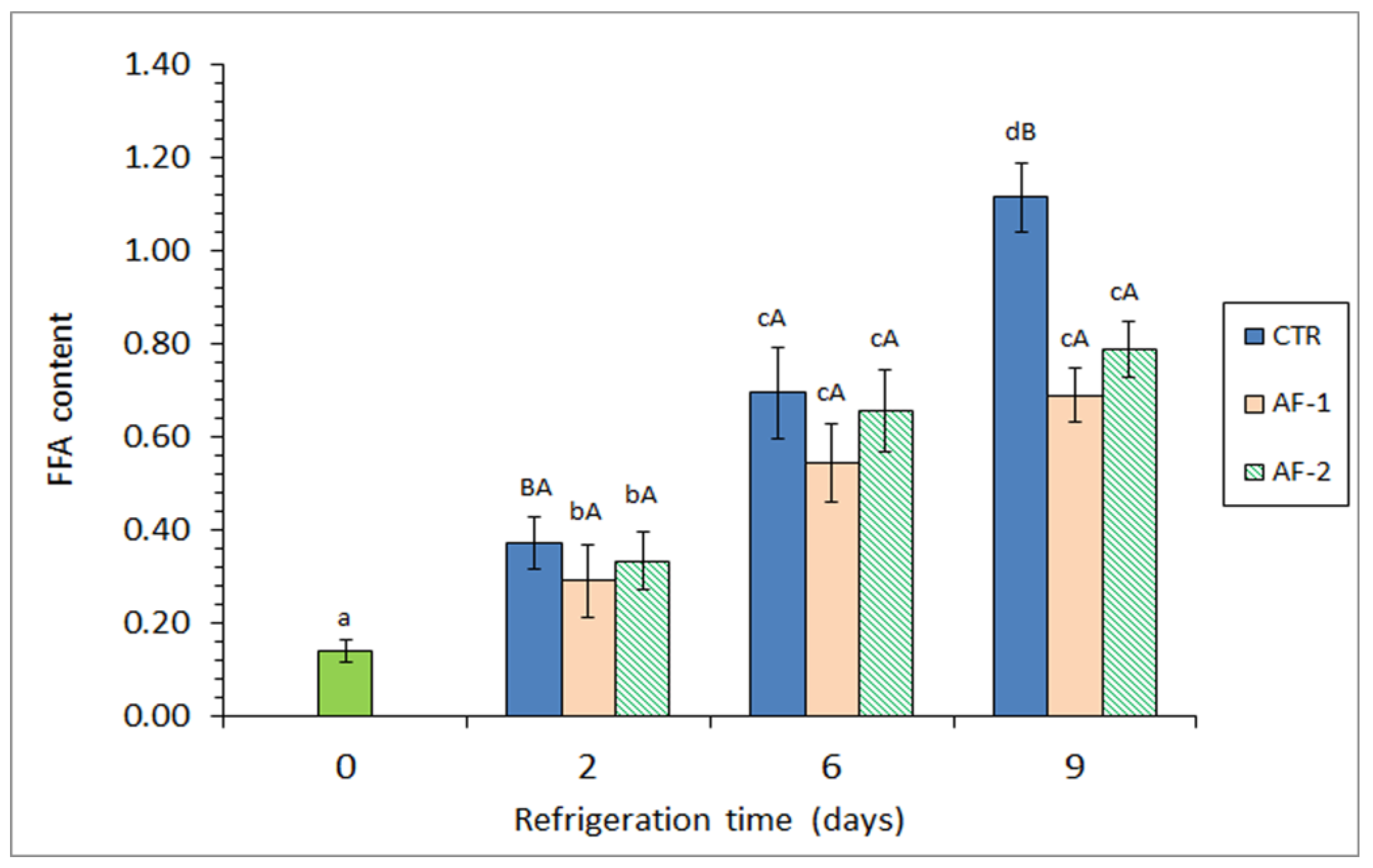
Lipid hydrolysis was assessed via the FFA value (Figure 3). The formation of this catabolic end product significantly increased (p < 0.05) over the storage period across all batches. An inhibitory effect (p < 0.05) on FFA formation was noted at the end of the storage period in the fish samples, where the alga flour extract was included in the packaging medium. However, no significant differences (p > 0.05) were detected between batches containing the alga flour.

3.5. Determination of Lipid Oxidation Evolution

The progression of lipid oxidation was evaluated by analyzing the formation of primary (PV), secondary (TBA-i), and tertiary (FR) compounds, as well as by assessing the PI. Special attention was given to comparisons between the treated and CTR batches.
Peroxide formation in this study was minimal [38,54]. Across all batches, the peroxide content ranged from 0.68 to 5.03 meq·kg−1 lipids throughout the study (Table 3). No significant trend with storage time was observed (p > 0.05), although the highest average values were recorded at the end of the storage period in all batches. An inhibitory effect (p < 0.05) on peroxide formation was noted on day 9 due to the presence of flour extract in the packaging medium; this effect was more pronounced (p < 0.05) with higher concentrations of the alga extract.
A significant increase (p < 0.05) in TBARS formation was observed in all batches during the 0–6-day period (Table 3). However, by the end of the storage period, a decrease in the average value was noted in samples from batches containing the alga extract. On day 9, an inhibitory effect (p < 0.05) on TBARS formation was observed in the AF-2 batch compared with the CTR batch.
A significant increase (p < 0.05) in FR values with storage time was detected in all batches (Table 3). Additionally, an inhibitory effect (p < 0.05) on fluorescent compound formation was observed on day 9 due to the inclusion of alga flour extract in the packaging medium (AF-1 and AF-2 batches). This effect was more pronounced (p < 0.05) with higher concentrations of the alga extract.
A progressive decrease in the PI was detected in most cases due to refrigerated storage (Table 3). Throughout the refrigeration period, higher average values were observed in the fish samples treated with the flour extract. Significant differences (p < 0.05) were found after 2 and 6 days when the high-concentration (AF-2) packaging conditions were considered.

4. Discussion

4.1. Antimicrobial Activity

The results of microbiological analyses revealed that incorporating bioactive algal extracts into gelatin packaging films inhibited the growth of all five microbial groups investigated in Atlantic mackerel stored for 9 d under refrigeration conditions. Notably, the differences in three of the five microbial parameters investigated among batches were statistically significant (p < 0.05). These results provide a promising strategy for developing active films incorporating algal extracts to achieve better retention of fish quality by delaying microbial breakdown. These results agree with those of previous studies on the preservative effect of Gelidium sp. flour against the most common food pathogenic and spoilage bacteria [38]. Moreover, an antimicrobial effect was reported in such a study when an aqueous extract of Gelidium flour was added to the icing medium employed for the chilled storage of Atlantic mackerel (S. scombrus) [49].
The inhibitory effect of the current alga flour on microbial growth can be attributed to the presence of various bioactive compounds. Additionally, the combination of gelatin with bioactive compounds has been shown to better protect packaged foods and extend their shelf life [55]. This combination has been reported to enhance the mechanical properties and barrier functions of the film, which are crucial for controlling the release of bioactive components [55]. In addition, the incorporation of alga flour into gelatin films can affect the release rate of bioactive compounds, which is beneficial for maintaining the efficacy of bioactive components over time [56,57]. These interactions highlight the potential of using Gelidium sp. alga flour and gelatin films in food packaging to increase the delivery and effectiveness of bioactive components, contributing to better food preservation and safety.
In the present study, further investigations of the antimicrobial compounds were not conducted. However, owing to the use of an aqueous extract of red alga flour, the observed antimicrobial effect is likely due to the presence of hydrophilic constituents. Hydrophilic compounds with preservative properties, such as sulfate polysaccharides, proteins, peptides, glycosides, low-molecular-weight organic acids, and salts, have been reported to have preservative effects [58,59]. Notably, previous research has documented the antimicrobial properties of hydrophilic constituents isolated from red algae and aqueous extracts obtained from red macroalgae in both in vitro and real seafood studies.
In previous studies, glycolipids isolated from the red algae Laurencia papillosa and Galaxaura cylindrica have demonstrated antimicrobial activities in vitro, which are attributed to their compositions with high contents of monosaccharides such as mannuronic acid, galactose, and rhamnose [60]. Seedevi et al. [27] reported that sulfated polysaccharides from Gracilaria corticata exhibited antibacterial effects against various bacterial pathogens, including Salmonella typhi, Salmonella paratyphi, Staphylococcus aureus, Vibrio cholerae, and Klebsiella oxytoca. Additionally, sulfated polysaccharides from Gelidium pacificum showed potential beneficial effects on mice by means of the recovery of their gut microbiota and improving mucosal barrier function [36].
Both cold and hot aqueous extracts obtained from Pterocladia capillacea displayed significant antimicrobial effects in vitro [61]. The cold-water extract was notably rich in glucuronic acid, arabinose, and glucose, whereas the hot extract was rich in glucuronic acid and fructose. Agarwal et al. [34] reported that both aqueous and ethanolic extracts of the red alga Gelidium pusillum inhibited the marine pathogen Aeromonas caviae in vivo. Other studies have also shown the in vitro antibacterial effects of ethanol and aqueous extracts from various red algae (Gracilaria chilensis, Gelidium chilense, Iridaea larga, Gigartina chamissoi, Gigartina skottsbergii, and Gigartina radula) against Salmonella enteritidis, Bacillus cereus, and Escherichia coli [37].
Previous studies have reported that the presence of macroalage in different types of packaging has inhibitory effects on microbial activity in seafood systems. For example, antimicrobial activity was observed in fish paste inoculated with E. coli O157:H7, Listeria monocytogenes, and Salmonella typhimurium via an active packaging strategy with a Gelidium corneum–whey protein isolate film [35]. The inclusion of an aqueous extract of the red macroalga G. corneum in an edible film enhanced its physical properties (tensile strength, elongation at break, and water vapor permeability) and provided antimicrobial activity [62]. A reduction in microbial activity (lower aerobe counts, psychrotrophs, and TMA-N formation) was noted in refrigerated megrim (Lepidorhombus whiffiagonis) by incorporating lyophilized Fucus spiralis into a polylactic acid film [63]. The use of an icing medium containing ethanolic and aqueous extracts of the red alga Gracilaria gracilis also inhibited TMA formation during the chilled storage of hakes (Merluccius merluccius) [64]. Additionally, soaking chilled black tiger shrimp (Penaeus monodon) in ethanolic–aqueous extracts of red seaweed (Hypnea musciformis and Acanthophora muscoides) resulted in lower biogenic amine formation and extended shelf life [65]. Recently, a reduction in microbial activity (aerobic mesophilic and psychrotrophic counts) was observed in refrigerated mackerel (S. scombrus) by incorporating F. spiralis powder into a gelatin-based active film [41].

4.2. Lipid Oxidation Development

According to the present results for peroxide, TBARS, fluorescent compounds, and polyene values, an inhibitory effect (p < 0.05) on lipid oxidation development was detected when the flour extract was incorporated into the packaging films. This effect was more prominent (p < 0.05) as the flour extract content in the active packaging medium increased. Closely related to the current research, an antioxidant effect of aqueous extracts of Gelidium flour was reported in a study considering a heated fish muscle system [39]. In this study, the inclusion of flour extracts resulted in the reduced formation of fluorescent compounds in both the aqueous and organic fractions obtained from the lipid extraction of fish muscle.
Lipid oxidation mechanisms can be viewed as multistep processes in which various compounds are produced sequentially. Compounds formed in the initial stages (e.g., peroxide compounds) are more unstable and breakdown into lower-molecular-weight compounds (e.g., carbonyl compounds). In the advanced stages of lipid oxidation, both types of electrophilic compounds (peroxides and carbonyls) can react with other molecules (e.g., nucleophilic groups such as -NH2 or -SH) present in the fish muscle, leading to the formation of interaction compounds (tertiary lipid oxidation compounds), which can be identified on the basis of their fluorescent properties [52,66].
Seeds are generally known to be rich sources of antioxidant compounds [25,67]. The inhibitory effect on lipid oxidation observed in this study can be attributed to the presence of antioxidant compounds in the flour extract. No further investigations were conducted in the present study to analyze the specific compounds responsible for this preservative effect. However, given that a water extract of red algae flour was used, the observed antioxidant activity is likely due to the presence of hydrophilic constituents in the flour extracts. These bioactive compounds inhibit the formation of primary oxidation compounds (e.g., peroxides) and reduce the formation of secondary oxidation compounds (e.g., carbonyls). Consequently, a lower content of primary and secondary oxidation compounds results in fewer interactions with nucleophilic compounds present in the muscle.
Previous works documented the antioxidant effects of aqueous extracts of red algae in both in vitro and real seafood studies, which aligns with the present findings. Thus, previous in vitro studies have shown the antioxidant behavior of water extracts obtained from Hypnea flagelliformis [68], Gracilaria verrucosa [69], and G. gracilis [70]. In such studies, these effects were linked to the presence of phenolic compounds (alkaloids, flavonoids, etc.), carbohydrates, etc. Among such preservative molecules, carbohydrates have attracted special attention. Thus, polysaccharide compounds obtained from Gracilaria corticata [27] and Porphyra yezoensis [71] were found to be responsible for their remarkable antioxidant activities during the development of in vitro studies.
Various studies have analyzed the carbohydrate composition of different red algal species. For example, cold aqueous extracts obtained from the red alga Pterocladia capillacea were found to be rich in glucuronic acid, arabinose, and glucose, whereas hot water extracts contained high levels of glucuronic acid and fructose [61]. Pei et al. [28] identified rhamnose, glucuronic acid, glucose, galactose, xylose, and L-fucose as the main components in Gelidium pristoides. Similarly, Olasehinde et al. [72] reported glucose, galactose, fucose, arabinose, and xylose as the primary monosaccharides in sulfated polysaccharides from G. pristoides.
Previous research has also highlighted the antioxidant effects of incorporating algae extracts into packaging films. For example, including lyophilized F. spiralis in a polylactic acid biodegradable film prevented lipid oxidation (peroxide and fluorescent compound formation) in refrigerated megrim (L. whiffiagonis) [63]. Alginate-based films made from the red macroalga Sargassum fulvellum combined with black chokeberry demonstrated antioxidant properties (ABTS and DPPH assays) [73]. The inclusion of a protein concentrate from Spirulina platensis in packaging films improved the retention of polyunsaturated FAs during the refrigerated storage (4 °C) of hake (M. merluccius) [74]. Recently, incorporating F. spiralis powder into a gelatin-based film reduced the formation of fluorescent compounds in refrigerated mackerel (S. scombrus) [41].

4.3. Lipid Hydrolysis Development

In this study, the inclusion of alga flour extract in the gelatin film resulted in an inhibitory effect on the FFA content. FFA formation in fish muscle during refrigerated storage is attributed to both endogenous and microbial enzyme activities [3,5,75]. Initially, before the microbial lag phase ends, FFA formation is primarily due to endogenous enzyme activity (e.g., lipases and phospholipases). Later, microbial extracellular lipases become the dominant mechanism for FFA generation. Given the significant increase in FFA formation observed during extended storage periods (6–9 days), microbial activity appears to be the main contributor to FFA formation. Therefore, the inhibition of FFA formation in the AF-1 and AF-2 batches at the end of the experiment can be attributed to the inhibition of microbial growth. As previously discussed in Section 4.1, the presence of antimicrobial compounds obtained from the aqueous extraction of Gelidium flour in the packaging system led to reduced lipid hydrolysis. This effect can be explained by the creation of less favorable conditions for the interaction between microbial enzymes and lipids in fish muscle [3,5].
The determination of FFA values is of great interest because of their significant quality implications. The accumulation of FFAs can lead to detrimental sensory properties and negatively impact consumer acceptability of seafood [3,4]. These negative changes include texture alterations and the development of off-odors and off-tastes. Additionally, FFA formation has a pronounced effect on lipid oxidation, as FFAs have lower oxidative stability than their corresponding TAGs and PLs do because of reduced steric hindrance to oxidative reactions [76,77].
In the present study, the effect of the addition of Gelidium flour powder to a heated fish muscle system on lipid hydrolysis development was investigated [39]. As a result, a preservative effect on the FFA content was inferred, and this effect increased as the flour concentration increased. When red macroalgae and seaweed are considered in general, a wide variety of different and contradictory results have been reported regarding their effects on FFA development. In agreement with the results of the present study, the addition of lyophilized F. spiralis to a gelatin film inhibited FFA formation in refrigerated mackerel (S. scombrus) [41]. However, a previous treatment with an aqueous extract from Polysiphonia fucoides led to enhanced lipid hydrolysis development in chilled minced Atlantic mackerel (S. scombrus) [78]. A similar increasing effect was observed in chilled hake (M. merluccius) by including a water extract of F. spiralis in the icing medium [79]. In contrast, no effect on FFA formation was detected in chilled megrim (L. whiffiagonis) [80] or hake (M. merluccius) [79] when an ethanol extract of alga F. spiralis powder was included in the icing medium. Similarly, the inclusion of G. gracilis extracts in the icing medium did not lead to a definite effect on FFA formation during refrigerated storage of hake (M. merluccius) [64].

5. Conclusions

A preservative effect resulting from the incorporation of Gelidium alga flour into a gelatin-based film was observed, implying better retention of fish quality and safety properties. Compared to the CTR batch, the alga flour extract in the active film had an inhibitory effect (p < 0.05) on microbial activity (total aerobes, psychrotrophs, and proteolytic bacteria), lipid oxidation (peroxide, TBA, fluorescence and polyene indexes), lipid hydrolysis (formation of FFAs), and pH increase in refrigerated mackerel muscle. In contrast, no effect (p > 0.05) was observed on TMA formation, Enterobacteriaceae, or lipolytic bacterial counts. The inhibitory effect on microbial activity was found to be greater in the batch with a lower flour extract concentration; however, lipid damage (as determined by PV, TBA-i, and FR) was found to be better inhibited by the use of the higher concentration of flour extract.
This study opens the way for the beneficial use of Gelidium sp. flour for enhancing the quality of refrigerated fish and, therefore, for its potential shelf life extension. To the best of our knowledge, this study provides a novel approach for the use of red alga waste. On the basis of the abundance of Gelidium sp., this strategy aligns with current global interest in sustainable food technology in the search for new strategies, including preservative compounds obtained from natural and underutilized sources. To optimize the use of alga flour extract, further research carrying out an optimized design study (i.e., response surface methodology) and considering the different variables of the process should be conducted.
Research should also consider the detailed analysis of the flour extract employed to assess the active preservative compounds present in it. Additionally, the physico-chemical characterization (DSC, SEM, and biodegradable tests) of the proposed active film should be performed, as should the possible interactions with gelatin and the food substrate.

Author Contributions

Conceptualization, J.M.M., J.B.-V. and S.P.A.; methodology, L.L., A.G. and M.T.; data curation, L.L., A.G. and M.T.; writing—original draft preparation, S.P.A.; writing—review and editing, J.M.M., J.B.-V. and S.P.A. All authors have read and agreed to the published version of the manuscript.

Funding

This research received no external funding.

Institutional Review Board Statement

Not applicable.

Informed Consent Statement

Not applicable.

Data Availability Statement

The data used to support the findings of this study are available from the corresponding author upon request.

Acknowledgments

Industrias Roko S.A. (Llanera, Asturias, Spain) is gratefully acknowledged for kindly providing the algae flour.

Conflicts of Interest

The authors declare no conflicts of interest.

References

  1. Ofosu, F.K.; Daliri, E.B.M.; Lee, B.H.; Yu, X. Current trends and future perspectives on omega-3 fatty acids. Res. J. Biol. 2017, 5, 11–20. [Google Scholar]
  2. Tilami, S.K.; Sampels, S. Nutritional Value of Fish: Lipids, Proteins, Vitamins, and Minerals. Rev. Fish. Sci. Aquac. 2018, 26, 243–253. [Google Scholar] [CrossRef]
  3. Sikorski, Z.E.; Kolakowski, E. Endogenous enzyme activity and seafood quality: Influence of chilling, freezing, and other environmental factors. In Seafood Enzymes. Utilization and Influence on Postharvest Seafood Quality; Haard, N.F., Simpson, B.K., Eds.; Marcel Dekker: New York, NY, USA, 2000; pp. 451–487. [Google Scholar]
  4. Özoğul, Y. Methods for freshness quality and deterioration. In Handbook of Seafood and Seafood Products Analysis; Nollet, L., Toldrá, F., Eds.; CRC Press, Taylor & Francis Group: Boca Raton, FL, USA, 2010; pp. 189–214. [Google Scholar]
  5. Ghali, A.; Dave, D.; Budge, S.; Brooks, M. Fish spoilage mechanisms and preservation: Review. Amer. J. Appl. Sci. 2010, 7, 859–877. [Google Scholar] [CrossRef]
  6. Gokoglu, N. Novel natural food preservatives and applications in seafood preservation: A review. J. Sci. Food Agric. 2019, 99, 2068–2077. [Google Scholar] [CrossRef] [PubMed]
  7. Mei, J.; Ma, X.; Xie, J. Review on natural preservatives for extending fish shelf life. Foods 2019, 8, 490. [Google Scholar] [CrossRef] [PubMed]
  8. Tabanelli, G.; Barbieri, F.; Montanari, C.; Gardini, F. Application of natural antimicrobial strategies in seafood preservation. In Innovative Technologies in Seafood Processing; Özogul, Y., Ed.; CRC Press, Taylor and Francis: Boca Raton, FL, USA, 2020; pp. 243–262. [Google Scholar]
  9. Cai, L.; Li, X.; Wu, X.; Lv, Y.; Liu, X.; Li, J. Effect of chitosan coating enriched with ergothioneine on quality changes of Japanese sea bass (Lateolabrax japonicas). Food Bioprocess Technol. 2014, 7, 2281–2290. [Google Scholar] [CrossRef]
  10. Yerlikaya, P.; Aydan Yatmaz, H.; Kadir Topuz, O. Applications of edible films and coatings in aquatic foods. In Innovative technologies in seafood processing; Özoğul, Y., Ed.; CRC Press, Taylor & Francis Group: Boca Raton, FL, USA, 2020; pp. 71–91. [Google Scholar]
  11. Tsoukalas, D.; Kendler, S.; Lerfall, J.; Nordeng Jakobsen, A. The effect of fishing season and storage conditions on the quality of European plaice (Pleuronectes platessa). LWT-Food Sci. Technol. 2022, 170, 114083. [Google Scholar] [CrossRef]
  12. Mihindukulasuriya, S.D.F.; Lim, L.T. Nanotechnology development in food packaging. Trends Food Sci. Technol. 2014, 40, 149–167. [Google Scholar] [CrossRef]
  13. Dehghani, S.; Hosseini, S.V.; Regenstein, J.M. Edible films and coatings in seafood preservation: A review. Food Chem. 2018, 240, 505–513. [Google Scholar] [CrossRef] [PubMed]
  14. Mohan, C.O.; Ravishankar, C.N.; Gopal, T.S. Active packaging of fishery products. Fish. Technol. 2010, 47, 1–18. [Google Scholar]
  15. Gómez-Estaca, J.; López-de-Dicastillo, C.; Hernández-Muñoz, P.; Catalá, R.; Gavara, R. Advances in antioxidant active food packaging. Trends Food Sci. Technol. 2014, 35, 42–51. [Google Scholar] [CrossRef]
  16. Biji, K.B.; Ravishankar, C.N.; Mohan, C.O.; Gopal, T.S. Smart packaging systems for food applications. J. Food Sci. Technol. 2015, 52, 6125–6135. [Google Scholar] [CrossRef] [PubMed]
  17. Kuley, E.; Özoğul, F.; Polat, A. Advances in Packaging. In Innovative Technologies in Seafood Processing; Özoğul, Y., Ed.; CRC Press, Taylor & Francis Group: Boca Raton, FL, USA, 2020; pp. 45–69. [Google Scholar]
  18. Dutta, P.K.; Tripathi, S.; Mehrotra, G.K.; Dutta, J. Perspectives for chitosan based antimicrobial films in food applications. Food Chem. 2009, 114, 1173–1182. [Google Scholar] [CrossRef]
  19. Aider, M. Chitosan applications for active biobased films production and potential in the food industry: Review. LWT-Food Sci. Technol. 2010, 43, 837–842. [Google Scholar] [CrossRef]
  20. Andrade, P.; Barbosa, M.; Pedro Matos, R.; Lopes, G.; Vinholes, J.; Mouga, T.; Valentão, P. Valuable compounds in macroalgae extracts. Food Chem. 2013, 138, 1819–1828. [Google Scholar] [CrossRef] [PubMed]
  21. Jafarzadeh, S.; Jafari, S.M.; Salejhabadi, A.; Nafchi, A.M.; Kumar, S.U.; Khalil, H.P.S. Biodegradable green packaging with antimicrobial functions based on the bioactive compounds from tropical plants and their byproducts. Trend Food Sci. Technol. 2020, 100, 262–277. [Google Scholar] [CrossRef]
  22. Cazón, P.; Velázquez, G.; Ramírez, J.A.; Vázquez, M. Polysaccharide-based films and coatings for food packaging. Food Hydroc. 2017, 68, 136–148. [Google Scholar] [CrossRef]
  23. Etxabide, A.; Uranga, J.; Guerrero, P.; de la Caba, K. Development of active gelatin films by means of valorization of food processing waste. Food Hydroc. 2017, 68, 192–198. [Google Scholar] [CrossRef]
  24. Sandsdalen, E.; Haug, T.; Stensvag, K.; Styrvold, O. The antibacterial effect of a polyhydroxylated fucophlorethol from the marine brown alga, Fucus vesiculosus. World J. Microb. Biotechnol. 2003, 19, 777–782. [Google Scholar] [CrossRef]
  25. Gupta, S.; Abu-Ghannam, N. Bioactive potential and possible health effects of edible brown seaweeds. Trends Food Sci. Technol. 2011, 22, 315–326. [Google Scholar] [CrossRef]
  26. Arulkumar, A.; Rosemary, T.; Paramasivam, S.; Rajendran, R.B. Phytochemical composition, in vitro antioxidant, antibacterial potential and GC–MS analysis of red seaweeds (Gracilaria corticata and Gracilaria edulis) from Palk Bay, India. Biocatal. Agricult. Biotechnol. 2018, 15, 63–71. [Google Scholar] [CrossRef]
  27. Seedevi, P.; Moovendhan, M.; Viramani, S.; Shanmugam, A. Bioactive potential and structural chracterization of sulfated polysaccharide from seaweed (Gracilaria corticata). Carb. Polym. 2017, 155, 516–524. [Google Scholar] [CrossRef] [PubMed]
  28. Pei, R.; Zhai, H.; Qi, B.; Hao, S.; Huang, H.; Yang, X. Isolation, purification and monosaccharide composition analysis of polysaccharide from Gelidium amansii. Food Ferment. Indust. 2020, 7, 57–62. [Google Scholar]
  29. Mostafavi, F.S.; Zaeim, D. Agar-based edible films for food packaging applications—A review. Int. J. Biol. Macrom. 2020, 159, 1165–1176. [Google Scholar] [CrossRef] [PubMed]
  30. Yu, G.; Zhang, Q.; Wang, Y.; Yang, Q.; Yu, H.; Li, H.; Chen, J.; Fu, L. Sulfated polysaccharides from red seaweed Gelidium amansii: Structural characteristics, antioxidant and anti-glycation properties, and development of bioactive films. Food Hydroc. 2021, 119, 106820. [Google Scholar] [CrossRef]
  31. Martínez-Sanz, M.; Gómez-Barrio, L.P.; Zhao, M.; Tiwari, B.; Knutsen, S.H.; Ballance, S.; Zobel, H.K.; Nilsson, A.E.; Krewer, C.; Östergren, K.; et al. Alternative protocols for the production of more sustainable agar-based extracts from Gelidium sesquipedale. Algal Res. 2021, 55, 102254. [Google Scholar] [CrossRef]
  32. Ferreira, M.; Ramos-Oliveira, C.; Magalhães, R.; Martins, N.; Ozório, R.O.A.; Salgado, J.M.; Belo, I.; Oliva-Teles, A.; Peres, H. Fermented agar byproduct and sunflower cake mixture as feedstuff for European seabass (Dicentrarchus labrax). Anim. Feed Sci. Technol 2024, 315, 116048. [Google Scholar] [CrossRef]
  33. Mouga, T.; Fernandes, I.B. The red seaweed giant Gelidium (Gelidium corneum) for new biobased materials in a circular economy framework. Earth 2022, 3, 788–813. [Google Scholar] [CrossRef]
  34. Agarwal, P.; Kayala, P.; Chandrasekaran, N.; Mukherjee, A.; Shah, S.; Thomas, J. Antioxidant and antibacterial activity of Gelidium pusillum (Stackhouse) against Aeromonas caviae and its applications in aquaculture. Aquac. Int. 2021, 29, 845–858. [Google Scholar] [CrossRef]
  35. Lim, G.O.; Hong, Y.H.; Song, K.B. Incorporating grapefruit seed extract into Gelidium corneum-whey protein isolate blend packaging film increases the shelf life of fish paste. J. Food Sci. Nutr. 2008, 13, 370–374. [Google Scholar] [CrossRef]
  36. Cui, M.; Zhou, R.; Wang, Y.; Zhang, M.; Liu, K.; Ma, C. Beneficial effects of sulfated polysaccharides from the red seaweed Gelidium pacificum Okamura on mice with antibiotic-associated diarrhea. Food Funct. 2020, 11, 4625–4637. [Google Scholar] [CrossRef] [PubMed]
  37. Ortiz-Viedma, J.; Aguilera, J.M.; Flores, M.; Lemus-Mondaca, R.; Larrazabal, M.J.; Miranda, J.M.; Aubourg, S.P. Protective effect of red algae (Rhodophyta) extracts on essential dietary components of heat-treated salmon. Antioxidants 2021, 10, 1108. [Google Scholar] [CrossRef]
  38. Miranda, J.M.; Trigo, M.; Barros-Velázquez, J.; Aubourg, S.P. Antimicrobial activity of red alga flour (Gelidium sp.) and its effect on quality retention of Scomber scombrus during refrigerated storage. Foods 2022, 11, 904. [Google Scholar] [CrossRef] [PubMed]
  39. Barbosa, R.G.; Trigo, M.; Zhang, B.; Aubourg, S.P. Effect of alga flour extract on lipid damage evolution in heated fish muscle system. Antioxidants 2022, 11, 807. [Google Scholar] [CrossRef] [PubMed]
  40. AOAC. Official Methods for Analysis of the Association of Analytical Chemistry, 15th ed.; Association of Official Chemists. Inc.: Arlington, VA, USA, 1990; pp. 931–937. [Google Scholar]
  41. Trigo, M.; Nozal, P.; Miranda, J.M.; Aubourg, S.P.; Barros-Velázquez, J. Antimicrobial and antioxidant effect of lyophilized Fucus spiralis addition on gelatin film during refrigerated storage of mackerel. Food Cont. 2022, 131, 108416. [Google Scholar] [CrossRef]
  42. Balakrishnan, B.; Lesieur, S.; Labarre, D.; Jayakrishnan, A. Periodate oxidation of sodium alginate in water and in ethanol–water mixture: A comparative study. Carb. Polym. 2005, 340, 1425–1429. [Google Scholar] [CrossRef]
  43. Ben-Gigirey, B.; Vieites Baptista de Sousa, J.; Villa, T.; Barros-Velázquez, J. Histamine and cadaverine production by bacteria isolated from fresh and frozen albacore (Thunnus alalunga). J. Food Prot. 1999, 62, 933–939. [Google Scholar] [CrossRef]
  44. Ben-Gigirey, B.; Vieites Baptista de Sousa, J.; Villa, T.; Barros-Velázquez, J. Changes in biogenic amines and microbiological analysis in albacore (Thunnus alalunga) muscle during frozen storage. J. Food Prot. 1998, 61, 608–615. [Google Scholar] [CrossRef] [PubMed]
  45. Ben-Gigirey, B.; Vieites Baptista de Sousa, J.; Villa, T.; Barros-Velázquez, J. Characterization of biogenic amine-producing Stenotrophomonas maltophilia strains isolated from white muscle of fresh and frozen albacore tuna. Int. J. Food Microb. 2000, 57, 19–31. [Google Scholar] [CrossRef]
  46. Tozawa, H.; Erokibara, K.; Amano, K. Proposed modification of Dyer’s method for trimethylamine determination in codfish. In Fish Inspection and Quality Control; Kreuzer, R., Ed.; Fishing News Books Ltd.: London, UK, 1971; pp. 187–190. [Google Scholar]
  47. Bligh, E.; Dyer, W. A rapid method of total extraction and purification. Can. J. Biochem. Physiol. 1959, 37, 911–917. [Google Scholar] [CrossRef]
  48. Herbes, S.E.; Allen, C.P. Lipid quantification of freshwater invertebrates: Method modification for microquantitation. Can. J. Fish. Aquat. Sci. 1983, 40, 1315–1317. [Google Scholar] [CrossRef]
  49. Lowry, R.; Tinsley, I. Rapid colorimetric determination of free fatty acids. J. Am. Oil Chem. Soc. 1976, 53, 470–472. [Google Scholar] [CrossRef]
  50. Chapman, R.; McKay, J. The estimation of peroxides in fats and oils by the ferric thiocyanate method. J. Am. Oil Chem. Soc. 1949, 26, 360–363. [Google Scholar] [CrossRef]
  51. Vyncke, W. Direct determination of the thiobarbituric acid value in trichloroacetic acid extracts of fish as a measure of oxidative rancidity. Fette. Seifen. Anstrichm. 1970, 72, 1084–1087. [Google Scholar] [CrossRef]
  52. Aubourg, S.P.; Medina, I.; Pérez-Martín, R. A comparison between conventional and fluorescence detection methods of cooking-induced damage to tuna fish lipids. Z. Lebensm. Unters. Forsch. 1995, 200, 252–255. [Google Scholar] [CrossRef]
  53. Aubourg, S.P.; Medina, I.; Pérez-Martín, R. Polyunsaturated fatty acids in tuna phospholipids: Distribution in the sn-2 location and changes during cooking. J. Agric. Food Chem. 1996, 44, 585–589. [Google Scholar] [CrossRef]
  54. Rustad, T. Lipid oxidation. In Handbook of Seafood and Seafood Products Analysis; Nollet, L., Toldrá, F., Eds.; CRC Press: Boca Raton, FL, USA, 2010; pp. 87–95. [Google Scholar]
  55. Martins, V.F.R.; Pintado, M.E.; Morais, R.M.S.C.; Morais, A.M.M.B. Recent highlights in sustainable biobased edible films and coatings for fruit and vegetable applications. Foods 2024, 13, 318. [Google Scholar] [CrossRef] [PubMed]
  56. Said, N.S.; Sarbon, N.M. Physical and mechanical characteristics of gelatin-based films as a potential food packaging material: A review. Membranes 2022, 12, 442. [Google Scholar] [CrossRef] [PubMed]
  57. Díaz-Montes, E. Polysaccharide-based biodegradable films: An alternative in food packaging. Polysaccharides 2022, 3, 761–775. [Google Scholar] [CrossRef]
  58. Kuda, T.; Ikemori, T. Minerals, polysaccharides and antioxidant properties of aqueous solutions obtained from macroalgal beach-casts in the Noto Peninsula, Ishikawa, Japan. Food Chem. 2009, 112, 575–581. [Google Scholar] [CrossRef]
  59. Pereira, L.; Amado, A.; Critchley, A.; Van de Velde, F.; Ribeiro-Claro, P. Identification of selected seaweed polysaccharides (phycocolloides) by vibrational spectroscopy (FTIR-ATR and FT-Raman). Food Hydroc. 2009, 23, 1903–1909. [Google Scholar] [CrossRef]
  60. El-Baroty, G.S.; El-Baz, F.K.; Abd-Elmoein, A.; El-Baky, H.H.A.; Ali, M.M.; Ibrahim, A.E. Evaluation of glycolipids of some Egyptian marine algae as a source of bioactive substances. Electr. J. Environm. Agric. Food Chem. 2011, 10, 2114–2128. [Google Scholar]
  61. Zeid, A.H.A.; Aboutabl, E.A.; Sleem, A.A.; El-Rafie, H.M. Water soluble polysaccharides extracted from Pterocladia capillacea and Dictyopteris membranacea and their biological activities. Carb. Polym. 2014, 113, 62–66. [Google Scholar] [CrossRef]
  62. Jo, W.; Song, N.; Lee, J.; Song, K. Physical properties and antimicrobial activities of a persimmon peel/red algae composite film containing grapefruit seed extract. Food Sci. Biotechnol. 2014, 23, 1169–1172. [Google Scholar] [CrossRef]
  63. García-Soto, B.; Miranda, J.; Rodríguez-Bernaldo de Quirós, A.; Sendón, R.; Rodríguez-Martínez, A.; Barros-Velázquez, J.; Aubourg, S.P. Effect of biodegradable film (lyophilized alga Fucus spiralis and sorbic acid) on quality properties of refrigerated megrim (Lepidorhombus whiffiagonis). Int. J. Food Sci. Technol. 2015, 50, 1891–1900. [Google Scholar] [CrossRef]
  64. Barbosa, R.G.; Trigo, M.; Dovale, G.; Rodríguez, A.; Aubourg, S.P. Antioxidant and antimicrobial behavior of alga Gracilaria gracilis extracts during hake (Merluccius merluccius) chilled storage. Bulg. Chem. Comm. 2018, 50, 118–124. [Google Scholar]
  65. Arulkumar, A.; Satheeshkumar, K.; Paramasivam, S.; Rameshthangam, P.; Miranda, J.M. Chemical biopreservative effects of red seaweed on the shelf life of black tiger shrimp (Penaeus monodon). Foods 2020, 9, 634. [Google Scholar] [CrossRef]
  66. Pokorný, J. Browning from lipid-protein interactions. Prog. Food Nutr. Sci. 1981, 5, 421–428. [Google Scholar]
  67. Farvin, K.; Jacobsen, C. Phenolic compounds and antioxidant activities of selected species of seaweeds from Danish coast. Food Chem. 2013, 138, 1670–1681. [Google Scholar] [CrossRef]
  68. Jassbi, A.R.; Mohabati, M.; Eslami, S.; Sohrabipour, J.; Miri, R. Biological activity and chemical constituents of red and brown algae from the Persian Gulf. Iran. J. Pharm. Res. 2013, 12, 339–348. [Google Scholar]
  69. Widowati, I.; Lubac, D.; Puspita, M.; Bourgougnon, N. Antibacterial and antioxidant properties of the red alga Gracilaria verrucosa from the North coast of Java, Semarang, Indonesia. Int. J. Latest Res. Sci. Technol. 2014, 3, 179–185. [Google Scholar]
  70. Reboleira, J.; Ganhão, R.; Mendes, S.; Adão, P.; Andrade, M.; Vilarinho, F.; Sanches-Silva, A.; Sousa, D.; Mateus, A.; Bernardino, S. Optimization of extraction conditions for Gracilaria gracilis extracts and their antioxidative stability as part of microfiber food coating additives. Molecules 2020, 25, 4060. [Google Scholar] [CrossRef] [PubMed]
  71. Wang, F.; Kong, L.M.; Xie, Y.Y.; Wang, C.; Wang, X.L.; Wang, Y.B.; Fu, L.L.; Zhou, T. Purification, structural characterization, and biological activities of degraded polysaccharides from Porphyra yezoensis. J. Food Biochem. 2021, 45, e13661. [Google Scholar] [CrossRef] [PubMed]
  72. Olasehinde, T.A.; Olaniran, A.O.; Okoh, A.I. Cholinesterase inhibitory activity, antioxidant properties, and phytochemical composition of Chlorococcum sp. extracts. J. Food Biochem. 2021, 45, e13395. [Google Scholar] [CrossRef] [PubMed]
  73. Kim, S.; Back, S.; Song, K. Physical and antioxidant properties of alginate films prepared from Sargassum fulvellum with black chokeberry extract. Food Pack. Shelf Life 2018, 18, 157–163. [Google Scholar] [CrossRef]
  74. Stejskal, N.; Miranda, J.M.; Martucci, J.F.; Ruseckaite, R.A.; Barros-Velázquez, J.; Aubourg, S.P. Quality enhancement of refrigerated hake muscle by active packaging with a protein concentrate from Spirulina platensis. Food Bioprocess Technol. 2020, 13, 1110–1118. [Google Scholar] [CrossRef]
  75. Aubourg, S.P.; Quitral, V.; Larraín, M.A.; Rodríguez, A.; Gómez, J.; Maier, L.; Vinagre, J. Autolytic degradation and microbiological activity in farmed Coho salmon (Oncorhynchus kisutch) during chilled storage. Food Chem. 2007, 104, 369–375. [Google Scholar] [CrossRef]
  76. Labuza, T. Kinetics of lipid oxidation in foods. CRC Crit. Rev. Food Technol. 1971, 2, 355–405. [Google Scholar] [CrossRef]
  77. Miyashita, K.; Takagi, T. Study on the oxidative rate and prooxidant activity of free fatty acids. J. Am. Oil Chem. Soc. 1986, 63, 1380–1384. [Google Scholar] [CrossRef]
  78. Babakhani, A.; Farvin, K.; Jacobsen, C. Antioxidative effect of seaweed extracts in chilled storage of minced Atlantic mackerel (Scomber scombrus): Effect on lipid and protein oxidation. Food Bioprocess Technol. 2016, 9, 352–364. [Google Scholar] [CrossRef]
  79. Barros-Velázquez, J.; Miranda, J.M.; Ezquerra-Brauer, J.M.; Aubourg, S.P. Impact of icing systems with aqueous, ethanolic and ethanolic-aqueous extracts of alga Fucus spiralis on microbial and biochemical quality of chilled hake (Merluccius merluccius). Int. J. Food Sci. Technol. 2016, 51, 2081–2089. [Google Scholar] [CrossRef]
  80. Miranda, J.M.; Trigo, M.; Barros-Velázquez, J.; Aubourg, S.P. Effect of an icing medium containing the alga Fucus spiralis on the microbiological activity and lipid oxidation in chilled megrim (Lepidorhombus whiffiagonis). Food Cont. 2016, 59, 290–297. [Google Scholar] [CrossRef]
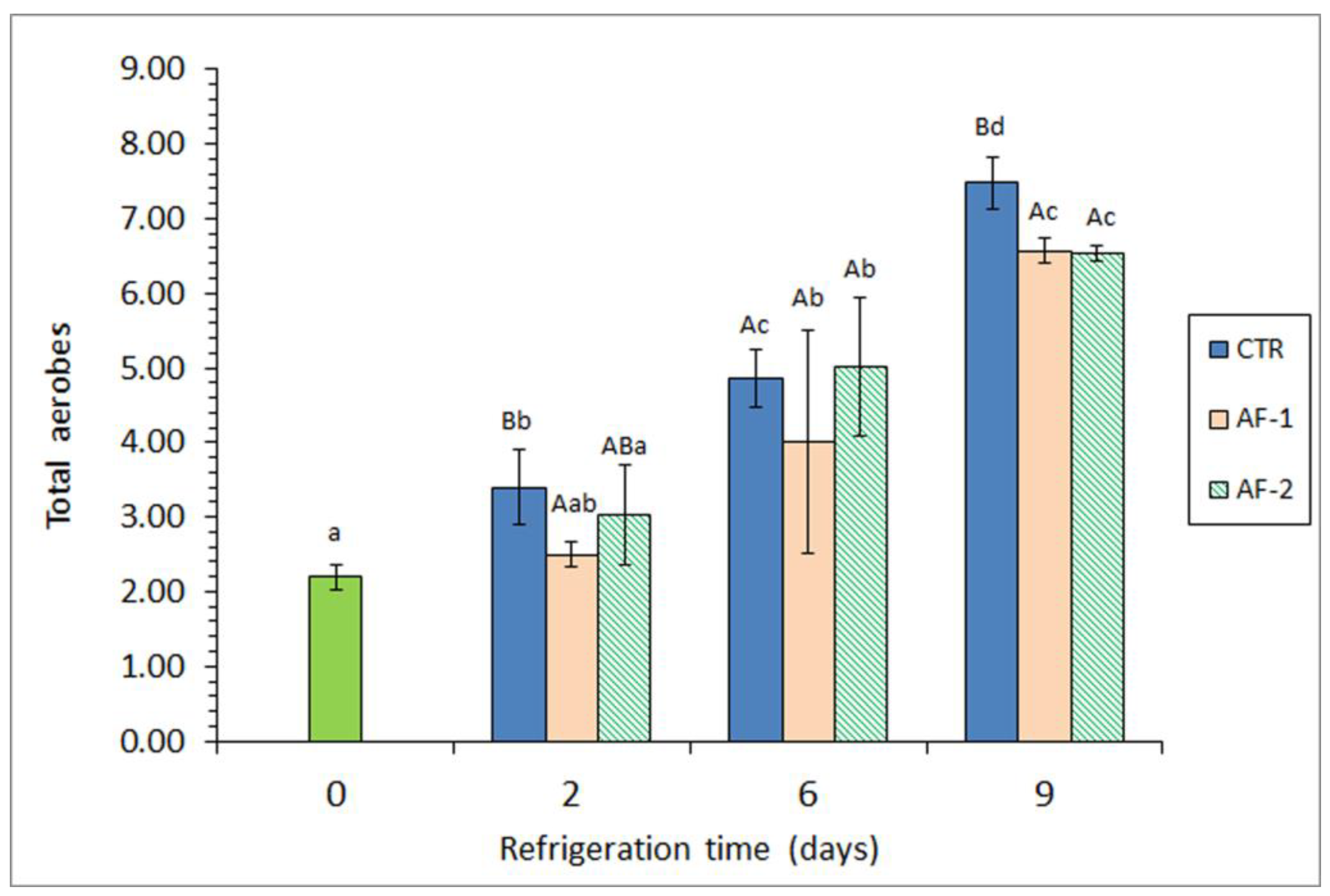
Figure 1. Determination of aerobic counts (log CFU·g−1 muscle) in refrigerated mackerel subjected to different packaging conditions. Average values ± standard deviations (n = 3). The packaging conditions are expressed in Table 1. Different lowercase letters denote significant differences (p < 0.05) with chilling time; different capital letters denote significant differences (p < 0.05) as a result of the packaging condition. Time 0 corresponds to initial fish.
Applsci 14 08817 g001
Figure 2. Determination of proteolytic counts (log CFU·g−1 muscle) in refrigerated mackerel subjected to different packaging conditions. Average values ± standard deviations (n = 3). The packaging conditions are expressed in Table 1. Different lowercase letters denote significant differences (p < 0.05) with chilling time; different capital letters denote significant differences (p < 0.05) as a result of the packaging condition. Time 0 corresponds to initial fish.
Applsci 14 08817 g002
Figure 3. Determination of the free fatty acid (FFA; g·kg−1 lipids) content in refrigerated mackerel subjected to different packaging conditions. Average values ± standard deviations (n = 3). The packaging conditions are expressed in Table 1. Different lowercase letters denote significant differences (p < 0.05) with chilling time; different capital letters denote significant differences (p < 0.05) as a result of the packaging condition. Time 0 corresponds to initial fish.
Applsci 14 08817 g003
Table 1. Determination of Enterobacteriaceae, psychrotrophs, and lipolytic bacterial counts (log CFU·g−1 muscle) * in refrigerated mackerel subjected to different packaging conditions **.
Microbial GroupPackaging ConditionRefrigeration Time (Days)
0269
EnterobacteriaceaeCTR1.00 ± 0.00 a1.86 ± 0.52 Ab2.33 ± 0.35 Ab4.21 ± 0.65 Ac
AF-11.00 ± 0.00 a1.53 ± 0.20 Ab1.73 ± 0.88 Ab3.23 ± 0.35 Ac
AF-21.00 ± 0.00 a1.75 ± 0.64 Ab2.28 ± 0.87 Ab3.73 ± 0.41 Ac
PsychrotrophsCTR2.82 ± 0.24 a4.68 ± 0.79 Bb6.32 ± 0.32 Bc8.78 ± 0.15 Bd
AF-12.82 ± 0.24 a2.93 ± 0.35 Aa5.16 ± 0.32 Ab7.83 ± 0.30 Ac
AF-22.82 ± 0.24 a4.68 ± 0.39 Bb6.44 ± 0.15 Bc7.54 ± 0.55 Ad
Lipolytic bacteriaCTR2.00 ± 0.00 a2.26 ± 0.24 Aa3.13 ± 0.41 Ab4.95 ± 1.09 Ac
AF-12.00 ± 0.00 a2.66 ± 0.58 Aab2.43 ± 0.51 Aab3.64 ± 1.05 Ab
AF-22.00 ± 0.00 a2.16 ± 0.28 Aab3.00 ± 0.89 Abc3.63 ± 0.46 Ac
* Average values ± standard deviations (n = 3). In each row, different lowercase letters denote significant differences (p < 0.05) with refrigeration time; in each column, different capital letters denote significant differences (p < 0.05) as a result of the packaging conditions. ** Packaging conditions: CTR (Control; gelatin films prepared without alga flour extract); AF-1 and AF-2 correspond to low and high concentrations of alga flour extracts, respectively, in gelatin films.
Table 2. Evolution of the pH and trimethylamine (TMA; mg TMA-N·kg−1 muscle) values * in refrigerated mackerel subjected to different packaging conditions **.
Quality IndexPackaging ConditionRefrigeration Time (Days)
0269
pHCTR6.25 ± 0.02 a6.51 ± 0.02 Bb6.58 ± 0.15 Ab6.59 ± 0.02 Bb
AF-16.25 ± 0.02 a6.39 ± 0.10 ABab6.40 ± 0.16 Aab6.42 ± 0.08 Ab
AF-26.25 ± 0.02 a6.29 ± 0.08 Aa6.44 ± 0.11 Aab6.50 ± 0.03 ABb
TMACTR1.60 ± 0.42 a1.87 ± 0.47 Aa12.80 ± 1.7 Ab61.50 ± 15.7 Ac
AF-11.60 ± 0.42 a2.60 ± 0.78 Aa11.57 ± 2.3 Ab52.67 ± 23.6 Ac
AF-21.60 ± 0.42 a2.80 ± 0.79 Aa11.00 ± 4.6 Ab50.20 ± 13.1 Ac
* Average values ± standard deviations (n = 3). In each row, different lowercase letters denote significant differences (p < 0.05) with refrigeration time; in each column, different capital letters denote significant differences (p < 0.05) as a result of the packaging conditions. ** Packaging conditions as expressed in Table 1.
Table 3. Determination of lipid oxidation evolution * in refrigerated mackerel subjected to different packaging conditions **.
Quality Index ***Packaging ConditionRefrigeration Time (Days)
0269
PVCTR0.98 ± 0.47 a1.24 ± 0.43 Aa3.05 ± 1.70 Aab5.03 ± 1.61 Cb
AF-10.98 ± 0.47 a0.72 ± 0.35 Aa2.11 ± 1.66 Aab2.27 ± 0.15 Bb
AF-20.98 ± 0.47 ab1.57 ± 0.44 Aab0.68 ± 0.46 Aa1.60 ± 0.33 Ab
TBA-iCTR0.42 ± 0.12 a0.91 ± 0.17 Ab2.86 ± 1.07 Ac4.77 ± 0.76 Bd
AF-10.42 ± 0.12 a1.13 ± 0.45 Ab3.25 ± 1.39 Ac3.10 ± 1.77 ABc
AF-20.42 ± 0.12 a1.18 ± 0.42 Ab1.84 ± 0.42 Ab1.77 ± 0.27Ab
FRCTR3.69 ± 1.56 a4.85 ± 0.77 Aa6.39 ± 0.65 Ab7.22 ± 0.29 Cc
AF-13.69 ± 1.56 a4.89 ± 0.17 Aa5.40 ± 0.61 Aab6.16 ± 0.58 Bb
AF-23.69 ± 1.56 a4.44 ± 0.94 Aa5.84 ± 1.32 Aa4.86 ± 0.16 Aa
PICTR2.27 ± 0.16 a2.04 ± 0.23 Aa1.97 ± 0.21 Aa1.88 ± 0.47 Aa
AF-12.27 ± 0.16 a2.20 ± 0.16 ABa1.98 ± 0.10 Aa1.94 ± 0.53 Aa
AF-22.27 ± 0.16 a2.40 ± 0.10 Ba2.25 ± 0.02 Ba2.14 ± 0.12 Aa
* Average values ± standard deviations (n = 3). In each row, different lowercase letters denote significant differences (p < 0.05) with refrigeration time; in each column, different capital letters denote significant differences (p < 0.05) as a result of the packaging conditions. ** Packaging conditions as expressed in Table 1. *** Abbreviations and units: PV (peroxide value; miliequivalents·kg−1 lipids), TBA-i (thiobarbituric acid index; mg malondialdehyde·kg−1 muscle), FR (fluorescence ratio) and PI (polyene index).
Disclaimer/Publisher’s Note: The statements, opinions and data contained in all publications are solely those of the individual author(s) and contributor(s) and not of MDPI and/or the editor(s). MDPI and/or the editor(s) disclaim responsibility for any injury to people or property resulting from any ideas, methods, instructions or products referred to in the content.