Article

Developmental Toxicity of C10 Massoia Lactone, the Main Constituent of Cryptocarya massoia, on Zebrafish (Danio rerio) Embryos

1
Department of Integrative Biology, Kyungpook National University, Daegu 41566, Republic of Korea
2
Department of Applied Biosciences, Kyungpook National University, Daegu 41566, Republic of Korea
3
Department of Environmental Engineering, Kumoh National Institute of Technology, Gumi 39177, Republic of Korea
4
Department of Energy Engineering Convergence, Kumoh National Institute of Technology, Gumi 39177, Republic of Korea
*
Authors to whom correspondence should be addressed.
These authors contributed equally to this work.
These authors also contributed equally to this work.
Appl. Sci. 2024, 14(2), 538; https://doi.org/10.3390/app14020538
Submission received: 30 November 2023 / Revised: 31 December 2023 / Accepted: 5 January 2024 / Published: 8 January 2024
(This article belongs to the Section Agricultural Science and Technology)

Abstract

:

Featured Application

C10 massoia lactone possessed developmental toxicity on cardiac formation of zebrafish embryos.

Abstract

C10 massoia lactone (C10) is the main component of massoia essential oil derived from Cryptocarya massoia plant bark, which is used as natural flavoring agent of “generally recognized as safe” status. In this study, the developmental toxicity of C10 was evaluated on zebrafish (Danio rerio) embryos at an exposure level of 0–2000 µg·L−1, and acute toxicity was determined with respect to lethal effects, hatching rates, and morphological changes. Additionally, morphological changes were determined for the endpoints as the occurrence of yolk edema, pericardial edema, spine curvature, and shortened body length after treatment until 96 h post-fertilization (hpf). The complete lethality of C10 was achieved with embryos treated at 2000 µg·L−1, and most embryos treated at 1000 µg·L−1 developed pericardial edemas with some spine curvature. Some embryos exhibited delayed development with shortened body length when compared with the control. Hatchability was completely accomplished at the tested dose of 1000 µg·L−1, and cardiac malformation was observed using a transgenic zebrafish line Tg(cmlc:EGFP), with a lower heartbeat rate in embryos treated with C10 for 72 hpf. After 96 hpf, heartbeat rates were normalized when compared with the control group, and two cardiac development-related genes such as nppa and canca1 were differently expressed in C10-treated embryos by 2.3-fold and 0.4-fold, respectively. Therefore, C10 must be studied further in other higher organisms for its risk.

1. Introduction

Recently, the Flavor and Extract Manufacturers Association (FEMA) started to review the safety of more than 250 naturally occurring flavor complexes (NFCs) currently utilized in food industry. In this review, 23 NFCs have been confirmed as generally recognized as safe (GRAS) when they are used as flavor ingredients in accordance with the examination of each NFC on the basis of the view of threshold of toxicological apprehensiveness [1]. Among the 23 NFCs, highly produced oils such as castor oil (6690 kg), lemongrass oil (3980 kg), and citronella oil (1830 kg) are included in the GRAS-affirmed list; moreover, the lowest produced, NFC massoia oil (MO, 0.4 kg), is also in the list [1]. However, this review has not considered the safety of other purposed uses as dietary supplements or other ingestible preparations. Furthermore, more scientific information to properly determine NFC’s toxicity is necessary to understand the role of NFC in epigenetic modifications rather than genotoxic changes in humans or other mammals. This is because the evaluation process of toxicity includes the steps to find the exposure routes and possible toxicity of the recognized components [1,2,3].
MO is derived from the bark of Massoia aromatica (Becc, Lauraceae), which is native to Indonesia, Papua New Guinea, and Australia. MO possesses anti-Candida properties toward Candida albicans with 0.074% (v/v) of IC50 [4] and an antiaflatoxigenic effect on Aspergillus flavus at the 0.5 ppm level [5]. These biological activities are derived from the main component of MO, known as C10 massoia lactone (C10), as it inhibits the hyphal formation and hyphal growth of C. albicans as well as the mycelial growth of A. flavus [4,5]. C10 has been quantitatively determined using a chromatographic analysis such as gas chromatography–mass spectrometry (GC–MS), accounting for over 40% of the total components in MO [5]. The antifungal activities of other main lactones such as C12 massoia lactone (36.7%) and C14 massoia lactone (1.4%) were excluded from this study because researchers can hardly obtain these compounds as they are commercially not available. Moreover, in inhibiting the growth of 21 plant pathogens, C10 decreased ergosterol formation, which is an important component of fungal cell structure with an increasing amount of reactive oxygen species in fungal cells [6]. On the contrary, C10 and C12 were isolated from MO, and C10 showed larvicidal activities on Aedes albopictus with 100% mortality at a treatment level of 50 ppm, whereas C12 showed 100% lethal effect on the mosquito larvae at a treatment level of 25 ppm [7]. According to [7], C10 was the major lactone in MO as it reached 79.97% as analyzed by GC–MS.
The developmental toxicity of various chemicals has been assessed using zebrafish as they are easy to study because of the transparency of their eggs, high similarity with human genomes, formation of all organs, and large numbers of offsprings after mating [8,9]. To evaluate the developmental toxicity, the transgenic lines of zebrafish can be used to understand the phenomenal modification on organ development, including the heart, liver, brain, and blood vessels with blood flow [10,11,12]. Recently, piperlongumine as one of potential anticancer drug caused severe inhibition on heart and liver development of zebrafish embryos, although its efficacy to control cancer cells has been well reported [13,14]. Therefore, we believe that preclinical examination using zebrafish embryos can bridge the gap between in vitro to in vivo tests, and studies such as ours provide more useful information when a scientific panel decides to use some natural products as GRAS in food industries [15].
In this study, C10 was used to evaluate its developmental toxicity on zebrafish embryos. First, acute toxicities were determined with the two endpoints as lethal effects hatching rates of C10-treated zebrafish embryo groups. Morphological modifications were examined in the C10-treated embryo groups as the whole-body length, spine curvature, and edema occurrences on the heart and yolk. Heartbeat was also checked to examine proper heart function, and the transgenic line on the myosin light chain proteins in the heart, Tg(cmlc:EGFP), was used to determine abnormal heart development. Finally, RT-qPCR was performed to examine gene expression in relation to abnormal heart formation and heart dysfunction.

2. Materials and Methods

2.1. Chemicals

6-pentyl-5,6-dihydropyran-2-one with a purity of greater than 95%, known as C10 massoia lactone, was provided by Sigma-Aldrich Co. (St. Louis, MO, USA). C10 massoia lactone was initially dissolved in dimethyl sulfoxide with a cell culture grade (Sigma-Aldrich) to produce a stock solution. The initial concentration of stock solution was 500 mg·mL−1. Analytical grade chemicals were used in this study unless further stated.

2.2. Zebrafish Maintenance

A wild-type and a transgenic zebrafish line Tg(cmlc:EGFP) was generously supplied by Emeritus Prof. Tae-Lin Huh, Kyungpook National University (Daegu, Republic of Korea), and stored in our research lab for over 20 generations. The zebrafish were kept in a circulated maintenance system filtered by using a three-staged carbon-sediment filter. Biological filtration was performed using a sponge-filled sump tank, accommodating a bio-filter medium, which was consisted of a Sera Siporax bio active supplied by SERA (Heinsberg, Germany) and EHEIM SUBSTRATpro supplied by Eheim GmbH & Co. KG (Deizisau, Germany). The water sterilization was performed by a UV sterilizer, and the temperature of water was around 26 °C, controlled by a temperature control system (DBM-250) supplied by Daeil (Busan, Republic of Korea). The fish nurturing room was set to a light/dark cycle of 14:10 h (day:night) under control. Tetra fresh brine shrimps (Tetra, Blacksburg, VA, USA) were continuously provided three times per day.

2.3. Acute Toxicity of C10 Massoia Lactone to Zebrafish Embryos

The acute toxicity of C10 was evaluated in accordance with the OECD guideline 236. A day before the test, healthy fish (10 pairs) were left in the mating cage for the collection of laying embryos. On the day of the test, the embryos were washed using methylene blue in an E3 medium consisting of NaCl (5 mM), KCl (0.17 mM), CaCl2 (0.33 mM), and MgSO4 (0.33 mM). And then, the washed embryos were stored in an E3 medium set to 26 °C for selecting healthy eggs. The selection of healthy embryos was performed by a stereomicroscope (Stemi 305, Zeiss, Oberkochen, Germany) at 3 h post-fertilization (hpf). At least 15 selected healthy eggs were left in each well of a six-well plate, consisting of the control group in 0.1% DMSO in E3 medium or/and the treated groups using C10 massoia lactone (0, 125, 250, 500, 1000, and 2000 µg·L−1). In measuring the acute toxicity, C10 massoia lactone-treated embryos were exposed until 96 hpf without further replacement of test solution. The three end points such as hatching rates, morphological changes, and lethal effects were examined and recorded at every 24 h until 96 hpf during the test period. Using SAS statistical software (Version 9.4) developed by SAS Institute Inc. (Cary, NC, USA), the LC50 value of C10 massoia lactone was calculated, and morphological changes during the development were captured by an BX53 (Olympus, Tokyo, Japan) arrayed with a DP80 monochrome/color dual CCD camera.

2.4. Assessment of the Developmental Defects in Heart

In the Tg(cmlc:EGFP) line, heterogenotyped female fish were left in the mating chamber with the wild-type male fish the night before use, and they were kept overnight. The next morning, zebrafish eggs were promptly collected and, among the eggs, healthy embryos were selected in accordance with the abovementioned guidelines. Thirty healthy embryos were placed in each well of six-well plates with an E3 medium. Then, the egg-containing well was treated with a series of C10 concentrations, including 0, 125, 250, 500, 1000, and 2000 µg·L−1. Heterogenotyped embryos were picked at 48 hpf, and the homogenotyped embryos were discarded. Pictures of heterogenotyped embryos were fluorescently captured using the BX53 microscope with the DP80 CCD. For Tg(cmlc:EGFP), cardial morphological changes of the C10-treated embryos were recorded as images at 72 and 96 hpf and compared with those of the control group. Heartbeat rates of each C10-treated group, including the control group, were recorded at 48, 72, and 96 hpf for 30 s, and then multiplied two times for reaching heartbeat rates per minute (n = 10). The dead embryos were discarded to maintain clean environments and protect all tested embryos from bacterial infection.

2.5. Analysis Gene Expression Using RT-qPCR

In analyzing gene expression, RT-qPCR was employed and performed according to the previously published report [16]. Fifty embryos of each C10-treated group were selected at 96 hpf and washed twice using Ambion DEPC-treated water supplied by Thermo Fisher Scientific (Waltham, MA, USA). Afterward, TRIzol solution (1 mL, Thermo Fisher Scientific) was treated to the sample embryos, and left on ice for 15 min. Next, chloroform (200 µL) was added to the sample solution, and the chloroform mixture was vortexed for 15 s with extra incubation for 15 min. The centrifugation for the incubated samples was conducted for 15 min at 13,000× g, and then the supernatants were cautiously collected and transferred to the new e-tubes. During RNA aggregation, isopropanol (500 µL) was added into each sample tube with gentle mixing. The mixed sample-containing tubes were placed on ice for 10 min, and then further centrifugation was performed for 15 min. Eventually, the extracted RNA with white pellets were redissolved in DEPC-treated water, and quality control was conducted on the sampled RNAs. Furthermore, the sampled RNAs were analyzed using the μDrop™ plate system (Thermo Fisher Scientific) equipped with a Multiskan Go microplate reader (Thermo Fisher Scientific). Using the extracted total RNA, cDNA was prepared by the Maxima First Strand cDNA Synthesis Kit (Thermo Fisher Scientific) in accordance with the manufacturer’s guidelines. The mRNA level was analyzed by Quantstudio 3 (Thermo Fisher Scientific) with a Luna Universal qPCR Master Mix (New England BioLabs Inc., Ipswich, MA, USA). For the amplification, running conditions were used as 40 cycles of 95 °C for 10 s, 60 °C for 15 s, and 72 °C for 20 s. For the analysis of the gene expression level, the 2−∆ΔCT method was utilized to analyze the raw data, and the gapdh level was used as a normalizer. The primers used in this study are listed in Table S1.

2.6. Statistical Analysis

Statistical analysis to evaluate all data in this study was conducted by GraphPad Prism 8.0 (GraphPad Software, San Diego, CA, USA). Another statistical platform (IBM SPSS Statistics 26.0) was also employed and used in this study. Furthermore, one-way ANOVA with post hoc Tukey tests were used for multiple comparison among C10-treated groups. Data were expressed as the mean with standard deviations.

3. Results

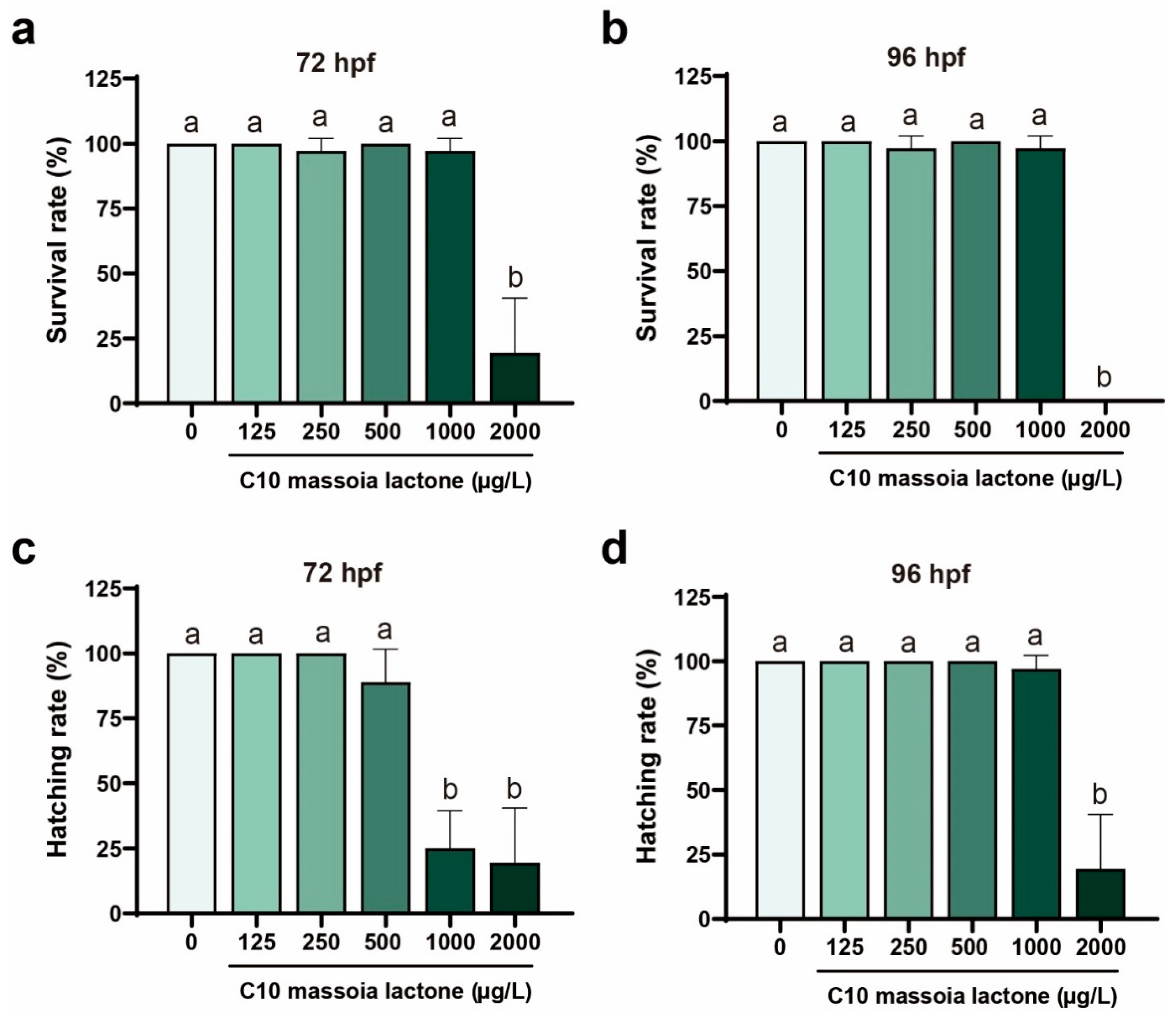
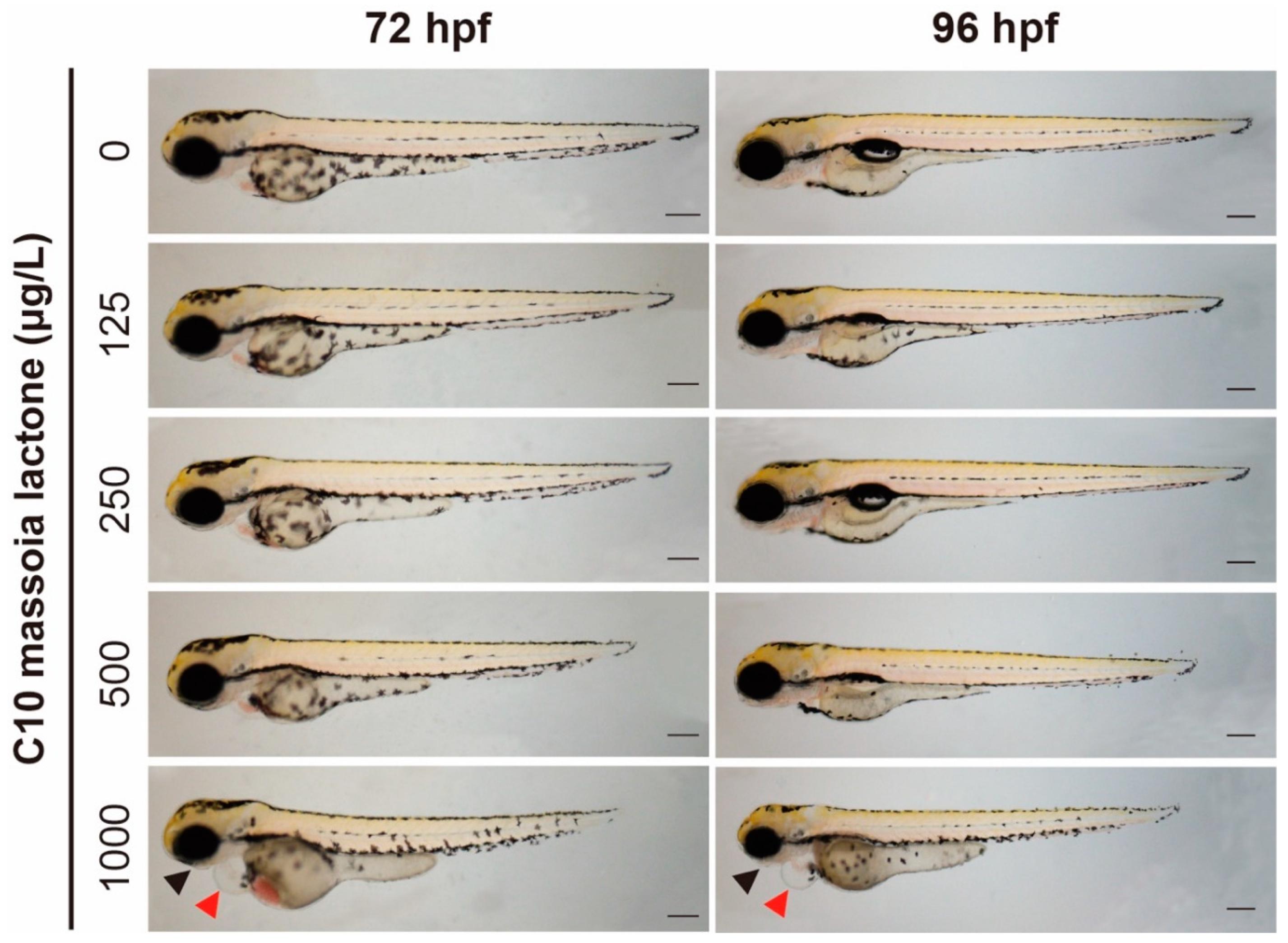
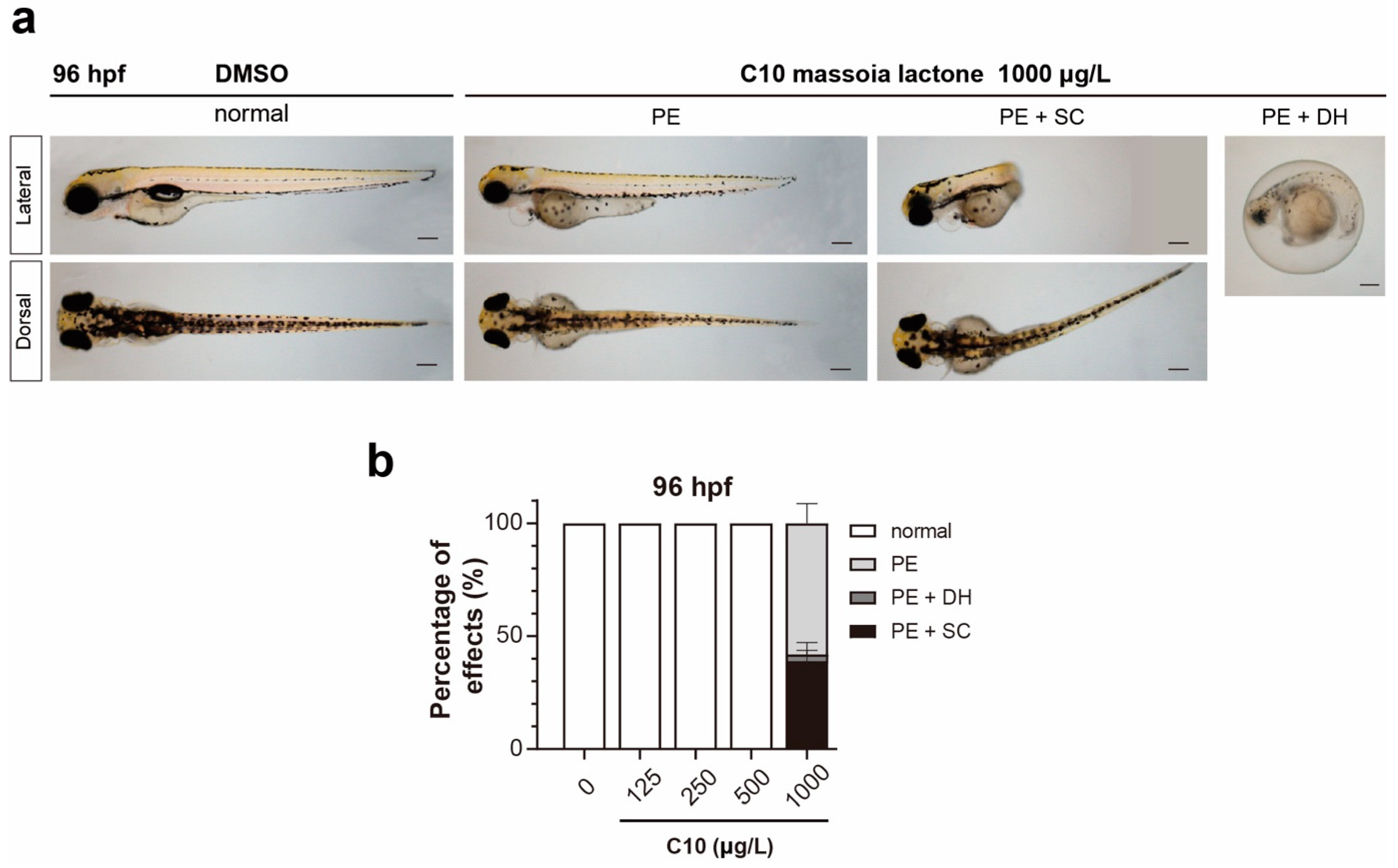
3.1. Acute Toxicity of C10 on Zebrafish Embryos and Phenotypic Alterations

Zebrafish embryos were treated with massoia lactone (C10) in the dose range of 0 to 2000 µg·L−1 during 96 h exposure. The lethal effects of C10 were observed at 72 and 96 hpf after treatments (Figure 1), and 20% of the embryos in the total C10-treated group lived at a treated dose of 2000 µg·L−1 after 72 hpf (Figure 1a). At the same concentration, a 100% mortality was found in C10 embryos after 96 hpf (Figure 1b). No mortality was observed in groups at a treated concentration of less than 1000 µg·L−1 (Figure 1a,b). In the case of hatching rates, 100% hatching rates were measured at treated concentrations of 125 and 250 µg·L−1, whereas 85% and lower than 25% hatching rates were found at 500–2000 µg·L−1 for 72 hpf (Figure 1c,d). However, a huge difference was determined in the hatching rates of the C10-treated groups at 1000 µg·L−1 after 72 hpf (Figure 1c), as most embryos in the same group were hatched at 96 hpf (Figure 1d). On the contrary, the C10-treated group at 2000 µg·L−1 was all dead without proper hatching, indicating that C10 may penetrate the chorion membrane of embryos.
A severe shortened size on the whole-body length was measured in the C10-treated groups at 500 and 1000 µg·L−1 (Figure 2). Developmental delay occurred after C10 treatment on zebrafish embryos. Thus, various malformations were found in C10-treated embryos at a concentration of 1000 µg·L−1 (Figure 3). Three different types of malformations were discovered, such as pericardial edema (PE), SC, and the combination of PE + SC (Figure 3a).
Additionally, PE with delayed hatching (DH) was measured because PE was observed in the embryos through DH. No malformation was found in the embryos at a C10-treated concentration of 0–500 µg·L−1 after 96 hpf (Figure 3b). At a treated concentration of 1000 µg·L−1, 55% of the treated embryos exhibited PE, less than 4% of the treated embryos showed PE with DH, and approximately 40% of the treated embryos had PE with SC (Figure 3b). Furthermore, all C10-treated embryos exhibited PEs with a low percentage of SC.

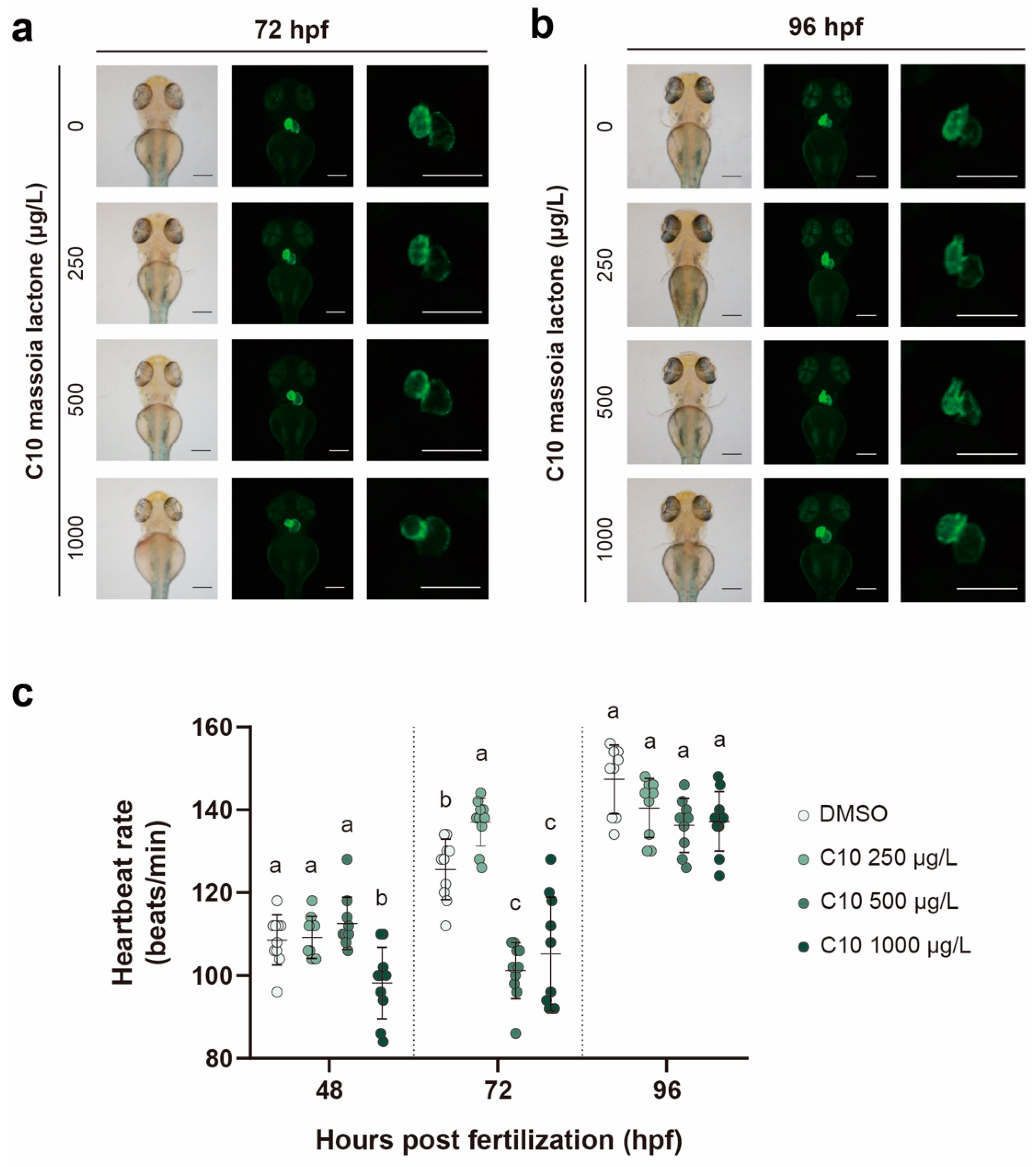
3.2. Heart Development with the Measurement of the Heartbeat Rate

The healthy embryos of Tg(cmlc:EGFP) were employed to understand the cardiac development in C10-treated embryos (Figure 4a). This transgenic line was prepared for zebrafish expressing GFP proteins in the cmlc gene promoter. Using these transgenic embryos, C10 was treated at three different concentrations (250, 500, and 1000 µg·L−1), and the formation of the heart of embryos was visualized, particularly images of the C10-treated embryos after 72 and 96 hpf at 250, 500, and 1000 µg·L−1 treatments (Figure 4a,b).
At 72 hpf after C10 treatment, the embryos at the C10-exposed concentrations of 250 and 500 µg·L−1 did not show any malformation of the heart (Figure 4a). At 96 hpf after C10 treatment, the embryos at C10-exposed concentrations of 250 and 500 µg·L−1 did have not altered heart formation (Figure 4b). In the case of the C10-treated embryos at 1000 µg·L−1, a larger ventricle and atrium of the heart in C10-treated embryos were observed, but this change did not cause any dysfunction of the heart because no difference was observed among heartbeat rates (Figure 4c). However, the heartbeat rates significantly decreased in the C10-exposed groups at 1000 µg·L−1 after 48 hpf. Furthermore, the heartbeat rates significantly increased in the C10-exposed groups at 250 µg·L−1 with decreasing heartbeat rates in the treated embryos at 500 and 1000 µg·L−1 after 72 hpf. Finally, all C10-treated embryos showed no significantly different heartbeat rate in comparison to the control group after 96 hpf (Figure 4c).

3.3. Heart Malformation and the Relative mRNA Expression

Heart formation was observed in C10-treated embryos in comparison to the control group. Among the eight genes in the C10-exposed embryos, nppa and cacna1c genes were expressed by about 2.3-fold and 0.4-fold, respectively, at a treated concentration of 1000 µg·L−1 after 96 hpf when compared with the control group (Figure 5).
The genes nppa and cacna1c are generally recognized for encoding natriuretic peptide A and a voltage-dependent calcium channel, respectively. Therefore, alterations of the expression of these two genes may cause the cardiac dysfunction as changes in blood pressure regulation in the heart and influx of calcium ions into the heart cells. However, the other six tested genes were not considered because of the cutoff value of two-fold differences. Under the same standard on the gene expression, the other nine genes were not significantly considered because of the cutoff value. Such genes are involved in the detoxification or defense, inflammation, apoptosis, and endoplasmic reticulum (Figure S1).

4. Discussion

4.1. Lethal Effect and Inhibition of the Hatching Rates by C10 toward Zebrafish Embryos

The ecotoxicological toxicity of plant essential oils (EO) and their monoterpenoids in aquatic organisms, including various fish species, has been studied. Three EO derived from Aloysia triphylla, Lippia gracilis, and Piper aduncum showed lethal effects on juvenile tambaqui (Colossoma macropomum) with LC50 values (4 h of exposure) of 109.57, 41.63, and 48.17 mg·L−1 for A. triphylla EO, L. gracilis EO, and P. aduncum EO, respectively [17]. These EOs caused severe damage to the gills of the tested fish larvae. However, the 96 h-LC50 value for EO of thyme plant, thymol, EO of cumin seeds, and EO of caraway seeds was reported using a 30-day-old rainbow trout as 6.6, 2.6, 35, and 14 mg·L−1, respectively [18]. Similarly, two monoterpenes, namely, thymol and 1,8-cineol, were assessed to determine their LC50 values toward the insectivorous guppy fish Poecilia reticulate [19]. The 24 h-LC50 values of thymol toward female and male guppy fish were 12.51 and 10.99 mg·L−1, respectively, whereas the LC50 values of 1,8-cineol toward female and male guppy fish were 3997.07 and 1701.93 mg·L−1, respectively [19]. These two monoterpenes showed 319.5-fold and 154-9-fold differences for female guppy and male guppy, respectively [19].
In our study, C10 massoia lactone (C10) showed 100% mortality at a treated dose of 2 mg·L−1, and 100% paralyzed larvae were observed at 1 mg·L−1. However, no lethal effect on zebrafish embryos was found until 96 h exposure at 1 mg·L−1. This result was not directly different from the LC50 value in previous reports because of the use of different fish organisms and their developmental stages, but this lactone exhibited strong mortality toward zebrafish embryos at a lower treatment concentration to kill 100% tested organisms. Thus, Zingiber cassumunar EO showed 100% mortality at 100 mg·L−1 against zebrafish embryos [20], and Port Orford cedar heart oil showed 100% mortality toward O. mykiss at 5.0 mg·L−1 [21]. Therefore, C10 was stronger than EO of Z. cassumunar on zebrafish embryos for 96 h exposure.
In the case of hatching rates, no direct comparison is performed between C10 and other EOs and their constituents using zebrafish embryos. However, there are many reports on effect of pesticides on hatching rates using the same organism. Fipronil (FIL), a phenylpyrazole insecticide, exhibited about 70% mortality and 85% hatchability in zebrafish embryos treated with 5.0 mg·L−1 of FIL [22]. In our study, 100% mortality was achieved in zebrafish embryos exposed to 2.0 mg·L−1 of C10, and at the same treatment concentration, hatchability was completely suppressed. In this regard, C10 caused a stronger inhibitory effect on hatchability when compared with the two insecticides, CPM, and FIL.

4.2. Morphological Changes in C10-Treated Zebrafish Embryos

With C10 treatment on zebrafish embryos, a significant change on their morphologies is related to the occurrence of PE in the whole treated embryos at the concentration of 1.0 mg·L−1. Most chemicals cause, separately or in-combination, SC, yolk edema, PE, and shortened body length. For example, FIL and its two metabolites as FIL-sulfite and FIL-sulfone did not induce PE [22], and pyraclostrobin (PS) induced 100% pericardial formation in zebrafish embryos at a treated concentration of 0.075 mg·L−1 [16]. The formation of PE may be related to the induction of cardiac malformation in zebrafish embryos. This issue has not been statistically supported, but many studies may prove this hypothesis. In the case of PS-treated zebrafish embryos, the formation of the atrium and ventricle was inhibited at a concentration of 0.075 mg·L−1 with a change in shape from circular to cylindrical, and the dysfunction of malformed hearts was confirmed by measuring the heartbeat rates [16]. Therefore, C10 may be considered as an enhancer of PE formation in zebrafish embryos similar to PS and 2-(bromomethyl)naphthalene (2-BMN), resulting in the further study of C10 on the verification of heart development.
Another morphological change after C10 treatment in zebrafish embryos is shortened body length. This change is related to developmental delay, thus most embryos with shortened body length return to the normal condition of life after the disappearance of stressful moments. For example, azoxystrobin (AZ) caused shortened whole-body length in zebrafish embryos, but the AZ-treated embryos returned to a survival rate of 94.9% with a hatching rate of 78.6% after 96 hpf at an AZ-treated concentration of 2.0 mg·L−1 [16]. Therefore, the shortened body length caused by the tested chemicals may indicate no severe damage on life, but the concurrent occurrence of other morphological changes may provide considerable malformation in development.

4.3. Heart Development in C10-Treated Zebrafish Embryos

By conducting C10 treatment, PE occurred in all C10-exposed zebrafish embryos at a concentration of 1.0 mg·L−1. The occurrence of this edema may be related to heart malformation. Therefore, a transgenic zebrafish line, Tg(cmlc2:EGFP), was utilized to find the relationship between C10 treatment and heart malformation. Many studies have supported the relationship between PE occurrence and heart malformation [16,22]. C10 caused slight differences in cardiac development when compared with the control, resulting in decreasing heartbeat rates at 48 and 72 hpf and then maintaining normal heartbeat rates at 96 hpf in C10-treated embryos. Based on these results, C10 showed no strong developmental toxicity on heart formation.
Using RT-qPCR analysis, the expression of eight genes was examined in C10-treated embryos, and nppa and cacna1c genes were significantly up- and down-regulated, respectively. In the nppa gene expression, C10-treated embryos exhibited a 2.3-fold increase and encoded natriuretic peptide A, which restricted the atrial location during embryonic development [23]. The deficiency of the nppa gene is related to the failure of blood pressure [24], so a higher expression in C10-treated embryos may cause heart dysfunction as our study observed differential heartbeat rates. In the PS-treated zebrafish embryos, the nppa gene was highly expressed by 8.6-fold when compared with the control group [16].
However, the decreased expression level of the cacna1 gene in C10-treated zebrafish embryos was observed, and this gene downregulation may be related to the dysfunction of heart contraction caused by calcium homeostasis impairment because it encodes a voltage-dependent calcium channel. Recently, a similar result has been reported in the bisphenol A-treated zebrafish embryo as an impairment of cardiac contraction and relaxation by the downregulation of the cacna1 gene [25].
Other genes were not differently expressed in relation to C10 treatment in zebrafish embryos. In zebrafish embryos treated with ginger oil, very low teratogenic effects were observed in the treatment range of 1 to 10 mg·L−1. Thus, ginger oil does not have embryotoxicity and teratogenicity at treated concentrations of less than 10 mg·L−1 [20]. The C10 compound is a major component of massoia EO; however, this compound shows high embryotoxicity as 100% of zebrafish embryos died at 2.0 mg·L−1 when compared with ginger oil, and it exhibits heart dysfunction until 72 hpf after C10 treatment.

5. Conclusions

Natural flavoring additives in foods are considered as GRAS because of their long-term use in human food without any hazardous effect on human health. Recently, some additives were re-evaluated and affirmed as GRAS for use in flavorings, including MO. In MO, C10 lactone is a major constituent, reaching over 40%. However, limited information is available regarding C10 toxicity toward various living organisms. In this study, the developmental toxicity of C10 was determined using zebrafish embryos as a comparison and prediction organism to human health because they are highly similar to human genome and organ development. C10 caused 100% PE in the tested embryos at 1.0 mg·L−1, and all tested embryos died at 2.0 mg·L−1. These results were compared with ginger oil toxicity, which is weaker in zebrafish embryos. Furthermore, C10 may cause cardiac malformation during heart development in the treated embryos, resulting in lower heartbeat rates. In this study, two important cardiac development-related genes were differently expressed in the C10-treated embryos. Therefore, C10 as a main component in MO, may cause high mortality and cardiac malformation to zebrafish embryos, thus its risk should be considered in other organisms such as humans.

Supplementary Materials

The following supporting information can be downloaded at: https://www.mdpi.com/article/10.3390/app14020538/s1, Table S1. Primers used for RT-qPCR analysis. Figure S1. Various gene expression levels related to detoxification process, inflammation, and apoptosis in zebrafish embryos treated with C10 massoia lactone at 96 hpf. The treated concentrations of C10 massoia lactone were 250, 500, and 1000 μg·L−1. Data were analyzed by a one-way ANOVA, and post hoc Tukey tests were used when there were differences among the means. Lowercase letters denote a significant difference between groups; a > b (p < 0.05). sod: superoxide dismutase. Gst: glutathione S-transferase. Cat: catalase. Il-6: interleukin-6. Il-1β: interleukin 1 beta. Bax: bcl-2-like protein 4. Bcl2a: B-cell lymphoma 2a. atf4: activating transcription factor 4. Atf6: activating transcription factor 6.

Author Contributions

Conceptualization, Y.L. and S.-E.L.; methodology, Y.L. and C.K.; software, Y.L. and C.K.; validation, Y.L., C.K., and S.-E.L.; formal analysis, Y.L. and C.K.; investigation, Y.L. and C.K.; resources, T.-O.K.; data curation, Y.L., C.K. and S.-E.L.; writing—original draft preparation, Y.L., T.-O.K., and S.-E.L.; writing—review and editing, Y.L., C.K., T.-O.K. and S.-E.L.; visualization, Y.L., C.K., and S.-E.L.; supervision, T.-O.K. and S.-E.L.; project administration, S.-E.L.; funding acquisition, T.-O.K. All authors have read and agreed to the published version of the manuscript.

Funding

This research was supported by Kumoh National Institute of Technology (202001120001).

Institutional Review Board Statement

Not applicable.

Informed Consent Statement

Not applicable.

Data Availability Statement

The data presented in this study are available on request from the corresponding author.

Conflicts of Interest

The authors declare no conflicts of interest.

References

  1. Rosol, T.J.; Cohen, S.M.; Eisenbrand, G.; Fukushima, S.; Gooderham, N.J.; Guengerich, F.P.; Hecht, S.S.; Rietjens, I.M.C.M.; Davidsen, J.M.; Harman, C.L.; et al. FEMA GRAS Assessment of Natural Flavor Complexes: Lemongrass Oil, Chamomile Oils, Citronella Oil and Related Flavoring Ingredients. Food Chem. Toxicol. 2023, 175, 113697. [Google Scholar] [CrossRef] [PubMed]
  2. Kroes, R.; Galli, C.; Munro, I.; Schilter, B.; Tran, L.A.; Walker, R.; Würtzen, G. Threshold of Toxicological Concern for Chemical Substances Present in the Diet: A Practical Tool for Assessing the Need for Toxicity Testing. Food Chem. Toxicol. 2000, 38, 255–312. [Google Scholar] [CrossRef] [PubMed]
  3. Cohen, S.M.; Eisenbrand, G.; Fukushima, S.; Gooderham, N.J.; Guengerich, F.P.; Hecht, S.S.; Rietjens, I.M.C.M.; Davidsen, J.M.; Harman, C.L.; Taylor, S.V. Updated Procedure for the Safety Evaluation of Natural Flavor Complexes Used as Ingredients in Food. Food Chem. Toxicol. 2018, 113, 171–178. [Google Scholar] [CrossRef] [PubMed]
  4. Hertiani, T.; Pratiwi, S.U.T.; Yuswanto, A.; Permanasari, P. Potency of Massoia Bark in Combating Immunosuppressed-Related Infection. Pharmacogn. Mag. 2016, 12, S363. [Google Scholar] [CrossRef] [PubMed]
  5. Lee, Y.; Park, S.J.; Kim, K.; Kim, T.O.; Lee, S.E. Antifungal and Antiaflatoxigenic Activities of Massoia Essential Oil and C10 Massoia Lactone against Aflatoxin-Producing Aspergillus Flavus. Toxins 2023, 15, 571. [Google Scholar] [CrossRef] [PubMed]
  6. Zhang, M.; Gao, Z.C.; Chi, Z.; Wang, Z.; Liu, G.L.; Li, X.F.; Hu, Z.; Chi, Z.M. Massoia Lactone Displays Strong Antifungal Property Against Many Crop Pathogens and Its Potential Application. Microb. Ecol. 2022, 84, 376–390. [Google Scholar] [CrossRef]
  7. Seo, S.M.; Lee, J.W.; Shin, J.; Tak, J.H.; Hyun, J.; Park, I.K. Development of Cellulose Nanocrystal-Stabilized Pickering Emulsions of Massoia and Nutmeg Essential Oils for the Control of Aedes Albopictus. Sci. Rep. 2021, 11, 12038. [Google Scholar] [CrossRef] [PubMed]
  8. Hoyberghs, J.; Bars, C.; Pype, C.; Foubert, K.; Ayuso Hernando, M.; Van Ginneken, C.; Ball, J.; Van Cruchten, S. Refinement of the Zebrafish Embryo Developmental Toxicity Assay. MethodsX 2020, 7, 101087. [Google Scholar] [CrossRef]
  9. Porretti, M.; Arrigo, F.; Di Bella, G.; Faggio, C. Impact of Pharmaceutical Products on Zebrafish: An Effective Tool to Assess Aquatic Pollution. Comp. Biochem. Physiol. Part C Toxicol. Pharmacol. 2022, 261, 109439. [Google Scholar] [CrossRef]
  10. Song, Y.S.; Dai, M.Z.; Zhu, C.X.; Huang, Y.F.; Liu, J.; Zhang, C.D.; Xie, F.; Peng, Y.; Zhang, Y.; Li, C.Q.; et al. Validation, Optimization, and Application of the Zebrafish Developmental Toxicity Assay for Pharmaceuticals Under the ICH S5(R3) Guideline. Front. Cell Dev. Biol. 2021, 9, 721130. [Google Scholar] [CrossRef]
  11. Wang, R.; Liu, K.; Zhang, Y.; Chen, X.; Wang, X. Evaluation of the Developmental Toxicity Induced by E804 in Zebrafish Embryos. Front. Pharmacol. 2020, 11, 493657. [Google Scholar] [CrossRef] [PubMed]
  12. Hema, T.; Poopal, R.K.; Ramesh, M.; Ren, Z.; Li, B. Developmental Toxicity of the Emerging Contaminant Cyclophosphamide and the Integrated Biomarker Response (IBRv2) in Zebrafish. Environ. Sci. Process Impacts 2023, 25, 1391–1406. [Google Scholar] [CrossRef] [PubMed]
  13. Jeon, H.J.; Kim, C.; Kim, K.; Lee, S.E. Piperlongumine Treatment Impacts Heart and Liver Development and Causes Developmental Delay in Zebrafish (Danio rerio) Embryos. Ecotoxicol. Environ. Saf. 2023, 258, 114995. [Google Scholar] [CrossRef] [PubMed]
  14. Tripathi, S.K.; Biswal, B.K. Piperlongumine, a Potent Anticancer Phytotherapeutic: Perspectives on Contemporary Status and Future Possibilities as an Anticancer Agent. Pharmacol. Res. 2020, 156, 104772. [Google Scholar] [CrossRef] [PubMed]
  15. Cassar, S.; Adatto, I.; Freeman, J.L.; Gamse, J.T.; Iturria, I.; Lawrence, C.; Muriana, A.; Peterson, R.T.; Van Cruchten, S.; Zon, L.I. Use of Zebrafish in Drug Discovery Toxicology. Chem. Res. Toxicol. 2020, 33, 95–118. [Google Scholar] [CrossRef]
  16. Kim, C.; Choe, H.; Park, J.; Kim, G.; Kim, K.; Jeon, H.J.; Moon, J.K.; Kim, M.J.; Lee, S.E. Molecular Mechanisms of Developmental Toxicities of Azoxystrobin and Pyraclostrobin toward Zebrafish (Danio rerio) Embryos: Visualization of Abnormal Development Using Two Transgenic Lines. Environ. Pollut. 2021, 270, 116087. [Google Scholar] [CrossRef] [PubMed]
  17. Santos, P.R.; Andrade-Porto, S.M.; Oliveira, M.I.B.; Brandão, F.R.; Matos, L.V.; Velásquez, J.G.R.; Farias, C.F.S.; Carpio, K.C.R.; Chaves, F.C.M.; Chagas, E.C. Acute Toxicity of Essential Oils of Aloysia triphylla (L’Hér.) Britton, Lippia Gracilis Schauer, and Piper aduncum L. in Colossoma macropomum (Cuvier, 1818). Braz. J. Biol. 2023, 83, 272853. [Google Scholar] [CrossRef]
  18. Tabarraei, H.; Hassan, J.; Mosavi, S.S. Determination of LD50 of Some Essential Oils and Histopathological Changes in Short-Term Exposure to One of Them in Rainbow Trout (Oncorhynchus mykiss). Toxicol. Res. Appl. 2019, 3, 239784731882071. [Google Scholar] [CrossRef]
  19. Bullangpoti, V.; Mujchariyakul, W.; Laksanavilat, N.; Junhirun, P. Acute Toxicity of Essential Oil Compounds (thymol and 1,8-cineole) to Insectivorous Guppy, Poecilia reticulata Peters, 1859. Agric. Nat. Resour. 2018, 52, 190–194. [Google Scholar] [CrossRef]
  20. Mektrirat, R.; Yano, T.; Okonogi, S.; Katip, W.; Pikulkaew, S. Phytochemical and Safety Evaluations of Volatile Terpenoids from Zingiber cassumunar Roxb. on Mature Carp Peripheral Blood Mononuclear Cells and Embryonic Zebrafish. Molecules 2020, 25, 613. [Google Scholar] [CrossRef]
  21. Duringer, J.M.; Swan, L.R.; Walker, D.B.; Craig, A.M. Acute Aquatic Toxicity of Western Juniper (Juniperus Occidentalis) Foliage and Port Orford Cedar (Chamaecyparis lawsoniana) Heartwood Oils. Environ. Monit. Assess 2010, 170, 585–598. [Google Scholar] [CrossRef] [PubMed]
  22. Kim, C.; Lee, S.E. Developmental Toxicity of Fipronil and Its Two Metabolites towards Zebrafish (Danio rerio) Embryos. Environ. Pollut. 2023, 333, 122119. [Google Scholar] [CrossRef] [PubMed]
  23. Goetze, J.P.; Bruneau, B.G.; Ramos, H.R.; Ogawa, T.; de Bold, M.K.; de Bold, A.J. Cardiac Natriuretic Peptides. Nat. Rev. Cardiol. 2020, 17, 698–717. [Google Scholar] [CrossRef] [PubMed]
  24. Salo, P.P.; Havulinna, A.S.; Tukiainen, T.; Raitakari, O.; Lehtimäki, T.; Kähönen, M.; Kettunen, J.; Männikkö, M.; Eriksson, J.G.; Jula, A.; et al. Genome-Wide Association Study Implicates Atrial Natriuretic Peptide Rather Than B-Type Natriuretic Peptide in the Regulation of Blood Pressure in the General Population. Circ. Cardiovasc. Genet. 2017, 10, e001713. [Google Scholar] [CrossRef]
  25. Arrokhman, S.; Luo, Y.H.; Lin, P. Additive Cardiotoxicity of a Bisphenol Mixture in Zebrafish Embryos: The Involvement of Calcium Channel and Pump. Ecotoxicol. Environ. Saf. 2023, 263, 115225. [Google Scholar] [CrossRef]
Figure 1. Acute toxicity of C10 massoia lactone in the range of treated concentrations from 0–2000 µg·L−1 against zebrafish embryos (Danio rerio). (a) Survival rate of C10 massoia lactone towards the embryos at the 72 hpf. (b) Survival rate of C10 massoia lactone towards zebrafish embryos at the 96 hpf. (c) Hatching rate of C10 massoia lactone-treated embryos at the 72 hpf. (d) Hatching rate of C10 massoia lactone-treated embryos at the 96 hpf. Data analyses were undertaken using a one-way ANOVA, and the differences in the means between control group and the treated groups were analyzed using post hoc Tukey tests (a > b, p < 0.05).
Applsci 14 00538 g001
Figure 2. Phenotypes of C10 massoia lactone-exposed zebrafish embryos at the developmental time points of 72 and 96 hpf after treatment. Phenotypic assessments of C10 massoia lactone were undertaken in the range of concentrations from the 0 to 1000 µg·L−1 levels against zebrafish embryos. Body length (μm) of the control embryos and C10 massoia lactone-treated embryos was measured after the treatment at 72 hpf and 96 hpf. The inserted bar length is set to 200 μm. Assessment for body length used 15 C10 massoia lactone-exposed zebrafish embryos for each treated concentration. The black triangle indicates non-developmental jaws in the embryos after exposure to the treated chemical. The red triangle indicates occurrence of pericardial edema in C10 massoia lactone-treated embryos.
Applsci 14 00538 g002
Figure 3. Malformed phenotypic embryos treated with C10 massoia lactone at 96 hpf after exposure (a). Phenotypic assessments of C10 massoia lactone were undertaken in the range of concentrations from 0–1000 µg·L−1 against the embryos. The inserted bar length is set to 200 µm. PE: pericardial edema. SC: spine curvature. DH: delayed hatching. Measurements of malformed phenotypes used fifteen C10 massoia lactone-exposed embryos for each exposed concentration at 96 hpf after the treatment (b).
Applsci 14 00538 g003
Figure 4. Cardiac development and heartbeat rates in C10 massoia lactone-treated Tg(cmlc2:EGFP) zebrafish embryos. The treated concentrations of C10 massoia lactone were 250, 500, and 1000 µg·L−1 level. (a) Morphology of the heart in the embryos at each exposed concentration at 72 hpf. (b) Morphology of the heart in embryos at each exposed concentration at 96 hpf. Two scale bars apply to measure heart sizes and they indicate 200 μm. (c) The mean of heart beat rates of the C10 massoia lactone-treated embryos were obtained after multiplied to minute, and 10 embryos at each treated concentration were used for the heartbeat measurements at 48, 72, and 96 hpf (c < b < a, p < 0.05).
Applsci 14 00538 g004
Figure 5. Graphs of the expression levels of cardiac development-related genes in C10 massoia lactone-treated zebrafish embryos at 96 hpf. C10 massoia lactone was treated as 250, 500, and 1000 µg·L−1 against the embryos. Data analyses were undertaken using a one-way ANOVA, and the differences in the means between control group and the treated groups were analyzed using post hoc Tukey tests (a > b > c, p < 0.05). Lowercase letters indicate a significant difference between the control and the C10 massoia lactone-treated groups; a > b > c (p < 0.05). The genes nppa, myh7, myh6, tnnt2a, myl7, gata4, nkx-2.5, and cacna1c are responsible for encoding natriuretic peptide A, myosin heavy chain 7, myosin heavy chain 6, troponin T type 2a (cardiac), myosin heavy chain 7, GATA binding protein 4, NK2 homeobox 5, and calcium voltage-gated channel subunit alpha 1C, respectively.
Applsci 14 00538 g005
Disclaimer/Publisher’s Note: The statements, opinions and data contained in all publications are solely those of the individual author(s) and contributor(s) and not of MDPI and/or the editor(s). MDPI and/or the editor(s) disclaim responsibility for any injury to people or property resulting from any ideas, methods, instructions or products referred to in the content.

