Article

Assessment of Surrogate Models for Research on Resistance and Deformation of Repairs of the Human Meniscal Roots: Porcine or Older Human Models?

by
Alejandro Peña-Trabalon
,
Ana Perez-Blanca
,
Salvador Moreno-Vegas
,
M. Belen Estebanez-Campos
and
Maria Prado-Novoa
*
Laboratory of Clinical Biomechanics of Andalusia (BIOCLINA), 29071 Málaga, Spain
*
Author to whom correspondence should be addressed.
Appl. Sci. 2024, 14(2), 670; https://doi.org/10.3390/app14020670
Submission received: 29 November 2023 / Revised: 8 January 2024 / Accepted: 10 January 2024 / Published: 12 January 2024
(This article belongs to the Special Issue Biomechanics of Soft and Hard Tissues)

Abstract

:

Featured Application

This study provides data and rationales to aid researchers in the selection of surrogate models for in vitro assessments of surgically repaired meniscal roots. The results are potentially applicable in experimental in vitro investigations aimed at evaluating the performance of surgical repair both in existing approaches and emerging techniques. It could also be of interest for studies seeking to adjust material models of the meniscal tissue around the suture area for incorporation into computational models.

Abstract

Meniscal root repair is not routinely recommended for patients over 75 years old, yet surrogate age-unrestricted human or porcine models are used for its evaluation. This study assesses the suitability of older human or porcine meniscus models for in vitro testing of the sutured meniscal horn. Three groups of menisci underwent a load-to-failure test with continuous monitoring of the traction force and deformation around the suture: human < 75 years, human ≥ 75 years, and porcine. Both surrogate models were compared to the younger group. The porcine group exhibited a 172.1%-higher traction force before tearing (p < 0.001) and a 174.1%-higher ultimate force (p < 0.001), without there being differences between the human groups. At tissue level, the older group had a 28.7%-lower cut-out stress (p = 0.012) and the porcine group had a 57.2%-higher stress (p < 0.001). Regarding elasticity at the sutured area, a 48.1%-greater deformation rate was observed in the older group (p < 0.001), without difference for the porcine group. In conclusion, neither the porcine nor the older human model demonstrated a clear advantage as a surrogate model for young human sutured meniscal horns. The older human meniscus is preferable for resistance at the specimen level, while the porcine model better represents deformation in the sutured zone.

1. Introduction

The menisci of the knee are fixed by ligamentous attachments that retain them in the intra-articular zone while allowing the necessary mobility (up to 10 mm [1]) to be placed between the tibia and femur at the contact area, which shifts during flexion–extension. The meniscal roots are the only ligamentous attachments with direct insertion into the bone, playing a crucial role in meniscal function. Complete meniscal root avulsion has been shown to cause significant changes in the contact biomechanics of the knee, increasing pressure and decreasing the contact area on the cartilage of the affected compartment, changes that may be similar to those observed for total meniscectomy [2,3,4]. Biomechanical studies have also highlighted the role of meniscal roots as secondary stabilizers of knee kinematics, contributing to the restriction of anteroposterior translation and internal–external rotation [5,6,7]. From a clinical point of view, a meniscal root detachment can lead to the rapid development of arthritis [8] or osteonecrosis [9] as a consequence of biomechanical alterations. The surgical treatment of meniscal root avulsions has currently evolved from partial meniscectomy to meniscal root reinsertion [10]. The surgical repair can be performed using two techniques: transtibial [11,12] or in situ fixation [4,13]. In both approaches, sutures are passed through the horn of the injured meniscus to hold it in place.
Some research studies have subjected sutured meniscal horn specimens to in vitro tests to evaluate the performance of surgical suture materials [14,15,16,17], the effectiveness of suture techniques [12,18,19,20,21], the validity of suturing or fixation devices [21,22,23,24], and the suitability of surgical approaches [18,25,26,27]. Both the resistance of the meniscal horn to suture traction without tear initiation and deformation at the suture area are assessed in these studies. Knowledge of the resistance to traction of the repaired meniscus is important in preventing the failure of surgical intervention, while monitoring post-repair displacements generated by suture traction is needed to ensure that they remain within clinically acceptable limits [28].
Porcine models are often chosen in experimental constructs [12,15,16,17,18,19,21,23,26,27,28,29] because of their reduced variability and their similarities with human menisci in terms of anatomical structure, vascularity, volume, and weight, although the width of porcine menisci is greater [30]. In addition, the kinematics of the porcine knee reasonably approximates that of the human knee. Consequently, the porcine model is a practical and economically feasible choice that finds extensive use in the biomechanical testing of meniscal repair and replacement techniques and allows for the reasonable translation of the results to clinical applications [31,32,33,34]. However, it is not proven that the entire set of biomechanical properties of the porcine meniscus matches those of the human meniscus [33,34,35], raising concerns about the use of this model as a human surrogate and underscoring the need to evaluate its applicability in relation to the specific parameters intended to be studied.
Other in vitro studies use human menisci [4,36,37,38,39], despite their greater heterogeneity among donors. And perhaps more importantly, although meniscal root repair is indicated for non-elderly patients, with the age cut-off being increasingly high in new paradigms [40,41,42], donors are not limited by age in many in vitro biomechanical studies. Therefore, the studies sometimes involve tissue from donors of a very advanced age.
Another interesting use of experimental models of the meniscus is in computational modeling, where material models of meniscal tissue are adjusted from properties obtained from in vitro tests of tissue samples from various origins, including animals and older humans. In this context, the analysis and optimization of different suturing techniques for meniscal root repair could greatly benefit from a tissue model that incorporates the effect induced by thread traction around the suture hole. In order to have an adequate model, it is necessary to perform a large number of experiments, with tissue availability being a major issue.
It is currently unknown as to whether the use of porcine or aged cadaveric material is appropriate for studying the behavior of repaired meniscal roots in adults. To our knowledge, their suitability as substitutes for younger human tissue has not been compared. The objective of this study is to assess the suitability of these two models for in vitro testing of meniscal root repair in terms of suture pull-out resistance and suture traction-induced displacements around the suture site. Our working hypotheses in studies on the mechanical behavior of the sutured meniscal horns in adults are the following: (1) meniscal tissue from older donors (≥75 years old) does not adequately represent the biomechanical behavior of the younger meniscus; (2) the porcine meniscus is a good surrogate for the human meniscus at an age routinely eligible for repairing.

2. Materials and Methods

To test the working hypotheses, three groups of isolated sutured menisci were considered: human <75 years, human ≥75 years, and porcine. The human <75 years group acted as the control group, while the human ≥75 years and porcine groups represented the experimental models under analysis. Isolated meniscus models were tested to focus the study on the behavior of the tissue–suture interface.
After approval by the Ethical Committee of Experimentation of the University of Malaga, to extract the menisci, 38 cryopreserved human knees from the mid-femur to the mid-tibia with no previous history of knee osteoarthritis, knee fractures, or surgery on the meniscus or tibial plateau, provided by a specialized company, and 22 stifle joints of adult pigs donated by a Spanish local slaughterhouse, also from the mid-femur to the mid-tibia, were used, complying with all legal and ethical requirements.

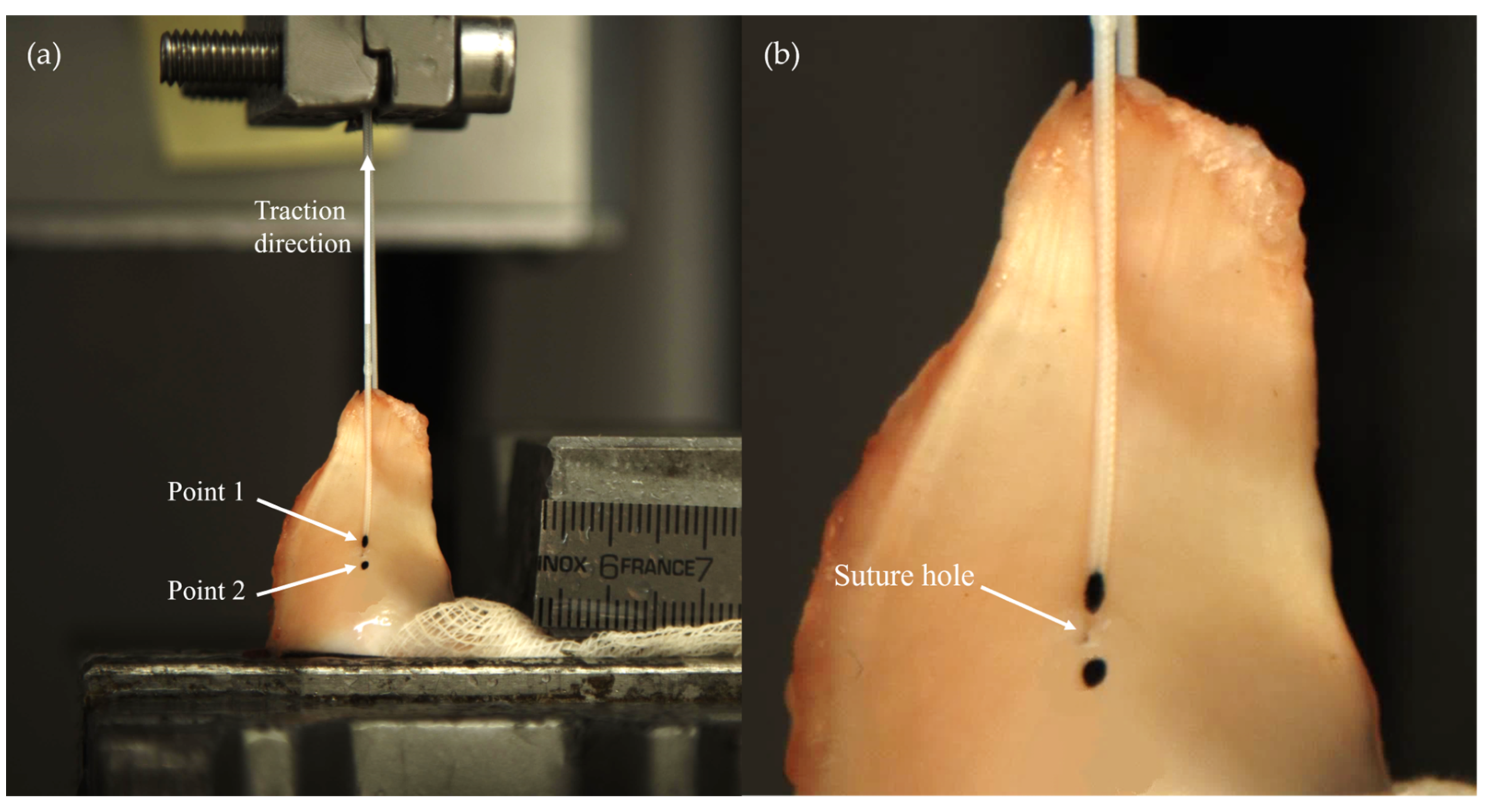
2.1. Specimen Preparation

Adult human knee joints were classified in two age groups, with an age limit of 75 years old. They were individually stored at a temperature of −20 °C in sealed plastic bags. The age limit of 75 years old was selected, taking into account recent recommendations on age extension for meniscal repair surgery.
The day prior to testing, the knee was left to thaw at room temperature, wrapped in dampened gauze. Once thawed, the knee was dissected, and the menisci were extracted. They were visually inspected for any potential damage or pathology, specifically at the roots and horns. The eligibility criteria required a minimum macroscopic quality grade of 3, according to the scale of Pauli et al. [43]. Upon meeting this criterion, the menisci were randomly assigned to undergo either anterior or posterior surgery. Finally, each meniscus was wrapped in a dampened gauze and individually packed in a sealed plastic bag to keep it hydrated in the cooler until the time of testing. Sixty-six human menisci, divided into two groups, were included in the study.
Porcine menisci were harvested, preserved, and prepared following the same procedure described for human specimens. The samples were obtained from skeletally mature, freshly slaughtered 6-month-old pigs weighing approximately 100 kg. The required inclusion criteria were the absence of macroscopic degenerative changes and traumatic damages.
At the time of testing, the meniscus was taken from the freezer and removed from the plastic bag. Then, a surgical suture of the horn was simulated by a specialized surgeon inserting a #2 non-absorbable, high-resistance, 100% UHMWPE, braided fiber thread (Force Fiber™ #2, Stryker Iberia, Madrid, Spain) using the attached ½ circle tapered needle. A single simple suture was chosen for the surgical simulation. The puncture was made at 5 mm from the internal meniscal edge and from the end of its root to be consistent with the zone where the surgical hole is made during the surgical procedure [29,44,45]. Although it is known that a single simple stitch is neither the most resistant nor the most commonly applied method for repairing a complete meniscal horn tear, it was selected because the use of multiple threads introduces uncertainty in load distribution, which hinders accurate load determination at the meniscus–suture interface. The use of a single suture creates the simplest tissue–suture interface, making it easier to quantify the resistant area of the tissue.
After suturing, the meniscus thickness was measured with a manual caliper at the point where the needle had been inserted. Then, the suture–meniscus set was wrapped again in a dampened gauze to keep it hydrated.

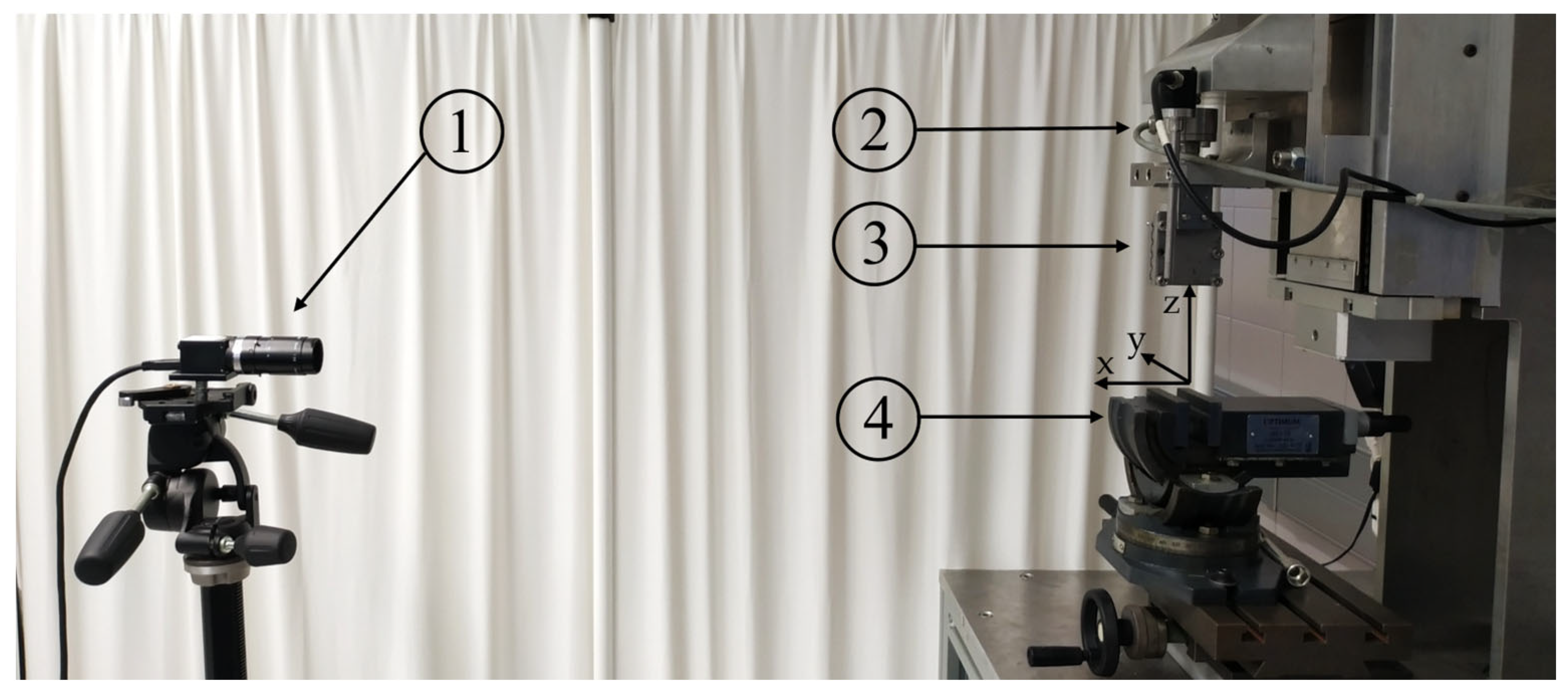
2.2. Biomechanical Testing

A single-axis testing machine (Figure 1), specially designed for biomechanical tests [46], was used to subject the sutured meniscus to a load-to-failure test. Three orthogonal axes are defined in the testing machine, with the z-axis coinciding with the servomotor screw that sets the direction of traction (Figure 1). The suture–meniscus set was placed in the testing machine in two phases. First, the meniscus was fixed with a clamp at approximately 8 mm from the suture point, using sandpaper surrounding the clamping area to increase friction. The meniscus was carefully oriented with the cranial surface facing the outer side of the testing machine, the longitudinal fibers of the horn were aligned to the loading direction, and the suture hole was centered with the actuator head in the loading direction. Second, the two free ends of the suture were also wrapped in sandpaper and attached to a mechanical clamp on the machine head, with 55 mm between the puncture point and the limit of the clamp when the suture is pulled manually, with just enough traction to keep it vertical. Fifty-five millimeters were selected as representative of the expected suture length in a transtibial meniscal horn repair [14,47]. An electronic inclinometer (Bubble Level 3D, v. 2.2.4, Maleirbag, 2022, Puebla, Mexico, installed on the smartphone M2002F4LG, Xiaomi, Beijing, China) was used to orient the meniscus so that its cranial and caudal surfaces were within the plane defined by the z-axis (traction direction) and the y-axis (transverse direction) of the testing machine as close as possible. Once the meniscus–suture set had been placed on the testing machine, two ink points were marked with a surgical pen on the cranial side. Aided by the inclinometer to identify the direction of traction on the meniscal surface, the points were located along a line in that direction passing through the suture hole: Point1 on the suture limb coincident with the meniscus–suture interface; and Point2 on the opposite side of the hole in its immediate surroundings (Figure 2). These marks were used for videogrammetric analysis of the displacements around the hole in the direction of traction.
The servo controller of the testing machine (SGDH-15AE-S-OY, Yaskawa Electric, Fukuoka, Japan) recorded the actuator displacement in the pull direction, and a 2000 N load cell of accuracy class 0.1 (U2B, HBM, Darmstadt, Germany) registered the applied traction force. Both signals were sampled at 1000 Hz. For videogrammetric analysis, a digital camera (VCXU-124C, Baumer, Frauenfeld, Switzerland) (Figure 1), synchronized with the sensors of the testing machine, was placed facing the cranial surface of the meniscus. For this purpose, the camera was mounted on a tripod (808RC4, Manfrotto, Cassola, Italy) featuring three rotating orthogonal mechanisms (Figure 1), and, using the electronic inclinometer, it was positioned to align its image plane with the plane containing the z and y axes of the testing machine, i.e., approximately parallel to the mid transverse plane of the meniscus. The camera was equipped with a 2.8/75 mm lens (C7528-M, Pentax, Tokyo, Japan). Images were acquired with a 250 ms sampling period.
The sutured meniscus was initially pulled to 1 N at 0.05 mm/s, held at this load level for 5 s to allow the specimen to stabilize, and then subjected to a displacement-controlled load-to-failure test at 0.1 mm/s.

2.3. Data Analysis

To assess the resistance at the specimen level, the force that initiated this meniscal tear, i.e., the meniscal cut-out force, F C , was registered for each specimen. The initiation of meniscal tissue cut-out was identified as the point of change in the slope of the curve representing the displacement of Point1 with respect to Point2, D , as a function of the traction force [46]. The tearing initiation was also verified by checking the video images to validate the accuracy of the detection. The maximum force borne by the specimen in the load-to-failure test, i.e., the ultimate force, F u , was also registered.
At the tissue level, the tissue cut-out stress of the meniscal horn, S c , was calculated as the engineering stress at the suture–meniscal interface at the tearing initiation as follows:
S c = F C ϕ · h ,
where ϕ is the nominal diameter of the suture, and h is the meniscal thickness at the hole, i.e., the denominator is the projected area at the meniscus–suture interface. Equation (1) may underestimate the contact surface at the beginning of the test since the suture passage may not be perfectly orthogonal to the meniscal surfaces despite being subjected to a tensile force of 1 N. However, as the test progresses, the thread will rapidly orient itself to minimize the contact area.
To evaluate tissue deformation around the suture site, an equivalent stiffness modulus on the traction direction of the suture, m , was calculated as the slope of the linear approximation of the stress–strain curve, σ ε , in the range of ε = [ 0 ,   0.3 ] . The engineering stress in the traction direction at the meniscus–suture interface was calculated for the test points prior to the initiation of tearing with an expression similar to the one used in Equation (2), i.e.,
σ = F ϕ · h ,
where F is the traction force recorded by the load cell during the test. And the strain was computed as follows:
ε = D D 0 1 ,
where D 0 is the distance between Point1 and Point2 computed in the first frame of the load-to-failure test, and D is the distance between them computed in subsequent frames.

2.4. Statistical Analyses

Sample size was determined using G*Power 3.1.9.7 software [48] for a minimum detectable difference of 15 N between the older human group and the younger human group as the control group, using a t-test at α = 0.05 with a power of 0.80 and an allocation ratio of N / N c = 0.5 and a standard deviation of 21 N estimated from a preceding work [49]. The resulting minimum control group size was N c = 41 . The difference of the 15 N selected represents 25% of the F c of the 60 N estimated for the younger human group from the data of the aforementioned previous work. This difference was considered sufficiently large so the adequacy of the experimental models analyzed, i.e., the human ≥75 years and porcine groups, could not be considered representative of the control group, i.e., human <75 years. Finally, conservative group sizes of N c = 44 for the control group and N = 22 for the test groups were chosen to allow for the exclusion of specimens and samples that did not meet the criteria for parametric testing.
Descriptive statistics and comparative tests were conducted using IBM® SPSS® Statistics, v.23 (IBM, Chicago, IL, USA). To assess differences in the mechanical properties at the suture site between the younger human group and each of the groups representing a different experimental model, non-parametric Kruskal–Wallis omnibus tests for independent samples were carried out. When differences were detected, post hoc analysis consisted of pre-planned comparisons of each model with the control group using Dunn–Bonferroni tests with correction for two comparisons. These nonparametric tests were chosen because all samples failed either the normality test or the homogeneity of variances test. p values ≤ 0.05 were regarded as significant.

3. Results

The three study groups included in the study showed the following characteristics:
  • Human <75 years group: 45 younger-than-75-years-old menisci (mean age 51.00 years, SD 13.56, range 28–67 years) obtained from 24 cadaveric knees of 24 donors after discarding 3 menisci due to not meeting the inclusion criteria. The group consisted of 27 menisci from men and 18 from women, 23 medial and 22 lateral menisci, 22 for anterior and 23 for posterior surgery.
  • Human ≥75 years group: 21 75-years-old-or-older specimens (mean age 84.86 years, SD 3.82, range 82–95 years) obtained from 14 cadaveric knees of 14 donors after discarding 7 menisci due to not meeting the inclusion criterion. The group consisted of 9 menisci from men and 12 from women, 11 medial and 10 lateral menisci, 9 for anterior and 12 for posterior surgery.
  • Porcine group: 21 menisci from 22 6-month-old pigs. All of them met the inclusion criteria. The group consisted of 11 medial and 11 lateral menisci, 10 for anterior and 12 for posterior surgery.

3.1. Meniscal Horn Thickness at the Suture Point, h

At the suture site, older human menisci were thicker than the younger menisci (p = 0.012), with a 24.7%-greater mean value. The difference in thickness was even more pronounced in the porcine group, which was 83.8% thicker (p < 0.001) (Figure 3 and Table 1).

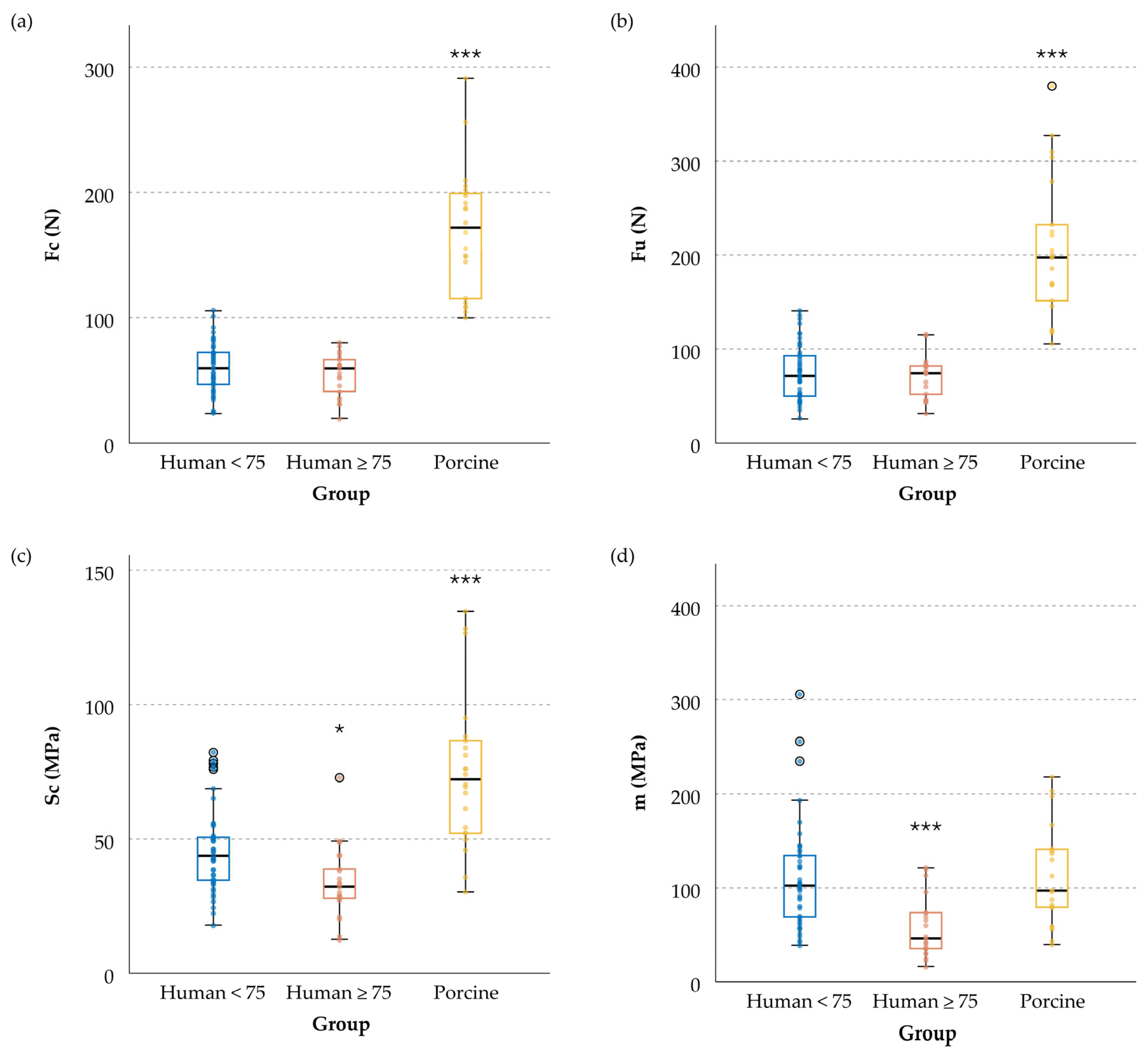
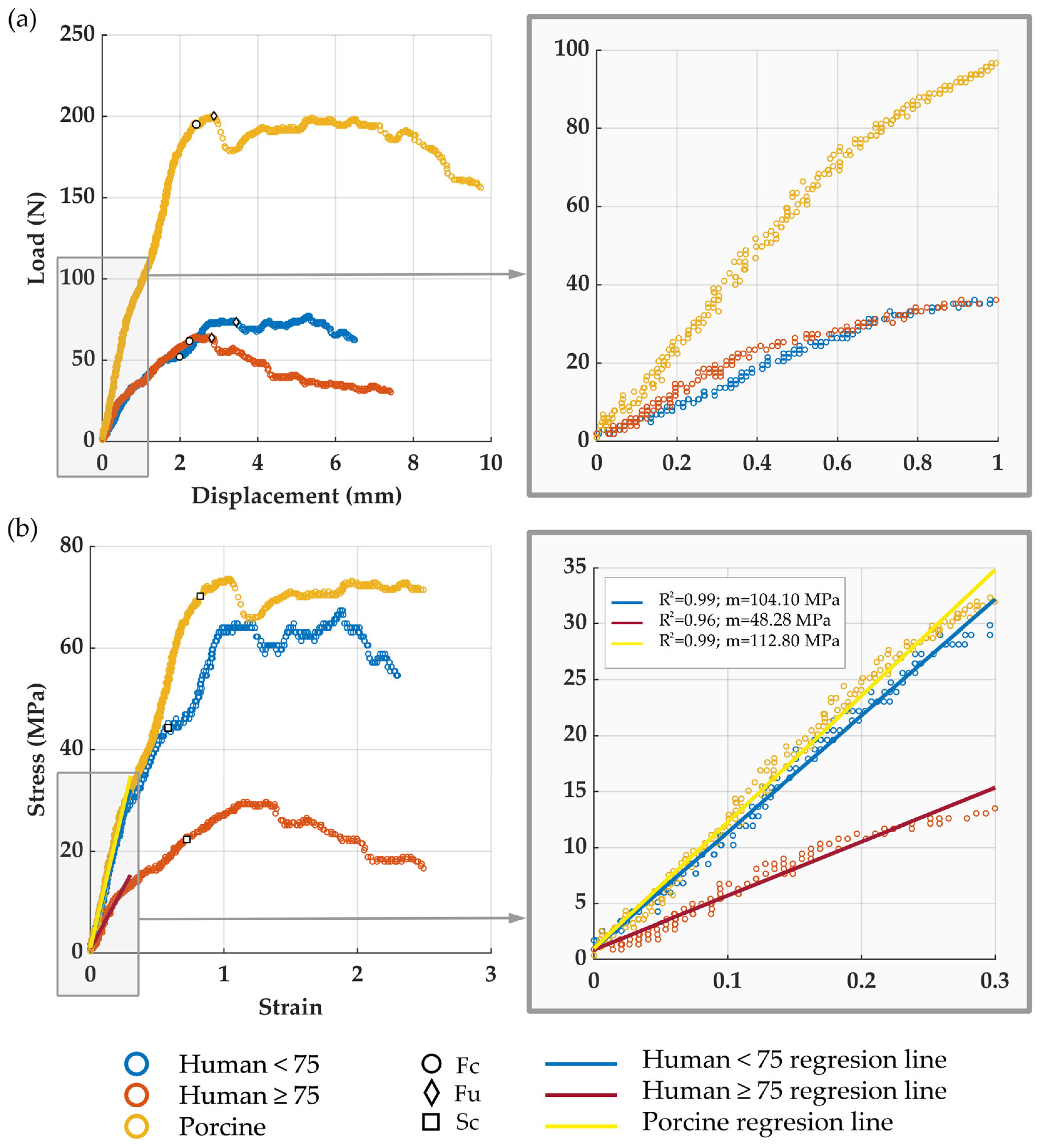
3.2. Meniscal Resistance to Cut-Out Initiation, Fc

Compared to the younger human group, the specimen resistance to cut-out initiation by suture traction, F C , was significantly higher in the porcine group (p < 0.001), with a 172.1% increase in the mean value (Table 2a and Figure 4a). However, no significant difference was found between the younger and older human groups.
Analysis of the video recordings confirmed that tearing was initiated slightly before the first local maximum of the force–deformation curve, consistent with previous reports [46].

3.3. Meniscal Ultimate Force, Fu

The results of the comparison between the testing groups and the control for the ultimate meniscal force were similar to those for F c (Table 2b and Figure 4b): the older human group was quite similar to the younger human group (the mean values differed by only by 6.1%), while the porcine model was 174.1% larger (p < 0.001).
Comparing the mean values of the ultimate force and the resistance to cut-out initiation for each group, F u was 24.9% higher than F c in the younger human group, 29.3% in the older human group, and 21.9% higher in the porcine model.

3.4. Tissue Cut-Out Resistance, Sc

When comparing the human meniscus groups, the sutured tissue in the older group was less resistant, with a mean S C 28.7% lower than in the younger group (p = 0.012). On the other hand, porcine tissue was more resistant than younger human tissue (p < 0.001), requiring a 57.2%-higher mean stress value to reach the cut-in point (Table 2c and Figure 4c).

3.5. Tissue Equivalent Stiffness Modulus, m

Figure 5a illustrates typical load–displacement curves for each of the sample types included in the study. Figure 5b displays the corresponding stress–strain curves for these representative samples, along with their linear adjustments in the strain interval [0, 0.3], and indicates the adjusted R-squared values of the fittings. Similar to the curves in Figure 5b, all meniscal horns showed highly linear behavior in the strain range [0, 0.3]. Specifically, the adjusted R-squared values for the linear fitting were within the range [0.91, 0.99] for human <75 and human ≥75 models, and in the range [0.92, 0.99] for the porcine model. As justified in the Materials and Methods section, the equivalent stiffness modulus, m, was computed as the slope of the aforementioned linear adjustments, resulting in the mean values in Table 2d.
Meniscal tissue at the sutured area was more elastic in the older group than in the younger group (p < 0.001 for both comparisons), with m being 48.1% greater (Table 2d and Figure 4d). In porcine specimens, the elasticity was very similar to younger human menisci, with almost identical mean m values (difference less than 0.6%).

4. Discussion

The main result of our study is that neither the porcine model nor the human model with cadaveric tissue from donors of advanced age (over 75 years old) can provide accurate representations of all the mechanical properties of adult human specimens under 75 years old. However, while neither of the two models offers a clear advantage for a general use in biomechanical in vitro tests, each could be adequate for examining specific characteristics of the sutured meniscal horn.
Regarding the resistance to suture-induced traction at the specimen level, no difference was observed between the older and younger human meniscus groups, which partially contradicts our first hypothesis that meniscal tissue from older donors (≥75 years) does not adequately represent that of the younger menisci. On the other hand, the porcine model was significantly more resistant than the younger human model at the specimen level, with differences of 172.1% in F c and 174.1% in F u , as opposed to our second hypothesis. Upon examining resistance at the tissue level in terms of tissue cut-out stress at the meniscus–suture interface, S c , both models exhibited significant differences from the younger human group. Specifically, porcine specimens exhibited tissue with a 57.2%-greater resistance to suture-induced tearing, whereas specimens from donors over 75 years old showed less-resistant tissue, with a 28.7% reduction compared to the human meniscus from younger donors. Regarding deformation, the findings from the comparison between the models opposed those observed in specimen resistance. In particular, the equivalent stiffness modulus, m , of the porcine model did not differ from that of the younger human models, while the human ≥75 years model showed a 48.1% increase in this elastic parameter compared to the human meniscus <75 years.
Consequently, the adequacy of these two models depends on the interest of the study, a result that partially contradicts both of our initial hypotheses that the meniscal tissue from older donors (≥75 years) does not adequately represent that of the younger menisci and that the porcine meniscus is a good surrogate for the human meniscus at an age routinely eligible for repairing. In research on mechanical properties at the specimen level, to analyze the resistance of sutured meniscal roots, the older human meniscus model resulted to be a preferable surrogate model to the porcine model; conversely, if the focus is on issues related to deformation before cut-out initiation in the sutured root zone, the porcine model offers a more accurate representation. For studies at the tissue level, as experimental tests to determine material properties around the sutured root area, neither model resulted to be a reliable surrogate in terms of resistance; but in tests where only tissue deformation prior to cut-out initiation is of interest, opting for the porcine model may be preferable. However, caution is advised in the latter case, as the tissue stresses would be inadequately characterized.
The differences found in the suitability of the surrogate models when used in studies evaluating meniscal root resistance at the specimen level or tissue level may be partially attributed to differences in thicknesses observed among the groups. The porcine model offers a surrogate for the human meniscus <75 years, which has a more resistant tissue, with a 57.2% higher S c . This difference is further accentuated at the specimen level due to the thicker root of the porcine meniscus, resulting in a 172.1%-higher F c and a 174.1%-higher F u . Conversely, when employing an older human meniscus model as a surrogate, although the tissue is less resistant, with a 28.7%-lower S c , this effect is offset at the specimen level. Likely, the reason is that the meniscal root of the older group was thicker than that of the younger group, resulting in the vanishing of differences at the specimen level.
Large animal models are valuable tools in biomechanical engineering, finding applications in both preliminary in vitro testing and in vivo testing. These models, including dogs, rabbits, cows, sheep, and pigs, have been extensively utilized in research on the natural meniscus, the study of its kinematic behavior, the assessment of the biomechanics of knee joint contact, the validation of engineered tissues proposed for total or partial replacement of the meniscus, and the evaluation of the efficacy of surgical meniscal repairs. The canine model, historically employed in studies, has experienced a decline in usage, potentially due to increasing pressure from animal advocacy groups. Moreover, from a technical perspective, the majority of surgical procedures on the canine meniscus require arthrotomy, often accompanied by medial collateral ligament rupture, deviating from arthroscopic procedures in humans. The rabbit model exhibits significant differences in vascularity, collagen orientation, and glycosaminoglycan content of the meniscus compared to human menisci. Additionally, the kinematics of the rabbit’s femorotibial joint significantly differ from those of the human knee [31,34,50]. The bovine model, significantly larger than the human meniscus, has limited application in biomechanical studies due to the size discrepancy [34,50]. The ovine meniscus, while anatomically similar to the human meniscus, differs in kinematics and contact force distribution, making it unsuitable for non-isolated studies of the repaired meniscus [30,31,34]. Regarding the porcine model, it closely resembles the human model in terms of anatomical structure, vascularity, volume, and weight [30,31,34], although the width of the meniscus is greater, as demonstrated in the present study. The kinematics of the porcine knee reasonably approximate those of the human knee. As a result of the aforementioned arguments, the porcine model is the predominant surrogate for in vitro tests of the young human meniscus [12,15,16,17,18,19,21,23,26,27,28,29], widely applied in biomechanical testing for meniscal repair and replacement techniques, allowing for effective translation to clinical applications.
Surgical repair of meniscal root avulsions has been traditionally limited to patients under the age of 35–40. However, there is a paradigm shift, with a contemporary inclination towards meniscus preservation through the utilization of repair techniques whenever feasible. The 2019 consensus of the European Society for Sports Traumatology, Knee Surgery and Arthroscopy (ESSKA) [42], drawing from a compilation of studies encompassing patients from 9 to 58 years of age, ‘clearly states that numerous meniscus tears that were considered irreparable should be repaired, e.g., older tears’. This shift is substantiated by recent follow-up studies that report the advantages of extending the range for surgical intervention to over 60 years of age [41,51]. Hence, this matter was carefully considered, and we decided to expand the age range of the young group to 75 years old, aligning with the aforementioned findings and to conform to the criteria of the orthopedic surgeons who are part of our research team. We consider that menisci from donors who are older than 75 years of age are representative of a group of patients who are too advanced in age to be routinely considered eligible for meniscal root repair surgery.
The experimental design involved testing isolated meniscus–suture constructs instead of menisci attached to tibial bones. This setup is consistent with prior works on the same topic [29,37,38,52]. It was selected to specifically focus on the behavior of the tissue–suture interface, excluding factors dependent on tibial fixation. Also in line with previous studies [29,37,38,52], the suture was oriented with the loading direction, eliminating shear friction and concentrating the tensile effect on the meniscus–suture interface, which was our area of interest. This orientation of the meniscal horn aimed to maximize tissue damage, representing a conservative criterion for detecting the maximum load the repair could withstand.
A single simple suture was utilized at the meniscal horn, deviating from common surgical options. This choice facilitates computation of the forces acting on the tissue–suture interface. Multiple stitches distribute the pulling load among them, posing challenges for achieving uniform distribution under laboratory conditions. Despite neither being the most resistant nor the most common suture technique for meniscal root repair, the use of a simple stitch eases the quantification of the meniscus–thread contact area, essential for calculating stress at the suture–meniscus interface. This simplicity minimizes the impact of meniscus anatomy or surgeon performance compared to more complex suture techniques.
To accurately detect cut-out initiation, automatic detection using a previously validated method [46] was utilized and further verified by checking the recorded video images. The results confirmed that, as formerly reported [46], tearing was initiated slightly before the first local maximum of the load–deformation curve. In the scientific literature, the maximum force borne by the specimen, F u , has usually been selected as the parameter to characterize the failure of repaired menisci under suture-induced traction. The authors consider it more relevant to study the cut-out initiation point, i.e., F c at the specimen level or S c at the tissue level, although F u has also been calculated for comparison purposes. F u was found to be higher than F c by nearly 30% for older human menisci and approximately 20% for the porcine group. After cut-out initiation, the progression of the tear may follow different paths within the meniscus of variable thickness. Consequently, the load–deformation curve exhibited a highly heterogeneous pattern from cut-out initiation to the absolute maximum force. This heterogeneity under laboratory conditions is expected to be more pronounced In a repaired knee subjected to physiological loads. Therefore, we believe that the cut-out initiation provides a better basis for comparing results between different groups because it is the starting point of the loss of structural integrity of the sample and it is less influenced by the post-tearing initiation trajectory.
Cryoprotectant solutions have been proposed to preserve biological tissues from damage caused by freezing, but we discarded their use for two main reasons. Primarily, they are not customarily used for preserving specimens in either of the two surrogate models of the human meniscus that our study focuses on. Additionally, there is a lack of data on their ability to maintain the mechanical properties of the suture meniscal horn better than the preservation method we used, particularly concerning elasticity and resistance.
Some studies have examined the resistance of meniscal horns to suture-induced traction, focusing on assessing various suturing techniques and devices for root reattachment. Antz et al. conducted a load-to-failure test on human menisci aged 46–64 years using two simple stitches with a #2 UHMWPE surgical suture on the posterior horn [37]. Despite differences in testing protocols, their reported values align with our findings for the young group, with a mean initial peak force of 137 ± 49 N for two stitches compared to our F c of 60.13 ± 19.91 N with a single stitch. In accordance with our results, the initial peak force must be very close to our F c . Vertullo et al. reported an ultimate load of 94.29 ± 7.99 N for a group of posterior medial menisci with a mean age of 54 ± 4 years using two simple #2 sutures [52]. This result is also compatible with our findings, considering F u values of 75.08 ± 26.62 N for the young group and 70.49 ± 16.92 N for the older group, acknowledging the use of two stitches and prior sample weakening by 1000 cycles of under-critical load. It must be highlighted that in cases where more than one suture is employed, inevitable asymmetries in traction distribution may occur. Consequently, when the interface of one stitch reaches the tension threshold, initiating tearing, it is highly probable that the other stitches are at a lower load level. Mitchell et al. found a mean ultimate force of 58.2 ± 29.6 N when testing posterior horns of human medial menisci (ages 48 to 88 years) with one simple stitch [38]. The lower values observed may be attributed to their use of a #0 UHMWPE surgical suture, implying a thinner suture diameter and resistant area than in our study. Unfortunately, meniscus thickness was not reported, which does not permit us to estimate S c in that study. Kopf et al. tested posterior human menisci aged 18–51 years in a load-to-failure test with two #2 single UHMWPE sutures, finding an ultimate load of 64.10 ± 22.50 N [53]. This value is considerably lower than other works and our younger group, factoring in the difference from the use of a single stitch.
Concerning meniscal horn elasticity, few studies have specifically addressed deformation at the sutured horn [29,46]. While some biomechanical works have reported on meniscus–suture construct stiffness, the results vary widely, even when restricted to UHMWPE threads. Stiffness for two simple sutures ranged from 774 ± 730 N/mm [38] using suture #0 to 24.55 ± 4.05 N/mm [52] with #2 sutures. Dispersion may be due to differences in suture thread lengths, which are often unreported, and varied cyclic loading histories before the load-to-failure test, which are known to influence the elasticity of surgical sutures if insufficient resting time is allowed [54].
Our results for the porcine model fall within the previously published range of failure loads. However, it is noteworthy that the range of published data is considerably wide. In a study where a meniscus was also sutured with a single simple stitch, utilizing a UHMWPE #2 suture [17], an ultimate load of 169 ± 73.4 N was reported. The result aligns with our outcome of 206.79 ± 74.31 N, considering that in that study, the specimens were pre-weakened with 1000 cycles of load between 5 and 20 N. In contrast, other tests involving porcine models yielded results for two sutures ranging from 129.8 ± 14.6 N [19] to 221.67 ± 43 N [29], although various suturing techniques and suture materials were employed. Beyond the variations attributed to different testing protocols, these disparities may be attributed to the diverse origin of the animal. While animal surrogate models provide the advantage of ensuring uniformity among donors, variations in breeds, types of feeding, stabling conditions, etc., can introduce significant differences between groups of diverse origins, even if they belong to the same species. This concern has been previously raised, and we believe it warrants further investigation [55].
In a study by Camarda et al., a comparison of the cyclic stiffness and ultimate load in load-to-failure tests on posterior horns of humans aged 50–70 years and porcine samples was undertaken [35]. Specimens sutured with three simple stitches using #2 UHMWPE threads were subjected to 1000 cycles of loading ranging from 10 N to 30 N, followed by a load-to-failure test. The study reports a 2.4-times-higher ultimate load for the porcine model compared to the human model, which is consistent with our findings of a 2.7-times-higher F u for the porcine model compared to the younger human model, with no previous cyclic loading that may have weakened the tissue. Their human model also exhibited lower cyclic stiffness of the meniscus–suture construct in contrast to our results, which did not find any differences in deformation. However, the parameters are not comparable because our study was limited to the meniscus around the suture area, and we performed a direct load-to-failure test.
The mechanical properties of sutured meniscal tissue have not been studied so far. With the data extracted from this study, it becomes possible to fit the material properties of meniscal tissue of different origins in the area surrounding the suture hole, thus contributing to the development of computational models with more realistic behavior. On the other hand, the study highlights the need for further research to analyze the mechanical behavior of the sutured meniscal horn in situations where the viscoelastic nature of the meniscal tissue has a significant influence, such as in the case of dynamic impact loads or quasi-stationary loads maintained over a long period of time. Additionally, the authors maintain it will be interesting to conduct a thorough study of the alteration that the suture produces in the mechanical behavior of the meniscal root in the immediate postoperative period. Furthermore, in vivo studies could allow for an assessment of the progress of the mechanical properties of the sutured root as healing progresses, evaluating whether its natural meniscal properties can be fully restored.
This study is subjected to inherent limitations associated with in vitro testing, notably the absence of the healing effect observed over time. The use of an isolated meniscal setup implies a deliberate exclusion of the influences from surrounding soft tissues, ligaments, cartilage, and bones. The rationale for this decision has been discussed above. A single simple suture was employed, as also previously justified. Therefore, the cut-out and ultimate forces reported in the results section do not quantitatively reflect the maximum traction forces that the clinically repaired meniscus, using other more-common suturing techniques, might withstand. The load-to-failure test was conducted at 0.1 mm/s, representing a quasi-static displacement velocity lower than expected for knee movement in daily activities [54]. However, given the viscoelastic behavior of the meniscus reported in compression tests [55,56], further research is warranted to comprehensively understand the dynamic response of the sutured meniscal tissue.
This minimizes the potential influence of viscous effects on the test, a characteristic not addressed in this study. All the animals came from the same local slaughterhouse, which may raise questions about whether porcine models from other origins would give different results [57].

5. Conclusions

In in vitro experimental tests to analyze the mechanical behavior of the sutured horn of the adult human meniscus at an eligible age to be routinely repaired (below 75 years old), neither the porcine tissue nor the cadaveric tissue from donors of advanced age (over 75 years old) could provide accurate models. However, each one could be adequate for examining specific characteristics depending on the interest of the study; the older human meniscus model is a preferable surrogate model in studies on the resistance of sutured meniscal root specimens; conversely, porcine models provide a more accurate representation in works involving deformation-related properties of the meniscus around the suture site at the tissue level.

Author Contributions

Conceptualization, A.P.-T., A.P.-B. and M.P.-N.; methodology, A.P.-T., A.P.-B. and M.P.-N.; software, M.B.E.-C. and S.M.-V.; validation, A.P.-T., M.B.E.-C. and S.M.-V.; formal analysis, A.P.-T.; investigation, all authors; resources, all authors; data curation, all authors; writing original draft preparation, A.P.-T., A.P.-B. and M.P.-N.; writing—review and editing, all authors; visualization, S.M.-V. and M.B.E.-C.; supervision, A.P.-B. and M.P.-N.; project administration, A.P.-B. and M.P.-N.; funding acquisition, M.P.-N. All authors have read and agreed to the published version of the manuscript.

Funding

This research was funded by Junta de Andalucía, Spain, grant number UMA20-FEDERJA-116.

Institutional Review Board Statement

The study was conducted in accordance with the Declaration of Helsinki and approved (including the animal study protocol) by the Institutional Ethics Committee of the University of Málaga, Spain (Comité Ético de Experimentación de la Universidad de Málaga, CEUMA) (protocol code 69-2018-H 2 October 2018).

Informed Consent Statement

Patient consent was waived due to the knees being obtained from an authorized provider. The provider certified that the knees came from donors who voluntarily had donated their bodies to science without financial compensation, in compliance with the legislation of the country of origin, and who had previously signed the corresponding consent forms.

Data Availability Statement

The data presented in this study are available on request from the corresponding author. The data are not publicly available because they include the demographic data of the donors.

Acknowledgments

We thank A. Espejo-Baena and A. Espejo-Reina, orthopedic surgeons of the BIOCLINA group, for their invaluable collaboration.

Conflicts of Interest

The authors declare no conflicts of interest.

References

  1. Fox, A.J.S.; Bedi, A.; Rodeo, S.A. The basic science of human knee menisci: Structure, composition, and function. Sports Health 2012, 4, 340–351. [Google Scholar] [CrossRef]
  2. Perez-Blanca, A.; Espejo-Baena, A.; Amat Trujillo, D.; Prado Nóvoa, M.; Espejo-Reina, A.; Quintero López, C.; Ezquerro Juanco, F. Comparative Biomechanical Study on Contact Alterations after lateral meniscus posterior root avulsion, transosseous reinsertion, and total meniscectomy. Arthroscopy 2016, 32, 624–633. [Google Scholar] [CrossRef]
  3. Allaire, R.; Muriuki, M.; Gilbertson, L.; Harner, C.D. Biomechanical consequences of a tear of the posterior root of the medial meniscus: Similar to total meniscectomy. J. Bone Jt. Surg. Am. 2008, 90, 1922–1931. [Google Scholar] [CrossRef]
  4. Espejo-Reina, A.; Prado-Novoa, M.; Espejo-Baena, A.; Peña-Trabalon, A.; Perez-Blanca, A. Biomechanical consequences of anterior root detachment of the lateral meniscus and its reinsertion. Sci. Rep. 2022, 12, 6182. [Google Scholar] [CrossRef] [PubMed]
  5. Shiwaku, K.; Kamiya, T.; Suzuki, D.; Yamakawa, S.; Otsubo, H.; Suzuki, T.; Takahashi, K.; Okada, Y.; Teramoto, A.; Ohnishi, H.; et al. The role of the medial meniscus in anterior knee stability. Orthop. J. Sport Med. 2022, 10, 23259671221132845. [Google Scholar] [CrossRef]
  6. Musahl, V.; Citak, M.; O’Loughlin, P.; Choi, D.; Bedi, A.; Pearle, A. The effect of medial versus lateral meniscectomy on the stability of the anterior cruciate ligament-deficient knee. Am. J. Sports Med. 2010, 38, 1591–1597. [Google Scholar] [CrossRef] [PubMed]
  7. Shoemaker, S.; Markolf, K. The role of the meniscus in the anterior-posterior stability of the loaded anterior cruciate-deficient knee. Effects of partial versus total excision. J. Bone Jt. Surg. Am. 1986, 68, 71–79. [Google Scholar] [CrossRef]
  8. Krych, A.; Reardon, P.; Johnson, N.; Mohan, R.; Peter, L.; Levy, B.; Stuart, M.J. Non-operative management of medial meniscus posterior horn root tears is associated with worsening arthritis and poor clinical outcome at 5-year follow-up. Knee Surg. Sports Traumatol. Arthrosc. 2017, 25, 383–389. [Google Scholar] [CrossRef]
  9. Hussain, Z.B.; Chahla, J.; Mandelbaum, B.R.; Gomoll, A.H.; LaPrade, R.F. The role of meniscal tears in spontaneous osteonecrosis of the knee: A systematic review of suspected etiology and a call to revisit nomenclature. Am. J. Sports Med. 2019, 47, 501–507. [Google Scholar] [CrossRef]
  10. Pache, S.; Aman, Z.S.; Kennedy, M.; Nakama, G.Y.; Moatshe, G.; Ziegler, C.; LaPrade, R.F. Meniscal root tears: Current concepts review. Arch. Bone Jt. Surg. 2018, 6, 250–259. [Google Scholar]
  11. Ahn, J.H.; Wang, J.H.; Yoo, J.C.; Noh, H.K.; Park, J.H. A pull out suture for transection of the posterior horn of the medial meniscus: Using a posterior trans-septal portal. Knee Surg. Sports Traumatol. Arthrosc. 2007, 15, 1510–1513. [Google Scholar] [CrossRef] [PubMed]
  12. Feucht, M.J.; Grande, E.; Brunhuber, J.; Burgkart, R.; Imhoff, A.B.; Braun, S. Biomechanical evaluation of different suture techniques for arthroscopic transtibial pull-out repair of posterior medial meniscus root tears. Am. J. Sports Med. 2013, 41, 2784–2790. [Google Scholar] [CrossRef]
  13. Balke, M.; Akoto, R.; Offerhaus, C.; Hoeher, J. Suture anchor refixation of meniscal root tears without an additional portal. Arthrosc. Tech. 2018, 7, e511–e515. [Google Scholar] [CrossRef] [PubMed]
  14. Espejo-Reina, A.; Prado-Novoa, M.; Espejo-Reina, M.J.; Gómez-Cáceres, A.; Dalla Rosa-Nogales, J.; Espejo-Baena, A. Non anatomic reinsertion after amputation of the anterior horn of the lateral meniscus. Orthop. Traumatol. Surg. Res. 2019, 105, 1115–1118. [Google Scholar] [CrossRef]
  15. Robinson, J.R.; Frank, E.G.; Hunter, A.J.; Jermin, P.J.; Gill, H.S. The strength of transosseous medial meniscal root repair using a simple suture technique is dependent on suture material and position. Am. J. Sports Med. 2018, 46, 924–932. [Google Scholar] [CrossRef]
  16. Wu, J.L.; Lee, C.H.; Yang, C.T.; Chang, C.M.; Li, G.; Cheng, C.K.; Chen, C.H.; Huang, H.S.; Lai, Y.S. Novel technique for repairing posterior medial meniscus root tears using porcine knees and biomechanical study. PLoS ONE 2018, 13, e0192027. [Google Scholar] [CrossRef]
  17. Feucht, M.J.; Grande, E.; Brunhuber, J.; Rosenstiel, N.; Burgkart, R.; Imhoff, A.B.; Braun, S. Biomechanical evaluation of different suture materials for arthroscopic transtibial pull-out repair of posterior meniscus root tears. Knee Surg. Sports Traumatol. Arthrosc. 2015, 23, 132–139. [Google Scholar] [CrossRef]
  18. Chung, K.S.; Choi, C.H.; Bae, T.S.; Ha, J.K.; Jun, D.J.; Wang, J.H.; Kim, J.G. Comparison of tibiofemoral contact mechanics after various transtibial and all-inside fixation techniques for medial meniscus posterior root radial tears in a porcine model. Arthroscopy 2018, 34, 1060–1068. [Google Scholar] [CrossRef]
  19. Fujii, M.; Furumatsu, T.; Xue, H.; Miyazawa, S.; Kodama, Y.; Hino, T.; Kamatsuki, Y.; Ozaki, T. Tensile strength of the pullout repair technique for the medial meniscus posterior root tear: A porcine study. Int. Orthop. 2017, 41, 2113–2118. [Google Scholar] [CrossRef] [PubMed]
  20. Rosslenbroich, S.B.; Borgmann, J.; Herbort, M.; Raschke, M.J.; Petersen, W.; Zantop, T. Root tear of the meniscus: Biomechanical evaluation of an arthroscopic refixation technique. Arch. Orthop. Trauma Surg. 2013, 133, 111–115. [Google Scholar] [CrossRef]
  21. Okimura, S.; Mae, T.; Tachibana, Y.; Iuchi, R.; Nakata, K.; Yamashita, T.; Shino, K. Biomechanical comparison of meniscus-suture constructs for pullout repair of medial meniscus posterior root tears. J. Exp. Orthop. 2019, 6, 17. [Google Scholar] [CrossRef] [PubMed]
  22. Forkel, P.; Foehr, P.; Meyer, J.C.; Herbst, E.; Petersen, W.; Brucker, P.U.; Burgkart, R.; Imhoff, A.B. Biomechanical and viscoelastic properties of different posterior meniscal root fixation techniques. Knee Surg. Sports Traumatol. Arthrosc. 2017, 25, 403–410. [Google Scholar] [CrossRef] [PubMed]
  23. Wu, S.-H.; Yeh, T.-T.; Hsu, W.-C.; Wu, A.T.; Li, G.; Chen, C.-H.; Lee, C.-H.; Wu, J.-L. Biomechanical comparison of four tibial fixation techniques for meniscal root sutures in posterior medial meniscus root repair: A porcine study. J. Orthop. Transl. 2020, 24, 144–149. [Google Scholar] [CrossRef]
  24. Prado-Novoa, M.; Perez-Blanca, A.; Espejo-Reina, A.; Espejo-Reina, M.J.; Espejo-Baena, A. Initial biomechanical properties of transtibial meniscal root repair are improved by using a knotless anchor as a post-insertion tensioning device. Sci. Rep. 2020, 10, 1748. [Google Scholar] [CrossRef]
  25. Feucht, M.J.; Grande, E.; Brunhuber, J.; Rosenstiel, N.; Burgkart, R.; Imhoff, A.B.; Braun, S. Biomechanical comparison between suture anchor and transtibial pull-out repair for posterior medial meniscus root tears. Am. J. Sports Med. 2014, 42, 187–193. [Google Scholar] [CrossRef]
  26. Kim, Y.M.; Joo, Y.B.; Noh, C.K.; Park, I.Y. The optimal suture site for the repair of posterior horn root tears: Biomechanical evaluation of pullout strength in porcine menisci. Knee Surg. Relat. Res. 2016, 28, 147–152. [Google Scholar] [CrossRef] [PubMed]
  27. Seo, J.H.; Li, G.; Shetty, G.M.; Kim, J.H.; Bae, J.H.; Jo, M.L.; Kim, J.S.; Lee, S.J.; Nha, K.W. Effect of Repair of Radial Tears at the Root of the Posterior Horn of the Medial meniscus with the pullout suture technique: A biomechanical study using porcine knees. Arthroscopy 2009, 25, 1281–1287. [Google Scholar] [CrossRef]
  28. Stärke, C.; Kopf, S.; Gröbel, K.H.; Becker, R. The Effect of a Nonanatomic repair of the meniscal horn attachment on meniscal tension: A biomechanical study. Arthroscopy 2010, 26, 358–365. [Google Scholar] [CrossRef]
  29. Perez-Blanca, A.; Prado Nóvoa, M.; Lombardo Torre, M.; Espejo-Reina, A.; Ezquerro Juanco, F.; Espejo-Baena, A. The role of suture cutout in the failure of meniscal root repair during the early post-operative period: A biomechanical study. Int. Orthop. 2018, 42, 811–818. [Google Scholar] [CrossRef]
  30. Takroni, T.; Laouar, L.; Adesida, A.; Elliott, J.A.W.; Jomha, N.M. Anatomical study: Comparing the human, sheep and pig knee meniscus. J. Exp. Orthop. 2016, 3, 35. [Google Scholar] [CrossRef]
  31. Deponti, D.; Di Giancamillo, A.; Scotti, C.; Peretti, G.M.; Martin, I. Animal models for meniscus repair and regeneration. J. Tissue Eng. Regen. Med. 2015, 9, 512–527. [Google Scholar] [CrossRef] [PubMed]
  32. Kyle Martin, R.; Gillis, D.; Leiter, J.; Shantz, J.S.; MacDonald, P. A porcine knee model is valid for use in the evaluation of arthroscopic skills: A pilot study. Clin. Orthop. Relat. Res. 2016, 474, 965–970. [Google Scholar] [CrossRef] [PubMed]
  33. Sweigart, M.A.; Zhu, C.F.; Burt, D.M.; Deholl, P.D.; Agrawal, C.M.; Clanton, T.O.; Athanasiou, K.A. Intraspecies and interspecies comparison of the compressive properties of the medial meniscus. Ann. Biomed. Eng. 2004, 32, 1569–1579. [Google Scholar] [CrossRef]
  34. Polito, U.; Andreis, M.; Di Giancamillo, A.; Modina, S.; Scurati, R.; Marmotti, A.; Michielon, G.; Domenicucci, M.; Lombardo, M.; Di Giancamillo, M.; et al. Clinical anatomy of the meniscus in animal models: Pros and cons. J. Biol. Regul. Homeost. Agents 2020, 34, 197. [Google Scholar]
  35. Camarda, L.; Bologna, E.; Pavan, D.; Morello, F.; Monachino, F.; Giacco, F.; Zingales, M. Posterior meniscal root repair: A biomechanical comparison between human and porcine menisci. Muscle Ligaments Tendons J. 2019, 9, 76–81. [Google Scholar] [CrossRef]
  36. Espejo-Reina, A.; Prado-Novoa, M.; Espejo-Baena, A.; Estebanez, B.; Perez-Blanca, A. Improved tibiofemoral contact restoration after transtibial reinsertion of the anterior root of the lateral meniscus compared to in situ repair: A biomechanical study. Int. Orthop. 2023, 47, 2419–2427. [Google Scholar] [CrossRef]
  37. Anz, A.W.; Branch, E.A.; Saliman, J.D. Biomechanical comparison of arthroscopic repair constructs for meniscal root tears. Am. J. Sports Med. 2014, 42, 2699–2706. [Google Scholar] [CrossRef]
  38. Mitchell, R.; Pitts, R.; Kim, Y.M.; Matava, M.J. Medial meniscal root avulsion: A biomechanical comparison of 4 different repair constructs. Arthroscopy 2016, 32, 111–119. [Google Scholar] [CrossRef]
  39. Krych, A.J.; Johnson, N.R.; Wu, I.T.; Smith, P.A.; Stuart, M.J. A simple cinch is superior to a locking loop for meniscus root repair: A human biomechanical comparison of suture constructs in a transtibial pull-out model. Knee Surg. Sports Traumatol. Arthrosc. 2018, 26, 2239–2244. [Google Scholar] [CrossRef] [PubMed]
  40. Poland, S.; Everhart, J.S.; Kim, W.; Axcell, K.; Magnussen, R.A.; Flanigan, D.C. Age of 40 years or older does not affect meniscal repair failure risk at 5 years. Arthroscopy 2019, 35, 1527–1532. [Google Scholar] [CrossRef]
  41. Ventura, M.; Seabra, P.; Oliveira, J.; Sousa, P.; Quesado, M.; Sousa, H.; Pereira, R.; Costa, A.; Carvalho, P. Meniscal Injuries in Patients Aged 40 Years or Older: A comparative study between meniscal repair and partial meniscectomy. Cureus 2023, 15, e33270. [Google Scholar] [CrossRef] [PubMed]
  42. Kopf, S.; Beaufils, P.; Hirschmann, M.T.; Rotigliano, N.; Ollivier, M.; Pereira, H.; Verdonk, R.; Darabos, N.; Ntagiopoulos, P.; Dejour, D.; et al. Management of traumatic meniscus tears: The 2019 ESSKA meniscus consensus. Knee Surg. Sports Traumatol. Arthrosc. 2020, 28, 1177–1194. [Google Scholar] [CrossRef] [PubMed]
  43. Pauli, C.; Grogan, S.; Patil, S.; Otsuki, S.; Hasegawa, A.; Koziol, J.; Lotz, M.; D’Lima, D.D. Macroscopic and histopathologic analysis of human knee menisci in aging and osteoarthritis. Osteoarthr. Cartil. 2011, 19, 1132–1141. [Google Scholar] [CrossRef] [PubMed]
  44. Kim, Y.M.; Rhee, K.J.; Lee, J.K.; Hwang, D.S.; Yang, J.Y.; Kim, S.J. Arthroscopic pullout repair of a complete radial tear of the tibial attachment site of the medial meniscus posterior horn. Arthroscopy 2006, 22, 795.e1–795.e4. [Google Scholar] [CrossRef] [PubMed]
  45. Menge, T.J.; Chahla, J.; Dean, C.S.; Mitchell, J.J.; Moatshe, G.; LaPrade, R.F. Anterior meniscal root repair using a transtibial double-tunnel pullout technique. Arthrosc. Tech. 2016, 5, e679–e684. [Google Scholar] [CrossRef] [PubMed]
  46. Pérez de la Blanca, A. Biomecánica de la Reinserción Transtibial de la raíz Posterior del Menisco Lateral de la Rodilla: Avances en la Técnica de Reparación. Ph.D. Thesis, University of Málaga, Málaga, Spain, 2019. [Google Scholar]
  47. Cerminara, A.J.; LaPrade, C.M.; Smith, S.D.; Ellman, M.B.; Wijdicks, C.A.; LaPrade, R.F. Biomechanical evaluation of a transtibial pull-out meniscal root repair challenging the bungee effect. Am. J. Sports Med. 2014, 42, 2988–2995. [Google Scholar] [CrossRef]
  48. Faul, F.; Erdfelder, E.; Lang, A.G.; Buchner, A. G*Power 3: A flexible statistical power analysis program for the social, behavioral, and biomedical sciences. Behav. Res. Methods. 2007, 39, 175–191. [Google Scholar] [CrossRef] [PubMed]
  49. Peña-Trabalon, A.; Perez-Blanca, A.; Moreno-Vegas, S.; Estebanez Campos, M.B.; Prado-Novoa, M. Age influence on resistance and deformation of the human sutured meniscal horn in the immediate postoperative period. Front. Bioeng. Biotechnol. 2024, 11, 1249982. [Google Scholar] [CrossRef]
  50. Bansal, S.; Keah, N.M.; Neuwirth, A.L.; O’Reilly, O.; Qu, F.; Seiber, B.N.; Mandalapu, S.; Mauck, R.L.; Zgonis, M.H. large animal models of meniscus repair and regeneration: A systematic review of the state of the field. Tissue Eng. Part C Methods 2017, 23, 661. [Google Scholar] [CrossRef]
  51. Husen, M.; Kennedy, N.I.; Till, S.; Reinholz, A.; Stuart, M.J.; Krych, A.J.; Saris, D.B. Benefits of meniscal repair in selected patients aged 60 years and older. Orthop. J. Sport Med. 2022, 10, 23259671221117492. [Google Scholar] [CrossRef]
  52. Vertullo, C.J.; Cadman, J.; Dabirrahmani, D.; Appleyard, R. Biomechanical comparison of an all-inside meniscal repair device construct versus pullout sutures for arthroscopic transtibial repair of posterior medial meniscus root tears a matched-pair cadaveric study. Orthop. J. Sport Med. 2021, 9, 23259671211000464. [Google Scholar] [CrossRef] [PubMed]
  53. Kopf, S.; Colvin, A.C.; Muriuki, M.; Zhang, X.; Harner, C.D. Meniscal root suturing techniques implications for root fixation. Am. J. Sports Med. 2011, 39, 2141–2146. [Google Scholar] [CrossRef] [PubMed]
  54. Prado-Novoa, M.; Perez-Sanchez, L.; Estebanez, B.; Moreno-Vegas, S.; Perez-Blanca, A. influence of loading conditions on the mechanical performance of multifilament coreless UHMWPE sutures used in orthopaedic surgery. Materials 2022, 15, 2573. [Google Scholar] [CrossRef] [PubMed]
  55. Norberg, C.; Filippone, G.; Andreopoulos, F.; Best, T.M.; Baraga, M.; Jackson, A.R.; Travascio, F. Viscoelastic and equilibrium shear properties of human meniscus: Relationships with tissue structure and composition. J. Biomech. 2021, 120, 110343. [Google Scholar] [CrossRef]
  56. Morejon, A.; Mantero, A.M.A.; Best, T.M.; Jackson, A.R.; Travascio, F. Mechanisms of energy dissipation and relationship with tissue composition in human meniscus. Osteoarthr. Cartil. 2022, 30, 605–612. [Google Scholar] [CrossRef]
  57. de Roy, L.; Morejon, A.; Jackson, A.R.; Travascio, F.; Seitz, A.M. A comparative study on the biomechanical properties of porcine meniscus in confined compression. In Proceedings of the 28th Congress of the European Society of Biomechanics, Maastricht, The Netherlands, 9–12 July 2023. [Google Scholar]
Figure 1. Uniaxial testing machine and the videogrammetric system: (1) digital camera; (2) load cell; (3) clamp on the head of the testing machine to fix the suture; (4) clamp on the base of the testing machine to fix the meniscus.
Applsci 14 00670 g001
Figure 2. (a) Meniscus specimen on the testing machine. The marks for videogrammetric tracking can be observed. (b) Detail of the suture hole area.
Applsci 14 00670 g002
Figure 3. Box plot showing the meniscal horn thickness at the sutured area, h , for the test groups. Each box represents the interquartile range (IQR) of the respective group, i.e., the 25–75% percentile, with the central horizontal line indicating the median value. The upper and lower limits of the vertical line correspond to the maximum and minimum values excluding outliers, i.e., the points outside 1.5 times the IQR. Dots represent all data points. Significant difference compared to human <75 years are indicated as * p < 0.05; *** p < 0.001.
Applsci 14 00670 g003
Figure 4. Box plots of the test groups showing (a) specimen cut-out force, F C ; (b) specimen ultimate force, F u ; (c) tissue cut-out resistance, S C ; (d) tissue equivalent stiffness modulus, m . For each group, the box represents the interquartile range (IQR) of the respective group, i.e., 25–75% percentile, with the central horizontal line indicating the median value. The upper and lower limits of the vertical line correspond to the maximum and minimum values excluding outliers, i.e., the points outside 1.5 times the IQR. Small circles correspond to outliers, if found. Dots represent all data points. Significant difference compared to human <75 years are indicated as * p < 0.05; *** p < 0.001.
Applsci 14 00670 g004
Figure 5. Typical curves for each of the sample types included in the study: (a) load–displacement curves with magnification of the displacement interval [0, 1]; (b) corresponding stress–strain curves with magnification of the strain interval [0, 0.3], along with their linear fitting within this interval, indicating the adjusted R-squared values.
Applsci 14 00670 g005
Table 1. Meniscus thickness of the meniscal horn at the sutured area.
Human <75 Years Human ≥75 YearsPorcine
Mean2.71 mm3.38 mm 4.98 mm ***
SD0.60 mm0.85 mm1.13 mm
Significant difference compared to human <75 years: p = 0.02; *** p < 0.001.
Table 2. Mechanical properties of the meniscal horns at the suture insertion point area: (a) specimen cut-out force; (b) specimen ultimate force; (c) tissue cut-out resistance; (d) tissue equivalent stiffness modulus.
Human <75 YearsHuman ≥75 YearsPorcine
(a)Fc (N)Mean60.154.5168.9 ***
SD19.916.950.4
(b)Fu (N)Mean75.170.5205.8 ***
SD29.621.974.3
(c)Sc (MPa)Mean47.433.8 *74.5 ***
SD14.113.127.5
(d)m (MPa)Mean110.757.5 ***111.3
SD55.632.052.1
Significant difference compared to human <75 years: * p < 0.05, *** p < 0.001.
Disclaimer/Publisher’s Note: The statements, opinions and data contained in all publications are solely those of the individual author(s) and contributor(s) and not of MDPI and/or the editor(s). MDPI and/or the editor(s) disclaim responsibility for any injury to people or property resulting from any ideas, methods, instructions or products referred to in the content.

