Article

Quality Assessment of Edible Plant-Based Fixed Oils Using Different Analytical Techniques and Machine Learning Approaches

by
Irini F. Strati
1,*,
Konstantinos Tsiantas
1,
Angeliki Psouni
1,
Georgia Ladika
1,
Dionisis Cavouras
2 and
Vassilia J. Sinanoglou
1
1
Laboratory of Chemistry, Analysis & Design of Food Processes, Department of Food Science and Technology, University of West Attica, Agiou Spyridonos, 12243 Egaleo, Greece
2
Department of Biomedical Engineering, University of West Attica, Agiou Spyridonos, 12243 Egaleo, Greece
*
Author to whom correspondence should be addressed.
Appl. Sci. 2024, 14(22), 10305; https://doi.org/10.3390/app142210305
Submission received: 6 October 2024 / Revised: 26 October 2024 / Accepted: 7 November 2024 / Published: 9 November 2024

Abstract

:
Plant-based carrier oils are constantly gaining popularity for their beneficial health effects on human organisms, thus shifting consumers’ preferences to alternative options in the oil market. The purpose of the study was to evaluate the quality characteristics of twenty-six edible plant-based fixed oils via chromatographic, chromatic, and spectroscopic techniques, suggesting their potential use as complementary edible oil sources. Palmitic, stearic, oleic, linoleic, and a-linolenic acids were found in considerable proportions, whereas the majority of oils possessed unsaturated (UFA)/saturated (SFA) fatty acid ratio greater than 1.6, suggesting their function in lowering blood pressure and preventing cardiovascular disease. Linseed, chia seed, macadamia, and canola oils provide a balanced intake of n-6 and n-3 polyunsaturated fatty acids (PUFA) within the range of 1:1 to 5:1. Oxidative stability was inversely related to oils’ PUFA content, with linseed, chia seed, pine cone, and walnut oils being the least stable oils against oxidation. Chlorophyll content in all oils was below the limit (50 mg/kg), preventing oxidation in the presence of light, whereas the highest values of b-carotene were noticed in soybean, linseed, and canola oils (61.18, 60.42, and 60.12 ppm, respectively). The application of machine learning algorithms for analyzing ATR-FTIR band intensities and FA proportions via discriminant analysis succeeded in discriminating pulp from seed oils, with a classification accuracy of 96.0% and 88.0%, respectively.

1. Introduction

Carrier oils, also known as fixed oils, “vegetable” oils, macerated oils, or oily extracts, are typically used in the cosmetic and pharmaceutical industries; however, with increasing consumer demand for healthier and alternative plant-based oils, edible plant-based fixed oils are becoming increasingly popular in the food industry. According to Athar and Nasir (2005) [1], the plant species yielding vegetable oils belong to 74 genera and 45 plant families. Fixed oils are usually obtained from the fatty parts of a plant (the kernels of nuts or seeds) via maceration, centrifugation, cold pressing, or extraction; the term “fixed oils” refers to a liquid mixture of non-volatile lipophilic substances remaining in the aroma-free material after the distillation of essential oils, consisting mainly of fats, resins, waxes, vitamins, and minerals [2,3,4].
Plant-based fixed oils are the principal source of essential fatty acids for the human organism, such as linoleic and α-linolenic acids, which are vital for proper growth, development, inflammation control, and reduction in risk factors associated with cardiovascular disease, cancer, and arthritis. Moreover, vegetable oils are a rich source of phenolic compounds, vitamins, tocopherols, pigments, and minerals that are beneficial to human health; they also occasionally demonstrate antibiotic as well as antimicrobial properties [5,6,7].
In recent years, improving living standards and increased public awareness of nutrition and health care have led to remarkable changes in consumer choice of edible oils. Apart from the common edible oils, many healthy and nutritious edible plant-based fixed oils have emerged and become popular, complementing and augmenting other edible oil sources [8]. Research to date has mainly focused either on the nutritional value of some common vegetable oils or on the analysis of the chemical composition of specific edible oils and their comparison with those of common edible oils [6,7]. However, more data are needed on the direct comparison of edible plant-based fixed oils in order to collect information and evaluate their added health value, enabling their use as new alternative sources of edible oils within foods.
The aim of the present research study was to characterize and assess the composition, quality characteristics, and oxidative stability of twenty-six edible plant-based fixed oils, including walnut, linseed, sesame, almond, hazelnut, pumpkin seed, avocado, black cumin, coffee bean, pine cone, evening primrose, grape seed, plum, pomegranate, mustard seed, sea buckthorn, chia seed, carrot, peanut, apricot, milk thistle, macadamia, canola, soybean, poppy seed, and virgin coconut oils, based on chromatographic, chromatic, and spectroscopic techniques coupled with chemometrics. In a further step, the research is focused on the nutritional quality of the aforementioned oils, which can be consumed for their health benefits or can enrich and supplement other vegetable oils. Additionally, the present study can contribute to the cultivation of diverse oil crops, enhancing new market trends and leading to the development of healthier food products with improved quality characteristics.

2. Materials and Methods

2.1. Edible Plant-Based Fixed Oil Samples

Walnut oil, linseed oil, sesame oil, almond oil, hazelnut oil, pumpkin seed oil, avocado oil, and black cumin oil were obtained from “Kirpitsas Ingredients” company (Serres, Greece, www.kirpitsas.gr, accessed on 25 October 2024) in dark colored glass bottles of 250 mL. Coffee bean oil, pine cone oil, evening primrose oil, grape seed oil, plum oil, pomegranate oil, mustard seed oil, sea buckthorn oil, chia seed oil, carrot oil, peanut oil, apricot oil, milk thistle oil, macadamia oil, canola oil, soybean oil, poppy seed oil, and virgin coconut oil were provided by HERBSTORE company (Ammos Emathias, Veroia, Greece, www.herbstore.gr, accessed on 25 October 2024) in dark colored glass bottles of 100 and 200 mL, whereas the raw virgin coconut oil was packed in a glass jar containing 200 g (216 mL) of product. All edible plant-based fixed oil samples from both companies were collected in two consequent production years during 2022 and 2023 (N = 26 oil samples × 2 years). The plant information of the examined edible fixed oils is presented in Table 1. All fixed oils were characterized as cold-pressed and organic. Upon delivery, all samples were stored at 4 °C until further analysis.

2.2. Fatty Acid Composition Analysis by Gas Chromatography–Flame Ionization Detector (GC-FID)

The fatty acid composition of all edible fixed oils was determined as fatty acid methyl esters (FAME) using an Agilent 6890 series gas chromatograph (Agilent Technologies, Palo Alto, CA, USA) equipped with a flame ionization detector. Fatty acid methyl esters (FAME) of oils were prepared according to the procedure described by Sinanoglou and Miniadis-Meimaroglou [9]. Briefly, 10 mg of edible fixed oil samples were dissolved in 0.75 mL of n-hexane; then, 0.1 mL of 2 M potassium hydroxide in methanol was added, and the solution was mixed for 2 min in a vortex mixer (Velp Scientifica, Usmate Velate, Italy), dried over anhydrous sodium sulfate and left for 5 min. After phase separation, 1 μL of the upper layer of n-hexane containing the fatty acid methyl esters was immediately injected into a DB-23 capillary column (60 m × 0.25 mm i.d. 0.15 μm film) [50%-Cyanopropyl-methylpolysiloxane] (Agilent Technologies, Catalogue No.: 122–2361). The analysis was split injection (split ratio 1:10), and it was performed according to Sinanoglou et al. [10], with helium as a carrier gas, under the following column oven temperature program. The initial temperature was programmed at 100 °C for 0 min, raised from 100 to 150 °C by a rate of 10 °C min−1, and held constant at 150 °C for 0 min. It was then raised from 150 to 195 °C at a rate of 2 °C min−1 and held constant at 195 °C for 5 min. It was then raised from 195 to 210 °C at a rate of 1 °C min−1 and held constant at 210 °C for 0 min and finally raised from 210 to 240 °C by a rate of 10 °C min−1 and held constant at 240 °C for 5 min. Injector and detector temperatures were maintained at 250 °C and 260 °C, respectively. The analysis time was 55.50 min. Hewlett-Packard Chem Station Software (ChemStation Rev. A.10.02) was used to calculate peak areas and retention times. Peak areas of FAME were normalized as the relative percentage of total methyl esters corresponding to their fatty acids.

2.3. Rancimat Method

The oxidative stability of edible fixed oils was determined by the Rancimat method using the modified AOCS official method Cd 12b-92 (2017c) [11]. The Rancimat method assesses stability by measuring the oxidation induction time (OIT) using the Rancimat apparatus (Metrohm 743, Herisau, Switzerland), which can operate over a temperature range of 50 to 220 °C. Oil samples (3 ± 0.2 g) were placed in Rancimat vessels through which air flowed at a rate of 20 L/h and then placed in an electric heating block at 120 °C. The exhaust air containing the volatile by-products of lipid oxidation from the oil samples was collected in measuring vessels filled with 60 mL of deionized water. The resulting increase in water conductivity was measured and plotted against time. The OIT (in hours), also defined as the OSI (oxidative stability index), is the time taken for a sharp increase in conductivity to occur, determined as the inflection point on the conductivity versus time curve.

2.4. Color Measurement

The chromatic characteristics of edible fixed oil samples were defined by the colorimetric coordinates L* (lightness), a* (redness/greenness), b* (yellowness/blueness), and h* (hue angle in degrees). The above values were measured using a tristimulus chromatometer (model CR-400, Minolta, Tokyo, Japan) calibrated with a standard white plate (L*: 97.83, a*: −0.45, b*: +1.88). Three random readings per sample were taken and averaged. Additionally, the color of oil samples was measured in glass cells using a Lovibond® Model Fx spectrocolorimeter (The Tintometer Limited, Lovibond House, Sun Rise Way, Amesbury, UK), providing color data according to the Lovibond® RYBN, chlorophyll and b-carotene color scales, with color units expressed as neutral (N), red (R), yellow (Y), and blue (B), according to the AOCS Official Method Cc 13e-92 [12]. The Lovibond scale is based on the color absorbance of different glasses, where the Lovibond degrees are determined by comparing the color of light transmitted through an optical path of the sample with the color of light that exits from the same source after passing through a series of standard colored glass filters. The combination of three primary colors (red, yellow, and blue) and neutral filters that matches the sample absorption defines the RYBN Lovibond color.

2.5. Attenuated Total Reflectance–Fourier Transform Infrared Spectroscopy (ATR-FTIR)

The sample of edible oils was evaluated using Fourier transform infrared spectroscopy with attenuated total reflectance (ATR-FTIR) at room temperature, using an ATR spectrometer (Shimadzu, IRAffinity-1S FTIR spectrometer, Kyoto, Japan). Data processing and analysis were conducted using LabSolutions IR software (version 2.21, Shimadzu, IRAffinity-1S FTIR spectrometer, Kyoto, Japan) [13].

2.6. Statistical Analysis

Experimental data were evaluated using analysis of variance (ANOVA), and significant differences among the means of three replicates (p < 0.05) per sample were determined by Duncan’s multiple range test, using the Statistica package (STATISTICA version 7.0, software Statsoft Inc., 2004).
Discriminant analysis was utilized to forecast the origin of the oil samples by analyzing ATR-FTIR spectra, fatty acid profiles, and color parameters. Various machine learning classifiers from the sklearn library in Python were employed for this purpose, including linear discriminant analysis (LDA), random forest (RF), multi-layer perceptron (MLP), and K-nearest neighbor (KNN) classifiers. The precision of classification for each algorithm refers to its ability to accurately differentiate oil samples into distinct classes, such as pulp and seed oil. According to Ioannou et al. [13], a machine learning (ML) model was crafted using a classification algorithm, blending selected features from measured parameters. Precision testing was conducted through cross-validation, employing a leave-one-out (LOO) method to gauge accuracy. To prevent overfitting, each feature combination was limited to a maximum of five features. These combinations were transformed into principal component analysis (PCA) components (PC1 and PC2), serving as input for the classifier. Scatter diagrams were generated from PCA, aiding in selecting the ML model design with the best data discrimination capability. This procedure was repeated for all classification algorithms and showed that the ML-model design using the LDA (for ATR-FTIR data) and the RF (for fatty acids and color data) algorithms produced optimum precision.

3. Results and Discussion

3.1. Fatty Acid Composition

The fatty acid composition of the examined edible fixed oils is presented in Table 2. In the examined oils, the proportion of saturated fatty acids varied widely, ranging from 9.39% in pine cone oil to 94.10% in virgin coconut oil. Among the saturated fatty acids, palmitic acid (C16:0) was predominant in most oils (ranging from 4.37% in canola oil to 33.44% in sea buckthorn oil), followed by stearic (C18:0), pentadecanoic (C15:0), myristic (C14:0), and lauric (C12:0) acids (Table S1). Capric (C10:0), margaric (C17:0), and arachidic (C20:0) acids were present in trace amounts in most oil samples compared to the other fatty acids. In virgin coconut oil, lauric acid accounted for 55.53% of total FAs, followed by myristic (19.34%) and palmitic (7.76%) acids, whereas pentadecanoic acid was the main saturated fatty acid in macadamia and evening primrose oils (Table S1).
As observed in Table 2, the monounsaturated fatty acid (MUFA) proportion of the examined edible fixed oils showed a great variation from 4.95% in virgin coconut oil to 73.36% in hazelnut oil. Oleic acid (C18:1 n-9) was the primary fatty acid found in all oils at considerable levels, ranging from 4.30% in virgin coconut oil to 71.61% in hazelnut oil (Table S2). Other monounsaturated fatty acids present in lesser amounts were cis-vaccenic acid (C18:1 n-7), gondoic acid (C20:1 n-9), erucic acid (C22:1 n-9), and cis-7-hexadecenoic acid (C16:1 n-9), a positional isomer of palmitoleic acid (Table S2). Interestingly, the major monounsaturated fatty acids in sea buckthorn oil were cis-7-hexadecenoic and oleic acids (29.0% and 28,50%, respectively), in accordance with previous research [14].
The polyunsaturated fatty acid (PUFA) proportion differed considerably among the edible fixed oils studied, ranging from 0.94% in virgin coconut oil to 75.77% in evening primrose oil (Table 2). Linoleic (C18:2 n-6) and α-linolenic (C18:3 n-3) acids were the prevailing polyunsaturated fatty acids, followed by γ-linolenic (C18:3 n-6), eicosapentaenoic (EPA) (C20:5 n-3), and docosapentaenoic (DPA) (C22:5 n-6) acids (Table S3). The highest proportion of linoleic acid (75.38%) was observed in evening primrose oil and the lowest in coconut oil (0.90%), whereas the respective percentages of α-linolenic acid were noticed in chia seed oil (54.27%) and in apricot oil (0.10%), respectively (Table S3). All the results obtained concerning the fatty acid proportion of the edible fixed oils studied are in good correlation with those earlier published in previous studies [5,15,16,17,18,19,20,21,22,23,24,25].
To go a step further, the nutritional value and health benefits of edible fixed oils can be evaluated by their fatty acid proportion. According to the World Health Organization (WHO), the ratio of SFA, MUFA, and PUFA in dietary fats is recommended to be 1:1.5:1 for maintaining good health [26]; however, no natural oil possesses such a balanced composition. In our study, all edible fixed oils (with the exception of virgin coconut oil) had UFA/SFA values greater than 1.6 (Table 2), suggesting their function in lowering blood lipids, reducing blood pressure, and preventing cardiovascular diseases [5]. PUFA/SFA ratios differed considerably among the fixed oils studied, ranging from 0.0 in virgin coconut oil to 6.9 in pine cone oil (Table 2). Diets rich in PUFAs are considered an option for low-fat diets and may reduce blood cholesterol levels, regulate immune function, reduce the susceptibility of LDL cholesterol to oxidation, and improve the fluidity of HDL cholesterol [27].
Moreover, the n-6/n-3 fatty acid ratio is considered an important index for assessing the nutritional quality of fats. In our research, the lowest n-6/n-3 ratios were found for linseed oil (0.33) and chia seed oil (0.39), whereas the highest ratios were observed in poppy seed oil (364), pine cone oil (232), apricot oil (148), pumpkin seed oil (123), hazelnut oil (116), and carrot oil (100). The high proportion of α-linolenic acid C18:3n3, as the main FA in linseed and chia seed oils, resulted in a low n-6/n-3 ratio, whereas, on the other hand, the n-6/n-3 ratios of poppy seed, pine cone, apricot, pumpkin seed, hazelnut, and carrot oils were attributed to high levels of linoleic acid C18:2n6 and poorer α-linolenic C18:3n3 and docosahexaenoic C22:6n3 acids.
Nutrition interventions often involve specific adjustments, such as controlling caloric intake and managing the distribution of macronutrients, including a balanced n-6 to n-3 PUFA ratio, as recommended for the management of obesity [28,29]. It has been proposed that an optimal n-6:n-3 ratio ranging from 1:1 to 5:1 may be essential for good health.. However, typical Western diets are characterized by excessive n-6 consumption, leading to n-6/n-3 ratios ranging from 10:1 to 20:1, thus increasing the risk of inflammatory diseases like obesity [30]. Accordingly, if we include the above recommendation for the n-6/n-3 ratio as a selection parameter, linseed, chia seed, macadamia, and canola oils could be considered as potential sources of a balanced intake of n-6 and n-3 PUFAs.

3.2. Oxidative Stability (OS) of the Edible Fixed Oils

The oxidation induction times (OIT) of oil samples were determined by Rancimat measurements and used as an index for the OS of edible fixed oils. The oxidation induction times of the edible fixed oils are provided in Table 2. The oxidation induction times of oil samples ranged from 0.12 to 69.3 h. Virgin coconut oil and sea buckthorn oil had the highest OIT values (69.3 h and 31.9 h, respectively) among the other oils, mainly due to the considerable percentage of saturated fatty acids (94.10% and 36.91%, respectively). Linseed, chia seed, pine cone, and walnut oils were the least stable oils against oxidation, probably attributed to their higher polyunsaturated fatty acid content (>60%) (Table 2). Interestingly, despite their high polyunsaturated fatty acid percentage, evening primrose and grape seed oils had similar OIT values (1.15 h and 1.03 h, respectively) and were higher than the above-mentioned oils, possibly due to the presence of natural antioxidants such as tocopherol and b-carotene [18,31,32].
The existing differences in the fatty acid composition of the edible fixed oils may affect their oxidative stability since edible oils with a high amount of polyunsaturated fatty acids are more susceptible to oxidation than oils with monounsaturated or saturated fatty acids. Monounsaturated fatty acids have fewer bonds available to react with oxygen and, therefore, oxidize [33]. More specifically, the rate of oxidation increases in relation to the degree of PUFAs, with linolenic acid oxidizing twice as fast as linoleic acid and 25 times faster than oleic acid [34]. In the case of the present study, almond, hazelnut, avocado, and macadamia oils containing more than 70% monounsaturated fatty acids presented relatively high OIT values (3.44 h, 2.60 h, 10.2 h, and 7.75 h, respectively).

3.3. Color Measurement of Edible Fixed Oils

The determination of color is an important measurement to predict the quality and consumer acceptability of edible oils. Table 3 presents the color parameters (CIE L*, a*, b*, and hue), as well as the R (red), Y (yellow), B (blue), N (neutral), chlorophyll, and b-carotene values in the Lovibond color scale for all the examined edible fixed oils. The lightness (L*) values (0 = black to 100 = white) differed significantly among the examined oils, ranging from 26.03 in virgin coconut oil to 67.07 in pine cone oil. Linseed oil (60.04), black cumin oil (60.05), coffee bean oil (59.89) as well as plum oil (62.71), macadamia oil (63.30), and canola oil (63.17) showed comparable lightness values. Moreover, the lightness values of poppy seed, chia seed, and grape seed oils did not differ significantly. For the majority of oils, the a* values were negative (green), ranging from −0.63 in peanut oil to −5.92 in linseed oil, whereas sea buckthorn oil had the highest positive (red) a* value (+8.17), followed by pumpkin seed oil (+3.26) and avocado oil (+0.01). All the fixed oils examined had positive b* values (yellow), varying from +3.83 (peanut oil) to +43.69 (canola oil), indicating an increase in the yellow proportion in color. On the other hand, the negative a* (−0.14) and b* (−0.55) values for virgin coconut oil pointed out colorless or very pale color.
The hue angle parameter describes the dominant spectral color component (such as red, green, or blue), whose value ranges from 0° to 360°. Each angle corresponds to one color: red-purple color at an angle of 0°, yellow color at 90°, bluish-green color at 180°, and blue color at 270° [35]. Hue values measured for sea buckthorn and pumpkin seed oils were 31.55° and 55.43°, respectively, thus imparting a reddish color, whereas, for the rest of the fixed oils, the predominant color was yellowish to slightly green due to hue angle values (89.97° in avocado oil to 108.68° in pomegranate oil).
The grading of oil based on the Lovibond color scale is mainly related to yellow and red. According to Table 3, red values (R) and yellow values (Y) of the Lovibond color scale were in line with a* and b* values measured in the CIELab color space, respectively. More specifically, the highest R values were observed in sea buckthorn (18.2) and pumpkin seed (10.9) oils, whereas the highest Y values were found in canola (69.0), followed by soybean (68.0) and avocado and linseed oils (60.0). In their research study, Gao et al. [36] have proposed a new method to determine oil color by spectrophotometry, proving that there is a strong correlation between Lovibond red (R) and yellow (Y) values and the chroma indices a* and b*.
Chlorophyll and b-carotene contents (in ppb) were determined using the Lovibond® color scale and their results are also presented in Table 3. Chlorophyll is a plant photosensitizer pigment for singlet oxygen formation in the presence of light and has a significant effect on the oxidative stability of oils, particularly when exposed to light. Chlorophyll is also undesirable because it gives the oil an unpleasant green or brown color. The chlorophyll content is a measure of seed maturity and quality but also indicates the use of improper pressing conditions [37,38].
The chlorophyll content in the examined oils varied from 0.0 (almond, pumpkin seed, coffee bean, pine cone, mustard seed, chia seed, apricot, macadamia, and virgin coconut oils) to 30.0 mg/kg (sea buckthorn oil), followed by avocado oil (17.6 mg/kg). Black cumin, evening primrose, grape seed, pomegranate, canola, soybean, and poppy seed oils had chlorophyll contents greater than 1000 ppb. In the other oils, the level of chlorophyll did not exceed 800 ppb. Chlorophyll levels in edible oils should not exceed 50 mg/kg to prevent its rapid oxidation in the presence of light [39].
Contrary to chlorophyll, b-carotene, belonging to the carotenoid group, is desirable due to its antioxidant and coloring properties. Soybean oil contained the highest content of b-carotene (61,176 ppb), followed by linseed (60,421 ppb) and canola (60,121 ppb) oils. In the other oils, the content of b-carotene ranged from 12.9 to 36.7 mg/kg, with the exception of avocado oil (55.0 mg/kg). The content of b-carotene in almond, pine cone, pomegranate, mustard seed, chia seed, peanut, apricot, macadamia, sesame, and virgin coconut oils did not exceed 9.0 mg/kg.

3.4. FTIR Spectra Interpretation of Edible Fixed Oils

The analyzed infrared spectra contain fundamental and characteristic bands whose frequencies and intensities can clearly determine the relevant functional groups in the investigated oils. The assignment of the absorption bands of ATR-FTIR spectra of edible fixed oils at the spectral range of 3300–500 cm−1 (Figure 1) was performed on the basis of data given in the literature. The bands at 3640–3530 and 3300–3400 cm−1 are associated with the nonbonded and bonded O-H stretching vibrations, respectively [40]. The bands at 2954, 2924, and 2954 cm−1 are ascribed to the asymmetric and symmetrical C(sp3)-H stretching vibrations of methyl- and methylene groups of lipids, whereas the bands at 1462 and 1377 cm−1 are related to their bending vibrations [20,41,42]. The strong band at 1743 cm−1 has been assigned to the C=O stretching vibration of ester groups in triglycerides [20,43]. The bands at 3008 and 1654 cm−1 are attributed to the C(sp2)-H and C=C(cis) stretching vibrations, respectively, while the bands at 1417 and 1396 cm−1 are attributed to the C-H bending vibrations of cis-olefinic groups [20,43]. The band at 966 cm−1 is assigned to the –HC=CH– (trans) out-of-plane bending vibrations, whereas the bands at 914, 866, and 721 are attributed to the –HC=CH– (cis) out-of-plane bending vibrations [44,45,46]. The band at 846 cm−1 corresponds to C–H bending vibrations of para-substituted aromatic rings [40]. The bands at 1238, 1159, and 1118 cm–1 are associated with the asymmetric and symmetrical stretching vibrations of the C–O bond of the ester group [13,20,43,45], whereas the bands at 1099 and 1027 cm–1 are attributed to the C-O stretching vibrations in secondary and primary alcohols, respectively [40].
Although the FTIR spectra of most of the edible fixed oils appear similar, minor differences exist in the specific band intensities at characteristic wavenumbers of edible oils spectra (Table S4) due to the variation in their composition. More specifically, the relative intensity of the band at 3008 cm−1 depends on the oil composition, as oils with a high proportion of polyunsaturated fatty acids show higher values in this band than those with a high proportion of monounsaturated fatty acids. This finding was clearly evidenced by Poiana et al. [45], who implemented the changes in the absorbance at 3006 cm–1 (A3006) and in the ratio of the maximum heights of the bands at 3006 and 2925 cm−1 (A3006/A2925) in order to detect the adulteration of extra virgin olive oil by the addition of soybean oil.
Intensity variations at wavenumbers associated with monounsaturated bonds (3008 cm−1, 1654 cm−1, 1417 cm−1, and 721 cm−1) are related to the fatty acid composition of the oils. Oils rich in monounsaturated fatty acids (MUFA), such as hazelnut, almond, avocado, plum, pomegranate, mustard seed, and apricot oils, exhibit lower intensities at these wavenumbers, reflecting their higher percentage of oleic acid, a predominant MUFA. On the other hand, oils rich in polyunsaturated fatty acids (PUFAs), such as evening primrose, chia seed, walnut, pine cone, and linseed oils, exhibited higher intensities, reflecting their higher concentrations of linoleic acid or α-linolenic acid.

3.5. Discriminant Analysis of Edible Fixed Oils

Deeping further, machine learning algorithms were applied to analyze FTIR band intensities, fatty acids proportions, and color values via discriminant analysis, aiming to pinpoint optimal feature sets for discerning between pulp and seed oils.
A classifier is a linear decision-making algorithm developed using the Bayes’ rule and the data distributions of each class of labeled data. For each new case (observation), the probability of belonging to each class is calculated, and it is classified as the class with the highest probability. Moreover, the RF classifier is designed by constructing many decision trees from random samples of the class-labeled data. The RF classifier places a new observation in the class indicated by the majority of the decision trees.
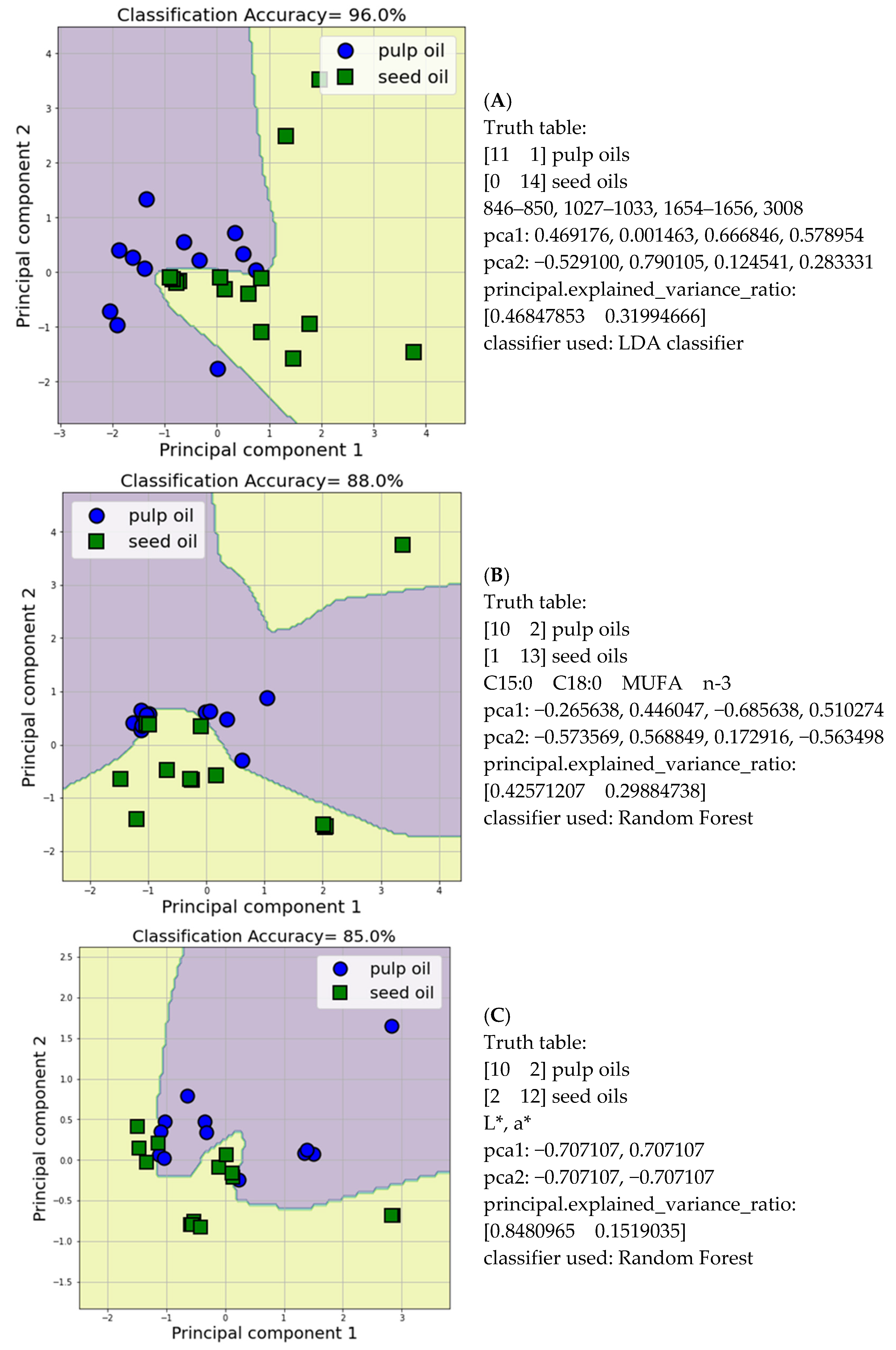
Illustrated in Figure 2A, the scatter diagram reveals the distinction between pulp and seed oils with an impressive 96.0% overall accuracy in discrimination. This discrimination was achieved through the utilization of FTIR bands at 846–850, 1027–1033, 1654–1656, and 3008 cm–1 as key features. In the scatter diagram, the initial cluster representing the pulp oil samples (depicted as blue circles) is situated on the left side, while the subsequent cluster, encompassing seed oil samples (illustrated as green squares), encapsulates the right side. Using this specific combination of four features in the development of a robust machine learning framework resulted in accurately classifying 11 out of 12 pulp oil samples (with one misclassification) and all 14 seed oil samples correctly. Interestingly, the FTIR absorption bands that successfully discriminated pulp from seed oils were associated with the presence of Csp2 bonds (3008 and 1654 cm−1) and minor components, such as primary alcohols (1027–1033 cm–1) and aromatic compounds (846–850 cm−1). It is important to point up that the unsaturation degree of the oils significantly affects their discrimination pattern. Moreover, in accordance with the above-mentioned findings, Lerma-García et al. [47] reported that vegetable oils contain primary fatty and triterpene alcohols, which are characteristic of the biological origin of the oil. Their composition is influenced by environmental factors, the origin of the pulp or seeds, and the method of oil extraction. These alcohols can serve as unique markers for identifying different types of oils. Additionally, Yu et al. [48] found that phenolic compounds are present in various oilseeds such as olive, rapeseed, sesame, soybean, and peanut. The quantity of these compounds primarily depends on the type of seed and the method used to extract the oil.
Additionally, Figure 2B shows the discrimination between pulp (blue circles) and seed (green squares) oils with 88.0% overall discrimination accuracy via the C15:0, C18:0, MUFA, and n-3 proportions. The above four-feature combination classified 9 out of 12 pulp oil samples (three samples were misclassified as seed oils) and 13 out of 14 seed oil samples (one sample was misclassified as pulp oils). It is of value that the most critical fatty acid sums for oil discrimination were MUFA and n-3 PUFA, as well as pentadecanoic (C15:0) and stearic (C18:0) acids. Notably, previous research has highlighted the multifaceted benefits of C15:0, suggesting its significance as an essential fatty acid. These benefits encompass diverse attributes such as pleiotropic activities, mitochondrial function restoration, enhancement of red blood cell stability, regulation of glucose metabolism, and suppression of cancer cell proliferation [49].
Finally, Figure 2C presents the pulp (blue circles) and seed (green squares) oil discrimination with an overall discrimination accuracy of 85.0%, via the L* and a* values. According to Vucāne et al. (2022) [50], the color characteristics of vegetable oils are influenced by factors such as the quality of the raw materials, the botanical variety, and production methods.
From the Machine Learning classifiers employed, the ML-model design produced the optimum precision using the Linear Discriminant Analysis, LDA, (for ATR-FTIR data) and the Random Forest, RF, (for fatty acids and color data) algorithms. Specifi-cally, the LDA classifier is a linear decision-making algorithm developed using the Bayes’ rule and the data distributions of each class of labelled data. For each new case (observation), its probability of belonging to each class is calculated and it is classified in the class with the highest probability. Moreover, the RF classifier is designed by con-structing many decision trees from random samples of the class-labelled data. The RF classifier places a new observation in the class indicated by the majority of the decision trees.
Illustrated in Figure 2A, the scatter diagram reveals the distinction between pulp and seed oils with an impressive 96.0% overall accuracy in discrimination. This dis-crimination was achieved through the utilization of FTIR bands at 846-850, 1027-1033, 1654-1656, and 3008 cm–1 as key feature. In the scatter diagram, the initial cluster rep-resenting the pulp oil samples (depicted as blue circles) is situated on the left side, while the subsequent cluster, encompass-ing seed oils’ samples (illustrated as green squares), encapsulates the right side. Employing this specific four-feature combination in the development of a robust machine-learning framework resulted in accurately classify-ing 11 out of 12 pulp oils’ samples (with one misclassification) and all 14 seed oils’ samples correctly. Interestingly, the FTIR absorption bands that successfully discrimi-nated pulp from seed oils were associated with the presence of Csp2 bonds (3008 and 1654 cm–1) and minor components, such as primary alcohols (1027–1033 cm–1) and ar-omatic com-pounds (846–850 cm–1). It is important to point up that the unsaturation de-gree of the oils significantly affects their discrimination pattern. Moreover, in accord-ance to the above mentioned findings, Lerma-García et al. [47] reported that vegetable oils contain primary fatty and triterpene alcohols, which are characteristic of the bio-logical origin of the oil. Their composition is influenced by environmental factors, the origin of the pulp or seeds, and the method of oil extraction. These alcohols can serve as unique markers for identifying different types of oils. Additionally, Yu et al. [48] found that phenolic compounds are present in various oilseeds such as olive, rapeseed, sesa-me, soybean, and peanut. The quantity of these compounds primarily depends on the type of seed and the method used to extract the oil.
Additionally, Figure 2B shows the discrimination between pulp (blue circles) and seed (green squares) oils with 88.0% overall discrimination accuracy, via the C15:0, C18:0, MUFA and n-3 proportions. The above four-feature combina-tion classified 9 out of 12 pulp oils’ samples (three samples were misclassified as seed oils), and 13 out of 14 seed oils’ samples (one sample was misclassified as pulp oils). It is of value that the most critical fatty acids’ sums for oils discrimination were MUFA and n-3 PUFA as well as pentadecanoic (C15:0) and stearic (C18:0) acids. Notably, pre-vious research has highlighted the multifaceted benefits of C15:0, suggesting its significance as an essen-tial fatty acid. These benefits encompass diverse attributes such as pleiotropic activi-ties, mitochondrial function restoration, enhancement of red blood cell stability, regu-lation of glucose metabolism, and suppression of cancer cell prolifera-tion [49].
Finally, Figure 2C presents the pulp (blue circles) and seed (green squares) oils’ discrimination with an overall dis-crimination accuracy of 85.0%, via the L* and a* values. According to Vucāne et al. (2022) [50], the color characteris-tics of vegetable oils are influenced by factors such as the quality of the raw materials, the botanical variety, and production methods.

4. Conclusions

The present study assessed the quality characteristics of twenty-six edible fixed oils originating from the fatty parts of a plant (pulp or seeds) in order to highlight their healthy added value and application in foods. The saturated fatty acid profile of the examined oils revealed the presence of palmitic (C16:0), followed by stearic (C18:0), pentadecanoic (C15:0), myristic (C14:0), and lauric (C12:0) acids. Oleic acid (C18:1 n-9) was the predominant MUFA in all the oils, and linoleic (C18:2 n-6) and α-linolenic (C18:3 n-3) acids were the major PUFA. Considering the nutritional quality of the oils, the ratios of UFA/SFA were greater than 1.6, whereas the lowest and the highest n-6/n-3 ratios were noticed in linseed, chia seed, canola, and macadamia oils and in poppy seed, pine cone, apricot, pumpkin seed, hazelnut, and carrot oils, respectively. Oxidative stability was inversely related to the polyunsaturated fatty acid content. Regarding the color parameter measurement, L* values differed significantly; a* values were negative (green) except for sea buckthorn, pumpkin seed, and avocado oils, whereas positive b* values (yellow) varied from +3.83 (peanut oil) to +43.69 (canola oil), indicating an increase in the yellow proportion in color. Chlorophyll levels in edible oils did not exceed 50 mg/kg, preventing the oxidation in the presence of light. B-carotene, desirable due to its antioxidant and coloring properties, had rich content in soybean, linseed, and canola oils. The results of the ATR-FTIR analysis pointed out that the intensity changes in the wavenumbers associated with monounsaturated bonds (3008 cm−1, 1654 cm−1, 1417 cm−1, and 721 cm−1) were related to the fatty acid profile of the oils. More specifically, hazelnut, almond, avocado, plum, pomegranate, mustard seed, and apricot oils, rich in MUFA, exhibit lower intensities at those wavenumbers compared to oils rich in PUFA, such as evening primrose, chia seed, walnut, pine cone, and linseed oils. Finally, the application of machine learning algorithms for analyzing ATR-FTIR band intensities and FA proportions via discriminant analysis was achieved to discriminate pulp from seed oils, with 96.0% and 88.0% classification accuracy, respectively. The research outcomes of the present study highlight the quality and nutritional superiority of the majority of the studied edible plant-based carrier oils, which in turn can be used as alternative oils for consumption or as fortifying agents in low-quality vegetable oils. In addition, the practical importance of the study could be of economic and nutritional interest to countries that rely on oilseed crops to support farmers and local industries.

Supplementary Materials

The following supporting information can be downloaded at: https://www.mdpi.com/article/10.3390/app142210305/s1, Table S1: Saturated fatty acid (SFA) composition (% of total) of edible fixed oils; Table S2: Monounsaturated fatty acid (MUFA) composition (% of total) of edible fixed oils; Table S3: Polyunsaturated fatty acid (PUFA) composition (% of total) of edible fixed oils; Table S4: Relative intensities% of ATR-FTIR spectra bands of edible fixed oils.

Author Contributions

Conceptualization, I.F.S. and V.J.S.; methodology, I.F.S., K.T., A.P. and G.L.; software, K.T. and D.C.; validation, I.F.S., K.T., G.L. and V.J.S.; formal analysis, K.T. and A.P.; investigation, I.F.S., A.P. and G.L.; resources, D.C. and V.J.S.; data curation, K.T. and D.C.; writing—original draft preparation, I.F.S. and V.J.S.; writing—review and editing, I.F.S., K.T., G.L. and V.J.S.; visualization, K.T., D.C. and G.L.; supervision, I.F.S. and V.J.S.; project administration, I.F.S. and V.J.S. All authors have read and agreed to the published version of the manuscript.

Funding

This research received no external funding.

Informed Consent Statement

Not applicable.

Data Availability Statement

The original contributions presented in the study are included in the article, further inquiries can be directed to the corresponding author.

Acknowledgments

The authors also acknowledge the undergraduate student Evangelia Papakostopoulou for her valuable contribution to this work.

Conflicts of Interest

The authors declare no conflicts of interest.

References

  1. Athar, M.; Nasir, S.M. Taxonomic Perspective of Plant Species Yielding Vegetable Oils Used in Cosmetics and Skin Care Products. Afr. J. Biotechnol. 2005, 4, 36–44. [Google Scholar]
  2. Ali, S.; Ekbbal, R.; Salar, S.; Yasheshwar; Ali, S.A.; Jaiswal, A.K.; Singh, M.; Yadav, D.K.; Kumar, S. Gaurav Quality Standards and Pharmacological Interventions of Natural Oils: Current Scenario and Future Perspectives. ACS Omega 2023, 8, 39945–39963. [Google Scholar] [CrossRef] [PubMed]
  3. Iqbal, A.; Begum, N.; Rabbi, F.; Akhtar, N.; Rahman, K.U.; Khan, W.M.; Shah, Z. In-Vitro Antimicrobial, Antioxidant and Enzyme Inhibitory Activities of Fixed Oil Extracted from Stem Bark of Tamarix Aphylla. Pharm. Chem. J. 2022, 56, 1116–1122. [Google Scholar] [CrossRef]
  4. Savic, I.; Savic Gajic, I.; Gajic, D. Physico-Chemical Properties and Oxidative Stability of Fixed Oil from Plum Seeds (Prunus domestica Linn.). Biomolecules 2020, 10, 294. [Google Scholar] [CrossRef] [PubMed]
  5. Xue, L.; Yang, R.; Wang, X.; Ma, F.; Yu, L.; Zhang, L.; Li, P. Comparative Advantages of Chemical Compositions of Specific Edible Vegetable Oils. Oil Crop Sci. 2023, 8, 1–6. [Google Scholar] [CrossRef]
  6. Jimenez-Lopez, C.; Carpena, M.; Lourenço-Lopes, C.; Gallardo-Gomez, M.; Lorenzo, J.M.; Barba, F.J.; Prieto, M.A.; Simal-Gandara, J. Bioactive Compounds and Quality of Extra Virgin Olive Oil. Foods 2020, 9, 1014. [Google Scholar] [CrossRef]
  7. Yang, R.; Zhang, L.; Li, P.; Yu, L.; Mao, J.; Wang, X.; Zhang, Q. A Review of Chemical Composition and Nutritional Properties of Minor Vegetable Oils in China. Trends Food Sci. Technol. 2018, 74, 26–32. [Google Scholar] [CrossRef]
  8. Toishimanov, M.; Nurgaliyeva, M.; Serikbayeva, A.; Suleimenova, Z.; Myrzabek, K.; Shokan, A.; Myrzabayeva, N. Comparative Analysis and Determination of the Fatty Acid Composition of Kazakhstan’s Commercial Vegetable Oils by GC-FID. Appl. Sci. 2023, 13, 7910. [Google Scholar] [CrossRef]
  9. Sinanoglou, V.J.; Miniadis-Meimaroglou, S. Fatty Acid of Neutral and Polar Lipids of (Edible) Mediterranean Cephalopods. Food Res. Int. 1998, 31, 467–473. [Google Scholar] [CrossRef]
  10. Sinanoglou, V.J.; Strati, I.F.; Bratakos, S.M.; Proestos, C.; Zoumpoulakis, P.; Miniadis-Meimaroglou, S. On the Combined Application of Iatroscan TLC-FID and GC-FID to Identify Total, Neutral, and Polar Lipids and Their Fatty Acids Extracted from Foods. ISRN Chromatogr. 2013, 2013, 1–8. [Google Scholar] [CrossRef]
  11. AOCS. Official Methods and Recommended Practices-Oil Stability Index, 6th ed.; American Oil Chemists’ Society: Champaign, IL, USA, 2017; Method Cd 12b–92. [Google Scholar]
  12. AOCS. Official Methods and Recommended Practices-Color of Fats and Oil, Lovibond, 6th ed.; American Oil Chemists’ Society: Champaign, IL, USA, 2017; Method Cc 13e–92. [Google Scholar]
  13. Ioannou, A.G.; Kritsi, E.; Sinanoglou, V.J.; Cavouras, D.; Tsiaka, T.; Houhoula, D.; Zoumpoulakis, P.; Strati, I.F. Highlighting the Potential of Attenuated Total Reflectance—Fourier Transform Infrared (ATR-FTIR) Spectroscopy to Characterize Honey Samples with Principal Component Analysis (PCA). Anal. Lett. 2023, 56, 789–806. [Google Scholar] [CrossRef]
  14. Zielińska, A.; Nowak, I. Abundance of Active Ingredients in Sea-Buckthorn Oil. Lipids Health Dis. 2017, 16, 95. [Google Scholar] [CrossRef] [PubMed]
  15. Amit; Jamwal, R.; Kumari, S.; Dhaulaniya, A.S.; Balan, B.; Singh, D.K. Application of ATR-FTIR Spectroscopy along with Regression Modelling for the Detection of Adulteration of Virgin Coconut Oil with Paraffin Oil. LWT 2020, 118, 108754. [Google Scholar] [CrossRef]
  16. Durmaz, G.; Karabulut, İ.; Topçu, A.; Asiltürk, M.; Kutlu, T. Roasting-Related Changes in Oxidative Stability and Antioxidant Capacity of Apricot Kernel Oil. J. Am. Oil Chem. Soc. 2010, 87, 401–409. [Google Scholar] [CrossRef]
  17. Maldonado, A.; Riquelme, N.; Muñoz-Fariña, O.; García, O.; Arancibia, C. Stability and Bioaccessibility of α-Tocopherol-Enriched Nanoemulsions Containing Different Edible Oils as Carriers. LWT 2023, 174, 114419. [Google Scholar] [CrossRef]
  18. Aktas, A.B.; Dastan, T.; Katin, K.P.; Kaya, S. Determination of Oxidative Stability of Different Vegetable Oils by Means of Middle Infrared Spectroscopy and DFT Calculations. Microchem. J. 2023, 194, 109232. [Google Scholar] [CrossRef]
  19. Kaur, A.; Singh, B.; Kaur, A.; Singh, N. Chemical, Thermal, Rheological and FTIR Studies of Vegetable Oils and Their Effect on Eggless Muffin Characteristics. J. Food Process Preserv. 2019, 43, e1397. [Google Scholar] [CrossRef]
  20. Vladimír, M.; Matwijczuk, A.P.; Niemczynowicz, A.; Kycia, R.A.; Karcz, D.; Gładyszewska, B.; Ślusarczyk, L.; Burg, P. Chemometric Approach to Characterization of the Selected Grape Seed Oils Based on Their Fatty Acids Composition and FTIR Spectroscopy. Sci. Rep. 2021, 11, 19256. [Google Scholar] [CrossRef]
  21. Lucarini, M.; Durazzo, A.; Kiefer, J.; Santini, A.; Lombardi-Boccia, G.; Souto, E.; Romani, A.; Lampe, A.; Ferrari Nicoli, S.; Gabrielli, P.; et al. Grape Seeds: Chromatographic Profile of Fatty Acids and Phenolic Compounds and Qualitative Analysis by FTIR-ATR Spectroscopy. Foods 2019, 9, 10. [Google Scholar] [CrossRef]
  22. Vadalà, R.; Nava, V.; Turco, V.L.; Potortì, A.G.; Costa, R.; Rando, R.; Ben Mansour, H.; Ben Amor, N.; Beltifa, A.; Santini, A.; et al. Nutritional and Health Values of Tunisian Edible Oils from Less-Used Plant Sources. Agriculture 2023, 13, 1096. [Google Scholar] [CrossRef]
  23. Maszewska, M.; Florowska, A.; Dłużewska, E.; Wroniak, M.; Marciniak-Lukasiak, K.; Żbikowska, A. Oxidative Stability of Selected Edible Oils. Molecules 2018, 23, 1746. [Google Scholar] [CrossRef] [PubMed]
  24. Soleimanifar, M.; Niazmand, R.; Jafari, S.M. Evaluation of Oxidative Stability, Fatty Acid Profile, and Antioxidant Properties of Black Cumin Seed Oil and Extract. Food Meas. 2019, 13, 383–389. [Google Scholar] [CrossRef]
  25. Król, K.; Gantner, M.; Piotrowska, A. The Quality Characteristic and Fatty Acid Profile of Cold-Pressed Hazelnut Oils during Nine Months of Storage. Agronomy 2021, 11, 2045. [Google Scholar] [CrossRef]
  26. WHO. Interim Summary of Conclusions & Dietary Recommendations on Total Fat & Fatty Acids. In The Joint FAO/WHO Expert Consultation on Fats & Fatty Acids in Human Nutri-Tion; WHO: Geneva, Switzerland, 2008; pp. 1–14. [Google Scholar]
  27. Lamine, M.; Mlikia, A. Nutritional Quality Perceptions through Fatty Acid Profiling, Health Lipid Indices and Antioxidant Potentialities. Open J. Nutr. Food Sci. 2021, 3, 1016. [Google Scholar]
  28. Simopoulos, A.P.; DiNicolantonio, J.J. The Importance of a Balanced ω-6 to ω-3 Ratio in the Prevention and Management of Obesity. Open Heart 2016, 3, e000385. [Google Scholar] [CrossRef]
  29. D’Angelo, S.; Motti, M.L.; Meccariello, R. ω-3 and ω-6 Polyunsaturated Fatty Acids, Obesity and Cancer. Nutrients 2020, 12, 2751. [Google Scholar] [CrossRef]
  30. Gonzalez-Becerra, K.; Barron-Cabrera, E.; Muñoz-Valle, J.F.; Torres-Castillo, N.; Rivera-Valdes, J.J.; Rodriguez-Echevarria, R.; Martinez-Lopez, E. A Balanced Dietary Ratio of N-6:N-3 Polyunsaturated Fatty Acids Exerts an Effect on Total Fatty Acid Profile in RBCs and Inflammatory Markers in Subjects with Obesity. Healthcare 2023, 11, 2333. [Google Scholar] [CrossRef]
  31. Farag, M.A.; Reda, A.; Nabil, M.; Elimam, D.M.; Zayed, A. Evening Primrose Oil: A Comprehensive Review of Its Bioactives, Extraction, Analysis, Oil Quality, Therapeutic Merits, and Safety. Food Funct. 2023, 14, 8049–8070. [Google Scholar] [CrossRef]
  32. Yang, C.; Shang, K.; Lin, C.; Wang, C.; Shi, X.; Wang, H.; Li, H. Processing Technologies, Phytochemical Constituents, and Biological Activities of Grape Seed Oil (GSO): A Review. Trends Food Sci. Technol. 2021, 116, 1074–1083. [Google Scholar] [CrossRef]
  33. Domínguez, R.; Pateiro, M.; Gagaoua, M.; Barba, F.J.; Zhang, W.; Lorenzo, J.M. A Comprehensive Review on Lipid Oxidation in Meat and Meat Products. Antioxidants 2019, 8, 429. [Google Scholar] [CrossRef]
  34. Vever-Bizet, C.; Dellinger, M.; Brault, D.; Rougee, M.; Bensasson, R.V. Singlet molecular oxygen quenching by saturated and unsaturated fatty-acids and by cholesterol. Photochem. Photobiol. 1989, 50, 321–325. [Google Scholar] [CrossRef] [PubMed]
  35. Fan, Y.; Li, J.; Guo, Y.; Xie, L.; Zhang, G. Digital Image Colorimetry on Smartphone for Chemical Analysis: A Review. Measurement 2021, 171, 108829. [Google Scholar] [CrossRef]
  36. Gao, H.; Sun, D.; Zhou, Y.; Zeng, L.; Shi, X. Experimental Study on Oil Colour Grading Based on Uniform Colour Space. J. Phys. Conf. Ser. 2023, 2428, 012007. [Google Scholar] [CrossRef]
  37. Matthäus, B.; Brühl, L. Quality of cold-pressed edible rapeseed oil in Germany. Food/Nahrung 2003, 47, 413–419. [Google Scholar]
  38. Górnaś, P.; Siger, A.; Juhņeviča, K.; Lācis, G.; Šnē, E.; Segliņa, D. Cold-pressed Japanese quince (Chaenomeles japonica (Thunb.) Lindl. ex Spach) seed oil as a rich source of α-tocopherol, carotenoids and phenolics: A comparison of the composition and antioxidant activity with nine other plant oils. Euro. J. Lipid Sci. Tech. 2014, 116, 563–570. [Google Scholar]
  39. Erickson, D.R. Edible Fats and Oils Processing Basic Principles and Modern Practices; American Oil Chemists’ Society: Champaign, IL, USA, 1990. [Google Scholar]
  40. Nandiyanto, A.B.D.; Oktiani, R.; Ragadhita, R. How to Read and Interpret FTIR Spectroscope of Organic Material. Indones. J. Sci. Technol. 2019, 4, 97. [Google Scholar] [CrossRef]
  41. do Nascimento, T.A.; Lopes, T.I.B.; Nazario, C.E.D.; Oliveira, S.L.; Alcantara, G.B. Vegetable Oils: Are They True? A Point of View from ATR-FTIR, 1H NMR, and Regiospecific Analysis by 13C NMR. Food Res. Int. 2021, 144, 110362. [Google Scholar] [CrossRef]
  42. Uzun, H.; Kaynak, E.G.; Ibanoglu, E.; Ibanoglu, S. Chemical and Structural Variations in Hazelnut and Soybean Oils after Ozone Treatments. Grasas y Aceites 2018, 69, 253. [Google Scholar] [CrossRef]
  43. de Souza, T.R.P.; Olenka, L.; Peternella, W.S. A Study of Degradation in Vegetable Oils by Exposure to Sunlight Using Fourier Transform Infrared Spectroscopy. Mater. Sci. Appl. 2020, 11, 678–691. [Google Scholar] [CrossRef]
  44. Mukhametov, A.; Mamayeva, L.; Kazhymurat, A.; Akhlan, T.; Yerbulekova, M. Study of Vegetable Oils and Their Blends Using Infrared Reflectance Spectroscopy and Refractometry. Food Chem. X 2023, 17, 100386. [Google Scholar] [CrossRef]
  45. Poiana, M.-A.; Alexa, E.; Munteanu, M.-F.; Gligor, R.; Moigradean, D.; Mateescu, C. Use of ATR-FTIR Spectroscopy to Detect the Changesin Extra Virgin Olive Oil by Adulteration Withsoybean Oil and High Temperature Heat Treatment. Open Chem. 2015, 13, 000010151520150110. [Google Scholar] [CrossRef]
  46. Arslan, F.N. Atr–ftir spectroscopy combined with chemometrics for rapid classification of extra virgin olive oils and edible oils from different cultivars available on the turkish markets. Eskişehir Tech. Univ. J. Sci. Technol. A-Appl. Sci. Eng. 2018, 19, 926–947. [Google Scholar] [CrossRef]
  47. Lerma-García, M.J.; Ramis-Ramos, G.; Herrero-Martínez, J.M.; Gimeno-Adelantado, J.V.; Simó-Alfonso, E.F. Characterization of the Alcoholic Fraction of Vegetable Oils by Derivatization with Diphenic Anhydride Followed by High-Performance Liquid Chromatography with Spectrophotometric and Mass Spectrometric Detection. J. Chromatogr. A 2009, 1216, 230–236. [Google Scholar] [CrossRef] [PubMed]
  48. Yu, X.; Yu, L.; Ma, F.; Li, P. Quantification of Phenolic Compounds in Vegetable Oils by Mixed-Mode Solid-Phase Extraction Isotope Chemical Labeling Coupled with UHPLC-MS/MS. Food Chem. 2021, 334, 127572. [Google Scholar] [CrossRef] [PubMed]
  49. Venn-Watson, S.K.; Butterworth, C.N. Broader and Safer Clinically-Relevant Activities of Pentadecanoic Acid Compared to Omega-3: Evaluation of an Emerging Essential Fatty Acid across Twelve Primary Human Cell-Based Disease Systems. PLoS ONE 2022, 17, e0268778. [Google Scholar] [CrossRef] [PubMed]
  50. Vucāne, S.; Cinkmanis, I.; Šabovics, M. Colorimetric Measurements of Vegetable Oils by Smartphone-Based Image Analysis. Proc. Latv. Acad. Sciences. Sect. B Nat. Exact Appl. Sci. 2022, 76, 110–115. [Google Scholar] [CrossRef]
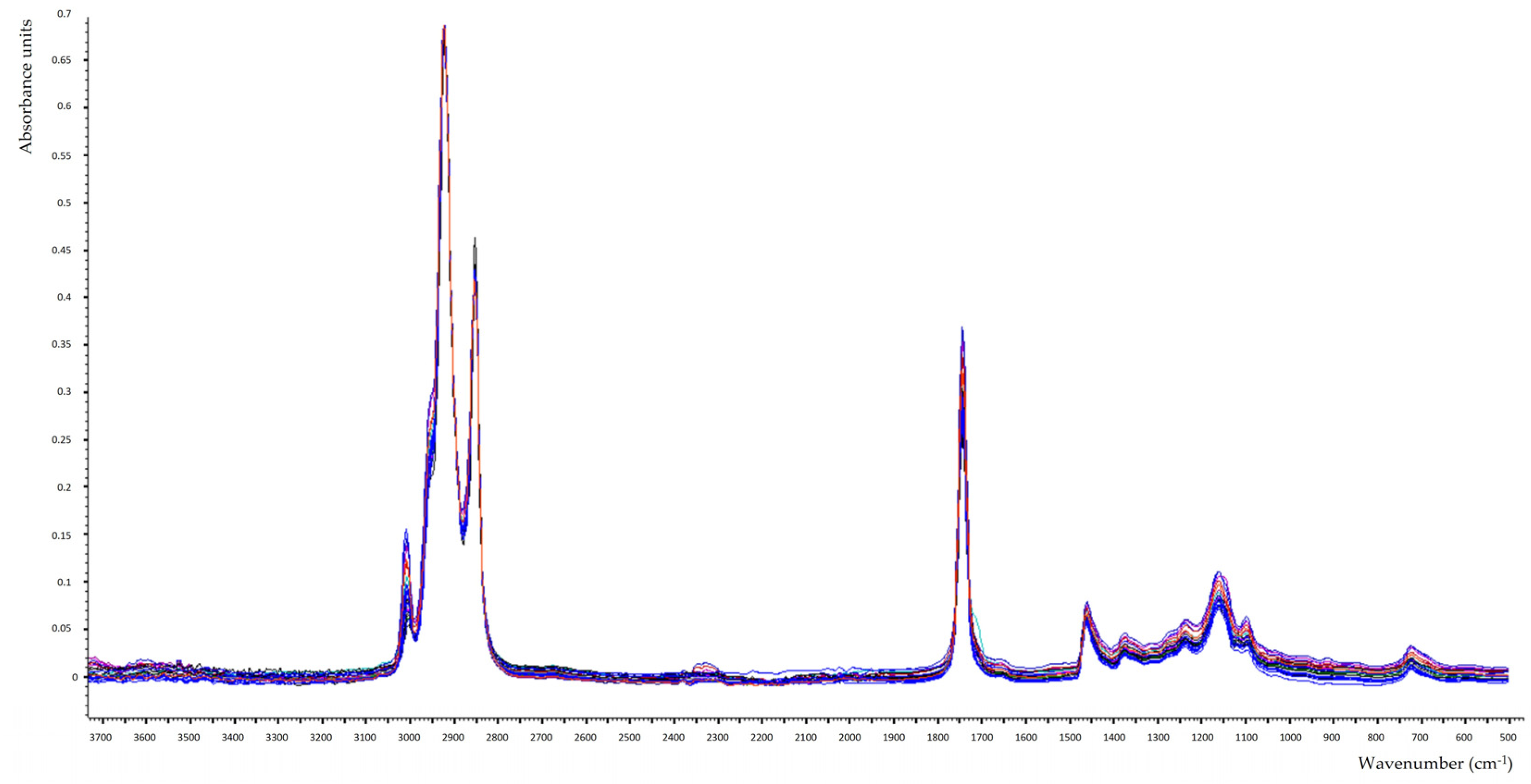
Figure 1. ATR-FTIR spectra of the examined edible fixed oils.
Applsci 14 10305 g001
Figure 2. Scatter diagram of the discrimination, employing selected (A) FTIR bands and (B) fatty acid proportions and (C) color parameters between pulp and seed oil samples.
Applsci 14 10305 g002
Table 1. Plant information of the examined edible fixed oils.
Edible Fixed OilsINCI
(International Nomenclature of Cosmetic Ingredients)
Plant Family
Walnut oil (1) *Juglans regia Seed Oil Juglandaceae
Linseed oil (2)Linum usitatissimum L. Seed OilLinaceae
Sesame oil (2)Sesamum indicum L. Seed OilPedaliaceae
Almond oil (1)Prunus amygdalus var. dulcis OilRosaceae
Hazelnut oil (1)Corylus avellanaBetulaceae
Pumpkin seed oil virgin unroasted (2)Cucurbita pepo seed OilCucurbitaceae
Avocado oil (1) Persea gratissima OilLauraceae
Black cumin oil (2)Nigella sativa L. Seed OilRanunculaceae
Coffee bean base oil (1)Coffea arabica Seed OilRubiaceae
Pine cone base oil (2)Pinus sibirica Seed OilPinaceae
Evening Primrose base oil (2)Oenothera biennis OilOnagraceae
Grape seed base oil (2)Vitis vinifera Seed OilVitaceae
Plum base oil (1)Prunus domestica Seed OilRosaceae
Pomegranate base oil (2)Punica granatum L. Seed OilLythraceae
Mustard seed base oil (2)Brassica juncea Seed ExtractBrassicaceae
Sea Buckthorn base oil (1)Hippophae rhamnoides Fruit ExtractElaeagnaceae
Chia seed base oil (2)Salvia hispanica Seed OilLamiaceae
Carrot base oil (1)Daucus carota subsp. sativus Root ExtractApiaceae
Peanut base oil (1)Arachis hypogaea L. OilFabaceae
Apricot base oil (1)Prunus armeniaca Kernel OilRosaceae
Milk thistle base oil (2)Silybum marianum Seed OilAsteraceae
Macadamia base oil kernel (1)Macadamia integrifolia Seed OilProteaceae
Canola base oil (2)Brassica campestris L. Seed OilBrassicaceae
Soybean base oil (2)Glycine soja OilFabaceae
Poppy seed base oil (2)Papaver somniferum Seed OilPapaveraceae
Coconut Virgin oil raw (1)Cocos nucifera L. OilArecaceae
* (1) Pulp oils and (2) seed oils.
Table 2. Fatty acid composition (% of total), UFA/SFA, SFA:MUFA:PUFA, n-6/n-3 ratios, and oxidation induction time (OIT) of examined edible fixed oils.
Edible Fixed OilSFAMUFAPUFAUFA/SFASFA:MUFA:PUFAn-6/n-3OIT (h)
Walnut oil14.50 ± 0.13 a16.54 ± 0.2368.96 ± 0.455.90 ± 0.02 a1:1.1:4.85.770.47 ± 0.02
Linseed oil14.35 ± 0.15 a21.08 ± 0.1764.57 ± 0.42 a5.97 ± 0.03 a1:1.5:4.50.330.12 ± 0.01
Almond oil14.98 ± 0.1270.28 ± 0.6814.74 ± 0.36 b5.68 ± 0.021:4.7:1.0--3.44 ± 0.01
Hazelnut oil12.59 ± 0.0873.36 ± 0.5714.05 ± 0.41 b6.94 ± 0.031:5.8:1.11162.60 ± 0.01
Pumpkin seed oil24.20 ± 0.2327.57 ± 0.2248.23 ± 0.293.13 ± 0.011:1.1:2.01233.14 ± 0.01
Avocado oil21.52 ± 0.1269.81 ± 0.388.84 ± 0.173.65 ± 0.021:3.2:0.413.010.2 ± 0.01
Black cumin oil23.76 ± 0.1623.00 ± 0.16 a53.24 ± 0.453.21 ± 0.011:1.0:2.294.12.15 ± 0.01 **
Coffee bean oil20.06 ± 0.2423.35 ± 0.18 a56.58 ± 0.263.98 ± 0.031:1.2:2.872.51.99 ± 0.01
Pine cone oil9.39 ± 0.11 c25.46 ± 0.1965.12 ± 0.37 ac9.65 ± 0.03 c1:2.7:6.92320.33 ± 0.01
Evening primrose 16.57 ± 0.087.67 ± 0.1275.77 ± 0.31 e5.04 ± 0.021:0.5:4.6--1.15 ± 0.01 *
Grape seed oil11.16 ± 0.10 b23.41 ± 0.26 a65.87 ± 0.62 c8.00 ± 0.02 b1:2.1:5.9--1.14 ± 0.02 *
Plum oil11.05 ± 0.07 b68.77 ± 0.35 b20.19 ± 0.248.05 ± 0.03 b1:6.2:1.8--1.03 ± 0.01
Pomegranate oil20.72 ± 0.1468.35 ± 0.41 b10.94 ± 0.15 d3.83 ± 0.011:3.3:0.577.10.64 ± 0.01
Mustard seed oil21.84 ± 0.1567.58 ± 0.4810.59 ± 0.18 d3.58 ± 0.031:3.1:0.514.80.82 ± 0.01
Sea buckthorn oil36.91 ± 0.2258.54 ± 0.36 c4.57 ± 0.111.71 ± 0.011:1.6:0.166.831.9 ± 0.01
Chia seed oil16.00 ± 0.138.86 ± 0.0875.30 ± 0.85 e5.26 ± 0.021:0.6:4.70.390.15 ± 0.02
Carrot oil19.87 ± 0.2139.48 ± 0.3340.63 ± 0.344.03 ± 0.021:2.0:2.01005.73 ± 0.01
Peanut oil13.96 ± 0.1458.51 ± 0.35 c27.53 ± 0.466.16 ± 0.031:4.2:2.026.21.67 ± 0.01
Apricot oil9.42 ± 0.06 c66.71 ± 0.4223.85 ± 0.229.61 ± 0.03 c1:7.1:2.51484.79 ± 0.02
Milk thistle oil22.57 ± 0.3121.65 ± 0.2755.79 ± 0.283.43 ± 0.021:1.0:2.527.52.05 ± 0.01
Macadamia oil25.34 ± 0.2671.74 ± 0.512.94 ± 0.062.95 ± 0.021:2.8:0.13.907.75 ± 0.01
Canola oil11.82 ± 0.0956.19 ± 0.3031.96 ± 0.397.45 ± 0.011:4.8:2.72.081.50 ± 0.01
Soybean oil18.63 ± 0.1126.38 ± 0.2155.00 ± 0.344.37 ± 0.011:1.4:3.07.782.16 ± 0.01 **
Poppy seed oil21.21 ± 0.1642.27 ± 0.2736.53 ± 0.413.72 ± 0.001:2.0:1.73640.81 ± 0.01
Sesame oil18.06 ± 0.1837.20 ± 0.1744.72 ± 0.534.54 ± 0.011:2.1:2.580.32.75 ± 0.01
Virgin coconut oil94.10 ± 0.424.95 ± 0.060.94 ± 0.050.06 ± 0.001:0.1:0.022.569.3 ± 0.02
SFA: saturated fatty acid; MUFA: monounsaturated fatty acid; PUFA: polyunsaturated fatty acid; UFA: unsaturated fatty acid; OIT: oxidation induction time. OIT values with similar asterisks are not considered statistically different (p > 0.05). Means in the same column bearing the same letter do not differ significantly (p > 0.05).
Table 3. Color parameters (L*, a*, b*, and hue) in CIELAB color space and RYBN, chlorophyll (ppb), and b-carotene (ppb) in Lovibond® color scale.
Edible Fixed OilL*a*b*Hue (°)RYBNChlorophyll (ppb)b-carotene (ppb)
Walnut oil56.05 ± 0.12 ef−3.04 ± 0.02 h13.21 ± 0.20 h102.99 ± 0.15 ef1.03.90.00.0256.012,891.0
Linseed oil60.04 ± 0.22 h−5.92 ± 0.10 a35.76 ± 0.20 o99.38 ± 0.12 de2.360.00.01.9294.060,421.0
Almond oil64.17 ± 0.12 mn−2.26 ± 0.04 ij8.56 ± 0.05 g104.80 ± 0.22 fg0.41.20.00.20.04054.0
Hazelnut oil55.62 ± 0.04 e−3.44 ± 0.05 g14.95 ± 0.41 ij102.96 ± 0.23 ef1.27.20.00.0161.019,119.0
Pumpkin seed oil41.46 ± 0.05 c3.26 ± 0.11 o4.73 ± 0.19 c55.43 ± 1.92 b10.98.66.10.00.015,324.0
Avocado oil46.01 ± 0.32 d0.01 ± 0.08 n15.83 ± 0.54 k89.97 ± 0.28 c2.360.01.40.017,611.055,022.0
Black cumin oil60,05 ± 0.05 h−5.19 ± 0.05 b23.46 ± 0.13 n102.48 ± 0.17 ef1.613.50.00.82440.036,582.0
Coffee bean oil59.89 ± 0.17 h−2.52 ± 0.07 i19.80 ± 0.16 m97.27 ± 0.22 d2.010.82.10.00.031,241.0
Pine cone oil67.07 ± 0.27 p−2.24 ± 0.05 ij7.32 ± 0.21 f107.05 ± 0.22 fg0.31.40.00.00.05853.0
Evening Primrose oil63.91 ± 0.20 lm−5.22 ± 0.04 b23.94 ± 0.17 n103.30 ± 1.69 ef1.821.00.00.92945.036,740.0
Grape seed oil64.85 ± 0.54 no−4.45 ± 0.02 d14.30 ± 0.23 i107.26 ± 0.19 fg0.77.10.00.03922.023,654.0
Plum oil62.71 ± 0.18 k−3.80 ± 0.07 f15.77 ± 0.15 jk103.54 ± 0.13 ef1.26.40.00.5226.018,269.0
Pomegranate oil55.61 ± 0.14 e−2.43 ± 0.07 ij7.20 ± 0.26 f108.68 ± 0.70 g0.42.40.00.01140.08569.0
Mustard seed oil56.64 ± 0,12 f−1.69 ± 0.06 k6.18 ± 0.08 e105.33 ± 0.43 fg0.41.61.00.20.05843.0
Sea buckthorn oil39,90 ± 0.43 b8.17 ± 0.15 p5.01 ± 0.04 cd31.55 ± 0.57 a18.22.410.10.030,000.030,000.0
Chia seed oil65.15 ± 0.16 o−1.59 ± 0.05 kl5.34 ± 0.08 cde106.59 ± 0.27 fg0.51.80.00.00.06691.0
Carrot oil60.94 ± 0.10 i−4.17 ± 0.03 de17.95 ± 0.09 l103.07 ± 0.03 ef1.511.80.00.8789.025,196.0
Peanut oil64.23 ± 0.16 mn−0.63 ± 0.34 m3.83 ± 0.20 b99.38 ± 5.39 de0.00.30.00.03.01443.0
Apricot oil56.61 ± 0.35 f−1.41 ± 0.04 kl5.72 ± 0.19 de103.83 ± 0.80 efg0.81.90.00.40.06321.0
Milk thistle oil61.80 ± 0.22 j−3.92 ± 0.01 ef14.80 ± 0.09 i104.85 ± 0.08 fg1.46.00.00.4179.017,211.0
Macadamia oil63.30 ± 0.18 kl−1.35 ± 0.08 l4.99 ± 0.11 cd105.09 ± 0.73 fg0.30.80.00.00.03472.0
Canola oil63.17 ± 0.59 kl−4.79 ± 0.11 c43.69 ± 0.74 q96.25 ± 0.18 d2.569.00.01.71103.060,121.0
Soybean oil58.68 ± 0.19 g−4.03 ± 0.07 ef36.86 ± 0.61 p96.23 ± 0.04 d2.868.00.01.81822.061,176.0
Poppy seed oil65.43 ± 0.33 o−4.11 ± 0.12 ef14.97 ± 0.17 ijk105.34 ± 0.27 fg1.07.10.00.12843.019,001.0
Sesame oil56.56 ± 0.25 f−2.18 ± 0.11 j8.58 ± 0.19 g104.26 ± 0.39 efg0.82.40.00.254.08049.0
Virgin Coconut oil26.03 ± 0.01 a−0.14 ± 0.05 n−0.55 ± 0.02 a255.77 ± 5.24 h0.40.51.50.00.02293.0
Mean values bearing different letters in the same column are statistically different (p < 0.05) (a < b < c <….n < o < p < q).
Disclaimer/Publisher’s Note: The statements, opinions and data contained in all publications are solely those of the individual author(s) and contributor(s) and not of MDPI and/or the editor(s). MDPI and/or the editor(s) disclaim responsibility for any injury to people or property resulting from any ideas, methods, instructions or products referred to in the content.