Article

Synthesis and Characterization of Hydroxyapatite Assisted by Microwave-Ultrasound from Eggshells for Use as a Carrier of Forchlorfenuron and In Silico and In Vitro Evaluation

by
Benjamín I. Romero-De La Rosa
1,
Silvia P. Paredes-Carrera
1,*,
Jorge A. Mendoza-Pérez
2,
Dulce E. Nicolás-Álvarez
3,
Vicente Garibay-Febles
4 and
Carlos A. Camacho-Olguin
5
1
Laboratorio de Nanomateriales Sustentables, Escuela Superior de Ingeniería Química e Industrias Extractivas, Instituto Politécnico Nacional S/N, Unidad Profesional Adolfo López Mateos, Mexico City 07708, Mexico
2
Departamento de Energías Renovables, Escuela Nacional de Ciencia Biológicas, Instituto Politécnico Nacional S/N, Unidad Profesional Adolfo López Mateos, Mexico City 07738, Mexico
3
Laboratorio de Hormonas y Conducta, Departamento de Fisiología, Escuela Nacional de Ciencia Biológicas, Instituto Politécnico Nacional S/N, Unidad Profesional Adolfo López Mateos, Mexico City 07738, Mexico
4
Instituto Mexicano del Petróleo, Eje Central Lázaro Cárdenas Norte 152, Gustavo A. Madero 07730, Mexico
5
Departamento de Mecatrónica, Universidad Politécnica del Valle de México, Av. Mexiquense, esq, Av. Universidad Politécnica S/N, Los Portales, Fuentes del Valle 54910, Mexico
*
Author to whom correspondence should be addressed.
Appl. Sci. 2024, 14(24), 11522; https://doi.org/10.3390/app142411522
Submission received: 13 November 2024 / Revised: 3 December 2024 / Accepted: 4 December 2024 / Published: 11 December 2024
(This article belongs to the Section Agricultural Science and Technology)

Abstract

:
This study utilized eggshell biomass as a calcium precursor for synthesizing hydroxyapatite (Hap) through a co-precipitation method assisted by a combined microwave-ultrasound (Mu/Us) crystallization process. Different milling techniques (mortar, high-energy mill, and sieving) were employed to prepare the eggshell biomass and identify the most effective calcium precursor. The precursor derived from high-energy milling, followed by sieving and thermal treatment at 750 °C (designated as Sample Hap-H3 750), was selected due to its higher porosity, enhanced crystallinity, and smaller particle size than other synthesized materials. This sample was subsequently used as a carrier for the plant hormone forchlorfenuron (FCF), forming the composite Hap-FCF. Comprehensive characterization was conducted using X-ray diffraction (XRD), Fourier-transform infrared spectroscopy (FTIR), specific surface area analysis (BET method), zeta potential (ZP), scanning electron microscopy (SEM), and bright-field transmission electron microscopy (BFTEM), ensuring reliable and robust data. The in silico evaluation of the phytohormone FCF with two receptors, gibberellin (GA3Ox2) and auxin (IAA7), produced notable results. Docking and molecular dynamics (MD) simulations demonstrated that the gibberellin receptor was preferentially stimulated, as shown by the higher binding affinity and the receptor’s sustained stability during the MD simulations. These findings underscore the potential applications of this research, emphasizing its significance in materials science and biochemistry. Moreover, the in vitro assessment of Hap-H3 750, Hap-FCF, FCF, and the control (distilled water) on the germination and growth of butterhead lettuce seeds (Lactuca sativa) over 30 days revealed that Hap-H3 750 and Hap-FCF promoted plant growth by 275–330% relative to the control. This effect was attributed to the preferential stimulation of the gibberellin receptors responsible for stem and root elongation. These results suggest that HAP nanoparticles could facilitate the controlled delivery of FCF in the agricultural sector, promising to be an effective nanofertilizer.

1. Introduction

The development of eco-friendly nanomaterials using agro-industrial waste has progressed significantly due to the environmental concerns linked with chemically synthesized nanomaterials [1]. Agro-industrial waste utilization enables resource valorization and reduces waste disposal costs. This study introduces bird eggshells as a calcium precursor for synthesizing hydroxyapatite through a combined microwave-ultrasonic method [2]. Currently, eggs are widely utilized as food additives, generating around 250,000 tons of eggshell waste annually [3]. Although biodegradable, eggshells require four to six weeks to decompose completely, often emitting unpleasant odors and carrying pathogens like Salmonella [4]. Recently, interest has grown in employing eggshell waste for chemical synthesis, particularly as a precursor for hydroxyapatite Ca10(PO4)6(OH)2 (Hap) through hydrothermal and sol-gel methods [5]. This approach yields a material with superior resistance and crystallinity compared to that derived from 4H2O-Ca(NO3)2 [6], showing promise as a nanofertilizer (supplying nutrients like Ni, Ca, P, and K) and as a phytohormone carrier [7]. As a bioceramic, Hap is rich in calcium and phosphorus, promoting plant growth due to its nutrient availability, non-toxicity, and biocompatibility.
Phytohormones further stimulate gibberellin receptors for stem and root elongation and auxin receptors for fruit growth. Forchlorfenuron (FCF) is an effective growth-regulating hormone that enhances cell division, leading to increased fruit size and synergistic action with auxins and gibberellins, thus promoting more pronounced lateral growth than traditional growth methods [8,9]. According to Maoz et al., phytohormones at concentrations of 5% or lower pose no health risk to consumers [10]. Moreover, advanced technologies such as simultaneous microwave-ultrasonic (MO-US) applications in Hap synthesis offer reduced synthesis times (from hours to minutes) and precise control over texture and morphology, dependent on irradiation time and power. It is noteworthy that the MO-US method has not yet been reported. While the MO-US method offers significant advantages, it is important to note that it also carries potential risks, such as forming reactive species that could be harmful if not properly managed. Microwave irradiation aligns molecular dipoles with electric and/or magnetic fields, inducing molecular movement and dipole rotation, leading to overheating and forming reactive species (free radicals, anions, and cations) that catalyze reactions quickly [11]. Ultrasonic irradiation generates high pressures and temperatures at localized points within a solution, caused by the formation, expansion, and implosion of bubbles (cavitation) due to ultrasonic wave expansion and compression in the liquid medium, producing reactive species that drive chemical reactions [11,12]. This research proposes a high-potential hydroxyapatite fertilizer synthesized using MO-US and loaded with forchlorfenuron (Hap-FCF) in suspension, allowing soil mobility and root penetration via the mass flow of water due to transpiration. This increases the availability of phosphorus, calcium, and forchlorfenuron to plants, preventing soil fixation and reducing fertilizer requirements. Such approaches foster sustainable and environmentally friendly agriculture by minimizing input needs and nutrient loss [1]. Growth stimulators offer high biocompatibility and biodegradability; their biological profile ensures the safety of human and environmental health when applied in open fields. In silico methods are increasingly used to predict and validate experimental techniques, assess interactions between organic molecules and biological proteins, and identify stable conformations and potential interaction sites [13].
In contrast, molecular prediction tools have been extensively applied in the pharmaceutical industry to identify new biological targets. In this study, however, docking and molecular dynamics simulations [14] are employed to predict both the affinity energy and chemical stability between forchlorfenuron (FCF) and the gibberellin (GA3ox2) and auxin (IAA7) receptors. This approach aims to demonstrate the potential for chemical interaction between these two molecules upon contact, thereby amplifying their effects on plants.

2. Materials and Methods

2.1. Materials

Eggshell, prepared as described in Section 2.2, forchlorfenuron at 98% (ReteNum Agroenzimas, Tlalnepantla, Mexico), phosphoric acid at 85% purity (Fermont, Monterrey, N.L, Mexico), and ethanol at 96% (Sigma-Aldrich, St. Louis, MO, USA) were used as received, without additional purification.

2.2. Preparation of Eggshell Precursor

Eggshells from domestic waste and bakeries in the State of Mexico were collected and manually washed with detergent to remove membranous residues and contaminants. The cleaned materials were rinsed with double-distilled water, dried at 60 °C, and subsequently crushed.

2.3. Different Methods of Eggshell Grinding

Three grinding methods were used for eggshell preparation: (a) mortar grinding (sample H1), where 5 g of eggshells were ground in a porcelain mortar for 45 min; (b) high-energy milling (sample H2), where eggshells were pre-ground for three minutes in a Hamilton Beach grinder (model 8036), followed by high-energy milling in a Pulverisette 7 premium line mill, as per Arredondo et al. [15]. The milling protocol included three cycles of 50 min each with 10 min intervals at 600 RPM, using agate chambers (45 cm3) loaded with 150 agate balls (5 mm diameter) and a sample mass of 2.5 g per chamber; (c) high-energy milling followed by sieving (sample H3), following the same high-energy milling procedure as H2, with an additional sieving step using a number 70 mesh (USA Standard Testing Sieve); (d) thermal treatment of sample H3 post-synthesis at 750 °C for 2 h (sample H3 750).

2.4. Synthesis of Hydroxyapatite (Hap)

The eggshells were calcined in a muffle furnace (Prendo, Puebla, Mexico model MF-3R) at 860 °C for 4 h. The calcined material was ground and mixed with distilled water, followed by stirring for 10 min. A 1M phosphoric acid (H3PO4) solution was added until the pH reached 8 [2]. The resulting white precipitate underwent simultaneous microwave-ultrasound irradiation in a Shanghai Xintuo model CW-2000A, Shangai, China reactor for 10 min, with a microwave power of 800 W and an ultrasound frequency of 40 kHz. The final product was washed five times with bi-distilled water, centrifuged at 12,000 RPM, and dried at 70 °C for 12 h in a Memmert model UN 55 oven, Bayern, Germany. This procedure was applied to the calcium precursors H1, H2, H3, and H3 750, resulting in samples labeled Hap-H1, Hap-H2, Hap-H3, and Hap-H3 750.

2.5. Formation of the Fertilizer Composite Hap-FCF

A 10 mg/L solution of FCF in ethanol was prepared and stirred at room temperature for 10 min. Then, 0.5 g of the Hap-H3 750 sample (the smallest particle size) was added, and the mixture was stirred for 2 h. The composite was dried in a Memmert model UN 55 oven at 60 °C for 24 h, and the resulting sample was designated as Hap-H3 750/FCF.

2.6. Characterization of Hap and Hap-FCF

All materials were characterized by X-ray diffraction (XRD), Fourier-transform infrared spectroscopy (FTIR), specific surface area (BET method), zeta potential (ZP), scanning electron microscopy (SEM), and bright-field transmission electron microscopy (BFTEM). XRD patterns were acquired using a Bruker model D8 Advance diffractometer with a Cu Kα radiation source ( λ = 1.5418 Å, 40 kV, and 30 mA), operating over an angular range of 2θ at a scanning speed of 2°·min−1. Crystal size (X_s) was calculated using the Scherrer equation:
X s = 0.9 λ β cos θ ,
Infrared spectra were recorded with a PerkinElmer Frontier spectrometer, Macedonio Alcala, CDMX, Mexico in transmission mode, covering the range from 400 to 4000 cm−1. The specific surface area (BET) was measured with a Micromeritics Gemini VII instrument, Norcross, GA, USA following a pretreatment of 2 h at 80 °C and 6 h at 180 °C. Zeta potential (ZP) was measured using a Zetasizer Nano ZSP instrument, Monterrey, Mexico, with samples diluted to a concentration of 0.1 g/mL in deionized water.
SEM micrographs were obtained using a FEI Nova NanoSEM 200, Madrid, Spain scanning electron microscope operating at 18 kV. TEM micrographs were captured using a JEOL JEM-ARM200CF, Napoles, CDMX, Mexico transmission electron microscope at an operating voltage of 200 kV.

2.7. In Silico Evaluation

Docking studies and molecular dynamics simulations were conducted using a comprehensive in silico evaluation to determine the binding energy and stability of the interaction between forchlorfenuron (FCF) and the gibberellin (GA3Ox2) and auxin (IAA7) receptors.
The 3D structures of the ligands were obtained from two publicly accessible databases. The PubChem database provided the ligand molecules: forchlorfenuron (FCF, ID: 93379), indole-3-acetic acid (IAA, ID: 802), and gibberellin 3A (GA3, ID: 6466), available at https://pubchem.ncbi.nlm.nih.gov/, accessed on 13 April 2023 [16].
Protein structures were sourced from the Protein Data Bank (PDB), specifically the auxin receptor (IAA7, ID: 2P1N) and gibberellin receptor (GA3Ox2, ID: 7EKD), accessible at https://www.rcsb.org/, accessed on 2 May 2023 [17].
Several software tools facilitated this analysis: Avogadro version 2.0 (2022) was used to visualize and optimize ligand molecules [18]. Chimera v.38.43.1129 was utilized to visualize and refine receptor proteins [19]. AutoDock Vina v.1.1.2 calculated the binding affinity between receptors and ligands [20,21]. After docking, the resulting data were visualized and analyzed with AutoDockTools v.4.2.6 [22]. The best docking configuration was then used in molecular dynamics simulations via Gromacs v.2022.4. Notably, all software used in this process is publicly accessible [23].
Molecular dynamic inputs were generated using the CHARMM-GUI v.3.8 server, accessible at https://charmm-gui.org/, accessed on 18 January 2024 [24]. The simulation box was cubic, encompassing the entire protein, and the system was neutralized with a 0.15 M concentration of KCl and NaCl ions. Following this, all systems underwent an NVT (constant number of particles, volume, and temperature) equilibration phase for 125,000 steps. Molecular dynamics simulations were conducted over 100 ns using the CHARMM36m force field, a widely used force field in biomolecular simulations due to its accuracy and reliability. Gromacs utilities evaluated parameters such as the root mean square deviation (RMSD) of the complexes, radius of gyration (RG), fluctuation (RMSF), and hydrogen bonding. The resulting data were plotted using the GRACE program.

2.8. In Vitro Evaluation

To assess plant growth using FCF, Hap-H3 750, and Hap-FCF as fertilizers, lettuce seeds (Lactuca sativa) were germinated (seeds obtained from Hortaflor, Cuernavaca, Mexico). To each 10 mL test tube, 2 mL of bi-distilled water and 10 mg of the tested fertilizer (FCF, Hap-H3 750, or Hap-FCF) were added. For the control, only 2 mL of bi-distilled water was added. All experiments were performed in triplicate. The height of lettuce seedlings was measured daily over 30 days under ambient conditions. Four seeds were used per treatment, totaling 12 seeds across all treatments. Germination percentage, root length, and germination rate were calculated using the control as a reference. Data were meticulously analyzed using two-way Analysis of Variance (ANOVA), and post-hoc Student-Newman-Keuls (SNK) tests were conducted to determine minimal significant differences between groups as identified by ANOVA.

3. Results and Discussion

The synthesized hydroxyapatite samples were obtained using calcium precursors derived from eggshells through various milling methods (H1, H2, H3, and H3-750). Smaller particle sizes and greater material homogeneity observed in the processed eggshells enhanced the conversion efficiency to Hap nanoparticles. Therefore, for composite formation with FCF, the Hap-H3 750 sample prepared using a calcium precursor obtained via high-energy milling, followed by sieving and thermal treatment at 750 °C, was selected and identified as Hap-FCF.

3.1. Material Characterization

3.1.1. X-Ray Diffraction (XDR)

The XRD patterns for the Hap series (Hap-H1, Hap-H2, Hap-H3, and Hap-H3 750), FCF, and Hap-FCF are displayed in Figure 1, where the materials, except for FCF, were synthesized using the co-precipitation method, assisted during the crystallization stage by simultaneous Microwave-Ultrasound Irradiation (MO-US).
For the Hap series, XRD patterns show reflections at 2θ values of (26, 32, 33, 34, 39, 47, 49, and 54) degrees corresponding to the (002), (211), (300), (202), (310), (222), (213), and (004) planes, respectively, which are characteristic of hydroxyapatite according to the card (JPCDS No. 09-0432) [25], corresponding to a compact hexagonal unit cell.
Based on the (211) peak, the Hap-H3 750 sample demonstrates the highest crystallinity, followed by the Hap-H2, Hap-H3, and Hap-H1 samples. Applying the Scherrer equation to the width of this peak, the smallest crystal size was calculated for the Hap-H3 sample (17 nm), followed by Hap-H3 750 (18 nm), Hap-H2 (29 nm), and Hap-H1 (45 nm).
In the case where the Hap-H3 sample was heated to 750 °C to form the Hap-H3 750 sample, no phase transition is observed as no new peaks appear. Instead, an increase in intensity is observed in the (002), (210), (211), (300), (222), and (213) planes, indicating that the material only underwent a crystallographic rearrangement due to the temperature effect.
The observed crystal sizes correlate with the milling method applied to the precursor material, as all samples were synthesized under identical microwave/ultrasound power and irradiation conditions. The calcium precursor sieved to a smaller particle size (mesh #170) yielded the smallest crystal size of 17 nm (Hap-H3); this sample subsequently underwent thermal treatment at 750 °C, following the procedure by Lucero et al. [26], resulting in the Hap-H3 750 sample with a crystal size of 18 nm. This treatment improved the material’s ordering compared to Hap-H3, as indicated by the higher intensity of the (002) peak (Figure 1).
It can be observed that, through the simultaneous MO–US irradiation method, pure Hap free of crystalline impurities was obtained in all cases, reducing the synthesis times from the hours reported using the conventional sol-gel method [27] and the neutralization method [28] to just 10 min. Scaling up to an industrial level is entirely feasible, and some industries already use commercial reactors with microwave irradiation for the synthesis, extraction, cleaning, drying, and calcination of materials. However, the combined use of these technologies (MO-US) with ultrasonics has not yet been implemented on an industrial scale. One of the main challenges in implementing such synthesis technologies is the need for more knowledge regarding the existence of alternative heating sources. For their implementation, a cost–benefit analysis would be necessary, which was not the focus of this study. Nevertheless, by reducing the synthesis time from hours to minutes, energy costs decrease significantly in addition to the ability to manipulate the texture of the resulting materials, which depends on the time and microwave-ultrasonic irradiation power.
For forchlorfenuron, characteristic peaks at the (102), (200), (140), and (230) planes, identified using the methods described in Inoue et al. [29] and HighScore Plus software version 3.0, were confirmed. In the Hap-FCF sample, peaks from both components are observed, with Hap signals predominating, indicating the effective integration of the two, with no additional peaks detected. The composite exhibits a 10° peak shift for both compounds, likely due to structural changes from their interaction, as suggested by Hernandez et al. [30], along with decreased peak intensities reflecting a reduction in crystallinity.

3.1.2. Fourier-Transform Infrared Spectroscopy (FTIR)

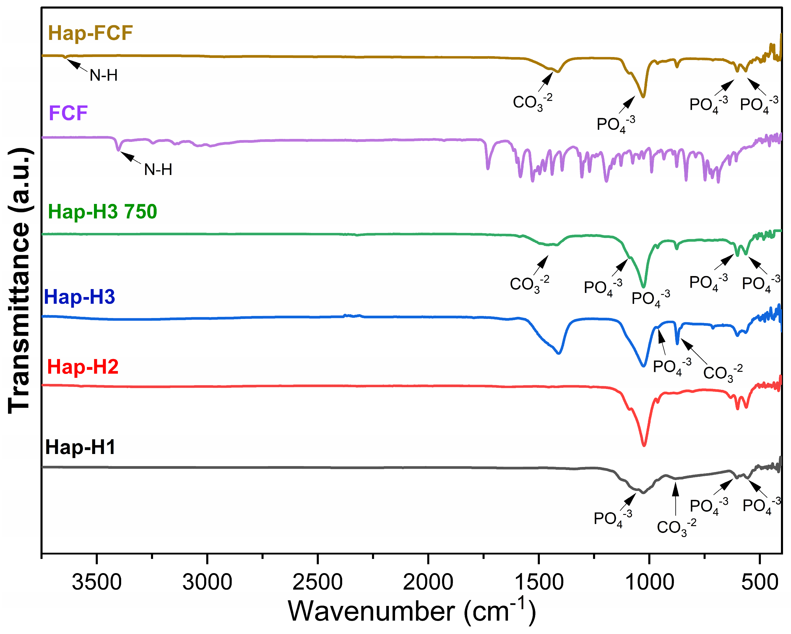
Figure 2 displays the FTIR spectra of the Hap series (Hap-H1, Hap-H2, Hap-H3, and Hap-H3 750), FCF, and the Hap-FCF composite.
For the Hap series samples, the FTIR spectra reveal bands associated with the carbonate (CO32−) and phosphate (PO43−) groups, characteristic of hydroxyapatite [31]. The bands linked to the ν3 vibration mode, attributed to O-C-O bonds, are observed at 874 cm−1. Additionally, a band at 1413 cm−1 appears due to the C-O bond vibration in the ν3 mode of carbonates (CO32−). Bands corresponding to the ν4 vibration mode of the O-P-O bond are evident at 563 and 601 cm−1. The P-O bond’s symmetric ν1 and ν3 vibration modes are detected at 960 cm−1 and 1021 cm−1, respectively, while the antisymmetric ν3 degenerate mode is observed at 1090 cm−1 [32,33,34,35]. These findings provide strong reassurance of the successful synthesis of hydroxyapatite in all cases, validating our research.
In the FCF spectrum, a peak at 3409 cm−1 corresponds to the N-H stretching, a characteristic feature of the FCF structure, as reported by Ren et al. [36]. In the Hap-FCF composite, the precise detection of the N-H bond band of FCF at 3641 cm−1, alongside the characteristic bands of hydroxyapatite, further validates the presence of both components within the composite, demonstrating the precision of our research.

3.1.3. Specific Surface Area (BET)

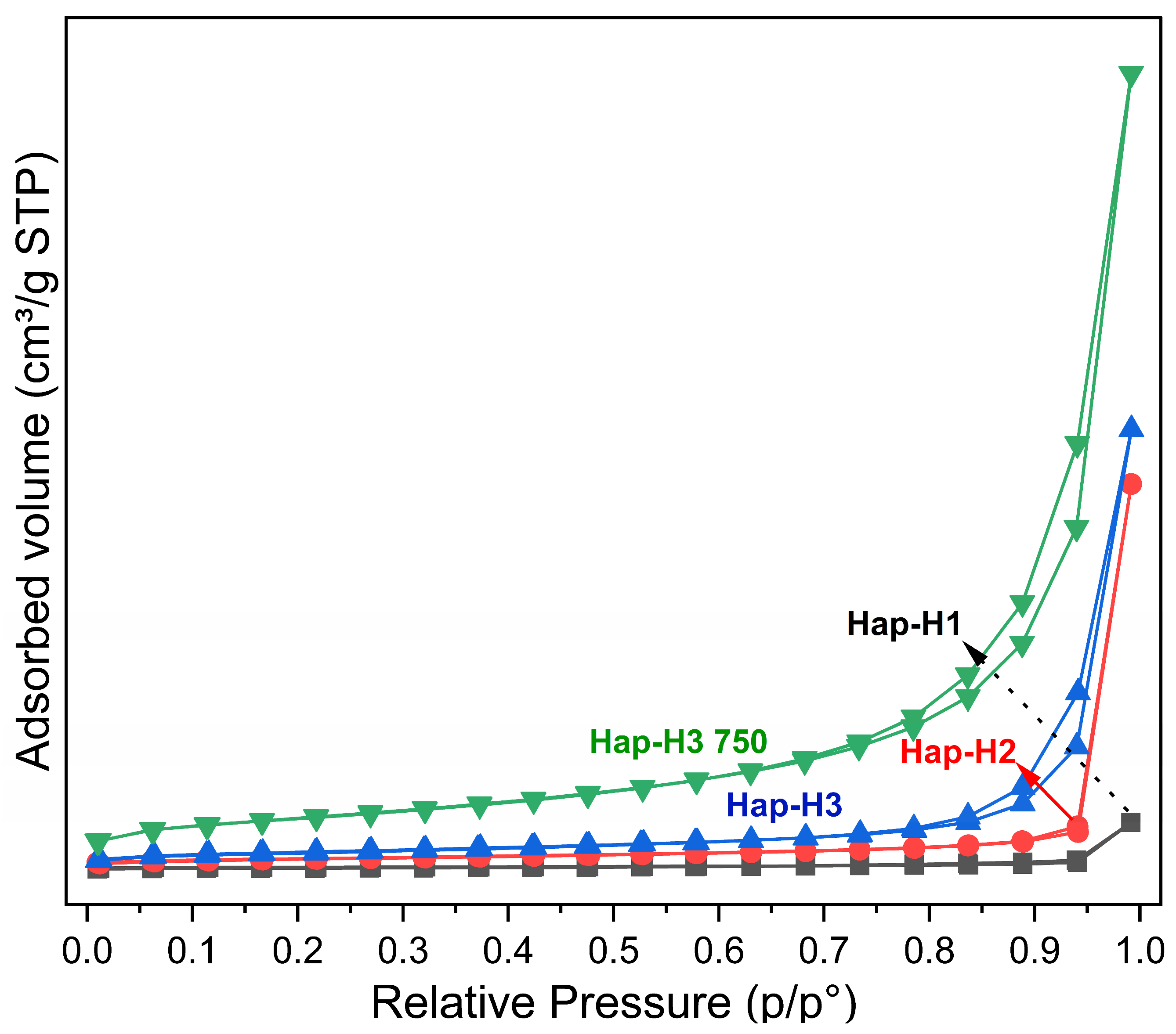
Figure 3 shows the specific surface area for the Hap series samples (Hap-H1, Hap-H2, Hap-H3, and Hap-H3 750), calculated from N2 adsorption isotherms using the BET (Brunauer–Emmett–Teller) method.
The N2 adsorption–desorption isotherms for the Hap series samples are depicted in Figure 3. The isotherms reveal that all samples are characterized as type IV, indicating initial monolayer formation within the pores followed by multilayer formation at higher relative pressures, typical of mesoporous materials [11]. The Hap series samples also display H3-type hysteresis, commonly associated with plate-like pores within a pore size range from 2 to 50 nm [37]. Specific surface area measurements obtained via the BET method show the highest value for the Hap-H3 750 sample at 128.41 m2/g, followed by Hap-H3 (41.46 m2/g), Hap-H2 (24.72 m2/g), and the lowest for Hap-H1 (4.51 m2/g).
Pore volume and pore diameter, calculated using the BJH (Barrett–Joyner–Halenda) model, are summarized in Table 1. The largest pore diameter was observed in the Hap-H2 sample (36.73 nm) with a pore volume of 0.41 cm3/g, followed by Hap-H1 (35.16 nm) with a pore volume of 0.05 cm3/g, Hap-H3 (31.63 nm) with a pore volume of 0.47 cm3/g, and finally, Hap-H3 750 (20.95 nm) with the highest pore volume of 0.86 cm3/g, contributing to its increased specific surface area.
Notably, the only difference between Hap-H3 and Hap-H3 750 lies in the thermal treatment applied to Hap-H3 750 after synthesis. Consequently, based on the X-ray diffraction results and the (211) peak (Figure 1), a reordering effect is observed in Hap-H3 750, leading to an increase in crystal size. As a result, pore diameter decreased while pore volume increased, enhancing the specific surface area from 41.46 m2/g in Hap-H3 to 128.41 m2/g in Hap-H3 750.

3.1.4. Zeta Potential (ZP)

The zeta potential of each Hap sample was measured to assess the stability of the Hap dispersion in water. The results, shown in Table 2, indicate that the synthesized Hap samples exhibit low stability in water as they precipitate rapidly. The measured zeta potential values were −18.6 mV for Hap-H1, −19.1 mV for Hap-H2, −21.9 mV for Hap-H3, −22.6 mV for Hap-H3 750, and −22.8 mV for Hap-FCF. This finding aligns with de la Vega et al. [38], who also reported the poor aqueous stability of hydroxyapatite. Generally, smaller Hap particles tend to exhibit higher stability in aqueous media; however, a suspension is typically considered stable only when zeta potential values exceed ± 30 mV [39].

3.1.5. Scanning Electron Microscopy (SEM)

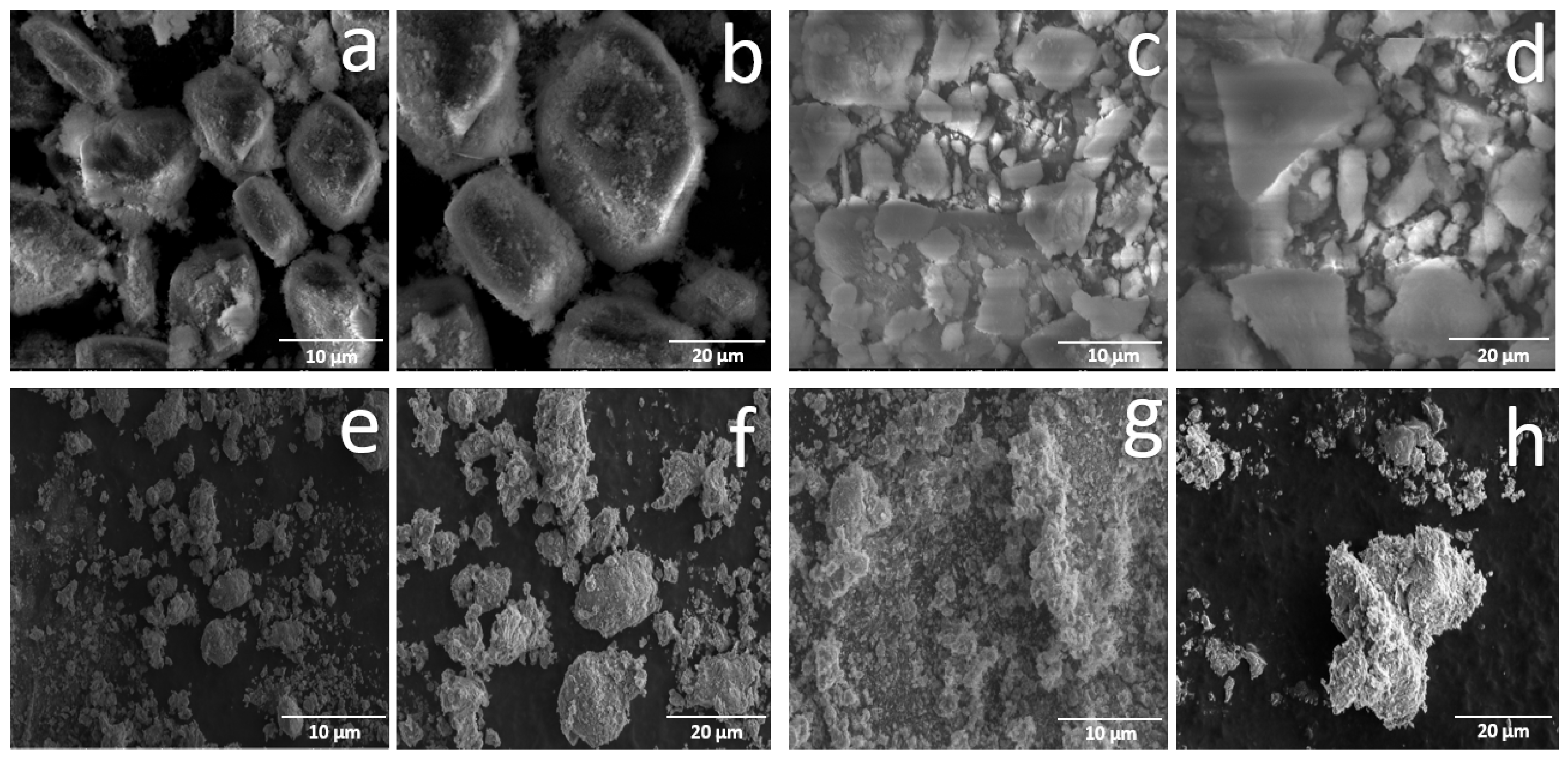
Figure 4 shows the scanning electron microscopy (SEM) micrographs of the Hap series at 500X and 1500X magnifications.
The sample synthesized using a manually ground calcium precursor (Hap-H1) at 500X magnification (a) reveals monolithic agglomerates of varying sizes and shapes, displaying more uniformity compared to other samples (c, e, and g). At 1500X magnification (b), these monolithic agglomerates are observed to be covered by clusters of smaller particles.
For the Hap-H2 sample, generated using a calcium precursor processed through high-energy milling, agglomerates of diverse sizes and shapes are observed, with a more pronounced level of heterogeneity than in the Hap-H1 sample (b). At 1500X magnification (d), these agglomerates are observed to be sparsely covered by clusters of smaller particles, further highlighting the unique characteristics of this sample.
When the calcium precursor was sieved to 8 μ m , the resulting hydroxyapatite sample (Hap-H3) exhibited laminar monoliths of varying sizes and shapes. The agglomerates observed are smaller than the Hap-H1 and Hap-H2 samples (e). The 1500X magnification micrograph provides a closer view of the size heterogeneity of these agglomerates, where smaller particles predominate. Upon subjecting this calcium precursor to a crucial heat treatment at 750 °C, the resulting sample (Hap-H3 750) exhibits a notable reduction in laminar monolith size, with enhanced disaggregation and size uniformity compared to its counterpart, Hap-H3 (g and h).

3.1.6. Bright-Field Transmission Electron Microscopy (BFTEM)

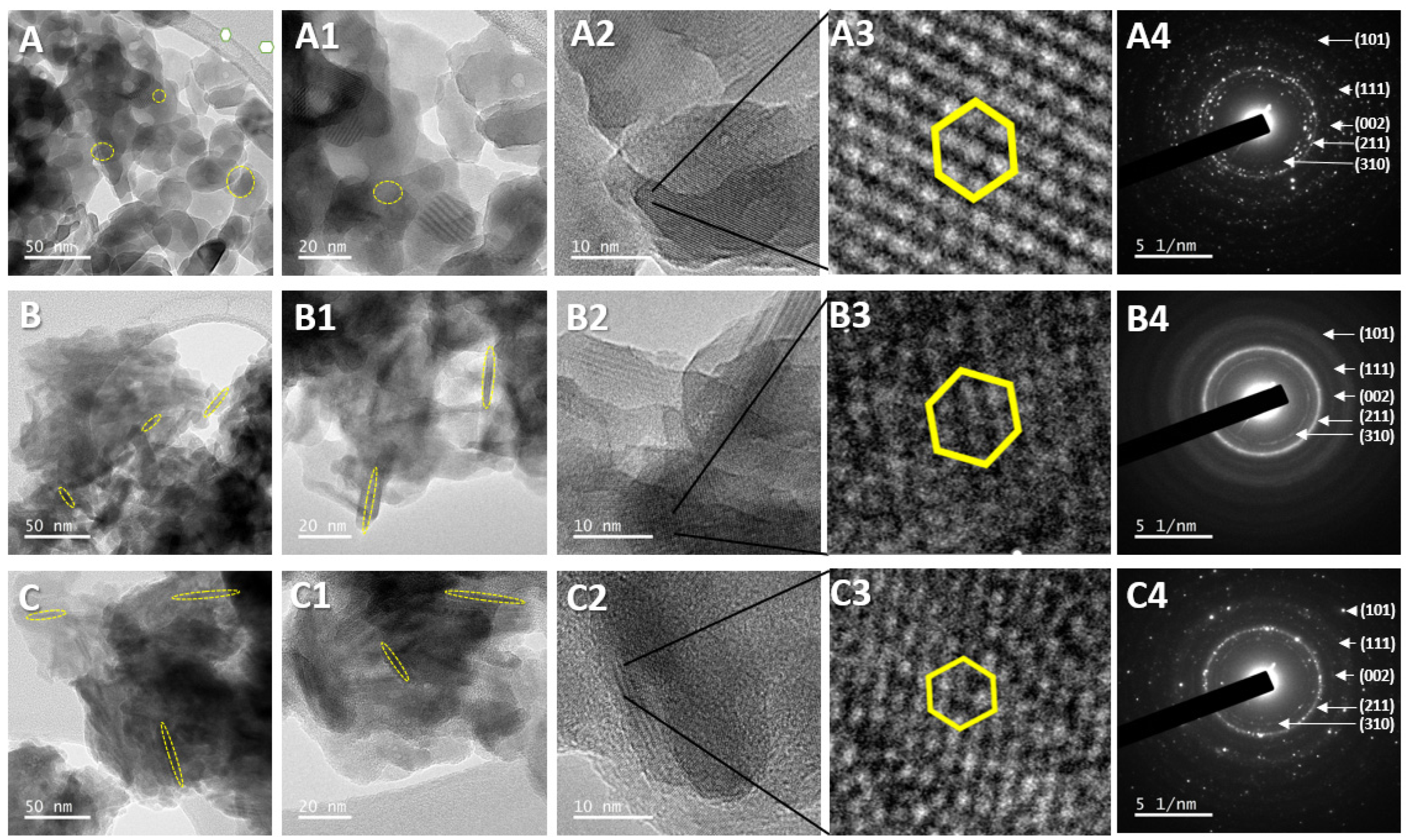
Figure 5 presents the bright field transmission electron microscopy (BFTEM) micrographs for Hap-H3, Hap-H3 750, and Hap-FCF samples.
In the Hap-H3 sample, with a scale bar of 50 nm, irregular agglomerates of nanoparticles showing a semispherical morphology are observed (Figure 5A) with a particle size of 41.5 ± 12.5 nm (Figure 5(A1)). In contrast, the Hap-H3 750 sample (Figure 5(B1)), also shown at 50 nm scale, exhibits agglomerates with a needle-like morphology (Figure 5B) measuring 5.2 ± 19.6 nm in width and length, suggests a morphological transformation induced by the thermal treatment.
The sample loaded with forchlorfenuron (Hap-FCF) at a scale of 50 nm reveals agglomerates that retain the needle-like morphology observed in Hap-H3 750 (Figure 5C). The addition of the phytohormone increases the needle length from 5.2 ± 19.6 nm to 6.1 ± 31.2 nm (Figure 5(C1)). According to the XRD (Figure 1), the Hap-FCF sample exhibits a 10° shift to the right in the harmonic signals, accompanied by a reduction in crystallinity. The diffraction signals predominantly display hydroxyapatite, suggesting that FCF is incorporated within the Hap structure, promoting elongation of the needles. The 5X digital zoom for all samples (Figure 5(A3–C3)) confirms the compact hexagonal crystal lattice (highlighted in yellow), which primarily corresponds to hydroxyapatite, as reported in other studies [40] and identified in Figure 1, according to JPCDS No. 09-0432.
The ring diffraction patterns in Figure 5(A4–C4) indicate the polycrystalline nature of the hydroxyapatite nanoparticles in the Hap-H3 and Hap-H3 750 samples, as well as the polycrystalline composite of hydroxyapatite and forchlorfenuron (Hap-FCF). Each diffraction ring corresponds to a family of crystal planes, representing an interplanar distance [41] characteristic of the compact hexagonal crystal structure. Incorporating the phytohormone, as evidenced by XRD (Figure 1), results in reduced crystallinity, confirmed by the decreased definition in the ring pattern (Figure 5(C4)).

3.2. In Silico Evaluation Results

Computational tools have significantly advanced the prediction of interactions and chemical stability. Thus, a simulation model was developed to predict the interaction between forchlorfenuron (FCF), gibberellin receptors (GA3Ox2), and auxin receptors (IAA7). This simulation model served as the in silico evaluation. Additionally, we meticulously designed an in vitro growth model using the germination of lettuce seeds (Lactuca sativa) to corroborate the stimulation of these receptors, ensuring the reliability of our findings.

3.2.1. Docking Between GA3Ox2 and the Ligands GA3 and FCF

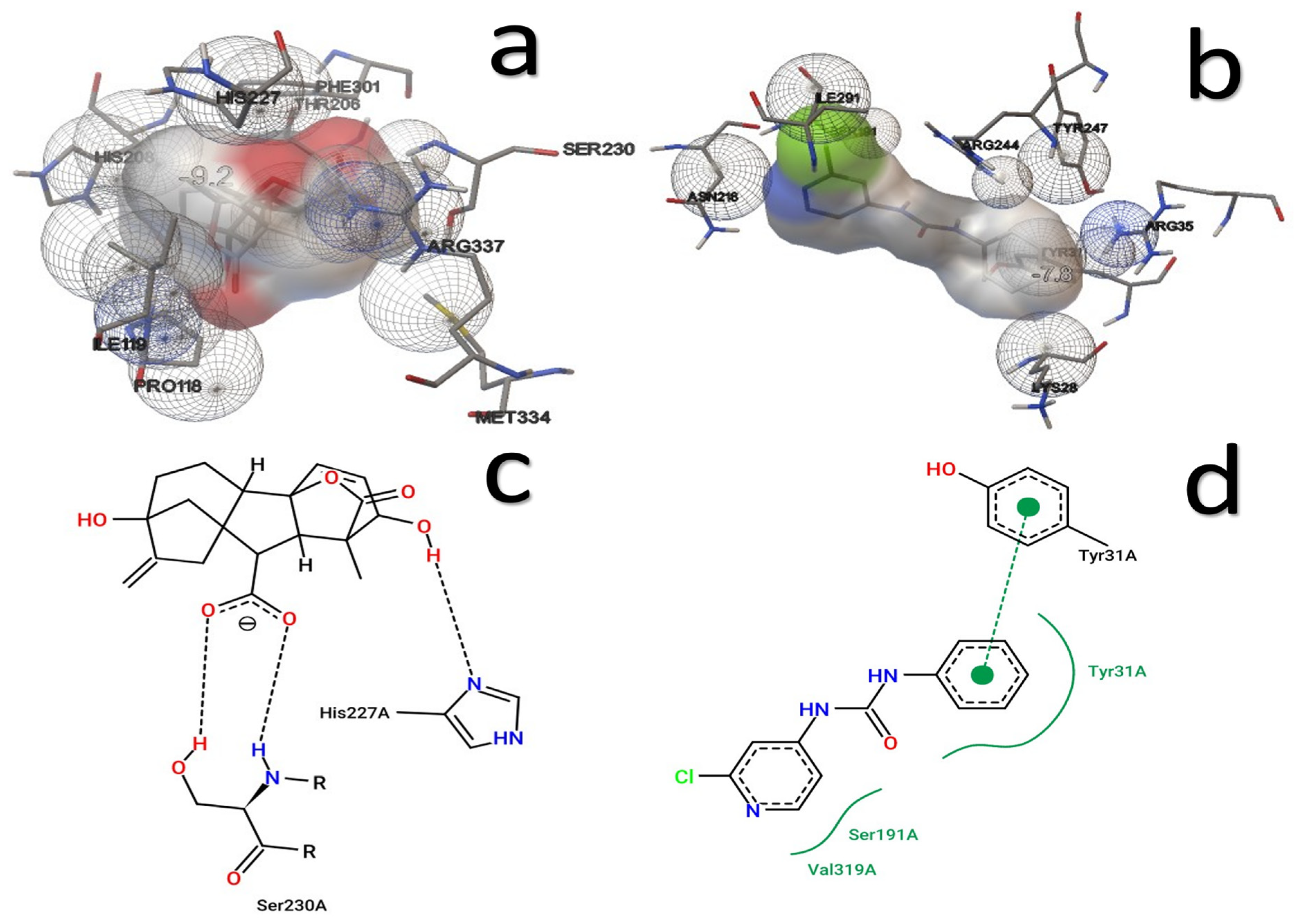
The binding affinity between GA3Ox2 and its endogenous ligand GA3 (GA3Ox2-GA3) was calculated at −9.2 kcal/mol, with interactions involving amino acids HIS208, HIS227, PHE310, THR206, SER230, ARG337, ILE119, PRO118, and MET334. In contrast, the docking analysis between the GA3Ox2 receptor and the ligand FCF revealed a binding energy of −7.8 kcal/mol, interacting with residues ASN210, ARG295, ASP229, HIS227, ILE119, LEU224, PHE301, SER297, TYR212, and VAL236. Figure 6 shows the 3D interaction maps of the gibberellin receptor (GA3Ox2) with its endogenous ligand GA3 (a) and with FCF (b) alongside the respective 2D interaction maps (c,d).
These findings strongly support the potential of the gibberellin receptor to bind with FCF, as evidenced by the observed interactions. To ensure the reliability of these results, we conducted an in-depth molecular dynamics study, which confirmed the stability of the interaction over time and reinforced the robustness of our conclusions.

3.2.2. Docking Between IAA7 and the Ligands IAA and FCF

The binding energy for the interaction between the auxin receptor IAA7 and its endogenous ligand IAA (IAA7-IAA) was −5.0 kcal/mol, involving interactions with amino acids ARG12, LYS13, VAL8, and ASN10. Conversely, docking analysis between IAA7 and FCF revealed a binding energy of −5.4 kcal/mol involving residues ARG12, LYS13, PRO6, ASN10, and TYR11. This indicates that IAA7 exhibits a slightly higher affinity for FCF than its endogenous ligand, underscoring the potential of FCF as an alternative ligand.
Figure 7 illustrates the 3D interaction models of the IAA7 receptor with (a) its endogenous ligand IAA and (b) FCF, as well as the corresponding 2D interaction maps (c,d).
These results suggest that FCF has a marginally higher affinity than the endogenous ligand and could potentially modulate the action mechanisms of the IAA7 receptor. Therefore, it is proposed that FCF may be recognized by the auxin (IAA7) and gibberellin (GA3Ox2) receptors, potentially eliciting a response in plants supporting root elongation and vegetative growth.
Table 3 summarizes the affinity energy and the amino acids involved in the interactions between each receptor and its ligands. Notably, amino acids HIS227 and ILE119 demonstrate recurrent interactions with the GA3Ox2 receptor, while ARG12, LYS13, and ASN10 typically interact with the IAA7 receptor, highlighted in bold.
As Foster et al. [42] noted, the gibberellin receptor (GA3Ox2) stimulates cell elongation, stem and root growth, seed germination, and dwarf plant development. The interaction with its endogenous ligand GA3 shows a higher affinity energy (−9.2 kcal/mol) than FCF (−7.8 kcal/mol), suggesting that while FCF binds with moderate affinity, it could still activate the receptor. Two of the interacting amino acids are shared between GA3 and FCF (bold in Table 3).
In contrast, the auxin receptor (IAA7) is associated with polar transport, callus formation, root initiation, delayed leaf abscission, and parthenocarpy induction [42]. The similar binding energies for the endogenous ligand and FCF (−5.0 and −5.4 kcal/mol, respectively) imply that FCF could effectively bind to IAA7, sharing three of the five essential interacting amino acids.
FCF shows molecular affinity for both GA3Ox2 and IAA7 receptors, suggesting its binding to the protein-ligand complex. A molecular dynamics study has been conducted to confirm the stability of the interaction over time.

3.2.3. Molecular Dynamics (MD)

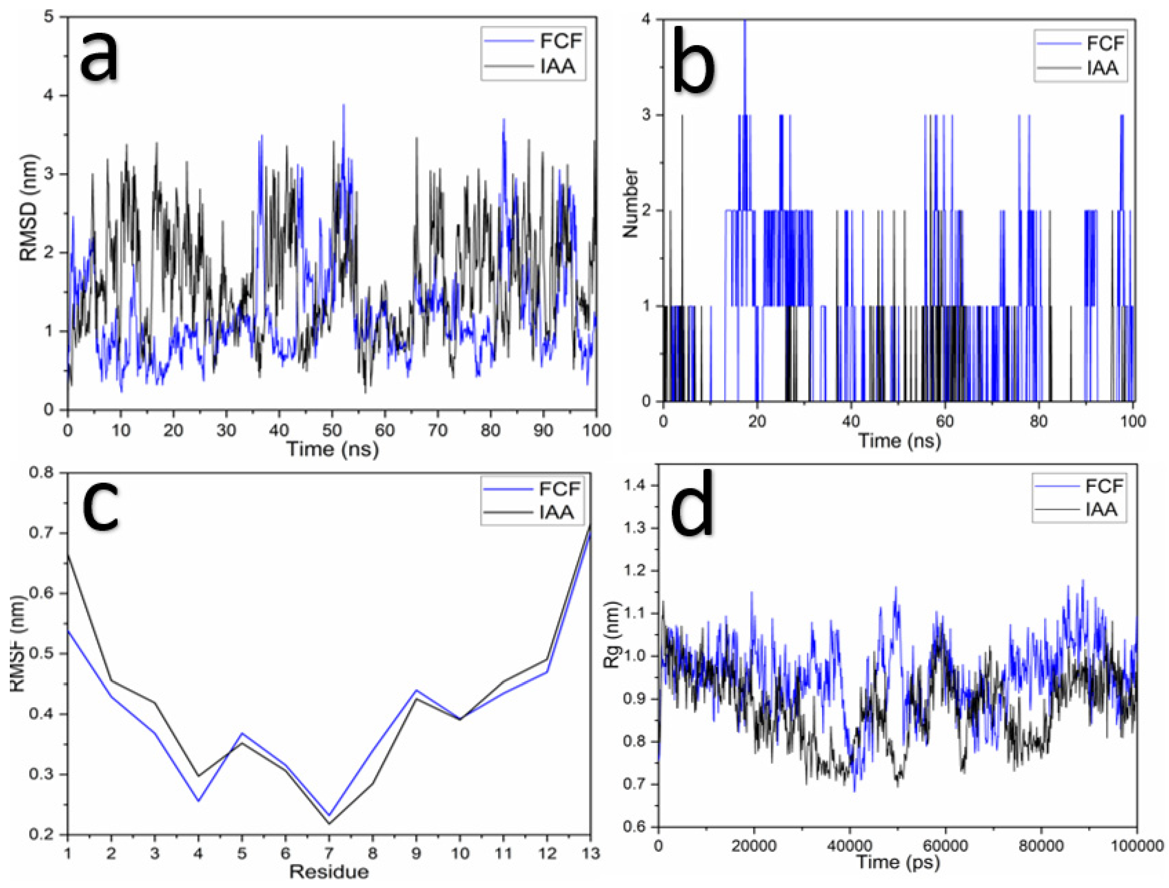
Figure 8 presents the results from the molecular dynamics (MD) simulations, showcasing the Root–Mean–Square Deviation (RMSD) between the GA3Ox2 receptor and the ligands GA3 and FCF (Figure 8a). Additionally, the figure illustrates hydrogen bond interactions (Figure 8b), Root–Mean–Square Fluctuation (RMSF; Figure 8c), and the Radius of Gyration (Rg; Figure 8d).
MD simulations confirmed the stability of the interaction between the GA3Ox2 receptor and the ligands GA3 and FCF. The RMSD values for both receptor–ligand complexes remained under 1 nm, indicating the strong affinity and stability of the interactions over time. Additionally, four hydrogen bonds for FCF and seven for GA3 were observed, providing notable stability at the binding site for both ligands. The RMSF analysis, which evaluates the motion of protein chain atoms, indicated that the region with the highest fluctuation corresponds to FCF, while GA3 exhibited more consistent behavior. The Rg values (Figure 8d) show a minimal variation of 0.04 nm, with ranges from 2.0 to 2.04 nm for GA3 and 2.09 to 2.13 nm for FCF, further confirming structural stability relative to the center of mass.
Figure 9 displays the MD simulation results for the auxin receptor (IAA7). The RMSD between the IAA7 receptor and the ligands IAA and FCF is shown in Figure 9a, hydrogen bond interactions in Figure 9b, RMSF in Figure 9c, and Rg in Figure 9d.
RMSD analysis between the IAA7 receptor and the ligands (IAA and FCF) suggests comparable stability over time, indicating that FCF binds similarly to IAA. A maximum of four hydrogen bonds for FCF and three for IAA were observed, which aids in stabilizing the docking (Figure 9b). The RMSF results show that the regions of fluctuation for both FCF and IAA are similar. The Rg values indicate a variation of 0.5 nm (ranging from 0.7 to 1.2 nm) for both ligands, further confirming structural stability during the simulation period.
FCF is a synthetic plant growth regulator, and although it is known that it plays this role in plants, it is not known how it interacts with them. On the other hand, natural phytohormones such as gibberellin 3A (GA3) and indole acetic acid (IAA) are known to play an important role in plant cell development and growth, acting together to achieve plant development. Therefore, it is interesting to investigate whether other molecules can stimulate these receptors and have positive impacts.
DM studies simulate the basic environmental conditions in a living system; in this case a solvation box was used containing water and ions, specifically 0.15 M KCl and 0.15 M NaCl, plus the protein (GA3Ox2 or IAA7), and the ligand (GA3, IAA or FCF). The entire system was run at room temperature (298.15 Kelvin grades).
In this study, the binding of FCF to both receptors (GA3Ox2 and IAA7) was evaluated, and we found that FCF has a greater stability over time when bound to gibberellin receptors; this implies that FCF is recognized by GA3Ox2 receptors and remains bound to them. This suggests that FCF, although not a natural phytohormone, can stimulate GA3Ox2 receptors and act as a gibberellin agonist, promoting similar effects such as cell elongation.
To determine the responses in the plant, an in vitro assay was carried out using lettuce, where the germination and elongation of the stem cells were evaluated. This trial allowed us to observe that germination and stem cell elongation increase when FCF-doped Hap treatments are used.

3.3. In Vitro Evaluation Results

To validate the findings from the docking and molecular dynamics simulations, lettuce seeds were exposed to FCF, Hap-H3 750, and Hap-FCF for 30 days to gather experimental data on the effects of these materials on plant growth.
Table 4 summarizes the in vitro growth results for lettuce plants (Lactuca sativa) at 10, 20, and 30 days: (a) control (distilled water) and seeds treated with (b) FCF, (c) Hap-H3 750, and (d) Hap-FCF.
The initial growth observed in the seeds is attributed to the nutrient content of the cotyledons, which explains the lack of significant changes during the first few days as germination primarily depends on the seed’s internal reserves (Tyler, 1998) [43]. Seeds in the control and FCF treatments germinated on day 8, while those treated with Hap-H3 750 germinated on day 7, and seeds treated with Hap-FCF germinated on day 6, making Hap-FCF the treatment with the fastest germination rate.
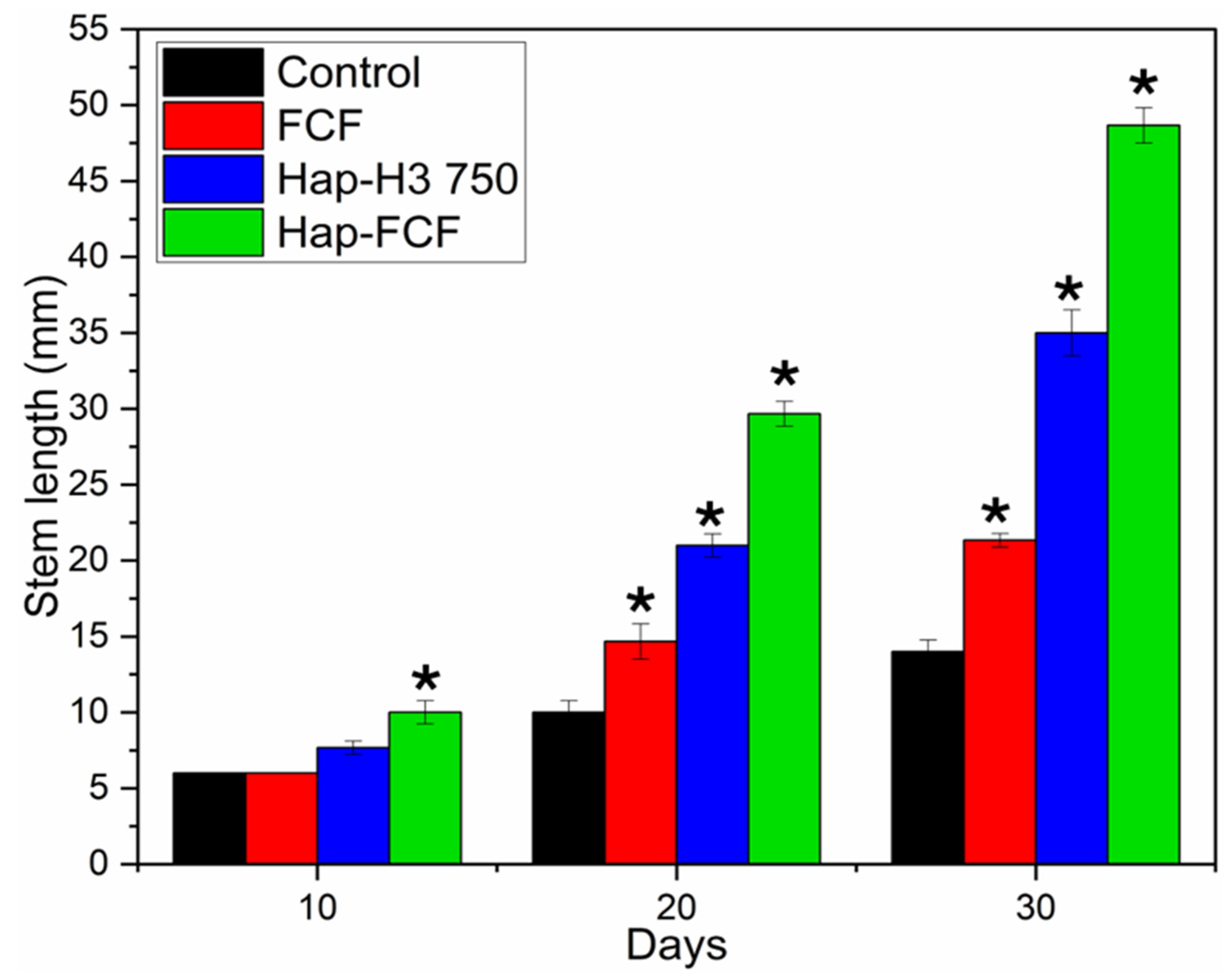
A two-way ANOVA and a post hoc Student–Newman–Keuls (SNK) test were performed. The asterisks in Figure 10 indicate statistically significant differences (* p < 0.05) compared to the control within each time group. At day 10, only the Hap-FCF treatment showed a significant difference relative to the control. At days 20 and 30, all treatments demonstrated a statistically significant increase in stem growth compared to the control, highlighting that the treatments containing forchlorfenuron and hydroxyapatite, especially Hap-FCF, promote plant growth. The statistical analysis supports that FCF, Hap-H3 750, and the composite Hap-FCF significantly enhance stem length. Characterization of the Hap-FCF composite confirms the effective interaction between the two components without new phase formation (XRD). As evidenced by statistical data, TEM analysis indicates increased needle-like structures with reduced crystallinity, contributing to the material’s efficacy.
Figure 11 depicts stem growth at day 30; where the control showed an elongation of 14 mm, FCF-treated plants reached 22 mm, Hap-H3 750-treated plants reached 35 mm, and Hap-FCF-treated plants reached 49 mm.
Plants treated with FCF, Hap-H3 750, and Hap-FCF exhibited more significant stem thickening than the control, with an increase of 15–20%. The findings indicate that Hap-H3 750 and Hap-FCF treatments significantly promote stem growth, increasing stem length by 275–330% relative to the control.
In vitro growth studies with butterhead lettuce demonstrate a cooperative effect between FCF and Hap treatments on stem cell development. This supports the relevance of molecular dynamics (MD) studies, as FCF exhibits effects akin to gibberellic acid (GA3), which accelerates germination and root and stem elongation, as shown in Figure 11. These results align with the docking and MD simulations, suggesting that Hap-FCF can function as a plant growth stimulator like gibberellins. The findings confirm that the gibberellin receptor (GA3Ox2) is effectively stimulated by FCF, with the Hap-FCF composite showing even greater stimulation, as demonstrated by the RMSD analysis (Figure 8a), which indicates the strong stability of FCF with the receptor compared to the reference ligand (GA3).

4. Conclusions

This study highlights that the calcium precursor size significantly influences the hydroxyapatite (Hap) crystal size, and that post-synthesis thermal treatment enhances its crystallinity. FTIR spectroscopy confirmed the successful synthesis of hydroxyapatite across all samples, with the Hap-FCF composite exhibiting an effective interaction between FCF and Hap without forming separate phases.
XRD and BET analyses demonstrated a structural rearrangement in the crystalline network when the Hap-H3 sample was heated to 750 °C, increasing both crystal size and pore volume. SEM and BFTEM microscopy further validated that the precursor size impacts the morphological characteristics of the resulting structures. Docking and molecular dynamics (MD) simulations revealed that FCF binds with a higher affinity to the gibberellin receptor (GA3Ox2) than to the auxin receptor (IAA7), showing stable interactions over the simulation period.
Experimental evidence from stem growth studies confirmed that the stimulation of the GA3Ox2 receptor promotes cell elongation. Treatments with Hap-H3 750 and Hap-FCF resulted in a 330% increase in stem growth compared to the control, demonstrating faster germination rates. These findings suggest that the combined use of hydroxyapatite and forchlorfenuron holds significant promise as a growth-promoting agent in agricultural applications.

Author Contributions

B.I.R.-D.L.R.: Conceptualization, Methodology, Data curation, Formal analysis, Visualization, Investigation, Writing—original draft preparation, Writing—reviewing and editing, Validation. S.P.P.-C.: Conceptualization, Methodology, Writing—reviewing and editing, Supervision. D.E.N.-Á.: Methodology, Data curation, Formal analysis, Visualization, Investigation. J.A.M.-P.: Methodology, Funding acquisition. V.G.-F.: SEM analysis. C.A.C.-O.: XRD analysis. All authors have read and agreed to the published version of the manuscript.

Funding

This research was made possible with the support of the Consejo Nacional de Humanidades, the Ciencias y Tecnologias (CONAHCyT), the IPN-ENCB.ESIQIE (Instituto Politecnico Nacional—Escuela Nacional de Ciencias Biologicas-Escuela Superior de Ingeniería Química e Insdustrias Extractivas), and the doctoral scholarship grant no. A210332.

Institutional Review Board Statement

Not applicable.

Informed Consent Statement

Not applicable.

Data Availability Statement

The original contributions presented in the study are included in the article, further inquiries can be directed to the corresponding author.

Conflicts of Interest

The authors declare no conflicts of interest.

References

  1. Li-Chan, E.C.Y.; Kim, H.O. Structure and Chemical Composition of Eggs; John Wiley & Sons: Hoboken, NJ, USA, 2008. [Google Scholar]
  2. Ravindran, C.; Panayam Parambil Kunnathulli, A.; Kunhikrishnan Maniath, J.; Isloor, A.M. Fabrication of eggshell derived hydroxyapatite polymer composite membrane for efficient removal of thorium ions from aqueous solutions. In Materials Today: Proceedings; Elsevier Ltd.: Amsterdam, The Netherlands, 2020; Volume 43, pp. 2140–2149. [Google Scholar] [CrossRef]
  3. Khandelwal, H.; Prakash, S. Synthesis and Characterization of Hydroxyapatite Powder by Eggshell. J. Miner. Mater. Charact. Eng. 2016, 4, 119–126. [Google Scholar] [CrossRef]
  4. Rivera, E.M.; Araiza, M.; Brostow, W.; Castaño, V.M.; Díaz-Estrada, J.; Hernández, R.; Rodríguez, J. Synthesis of Hydroxyapatite from Eggshells. Mater. Lett. 1999, 41, 128–134. [Google Scholar] [CrossRef]
  5. Juárez Robles, D.E.; Salas Avalos, N.S.; Chávez García, M.D.; García Mejía, T.A. Síntesis de Nano-Hidroxiapatita Por Los Métodos Sol-Gel y Coprecipitación; Universidad Autónoma Metropolitana: Mexico City, Mexico, 2017. [Google Scholar]
  6. de Lima, K.A.; Fernandes, E.F.d.S.; Pacheco, N.I.; Aguiar, E.S.; Lopes, D.C.; Mendes, L.A.P.P.F. Uma breve revisão sobre a hidroxiapatita: Uma biocerâmica promissora. Res. Soc. Dev. 2022, 11, e26411124767. [Google Scholar] [CrossRef]
  7. Repositorio, E.L. Repositorio Institucional Unasam Formato de Autorización para Publicación de Tesis y Trabajos de Investigación, Para a Optar Grados Académicos y Títulos Profesionales. Available online: https://www.google.com.hk/url?sa=t&source=web&rct=j&opi=89978449&url=https://investigacion.unasam.edu.pe/archivos/documentos/publicaciones/norma_9.docx&ved=2ahUKEwiBw-qY-5mKAxWEbvUHHaw8EwIQFnoECB8QAQ&usg=AOvVaw0vG8h9eez_dSQiWKjjmKBh (accessed on 2 December 2024).
  8. Cuesta, G.; Mondaca, E. Effect of an auxin-based bioregulator on growth of tomato seedlings. Rev. Chapingo Ser. Hortic. 2014, 20, 215–222. [Google Scholar] [CrossRef]
  9. Fisiología, Y.; Producción Vegetal, M.E.; Aranda González, P. Pontificia Universidad Católica de Chile Facultad de Agronomia e Ingenieria Forestal Dirección de Postgrado e Investigación. Available online: https://www.biologiachile.cl/biological_research/VOL44_2011/SupA/01_Biological_suplemento_interior.pdf (accessed on 12 November 2024).
  10. Maoz, I.; Bahar, A.; Kaplunov, T.; Zutchi, Y.; Daus, A.; Lurie, S.; Lichter, A. Effect of the cytokinin forchlorfenuron on tannin content of thompson seedless table grapes. Am. J. Enol. Vitic. 2014, 65, 230–237. [Google Scholar] [CrossRef]
  11. Garzón-Pérez, A.S.; Paredes-Carrera, S.P.; Martínez-Gutiérrez, H.; Cayetano-Castro, N.; Sánchez-Ochoa, J.C.; Pérez-Gutiérrez, R.M. Effect of combined microwave-ultrasound irradiation in the structure and morphology of hydrotalcite like compounds al/mg-ch3coo andits evaluation in the sorption of a reactive dye. Rev. Mex. Ing. Quim. 2020, 19, 363–375. [Google Scholar] [CrossRef]
  12. Salinas-Estevané, P.; Sánchez Cervantes, E.M. La Química Verde en la Síntesis de Nanoestructuras. Ingenierías 2012, 15, 7–16. [Google Scholar]
  13. De, U.; De, S.; En, A.; Analítica, R. Evaluación In Silico de las Interacciones Ligando-Receptor de Una Serie de Isoindolinas Derivadas de Aminoácidos y el Receptor del Ácido Gamma Aminobutírico Subtipo A (GABAA); Universidad Veracruzana: Xalapa, Mexico, 2018. [Google Scholar]
  14. Ballón Paucara, W.G.; Grados Torrez, R.E. Acomplamiento Molecular: Criterios Prácticos Para La Selección de Ligandos Biológicamente Activos e Identificación de Nuevos Blancos Terapéuticos. Rev. Con-Cienc. 2019, 7, 55–72. [Google Scholar]
  15. Arredondo-Tamayo, B.; Méndez-Méndez, J.V.; Chanona-Pérez, J.J.; Hernández-Varela, J.D.; González-Victoriano, L.; Gallegos-Cerda, S.D.; Martínez-Mercado, E. Study of Gellan Gum Films Reinforced with Eggshell Nanoparticles for the Elaboration of Eco-Friendly Packaging. Food Struct. 2022, 34, 100297. [Google Scholar] [CrossRef]
  16. Kim, S.; Chen, J.; Cheng, T.; Gindulyte, A.; He, J.; He, S.; Li, Q.; Shoemaker, B.A.; Thiessen, P.A.; Yu, B.; et al. PubChem 2023 update. Nucleic Acids Res. 2023, 51, D1373–D1380. [Google Scholar] [CrossRef]
  17. Berman, H.M.; Westbrook, J.; Feng, Z.; Gilliland, G.; Bhat, T.N.; Weissig, H.; Shindyalov, I.N.; Bourne, P.E. The Protein Data Bank. Nucleic Acids Res. 2000, 28, 235–242. [Google Scholar] [CrossRef] [PubMed]
  18. Hanwell, M.D.; Curtis, D.E.; Lonie, D.C.; Vandermeersch, T.; Zurek, E.; Hutchison, G.R. SOFTWARE Open Access Avogadro: An Advanced Semantic Chemical Editor, Visualization, and Analysis Platform. J. Cheminform. 2012, 4, 17. [Google Scholar] [CrossRef] [PubMed]
  19. Pettersen, E.F.; Goddard, T.D.; Huang, C.C.; Couch, G.S.; Greenblatt, D.M.; Meng, E.C.; Ferrin, T.E. UCSF Chimera—A visualization system for exploratory research and analysis. J. Comput. Chem. 2004, 25, 1605–1612. [Google Scholar] [CrossRef]
  20. Eberhardt, J.; Santos-Martins, D.; Tillack, A.F.; Forli, S. AutoDock Vina 1.2.0: New Docking Methods, Expanded Force Field, and Python Bindings. J. Chem. Inf. Model. 2021, 61, 3891–3898. [Google Scholar] [CrossRef]
  21. Trott, O.; Olson, A.J. AutoDock Vina: Improving the speed and accuracy of docking with a new scoring function, efficient optimization, and multithreading. J. Comput. Chem. 2010, 31, 455–461. [Google Scholar] [CrossRef]
  22. Morris, G.M.; Huey, R.; Lindstrom, W.; Sanner, M.F.; Belew, R.K.; Goodsell, D.S.; Olson, A.J. Software news and updates AutoDock4 and AutoDockTools4: Automated docking with selective receptor flexibility. J. Comput. Chem. 2009, 30, 2785–2791. [Google Scholar] [CrossRef] [PubMed]
  23. Hess, B.; Kutzner, C.; Van Der Spoel, D.; Lindahl, E. GRGMACS 4: Algorithms for highly efficient, load-balanced, and scalable molecular simulation. J. Chem. Theory Comput. 2008, 4, 435–447. [Google Scholar] [CrossRef]
  24. Brooks, B.R.; Brooks, C.L., III; MacKerell, A.D., Jr.; Nilsson, L.; Petrella, R.J.; Roux, B.; Won, Y.; Archontis, G.; Bartels, C.; Boresch, S.; et al. CHARMM: The biomolecular simulation program. J. Comput. Chem. 2009, 30, 1545–1614. [Google Scholar] [CrossRef]
  25. Gao, C.; Zhao, K.; Lin, L.; Wang, J.; Liu, Y.; Zhu, P. Preparation and characterization of biomimetic hydroxyapatite nanocrystals by using partially hydrolyzed keratin as template agent. Nanomaterials 2019, 9, 241. [Google Scholar] [CrossRef]
  26. Lucero, V.; Córdova, Z. Metodología Para La Fabricación de Andamios Porosos de Hidroxiapatita y Fluorapatita Mediante Lixiviación de Sales. Bachelor’s Thesis, Universidad Autónoma de Baja California, Tijuana, Mexico, 2016. [Google Scholar]
  27. AB Martínez-Valencia, H.; Esparza-Ponce, E.; Ortiz-Landeros, J. Caracterización estructural y morfológica de hidroxiapatita nanoestructurada: Estudio comparativo de diferentes métodos de síntesis. Superf. Vacío 2008, 21, 18–21. [Google Scholar]
  28. Prakash, K.H.; Kumar, R.; Ooi, C.P.; Cheang, P.; Khor, K.A. Apparent solubility of hydroxyapatite in aqueous medium and its influence on the morphology of nanocrystallites with precipitation temperature. Langmuir 2006, 22, 11002–11008. [Google Scholar] [CrossRef]
  29. Inoue, Y.; Hirano, A.; Murata, I.; Kobata, K.; Kanamoto, I. Assessment of the Physical Properties of Inclusion Complexes of Forchlorfenuron and γ-Cyclodextrin Derivatives and Their Promotion of Plant Growth. ACS Omega 2018, 3, 13160–13169. [Google Scholar] [CrossRef] [PubMed]
  30. Hernández-Navarro, C.; Díaz-león, J.L.; Louvier-Hernández, J.F.; Vázquez-López, J.A.; García-Bustos, E.; Flores-Flores, T.C.; Quintana-Hernández, P.A. Preparación y Caracterización de Un Composito de Tipo PVA/HAp. Mem. Congr. Nac. Ing. Bioméd. 2018, 5, 310–313. [Google Scholar]
  31. Vargas-Becerril, N.; Sánchez-Téllez, D.A.; Zarazúa-Villalobos, L.; González-García, D.M.; Álvarez-Pérez, M.A.; de León-Escobedo, C.; Téllez-Jurado, L. Structure of biomimetic apatite grown on hydroxyapatite (HA). Ceram. Int. 2020, 46 Part A, 28806–28813. [Google Scholar] [CrossRef]
  32. Zhou, H.; Yang, Y.; Yang, M.; Wang, W.; Bi, Y. Synthesis of mesoporous hydroxyapatite via a vitamin C templating hydrothermal route. Mater. Lett. 2018, 218, 52–55. [Google Scholar] [CrossRef]
  33. Muthu, D.; Kumar, G.S.; Gowri, M.; Prasath, M.; Viswabaskaran, V.; Kattimani, V.; Girija, E. Rapid synthesis of eggshell derived hydroxyapatite with nanoscale characteristics for biomedical applications. Ceram. Int. 2022, 48, 1326–1339. [Google Scholar] [CrossRef]
  34. Han, K.S.; Sathiyaseelan, A.; Saravanakumar, K.; Wang, M.H. Wound healing efficacy of biocompatible hydroxyapatite from bovine bone waste for bone tissue engineering application. J. Environ. Chem. Eng. 2022, 10, 106888. [Google Scholar] [CrossRef]
  35. Gusmão, S.B.S.; Ghosh, A.; de Menezes, A.S.; Pereira, A.F.M.; Lopes, M.T.P.; Souza, M.K.; Dittz, D.; Abreu, G.J.P.; Pinto, L.S.S.; Filho, A.L.M.M.; et al. Nanohydroxyapatite/Titanate Nanotube Composites for Bone Tissue Regeneration. J. Funct. Biomater. 2022, 13, 306. [Google Scholar] [CrossRef]
  36. Ren, Y.; Xiang, P.; Xie, Q.; Yang, H.; Liu, S. Rapid analysis of forchlorfenuron in fruits using molecular complex-based dispersive liquid–liquid microextraction. Food Addit. Contam. Part A Chem. Anal. Control Expo. Risk Assess. 2021, 38, 637–645. [Google Scholar] [CrossRef]
  37. Obregón, D. Estudio Comparativo de la Capacidad de Adsorción de Cadmio Utilizando Carbones Activados Preparados a Partir de Semillas de Aguaje y de Aceituna. Bachelor’s Thesis, Pontifical Catholic University of Peru, San Miguel, Peru, 2012. [Google Scholar] [CrossRef]
  38. Laura de la Vega-García, N.; Beatriz Peña-Valdivia, C.; del Carmen González-Chávez, M.A.; Padilla-Chacón, D.; Carrillo-González, R. Síntesis y Efecto de Nanopartículas de Hidroxiapatita en la Germinación y Crecimiento de Frijol Synthesis and Effect of Hydroxyapatite Nanoparticles on the Germination and Growth of Common Bean. Agrociencia 2020, 54, 1009–1029. [Google Scholar] [CrossRef]
  39. Pamela Agredo Sanín. Caracterización Fisicoquímica de Emulsiones Tipo O/W Estabilizadas Mediante Diferentes Mezclas de Surfactantes Catiónicos Monoméricos Y Poliméricos; Universidad Icesi: Cali, Colombia, 2017; Available online: https://repository.icesi.edu.co/biblioteca_digital/bitstream/10906/83426/1/T01254.pdf (accessed on 25 March 2024).
  40. Venkateswarlu, K.; Bose, A.C.; Rameshbabu, N. X-ray peak broadening studies of nanocrystalline hydroxyapatite by WilliamsonHall analysis. Phys. B Condens. Matter. 2010, 405, 4256–4261. [Google Scholar] [CrossRef]
  41. Paraguay-Delgado, F. Técnicas de microscopía electrónica usadas en el estudio de nanopartículas. Mundo Nano Rev. Interdiscip. Nanocienc. Nanotecnol. 2020, 13, 101–131. [Google Scholar] [CrossRef]
  42. Foster, A. Practical_Plant_Anatomy; Van Nostrand: New York City, NY, USA, 1949; pp. 142–143. [Google Scholar]
  43. Tyler, G.; Zohlen, A. Plant Seeds as Mineral Nutrient Resource for Seedlings-A Comparison of Plants from Calcareous and Silicate Soils. Ann. Bot. 1998, 81, 455–459. [Google Scholar] [CrossRef]
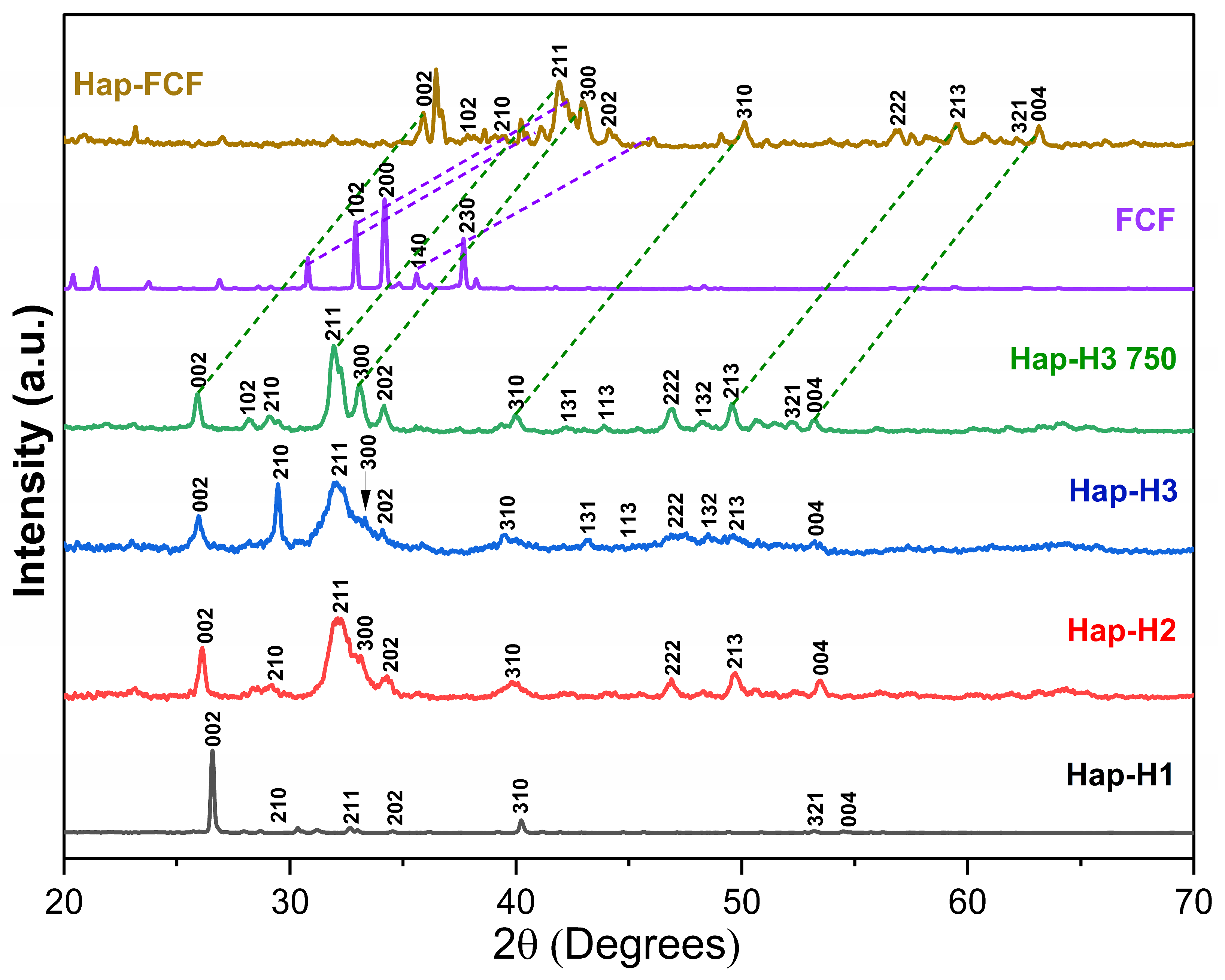
Figure 1. XRD patterns of the samples Hap, FCF, and Hap-FCF composite.
Applsci 14 11522 g001
Figure 2. FTIR spectrum analysis of the samples: Hap series, FCF, and Hap-FCF composite.
Applsci 14 11522 g002
Figure 3. N2 adsorption–desorption isotherms for the Hap series samples.
Applsci 14 11522 g003
Figure 4. SEM micrographs at 500X (10 μ m ) and 1500X (20 μ m ) for Hap series samples: Hap-H1 (a,b), Hap-H2 (c,d), Hap-H3 (e,f), and Hap-H3 750 (g,h).
Applsci 14 11522 g004
Figure 5. Bright field micrographs captured using transmission electron microscopy (BFTEM) for the samples Hap-H3 (AA3), Hap-H3 750 (BB3), Hap-FCF (CC3), and their ring diffraction patterns for the samples Hap-H3 (A4), Hap-H3 750 (B4), Hap-FCF (C4) [40].
Applsci 14 11522 g005
Figure 6. Docking results with the GA3Ox2 receptor. Interaction spheres with (a) GA3 and (b) FCF, and 2D interaction maps with (c) GA3 and (d) FCF.
Applsci 14 11522 g006
Figure 7. Docking analysis with the IAA7 receptor. Interaction spheres with (a) IAA and (b) FCF, and 2D interaction maps with (c) IAA and (d) FCF.
Applsci 14 11522 g007
Figure 8. Molecular dynamics results for GA3Ox2 and gibberellic acid GA3 (endogenous ligand, black line) and FCF (blue line); (a) RMSD, (b) Hydrogen bonds, (c) RMS fluctuation, and (d) Radius of gyration.
Applsci 14 11522 g008
Figure 9. Molecular dynamics results for IAA7 and IAA (endogenous ligand, black line) and FCF (blue line); (a) RMSD LIG after alignment to the backbone, (b) hydrogen bonds, (c) RMS fluctuation, and (d) radius of gyration.
Applsci 14 11522 g009
Figure 10. Stem length of Lactuca sativa at 10, 20, and 30 days of growth. Control (black), FCF (red), Hap-H3 750 (blue), and Hap-FCF (green). Bars represent mean and standard error (SE). Significant (p < 0.05) versus control treatment per day group. ANOVA, SNK test (* = significant).
Applsci 14 11522 g010
Figure 11. Stem length of fertilized lettuce plants after 30 days of growth: (a) control, (b) FCF, (c) Hap-H3 750, and (d) Hap-FCF.
Applsci 14 11522 g011
Table 1. Physisorption Isotherms of Hap Samples.
ID SampleSBET (m2/g)Pore Volume
BJH (cm3/g)
Pore Diameter
BJH (nm)
Hap-H14.510.0535.16
Hap-H224.720.4136.73
Hap-H341.460.4731.63
Hap-H3 750128.410.8620.95
SBET: The specific surface area calculated using the BET method.
Table 2. Average Zeta Potential (ZP) of the Hap Series and Hap-FCF Composite.
ID SampleZP (mV)
Hap-H1−18.6
Hap-H2−19.1
Hap-H3−21.9
Hap-H3 750−22.6
Hap-FCF−22.8
Table 3. Affinity energy and amino acid of interactions from docking GA3Ox2 and IAA7.
ReceptorLigandAffinity Energy
(kcal/mol)
Amino Acid of
Interaction
GA3Ox2GA3−9.2HIS208, HIS227, PHE310, THR206, SER230, ARG337, ILE119, PRO118, MET334
FCF−7.8ASN210, ARG295, ASP229, HIS227, ILE119, LEU224, PHE301, SER297, TYR212, VAL236
IAA7IAA−5.0ARG12, LYS13, PRO6, ASN10, TYR11
FCF−5.4ARG12, LYS13, VAL8, ASN10
Note: Endogenous ligands are shaded gray, and repeated amino acids in interactions are highlighted in bold.
Table 4. Growth assessment of lettuce plants at 10, 20, and 30 days treated with: (a) control, (b) FCF, (c) Hap-H3 750, and (d) Hap-FCF.
DayControlFCFHAP-H3 750Hap-FCF
0Applsci 14 11522 i001Applsci 14 11522 i002Applsci 14 11522 i003Applsci 14 11522 i004
10Applsci 14 11522 i005Applsci 14 11522 i006Applsci 14 11522 i007Applsci 14 11522 i008
20Applsci 14 11522 i009Applsci 14 11522 i010Applsci 14 11522 i011Applsci 14 11522 i012
30Applsci 14 11522 i013Applsci 14 11522 i014Applsci 14 11522 i015Applsci 14 11522 i016
Disclaimer/Publisher’s Note: The statements, opinions and data contained in all publications are solely those of the individual author(s) and contributor(s) and not of MDPI and/or the editor(s). MDPI and/or the editor(s) disclaim responsibility for any injury to people or property resulting from any ideas, methods, instructions or products referred to in the content.