Article

Color Changes of a Heat-Polymerized Polymethyl-Methacrylate (PMMA) and Two 3D-Printed Denture Base Resin Materials Exposed to Staining Solutions: An In Vitro Spectrophotometric Study

by
Vasileios Vergos
1,*,
Theodore Ganetsos
2,
Antreas Kantaros
2,*,
Antonios Theocharopoulos
1 and
Stavros Yannikakis
1
1
Department of Biomedical Sciences, University of West Attica, 12244 Athens, Greece
2
Department of Industrial Design and Production Engineering, University of West Attica, 12244 Athens, Greece
*
Authors to whom correspondence should be addressed.
Appl. Sci. 2024, 14(24), 11548; https://doi.org/10.3390/app142411548
Submission received: 16 October 2024 / Revised: 3 December 2024 / Accepted: 4 December 2024 / Published: 11 December 2024
(This article belongs to the Section Additive Manufacturing Technologies)

Abstract

:

Featured Application

The use of 3D printing technology in denture fabrication presents significant advancements over traditional methods. This technique allows for precise, customizable, and efficient manufacturing of denture bases, addressing key issues such as production time and cost. Recent studies focus on enhancing the properties of 3D-printed denture materials, such as their color stability, mechanical strength, and biocompatibility. These developments render 3D printing a critical tool for integrating advanced materials into routine dental practice, ultimately improving patient outcomes and the aesthetic longevity of dentures.

Abstract

Newly developed 3D-printed polymer materials are used for denture base fabrication. The aim of the present study was to evaluate the color stability of two new 3D-printed resins, a hard PPMA-based and a soft Urethane-based resin, in relation to a traditional heat-polymerized PMMA resin, which was used for comparison purposes. Specimens of the materials were immersed in five solutions (distilled water, red wine, black tea, coffee, and Coke®) for definite periods of time (one day, one week, and one month). The color measurements were carried out utilizing a spectrometer supported by a microscope and using special software. Color changes between immersion periods were calculated and statistically compared. The results showed that all types of resins were influenced during immersion periods. The heat-polymerized resin was influenced less than the others but with no significant difference to the 3D-printed hard PMMA resin. In respect to the materials compared, the discoloration effect for the 1 month immersion time was significantly more intense for the soft 3D-printed resin. In respect to the solutions’ staining effects, black tea and red wine significantly discolored all materials regardless of immersion periods. The new 3D-printed materials need further improvements for dental use.

1. Introduction

Complete dentures currently remain the main treatment of choice for edentulous patients, with heat-polymerizing PMMA (Polymethyl-methacrylate resin) serving as the fabrication material of choice since its’ introduction in 1936. In its improved form, PMMA presents low cost, good mechanical properties, almost natural appearance, easy handling, acceptable stability in the oral environment, and good, evidence-based, long-term performance. Nevertheless, it also presents some limitations, such as contact chemical irritation or allergic reactions, with consequent cytotoxicity, noteworthy polymerization shrinkage, moisture absorption, porosity, and possible discoloration. Additionally, a large number of clinical and laboratory sessions are required for the completion of a denture [1].
Three-dimensional printing, also known as additive manufacturing, has become a transformative technology across various fields, mainly due to its ability to fabricate complex, precise structures in a layer-by-layer way, unlocking a wealth of new possibilities. Cultural heritage preservation, archaeology, and museums are some relatively new application areas [2]. Exciting possibilities in healthcare and biomedicine are also now seen, ranging from prosthetics to regenerative medicine [3,4,5] and from surgical planning and education [6] to biomaterial development [7]. In the prosthetics field, and particularly in dentistry, it facilitates the rapid production of dental implants, crowns, and dentures with high precision, significantly reducing manufacturing time and costs [1,8,9].
PMMA-modified resins have been introduced for fabricating 3D-printed complete dentures, along with other polymer materials like urethane methacrylate polymers, styrene polymers, and polyesters [10,11,12,13]. The main type of these resins is PMMA resins, which are of similar flexural strength to heat-polymerizing resins. There are also some “Soft” resins for fabricating “flexible” dentures [14,15]. This type of material, used to treat some special cases, is based in urethane dimethacrylate (UDMA) or other molecules. It presents good dimensional stability and a lack of residual monomers, reducing the risk of chemical irritation or allergies [13,15]. There is a growing research trend aimed at improving 3D printing techniques and materials. Critical parameters that have to be improved for successful clinical performance are mechanical properties, biocompatibility, water absorption and solubility, and optical properties [16].
Color stability is crucial for esthetic reasons because its absence promotes patient dissatisfaction and reflects possible structural modification [17]. Intrinsic material features such as resin type, composition, polymerization mode, and aging, as well as oral environmental aspects, including food, beverages, wine, nicotine, and poor oral hygiene or use of denture cleaning agents, can influence the materials’ color stability. There are also specific denture-related parameters, such as surface abrasion, surface roughness, and water absorption [18].
Few studies compare traditional heat-polymerized resins with newer, 3D-printed materials for staining evaluation after exposure to different coloring media [12,19,20,21,22]. In most studies, specimens of the tested materials are immersed for definite periods of time in various potentially staining media (coffee, red wine, tea, Coke®, cleaning agents, etc.) in order to simulate the influence of daily use by the patient (references). According to the authors’ knowledge, no study has yet evaluated the soft urethane-based resins in such protocols.
The aim of the present in vitro study was to evaluate the influence of immersion time and coloring solution on the color stability of three denture base resins via spectrophotometry. The null hypothesis was that there is no difference in the color stability of the three resins immersed in five staining solutions for three immersion periods.

2. Materials and Methods

2.1. Resin Disc Specimen Production

Ninety (90) disc-shaped specimens (3.00 mm thickness, 10.0 mm diameter) were prepared using three different denture base polymer materials (see Table 1 for technical details) following the protocols described below.
Heat-polymerized resin specimens: Thirty (30) disc-shaped specimens were produced by the traditional technique. A PMMA denture base resin material (Meliodent, Kulzer Gmbh, Hanau, Germany) was used according to the manufacturer’s instructions. After mixing, it was packed in metal flasks into dental plaster disc-shaped molds, and a heat curing protocol was performed. The specimens were then de-flasked and prepared according to a standard preparation procedure, as described below.
Three-dimensional-printed specimens: A disc-shaped specimen was designed by dedicated 3D printer software (Asiga Composer Software, 2.0, ASIGA Co., Alexandria, Australia) using the proper dimensions (10 mm diameter, 3 mm thickness). An STL file was produced and sent to a DLP-type 3D printer unit (ASIGA Max UV-385, ASIGA Co., Alexandria, Australia) and thirty (30) specimens of each resin (hard and soft; see Table 1) were printed, (layer thickness: 75 μm; orientation: 0°; light intensity: 6.3 mW/cm2; exposure time: 31.00 s). After polymerizing, the specimens were removed from the platform and cleaned with isopropyl alcohol liquid (99.9% purity) in a special washing device for 10 min (Wash and Cure machine 3.0, Anycubic Technology Ltd., Shenzhen, China). After drying with a clean white absorbent paper, a post-curing process was followed for 15 min using a polymerization unit for achieving full polymerization (ASIGA FLASH CURE BOX, ASIGA Co., Alexandria, Australia) for both resins, according to the manufacturer’s recommendations.
All specimens were finished by grinding progressively using 600-grit, 800-grit, and 1200-grit waterproof silicon carbide paper, using a polishing device (Ecomet, Buehler Ltd., Lake Bluff, IL, USA). Final polishing was performed using a high-gloss agent (KMG, CANDULOR AG, Clattpark (Opfikon), Switzerland) on a white cotton yam wheel polishing machine (Tour A Polir MV2, Dentacier, Apelex-Bagneux, France), followed by washing under running water.
All specimens’ dimensions, especially thickness, were verified with a micrometer (measurement accuracy: 0.1 mm) and were stored in distilled water in room temperature (23 °C).
The ninety (90) specimens were randomly separated into five different groups per denture base polymer material. Fifteen subgroups were thus produced, with six specimens in each one (Table 2).

2.2. Staining Solution Preparation

A Greek red wine was used as received, at room temperature (23 °C ± 2). A black tea solution was prepared using three tea bags of black tea, seeped in 100 mL hot water (60 °C) for ten minutes, and the immersion was performed at 45 °C ± 2. The coffee solution was prepared by filtering 15 g of a filter coffee (3 teaspoons) with two cups (500 mL) of boiling water for 10 min. The immersion was performed at 45 °C ± 2. The Coke® was used as received, at room temperature, 23 °C ± 2; see Table 1. Distilled water was also used as the control solution for evaluating the effect of other solutions on the color stability of the resins tested. The temperature of all solutions during the time intervals was kept constant at 37 °C ± 2.

2.3. Immersion Protocol

After the initial storage in water, all specimens were washed in an ultrasonic bath with distilled water for 30 min at room temperature (23 °C ± 2). They were then dried and were stored for seven days in silica gel (Table 1) in a black opaque box for dehydration. This was done so that all specimens were at zero point in terms of water moisture prior to the experiment. The specimens were next stored in distilled water for 24 h, at 23 °C ± 2. Immediately after this storage period, the T0 color evaluation measurements, which served as the baseline color for the whole study, were performed. After that, the specimen groups were immersed in the staining solutions for 24 h, and the T1 color measurements were performed. T2 color measurements were performed after seven days of immersion in the solutions. Finally, T3 color evaluation measurements were performed after a one-month period of immersion. Before each measurement, all specimens were cleaned in an ultrasonic bath with distilled water for 30 min and were then dried using clean, soft absorbent paper. Fresh solutions were prepared daily for the duration of the study. All solution containers were placed in light-proof boxes to prevent potential light influence in each step of the experiment.

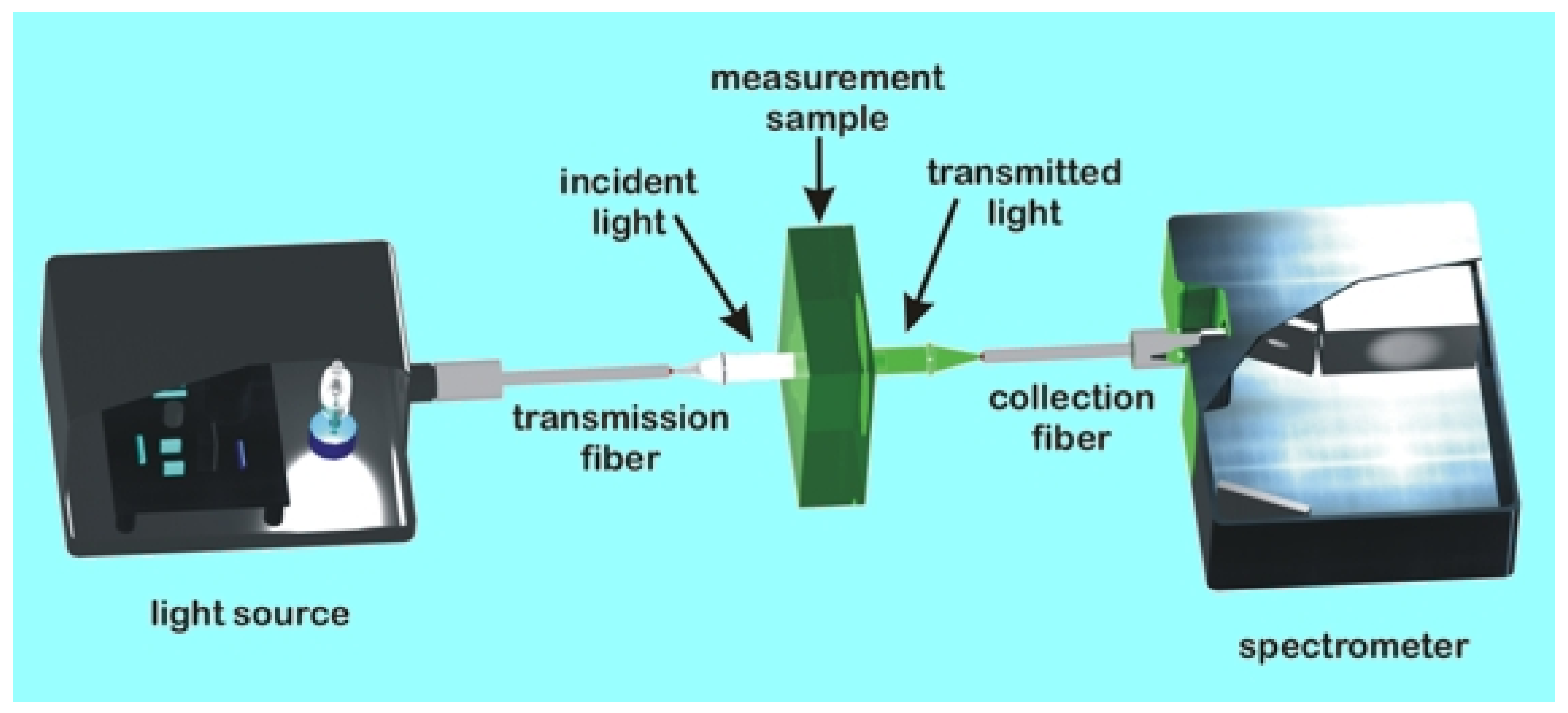
2.4. Color Measurement

An FR-μ Probe instrument was used in conjunction with a trinocular optical microscope (Figure 1). This setup incorporated two optical components: a spectrometer, which utilizes the microscope’s light source and operates in the spectral range the microscope supports, and a module that is mounted on the microscope’s C-port. Typically, for a 50× objective lens, the monitored area size is a ~5 μm diameter circle. Higher magnification or smaller aperture settings allow further sampling area reduction. Real-time specialized software (FR-Monitor®, ThetaMetrisis, Athens, Greece) processing is implemented in Visual C++ to allow film thickness (<10 nm up to 100 μm) and optical constant (n & k) calculations of the areas of interest [23,24,25]. Very fast computations of reflectance (absorbance, transmittance, and fluorescence are also available) spectral measurements of free-standing and supported (over transparent or partially/fully reflective substrates) stacks of (<10 layers) films are accurately calculated utilizing the White Light Reflectance Spectroscopy (WLRS) algorithm (ThetaMetrisisTM) [26,27,28], which were employed for the measurements in this study.
The definition of absorbance (A(λ)) is
A(λ) = −log10(Is/Io)
where Io is the reference light intensity, typically the emission light, e.g., with an empty cuvette, and Is is the light intensity through the sample of interest. For precise measurements, the dark spectrum should be subtracted from both sample and reference spectra; thus, Equation (1) becomes
A(λ) = −log10((Isam-dark)/(Iref-Idark))
where Iref is the reference light intensity, i.e., the light intensity of the light source used, as it is recorded by the spectrometer without passing through the sample of interest, e.g., with an empty cuvette for the liquid sample. Isam is the light intensity after passing through the sample, i.e., the light intensity recorded by the spectrometer when the light from the light source passes through the sample of interest.
Idark stands for the dark intensity, i.e., the light intensity recorded by the spectrometer with the light source switched off or blocked and without a sample. Usually, the absorbance value is in the [0, 2] range, reflecting the [0–99]% absorbance range. On the other hand, the definition of transmittance (T(λ)) is
T(λ) = Is/Io × 100%
and by considering the dark spectrum, as in the absorbance case
T(λ) = (Is/Io × 100%)
Figure 2 depicts the typical equipment configuration for optical measurements.
The workflow sequence consists of (a) the spectra recording, (b) specific wavelength(s) monitoring, and (c) the integration of a specific part of the spectrum. The FR-μProbe and FR-Monitor ver.1.3 software were used following the above description. The experimental average depth in 5 different positions of each denture resin disc sample was calculated [25]. Then, in order to define the real difference in color measurements, the values of ΔΕ1_0, ΔΕ2_0, and ΔΕ3_0 were presented in Arbitrary Units (U).

2.5. Statistical Analysis
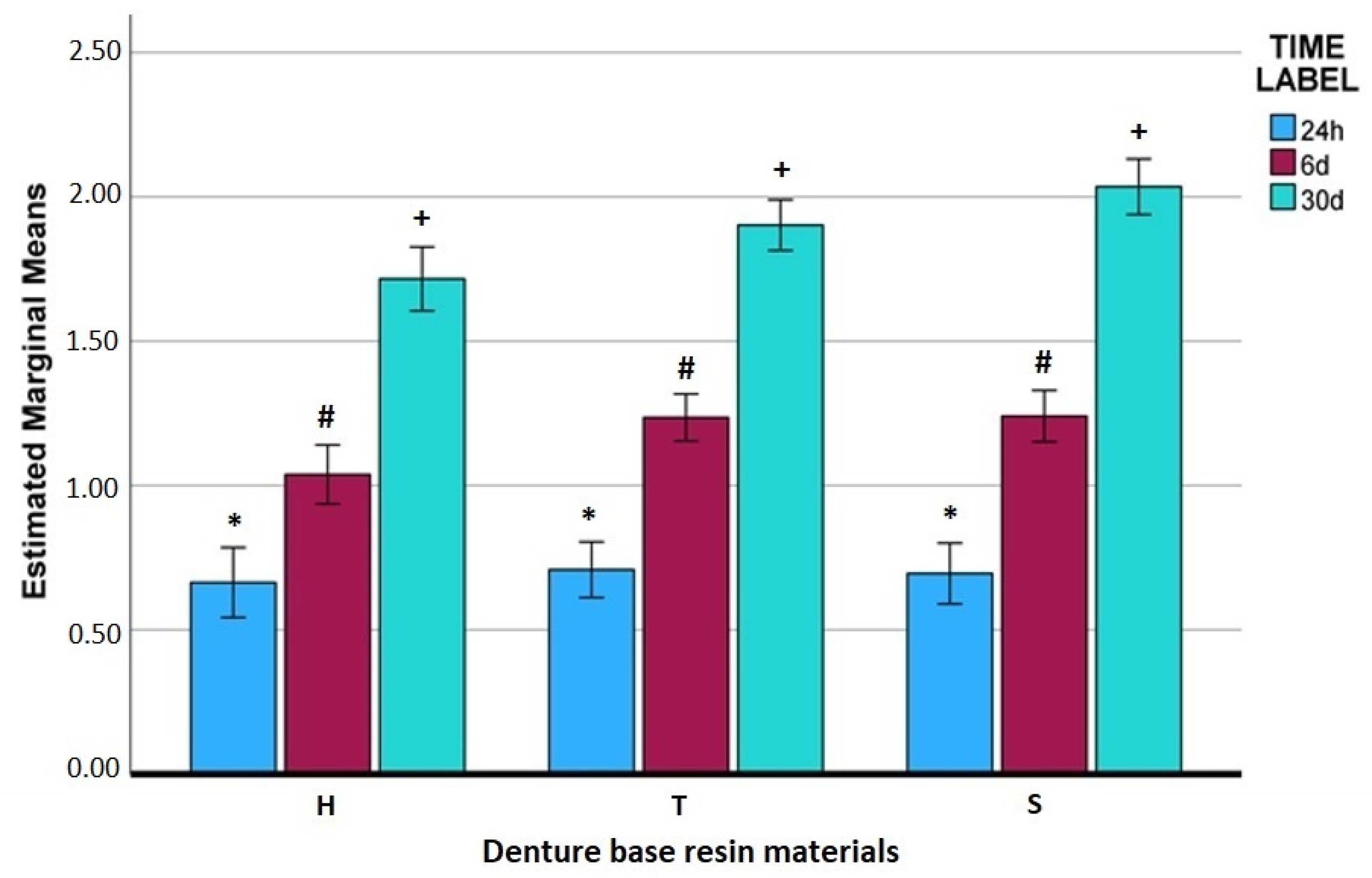
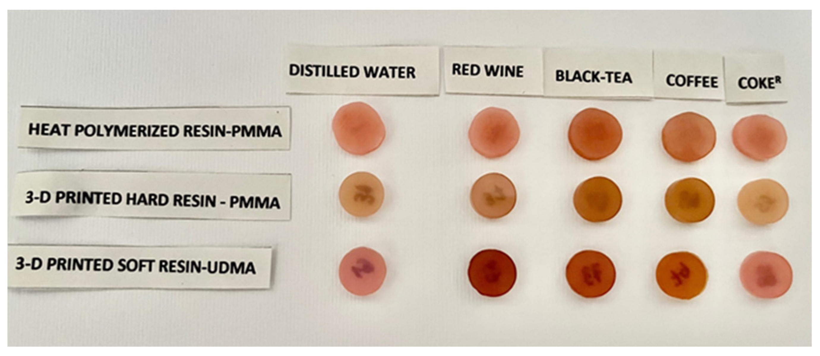
A two-way repeated measurement ANOVA was conducted in order to investigate the interactions of immersion time (within-subject, repeated measurements) and coloring solution type (between-subject, solutions-materials) on the color difference measurements of the three different resin materials tested (IBM SPSS Statistics, Version 29.0.1.0(171), IBM Inc., Chicago, IL, USA). The critical value was set at a 0.05 level of significance.

3. Results

Descriptive statistics (mean and SD) were used to describe the quantitative color difference (ΔΕ). The Bonferroni post hoc test was used for multiple comparisons between the various factors (Type resin/Staining Solution/Immersion Time). Also, interactive plots for mean color differences between the type of resin, staining solutions, and time periods with the same confidence interval (95%) were produced.
The Mean ΔΕ values with their confidence intervals of all denture base resin materials in the five solutions used for all immersion periods are listed in Table 3.
Statistical analysis of pairwise comparisons to assess possible interaction effects on the color stability between different parameters (denture material, staining solution, time) indicated that ΔΕ was significantly affected (Table 4).
A critical assumption is that time is a critical factor for all materials. In Figure 3, which represents the interaction between the type of material and immersion time, the highest values of ΔΕ were measured for the soft resin (S) in the 30-day period. A gradually increasing value for all materials was indeed shown, but the soft resin had the worst behavior (p < 0.05).
Moreover, the same behavior over time is evident for all staining solutions. In Figure 4, Figure 5, Figure 6, Figure 7 and Figure 8, it is evident that the ΔΕ parameter is related linearly with the immersion period, meaning that the color differences continue to increase over time.
This effect was more distinct for the black tea and red wine solutions. In respect to the staining solutions, the black tea and the red wine showed substantial and significantly (p < 0.05) different effects from the other solution values (Table 5). Coffee, Coke®, and distilled water had no significant (p > 0.05) differences between them.
Black tea’s effect was significantly (p < 0.05) different to that of the red wine. These two solutions affected all materials in all time periods at a statistically significant level. The other three solutions had a uniform behavior, with much lower values in all materials (Figure 9). Generally, black tea was by far the most staining solution for all denture base resin materials tested.
Figure 10 depicts a representative photograph of specimens from all groups at the 30-day period.
The soft resin showed the highest values of ΔΕ in almost all immersions, in all solutions, in all time periods. In particular, for the 30-day period, there were statistically significant (p < 0.05) differences as compared with the other two materials.

4. Discussion

This study evaluated the effect of four staining solutions (red wine, black tea, coffee, and Coke®) on the color stability of three denture base dental polymers (heat-polymerizing PMMA resin, hard-type 3D-printed PMMA resin, and urethane-based soft 3D-printed resin) in three immersion periods (one day, one week, and one month). The null hypothesis of this study was rejected by the results in most comparisons, indicating the significant influence of all parameters applied on the ΔΕ of all materials.
There is a direct and strong influence between immersion time and color change in the tested materials. This agrees with many studies that confirm the strong relationship between exposure time and coloring effect [18,22,29,30,31,32,33,34,35,36].
There was a slight increase of ΔΕ at the end of the 7-day period, which increased further at the end of the 30-day period (Figure 4, Figure 7, and Figure 8), but the values were at the 1.0 Unit level after immersing in water, coffee, and Coke®. The black tea and red wine solutions showed a stronger influence during all periods at the level of approximately 4.0 Units for black tea and 2.9 Units for the red wine at the end of the 30-day period. The same behavior was apparent in all time periods.
Many studies mention that water absorption is a common possible mechanism for discoloring denture base resins [19,31,32,37]. Berli et al. [37] found that 3D-printed resins absorbed water molecules, leading to an increase of surface solubility and roughness. The chemical bonds of the polymer network are decomposed from the hydrolysis effect of water, and a surface precipitation of coloring pigments becomes possible. So, the presence of water is critical over time and acts as a possible leader or a contributing factor to enhance various colorant’s deposition [31,38].
On the other side, some studies [19,39] found a decrease of ΔΕ values after a 7-day immersion period. They propose that, after a period, there is a stabilizing mechanism of PMMA resins through an aging process. Therefore, this point needs to be confirmed by clinical studies. Tieh et al. [29] mention that the discoloration process happens to a larger extent in the initial phases of exposure in stains, with coffee being the strongest discoloring media.
Concerning the different solutions, the results of this study showed an aggressive discoloring effect of black tea and red wine solutions on all materials. All materials in all immersion periods had an increasing ΔΕ, with the highest values shown in the 30-day period. In pairwise comparisons, there were significant (p < 0.05) differences for black tea and red wine compared with the other solutions and with each other. Otherwise, black tea was the strongest discoloring media, followed by the red wine solution.
Black tea contains color pigments with small (catechins, theaflavins) and large molecular weights (tannins, thearubigins). They also have an acidic nature and high-water solubility [18]. Therefore, under the presence of the water environment, which contributes to resin degradation and absorption, these colorants have a critical influence on the materials under this study’s conditions. This study corroborates the results of Zuo et al. [31], that tea discolors denture base resins over time, with lower values than red wine and Coke®. Kim et al. [17] also found that tea affects the color stability of 3D-printed resin over time.
Red wine is a well-established discoloring media in the oral environment. Many studies confirm this finding in comparison with various media (Coke®, coffee) [30,31,32,33,34,40]. Red wine has an acidic influence because it contains alcohol acids (pH~3.9), so it roughens the resin’s surface [33]. The alcohol molecules are absorbed by the organic matrix and have a plasticizing effect on the resin surface. The color pigments, like tannins, minerals, and anthocyanin, which is water soluble, can be more easily deposited. That explains why red wine has a stronger discoloring effect than Coke®, which is more acidic (pH~2.0) and contains citric acid. This behavior is confirmed by the results of this study (Table 5, Figure 9). Coke® had values similar to distilled water and coffee, with no significant differences during all periods (Table 5, Figure 9).
There is a general assumption by many studies [17,30,31,32,33,34,35,40] that coffee has a strong discoloring effect in dental resins compared with other staining agents. Coffee contains tannin and chlorogenic acids with a mild acidic pH (~5) [19]. The abovementioned hydrolytic action of water combined with these characteristics leads to a surface discoloration process, as these pigments are deposited on the resin’s surface. Kim et al. [17] mention in their review that coffee is the worst of all common beverages. In the present study, coffee exhibited a much lower discoloring effect than tea and red wine. It exhibited values that were not significantly different to distilled water and Coke®. This agrees with Alfouzan et al.’s [19] findings that coffee has a low discoloration potential compared with lemon juice and Coke®. The possible explanation they gave was that after the coffee layers have reached a certain thickness on the specimen surface, the layers are dislodged and dissolved in the water.
To summarize, in the present study there was clear evidence that tea and red wine have the strongest discoloring effect in all materials. All materials in the present study showed similar behavior in all staining media during all time periods. All three types of resins exhibited color changes over time (Figure 3) at a statistically significant level (Table 4), thus presenting a time-dependent pattern of ΔΕ changes. The highest values in the 30-day period were measured for the 3D-printed soft resin. The two PMMA resins (heat-polymerized and 3D-printed) exhibited a similar behavior. There are, however, contradictory findings in the literature.
Alfouzan et al. [19] found that 3D-printed denture resins demonstrated better color stability than heat-polymerized resins, which presented the highest ΔΕ. Dimitrova et al. [30] estimated that a 3D-printed resin performed better in color stability than a PMMA heat-polymerized resin, showing similar results with another study [11].
Moreover, Gruber et al. [32] found inferior color stability behavior in 3D-printed resins as compared to conventional heat-polymerized and CAD/CAM-milled PMMA resins. Shin et al. [22] reported that 3D-printed resins demonstrated color differences above acceptable clinical thresholds compared to CAD/CAM denture resins when exposed to curry, grape juice, coffee, and water. Çakmac et al. [21] showed that, during a coffee thermocycling test, CAD/CAM-milled PMMA showed better coloring behavior. After proper polishing, all materials presented clinically acceptable changes. Additively manufactured denture base resin had the highest color difference, and prepolymerized PMMA had the lowest color difference after coffee thermocycling; however, the first exhibited a perceivable color change. Significant differences in surface roughness were observed among tested materials within each time interval and among different time intervals.
Also, Koh et al. [20] found that 3D-printed acrylic teeth had no color differences from conventional acrylic teeth in different solutions (coffee, Coke®, erythrosine, and water). Tieh et al. [29], in a systematic review of the optical properties and color stability of different types of denture teeth, concluded that 3D-printed teeth showed contradictory findings, needing more research for improving the materials. On the other side, CAD/CAM teeth had similar properties to conventional heat-polymerized teeth. Coffee was the worst among common beverages.
In the present study, there was no clear differentiation between the denture base resin materials as, largely, no significant differences were detected. There was, however, a significant finding, where the soft resin exhibited the highest ΔΕ values in all groups. It seems that more research is needed in these materials for improving them. Dayan et al. [33] compared the discoloring behavior of a urethane-based denture resin, an autopolymerizing acrylic resin, a CAD/CAM PMMA resin, and a conventional heat-polymerized PMMA resin exposed to coffee, Coke®, and red wine solutions. They found that the urethane-based resin had the highest clinically unacceptable discoloring values. Zuo et al. [31]) estimated the same result. Polymers based one the urethane molecule [UDMA 1,6-bis(methacrylyloxy-2-ethoxycarbonylamino)-2,4,4-trimethylhexane] have a hydrophilic behavior because of the existence of two urethane groups. Thus, they have a tendency toward hygroscopic expansion, which leads to excess water sorption, hydrolytic degradation of the polymer matrix, marked surface roughening, and a subsequent discoloration process [41]. Surface morphology may influence the hydrophilic behavior of the material and the subsequent possible tendency toward discoloration [42].
The differences in results amongst the various studies reflect the different research methodologies/protocols applied. The discoloring behavior is material dependent. More research is therefore needed along with a call for standardization of both discoloring and evaluation protocols amongst researchers through recognized standards organizations (e.g., ISO).
This study has its limitations. It is an in vitro study. Variations in dietary habits, oral hygiene status, using of cleansing agents, pH fluctuations, and saliva type are also important in vivo influencing factors [32,36]. The temperature of the immersion solutions was kept stable at 37 °C in order to simulate the oral environment. In real oral function, however, the temperature varies over dining times. Another limitation is that there was no study of the specimens’ surface roughness and surface morphology, even though it is very important for the surface wettability and the hydrophilic behavior of the material. The purpose of the study was to evaluate the influence of the staining solutions in depth, not only on the surface area where the stains are easily cleaned. The most important limitation was that the measuring method used in this study provided arbitrary ΔE values, though they provided quantitative characteristics.
The CIE Lab System, which is proposed by ISO/TR 28642:2016, is the most commonly used method for evaluating the color changes of dental resins [43]. This system correlates the spectrophotometric data by using a special software that incorporates the human eye response to color changes. L * a * b * values are produced, and thresholds for perceptibility and acceptability by the human eye of the color changes are determined. This can be very useful from a practical perspective, but it might not be adequately specific and accurate as an evaluating method [32]. The light reflectance and the consequent color measurement are influenced by many factors. The surface texture (matt or glossy surface) affects the L * a * b * values; there are differences between specific system measuring modes (Specular Component Included—SCI; Specular Component Excluder—SCE). Finally, there seems to be no universal consensus for recording objective values.
In an attempt to improve the objectivity of these results, a more specific method that is more accurate in measuring the light reflectance of the resin surface without influence of the surface texture was used. The method, as explained in details in Section 2, measures the light reflectance from many points of the surface, at a shallow depth and without any influence from the surface texture. Even more, all observations were performed under the same conditions, so the results and comparisons were strong enough for evaluating the true differences in color changes by comparing the parameters internally. Experiments towards correlating the CIE Lab system methodology and other methods of measuring color differences are required. Until then, direct comparison of the results of this study with others cannot be made [1,7,9,19,44,45].

5. Conclusions

The findings of this study demonstrate several important aspects of the color stability of different denture base materials under various immersion conditions. It was observed that the color changes of all materials were time dependent, with increasing discoloration over extended immersion periods. The solutions containing tea and red wine had a significantly greater impact on color stability than other tested solutions, such as coffee and Coca-Cola. These results suggest that specific external agents, particularly those containing strong staining media, play a critical role in the staining process.
Moreover, the study indicates that color stability is highly dependent on the material composition. Heat-polymerized PMMA resins and hard 3D-printed PMMA resins exhibited comparable behavior, with both materials demonstrating moderate discoloration over time. This similarity may suggest that, in terms of resistance to staining, the 3D-printed PMMA material can be a viable alternative to conventional heat-polymerized resins for denture fabrication.
In contrast, the soft 3D-printed resin performed poorly, displaying the highest degree of discoloration across all immersion periods. Although the statistical analysis did not show a significant difference for this material, except in one comparison, the results clearly highlight its vulnerability to staining, indicating that further research is necessary to improve its formulation. Overall, these findings emphasize the importance of material selection in the context of dental applications, where color stability is a critical factor for long-term aesthetic performance. Further investigations should focus on optimizing the properties of soft 3D-printed resins to enhance their resistance to external discoloration agents.

Author Contributions

Conceptualization, V.V., S.Y. and T.G.; methodology, V.V. and T.G.; software, V.V., A.T. and T.G.; validation, V.V., A.T., A.K. and T.G.; formal analysis, V.V. and T.G.; investigation, V.V. and T.G.; resources, V.V., A.T., A.K. and T.G.; data curation, V.V., A.T., A.K. and T.G.; writing—original draft preparation, V.V. and T.G.; writing—review and editing, V.V., A.T., A.K. and T.G.; visualization, V.V., S.Y. and T.G.; supervision, V.V. and T.G.; project administration, T.G. All authors have read and agreed to the published version of the manuscript.

Funding

This research received no external funding.

Institutional Review Board Statement

Not applicable.

Informed Consent Statement

Not applicable.

Data Availability Statement

The data presented in this study are available in the article.

Conflicts of Interest

The authors declare no conflicts of interest.

References

  1. Anadioti, E.; Musharbash, L.; Blatz, M.B.; Papavasiliou, G.; Kamposiora, P. 3D printed complete removable dental prostheses: A narrative review. BMC Oral Health 2020, 20, 343. [Google Scholar] [CrossRef] [PubMed]
  2. Kantaros, A.; Soulis, E.; Alysandratou, E. Digitization of Ancient Artefacts and Fabrication of Sustainable 3D-Printed Replicas for Intended Use by Visitors with Disabilities: The Case of Piraeus Archaeological Museum. Sustainability 2023, 15, 12689. [Google Scholar] [CrossRef]
  3. Kantaros, A.; Ganetsos, T. From Static to Dynamic: Smart Materials Pioneering Additive Manufacturing in Regenerative Medicine. Int. J. Mol. Sci. 2023, 24, 15748. [Google Scholar] [CrossRef]
  4. Kantaros, A. Bio-Inspired Materials: Exhibited Characteristics and Integration Degree in Bio-Printing Operations. Am. J. Eng. Appl. Sci. 2022, 15, 255–263. [Google Scholar] [CrossRef]
  5. Kantaros, A. 3D Printing in Regenerative Medicine: Technologies and Resources Utilized. Int. J. Mol. Sci. 2022, 23, 14621. [Google Scholar] [CrossRef] [PubMed]
  6. Kantaros, A.; Petrescu, F.I.T.; Abdoli, H.; Diegel, O.; Chan, S.; Iliescu, M.; Ganetsos, T.; Munteanu, I.S.; Ungureanu, L.M. Additive Manufacturing for Surgical Planning and Education: A Review. Appl. Sci. 2024, 14, 2550. [Google Scholar] [CrossRef]
  7. Kantaros, A.; Ganetsos, T.; Petrescu, F.I.T. Transforming Object Design and Creation: Biomaterials and Contemporary Manufacturing Leading the Way. Biomimetics 2024, 9, 48. [Google Scholar] [CrossRef]
  8. Tian, Y.; Chen, C.; Xu, X.; Wang, J.; Hou, X.; Li, K.; Lu, X.; Shi, H.; Lee, E.-S.; Jiang, H.B. A Review of 3D Printing in Dentistry: Technologies, Affecting Factors, and Applications. Scanning 2021, 2021, 9950131. [Google Scholar] [CrossRef]
  9. Alhallak, K.; Hagi-Pavli, E.; Nankali, A. A review on clinical use of CAD/CAM and 3D printed dentures. Br. Dent. J. 2023, 1–5. [Google Scholar] [CrossRef]
  10. Khorsandi, D.; Fahimipour, A.; Abasian, P.; Saber, S.S.; Seyedi, M.; Ghanavati, S.; Ahmad, A.; De Stephanis, A.A.; Taghavinezhaddilami, F.; Leonova, A.; et al. 3D and 4D printing in dentistry and maxillofacial surgery: Printing techniques, materials, and applications. Acta Biomater. 2021, 122, 26–49. [Google Scholar] [CrossRef]
  11. Revilla-León, M.; Özcan, M. Additive Manufacturing Technologies Used for Processing Polymers: Current Status and Potential Application in Prosthetic Dentistry. J. Prosthodont. 2019, 28, 146–158. [Google Scholar] [CrossRef] [PubMed]
  12. Dimitrova, M.; Corsalini, M.; Kazakova, R.; Vlahova, A.; Chuchulska, B.; Barile, G.; Capodiferro, S.; Kazakov, S. Comparison between Conventional PMMA and 3D Printed Resins for Denture Bases: A Narrative Review. J. Compos. Sci. 2022, 6, 87. [Google Scholar] [CrossRef]
  13. Lin, C.-H.; Lin, Y.-M.; Lai, Y.-L.; Lee, S.-Y. Mechanical properties, accuracy, and cytotoxicity of UV-polymerized 3D printing resins composed of BisEMA, UDMA, and TEGDMA. J. Prosthet. Dent. 2020, 123, 349–354. [Google Scholar] [CrossRef]
  14. Mumcu, E.; Cilingir, A.; Gencel, B.; Sülün, T. Flexural properties of a light-cure and a self-cure denture base materials compared to conventional alternatives. J. Adv. Prosthodont. 2011, 3, 136–139. [Google Scholar] [CrossRef]
  15. Binaljadm, T.M. Flexible Denture: A Literature Review. Cureus 2024, 16, e55425. [Google Scholar] [CrossRef]
  16. Revilla-León, M.; Meyers, M.J.; Zandinejad, A.; Özcan, M. A review on chemical composition, mechanical properties, and manufacturing work flow of additively manufactured current polymers for interim dental restorations. J. Esthet. Restor. Dent. 2019, 31, 51–57. [Google Scholar] [CrossRef]
  17. Kim, M.-C.; Byeon, D.-J.; Jeong, E.-J.; Go, H.-B.; Yang, S.-Y. Color stability, surface, and physicochemical properties of three-dimensional printed denture base resin reinforced with different nanofillers. Sci. Rep. 2024, 14, 1842. [Google Scholar] [CrossRef]
  18. Kentrou, C.; Papadopoulos, T.; Lagouvardos, P. Color changes in staining solutions of four light-cured indirect resin composites. Odontology 2014, 102, 189–196. [Google Scholar] [CrossRef] [PubMed]
  19. Alfouzan, A.F.; Alotiabi, H.M.; Labban, N.; Al-Otaibi, H.N.; Al Taweel, S.M.; AlShehri, H.A. Color stability of 3D-printed denture resins: Effect of aging, mechanical brushing and immersion in staining medium. J. Adv. Prosthodont. 2021, 13, 160–171. [Google Scholar] [CrossRef]
  20. Koh, E.-S.; Cha, H.-S.; Kim, T.-H.; Ahn, J.-S.; Lee, J.-H. Color stability of three dimensional-printed denture teeth exposed to various colorants. J. Korean Acad. Prosthodont. 2020, 58, 1–6. [Google Scholar] [CrossRef]
  21. Çakmak, G.; Weber, F.A.; Donmez, M.B.; Kahveci, Ç.; Schimmel, M.; Yilmaz, B. Effect of coffee thermocycling on the surface roughness and stainability of denture base materials with different chemical compositions manufactured with additive and subtractive technologies. J. Esthet. Restor. Dent. 2024, 36, 453–459. [Google Scholar] [CrossRef] [PubMed]
  22. Shin, J.-W.; Kim, J.-E.; Choi, Y.-J.; Shin, S.-H.; Nam, N.-E.; Shim, J.-S.; Lee, K.-W. Evaluation of the color stability of 3D-printed crown and bridge materials against various sources of discoloration: An in vitro study. Materials 2020, 13, 5359. [Google Scholar] [CrossRef] [PubMed]
  23. Niakoula, D.; Raptis, I.; Goustouridis, D.; Argitis, P. Glass transition temperature monitoring in bilayer and patterned photoresist films. Jpn. J. Appl. Phys. Part 1 Regul. Pap. Short Notes Rev. Pap. 2004, 43, 5247–5248. [Google Scholar] [CrossRef]
  24. Marceau, S.; Tortai, J.-H.; Tillier, J.; Vourdas, N.; Gogolides, E.; Raptis, I.; Beltsios, K.; van Werden, K. Thickness-dependent glass transition temperature of thin resist films for high resolution lithography. Microelectron. Eng. 2006, 83, 1073–1077. [Google Scholar] [CrossRef]
  25. Manoli, K.; Goustouridis, D.; Chatzandroulis, S.; Raptis, I.; Valamontes, E.S.; Sanopoulou, M. Vapor sorption in thin supported polymer films studied by white light interferometry. Polymer 2006, 47, 6117–6122. [Google Scholar] [CrossRef]
  26. Vourdas, N.; Karadimos, G.; Goustouridis, D.; Gogolides, E.; Boudouvis, A.G.; Tortai, J.; Beltsios, K.; Raptis, I. Multiwavelength interferometry and competing optical methods for the thermal probing of thin polymeric films. J. Appl. Polym. Sci. 2006, 102, 4764–4774. [Google Scholar] [CrossRef]
  27. Drygiannakis, D.; Raptis, I.; Patsis, G.; Boudouvis, A.; Vanwerden, K. Processing effects on the dissolution properties of thin chemically amplified photoresist films. Microelectron. Eng. 2008, 85, 955–958. [Google Scholar] [CrossRef]
  28. Sarantopoulou, E.; Kollia, Z.; Cefalas, A.; Manoli, K.; Sanopoulou, M.; Goustouridis, D.; Chatzandroulis, S.; Raptis, I. Surface nano/micro functionalization of PMMA thin films by 157nm irradiation for sensing applications. Appl. Surf. Sci. 2008, 254, 1710–1719. [Google Scholar] [CrossRef]
  29. Tieh, M.T.; Waddell, J.N.; Choi, J.J.E. Optical Properties and Color Stability of Denture Teeth-A Systematic Review. J. Prosthodont. 2022, 31, 385–398. [Google Scholar] [CrossRef]
  30. Dimitrova, M.; Vlahova, A.; Hristov, I.; Kazakova, R.; Chuchulska, B.; Gladysheva, A. Color Changes of 3D Printed and Conventional Dental Resins for Removable Prosthodontics After Immersion in Different Staining Agents. J. Imab Annu. Proceeding (Sci. Pap.) 2023, 29, 4861–4865. [Google Scholar] [CrossRef]
  31. Zuo, W.; Feng, D.; Song, A.; Gong, H.; Zhu, S. Effects of organic-inorganic hybrid coating on the color stability of denture base resins. J. Prosthet. Dent. 2015, 115, 103–108. [Google Scholar] [CrossRef] [PubMed]
  32. Gruber, S.; Kamnoedboon, P.; Özcan, M.; Srinivasan, M. CAD/CAM Complete Denture Resins: An In Vitro Evaluation of Color Stability. J. Prosthodont. 2021, 30, 430–439. [Google Scholar] [CrossRef]
  33. Dayan, C.; Guven, M.C.; Gencel, B.; Bural, C. A color stability comparison of conventional and cad/cam polymethyl methacrylate denture base materials. Acta Stomatol. Croat. 2019, 53, 158–167. [Google Scholar] [CrossRef] [PubMed]
  34. Gregorius, W.C.; Kattadiyil, M.T.; Goodacre, C.J.; Roggenkamp, C.L.; Powers, J.M.; Paravina, R.D. Effects of ageing and staining on color of acrylic resin denture teeth. J. Dent. 2012, 40 (Suppl. S2), e47–e54. [Google Scholar] [CrossRef]
  35. Hipólito, A.C.; Barão, V.A.; Faverani, L.P.; Ferreira, M.B.; Assunção, W.G. Color degradation of acrylic resin denture teeth as a function of liquid diet: Ultraviolet-visible reflection analysis. J. Biomed. Opt. 2013, 18, 105005. [Google Scholar] [CrossRef] [PubMed]
  36. Polychronakis, N.; Lagouvardos, P.; Polyzois, G.; Sykaras, N.; Zoidis, P. Color changes of polyetheretherketone (PEEK) and polyoxymethelene (POM) denture resins on single and combined staining/cleansing action by CIELab and CIEDE2000 formulas. J. Prosthodont. Res. 2020, 64, 159–166. [Google Scholar] [CrossRef]
  37. Berli, C.; Thieringer, F.M.; Sharma, N.; Müller, J.A.; Dedem, P.; Fischer, J.; Rohr, N. Comparing the mechanical properties of pressed, milled, and 3D-printed resins for occlusal devices. J. Prosthet. Dent. 2020, 124, 780–786. [Google Scholar] [CrossRef]
  38. Dietschi, D.; Campanile, G.; Holz, J.; Meyer, J.-M. Comparison of the color stability of ten new-generation composites: An in vitro study. Dent. Mater. 1994, 10, 353–362. [Google Scholar] [CrossRef]
  39. Imirzalioglu, P.; Karacaer, O.; Yilmaz, B.; Ozmen Msc, I. color stability of denture acrylic resins and a soft lining material against tea, coffee, and nicotine. J. Prosthodont. 2010, 19, 118–124. [Google Scholar] [CrossRef]
  40. Papathanasiou, I.; Papavasiliou, G.; Kamposiora, P.; Zoidis, P. Effect of Staining Solutions on Color Stability, Gloss and Surface Roughness of Removable Partial Dental Prosthetic Polymers. J. Prosthodont. 2022, 31, 65–71. [Google Scholar] [CrossRef]
  41. Kerby, R.E.; Knobloch, L.A.; Schricker, S.; Gregg, B. Synthesis and evaluation of modified urethane dimethacrylate resins with reduced water sorption and solubility. Dent. Mater. 2009, 25, 302–313. [Google Scholar] [CrossRef] [PubMed]
  42. Gad, M.M.; Fouda, S.M.; Abualsaud, R.; Alshahrani, F.A.; Al-Thobity, A.M.; Khan, S.Q.; Akhtar, S.; Ateeq, I.S.; Helal, M.A.; Al-Harbi, F.A. Strength and Surface Properties of a 3D-Printed Denture Base Polymer. J. Prosthodont. 2022, 31, 412–418. [Google Scholar] [CrossRef] [PubMed]
  43. Luo, M.R.; Cui, G.; Rigg, B. The development of the CIE 2000 colour-difference formula: CIEDE2000. Color Res. Appl. 2001, 26, 340–350. [Google Scholar] [CrossRef]
  44. Al-Qarni, F.D.; Gad, M.M. Printing Accuracy and Flexural Properties of Different 3D-Printed Denture Base Resins. Materials 2022, 15, 2410. [Google Scholar] [CrossRef]
  45. Kantaros, A.; Ganetsos, T.; Petrescu, F.I.T.; Ungureanu, L.M.; Munteanu, I.S. Post-Production Finishing Processes Utilized in 3D Printing Technologies. Processes 2024, 12, 595. [Google Scholar] [CrossRef]
Figure 1. FR-μ probe installed on an optical microscope using an internal light source.
Applsci 14 11548 g001
Figure 2. The typical basic parts for optical measurements.
Applsci 14 11548 g002
Figure 3. Interactive plot for ΔΕ between type of material and immersion time (H: heat-polymerized resin; T: 3D PMMA resin; S: Urethane-based resin). The different symbol indicators (*, #, +) above each column indicate significant (p < 0.001) differences between the immersion times within the various material groups.
Applsci 14 11548 g003
Figure 4. Estimated ΔΕ means of all materials (H: heat-polymerized resin; T: 3D PMMA resin; S: Urethane-based resin) in distilled water.
Applsci 14 11548 g004
Figure 5. Estimated ΔΕ means of all materials (H: heat-polymerized resin; T: 3D PMMA resin; S: Urethane-based resin) in red wine.
Applsci 14 11548 g005
Figure 6. Estimated ΔΕ means of all materials (H: heat-polymerized resin; T: 3D PMMA resin; S: Urethane-based resin) in black tea.
Applsci 14 11548 g006
Figure 7. Estimated ΔΕ means of all materials (H: heat-polymerized resin; T: 3D PMMA resin; S: Urethane-based resin) in coffee.
Applsci 14 11548 g007
Figure 8. Estimated ΔΕ means of all materials (H: heat-polymerized resin; T: 3D PMMA resin; S: Urethane-based resin) in Coke®.
Applsci 14 11548 g008
Figure 9. Interactive plot for ΔΕ between type of material and staining solution (H: heat-polymerized resin; T: 3D PMMA resin; S: Urethane-based resin). The different symbol indicators (*, #, +) above each column indicate significant (p < 0.001) differences between the immersion solution effects within the various material groups.
Applsci 14 11548 g009
Figure 10. Representative photo of specimens of all groups in the T_30 measurement.
Applsci 14 11548 g010
Table 1. Technical details of the materials used.
Commercial NameCompositionLot No.Manufacturer
Meliodent pink (Heat-polymerized Denture Resin) Powder (polymethylmethacrylate)
Liquid (methylmethacrylate, dimetha crylate) PMMA
KO10062Kulzer Gmbh, Hanau, Germany
Denta Base pink (Hard 3D-printed resin for denture base)PMMA resin
7,7,9(or 7,9,9)-trimethyl-4,13-dioxo-3,14-dioxa-5,12-diazahexadecane-1,16-diyl bismethacrylate; Diphenyl(2,4,6-trimethylbenzoyl) phosphine oxide; Tetrahydrofurfuryl methacrylate
Not availableASIGA Pty Ltd., Alexandria, Australia
Flexo Denture Base V2 pink (Soft 3D-printed resin)Urethane dimethacrylate UDMA
(methacrylyloxy–2-ethoxy–carbonyl amino)-2,4, 4 trimethythexane. This is a reaction product of 2-hydroxyethylmethacrylate and 2, 4, 4-trimethyl-hexamethylenediisocyanate–UDMA (according to the manufacturer’s information, sent by mail)
SNR202300021Senertec 3D Yazici, Izmir, Turkey
Lipton Black Tea; Origin, Kenya Black tea 100%L4 076 1 J3 01
L4 076 2 J3 01
L4 076 3 J3 01
Ekaterra global Operations BV, Rotterdam, The Netherlands
Jacobs Kronung (filtered coffee)Ground coffee grainsLK00942063 01Jacobs Douwe Egberts, DK Amsterdam, The Netherlands
Akres 2023 (dry red wine)Alcohol 14% vol.L 240221Produced and bottled by Skouras Winery, Argos, Greece
Coke® Original Taste (Coca-Cola)Water, Sugar, Carbon dioxide, Caffeine, phosphoric acid, Other Aromatic substances20B43E29BCoca-Cola HBC Cyprus, Nicosia, Cyprus
Mallinckrodt Silica gel Silica gel 4000834Mallinckrodt Chemical Works, St. Louis, MO, USA
KMG POLISH LIQUIDAlcohols, C-16-18, ethoxylated Aquatic Acute 1, H400; Aquatic Chronic 3, H412 2.5Z02SNBCANDULOR AG, Clattpark (Opfikon), Switzerland
Table 2. Groups of the studied materials (n = 6).
Groups
Heat-polymerized resinH (Heat)
Distilled water (control)
Wine
Black Tea
Coffee
Coke®
3D-printed Hard ResinT (Tough)
Distilled water (control)
Wine
Black Tea
Coffee
Coke®
3D-printed Soft ResinS (Soft)
Distilled water (control)
Wine
Black Tea
Coffee
Coke®
Table 3. Mean ΔΕ values with their 95% confidence interval for all materials in four coloring solutions in all immersion periods. Distilled water was the control solution. H = heat-polymerized resin; T = 3D-printed hard PMMA resin; S = 3D-printed Urethane-based soft resin.
95% Confidence Interval
Time LabelSolution TypeMaterialMeanStd. ErrorLower BoundUpper Bound
24 hWaterH0.1030.136−0.1680.374
T0.0840.108−0.1310.299
S0.1160.118−0.1190.351
Red_WineH0.6670.1360.3960.937
T0.9550.1080.7401.170
S1.1010.1180.8661.336
Black_TeaH2.1640.1361.8942.435
T2.0960.1081.8812.311
S1.9530.1181.7172.188
CoffeeH0.2410.136−0.0300.512
T0.1790.108−0.0370.394
S0.2090.118−0.0260.445
Coke®H0.1440.136−0.1270.415
T0.2280.1080.0120.443
S0.0970.118−0.1380.332
6 daysWaterH0.1840.115−0.0450.413
T0.2520.0910.0700.434
S0.0930.100−0.1060.292
Red_WineH1.2700.1151.0411.499
T1.8950.0911.7132.077
S2.3480.1002.1492.547
Black_TeaH2.8550.1152.6263.083
T3.1190.0912.9373.301
S2.7440.1002.5462.943
CoffeeH0.4290.1150.2010.658
T0.3930.0910.2110.575
S0.5390.1000.3400.737
Coke®H0.4520.1150.2230.681
T0.5190.0910.3370.701
S0.4780.1000.2800.677
30 daysWaterH0.6020.1240.3550.849
T1.0750.0990.8791.272
S0.9920.1080.7781.207
Red_WineH1.9660.1241.7182.213
T2.7460.0992.5502.943
S2.9920.1082.7773.207
Black_TeaH4.0210.1243.7744.268
T3.8820.0993.6864.079
S4.1750.1083.9604.390
CoffeeH1.0650.1240.8181.312
T0.8330.0990.6371.030
S1.0110.1080.7961.226
Coke®H0.9250.1240.6781.172
T0.9730.0990.7761.169
S1.0050.1080.7901.220
Table 4. Bonferroni test based on observed means. The error term is Mean Square (Error) = 0.037.
Mean Difference
(I) Time Label (J) Time Label (I–J)
Std. ErrorSig.95% Confidence Interval
Lower BoundUpper Bound
24 h6 days−0.4823 *0.05012<0.001−0.6050−0.3595
30 days−1.1952 *0.05183<0.001−1.3221−1.0683
6 days24 h0.4823 *0.05012<0.0010.35950.6050
30 days−0.7129 *0.04762<0.001−0.8296−0.5963
30 days24 h1.1952 *0.05183<0.0011.06831.3221
6 days0.7129 *0.04762<0.0010.59630.8296
* The mean difference is significant at the 0.05 level.
Table 5. Bonferroni test based on observed means. The error term is Mean Square (Error) = 0.037.
(I) Solution Type(J) Solution Type(I–J)Std. ErrorSig.95% Lower Bound95% Upper Bound
WaterRed_Wine−1.4294 *0.06379<0.001−1.6139−1.2449
Black_Tea−2.6541 *0.06379<0.001−2.8387−2.4696
Coffee−0.16470.063790.118−0.34920.0198
Coke®−0.16060.063790.140−0.34510.0239
Red_WineWater1.4294 *0.06379<0.0011.24491.6139
Black_Tea−1.2247 *0.06379<0.001−1.4093−1.0402
Coffee1.2647 *0.06379<0.0011.08011.4492
Coke®1.2688 *0.06379<0.0011.08431.4533
Black_TeaWater2.6541 *0.06379<0.0012.46962.8387
Red_Wine1.2247 *0.06379<0.0011.04021.4093
Coffee2.4894 *0.06379<0.0012.30492.6739
Coke®2.4935 *0.06379<0.0012.30902.6781
CoffeeWater0.16470.063790.118−0.01980.3492
Red_Wine−1.2647 *0.06379<0.001−1.4492−1.0801
Black_Tea−2.4894 *0.06379<0.001−2.6739−2.3049
Coke®0.00410.063791.000−0.18040.1887
Coke®Water0.16060.063790.140−0.02390.3451
Red_Wine−1.2688 *0.06379<0.001−1.4533−1.0843
Black_Tea−2.4935 *0.06379<0.001−2.6781−2.3090
Coffee−0.00410.063791.000−0.18870.1804
* The mean difference is significant at the 0.05 level.
Disclaimer/Publisher’s Note: The statements, opinions and data contained in all publications are solely those of the individual author(s) and contributor(s) and not of MDPI and/or the editor(s). MDPI and/or the editor(s) disclaim responsibility for any injury to people or property resulting from any ideas, methods, instructions or products referred to in the content.

