Article

Porous Mg–Hydroxyapatite Composite Incorporated with Aloe barbadensis Miller for Scaphoid Fracture Fixation: A Natural Drug Loaded Orthopedic Implant

1
Department of Mechanical Engineering, Thapar Institute of Engineering and Technology, Patiala 147004, Punjab, India
2
Department of Biotechnology Engineering, Thapar Institute of Engineering and Technology, Patiala 147004, Punjab, India
3
Department of Mechanical Engineering, National University of Singapore, 9 Engineering Drive 1, Singapore 117576, Singapore
*
Authors to whom correspondence should be addressed.
Appl. Sci. 2024, 14(4), 1512; https://doi.org/10.3390/app14041512
Submission received: 23 December 2023 / Revised: 17 January 2024 / Accepted: 9 February 2024 / Published: 13 February 2024

Abstract

:
The current study focused on developing a biodegradable implant composite material that could work in a multitude of applications. The fabricated composite showcases a porous matrix of Mg–hydroxyapatite developed through the spacer-holder technique. The composite was incorporated with a natural medicinal plant, i.e., Aloe barbadensis miller, commonly known as the Aloe vera plant. The final composite was enveloped under a thin layer of PLA to work as an encapsulated drug as well as a composite material for implant applications. Further, the mechanical and microstructural properties were analyzed along with corrosion analysis through the weight loss method and pH change. The experiments showed an improvement in the corrosion rate when tested under cell culture medium. The antibacterial rates were experimented with under different aloe vera concentrations against Gram-positive B. subtilis and Gram-negative E. coli, and finally, a minimum inhibitory value was formulated for further experimentations. Hemocompatibility and surface wettability tests were also performed, which revealed improved surface hydrophilicity with a reduced hemolysis rate. An in vitro cell viability analysis was performed against the MG63 osteoblast cell line to indicate the cytotoxicity and cytocompatibility of the samples. This research proposed a novel composite material that provides antibacterial and non-toxic properties and retains its strength under a physiological environment.

1. Introduction

The last decade has witnessed an immense increase in carpal injuries, of which 60–70% of injuries seem to be scaphoid fractures of the carpal bone [1]. The development of metallic biomaterials for the regeneration of bone after fractures has proven an effective therapeutic solution. However, several side effects of the currently available biomedical materials have been encountered, such as tenovaginitis, arthrosis, and carpal tunnel syndrome from the scaphoid implantation procedure [2]. Therefore, the current implant industry requires a solution to deal with emerging bone-related issues such as osteoporosis, osteoarthritis, bone cancer, infection in the bones, etc.
For this, the search for multifunctional biomaterial composites has been under way, which could provide immediate strength to sustain the physiological environment and work as an antibiotic. The demand for a novel biomaterial for the orthopedic industry has brought together various brains with different expertise to brainstorm many solutions. Nonetheless, several conventionally used metals, such as Ti alloys [3,4], Fe and its alloys [5], etc., have been introduced for implant applications; their high strength (more significant than that of natural bone) leads to several negative factors. These include swelling and inflammation due to rigid movement of the bones, stress-shielding, high costs incurred in re-implantation, etc. [6]. In addition, several polymers and ceramics have also been investigated for the same, but their high corrosion rate and toxicity issues have limited their application [7]. In order to rectify such restrictions, the ideal implant ought to be biodegradable, biocompatible, and display the same mechanical properties as that of natural bone. Although ceramics have excellent corrosion resistance properties, their brittleness makes them challenging to use as load-bearing materials [8]. On the other hand, polymer-based scaffolds cannot resist mechanical loads and stresses during orthopedic implantation [9,10]. This has led to the development of porous metals with adequate mechanical strength to withstand stresses and provide a higher surface area to promote the ingrowth of the patient’s bone.
Among such metals, magnesium (Mg) and its alloys and composites have been one of the most promising materials for the same. These are lightweight and highly biocompatible with a density of 1.73 g/cm3 and a low modulus (41–45 GPa) [11,12], similar to that of natural bone, reducing the problems of multiple surgeries and stress-shielding. Despite its high biocompatibility, its low corrosion resistance has reduced its efficacy as a noble implant material. Several reinforcing elements have been introduced to improve the corrosion resistance of Mg and its osteoconductivity. Brown et al. [13] developed an Mg/PLGA composite scaffold using a solvent casting and salt leaching method. PLGA worked as a pH buffering agent by increasing the sustainability of the Mg scaffold for ten weeks. The scaffold improved osteogenesis and reduced the inflammatory response under in vivo studies. Another study by Dutta et al. [14] highlighted a powder metallurgy route for synthesizing a porous Mg scaffold by using naphthalene particles as a porogen. The scaffold provided a compressive strength of 24 ± 4.5 MPa to 184 ± 9.9 MPa. The strength seems to reduce with an increase in the concentration of the porogen. That study revealed that the corrosion of the scaffold was primarily dependent on the percentage of porogen addition.
Moreover, the synergy of ceramic and metal results in a material with superior mechanical strength. Metals like cobalt–chromium or titanium offer toughness and ductility, while ceramics like alumina or zirconia give high hardness and resistance to wear. The overall better mechanical performance and endurance of ceramic–metal composite implants are further enhanced by the elimination of stress shielding effects, an even load distribution, and enhanced corrosion resistance. Jia et al. [15] fabricated interconnective pores in an Mg scaffold through template replication of infiltration casting. Their study revealed that the regulation of mechanical strength, cell migration, and proliferation could be controlled through proper pore size distribution.
To improve the strength and corrosion resistance of a Mg matrix, several reinforcements have been investigated, including metals, ceramics, and polymers [16,17,18,19,20,21,22,23,24,25,26]. Parande et al. [27] studied the effect of low-cost eggshells on a Mg–Zn alloy’s mechanical and immersion properties. The addition of 5% eggshell particles to the matrix improved the corrosion resistance of the alloy with an increase in mechanical strength. Aida et al. [28] analyzed the effect of space holder content and sintering temperature on Mg foams. The Mg foams were produced using carbamide particles in the sintering dissolution process (SDP). The carbamide was added in different concentrations, and it was found that the optimum percentage achieved was 50%. The adequate compressive strength and density resulted was 5 MPa and 0.61 g/cm3 at 630 °C sintering temperature. In addition, another study proposed by Julmi et al. [29] utilized Mg alloys LAE442 (4 wt% Li, 4 wt% Al, 2 wt% rare earth mischmetal, and balance Mg) and MgLa2 (2 wt% La and balance Mg) as an open-pore biodegradable implant with different pore sizes. The LAE442 alloy turned out to be a better option in terms of casting and mechanical properties. Moreover, the alloys were coated with different coating materials for better bone ingrowth determination. Better biocompatibility and corrosion rates were observed with a MgF2 + PLA combination. Other techniques were also used to improve the anti-toxicity and osteoconductivity of porous implants by incorporating different drugs and natural extracts. Zhao et al. [30] studied the effect of heat treatment on gentamicin (GM) loaded polyelectrolyte multilayers on a Mg alloy. The alloy was constructed through spin-assisted layer-by-layer assembly. The corrosion rates seemed to be improved by heat-treated films, which also improved the long-term release of GM, leading to increased antibacterial activity. Khiabani et al. [31] analyzed a betamethasone sodium phosphate (BSP) drug as a plasma electrolytic coating layer for a Mg alloy. The corrosion rate was reduced by two or three orders of magnitude by adding a drug-loaded BSP layer. Several other studies have been performed by loading different drugs/antibiotics into a porous scaffold to add an anti-inflammatory response and to promote osteogenic activities [32].
Natural medicines have been considered one of the most substantially utilized therapeutic solutions for orthopedic problems. These have shown better outcomes than synthetic medicines from the early Romans and Ayurveda to the 21st century. Although several medicinal plants are available in our mother nature, their proper utilization for medical solutions is still a matter of research. In the orthopedic implant industry, natural medicines are untouched sections to date due to their unsettled extraction processes and desired concentrations for particular applications. Several natural medicinal materials, such as curcumin powder [33,34,35], ginseng [36], naringenin [37,38], thymol [39], etc., have been used to date for bone therapeutic applications. However, issues such as extraction, incorporation, and required concentration amounts for different applications remain unsolved. Their individual effects are high, but to use them for multiple applications, they must be incorporated within some rigid matrix to withhold them without losing their individual properties.
Different composite materials have been synthesized from these natural plant extracts. Mahmoud et al. [40] developed an LDH-curcumin nanocomposite as a novel wound healing and anti-inflammatory implant. Controlled drug release was obtained along with satisfactory biocompatibility and healing activity with collagen formation. Moreover, numerous physiologically active substances, including minerals, sugar, vitamins, lignin, amino acids, and enzymes, are found in aloe vera. These substances have the capacity to modulate the immune system, function as antioxidants, and reduce inflammation, all of which support the proliferation and differentiation of cells in tissue culture. Additionally, Aloe vera has been shown to improve wound care management and cell proliferation. Banerjee et al. [41] studied the effect of an aloe vera gel doped in a hydroxyapatite coated titanium implant. Acemannan extracted from aloe vera and chitosan loading in the coating proved an effective approach for improved osteointegration and in vivo bone formation. Raj et al. [42] produced a gentamicin sulfate-loaded chitosan combined with aloe vera on a Ti alloy through the electrodeposition method for treating osteomyelitis. Srividya et al. [43] synthesized a novel bone graft material containing biphasic calcium phosphate and chitosan fortified with aloe vera with improved osteoconductivity and mechanical properties.
The major complication in using synthetic drugs/antibiotics is maintaining their controlled release to avoid overdosage [44,45,46]. A higher concentration can cause cell necrosis as well as uneasiness in the patient’s health, resulting in several health problems. However, when it comes to natural solutions, several medicinal plant derivatives have been utilized for the same, but the issue remains unresolved. Loading these natural medicines and their optimum amount to resist bacterial growth and promote osteoconductivity is challenging. Therefore, the current study proposes a novel material for scaphoid fracture regeneration and effective tissue healing through the incorporation of a medicinal plant, i.e., Aloe barbadensis miller, commonly known as aloe vera, in a porous metal matrix. The matrix material used for retaining the aloe vera liquid was magnesium (Mg), reinforced with hydroxyapatite, synthesized via the powder metallurgy route. The porosity in the material was developed through the spacer-holder technique using carbamide (CH4N2O) particles. The porous material developed was loaded with aloe vera plant in diluted liquid form with an experimented dosage (in mg/mL) and was entrenched in the matrix by coating with poly-lactic acid (PLA). The samples produced were tested to analyze the antibacterial rate, hemocompatibility, and cell viability through in vitro experimentation. Moreover, the mechanical strength of the implant developed was analyzed along with its corrosion and surface properties to examine the material’s sustainability in the physiological environment during implantation. Based on the results obtained, the developed material has been proposed for carpal injuries, especially scaphoid fractures.

2. Materials and Methods

2.1. Materials

The magnesium (99.07% purity) was procured from Fine laboratory chemicals, Mumbai, India, whereas the hydroxyapatite (HA) ( C a 10 ( P O 4 )(OH ) 2 ) powder (99.12% purity) was purchased from Sigma Aldrich, USA with a particle size of ≤100 μm and 10 µm, respectively. The carbamide (CH4N2O)) powder (size 50 µm) was purchased from Sigma Aldrich, USA. The aloe vera plant leaves were obtained from the Thapar Institute of Engineering and Technology, India campus. In addition, the poly-lactic acid (PLA) granules were acquired from Nature tech, Tamil Nadu, India.

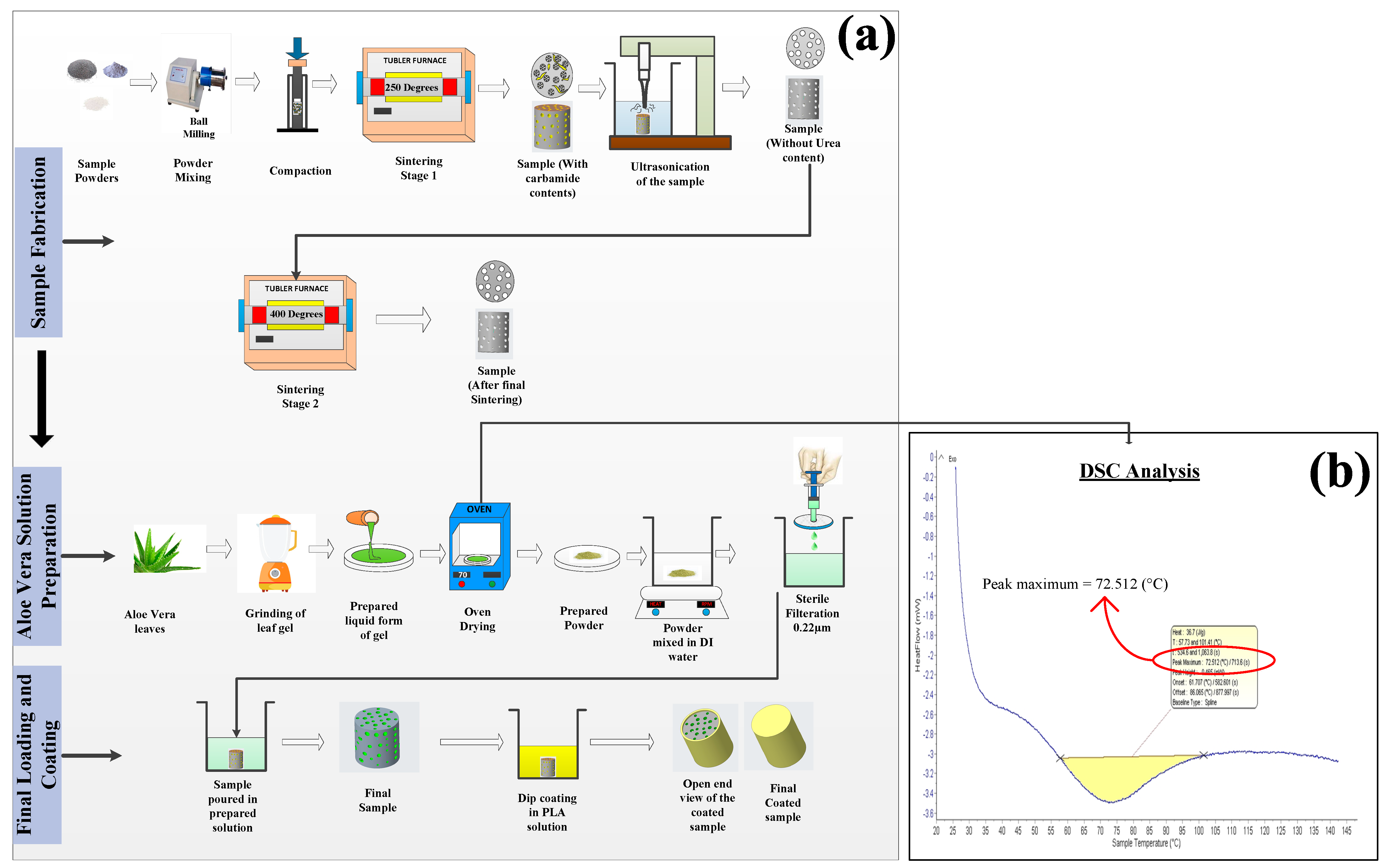
2.2. Sample Preparation

A pictorial view presenting the preparation of the samples is shown in Figure 1a. The powders were initially mixed together with the help of a ball milling setup for two hours at 90 rpm. Different samples were prepared by changing the concentration of carbamide powder (10%, 20%, and 30%) (Sigma Aldrich, USA). The milled powders were then compressed with a hydraulic pellet pressing machine (antsLAB, Maharashtra, India) at a pressure of 100 MPa for one minute. The obtained samples after compression were sintered under a vacuum tube furnace for 250 °C for 4 h (Stage 1 sintering). This resulted in the evaporation of 20% of the carbamide particles, forming pores inside the sample. The density of the samples was measured using a fluid displacement method based on the Archimedes principle. Table 1 shows the density and porosity of the developed samples with different spacer holder concentrations as well as aloe vera and PLA addition samples. The sample with 20% carbamide powder showed promising results as discussed in the further experimentation sections.
The as-obtained pores were then cleaned by flushing out the carbamide particles from the samples with an ultrasonication machine (Chrom Tech, USA) for 45 min. The cleaned sample was then dried using a dryer followed by 6 h desiccation under a desiccator. The dried sample was then kept in a vacuum furnace for 2 h at 400 °C for Stage 2 sintering. The obtained sample was then allowed to cool under vacuum for 12 h.

2.3. Aloe vera Solution Preparation

The incorporation of aloe vera into the developed porous composite sample was realized by dipping samples into the prepared solution of aloe vera as shown in Figure 1a. The as-received leaves of aloe vera were cleaned with 70% ethanol solution followed by distilled water washing. The pulp/gel present inside the leaves was then extracted through a spatula and placed in a grinder to convert it into a liquid form. The desired temperature required for converting aloe vera resin into powder without losing its properties was analyzed by examining the melting temperature of the resin. This was achieved through a differential scanning calorimetry (DSC) technique (Setaram, France) in an aluminum crucible under an inert nitrogen atmosphere as shown in Figure 1b. Finally, the data were generated through Calisto Data Acquisition software (Version 1.493). The optimum temperature required to convert the resin into vapor was observed to be 72 °C. Therefore, the prepared liquid resin was then kept in an oven at 70 °C to remove the water content from the liquid and convert it into a powder form.
For analyzing the minimum percentage of aloe vera required for further experimentation, the prepared powder was then dissolved in distilled water in different concentrations, i.e., 10 mg/mL, 20 mg/mL, 30 mg/mL, 100 mg/mL, and was filter sterilized (0.22 µm) to remove any microbial agents. Finally, the prepared metal sample was immersed in the sterilized aloe vera solution and was allowed to stay in it for 24 h with minimum agitation at room temperature under sterile conditions. The sample was then taken out of the solution flask and was allowed to dry under laminar air flow to maintain its sterility.

2.4. Coating Preparation

The coating of PLA over the porous metal sample was applied through a dip-coating method as shown in Figure 1a. The PLA granules were immersed in a chloroform (CHCl3) solvent with two different concentrations, i.e., 5% and 10%, using a magnetic stirrer at room temperature for 12 h. The developed sample, after aloe vera incorporation, was dip-coated for 1 min with a withdrawal speed of 35 cm/min. The withdrawal speed and coating layer does not affect the surface parameters of the coating [47,48,49]. The samples were then kept at 50 °C for 2 h to eliminate the traces of chloroform. The coating was allowed to settle, and the sample was then placed in a sterile environment to avoid any contamination.

2.5. Characterization

The procured materials were initially tested using Fourier transform infrared (FTIR) spectroscopy (Shimadzu-QATR 10, Kyoto, Japan) to identify different compounds present in the sample powders. Moreover, to analyze the characteristics and presence of several organic compounds and bonding behavior in the developed aloe vera powder, FTIR spectroscopy is a convenient technique. Field emission scanning electron microscopy (FESEM) (ZEISS, Sigma 500, Schaffhauserstrasse, Germany) was used to acquire images of the sample. In addition, the porosity developed in the samples was also observed using this technique. Along with this, microstructure characterization was also performed using FESEM assisted with energy dispersive spectroscopy (EDS) techniques (ZEISS, Sigma 500, Germany). The phase identification was achieved through the X-ray diffraction (XRD) technique (Malvern Panalytical, Malvern, UK) with a scanning range (2ϴ) from 20°–110° setting the K-alpha 2/K-alpha1 ratio at 0.5 and the step size at 0.01313. The measurements were performed through the X’Pert Highscore software (version 2.1) of PANalytical and the values for different phases were obtained using the ICSD cards available with the software.
Moreover, to analyze the chemical structure of the PLA coating developed over the substrate, Raman spectroscopy (Horiba, Montpellier, France) with an excitation source wavelength of 532 nm with a wavelength range of 500–3500 cm−1 was utilized. Moreover, the coating thickness was determined under FESEM microscopy by observing a cross-section of the sample.

2.6. Mechanical and Surface Behavior

Microhardness (HV) and ultimate compressive strength (MPa) tests were performed on the developed samples in triplicate and the average values were plotted. The microhardness was determined using a Vicker’s hardness tester (Metatech Industries, Haryana India) with a 20 gm load and a 15 s dwell time, whereas the compression tests were performed according to ASTM standard E9-89a. The specimens with size 13 mm × 5 mm were compressed via quasi-static compression under room temperature conditions with an automatic compression testing machine (AIMIL instrumentation and technologies) with a strain rate of 8.29 × 10 5   s 1 . All of the tests were performed in triplicates.
The surface properties of the samples were assessed using a surface roughness tester (Mitutoyo, SJ400, Kanagawa, Japan) with a measuring range of 800 µm. The surface hydrophobicity was examined using a sessile drop method with a contact angle measurement technique. A deionized water droplet of size 2 µm was allowed to drop over the sample surface and the final image was taken using a Nikon AF-S DX lens. The contact angle was measured through Image J software (1.54f) and a total of 20 readings were taken and an average value was plotted.

2.7. Immersion Studies

The immersion studies were performed under a cell culture medium due to its similarities with interstitial fluid. The cell culture medium consisted of 95% minimum essential medium (MEM) and 5% fetal bovine serum (FBS) (HIMEDIA, Maharashtra India). The samples were dipped in the solution for seven consecutive days and corresponding changes in weight and pH were analyzed. The samples were placed under a physiological environment, i.e., 37 °C for seven days. The corrosion rate was calculated according to Equation (1). The samples after every interval were removed from the solution in a sterile environment and cleaned with 5% H2CrO4 to remove unwanted debris. Debris could hinder the calculations; therefore, the samples were dipped for 2 min and then washed with sterilized distilled water and dried for 1 h. Afterward, the samples were placed back in the cell culture medium and placed in an incubator for another cycle of measurement. The change in pH of the medium was also calculated simultaneously to analyze the effect of metal degradation.
Corrosion   rate   ( C r ) = 2.1   Δ w A · t   mm / yr
In Equation (1), ‘Δw’ represents weight loss/change in weight in mg, ‘A’ is the surface area in c m 2 , and ‘t’ is the total immersion time in days.
Simultaneously, the samples immersed under corrosion medium were collected for analyzing the Mg ion release in mg/L with an atomic absorption spectrophotometer (AAS, GBC032AA, Australia). The samples were collected in 5 mL Eppendorf tubes for the 1st, 3rd, 5th, and 7th day, and their corresponding values were plotted.

2.8. Antibacterial Behavior

The antibacterial activity of the composite samples was tested against a Gram-negative bacterium, E. coli, and a Gram-positive bacterium, B. subtilis, procured from MTCC, Chandigarh, India. For experimental purposes, both bacterial species were inoculated aerobically in nutrient broth at 37 °C for 24 h. The developed samples were tested against pure E. coli and B. subtilis as controls for the experiment. Then, different concentrations were assessed to analyze the minimum concentration required to produce an antibacterial property with the aloe vera solution. The methodology used to analyze the antibacterial rate of the prepared samples was by calculating the zone of inhibition through the disc diffusion test method. For the growth and spreading of bacteria, nutrient agar (NA) with a ratio of 30 mL/plate was prepared. Further, heated agar was poured into Petri plates and allowed to cool down under a U.V. atmosphere for 3–4 h, and a solid layer was formed in the plates. The bacterial strain (100 µL of E. coli and B. subtilis) was swabbed uniformly over the NA plates with a micropipette (ThermoFisher, USA). Simultaneously, the specimens were placed over the spread bacteria and allowed to incubate for 24 h at 37 °C under physiological conditions. The examination of antibacterial activity was conducted by calculating the inhibition zone through Equation (2).
H = D d 2
where H denotes the inhibition zone diameter (in mm), D is the total diameter of the sample and inhibition zone (in mm), and d is the diameter of the sample in mm.

2.9. Hemocompatibility

To determine the blood compatibility of the prepared samples, hemolysis analysis was performed. A sample of 5 mL of fresh blood from a healthy donor was collected and stored in a tube with the addition of ethylenediaminetetraacetic acid (EDTA) as a decoagulant. The samples were soaked in 5 mL standard PBS solution in a test tube and kept at 37 °C for 45 min. After that, the diluted blood was held in a test tube and placed at 37 °C for one hour. Then, the blood-containing tube was taken out, and 3 mL of blood was added to 9 mL PBS and duly mixed. The mixed suspension was centrifuged for 10 min at 2400 rpm, and the supernatant was removed. This step was repeated four times. After the final wash, the supernatant was removed, and 200 µL of blood was added to 9.5 mL PBS solution after extracting it from the bottom of the tube and mixing properly. The blood suspension was taken and added to tubes containing the samples in a way that the samples were dipped completely. Finally, the optical density (OD) was obtained via UV–vis spectrophotometry (Thermo Fisher, USA) by adding the suspension to a 96-well plate separately. Normal saline with 0.2 mL diluted blood and distilled water with 0.2 mL diluted blood were taken as negative and positive controls, respectively. Henceforth, the % hemolysis ratio (HR) was calculated by using Equation (3).
%   Hemolysis   rate = [ O D t O D v e ] [ O D + v e O D v e ]   × 100
where O D t refers to the optical density of the test sample and O D + v e   a n d   O D v e denotes the optical density of the positive and negative control, respectively.

2.10. In Vitro Cytotoxicity Analysis

2.10.1. Cell Viability

Cell proliferation was analyzed through MTT assays against the MG-63 human osteosarcoma cell line. The cell line was procured from NCCS, Pune, India. The cells were maintained in DMEM high glucose media (AL111, HIMEDIA) supplemented with 10% FBS (RM10432, HIMEDIA) along with a 1% antibiotic–antimycotic solution in an atmosphere of 5% CO2, 18–20% O2 at 37 °C temperature in a CO2 incubator and sub-cultured every 2 days. A 100 µL cell suspension was seeded in a 12-well plate with a cell density of 50,000 cells per well without the test agent and the cells were allowed to grow for 24 h. The samples were sterilized by keeping them under UV radiation for 30 min and gently washed twice with 1× PBS. The samples were then placed in each well of a 12-well plate with sterile forceps and the wells were filled with fresh culture medium. Cells without any sample were considered as untreated and cells treated with doxorubicin with 1 uM/mL concentration were considered as a positive control for the study. Afterward, the plates were incubated for 48 h at 37 °C in a 5% CO2 atmosphere. After the incubation period, the plates were taken out from the incubator and the spent media was removed. MTT reagent (HIMEDIA) was added to a concentration of 0.5 mg/mL of the total volume. The plates were returned to the incubator and incubated for 3 h. After incubation, the MTT reagent was removed and a solubilization solution (DMSO) was added followed by gentle stirring in a gyratory shaker to enhance dissolution. The absorbance was calculated in an ELISA plate reader (Thermo Fisher) at 570 nm and the final cell viability was calculated using Equation (4).
%   Cell   Viability     M e a n   a b s .   o f   t r e a t e d   c e l l s M e a n   a b s .   o f   u n t r e a t e d   c e l l s × 100
The viability of the cells on the developed samples was confirmed through confocal microscopic analysis (Carl Zeiss, Oberkochen, Germany). Cells were cultured in a 12-well plate at a density of 2 × 10⁵ cells/2 mL and incubated in a CO2 incubator at 37 °C for 24 h. The samples were sterilized under UV radiation and then placed in each well with the help of sterile forceps. At the end of the treatment, the medium was removed from the wells and they were washed with PBS. Afterward, the cells were trypsinized with 300 µL trypsin-EDTA solution and were harvested into 2 mL Eppendorf tubes. The tubes were centrifuged at 2000 rpm for 5 min. The cells were stained with 200 µL of staining solution for 10 min. The staining solution was then removed by centrifuging the tubes at 1000 rpm for 5 min and washing with PBS to remove excess dye. Then, 100 µL of cell suspension was carefully loaded on a clean glass slide under a coverslip, and a drop of mounting medium was added before imaging. Observations were made under a fluorescence microscope with a filter cube with excitation 560/40 nm and emission 645/75 nm for EtBr and excitation 470/40 and emission 525/50 for acridine orange. Finally, the images were overlayed by ImageJ Software v1.48.

2.10.2. Cell Apoptosis/Necrosis

The cultured cells in a 12-well plate with a density of 0.5 × 10⁶ cells/2 mL were taken after 24 h incubation. The samples, after sterilization under UV light, were washed with 1× PBS 2 times to ensure sterility. The samples were kept in a 12-well plate where cells without any sample were considered untreated and cells with doxorubicin with 1 µM/mL concentration were considered as a positive control for the study. Afterward, the cells were incubated for 48 h, and at the end of treatment, the medium was removed from all of the wells and they were given a PBS wash. The PBS was removed and 200 μL of trypsin-EDTA solution was added and incubated for 3–4 min at 37 °C. Then, 2 mL culture medium was added, and the cells were harvested directly into 12 × 75 mm polystyrene tubes. The tubes were centrifuged for five minutes at 300× g at 25 °C and the supernatant was decanted carefully followed by washing the cells twice with PBS. Then, 5 μL of FITC Annexin V was added to the cells with gentle vortexing and they were incubated for 15 min at RT (25 °C) in the dark. Then, 5 μL of PI was added along with 400 μL of 1× Annexin Binding Buffer to each tube and vortexed gently. Finally, the analysis was performed by Flow Cytometry (BD FACS Calibur, Indianapolis, India) immediately after the addition of PI. The calibration of the results was carried out using Kaluza analysis 2.1 (Beckman Coulter, USA) software.

2.11. Statistical Analysis

The experimental results were obtained by considering different parameters. These parameters were tested against the obtained porosity through different amounts of spacer holder and compressive strength. The statistical analysis was performed by one-way analysis of variance (ANOVA) using MINITAB 18 statistical software. The statistical significance was defined as: p < 0.05 compared between groups.

3. Results

3.1. Characterization

The FESEM characterization of the samples is shown in Figure 2a–f, highlighting the surface morphology of the developed samples. A high level of micropores and macropores can be observed on the surface of the samples when analyzed under a microscope. However, no other surface damage was observed at the macro level. Nonetheless, when the sample was dipped under the aloe vera, the surface texture was somehow changed compared to the initial conditions. The dried aloe vera particles were observed under the microscope as a thin crusty layer covering the entire surface of the sample. After the coating, the entire surface looks like a defect-free surface, enveloped under a PLA film. The EDS analysis in Figure 2g–i revealed the presence of Mg and components of HA in the sample; however, these peaks tended to diminish after the coating with a new peak of Cl due to the availability of a small amount of chloroform in the coating.
Figure 3a shows the XRD patterns of the porous samples along with the aloe vera incorporated and PLA-coated samples. Mg and CaCO3 show prominent peaks in the analysis and are marked correspondingly. The Mg peaks were available at 32.1°, 34.6°, 37°, and 64.2°, whereas 44.2° and 57° were the peaks shown by CaCO3. These peaks are in agreement with the data available in references [50,51,52] as well as with the JCPDS cards for Mg (035-0821) and CaCO3 (47-1743). The absence of any other distinguishable peaks shows that during the sintering, no further interfacial reactions occurred between the molecules of the materials. Moreover, Mg being the highest quantity projected the maximum peak intensity followed by CaCO3, which tended to reduce after aloe vera incorporation and PLA coating.
In comparison to metals, polymers often have lower atomic masses. The polymer coating on the coated sample may be able to absorb X-rays more efficiently than the metal substrate underneath when they interact with it. The intensity of the metal-related XRD peaks decreased consequently. Another important factor is the polymer coating’s thickness. Greater attenuation of the X-rays by thicker coatings could result in a more noticeable peak intensity reduction. Therefore, the intensity of the peaks was reduced after coating with PLA; nonetheless, their presence was still discernible due to the low thickness of the polymer film.
This shows that the coating was highly effective without any defects as the electrons from the electron beam could not penetrate through it, which, if possible, could show some peaks of the base substrate. The observed behavior is suitable in the current scenario as the better the coating, the better the protection of the substrate material, leading to improved mechanical and corrosion properties. These properties are discussed briefly in the mechanical and corrosion properties section. Moreover, cross-sectional imaging revealed the coating thickness (Figure 3b). It can be seen that with 5% PLA, the coating thickness achieved was 39.28 µm, whereas, with 10% PLA, the thickness achieved was 78.56 µm.
The porosity of the samples was also observed under FESEM microscopy, as shown in Figure 4a–f. The microscopic characterization revealed two different types of pores developed in the sample, with an average size of 42.12 µm and 22.17 µm. However, when the sintering temperature was increased to 450 °C, the pores started to reduce, and as the temperature reached 500 °C, the material became solid with negligible porosity compared to 400 °C. The evolution of spacer holder particles from the substrate is shown in Figure 4g.

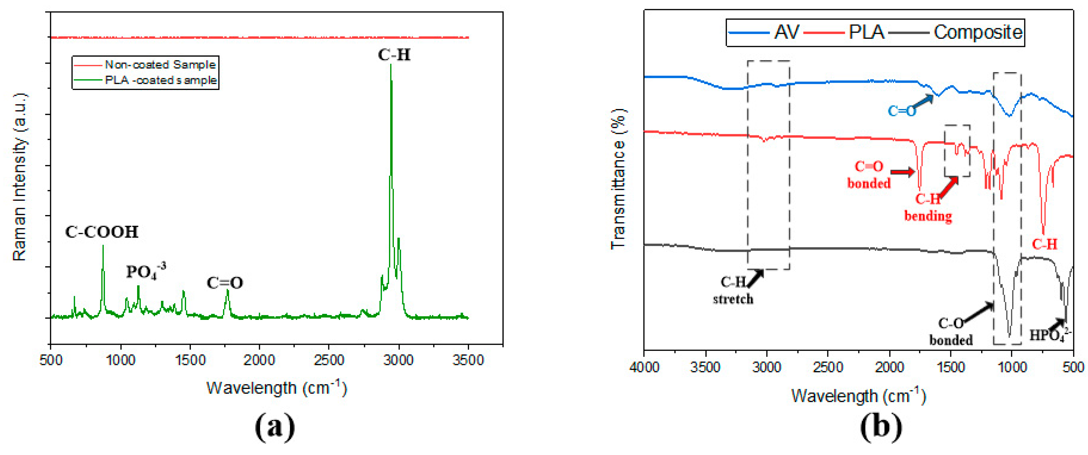
3.2. FTIR and Raman Spectroscopy

The chelation among the elements of the composite material developed and the coating formed over the surface were analyzed through FTIR and Raman spectroscopy techniques. As depicted in Figure 5a, the composite developed after coating showed similar FTIR peaks as that of pure aloe vera and PLA. The composite revealed the peak of C-O and C-H at 1823 cm−1 and 1919 cm−1, respectively. Furthermore, the composite sample had a collection of peaks at 600 cm−1 and 651 cm−1, nominal to HPO42−. These peaks corresponded to the results presented in previous studies [53,54]. The native materials were assessed to compare the available peaks with the developed composite peaks. The bonding mode can be determined by analyzing the presence of C=O and C-H bending and stretching vibrations, and their peak positions. The C=O peaks for aloe vera were observed at 1601 cm−1, whereas for PLA, they were slightly shifted to a higher frequency range and developed a sharp peak at 1746 cm−1. Meanwhile, a broad peak at 3445 cm−1 was observed for aloe vera and the synthesized composite, corresponding to the terminal hydroxyl group. Also, a characteristic C-H stretching frequency was observed for all of the samples, which agrees with the results obtained from the literature [55,56,57,58].
Raman spectroscopy was used to provide insight into the coating developed over the porous aloe vera-loaded substrate through dip-coating. According to Figure 5b, the PLA-coated sample showed characteristic bands at 2995 cm−1 and 2990 cm−1, which are in agreement with the results obtained in [59,60]. These bands are attributed to the symmetric and asymmetric stretching vibration of the C-H (νas/sCH3) bond of the PLA chain [61]. The stretching vibration of C=O (νC=O) can be observed at 1763 cm−1, whereas an asymmetric deformation vibration of the CH3asCH3) bond and stretching vibration of the C-COO (νC-COO) bond were seen at 1445 cm−1 and 888 cm−1, respectively. The presence of hydroxyapatite (with 15% conc.) in the sample highlighted an additional PO4−3 band at 1200 cm−1. The spectrum covered approximately all of the polymer-specific bands of PLA along with some minor bands of the substrate material. Another spectrum for non-coated material was also observed under Raman analysis, which showed no such intense peaks due to the presence of zero polymeric bonds. Although some microbands of PO4−3 were observed at 585 cm−1 and 425 cm−1, their intensity was relatively low.

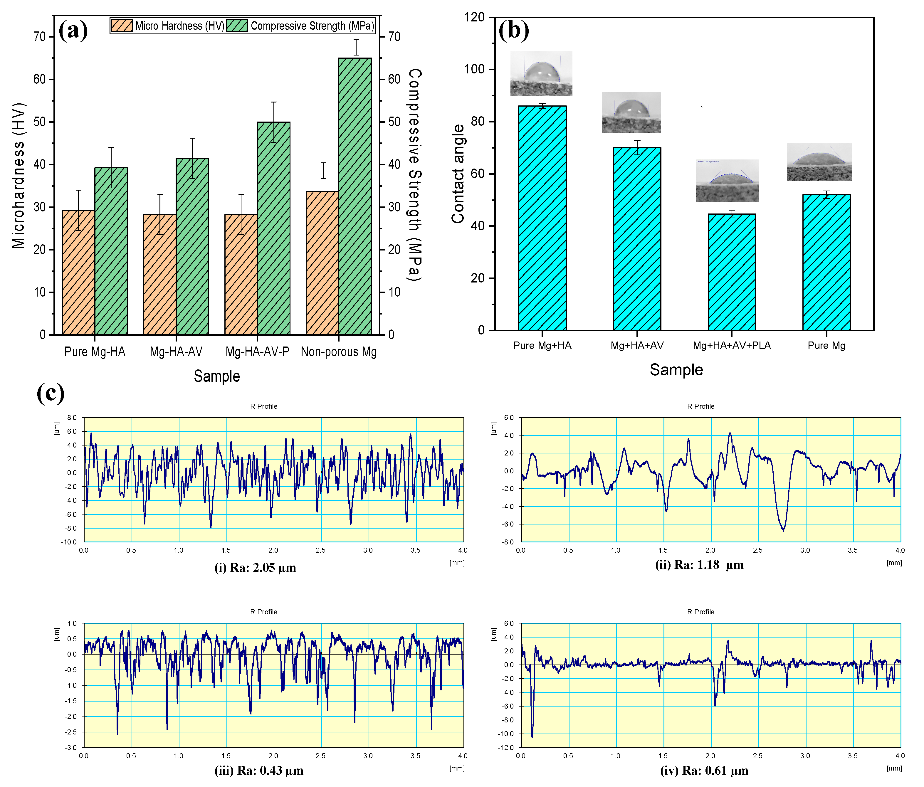
3.3. Mechanical and Surface Properties

The mechanical properties of the samples were analyzed in terms of microhardness and ultimate compressive strength (UCS). Figure 6a depicts the graphical representation of both experiments performed. It was observed that the microhardness and UCS values tended to decrease when compared to pure Mg. The minimum microhardness of 28.37 HV was displayed by the sample coated with PLA, whereas the non-coated sample showed a microhardness value of 29.35 HV. Moreover, the sample loaded with aloe vera solution did not show a significant change in the hardness value when tested under similar conditions. As compared to non-porous Mg, the values seemed to be quite low.
In addition, the ultimate compressive strength (UCS) of the sample was analyzed at a strain rate of 8.29 × 10 5 s−1. It was observed that the coating provided an impressive resistance against compressive load by enhancing the UCS value of the porous Mg sample to 50.1 MPa. The UCS value of the non-coated sample was seen to be 39.28 MPa and that of the aloe vera solution loaded at 41.5 MPa. The porosity of the samples plays an important role in strengthening the matrix; the higher the porosity, the lower the strength [62]. These results are consistent with the compressive characteristics of cancellous bone [63].
In addition, the surface properties were examined to analyze the effect of coating and aloe vera impregnation on the surface roughness and surface hydrophobicity of the samples. Figure 6b,c shows a graphical representation of the developed samples’ average surface roughness and contact angle values. It can be observed that the coating reduced the roughness (0.43 µm) of the surface immensely, with a corresponding reduction in surface hydrophilicity. However, the native sample showed a higher level of roughness value with a high amount of surface hydrophobicity. Adding aloe vera solution to the substrate material also helped subside the roughness values by 1.13 µm from 2.05 µm. The maximum hydrophobicity with a contact angle value of 88.2° was achieved with the base matrix material, whereas the minimum contact angle value of 45.2° was attained after coating with a layer of PLA. These properties are beneficial when considered for cell adhesion over the surface, as discussed in the forthcoming sections.

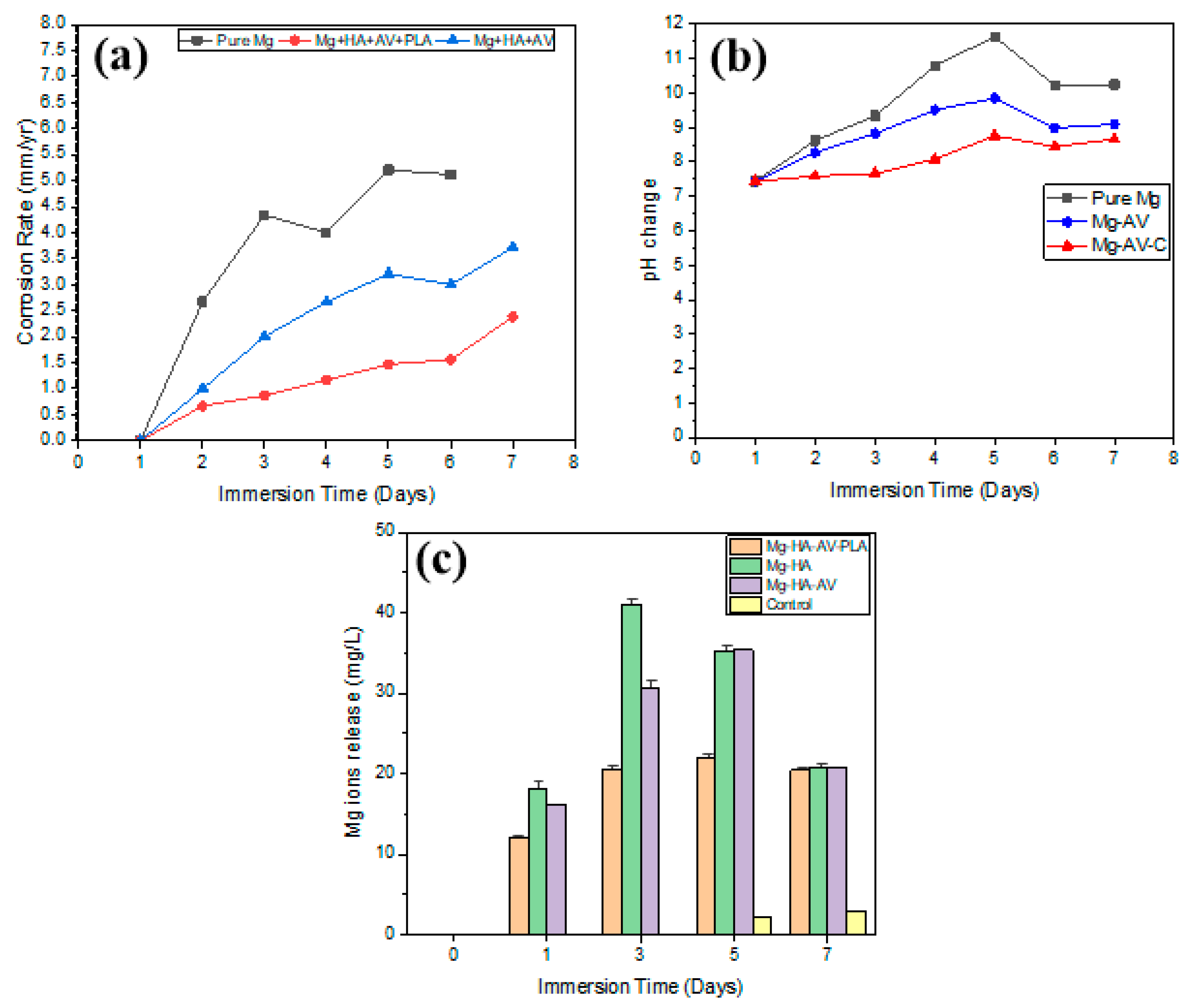
3.4. Corrosion Properties

The corrosion rates of the samples were analyzed through the weight loss technique by immersing them in a cell culture medium, as shown in Figure 7a. The corresponding change in pH (as shown in Figure 7b) was also assessed to develop an insight into the reaction of the samples with the culture medium. Cell culture medium was selected for the study to inspect the real-time corrosion rate under in vitro conditions. It was observed that the successful incorporation of aloe vera into the porous matrix helped bind the substrate particles together, reducing the corrosion rates. These rates seemed to be much lower when a layer of PLA was coated over the substrate, which helped restrict the composite’s rapid corrosion. The maximum corrosion rate achieved was 5.2 mm/yr with a pH value of 10.1 for the pure porous Mg sample, whereas the minimum value was 2.7 mm/yr with a pH change of 8.7 for the sample with aloe vera incorporation and PLA coating after the seventh day of degradation. Moreover, the barrier provided by the PLA resulted in reduced corrosion rates with a percentage reduction of approx. 40% as compared to the substrate material.
In addition, the dissolution of Mg into the corrosion medium was examined through the atomic absorption spectroscopy technique, as shown in Figure 7c. A high amount of leaching of metal ions into the medium would increase the solution’s pH level by releasing more hydrogen [64,65]. Therefore, the Mg ion concentration released into the corrosion medium was analyzed, and it was observed that the release rate increased with the immersion time. Nonetheless, the release of Mg ions tended to reduce when aloe vera solution was added to the porous substrate. This was seen to decline more after coating with PLA. The maximum release of 42 mg/L in the pure substrate was observed on the third day of immersion, followed by the aloe vera-loaded sample with 31.5 mg/L and the PLA-coated sample with a 20.7 mg/L Mg ions release rate. Afterward, this release rate seemed to reduce due to the oxide layer developed across the substrate, which restricted the Mg ions release into the immersion medium [66,67]. In the case of the aloe vera loaded sample, a possible reason for an initial burst of Mg ions can be the surface abrasion of the substrate when it was in contact with the corrosion medium. Once the surface layer was abraded, the aloe vera solution, which was infused inside the substrate, resisted the release of Mg ions into the medium. However, when PLA was coated over the substrate, the release rate was constant, showing lower Mg ions release due to effective coating development.

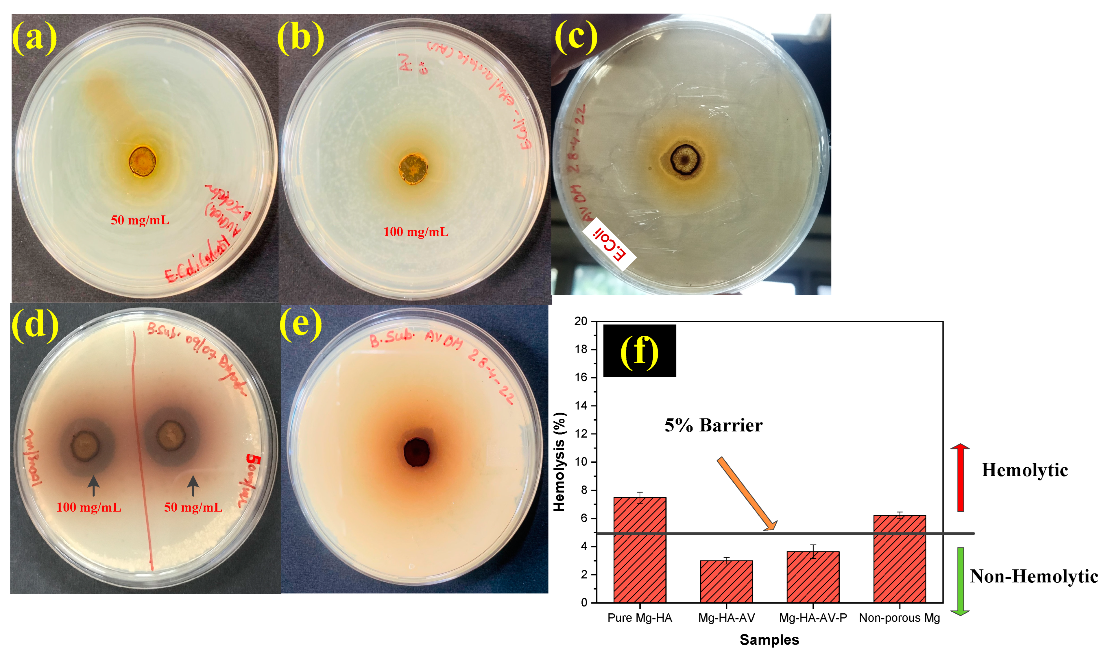
3.5. Antibacterial and Hemocompatibility Analysis

The antibacterial behavior of the developed composite material is shown in Figure 8a–e. The degradation of Mg in the physiological medium results in the dissolution of Mg2+ ions. This could work as an antibacterial function for Mg against different bacterial agents. However, excessive degradation could lead to implant failure at an early stage of implantation. Moreover, during carpal injuries, major issues during implantation are inflammation due to bacterial infection caused by the native material of the implant, despite the antitoxic properties of the material used [68,69]. Therefore, an infusion of aloe vera solution was applied to enhance the antibacterial rate of the composite developed in this study.
The aloe vera solution was initially tested without adding a porous substrate against Gram-positive B. subtilis and Gram-negative E. coli. The experiment was executed by considering the different concentrations of aloe vera solution mixed with DI water, taking a minimum concentration of 50 mg/mL and a maximum of 100 mg/mL. The concentration was tested against both strains of bacteria. It was assessed that aloe vera solution, in pure liquid form, was impressively effective against B. subtilis but did not show any inhibitory effect against E. coli. However, after incorporating it with the porous substrate and PLA coating, it showed a higher inhibitory effect against E. coli with an inhibitory zone of 30 mm. In contrast, this value was lower, i.e., 18.2 mm, against B. subtilis.
The hemocompatibility of the developed composite samples is shown in Figure 8f. A material is said to be hemocompatible if its hemolysis percentage is below 5% [70,71]. Therefore, in the current study, it can be observed that when aloe vera solution was added to the porous substrate, the hemolysis percentage was 3%, whereas after coating with PLA, it escalated to 3.7%. However, it was below the standard limit bar of 5%, making these samples hemocompatible. Of note, the native materials were hemolytic in nature, with porous Mg showing 7% and pure Mg 6% hemolysis percentage.

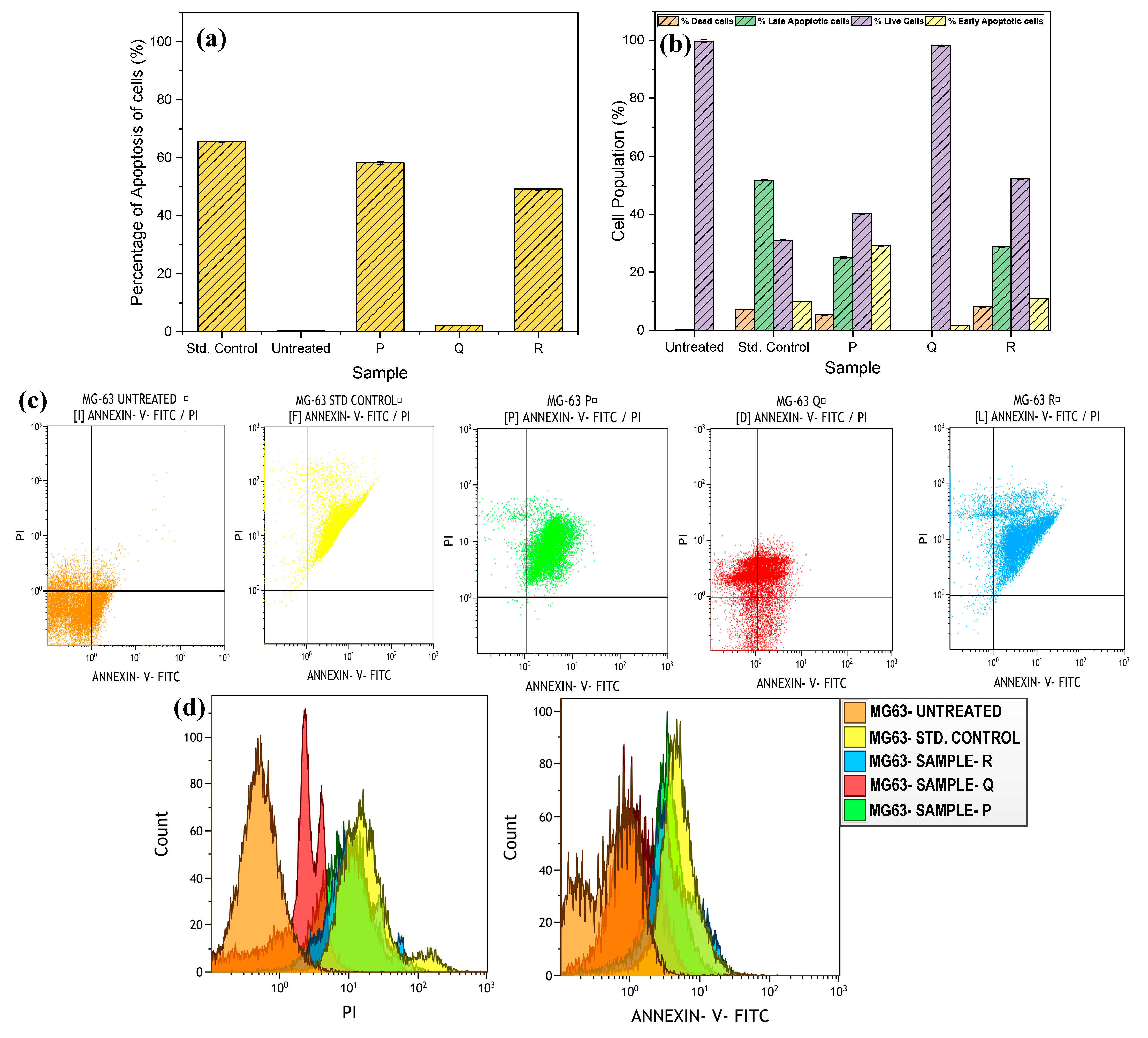
3.6. Cytotoxicity Analysis

The level of cytotoxicity and adequate cellular response are essential to define an implant material’s success rate. Here, the developed porous Mg-based composite samples interacted with osteoblast-like cells (MG63 cells) to assess the viability of the cells and their apoptotic reactions. The cell viability of the samples was evaluated using an MTT assay to explore the potential of any cytotoxicity of the composites. In addition, the live/dead cell ratio was discovered using the confocal microscopy technique, which provided a clear insight into the composite’s reaction with the cells. According to Figure 9, the samples consisting of aloe vera loading inside the porous substrate showed impressive cell viability of 98.64% followed by the composite coated with PLA with 84.18% cell viability. However, the sample without aloe vera inclusion and PLA coating showed a higher amount of cytotoxicity, with a cell viability of 48.37%. The corresponding confocal images highlight a similar trend of the live/dead cell ratio after reacting with the developed composite samples.
Another study was performed to examine cell necrosis and apoptosis due to interactions with the composite samples. The MG63 cells were incubated with the porous Mg–HA composite (Sample P), aloe vera loaded composite (Sample Q), and PLA coated composite (Sample R), along with a standard control (doxorubicin added to MG63 cells) and untreated MG63 cells for 48 h. The apoptosis was quantified by flow cytometry using cells cultured with the different samples and was stained with FITC Annexin V and PI.
Figure 10 shows a representative experiment conducted, which depicts the fluorescence intensity in MG63 cells treated with different samples. The percentage of apoptosis of cells for different samples is highlighted in Figure 10a, and it can be observed that sample Q, which consists of loaded aloe vera solution, showed only 2.11% of cells that were apoptotic, whereas, when the coating was applied (sample R), this percentage increased to 49.2%. Nonetheless, this rate was quite lower than that of the pure composite with aloe vera and coating, having a % apoptosis of 59.5% compared to the standard control and untreated MG63 cells. Moreover, Figure 10b depicts the data regarding the percentage of dead cells, live cells, early apoptosis, and late apoptosis of MG63 cells against different samples. It was observed that the aloe vera loaded sample (sample Q) had a maximum percentage of live cells, i.e., 98.29%, with an approx. negligible percentage of late apoptosis and dead cells, and only 1.71% of early apoptosis was shown by sample Q. In addition, adding a PLA layer over the substrate reduced the percentage of live cells to 51%, but it was comparatively higher than the pure porous substrate with only 39.9% live cells. Table 2 shows the data regarding the percentage of live/dead and early/late apoptosis of the cells.

4. Discussion

This study aimed to develop a porous Mg-based composite with HA as a reinforcing material for scaphoid fracture applications. This material was designed keeping in mind its multitude of capabilities via incorporating natural plant-based solutions that could work as a therapeutic drug. For this, the aloe vera plant was selected because of its extensive antibiotic properties and was infused into the porous matrix in liquid form. This material was coated with a layer of PLA to encapsulate this drug inside the substrate and protect against immediate degradation. The authors believe that this approach of developing a natural plant-based drug-loaded composite material with effective mechanical and biocompatibility properties is reported here for the first time. The fabrication of the porous Mg–HA composite has been successfully carried out through a powder metallurgy technique using carbamide as a spacer holder material.

4.1. Microstructure, Mechanical Properties, and Corrosion Behavior of the Developed Porous Composite

The current work focused on synthesizing three different types of composite samples: a porous Mg–HA substrate (sample name: Pure Mg) without any additions; an aloe vera loaded Mg–HA substrate (sample name: Mg-HA-AV); and a PLA-coated aloe vera loaded Mg–HA substrate (sample name: Mg-HA-AV-PLA). It can be observed through the EDS results that the peaks of Mg seemed to reduce as the aloe vera solution was added and diminished further after PLA coating. This can be due to a barrier provided by the PLA layer to X-ray signals generated by the electron beams striking the surface of the sample. Moreover, the surface developed through coating was crack-free and successfully covered the entire sample. In addition, the XRD results showed a reduction in peak intensity, which could be the hindrance developed by the aloe vera solution and PLA layer to the electron beams. Although no additional phases were seen to be formed after the sintering process, the development of a CaCO3 compound was observed with sharp peaks showing the crystallinity of the compound in the matrix. The presence of HA was also validated by the Raman intensity peaks of the PLA-coated sample with a corresponding PO4−3 stretching vibration. Moreover, the FTIR spectra of the composite developed and its native materials highlighted the presence of C-O bonded peaks that were in agreement with that of the PLA and pure aloe vera peaks. In addition, the presence of HPO4−2 depicted the successful reinforcement of HA in the matrix.
The developed composites were compared with the non-porous Mg material, and it was found that the presence of a high amount of porosity reduced the mechanical strength of the composites. Nonetheless, it was higher than the effective strength of cancellous bone [72]. The pores developed were seen to be of two types: micropores and macropores with a pore diameter of 44.72 µm and 26.12 µm, respectively. The addition of aloe vera did not improve the strength of the substrate material; however, when a coating layer of PLA was added to the substrate, it immensely enhanced the material’s compressive strength by 28% compared to the Pure Mg sample. A surprising phenomenon was also observed in this study: after the incorporation of aloe vera into the porous substrate, a sudden increase in the compressive strength of the material was encountered. This could be due to the sticky nature of the aloe vera that generated a binding factor in the material, increasing its strength relatively. The increase in strength of the composite after PLA coating could be due to resistance to the load provided by the polymeric layer to reach the substrate material, hence increasing its strength. Despite its higher strength, no change in microhardness was observed in the samples.
Although there was a trifling amount of reduction in microhardness values when aloe vera and PLA were added, the values were quite similar. Impressive surface properties were induced in the samples after coating with PLA. It was observed that the surface hydrophilicity was increased with a coating that is in good agreement for osteoblast cells to attach and proliferate over the surface, as discussed in the next section. The sample without coating or aloe vera addition consists of a highly porous surface, resulting in higher surface roughness, hence more hydrophobicity. Whereas, improved hydrophilicity also resulted in enhanced surface roughness due to the smooth surface developed after coating, as depicted in the FESEM micrographs. Surface roughness is a factor that is directly proportional to hydrophobicity. As the roughness increases, the hydrophobicity increases, leading to the more spherical shape of the water droplets [73,74,75]. The developed results are in agreement with the above statement.
To work as an effective bioimplant, the developed composite should withstand the corrosive physiological environment until the bone heals. In the current study, a porous Mg composite has been developed, resulting in rapid corrosion in the cell culture corrosion medium due to its porosity. However, when introduced to an aloe vera solution, its degradation reduced slightly, which might be due to the sticking of the substrate particles together with aloe vera working as an adhesive substance. However, it did not resist the Cl ions present in the medium to corrode the composite material, leading to an increase in the medium’s pH level. The PLA coating layer developed through dip coating provided a barrier to work against the corrosive media by reducing the corrosion rate of the composite material immensely. Nonetheless, as the PLA layer degraded, the corrosion proceeded similarly to the other samples. The PLA layer provided prolonged resistance toward the corrosion, which is necessary for an implant to work until the bone heals. Shi et al. [76] provided a mechanical interlocking phenomenon, showing that the Mg-O chemical bond provided a substantial binding property of PLA with the Mg surface. Based on the reported achievement, the developed layer of PLA worked in a similar way to resist the corrosion of the porous Mg substrate.
The release of Mg ions is an important phenomenon to analyze the corrosion behavior of the material. In the present study, the Mg ion release rate (mg/L) was examined through atomic absorption spectroscopy for the 1st, 3rd, 5th, and 7th days of immersion. The Mg ion release was relatively high on the third day of immersion, as related to the corrosion rate on the same day. Afterward, the release rate became constant. This can be due to the development of a Mg(OH)2 layer over the surface of the sample, which restricted the further release of Mg ions from the substrate [77,78]. Although the PLA layer resisted the Mg ion release impressively, a small amount of release was observed continuously. This could be due to the continuous degradation of the PLA layer slowly, leading to Cl ions penetrating the layer and reaching the Mg substrate, hence corroding the substrate and releasing Mg ions. However, this rate was very low as compared to the untreated Mg sample and aloe vera loaded sample, which proves the effectiveness of the coating in delaying the corrosion rate for a sustained amount of time. The PLA layer provides a blocking and covering effect by working as a sealing layer for the porous substrate for a certain amount of time. Figure 11a shows the detailed phenomenon of Mg ion release from the PLA-coated composite.

4.2. In-Vitro Biocompatibility Analysis

Along with an excellent degradation rate and better strength, an implant material should also possess a high level of biocompatibility. The non-toxic behavior of a biomaterial is an essential component to be considered for a standard material for bone therapeutic applications. However, the biodegradation of a material can cause a certain level of toxic effects in the body and inflammatory effects due to bactericidal actions. Maintaining a trade-off between strength, resistance against bacterial attack, and cytotoxicity is an essential factor where many studies seem to fail. The porous Mg–HA composite developed in the present study showed excellent biocompatibility and effective mechanical and surface properties. The addition of aloe vera as an anti-inflammatory and antibiotic solution fashioned the current composite into an antibacterial and cytocompatible biomaterial. Aloe vera has several antiseptic properties, holding components such as salicylic acid, urea nitrogen, sulfur, phenols, and cinnamomic acid. These components show inhibitory effects against fungi, bacteria, and viruses. Moreover, they inhibit the cyclooxygenase pathway and resist arachidonic acid from producing prostaglandin E2 [79]. This property helps develop anti-inflammatory effects for bone therapeutic applications by reducing the inflammatory issues triggered due to implant grafting.
The bacterial activity was analyzed against the different concentrations of aloe vera solution initially, i.e., 50% to 100%, to assess the minimum concentration of aloe vera solution required to resist bactericidal activity. It was found that a minimum of 50%, i.e., 50 mg/mL of aloe vera solution, is required to resist bacterial growth. However, these results were effective only against Gram-positive (B. subtilis) bacteria but not Gram-negative (E. coli) bacteria. Although the solution showed only contact inhibition against E. coli, the PLA coating had a higher inhibitory effect. In the case of B. subtilis, the solution and when infused in the substrate along with the PLA coated sample, showed similar effects. Various authors have highlighted a similar behavior of plant extracts against bacterial strains in their studies. These studies showed a higher inhibitory effect against Gram-positive bacteria as compared to Gram-negative [80,81]. This could be due to several factors, including the presence of many components in the aloe vera plant, such as phenols, polysaccharides, etc., and a different secondary metabolite. Moreover, aloe vera consists of certain other components named hydroxylated phenol, also known as pyrocatechol, which is known to have a highly toxic effect against microorganisms. These components are highly effective against Gram-positive bacteria. The Gram-negative bacteria’s outer membrane consists of lipopolysaccharides responsible for creating a barrier against these components. Moreover, the periplasmic space of enzymes in the Gram-negative bacteria tends to break down molecules entering from outside the bacteria wall, reducing their antibacterial effects [82,83].
Hemocompatibility is a crucial factor that validates the implant’s toxicity when encountering blood cells. In the current study, the developed material showed impressive hemolytic behavior. To be considered non-hemolytic, a material should possess a percentage hemolysis rate below 5%; above that, it is considered unsuitable for further biomedical applications [84]. The aloe vera addition to the porous Mg–HA substrate showed an excellent hemolysis rate relative to other samples. Nonetheless, when coated with PLA, this percentage increased by 22% but was entirely below the 5% barrier. The optimum percentage of PLA addition was ≤5%; if this value is exceeded, it will lead to a high level of hemolysis. Hemolysis consists of rupturing of erythrocytes (RBCs) and the release of hemoglobin in the plasma. It has been known that the lysis of the erythrocytes occurs due to osmotic imbalance or physical disturbance due to interactions with external agents [85]. The presence of aloe vera increases the collagen amount and collagen cross-linking in the body, hence promoting tissue healing [86]. In addition, aloe vera consists of several other healing substances, such as glycoproteins and polysaccharides, which help reduce pain and work. Therefore, aloe vera addition to the porous composite circumvents the lysis of erythrocytes, hence decreasing the hemolysis rate by providing a protective response to the cells.
A composite material designed for tissue engineering applications must possess minimum cytotoxicity. The current study showed that the porous Mg composite, consisting of an aloe vera solution as the therapeutic agent, had very low cytotoxicity, maintained even after coating with PLA. However, PLA coating reduced the cell viability by some amount, but further modifications and better sterilization could escalate these factors. Although this does not change the fact that the PLA coating does not show any immense signs of cytotoxicity with a viability percentage above 70%, the literature showed that this material is highly biocompatible and can be utilized in orthopedic applications [87,88]. The current study utilized MTT assays to analyze the mitochondrial activity, which is related to cell proliferation [89]. Confocal microscopy revealed that the growth of osteocytes was normal and this is a good sign of a healthy response to the developed material. Similar results were obtained by Carter et al. [90], where the presence of aloe vera enhanced the viability of 3T3 cells impressively. Many studies have considered aloe vera an added therapeutic agent and proven it is an excellent tissue-healing antibiotic material. Aloe vera in liquid form consists of 99% water, whereas in the dry state, it contains 55% polysaccharides, 17% sugars, 16% minerals, 7% proteins, 4% lipids, and 1% phenolic components [91]. These components play a significant role in tissue regeneration and provide therapeutic support to bone fragments’ healing process. The flow cytometry studies showed similar results with aloe vera-loaded implants exhibiting the lowest level of apoptosis (both early and late) and 98% live cells. The cytotoxicity studies in the present work developed an insight into the cell death by apoptosis due to the reaction with the developed composites. A significant cellular response, including large histiocytic infiltration and particle phagocytosis, is evoked when composite degradation is prevalent. This causes an increase in the apoptosis rate, hence reducing the percentage of live cells. However, aloe vera’s presence prevented this phenomenon from occurring by providing subsequent therapeutic shielding of the cells.
Aloe vera consists of several biological components that provide several transport and activities of biological factors required in tissue regeneration. A mannose-rich polysaccharide called glucomannan and a growth hormone called gibberellin interact with the fibroblast’s growth factor receptor to promote the activity and proliferation of the cell. Aloe vera based implants have also been promoted as an anti-cancerous drug in a study proposed by Sridhar et al. [91], where curcumin/aloe vera was used with PCL electrospun membranes. Suganya et al. [92] developed electrospun fiber mats with aloe vera blended with PCL as dermal substitutes with excellent cell proliferation produced by 10% aloe vera addition. Therefore, based on the above literature, the proposed implant presented in the current study proved an effective implant composite material. The mechanism showing an effect of aloe vera addition on the inhibition against bacterial cells and growth of bone regeneration cells is shown in Figure 11b. The incorporation of aloe vera in liquid form demonstrated impressive cell viability with reduced apoptosis. Moreover, the development of PLA coating over the substrate showed promising results to be considered a non-cytotoxic biocompatible material for application in scaphoid fractures. In addition, further research can also bring forth the proposed composite’s applications in other orthopedic injuries and tissue regeneration functions.

5. Conclusions

The current study witnessed the development of a multifunctional biomaterial composite with an extensive capability to provide therapeutic assistance for scaphoid fractures. The fabrication of a porous Mg–hydroxyapatite composite was successfully completed with the help of a powder metallurgy technique. The following conclusions can be drawn:
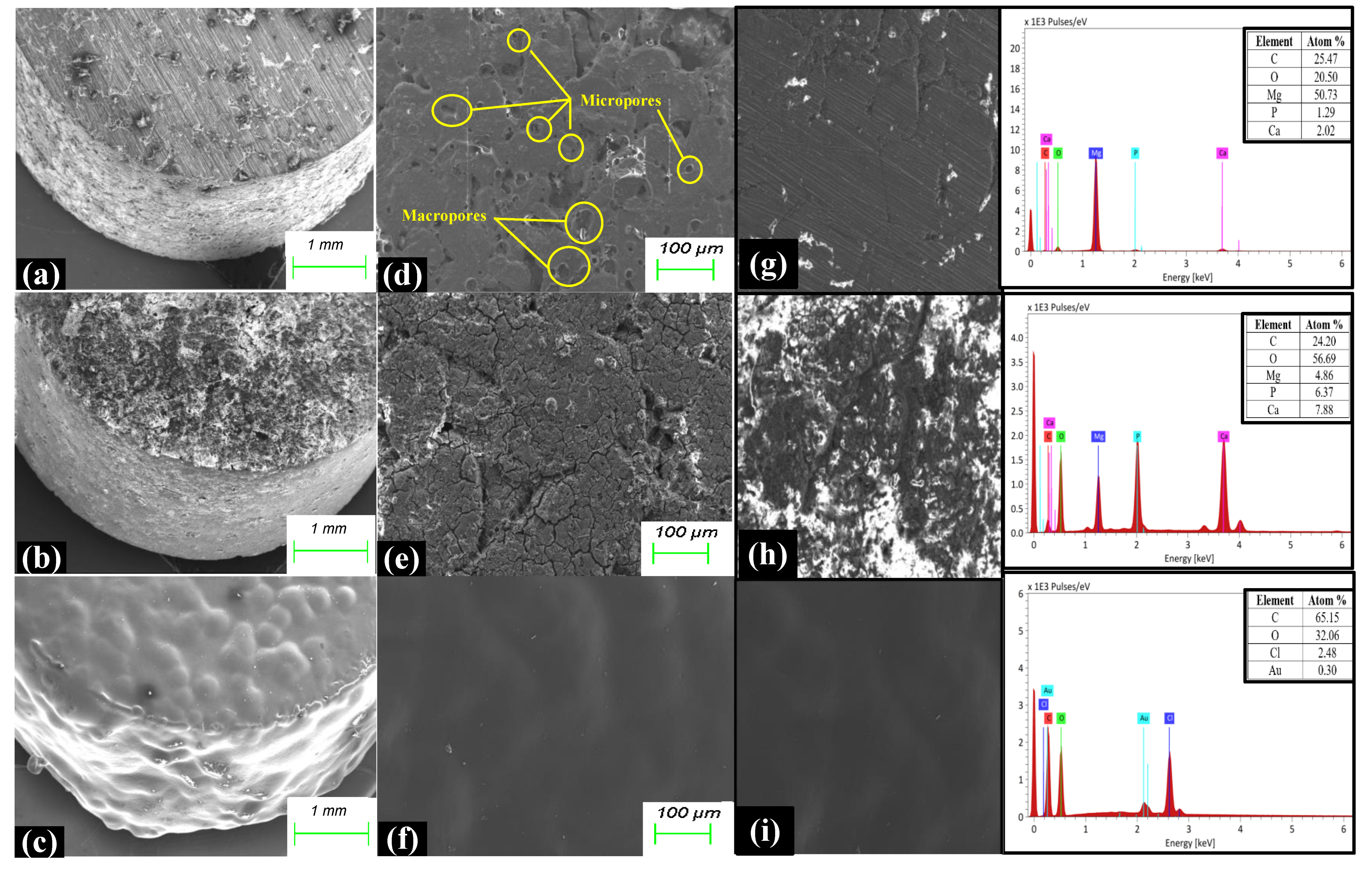
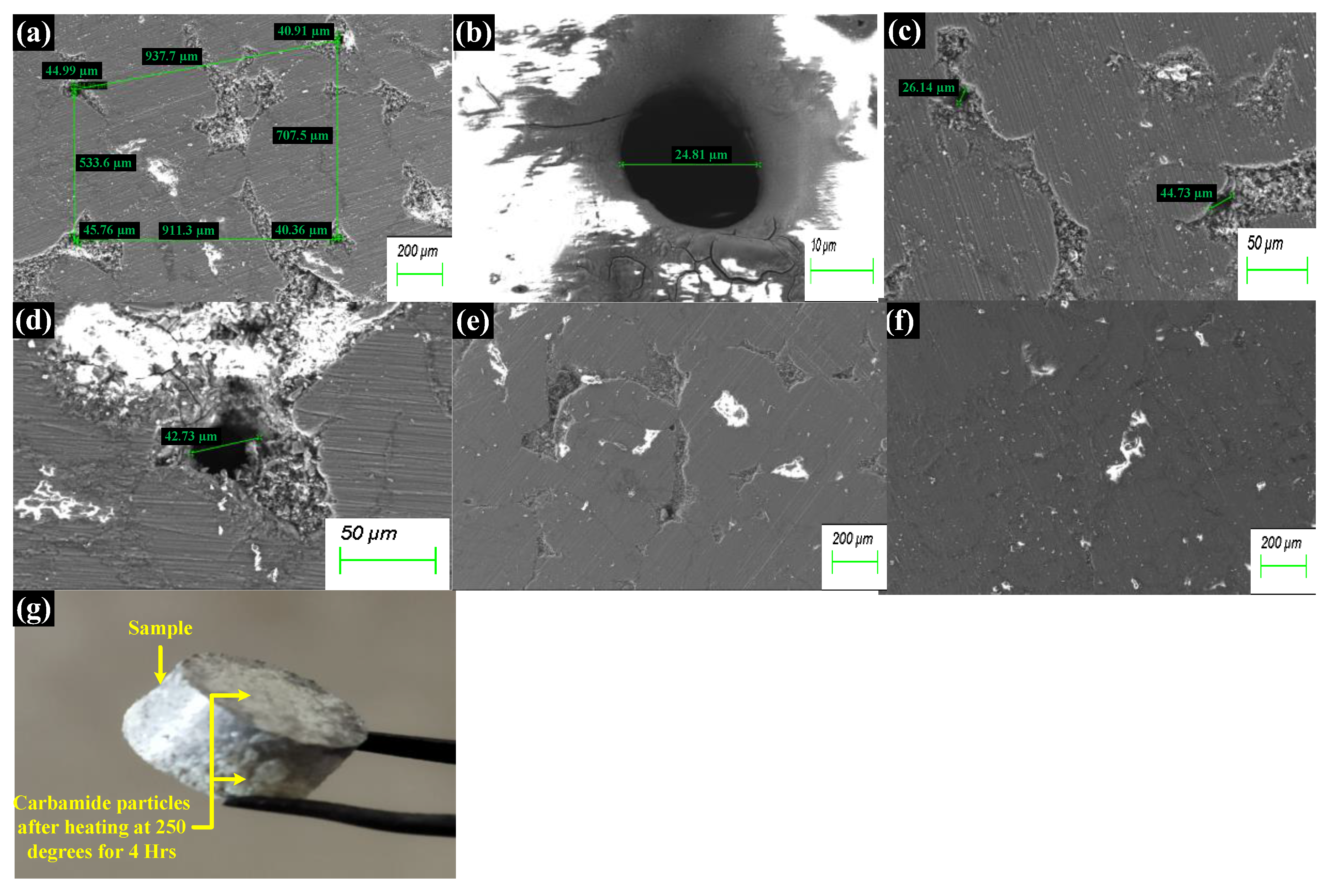
  • The microstructure images revealed two different pore sizes generated in the matrix using a spacer holder technique. The suitable porosity of 72.25% was optimized with 20% carbamide for aloe vera incorporation with an adequate strength.
  • The successful PLA coating and aloe vera incorporation led to an excellent microhardness of 28.37 HV and UCS value of 50.1 Mpa, with an impressive surface hydrophobicity and reduced surface roughness.
  • A reduction in corrosion rates was observed after PLA coating, leading to a decline in the release of Mg ions, hence increasing the longevity of the composite in the cell culture medium.
  • The minimum %age of aloe vera solution (in mg/mL) was formulated to be 50% (50 mg/mL) for successful antibacterial and hemocompatibility properties.
  • The in vitro cell cytotoxicity experiments depicted magnificent cell viability of 98% after the addition of aloe vera to the porous matrix. In addition, a decline in the cell apoptosis rates was observed with aloe vera samples as compared to the pure Mg–HA substrate.
Finally, it can be concluded that the developed composite sample with aloe vera working as a therapeutic drug can be proposed as an advanced multifunctional biomaterial composite for orthopedic applications, especially carpal injuries.

6. Future Work

These successful in vitro results will help the authors to move forward toward in vivo experimental models so as to develop a clear insight into the working of the developed composite in real physiological conditions.

Author Contributions

Conceptualization, D.A.; Methodology, D.A. and S.S.; software, D.A.; validation, D.A., S.S. and M.G.; formal analysis, D.A.; investigation, D.A.; resources, D.A. and S.S.; data curation, D.A.; writing—original draft preparation, D.A.; writing—review and editing, D.A.; visualization, D.A. and S.S.; supervision, S.S. and M.G.; project administration, S.S.; funding acquisition, not applicable. All authors have read and agreed to the published version of the manuscript.

Funding

No funding details available.

Institutional Review Board Statement

Not applicable.

Informed Consent Statement

Not applicable.

Data Availability Statement

The data presented in this study are available on request from the corresponding authors. The data are not publicly available due to privacy.

Acknowledgments

The authors would like to acknowledge the Thapar Institute of Engineering and Technology (TIET), Patiala, India, for the support provided for this research. Moreover, the authors are highly obliged to Dinesh Goyal, Department of Biotechnology, TIET, and his research laboratory (RL 3) for providing necessary assistance to conduct the biocompatibility experiments. Moreover, the authors would like to acknowledge Ashok Kumar Tiwary, Dean of Academics, Deptt of Pharmacy, Punjabi University, Patiala, Punjab, India for his valuable inputs and support for carrying out this research.

Conflicts of Interest

The authors declare no conflicts of interest.

Abbreviations

HA/HAPHydroxyapatite
MgMagnesium
AVAloe vera
PLAPolylactic acid
PLGAPoly-(l-lactic)- glycolic acid
S. aureusStaphylococcus aureus
E. coliEscherichia coli
B. subtilisBacillus subtilis
ABAntibacterial
CFUColony forming units
UCSUltimate compressive strength
HRHemolysis rate
EDTAEthylenediamine tetraacetic acid
PBSPhosphate buffered saline
DMEMDulbecco’s modified Eagle medium
IZDInhibition zone diameter
CCarbamide particle
NANutrient agar

References

  1. Rancy, S.; Zelken, J.; Lipman, J.; Wolfe, S. Scaphoid Proximal Pole Fracture Following Headless Screw Fixation. J. Wrist Surg. 2015, 5, 71–76. [Google Scholar] [CrossRef]
  2. Rossello, M.I. A case of total scaphoid titanium custom-made 3D-printed prostheses with one-year follow-up. Case Rep. Plast. Surg. Hand Surg. 2020, 7, 7–12. [Google Scholar] [CrossRef]
  3. Qin, J.; Ma, J.; Liang, Q.; Li, J.; Tang, B. Tribological, cytotoxicity and antibacterial properties of graphene oxide/carbon fibers/polyetheretherketone composite coatings on Ti–6Al–4V alloy as orthopedic/dental implants. J. Mech. Behav. Biomed. Mater. 2021, 122, 104659. [Google Scholar] [CrossRef]
  4. Soro, N.; Brodie, E.G.; Abdal-hay, A.; Alali, A.Q.; Kent, D.; Dargusch, M.S. Additive manufacturing of biomimetic Titanium-Tantalum lattices for biomedical implant applications. Mater. Des. 2022, 218, 110688. [Google Scholar] [CrossRef]
  5. Findik, F. Recent developments of metallic implants for biomedical applications. Gels 2020, 8, 323. [Google Scholar] [CrossRef]
  6. Agarwal, S.; Curtin, J.; Duffy, B.; Jaiswal, S. Biodegradable magnesium alloys for orthopaedic applications: A review on corrosion, biocompatibility and surface modifications. Mater. Sci. Eng. C 2016, 68, 948–963. [Google Scholar] [CrossRef]
  7. Hernández-Escobar, D.; Champagne, S.; Yilmazer, H.; Dikici, B.; Boehlert, C.J.; Hermawan, H. Current status and perspectives of zinc-based absorbable alloys for biomedical applications. Acta Biomater. 2019, 97, 1–22. [Google Scholar] [CrossRef] [PubMed]
  8. Alizadeh-Osgouei, M.; Li, Y.; Wen, C. A comprehensive review of biodegradable synthetic polymer-ceramic composites and their manufacture for biomedical applications. Bioact. Mater. 2019, 4, 22–36. [Google Scholar] [CrossRef] [PubMed]
  9. Kalantari, K.; Afifi, A.M.; Jahangirian, H.; Webster, T.J. Biomedical applications of chitosan electrospun nanofibers as a green polymer—Review. Carbohydr. Polym. 2019, 207, 588–600. [Google Scholar] [CrossRef] [PubMed]
  10. Iqbal, N.; Iqbal, S.; Iqbal, T.; Bakhsheshi-Rad, H.R.; Alsakkaf, A.; Kamil, A.; Abdul Kadir, M.R.; Idris, M.H.; Raghav, H.B. Zinc-doped hydroxyapatite—Zeolite/polycaprolactone composites coating on magnesium substrate for enhancing in-vitro corrosion and antibacterial performance. Trans. Nonferrous Met. Soc. China (Engl. Ed.) 2020, 30, 123–133. [Google Scholar] [CrossRef]
  11. Bordbar-Khiabani, A.; Yarmand, B.; Sharifi-Asl, S.; Mozafari, M. Improved corrosion performance of biodegradable magnesium in simulated inflammatory condition via drug-loaded plasma electrolytic oxidation coatings. Mater. Chem. Phys. 2020, 239, 122003. [Google Scholar] [CrossRef]
  12. Xue, K.; Liang, L.X.; Cheng, S.C.; Liu, H.P.; Cui, L.Y.; Zeng, R.C.; Li, S.Q.; Wang, Z.L. Corrosion resistance. antibacterial activity and drug release of ciprofloxacin-loaded micro-arc oxidation/silane coating on magnesium alloy AZ31. Prog. Org. Coat. 2021, 158, 106357. [Google Scholar] [CrossRef]
  13. Brown, A.; Zaky, S.; Ray, H.; Sfeir, C. Porous magnesium/PLGA composite scaffolds for enhanced bone regeneration following tooth extraction. Acta Biomater. 2015, 11, 543–553. [Google Scholar] [CrossRef]
  14. Dutta, S.; Bavya Devi, K.; Roy, M. Processing and degradation behavior of porous magnesium scaffold for biomedical applications. Adv. Powder Technol. 2017, 28, 3204–3212. [Google Scholar] [CrossRef]
  15. Jia, G.; Huang, H.; Niu, J.; Chen, C.; Weng, J.; Yu, F.; Wang, D.; Kang, B.; Wang, T.; Yuan, G.; et al. Exploring the interconnectivity of biomimetic hierarchical porous Mg scaffolds for bone tissue engineering: Effects of pore size distribution on mechanical properties, degradation behavior and cell migration ability. J. Magnes. Alloys 2021, 9, 1954–1966. [Google Scholar] [CrossRef]
  16. Li, H.F.; Shi, Z.Z.; Wang, L.N. Opportunities and challenges of biodegradable Zn-based alloys. J. Mater. Sci. Technol. 2020, 46, 136–138. [Google Scholar] [CrossRef]
  17. Zhang, J.; Dai, Y. Study on Properties of New Mg-Y-Nd-(La+Ce)-Zr Degradable Magnesium Alloy. In IOP Conference Series: Earth and Environmental Science; Institute of Physics Publishing: Bristol, UK, 2019. [Google Scholar] [CrossRef]
  18. Seitz, J.M.; Durisin, M.; Goldman, J.; Drelich, J.W. Recent Advances in Biodegradable Metals for Medical Sutures: A Critical Review. Adv. Healthc. Mater. 2015, 4, 1915–1936. [Google Scholar] [CrossRef]
  19. Rzychoń, T.; Michalska, J.; Kielbus, A. Corrosion Resistance of Mg-RE-Zr Alloys. J. Achiev. Mater. Manuf. Eng. 2007, 21, 304–308. [Google Scholar]
  20. Cifuentes, S.C.; Lieblich, M.; Saldaña, L.; González-Carrasco, J.L.; Benavente, R. In vitro degradation of biodegradable polylactic acid/Mg composites: Influence of nature and crystalline degree of the polymeric matrix. Materialia 2019, 6, 100270. [Google Scholar] [CrossRef]
  21. Bowen, P.K.; Drelich, J.; Goldman, J. Zinc exhibits ideal physiological corrosion behavior for bioabsorbable stents. Adv. Mater. 2013, 25, 2577–2582. [Google Scholar] [CrossRef] [PubMed]
  22. Liu, Y.; Lu, B.; Cai, Z. Recent Progress on Mg- And Zn-Based Alloys for Biodegradable Vascular Stent Applications. J. Nanomater. 2019, 2019, 1310792. [Google Scholar] [CrossRef]
  23. Glöckel, F.; Uggowitzer, P.J.; Felfer, P.; Pogatscher, S.; Höppel, H.W. Influence of Zn and Sn on the precipitation behavior of new Al-Mg-Si alloys. Materials 2019, 12, 2547. [Google Scholar] [CrossRef]
  24. Li, G.; Yang, H.; Zheng, Y.; Chen, X.H.; Yang, J.A.; Zhu, D.; Ruan, L.; Takashima, K. Challenges in the use of zinc and its alloys as biodegradable metals: Perspective from biomechanical compatibility. Acta Biomater. 2019, 97, 23–45. [Google Scholar] [CrossRef]
  25. Das Lala, S.; Deb, P.; Barua, E.; Deoghare, A.B.; Chatterjee, S. ScienceDirect Characterization of Hydroxyapatite Derived from Eggshells for Medical Implants. Mater. Today: Proc. 2019, 15, 323–327. [Google Scholar] [CrossRef]
  26. El-Mahallawy, N.; Palkowski, H.; Klingner, A.; Diaa, A.; Shoeib, M. Effect of 1.0 wt.% Zn addition on the microstructure, mechanical properties, and bio-corrosion behaviour of micro alloyed Mg-0.24Sn-0.04Mn alloy as biodegradable material. Mater. Today Commun. 2020, 24, 100999. [Google Scholar] [CrossRef]
  27. Parande, G.; Manakari, V.; Sharma Kopparthy, S.D.; Gupta, M. A study on the effect of low-cost eggshell reinforcement on the immersion, damping and mechanical properties of magnesium–zinc alloy. Compos. B Eng. 2020, 182, 107650. [Google Scholar] [CrossRef]
  28. Aida, S.F.; Zuhailawati, H.; Anasyida, A.S. The Effect of Space Holder Content and Sintering Temperature of Magnesium Foam on Microstructural and Properties Prepared by Sintering Dissolution Process (SDP) Using Carbamide Space Holder. Procedia Eng. 2017, 184, 290–297. [Google Scholar] [CrossRef]
  29. Julmi, S.; Krüger, A.K.; Waselau, A.C.; Meyer-Lindenberg, A.; Wriggers, P.; Klose, C.; Maier, H.J. Processing and coating of open-pored absorbable magnesium-based bone implants. Mater. Sci. Eng. C 2019, 98, 1073–1086. [Google Scholar] [CrossRef] [PubMed]
  30. Zhao, Y.; Chen, X.; Li, S.; Zeng, R.; Zhang, F.; Wang, Z.; Guan, S. Corrosion resistance and drug release profile of gentamicin-loaded polyelectrolyte multilayers on magnesium alloys: Effects of heat treatment. J. Colloid. Interface Sci. 2019, 547, 309–317. [Google Scholar] [CrossRef] [PubMed]
  31. Bordbar-Khiabani, A.; Yarmand, B.; Mozafari, M. Functional PEO layers on magnesium alloys: Innovative polymer-free drug-eluting stents. Surf. Innov. 2018, 6, 237–243. [Google Scholar] [CrossRef]
  32. Aggarwal, D.; Kumar, V.; Sharma, S. Drug-loaded biomaterials for orthopedic applications: A review. J. Control. Release 2022, 344, 113–133. [Google Scholar] [CrossRef]
  33. Chen, Y.C.; Shie, M.Y.; Wu, Y.H.A.; Lee, K.X.A.; Wei, L.J.; Shen, Y.F. Anti-inflammation performance of curcumin-loaded mesoporous calcium silicate cement. J. Formos. Med. Assoc. 2017, 116, 679–688. [Google Scholar] [CrossRef]
  34. Pengjam, Y.; Panichayupakaranant, P.; Tanrattanakul, V. Curcuminoid (CRE-Ter)/Liposome as delivery platform for anti-osteoclastogenesis via NF-κB/ERK pathways in RANKL-induced RAW 264.7 cells through PLA foams. Heliyon 2021, 7, e07823. [Google Scholar] [CrossRef]
  35. Daily, J.W.; Yang, M.; Park, S. Efficacy of Turmeric Extracts and Curcumin for Alleviating the Symptoms of Joint Arthritis: A Systematic Review and Meta-Analysis of Randomized Clinical Trials. J. Med. Food 2016, 19, 717–729. [Google Scholar] [CrossRef]
  36. Thangavelu, M.; Adithan, A.; John Peter, J.S.; Hossain, M.A.; Kim, N.S.; Hwang, K.C.; Khang, G.; Kim, J.H. Ginseng compound K incorporated porous Chitosan/biphasic calcium phosphate composite microsphere for bone regeneration. Int. J. Biol. Macromol. 2020, 146, 1024–1029. [Google Scholar] [CrossRef]
  37. Chen, R.; Qi, Q.L.; Wang, M.T.; Li, Q.Y. Therapeutic potential of naringin: An overview. Pharm. Biol. 2016, 54, 3203–3210. [Google Scholar] [CrossRef]
  38. Chen, K.Y.; Lin, K.C.; Chen, Y.S.; Yao, C.H. A novel porous gelatin composite containing naringin for bone repair. Evid.-Based Complement. Altern. Med. 2013, 2013, 283941. [Google Scholar] [CrossRef] [PubMed]
  39. Amirghofran, Z.; Ahmadi, H.; Karimi, M.H.; Kalantar, F.; Gholijani, N.; Malek-Hosseini, Z. In vitro inhibitory effects of thymol and carvacrol on dendritic cell activation and function. Pharm. Biol. 2016, 54, 1125–1132. [Google Scholar] [CrossRef]
  40. Mahmoud, R.; Safwat, N.; Fathy, M.; Mohamed, N.A.; El-Dek, S.; El-Banna, H.A.; Farghali, A.; Abo El-Ela, F.I. Novel anti-inflammatory and wound healing controlled released LDH-Curcumin nanocomposite via intramuscular implantation, in-vivo study. Arab. J. Chem. 2022, 15, 103646. [Google Scholar] [CrossRef]
  41. Banerjee, D.; Bose, S. Effects of aloe vera gel extract in doped hydroxyapatite-coated titanium implants on in vivo and in vitro biological properties. ACS Appl. Bio. Mater. 2019, 2, 3194–3202. [Google Scholar] [CrossRef] [PubMed]
  42. Raj, R.M.; Duraisamy, N.; Raj, V. Drug loaded chitosan/aloe vera nanocomposite on Ti for orthopedic applications. Mater. Today Proc. 2021, 51, 1714–1719. [Google Scholar] [CrossRef]
  43. Srividya, S.; Sastry, T.P.; Jeevitha, D.; Samiksha, N. Synthesis and Characterization of a Novel Bone Graft Material Containing Biphasic Calcium Phosphate and Chitosan Fortified with Aloe Vera. Int. J. Pharm. Pharm. Sci. 2014, 2, 358–361. [Google Scholar] [CrossRef]
  44. Singh, V. Medicinal plants and bone healing. Natl. J. Maxillofac. Surg. 2017, 8, 4. [Google Scholar] [CrossRef]
  45. Li, H.; Wu, R.; Yu, H.; Zheng, Q.; Chen, Y. Bioactive Herbal Extracts of Traditional Chinese Medicine Applied with the Biomaterials: For the Current Applications and Advances in the Musculoskeletal System. Front. Pharmacol. 2021, 12, 778041. [Google Scholar] [CrossRef]
  46. Kannan, M.B.; Liyanaarachchi, S. Hybrid coating on a magnesium alloy for minimizing the localized degradation for load-bearing biodegradable mini-implant applications. Mater. Chem. Phys. 2013, 142, 350–354. [Google Scholar] [CrossRef]
  47. Badv, M.; Bayat, F.; Weitz, J.I.; Didar, T.F. Single and multi-functional coating strategies for enhancing the biocompatibility and tissue integration of blood-contacting medical implants. Biomaterials 2020, 258, 120291. [Google Scholar] [CrossRef] [PubMed]
  48. Ma, J.; Thompson, M.; Zhao, N.; Zhu, D. Similarities and differences in coatings for magnesium-based stents and orthopaedic implants. J. Orthop. Translat. 2014, 2, 118–130. [Google Scholar] [CrossRef] [PubMed]
  49. Azizieh, M.; Mazaheri, M.; Balak, Z.; Kafashan, H.; Kim, H.S. Fabrication of Mg/Al12Mg17 in-situ surface nanocomposite via friction stir processing. Mater. Sci. Eng. A 2018, 712, 655–662. [Google Scholar] [CrossRef]
  50. Ikpi, M.; Dong, J.; Wei, J.; Ke, W.; Xu, S.; Ikpi, M.E.; Chen, N. Effects of Cadmium alloying on the Corrosion and Mechanical Properties of Magnesium. Int. J. Electrochem. Sci. 2012, 7, 4735–4755. [Google Scholar] [CrossRef]
  51. Luo, X.; Song, X.; Cao, Y.; Song, L.; Bu, X. Investigation of calcium carbonate synthesized by steamed ammonia liquid waste without use of additives. RSC Adv. 2020, 10, 7976–7986. [Google Scholar] [CrossRef]
  52. Chieng, B.W.; Ibrahim, N.A.; Yunus, W.M.Z.W.; Hussein, M.Z. Poly(lactic acid)/poly(ethylene glycol) polymer nanocomposites: Effects of graphene nanoplatelets. Polymers 2014, 6, 93–104. [Google Scholar] [CrossRef]
  53. Chauhan, P.; Kumar, A. Development of a microbial coating for cellulosic surface using aloe vera and silane. Carbohydr. Polym. Technol. Appl. 2020, 1, 100015. [Google Scholar] [CrossRef]
  54. Pavia, D.L.; Lampman, G.M.; Kriz, G.S. Introduction to Spectroscopy, 3rd ed.; Thomson Brooks/Cole: Pacific Grove, CA, USA, 2000. [Google Scholar]
  55. Fardsadegh, B.; Jafarizadeh-Malmiri, H. Aloe vera leaf extract mediated green synthesis of selenium nanoparticles and assessment of their in vitro antimicrobial activity against spoilage fungi and pathogenic bacteria strains. Green Process. Synth. 2019, 8, 399–407. [Google Scholar] [CrossRef]
  56. Chandrasekaran, A.; Suresh, S. Synthesis and characterization of nano-hydroxyapatite (n-HAP) using the wet chemical technique Synthesis and Characterization of Yttrium Stabilized Zirconia Nanoparticles View project Biodegradable superabsorbent material for application in personal disposable hygiene products View project. Artic. Int. J. Phys. Sci. 2013, 8, 1639–1645. [Google Scholar]
  57. Gheisari, H.; Karamian, E.; Abdellahi, M. A novel hydroxyapatite -Hardystonite nanocomposite ceramic. Ceram. Int. 2015, 41, 5967–5975. [Google Scholar] [CrossRef]
  58. Pandele, A.M.; Constantinescu, A.; Radu, I.C.; Miculescu, F.; Voicu, S.I.; Ciocan, L.T. Synthesis and characterization of PLA-micro-structured hydroxyapatite composite films. Materials 2020, 13, 274. [Google Scholar] [CrossRef] [PubMed]
  59. Relinque, J.J.; de León, A.S.; Hernández-Saz, J.; García-Romero, M.G.; Navas-Martos, F.J.; Morales-Cid, G.; Molina, S.I. Development of surface-coated Polylactic Acid/Polyhydroxyalkanoate (PLA/PHA) nanocomposites. Polymers 2019, 11, 400. [Google Scholar] [CrossRef] [PubMed]
  60. Jia, W.; Luo, Y.; Yu, J.; Liu, B.; Hu, M.; Chai, L.; Wang, C. Effects of high-repetition-rate femtosecond laser micromachining on the physical and chemical properties of polylactide (PLA). Opt. Express 2015, 23, 26932. [Google Scholar] [CrossRef]
  61. Seyedraoufi, Z.S.; Mirdamadi, S. Synthesis, microstructure and mechanical properties of porous Mg-Zn scaffolds. J. Mech. Behav. Biomed. Mater. 2013, 21, 1–8. [Google Scholar] [CrossRef]
  62. Walsh, W.R.; Pelletier, M.H.; Bertollo, N.; Lovric, V.; Wang, T.; Morberg, P.; Parr, W.C.H.; Bergadano, D. Bone ongrowth and mechanical fixation of implants in cortical and cancellous bone. J. Orthop. Surg. Res. 2020, 15, 177. [Google Scholar] [CrossRef]
  63. Lu, Y.; Wan, P.; Zhang, B.; Tan, L.; Yang, K.; Lin, J. Research on the corrosion resistance and formation of double-layer calcium phosphate coating on AZ31 obtained at varied temperatures. Mater. Sci. Eng. C 2014, 43, 264–271. [Google Scholar] [CrossRef] [PubMed]
  64. Chen, K.; Xie, X.; Tang, H.; Sun, H.; Qin, L.; Zheng, Y.; Gu, X.; Fan, Y. In vitro and in vivo degradation behavior of Mg–2Sr–Ca and Mg–2Sr–Zn alloys. Bioact. Mater. 2020, 5, 275–285. [Google Scholar] [CrossRef] [PubMed]
  65. Johari, M.; Tabaian, S.H.; Saeedi, S. Microstructural Characterization and Investigation on Corrosion Properties of Mg-Zn-RE-Ca Alloy as a Possible Biomedical Implant. Met. Mater. Int. 2022, 28, 1386–1400. [Google Scholar] [CrossRef]
  66. Bao, Y.; Chen, L.; Tang, J.; Li, Z.; Zhao, G.; Zhang, C. Investigation on corrosion behavior and mechanical properties of an extruded Mg-Zn-Al-Sn-Mn alloy. Mater. Charact. 2021, 180, 111439. [Google Scholar] [CrossRef]
  67. Polat, O.; Toy, S.; Kibar, B. Surgical outcomes of scaphoid fracture osteosynthesis with magnesium screws. Jt. Dis. Relat. Surg. 2021, 32, 721–728. [Google Scholar] [CrossRef]
  68. Allemann, F.; Halvachizadeh, S.; Rauer, T.; Pape, H.C. Clinical outcomes after carbon-plate osteosynthesis in patients with distal radius fractures. Patient Saf. Surg. 2019, 13, 30. [Google Scholar] [CrossRef]
  69. Liu, H.Y.; Du, L.; Zhao, Y.T.; Tian, W.Q. In vitro hemocompatibility and cytotoxicity evaluation of halloysite nanotubes for biomedical application. J. Nanomater. 2015, 2015, 384. [Google Scholar] [CrossRef]
  70. Li, J.; Zheng, W.; Zheng, Y.F.; Lou, X. Cell responses and hemocompatibility of g-HA/PLA composites. Sci. China Life Sci. 2011, 54, 366–371. [Google Scholar] [CrossRef]
  71. Roberts, T.T.; Rosenbaum, A.J. Bone grafts, bone substitutes and orthobiologics the bridge between basic science and clinical advancements in fracture healing. Organogenesis 2012, 8, 114–124. [Google Scholar] [CrossRef]
  72. Liu, S.; Ulugun, B.; DeFlorio, W.; Arcot, Y.; Yegin, Y.; Salazar, K.S.; Castillo, A.; Taylor, T.M.; Cisneros-Zevallos, L.; Akbulut, M. Development of durable and superhydrophobic nanodiamond coating on aluminum surfaces for improved hygiene of food contact surfaces. J. Food Eng. 2021, 298, 110487. [Google Scholar] [CrossRef]
  73. Thompson, D.O.; Chimenti, D.E. Review of Progress in Quantitative Nondestructive Evaluation: Kingston, Rhode Island, 26–31 July 2009; American Institute of Physics: College Park, MD, USA, 2010. [Google Scholar]
  74. Bayat, A.; Ebrahimi, M.; Moshfegh, A.Z. Correlation between surface roughness and hydrophobicity of GLAD RF sputtered PTFE/W/Glass nanorod thin films. Vacuum 2014, 101, 279–282. [Google Scholar] [CrossRef]
  75. Shi, P.; Niu, B.; Shanshan, E.; Chen, Y.; Li, Q. Preparation and characterization of PLA coating and PLA/MAO composite coatings on AZ31 magnesium alloy for improvement of corrosion resistance. Surf. Coat. Technol. 2015, 262, 26–32. [Google Scholar] [CrossRef]
  76. Mousa, H.M.; Abdal-Hay, A.; Bartnikowski, M.; Mohamed, I.M.A.; Yasin, A.S.; Ivanovski, S.; Park, C.H.; Kim, C.S. A Multifunctional Zinc Oxide/Poly(Lactic Acid) Nanocomposite Layer Coated on Magnesium Alloys for Controlled Degradation and Antibacterial Function. ACS Biomater. Sci. Eng. 2018, 4, 2169–2180. [Google Scholar] [CrossRef] [PubMed]
  77. Asadi, H.; Suganthan, B.; Ghalei, S.; Handa, H.; Ramasamy, R.P. A multifunctional polymeric coating incorporating lawsone with corrosion resistance and antibacterial activity for biomedical Mg alloys. Prog. Org. Coat. 2021, 153, 106157. [Google Scholar] [CrossRef]
  78. Subramaniam, G.; Yew, X.Y.; Sivasamugham, L.A. Antibacterial activity of Cymbopogon citratus against clinically important bacteria. South Afr. J. Chem. Eng. 2020, 34, 26–30. [Google Scholar] [CrossRef]
  79. Issazadeh, S.A.; Hatami, S.; Yavarmanesh, M. In vitro investigation of chemical composition and antibacterial activity of alcoholic, hydroalcoholic extracts, and essential oil of Spinacia oleracea leaves from Iran. J. Food Saf. 2021, 41, e12891. [Google Scholar] [CrossRef]
  80. Scavo, A.; Pandino, G.; Restuccia, C.; Parafati, L.; Cirvilleri, G.; Mauromicale, G. Antimicrobial activity of cultivated cardoon (Cynara cardunculus L. var. altilis DC.) leaf extracts against bacterial species of agricultural and food interest. Ind. Crops Prod. 2019, 129, 206–211. [Google Scholar] [CrossRef]
  81. García-Martínez, O.; De Luna-Bertos, E.; Ramos-Torrecillas, J.; Ruiz, C.; Milia, E.; Lorenzo, M.L.; Jimenez, B.; Sánchez-Ortiz, A.; Rivas, A. Phenolic compounds in extra virgin olive oil stimulate human osteoblastic cell proliferation. PLoS ONE 2016, 11, e0150045. [Google Scholar] [CrossRef]
  82. Rethinam, S.; Vijayan, S.; Aruni, A.W.; Basaran, B.; Alagumuthu, T.; Ramamoorthy, R. Enhanced tissue regeneration using an nano- bioactive scaffold- A novel perspective. Mater. Chem. Phys. 2020, 240, 122303. [Google Scholar] [CrossRef]
  83. Naveen, K.V.; Saravanakumar, K.; Sathiyaseelan, A.; Wang, M.H. Eco-friendly synthesis and characterization of Aloe vera/Gum Arabic/silver nanocomposites and their antibacterial, antibiofilm, and wound healing properties. Colloids Interface Sci. Commun. 2022, 46, 100566. [Google Scholar] [CrossRef]
  84. Mazzulla, S.; Sesti, S.; Schella, A.; Perrotta, I.; Anile, A.; Drogo, S. Protective Effect of Aloe vera (Aloe barbadensis Miller) on Erythrocytes Anion Transporter and Oxidative Change. Food Nutr. Sci. 2012, 3, 1697–1702. [Google Scholar] [CrossRef]
  85. Paul, S.; Modak, D.; Chattaraj, S.; Nandi, D.; Sarkar, A.; Roy, J.; Chaudhuri, T.K.; Bhattacharjee, S. Aloe vera gel homogenate shows anti-inflammatory activity through lysosomal membrane stabilization and downregulation of TNF-α and Cox-2 gene expressions in inflammatory arthritic animals. Futur. J. Pharm. Sci. 2021, 7, 12. [Google Scholar] [CrossRef]
  86. Carvalho, J.R.G.; Conde, G.; Antonioli, M.L.; Santana, C.H.; Littiere, T.O.; Dias, P.P.; Chinelatto, M.A.; Canola, P.A.; Zara, F.J.; Ferraz, G.C. Long-term evaluation of poly(Lactic acid) (PLA) implants in a horse: An experimental pilot study. Molecules 2021, 26, 7224. [Google Scholar] [CrossRef] [PubMed]
  87. da Silva, D.; Kaduri, M.; Poley, M.; Adir, O.; Krinsky, N.; Shainsky-Roitman, J.; Schroeder, A. Biocompatibility, biodegradation and excretion of polylactic acid (PLA) in medical implants and theranostic systems. Chem. Eng. J. 2018, 340, 9–14. [Google Scholar] [CrossRef] [PubMed]
  88. Wang, Z.; Yan, J.; Li, J.; Zheng, Q.; Wang, Z.; Zhang, X.; Zhang, S. Effects of biodegradable Mg-6Zn alloy extracts on apoptosis of intestinal epithelial cells. Mater. Sci. Eng. B Solid. State Mater. Adv. Technol. 2012, 177, 388–393. [Google Scholar] [CrossRef]
  89. Carter, P.; Rahman, S.M.; Bhattarai, N. Facile fabrication of aloe vera containing PCL nanofibers for barrier membrane application. J. Biomater. Sci. Polym. Ed. 2016, 27, 692–708. [Google Scholar] [CrossRef]
  90. Rahman, S.; Carter, P.; Bhattarai, N. Aloe Vera for Tissue Engineering Applications. J. Funct. Biomater. 2017, 8, 6. [Google Scholar] [CrossRef]
  91. Sridhar, R.; Ravanan, S.; Venugopal, J.R.; Sundarrajan, S.; Pliszka, D.; Sivasubramanian, S.; Gunasekaran, P.; Prabhakaran, M.; Madhaiyan, K.; Sahayaraj, A.; et al. Curcumin-and natural extract-loaded nanofibres for potential treatment of lung and breast cancer: In vitro efficacy evaluation. J. Biomater. Sci. Polym. Ed. 2014, 25, 985–998. [Google Scholar] [CrossRef]
  92. Suganya, S.; Venugopal, J.; Agnes Mary, S.; Ramakrishna, S.; Lakshmi, B.S.; Giri Dev, V.R. Aloe vera incorporated biomimetic nanofibrous scaffold: A regenerative approach for skin tissue engineering. Iran. Polym. J. (Engl. Ed.) 2014, 23, 237–248. [Google Scholar] [CrossRef]
Figure 1. (a) Pictorial representation of composite development and (b) differential scanning calorimeter (DSC) analysis.
Applsci 14 01512 g001
Figure 2. FESEM micrograph images of (a,d) porous Mg composite sample, (b,e) aloe vera loaded sample, (c,f) PLA coated sample, and (gi) EDS images of the respective samples.
Applsci 14 01512 g002
Figure 3. (a) XRD image of the developed composite samples, (b) FESEM cross-sectional images showing different coating thicknesses achieved over the porous samples.
Applsci 14 01512 g003
Figure 4. FESEM micrograph images showing the porosity developed in the samples. (a,c,d) Images depicting the distance between individual pores and pore size. (b) Close up view of a pore with a pore size of 24.81 µm. (c,e,f) Image showing low porosity due to a high sintering temperature. (g) Pictorial view of the evolution of carbamide particles and settling over the surface of the sample.
Applsci 14 01512 g004
Figure 5. (a) Raman spectroscopy of the coated and non-coated samples. (b) FTIR spectroscopy analysis of the developed samples.
Applsci 14 01512 g005
Figure 6. Graphical representation of (a) microhardness and compressive strength of the developed samples, (b) contact angle analysis, and (c) surface roughness values.
Applsci 14 01512 g006
Figure 7. Corrosion properties of Mg–HA based composite sample. (a) Corrosion rate through weight loss method under cell culture medium. (b) Change in pH. (c) Mg release rate analysis through atomic absorption spectroscopy.
Applsci 14 01512 g007
Figure 8. (a) Antibacterial analysis against E. coli revealing zone of inhibition of aloe vera solution with 50 mg/mL concentration, (b) with 100 mg/mL, and (c) PLA coated sample. (d) Antibacterial analysis against B. subtilis revealing zone of inhibition of aloe vera solution with 50 mg/mL concentration and 100 mg/mL and (e) PLA coated sample. (f) Hemocompatibility analysis of the developed composite samples.
Applsci 14 01512 g008
Figure 9. Cell viability analysis of the developed Mg–HA composite against the MG63 osteoblast cell line through MTT assays. (a) Graphical representation of %age cell viability. (bf) Confocal microscopy images of the live/dead cells. (gk) Optical microscopy images of the cells under the given experimental conditions.
Applsci 14 01512 g009
Figure 10. Cell apoptosis/necrosis study of the developed Mg–HA composite samples. (a) Graphical representation showing %age apoptotic cells. (b) Cell population in %age. (c,d) The flow cytometry test showing the validation of the phenotype of the segregated cells based on their unique cell surface markers.
Applsci 14 01512 g010
Figure 11. Pictorial representation showing (a) Mg–HA based substrate undergoing corrosion mechanism after coating with PLA and (b) effect of aloe vera on the bacterial cells and corresponding bone regeneration cells proliferation.
Applsci 14 01512 g011
Table 1. Density and porosity of the developed samples.
SampleCarbamide (C) wt%Density (g/cm3)Porosity (%)
Mg-HA01.7512 ± 0.0133.12 ± 0.131
Mg-HA-10C100.9251 ± 0.01251.1 ± 0.122
Mg-HA-20C200.6858 ± 0.04372.25 ± 0.024
Mg-HA-30C300.4557 ± 0.00581.5 ± 0.154
Mg-HA-20C-AV201.0052 ± 0.02570.25 ± 0.115
Mg-HA-20C-AV+PLA coating202.1250 ± 0.0022.18 ± 0.113
Table 2. Data showing MG63 Apoptosis study against different samples.
Sample% Dead Cells% Late Apoptotic Cells% Live Cells% Early Apoptotic Cells
Untreated00.1899.740.08
Std. Control 7.2251.7131.0710
P25.3525.240.2929.16
Q20098.291.71
R28.0728.7352.3110.89
Disclaimer/Publisher’s Note: The statements, opinions and data contained in all publications are solely those of the individual author(s) and contributor(s) and not of MDPI and/or the editor(s). MDPI and/or the editor(s) disclaim responsibility for any injury to people or property resulting from any ideas, methods, instructions or products referred to in the content.

