Article

Antibiotic Resistance in Seawater Samples from East Coast of Spain

by
Diego Dasí
1,
María Luisa Camaró-Sala
2,
Ana González
1,
Miguel García-Ferrús
1,
Ana Isabel Jiménez-Belenguer
1 and
María Ángeles Castillo
1,*
1
Centro Avanzado de Microbiología de Alimentos, Universitat Politècnica de València, C/Camí de Vera s/n, 46022 Valencia, Spain
2
Laboratorio de Microbiología de Salud Pública—Valencia, Avenida de Cataluña, 21, 46020 Valencia, Spain
*
Author to whom correspondence should be addressed.
Appl. Sci. 2024, 14(5), 1965; https://doi.org/10.3390/app14051965
Submission received: 4 January 2024 / Revised: 24 February 2024 / Accepted: 26 February 2024 / Published: 28 February 2024

Abstract

:
Seawater has been proposed as a reservoir for antibiotic-resistant bacteria and antibiotic resistance genes, thus representing a risk to public health. In this study, we evaluated the presence of antibiotic resistance determinants (bacteria and genes) in 77 seawater samples collected at different points along the coast of the Gulf of Valencia (Spain). Specifically, indicators of fecal contamination bacteria, Escherichia coli and Enterococcus sp., were isolated, and their antibiotic resistance profiles were analyzed through the use of the Sensititre® system, followed by the detection of the main antibiotic resistance genes (blaTEM, qnrS, tetW, sulI, and ermB). The highest frequencies of resistance in the E. coli isolates were detected for ampicillin (35.1%) and ciprofloxacin (17.5%), followed by sulfamethoxazole and trimethoprim (15.7%), while 23% of enterococci isolates showed resistance to a single antibiotic, 20% against tetracycline and 3% against daptomycin. Through PCR analysis, 93% of the E. coli strains showed the blaTEM and sulI resistance genes. Among the enterococci, the presence of the blaTEM gene was detected in 40% of the isolates, while the rest of the genes were present at very low rates. Among the water samples, 57% were positive for at least one of the tested genes, italic format with blaTEM being the most commonly found gene (47%), followed by the qnrS (33%) and sulI (23%) genes. These results show that seawater, in addition to being subjected to a high rate of fecal contamination, can contribute to the spread of antibiotic resistance.

1. Introduction

Antimicrobial resistance (AMR) is one of the world’s greatest public health challenges [1,2]. The spread of antibiotic-resistant bacteria (ARB) is directly linked to the overuse and inappropriate use of antibiotics in human and veterinary medicine, aquaculture, and agriculture, as well as inadequate disposal in several regions of the world [3]. Although most studies have focused on resistance in clinical settings, the increase in infections caused by ARB has stimulated interest in understanding the occurrence of antibiotic resistance in natural settings. The environment plays a crucial role as a potential reservoir of antibiotic resistance genes (ARGs) [4,5], contributing to their spread among environmental bacteria and human pathogens via horizontal gene transfer (conjugative plasmids and integrons) [6,7,8].
AMR is one of the central points of the “One Health” approach, and any strategy aimed at reducing the risk to human health posed by AMR must consider not only the health context but also all of the complex interrelationships that occur between all of the ecological niches that act as reservoirs and sources of dispersion of these resistance determinants [9,10]. Several studies have reported the presence of antibiotic-resistant bacteria and/or resistance genes in different environments such as soils [11], vegetables [12,13], livestock farms [14], and aquatic environments such as wastewater [15,16], drinking water [17,18,19,20], surface waters [21,22,23], irrigation ditches [24,25], and, to a lesser extent, the marine environment. However, it is well known that run-off from land-based sources and wastewater discharge can reach the sea, introducing resistant bacteria and their genes to this environment in turn [26]. Another important source of antimicrobial resistance is the use of antibiotics in activities related to marine aquaculture [27,28]. Therefore, seawater can diffuse ARGs not only through the fluidity of water bodies but also through transfer between bacterial species [29]. Furthermore, urban coastal beaches, which are widely used by humans for recreational purposes, may provide an ideal environment for resistance spread [30,31].
Although ARB and ARG have been recognized as emerging contaminants [32,33], insufficient information is available on their prevalence and abundance in the environment [34]. Therefore, information on the environmental occurrence of ARB and ARGs will help to fill existing gaps and support national and international action plans to reduce the spread of antimicrobial resistance [35].
Although, as mentioned above, ARGs could be transferred to the sea, reports on ARG pollution in coastal areas are very few. Hence, this study aimed to evaluate the antibiotic resistance profile of fecal indicator bacteria isolated from seawater from different coastal locations in Comunitat Valenciana (Spain) by combining phenotypic and molecular analyses. For the present study, we selected a series of ARGs that confer resistance to the most widely used antibiotics in the clinical practice: blaTEM (β-lactams), ermB (macrolides), qnrS (fluoroquinolones), sulI (sulfonamides), and tetW (tetracyclines). These genes are involved in different resistance mechanisms, such as antibiotic efflux, antibiotic inactivation, antibiotic target substitution, antibiotic target modification, and antibiotic target protection and have been shown to be significant in estuarine and marine environments [36].

2. Materials and Methods
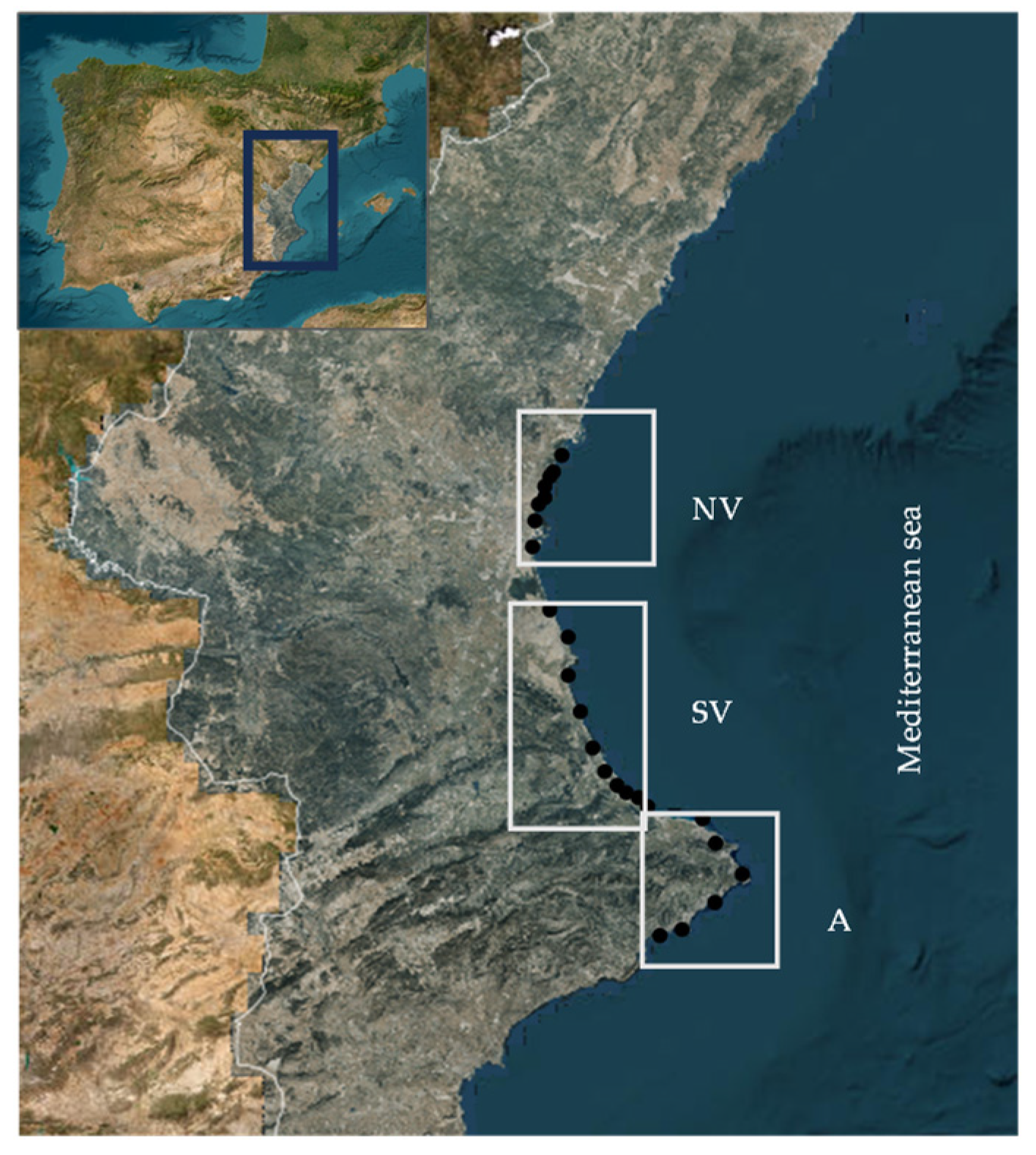
2.1. Samples

A total of 77 seawater samples were taken along 23 coastal locations of the Comunitat Valenciana (eastern Spain). Twenty-one seawater samples were collected from eight municipalities of the North Valencia region, thirty samples were collected from nine municipalities of the South Valencia region, and twenty-six samples were collected from six municipalities of the Alicante region. Figure 1 shows the sampling locations along the coast. All of the samples were collected aseptically, placed in a temperature-controlled box at 4 °C, transported to the laboratory, and processed without further delay.

2.2. Bacterial Analysis

Bacterial analyses were performed using Colilert and Enterolert test kits to detect Escherichia coli and Enterococcus sp., respectively, according to the manufacturer’s instructions. Both methods have been certified as alternative methods by the standards [37,38]. The density units were expressed as the most probable number (MPN) per 100 mL, according to the Quantry-Tray/2000 and Quanty-Tray systems, respectively (IDEXX Laboratories, Hooddorp, The Netherlands). E. coli and Enterococcus sp. colonies were isolated from positive wells and subcultured onto Tryptone Bile X-Glucuronide Agar (Oxoid, Ltd., Hampshire, UK) and Bile Esculine Azide Agar (Scharlau, Barcelona, Spain), respectively. Tentative colonies were then confirmed by the API phenotypic identification system (API20E strips, BioMèriux, Marcy-l’Étoile, France) and stored in refrigeration until their use.

2.3. Determination of Antibiotic Resistance

The antibiotic resistance of E. coli and Enterococcus sp. isolates was determined using the Sensititre System (Thermo Fischer Scientific, Madrid, Spain), an automatic system that uses a microplate format with a panel of different antimicrobial compounds that are accurately dosed at appropriate dilutions resembling the classical broth dilution method, according to the methods described by the Clinical and Laboratory Standards Institute [39], and accepted as a method of international reference [40]. EUVSEC plates were used for E. coli isolates, with the following antimicrobials to be tested: ampicillin (AMP), azithromycin (AZI), cefotaxime (FOT), ceftazidime (TAZ), ciprofloxacin (CIP), chloramphenicol (CHL), colistin (COL), gentamicin (GEN), meropenem (MER), nalidixic acid (NAL), sulfamethoxazole (SMX), tetracycline (TET), tigecycline (TGC), and trimethoprim (TMP). The enterococci isolates were tested against ampicillin (AMP), ciprofloxacin (CIP), chloramphenicol (CHL), daptomycin (DAP), erythromycin (ERY), gentamicin (GEN), linezolid (LIN), quinupristin/dalfopristin (QUIN/DAL), teicoplanin (TEI), tetracycline (TET), tigecycline (TGC), and vancomycin (VAN), using the EUVNEC plate. The test was performed following the manufacturer’s instructions. The results were interpreted by means of a digital visualization system, Sensititre Vizion (Thermo Scientific); for wells that showed bacterial growth, a sedimentation button was observed, indicating the resistance of the microorganism to the concentration of the antibiotic contained in the well. The well containing the lowest antimicrobial concentration where no growth was observed was the MIC (minimum inhibitory concentration). E. coli ATCC 25922 and E. fecalis ATCC 29212 were used as internal controls.

2.4. Detection of ARG

The analysis for the presence of ARG was carried out both in direct water samples and in bacteria isolated from these waters. For the DNA extraction from both sample types, the GenE-lute™ Bacterial Genomic DNA Kit (Sigma-Aldrich, Madrid, Spain) ref. NA2110) was used according to the manufacturer’s instructions. For the water samples, a volume of 250 mL was filtered through a 0.45 μm pore size membrane (Millipore) to retain the bacterial cells. The membranes were aseptically cut and suspended in 500 µL of lysis buffer included in the kit. Next, 80 mg of glass beads was added, and the mixture was shaken for 20 min at 3000 rpm (Disruptor Gene, USA Scientific, Ocala, FL, USA). For ARG detection from E. coli and enterococci isolates, bacterial cells from an overnight culture were harvested and suspended in 500 μL of TE buffer (10 mM Tris-HCl and 1 mM EDTA, pH 8), and then, DNA was extracted.
The ARGs chosen for this study were blaTEM, ermB, qnrS, sulI, and tetW, which confer resistance to β-lactam antibiotics, macrolides, quinolones, sulfonamides, and tetracyclines, respectively. These genes are involved in a variety of resistance mechanisms. These include antibiotic efflux, antibiotic inactivation, antibiotic target replacement, antibiotic target modification and antibiotic target protection.
The PCR mixture was prepared in a total volume of 50 μL containing 5 μL of the DNA template and 45 μL of the reaction mixture including 1X PCR buffer (Sigma-Aldrich), 1.5 mM of MgCl2 (Sigma, Aldrich), 0.2 mM of dNTPs, 0.25 μM of each primer (TIB MOLBIOL, Madrid, Spain), and 2.5 U of Taq polymerase (Sigma, Aldrich). The primer sequences and cycling conditions for five PCRs are shown in Table 1. MiliQ water was used as a negative control; as positive controls for each gene, E. coli isolates from our own collection tested in previous work were used. The PCR products were analyzed using gel electrophoresis on 1.2% agarose gel in 1xTAE buffer (40 mM of Tris, 20 mM of acetic acid, 1 mM of EDTA) plus 5 μL of RedSAfe (iNtRON Biotechnology, Ecogen, Madrid, Spain) per 100 mL. Ten μL of PCR product, previously mixed with 6x loading buffer, was used. Two molecular weight markers were included in each gel (GeneRuler 100 bp DNA Ladder, 0.5 μg/μL, Thermo Fischer Scientific, Madrid, Spain). Finally, the fragments were visualized in a transilluminator under UV light.

2.5. Statistical Analysis

Statistical analysis was performed by using Statgraphics (Centurion XVII). A multi-factor analysis of variance was carried out to compare the results obtained in E. coli and Enterococcus sp. counts for each sample, followed by a post hoc analysis using the Fisher’s least significance difference (LSD) method. Resistance gene detection was analyzed by a χ2 test, using contingency tables, in order to establish any possible dependence with location of sampling.

3. Results

3.1. Microbiological Determination of E. coli and Enterococcus sp. in Marine Water Samples

Among the 77 water samples analyzed, 74% showed contamination by E. coli, and 39% showed contamination by Enterococcus sp. The NMP of CFU/100 mL varied from 1 to 3.8 log10 and 1 to 2.9 log10 for E. coli and Enterococcus sp., respectively. The statistical analysis did not show significant differences in the contamination level among the three sampling zones. In accordance with the limits established in Spanish regulations (R.D. 1341/2007), 87% of the analyzed waters presented excellent quality (87%) as bathing waters, while the rest of them were classified as being in the good quality category.

3.2. Antibiotic Resistance Determination of Bacterial Isolates

A total of 87 strains were isolated from the seawater samples (57 E. coli strains, and 30 Enterococcus sp. strains) and subjected to the antibiotic sensitivity test. The resistance of the bacteria to antibiotics was determined according to the CLSI standards [39] for antimicrobial susceptibility testing.
Among the 57 isolates of E. coli (Table 2), 34 isolates (59.6%) were resistant to at least one antibiotic. Resistance to ampicillin was the most frequent observation (20 isolates) followed by ciprofloxacin (10 isolates), sulfamethoxazole, and trimethoprim with 9 isolates for each and tetracycline and nalidixic acid with 8 isolates (Supplementary Materials Table S1). The MIC values for these antibiotics were 32 - > 64 mg/L, 0.125–0.5 mg/L, >1024 mg/L, >32 mg/L, 32 - ≥ 64 mg/L, and >128 mg/L, respectively. There was no correlation (p > 0.05) between the number of strains resistant to any antibiotic and the area from which they were isolated. Multiple resistance (≥3 antibiotic classes) [45] was observed in 5 out of 34 resistant E. coli strains, (14.7%). Each of them presented a unique multi-resistance pattern. β-lactam resistance was present in all profiles, followed by sulfonamide and tetracycline resistance, present in four profiles, and quinolone resistance, observed in three profiles. Resistance to polymyxins (colistin) and aminoglycosides (gentamicin) was only present in one multi-resistance profile for each. Finally, none of the isolates showed resistance to chloramphenicol and tigecycline, with MIC ranges of ≤8–16 mg/L and ≤0.25–1 mg/L, respectively.
Only seven Enterococcus sp. isolates showed resistance to the tested antibiotics, specifically, six isolates against tetracycline and one against daptomycin (Table 3) (Supplementary Materials Table S4). The MIC ranges for these antibiotics were 64–128 mg/L and 16 mg/L. The statistical analysis showed no correlation (p > 0.05) between the resistance of the isolates to any antibiotic and the area from which they were isolated.

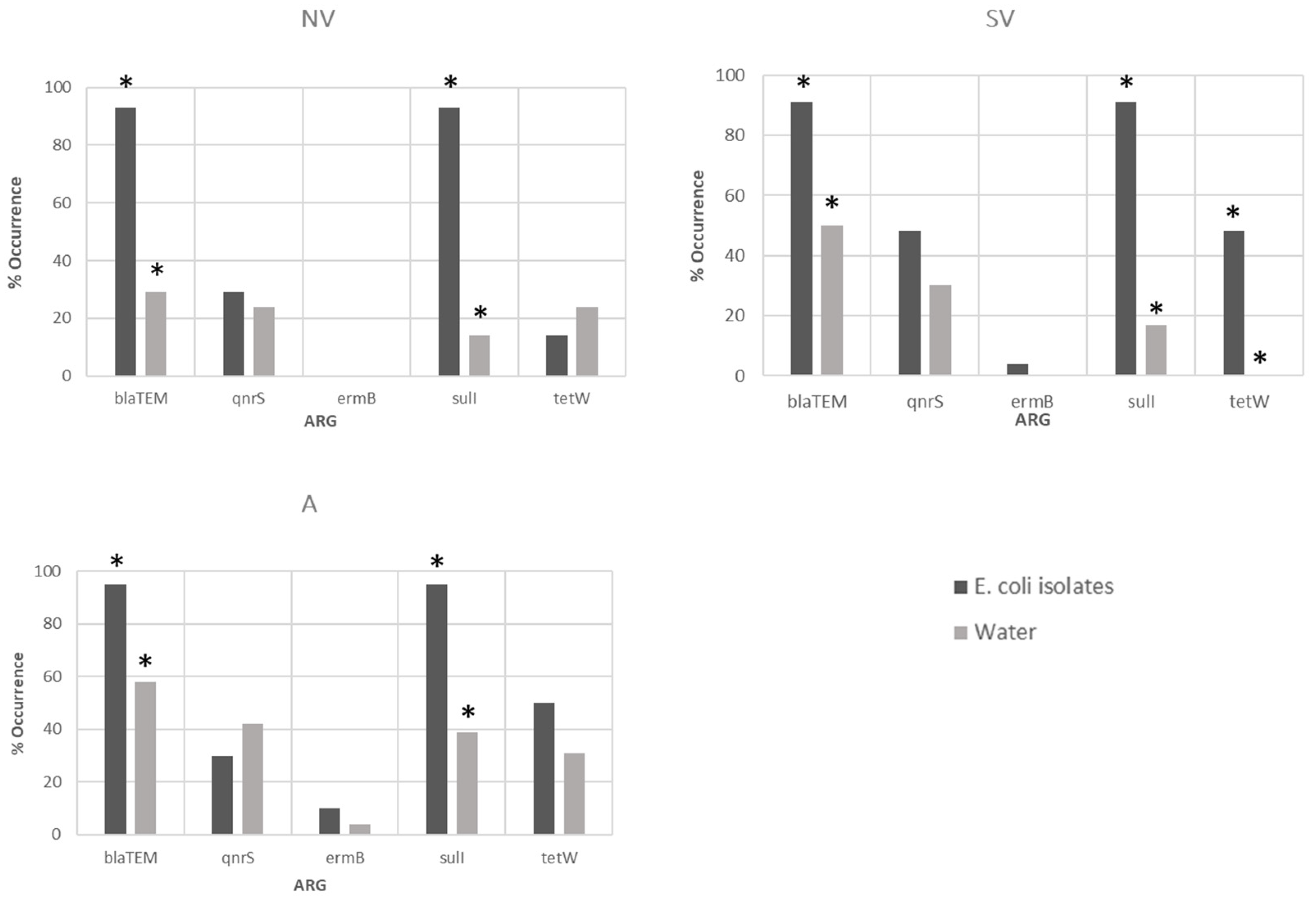
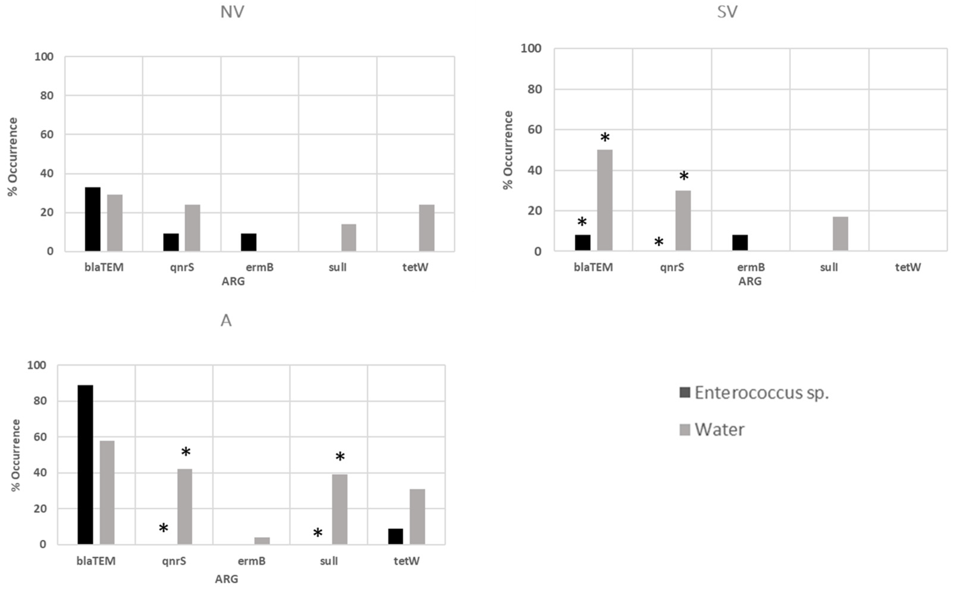
3.3. Antibiotic-Resistant Gene Determination in Water Samples and Bacterial Isolates

The presence of five antibiotic resistance genes in the 77 seawater samples was analyzed as described in the Section 2. Forty-four of the samples (57%) were positive for at least one of the genes. As shown in Table 4, the blaTEM gene was the most frequently detected gene (46.7%), followed by qnrS (32.5%) and sulI (23.4%); the tetW gene was detected in 17% of the samples, while ermB was only found to be present in one sample (Supplementary Materials Table S3). The statistical analysis only showed a significant relationship between the tetW gene and the region of Alicante (χ2 = 10.384, p < 0.05), and it was not detected in any of the samples from South Valencia. Combinations of two genes were detected in 24 seawater samples (31%), with the most abundant combinations being those containing the blaTEM gene. Three gene combinations were detected in nine samples (ca. 12%). Finally, four and five gene combinations were present in only one sample. A statistically significant dependence relationship was only detected between the tetW gene and the sampling zones of North Valencia and Alicante (χ2 = 10,384, p < 0.05) as this gene was not detected in any of the samples from South Valencia.
The blaTEM gene, together with the sulI gene, was the most frequently detected gene, with each of them being found in 53 E. coli strains (92.9%) (Table 5). They were followed in frequency by the tetW and qnrS genes, present in 40% and 36.8% of the samples, respectively. In contrast, the ermB gene was detected in only three isolates (5%) (Supplementary Materials Table S2). The statistical analysis showed no dependence relationship between the sampling area and the frequency of gene detection in E. coli isolates (p > 0.05).
The analysis of genes in enterococci (Table 6) again showed that the blaTEM gene was the most abundant, with it being detected in 40% of the isolates. The percentages of the other genes ranged from 3.3% for qnrS and tetW to 6.6% for ermB. It should be noted that sulI was not detected (Supplementary Materials Table S5). The statistical analysis showed no relationship between the sampling area and the occurrence of genes in the isolates (p > 0.05).
For most of the genes investigated, their presence was greater in the E. coli isolates than in the directly analyzed waters (Figure 2). In North Valencia, the blaTEM and sulI genes were detected at significantly higher levels in the E. coli isolates than in the seawater samples (χ2 = 13.988, p = 0.0002 and χ2 = 20.896, p = 0.000, respectively). In South Valencia, besides the blaTEM and sulI genes (χ2 = 10.194, p = 0.0014 and χ2 = 29.020, p = 0.000, respectively), significant differences were found in the tetW gene, which was not detected in any of the seawater samples (χ2 = 18.106, p = 0.0000). In Alicante, significant differences were found in the blaTEM and sulI genes, which were detected to a greater extent in the isolates (χ2 = 8.160, p = 0.0043 and χ2 = 15.120, p = 0.0001, respectively).
When comparing the presence of ARGs in the enterococci and water samples (Figure 3), the blaTEM and qnrS genes were detected at significantly higher levels in the seawater samples than in enterococci isolates from South Valencia (χ2 = 6.310, p = 0.0120 and χ2 = 4.582, p = 0.0323, respectively). In addition to the qnrS gene (χ2 = 5.553, p = 0.0184), significant differences were found in the sulI gene in the Alicante area (χ2 = 4.846, p = 0.0277), only detected in the water samples.

4. Discussion

In recent decades, humanity has been confronted with the growing problem of the spread of antibiotic-resistant bacteria (ARB) and the difficulty of treating associated infections. The presence of these bacteria in coastal environments, as well as the presence of ARGs circulating freely in the water, represents a serious problem, especially for individuals who visit such areas for leisure purposes [46]. However, data on their presence and importance in the marine environment are still limited.
To this end, 77 water samples from the coastal waters of Comunitat Valencia (eastern Spain) were analyzed using microbial and molecular methods, to determine the presence of ARB and ARGs. The main species studied included E. coli and Enterococcus sp., which were tested for resistance to 12–14 antibiotics. The detected levels of both microorganisms were within the range found in the compilation of several studies in marine waters worldwide encompassing four decades of research [47], ranging from 0.59 to 3.51 log10 CFU/100 mL for E. coli and 0.63 to 3.21 log10 CFU/100 mL for Enterococcus sp. In a macro study conducted in Spain between 2012 and 2015, covering 1392 beaches throughout the country, a mean of 1.4 log10 CFU/100 mL for E. coli and 1.32 log10 CFU/100 mL for Enterococcus sp. was found for the southern Mediterranean, where our samples were collected, with a national mean of 1.93 and 1.65 log10 CFU/100 mL for each of the microorganisms [48]. Our values were within these averages, although we found some samples with E. coli values of 4.4 log10 CFU/100 mL and some samples of Enterococcus sp. with values of 4.2 log10 CFU/100 mL; both results were above acceptable levels and could have been due to the spillage of fecal matter due to problems with the sewerage system at the location or run-off from rainfall. It should also be noted that these exceptionally polluted samples were taken in the summer, on beaches close to the city of Valencia, where not only members of the local population (Valencia and its metropolitan area comprise a population of around one and a half million inhabitants) but also many tourists bathed during this period. Some authors have already stated that beaches in urban environments, as in our case, are of lower quality than those in semi-urban or natural environments [49,50,51,52], which is related to the number of people using the beaches, as well as the number of pets and birds, especially seagulls, that have access to them [53,54,55]. It is, therefore, difficult to determine the origin of this contamination.
The search for ARB was directed toward the detection of E. coli and enterococci of fecal origin. Both microorganisms have been recognized as important actors in the spread of antibiotic resistance [56,57]. The percentage of E. coli strains resistant to at least one antibiotic (59.6%) was similar to that obtained in a study conducted on beaches in Brazil [58] and lower than that obtained in other studies in Portugal [56] or China [59], with percentages of up to 80% being observed. Ampicillin resistance was the most frequent observation, confirming that β-lactam resistance was the most prevalent form in the aquatic environment [60,61]. Other antibiotics, such as ciprofloxacin, nalidixic acid, tetracycline, and sulfamethoxazole, were found to have resistance rates ranging from 14% to 18%. Similar findings were made by Hernández et al. [62] in a study conducted in Antarctica, an extreme location where the human presence is due to the establishment of scientific bases and where the removal of fecal contamination is very strictly controlled. This reflects the general resistance to β-lactams worldwide, with varying rates of resistance to other antibiotics depending on various factors such as geographical location. This fact also demonstrates the ability of antimicrobial resistance to distribute homogeneously in seawater.
Nine percent of the E. coli strains showed multidrug resistance, and again, the β-lactam family was present in all of the resistance profiles. Comparing these data with those of other studies, we see very different percentages of multidrug resistance. In a study carried out on the coast of Sicily, [63] found levels of multidrug resistance in their isolates ranging from 7% (against five antibiotics) to 14% (against four antibiotics). Similar results were obtained in isolates from Brazil beaches with multi-resistance levels ranging from 0 to 8.3% [64]. This supports the idea that the majority of multidrug-resistant ARBs are not of environmental origin but rather originate in places of high selective pressure, such as hospitals or highly contaminated ecosystems. The levels found in each geographical area are very consistent with the antibiotic use policies in these areas [65].
The enterococci in our study showed a low level of resistance (23%) compared with other studies conducted on Mediterranean beaches, for example, in Italy, with 38% of resistant strains [66], in Morocco (ca. 65%) [57], or in Tunisia, where this resistance is as high as 75% [67]. In our study, only strains resistant to tetracycline (20%) and daptomycin (3%) were obtained, with the latter being a rarely used drug. However, it is important to note that daptomycin is used as a last line of therapy, especially in vancomycin-resistant enterococcal infections [68]; therefore, the emergence of daptomycin-resistant enterococci is of concern in healthcare settings. No multidrug-resistant Enterococcus spp. isolates were found. These data are encouraging as antibiotic-resistant enterococci found in coastal marine waters are often closely related to human clinical isolates, probably originating from urban wastewater [69,70].
PCR assays revealed that blaTEM was the most frequently detected gene in the analyzed seawater samples. These results were in line with other studies in the Mediterranean, where this gene was detected in almost all samples [56,63,71], confirming that β-lactam antibiotic resistance genes were widely distributed in seawater samples, detecting it in fishes and wild marine species [72]. In a macro study carried out on water samples from oceans around the world, the authors found a high abundance of ARGs encoding resistance to β-lactam antibiotics, probably due to the high use of these antibiotics, as stated by the WHO [73,74]. The presence of the qnrS, sulI, and tetW genes was also comparable with the results of some other works that attested to their prevalence in coastal environments [75,76], but this contrasts with the absence confirmed in other coastal waters, such as Sicily [63]. This observed variability in the presence and abundance of ARGs was highly dependent on antibiotic usage patterns, which was a key factor for the generation and propagation of ARGs [77].
Once again, the blaTEM gene was the most frequently detected gene in both E. coli (92.9%) and Enterococcus sp. isolates (40%). The remaining genes showed more variable percentages, with the exception of sulI in E. coli isolates, which also stood out with identical percentages to blaTEM. Other studies have demonstrated that blaTEM, tet, and sul genes are ubiquitous in E. coli isolates from estuarine and marine environments [32,36,78,79].
In the present study, the frequency with which ARGs were detected was higher in E. coli isolates than in direct water samples. These results contrast with those found in other types of water, such as the study carried out by Amato et al. [24] in water from ditches that flow into the sea, specifically in the region of Valencia Norte. This, in turn, would be consistent with some studies suggesting that culture-dependent antimicrobial resistance approaches could underestimate the frequency and diversity of genetic determinants of antimicrobial resistance in environmental samples [80,81]. However, in our case, this could be due to the fact that the marine environment is a diluting medium, so the filtrate could provide little genetic material for amplification, while for the detection of E. coli ARGs, pure colonies with a high DNA load are used as a starting point.
Antibiotic resistance is no longer merely a clinical problem [82]. The demonstration of the presence of antibiotic resistance (bacteria and genes) in this work highlights the role of the sea in the spread of AR genes and the need to carefully consider the risks to human health associated with fecal contamination. Coastal areas, where human recreation, in addition to commercial activities, is important, are ideal environments for the transmission of resistance to the human population.

5. Conclusions

This study provides a clearer perspective on the circulation of ARG in the marine environment and its relationship with fecal pollution in seawater. This could facilitate future interventions to reduce antimicrobial resistance in the environment as part of a holistic One Health approach.

Supplementary Materials

The following supporting information can be downloaded at: https://www.mdpi.com/article/10.3390/app14051965/s1. Table S1: Incidence of resistant E. coli isolates for each antimicrobial compound according to sampling zone. Table S2: Presence of antibiotic resistance genes in E. coli isolates according to sampling zone. Table S3: Presence of antibiotic resistance genes in water samples according to sampling zone. Table S4: Incidence of resistant Enterococcus sp. isolates for each antimicrobial compound according to sampling zone. Table S5: Presence of antibiotic resistance genes in Enterococcus sp. isolates according to sampling zone.

Author Contributions

Conceptualization: D.D. and M.Á.C.; methodology: A.G., M.G.-F. and M.Á.C.; resources: M.L.C.-S.; investigation: D.D. and M.L.C.-S.; funding acquisition, A.I.J.-B.; formal analysis: D.D. and M.Á.C.; writing—review and editing, M.Á.C. All authors have read and agreed to the published version of the manuscript.

Funding

This research was funded by CIAICO/2021/149. Generalitat Valenciana (Comunitat Valenciana, Spain).

Institutional Review Board Statement

Not applicable.

Informed Consent Statement

Not applicable.

Data Availability Statement

Data are contained within the article.

Conflicts of Interest

The authors declare no conflicts of interest.

References

  1. Kraemer, S.A.; Ramachandran, A.; Perron, G.G. Antibiotic Pollution in the Environment: From Microbial Ecology to Public Policy. Microorganisms 2019, 7, 180. [Google Scholar] [CrossRef]
  2. Waseem, H.; Williams, M.R.; Jameel, S.; Hashsham, S.A. Antimicrobial Resistance in the Environment. Water Environ. Res. 2018, 90, 865–884. [Google Scholar] [CrossRef]
  3. Bondarczuk, K.; Piotrowska-Seget, Z. Microbial diversity and antibiotic resistance in a final effluent-receiving lake. Sci. Total Environ. 2019, 650, 2951–2961. [Google Scholar] [CrossRef] [PubMed]
  4. Gibson, M.K.; Forsberg, K.J.; Dantas, G. Improved annotation of antibiotic resistance determinants reveals microbial resistomes cluster by ecology. ISME J. 2015, 9, 207–216. [Google Scholar] [CrossRef] [PubMed]
  5. Martínez, J.L. Antibiotics and antibiotic resistance genes in natural environments. Science 2008, 321, 365–367. [Google Scholar] [CrossRef] [PubMed]
  6. Alduina, R. Antibiotics and environment. Antibiotics 2020, 9, 202. [Google Scholar] [CrossRef] [PubMed]
  7. Peterson, E.; Kaur, P. Antibiotic resistance mechanisms in bacteria: Relationships between resistance determinants of antibiotic producers, environmental bacteria, and clinical pathogens. Front. Microbiol. 2018, 9, 2928. [Google Scholar] [CrossRef] [PubMed]
  8. Gillings, M.; Gaze, W.; Pruden, A.; Samalla, K.; Tiedje, J.M.; Zhu, Y.-G. Using the class 1 integron-integrase gene as a proxy for anthropogenic pollution. ISME J. 2015, 9, 1269–1279. [Google Scholar] [CrossRef] [PubMed]
  9. Cycón, M.; Mrozik, A.; Piotrowska-Seget, Z. Antibiotics in the soil environment—Degradation and their impact on microbial activity and diversity. Front. Microbiol. 2019, 10, 338. [Google Scholar] [CrossRef] [PubMed]
  10. Ahmad, N.; Joji, R.M.; Shahid, M. Evolution and implementation of One Health to control the dissemination of antibiotic-resistant bacteria and resistance genes: A review. Front. Cell Infect. Microbiol. 2023, 12, 1065796. [Google Scholar] [CrossRef]
  11. Wu, J.; Wang, J.; Li, Z.; Guo, S.; Li, K.; Xu, P.; Ok, Y.S.; Jones, D.L.; Zou, J. Antibiotics and antibiotic resistance genes in agricultural soils: A systematic analysis. Crit. Rev. Environ. Sci. Technol. 2023, 53, 847–864. [Google Scholar] [CrossRef]
  12. Jiménez-Belenguer, A.I.; Ferrús, M.A.; Hernández, M.; García-Hernández, J.; Moreno, Y.; Castillo, M.A. Prevalence and characterization of beta-lactam and carbapenem-resistant bacteria isolated from organic fresh produce retailed in Eastern Spain. Antibiotics 2023, 12, 387. [Google Scholar] [CrossRef]
  13. Hölzel, C.S.; Tetens, J.L.; Schwaiger, K. Unraveling the role of vegetables in spreading antimicrobial-resistant bacteria: A need for quantitative risk assessment. Foodborne Pathog. Dis. 2018, 15, 671–688. [Google Scholar] [CrossRef] [PubMed]
  14. Ji, X.; Shen, Q.; Liu, F.; Ma, J.; Xu, G.; Wang, Y.; Wu, M. Antibiotic resistance gene abundances associated with antibiotics and heavy metals in animal manures and agricultural soils adjacent to feedlots in Shanghai, China. J. Hazard. Mater. 2012, 235, 178–185. [Google Scholar] [CrossRef] [PubMed]
  15. Gwenzi, W.; Musiyiwa, K.; Mangori, L. Sources, behaviour and health risks of antimicrobial resistance genes in wastewaters: A hotspot reservoir. J. Environ. Chem. Eng. 2020, 8, 102220. [Google Scholar] [CrossRef]
  16. Bouki, C.; Venieri, D.; Diamadopoulos, E. Detection and fate of antibiotic resistant bacteria in wastewater treatment plants: A review. Ecotoxicol. Environ. Saf. 2013, 91, 1–9. [Google Scholar] [CrossRef] [PubMed]
  17. Daly, M.; Powell, J.; O’Connell, N.H.; Murphy, L.; Dunne, C.P. Antimicrobial resistance is prevalent in E. coli and other Enterobacterales isolated from public and private drinking water supplies in the Republic of Ireland. Microorganisms 2023, 11, 1224. [Google Scholar] [CrossRef] [PubMed]
  18. Odonkor, S.T.; Addo, K.K. Prevalence of multidrug-resistant Escherichia coli isolated from drinking water sources. Int. J. Microbiol. 2018, 2018, 7204013. [Google Scholar] [CrossRef] [PubMed]
  19. Fernando, D.M.; Tun, H.M.; Poole, J.; Patidar, R.; Li, R.; Mi, R.; Amarawansha, G.E.A.; Fernando, W.G.D.; Khafipour, E.; Farenhorst, A.; et al. Detection of antibiotic resistance genes in source and drinking water samples from a First Nations community in Canada. Appl. Environ. Microbiol. 2016, 82, 4767–4775. [Google Scholar] [CrossRef] [PubMed]
  20. Coleman, B.L.; Salvadori, M.I.; McGeer, A.J.; Sibley, K.A.; Neumann, N.F.; Bondy, S.J.; Gutmanis, I.A.; McEwen, S.A.; Lavoie, M.; Strong, D.; et al. The role of drinking water in the transmission of antimicrobial-resistant E. coli. Epidemiol. Infect. 2012, 140, 633–642. [Google Scholar] [CrossRef]
  21. Wang, R.-N.; Zhang, Y.; Cao, Z.-H.; Wang, X.-Y.; Ma, B.; Wu, W.-B.; Hu, N.; Huo, Z.-Y.; Yuan, Q.-B. Occurrence of super antibiotic resistance genes in the downstream of the Yangtze River in China: Prevalence and antibiotic resistance profiles. Sci. Total Environ. 2019, 651, 1946–1957. [Google Scholar] [CrossRef]
  22. Rodriguez-Mozaz, S.; Chamorro, S.; Marti, E.; Huerta, B.; Gros, M.; Sanchez-Melsio, A.; Borrego, C.M.; Barcelo, D.; Balcazar, J.L. Occurrence of antibiotics and antibiotic resistance genes in hospital and urban wastewaters and their impact on the receiving river. Water Res. 2015, 69, 234–242. [Google Scholar] [CrossRef] [PubMed]
  23. Stoll, C.; Sidhu, J.P.S.; Tiehm, A.; Toze, S. Prevalence of clinically relevant antibiotic resistance genes in surface water samples collected from Germany and Australia. Environ. Sci. Technol. 2012, 46, 9716–9726. [Google Scholar] [CrossRef] [PubMed]
  24. Amato, M.; Dasí, D.; González, A.; Ferrús, M.A.; Castillo, M.A. Occurrence of antibiotic resistant bacteria and resistance genes in agricultural irrigation waters from Valencia city (Spain). Agric. Water Manag. 2021, 256, 107097. [Google Scholar] [CrossRef]
  25. Araújo, S.; Silva, I.A.T.; Tacao, M.; Patinha, C.; Alves, A.; Henriques, I. Characterization of antibiotic resistant and pathogenic Escherichia coli in irrigation water and vegetables in household farms. Int. J. Food Microbiol. 2017, 257, 192–200. [Google Scholar] [CrossRef] [PubMed]
  26. Amos, G.C.A.; Hawkey, P.M.; Gaze, W.H.; Wellington, E.M. Wastewater effluent contributes to the dissemination of CTX-M-15 in the natural environment. J. Antimicrob. Chemother. 2014, 69, 1785–1791. [Google Scholar] [CrossRef] [PubMed]
  27. Yuan, X.; Lv, Z.; Zhang, Z.; Han, Y.; Liu, Z.; Zhang, H. A review of antibiotics, antibiotic resistant bacteria, and resistance genes in aquaculture: Occurrence, contamination, and transmission. Toxics 2023, 11, 420. [Google Scholar] [CrossRef] [PubMed]
  28. Amarasiri, M.; Sano, D.; Suzuki, S. Understanding human health risks caused by antibiotic resistant bacteria (ARB) and antibiotic resistance genes (ARG) in water environments: Current knowledge and questions to be answered. Crit. Rev. Environ. Sci. Technol. 2020, 50, 2016–2059. [Google Scholar] [CrossRef]
  29. Su, Z.; Li, A.; Chen, J.; Huang, B.; Mu, Q.; Chen, L.; Wen, D. Wastewater discharge drives ARGs spread in the coastal area: A case study in Hangzhou Bay, China. Mar. Pollut. Bull. 2020, 151, 110856. [Google Scholar] [CrossRef]
  30. Carney, R.L.; Labbate, M.; Siboni, N.; Tagg, K.A.; Mitrovic, S.M.; Seymour, J.R. Urban beaches are environmental hotspots for antibiotic resistance following rainfall. Water Res. 2019, 167, 11508. [Google Scholar] [CrossRef]
  31. Leonard, A.F.; Zhang, L.; Balfour, A.J.; Garside, R.; Gaze, W.H. Human recreational exposure to antibiotic resistant bacteria in coastal bathing waters. Environ. Int. 2015, 82, 92–100. [Google Scholar] [CrossRef]
  32. Makkaew, P.; Kongprajug, A.; Chyerochana, N.; Sresung, M.; Precha, N.; Mongkolsuk, S.; Sirikanchana, K. Persisting antibiotic resistance gene pollution and its association with human sewage sources in tropical marine beach waters. Int. J. Hyg. Environ. Health 2021, 238, 113859. [Google Scholar] [CrossRef]
  33. Pruden, A.; Pei, R.; Storteboom, H.; Carlson, K.H. Antibiotic resistance genes as emerging contaminants: Studies in northern Colorado. Environ. Sci. Technol. 2006, 40, 7445–7450. [Google Scholar] [CrossRef]
  34. World Health Organization. Global Action Plan on Antimicrobial Resistance. 2015. Available online: http://apps.who.int/iris/bitstream/10665/193736/1/9789241509763eng.pdf?ua=1 (accessed on 1 September 2023).
  35. Singer, A.C.; Shaw, H.; Rhodes, V.; Hart, A. Review of antimicrobial resistance in the environment and its relevance to environmental regulators. Front. Microbiol. 2016, 7, 1728. [Google Scholar] [CrossRef]
  36. Zheng, D.; Yin, G.; Liu, M.; Chen, C.; Jiang, Y.; Hou, L.; Zheng, Y. A systematic review of antibiotics and antibiotic resistance genes in estuarine and coastal environments. Sci. Total Environ. 2021, 777, 146009. [Google Scholar] [CrossRef]
  37. ISO 9308-2:2012; Water quality —Enumeration of Escherichia coli and coliform bacteria. Part 2: Most probable number method. International Organization for Standardization: Geneva, Switzerland, 2012.
  38. ISO 7899-1:1998/Cor 1:2000; Water quality Detection and enumeration of intestinal enterococci. Part 1: Miniaturized method (Most Probable Number) for surface and wastewater. International Organization for Standardization: Geneva, Switzerland, 2000.
  39. Clinical and Laboratory Standards Institute (CLSI). Performance Standards for Antimicrobial Susceptibility Testing. In Twenty-Fifth Informational Supplement; CLSI DocumentM100-S25; Committee for Clinical Laboratory Standards: Wayne, PA, USA, 2015. [Google Scholar]
  40. ISO 20776-1:2019; Susceptibility testing of infectious agents and evaluation of performance of antimicrobial susceptibility test devices. Part 1: Broth micro-dilution reference method for testing the in vitro activity of antimicrobial agents against rapidly growing aerobic bacteria involved in infectious diseases. International Organization for Standardization: Geneva, Switzerland, 2019.
  41. Xi, C.; Zhang, Y.; Marrs, C.F.; Ye, W.; Simon, C.; Foxman, B.; Nriagu, J. Prevalence of antibiotic resistance in drinking water treatment and distribution systems. Appl. Environ. Microbiol. 2009, 75, 5714–5718. [Google Scholar] [CrossRef]
  42. Chen, J.; Yu, Z.T.; Michel, F.C., Jr.; Wittum, T.; Morrison, M. Development and application of real-time PCR assays for quantification of erm genes conferring resistance to macrolides-lincosamides-streptogramin B in livestock manure and manure management systems. Appl. Environ. Microbiol. 2007, 73, 4407–4416. [Google Scholar] [CrossRef]
  43. Pei, R.; Kim, S.C.; Carlson, K.H.; Pruden, A. Effect of river landscape on the sediment concentrations of antibiotics and corresponding antibiotic resistance genes (ARG). Water Res. 2006, 40, 2427–2435. [Google Scholar] [CrossRef]
  44. Aminov, R.I.; Garrigues-Jeanjean, N.; Mackie, R.I. Molecular ecology of tetracycline resistance: Development and validation of primers for detection of tetracycline resistance genes encoding ribosomal protection proteins. Appl. Environ. Microbiol. 2001, 67, 22–32. [Google Scholar] [CrossRef]
  45. Magiorakos, A.-P.; Srinivasan, A.; Carey, R.B.; Carmeli, Y.; Falagas, M.E.; Giske, C.G.; Harbarth, S.; Hindler, J.F.; Kahlmeter, G.; Olsson-Liljequist, B.; et al. Multidrug-resistant, extensively drug-resistant and pan drug-resistant bacteria: An international expert proposal for interim standard definitions for acquired resistance. Clin. Microbiol. Infect. 2012, 18, 268–281. [Google Scholar] [CrossRef]
  46. Belding, C.; Boopathy, R. Presence of antibiotic-resistant bacteria and antibiotic resistance genes in coastal recreational waters of southeast Louisiana, USA. J. Water Supply Res. Technol. AQUA 2018, 67, 800–809. [Google Scholar] [CrossRef]
  47. Korajkic, A.; McMinn, B.R.; Harwood, V.J. Relationships between microbial indicators and pathogens in recreational water settings. Int. J. Environ. Res. Public Health 2018, 15, 2842. [Google Scholar] [CrossRef]
  48. Aragonés, L.; López, I.; Palazón, A.; López-Úbeda, R.; García, C. Evaluation of the quality of coastal bathing waters in Spain through fecal bacteria Escherichia coli and Enterococcus. Sci. Total Environ. 2016, 566–567, 288–297. [Google Scholar] [CrossRef]
  49. Ariza, E.; Jimenez, J.A.; Sarda, R.; Villares, M.; Pinto, J.; Fraguell, R.; Roca, E.; Marti, C.; Valdemoro, H.; Ballester, R. Proposal for an integral quality index for urban and urbanized beaches. Environ. Manag. 2010, 45, 998–1013. [Google Scholar] [CrossRef]
  50. McLellan, S.L. Genetic diversity of Escherichia coli isolated from urban rivers and beach water. Appl. Environ. Microbiol. 2004, 70, 4658–4665. [Google Scholar] [CrossRef]
  51. May, C.W.; Horner, R.R.; Karr, J.R.; Mar, B.W.; Welch, E.B. Effects of urbanization on small streams in the Puget Sound ecoregion. Watershed Prot. Tech. 1999, 2, 79–84. [Google Scholar]
  52. Winter, J.G.; Duthie, H.C. Effects of urbanization on water quality, periphyton and invertebrate communities in a southern Ontario stream. Can. Water Resour. J. 1998, 23, 245–257. [Google Scholar] [CrossRef]
  53. Abdelzaher, A.M.; Wright, M.E.; Ortega, C.; Solo-Gabriele, H.M.; Miller, G.; Elmir, S.; Newman, S.; Shih, P.; Bonilla, J.A.; Bonilla, T.D.; et al. Presence of pathogens and indicator microbes at a non-point source subtropical recreational marine beach. Appl. Environ. Microbiol. 2010, 76, 724–732. [Google Scholar] [CrossRef]
  54. Haugland, R.A.; Siefring, S.C.; Wymer, L.J.; Brenner, K.P.; Dufour, A.P. Comparison of Enterococcus measurements in freshwater at two recreational beaches by quantitative polymerase chain reaction and membrane filter culture analysis. Water Res. 2005, 39, 559–568. [Google Scholar] [CrossRef]
  55. Whitman, R.L.; Nevers, M.B.; Korinek, G.C.; Byappanahalli, M.N. Solar and temporal effects on Escherichia coli concentration at a Lake Michigan swimming beach. Appl. Environ. Microbiol. 2004, 70, 4276–4285. [Google Scholar] [CrossRef]
  56. Alves, M.S.; Pereira, A.; Araújo, S.M.; Castro, B.B.; Correia, A.C.M.; Henriques, I. Seawater is a reservoir of multi-resistant Escherichia coli, including strains hosting plasmid-mediated quinolones resistance and extended-spectrum beta-lactamases genes. Front. Microbiol. 2014, 5, 426. [Google Scholar] [CrossRef]
  57. Bennani, M.; Amarouch, H.; Oubrim, N.; Cohen, N. Identification and antimicrobial resistance of fecal Enterococci isolated in coastal Mediterranean environments of Morocco. Eur. J. Sci. Res. 2012, 70, 266–275. [Google Scholar]
  58. da Costa Andrade, V.; Zampieri Bdel, B.; Ballesteros, E.R.; Pinto, A.B.; de Oliveira, A.J. Densities and antimicrobial resistance of Escherichia coli isolated from marine waters and beach sands. Environ. Monit. Assess. 2015, 187, 342. [Google Scholar] [CrossRef]
  59. Na, G.; Zhang, W.; Zhou, S.; Gao, H.; Lu, Z.; Wu, X.; Li, R.; Qiu, L.; Cai, Y.; Yao, Z. Sulfonamide antibiotics in the Northern Yellow Sea are related to resistant bacteria: Implications for antibiotic resistance genes. Mar. Poll. Bull. 2014, 84, 70–75. [Google Scholar] [CrossRef]
  60. Letchumanan, V.; Yin, W.F.; Lee, L.H.; Chan, K.G. Prevalence and antimicrobial susceptibility of Vibrio parahaemolyticus isolated from retail shrimps in Malaysia. Front. Microbiol. 2015, 6, 33. [Google Scholar] [CrossRef]
  61. Şanlıtürk, G.; Güran, M. Monitoring of microbiological dynamics in beach sand and seawater samples from recreational and non-recreational beaches over a two-year period. Int. J. Environ. Health Res. 2022, 32, 1973–1985. [Google Scholar] [CrossRef]
  62. Hernández, F.; Calısto-Ulloa, N.; Gómez-Fuentes, C.; Gómez, M.; Ferrer, J.; González-Rocha, G.; Bello-Toledo, H.; Botero-Coy, A.M.; Boıx, C.; Ibáñez, M.; et al. Occurrence of antibiotics and bacterial resistance in wastewater and sea water from the Antarctic. J. Hazard. Mater. 2019, 363, 447–456. [Google Scholar] [CrossRef]
  63. Gambino, D.; Savoca, D.; Sucato, A.; Gargano, V.; Gentile, A.; Pantano, L.; Vicari, D.; Alduina, R. Occurrence of antibiotic resistance in the Mediterranean Sea. Antibiotics 2022, 11, 332. [Google Scholar] [CrossRef]
  64. Fernandes Cardoso de Oliveira, A.J.; Ranzani de França, P.T.; Pinto, A.B. Antimicrobial resistance of heterotrophic marine bacteria isolated from seawater and sands of recreational beaches with different organic pollution levels in southeastern Brazil: Evidences of resistance dissemination. Environ. Monit. Assess. 2010, 169, 375–384. [Google Scholar] [CrossRef]
  65. European Centre for Disease Prevention and Control. Antimicrobial Resistance in the EU/EEA (EARS-Net)-Annual Epidemiological Report 2021; ECDC: Stockholm, Sweden, 2022. [Google Scholar]
  66. Vignaroli, C.; Pasquaroli, S.; Citterio, B.; Di Cesare, A.; Mangiaterra, G.; Fattorini, D.; Biavasco, F. Antibiotic and heavy metal resistance in enterococci from coastal marine sediment. Environ. Pollut. 2018, 237, 406–413. [Google Scholar] [CrossRef]
  67. Alibi, S.; Crespo, D.; Navas, J. Plant-derivatives small molecules with antibacterial activity. Antibiotics 2021, 10, 231. [Google Scholar] [CrossRef]
  68. Li, G.; Walker, M.J.; De Oliveira, D.M.P. Vancomycin resistance in Enterococcus and Staphylococcus aureus. Microorganisms 2022, 11, 24. [Google Scholar] [CrossRef] [PubMed]
  69. Vignaroli, C.; Luna, G.M.; Pasquaroli, S.; Di Cesare, A.; Petruzzella, R.; Paroncini, P.; Biavasco, F. Epidemic Escherichia coli ST131 and Enterococcus faecium ST17 in coastal marine sediments from an Italian beach. Environ. Sci. Technol. 2013, 47, 13772–13780. [Google Scholar] [CrossRef] [PubMed]
  70. Di Cesare, A.; Luna, G.M.; Vignaroli, C.; Pasquaroli, S.; Tota, S.; Paroncini, P.; Biavasco, F. Aquaculture can promote the presence and spread of antibiotic-resistant Enterococci in marine sediments. PLoS ONE 2013, 8, e62838. [Google Scholar] [CrossRef]
  71. Sucato, A.; Vecchioni, L.; Savoca, D.; Presentato, A.; Arculeo, M.; Alduina, R. A Comparative Analysis of Aquatic and Polyethylene-Associated Antibiotic-Resistant Microbiota in the Mediterranean Sea. Biology 2021, 10, 200. [Google Scholar] [CrossRef]
  72. Alduina, R.; Gambino, D.; Presentato, A.; Gentile, A.; Sucato, A.; Savoca, D.; Filippello, S.; Visconti, G.; Caracappa, G.; Vicari, D.; et al. Is Caretta Caretta a Carrier of Antibiotic Resistance in the Mediterranean Sea? Antibiotics 2020, 9, 116. [Google Scholar] [CrossRef]
  73. Xu, N.; Qiu, D.; Zhang, Z.; Wang, Y.; Chen, B.; Zhang, Q.; Wang, T.; Hong, W.; Zhou, N.-Y.; Penuelas, J.; et al. A global atlas of marine antibiotic resistance genes and their expression. Water Res. 2023, 244, 120488. [Google Scholar] [CrossRef]
  74. World Health Organization. WHO Report on Surveillance of Antibiotic consumption: 2016–2018 Early Implementation; World Health Organization: Geneva, Switzerland, 2018. [Google Scholar]
  75. Pepi, M.; Focardi, S. Antibiotic-resistant bacteria in aquaculture and climate change: A challenge for health in the Mediterranean area. Int. J. Environ. Res. Public Health 2021, 18, 5723. [Google Scholar] [CrossRef] [PubMed]
  76. Niu, Z.-G.; Zhang, K.; Zhang, Y. Occurrence and distribution of antibiotic resistance genes in the coastal area of the Bohai Bay, China. Mar. Pollut. Bull. 2016, 107, 245–250. [Google Scholar] [CrossRef]
  77. Kristiansson, E.; Fick, J.; Janzon, A.; Grabic, R.; Rutgersson, C.; Weijdegard, B.; Söderstrom, H.; Joakim Larsson, D.G. Pyrosequencing of antibiotic-contaminated river sediments reveals high levels of resistance and gene transfer elements. PLoS ONE 2011, 6, 1–7. [Google Scholar] [CrossRef]
  78. Li, L.G.; Huang, Q.; Yin, X.; Zhang, T. Source tracking of antibiotic resistance genes in the environment—Challenges, progress, and prospects. Water Res. 2020, 185, 116127. [Google Scholar] [CrossRef]
  79. Divya, S.P.; Hatha, A.A.M. Screening of tropical estuarine water in south-west coast of India reveals emergence of ARGs-harboring hypervirulent Escherichia coli of global significance. Int. J. Hyg Environ. Health 2019, 222, 235–248. [Google Scholar] [CrossRef]
  80. Henriques, I.S.; Fonseca, F.; Alves, A.; Saavedra, M.J.; Correia, A. Occurrence and diversity of integrons and β-lactamase genes among ampicillin-resistant isolates from estuarine waters. Res. Microbiol. 2006, 157, 938–947. [Google Scholar] [CrossRef]
  81. Schwartz, T.; Kohnen, W.; Janses, B.; Obst, U. Detection of antibiotic-resistant bacteria and their resistance genes in wastewater, surface water, and drinking water biofilms. FEMS Microbiol. Ecol. 2003, 43, 325–335. [Google Scholar] [CrossRef]
  82. Forsberg, K.J.; Reyes, A.; Wang, B.; Selleck, E.M.; Sommer, M.O.A.; Dantas, G. The shared antibiotic resistome of soil bacteria and human pathogens. Science 2012, 337, 1107–1111. [Google Scholar] [CrossRef]
Figure 1. Coastal water sampling sites. NV: North Valencia; SV: South Valencia; A: Alicante.
Applsci 14 01965 g001
Figure 2. Comparison of ARG occurrence among the E. coli isolates and water from the different locations (NV: North Valencia; SV: South Valencia; A: Alicante). *: significant differences (p < 0.05) between gene occurrence in water samples and in the E. coli isolates.
Applsci 14 01965 g002
Figure 3. Comparison of ARG occurrence among the Enterococcus sp. isolates and water from the different locations (NV: North Valencia; SV: South Valencia; A: Alicante). *: significant differences (p < 0.05) between gene occurrence in water samples and in the Enterococcus sp. isolates.
Applsci 14 01965 g003
Table 1. PCR primer sequences, targets, and conditions of the reactions.
Target GeneSequenceConditionsProduct (bp)Reference
blaTEM5′-GCKGCCAACTTACTTCTGACAACG-3′
5′-CTTTATCCGCCTCCATCCAGTCTA-3′
95 °C 3 min (1 cycle); 95 °C 15 s and 60 °C 20 s (40 cycles); 72 °C 1 min247[41]
ermB5′-GATACCGTTTACGAAATTGG-3′
5′-GAATCGAGACTTGAGTGTGC-3′
95 °C 3 min (1 cycle); 95 °C 15 s and 58 °C 20 s (40 cycles); 72 °C 1 min364[42]
qnrS5′-GACGTGCTAACTTGCGTGAT-3′
5′-TGGCATTGTTGGAAACTTG-3′
95 °C 3 min (1 cycle); 95 °C 15 s and 62 °C 20 s (40 cycles); 72 °C 1 min240[22]
sulI5′-CGCACCGGAAACATCGCTGCAC-3′
5′-TGAAGTTCCGCCGCAAGGCTCG-3′
95 °C 3 min (1 cycle); 95 °C 15 s and 65 °C 20 s (40 cycles); 72 °C 1 min163[43]
tetW5′-GAGAGCCTGCTATATGCCAGC-3′
5′-CTTTATCCGCCTCCATCCAGTCTA-3′
95 °C 3 min (1 cycle); 95 °C 15 s and 60 °C 20 s (40 cycles); 72 °C 1 min168[44]
Reprinted with permission from Ref. [24]. Copyright 2021, Elsevier.
Table 2. Incidence resistant E. coli isolates for each antimicrobial compound separately and that of multi-resistance in three different zones of Comunitat Valenciana Coast.
ZoneNo. Tested IsolatesNumber of E. coli Resistant Isolates
SMXTMPCIPTETMERAZINALFOTCHLTGCTAZCOLAMPGENMDR a
NV 14232201300010412
SV234422134200411202
A 20324401100000401
Total5799108158200512015
% of resistant
isolates
15.815.817.510.41.88.8143.5008.81.835.11.814.7
SMX: sulfamethoxazole; TMP: trimethoprim; CIP: ciprofloxacin; TET: tetracycline; MER: meropenem; AZI: azithromycin; NAL: nalidixic acid; FOT: cefotaxime; CHL: chloramphenicol; TGC: tigecycline; TAZ: ceftazidime; COL: colistin; AMP: ampicillin; GEN: gentamicin. NV: North Valencia; SV: South Valencia; A: Alicante. a: MDR: resistance to ≥3 antibiotic classes.
Table 3. Incidence of resistant Enterococcus sp. isolates for each antimicrobial compound in three different zones of Comunitat Valenciana Coast.
ZoneNo. Tested IsolatesNumber of Enterococcus Resistant Isolates
AMPCHLCIPDAPERYGENLINQUIN/
DAL
TEITETTGCVAN
NV 9000000000000
SV12000010000400
A 9000000000200
Total30000010000600
% of resistant
isolates
00003.300006.600
AMP: ampicillin; CHL: chloramphenicol; CIP: ciprofloxacin; DAP: daptomycin; ERY: erythromycin; GEN: gentamicin; LIN: linezolid; QUIN/DAL: quinupristin/dalfopristin; TEI: teicoplanin; TET: tetracycline; TGC: tigecycline; VAN: vancomycin. NV: North Valencia; SV: South Valencia; A: Alicante.
Table 4. Presence of antibiotic resistance genes in water samples according to sampling zone.
ZoneNo. SamplesNumber of Positive Samples
blaTEMqnrSermBsulItetW
NV2165035
SV30159050
A2615111108
Total (%)7736 (46.7)25 (32.5)1 (1.2)18 (23.4)13 (16.9)
NV: North Valencia; SV: South Valencia; A: Alicante.
Table 5. Presence of antibiotic resistance genes in E. coli isolates according to sampling zone.
ZoneNo. IsolatesNumber of Positive Isolates
blaTEMqnrSermBsulItetW
NV141340132
SV23211112111
A2019621910
Total (%)5753 (92.9)21 (36.8)3 (5.2)53 (92.9)23 (45.1)
NV: North Valencia; SV: South Valencia; A: Alicante.
Table 6. Presence of antibiotic resistance genes in Enterococcus sp. isolates according to sampling zone.
ZoneNo. IsolatesNumber of Positive Isolates
blaTEMqnrSermBsulItetW
NV931100
SV1210100
A980001
Total (%)3012 (40.0)1 (3.3)2 (6.6)01 (3.3)
NV: North Valencia; SV: South Valencia; A: Alicante.
Disclaimer/Publisher’s Note: The statements, opinions and data contained in all publications are solely those of the individual author(s) and contributor(s) and not of MDPI and/or the editor(s). MDPI and/or the editor(s) disclaim responsibility for any injury to people or property resulting from any ideas, methods, instructions or products referred to in the content.

