Article

The Impact of High-Intensity Interval Exercise Including Acceleration/Deceleration Patterns on Redox Status of Healthy Male Adults

by
Eleanna Chalari
1,2,
Huw S. Jones
3,
Marios Hadjicharalambous
4,* and
Mark C. Fogarty
5
1
Department of Sport and Physical Education, Faculty of Health Sciences, Aegean College, 105 64 Athens, Greece
2
Department of Sport Health and Exercise Science, Faculty of Health Sciences, School of Life Sciences, University of Hull, Hull HU6 7RX, UK
3
School of Pharmacy and Medical Sciences, Faculty of Life Sciences, University of Bradford, Bradford BD7 1DP, UK
4
Human Performance Laboratory, Department of Life Sciences, School of Life and Health Sciences, University of Nicosia, Nicosia 1700, Cyprus
5
Leeds Trinity University, Leeds LS18 5HD, UK
*
Author to whom correspondence should be addressed.
Appl. Sci. 2024, 14(6), 2655; https://doi.org/10.3390/app14062655
Submission received: 6 February 2024 / Revised: 8 March 2024 / Accepted: 19 March 2024 / Published: 21 March 2024
(This article belongs to the Special Issue Exercise, Fitness, Human Performance and Health)

Abstract

:
High-intensity interval exercise (HIIE) is a type of structured physical training characterized by repeated bouts of high-intensity exercise interspersed with recovery periods. Although HIIE was found to improve physical performance in a relatively short period of time, there is emerging evidence suggesting that acute HIIE may induce oxidative stress. The purpose, therefore, of the present study was to examine the effect of intermittency and/or acceleration during HIIE on oxidative stress in male participants. Nine healthy males [(age: 21.0 ± 3.0 years; height: 180.0 ± 4.0 cm; body mass: 79.4 ± 7.9 kg; maximal oxygen uptake ( V ˙ O2max) 52.0 ± 6.0 mL·kg−1·min−1)] were recruited to perform six distinct exercise protocols of various intermittency (high, medium, and low) and acceleration (high, medium, and low) while a control session was also included. Blood samples were obtained to determine oxidative stress indices (lipid hydroperoxides, superoxide dismutase, and total glutathione) at rest, 1 h, 2 h, and 24 h following exercise on a non-motorized treadmill. The intra-individual variability of participants was observed in lipid hydroperoxides at baseline, ranging from 1.80 to 20.69 μmol·L−1. No significant differences among the six different exercise protocols in any of the oxidative stress indices evaluated were observed (p > 0.05). These results suggest that the influence of various intermittency levels and acceleration patterns upon exercise-induced oxidative stress is negligible.

1. Introduction

The concept of the damaging effects of exercise-induced oxidative stress during acute intense or prolonged exercise has been reported extensively in the literature over the years [1,2,3,4]. In terms of exercise-induced oxidative stress, there is accumulating evidence showing that there is an appropriate concentration of reactive oxygen species (ROS) with which positive physiological adaptations may occur; however, there is also evidence that ROS production can cause micromolecular structural damage or inflammation [5,6,7]. It has also been shown that specific training adaptations may occur as a result of chronic exercise, which promotes antioxidant activity [8,9].
Moreover, low levels of ROS during exercise are shown to be biologically significant in the signalling process, causing muscle adaptations and remodelling [10,11,12,13,14,15] as well as promoting muscle contractile function [3,16,17,18]. In addition, a recent systematic review showed that a running exercise may not produce a response to specific biomarkers of oxidative stress; instead, oxidative damage to the biomarkers of lipids, proteins, and various enzymatic and non-enzymatic antioxidants is expressed according to the training level of each individual [4]. Indeed, studies have shown that higher physical fitness levels are associated with decreased oxidative stress [19], and regular endurance physical exercise appears to improve antioxidant capacity [3,8].
The difficulty, perhaps, of determining oxidative stress levels during exercise may be due to the diversity of the different exercise components that potentially affect the overall result, since there is no single “universal exercise protocol” to be implemented for such studies, resulting in contradictions in the literature [4,20]. A widely used exercise protocol applied in the active population is high-intensity interval training (HIIT) [21,22]. This type of activity involves brief intervals of high-intensity exercise interspersed with periods of low-volume exercise or rest [23]. The use of intermittent exercise has been shown to be an effective approach for physiological adaptations and an efficient exercise approach that promotes benefits in healthy [24,25] and diseased individuals [21]. However, the manipulation of HIIT variables and the diversity of HIIT regimes that exists may directly affect physiological responses [26].
Although HIIT has mainly been utilized in the literature for training studies, the acute responses or the effect of intermittency and/or the rate of acceleration and deceleration phases during HIIT-induced oxidative stress have not been documented yet [27,28,29]. It is also not currently clear what, if any, impact the degree of intermittency (how often intensity is changing during exercise) and rate of acceleration may have on the generation of oxidative stress [4]. Understanding the contributions of these parameters to exercise-induced oxidative stress could provide an important insight and context for the design of future HIIT protocols, particularly in the context of use in diseased populations, such as patients with cardiovascular or metabolic health complications [30,31,32]. Consequently, the aim of the current study was to examine the impact of intermittency and acceleration/deceleration phases during HIIT-induced oxidative stress. It was hypothesized that different HIIE levels and acceleration/deceleration phases during running would produce different oxidative stress responses in healthy, physically active males.

2. Materials and Methods

2.1. Participants

A total of 9 healthy male, regularly active (participated in exercise sessions at least 3 times per week), non-smoker participants who were not taking any medication or supplements were recruited for the study. The decision to include healthy male participants was made on the basis to restrict variability, since there is no indication in the literature about oxidative stress variability between genders and the effects of different disease statuses. This study was conducted according to the Declaration of Helsinki guidelines, and all procedures were approved by the University of Hull Institutional Research Ethics Committee (protocol code 1314061, 5 October 2015). After acquiring informed consent from participants, height, body mass, body fat percentage, resting heart rate, and blood pressure were evaluated (Table 1).

2.2. Experimental Design

Maximal oxygen uptake ( V ˙ O2max) was determined on a motorized treadmill (Cosmos, H/P Cosmos, quasar, Rome, Italy) at a set 1% gradient and with an initial speed of 8 km/h, which increased by 1 km/h every minute until volitional exhaustion. Prior to engaging with the exercise intervention protocols, participants were familiarized with all experimental equipment and procedures. During familiarization with the non-motorized treadmill (NMT), participants walked (4 km∙h−1∙min−1), jogged (between 6–8 km∙h−1∙min−1), or ran (between 8–10 km∙h−1∙min−1) for 30 s, interspersed with 30 s rest, and this lasted for 15 min (4 min exercise, 1 min rest between each mode). During the 1 min rest period, participants dismounted from the NMT and walked around the laboratory. These sessions were repeated until participants were fully familiarized. Familiarization was considered complete when the participant was able to achieve and maintain the desired speed within a 2 s period.
To account for diurnal variations for accomplishing the exercise intervention protocols, a time frame commencing at 06:30–08:30 was provided to all participants and all treatments. During this time frame, each participant had to choose a specific time period during which they would attend the laboratory at the exact same time (±15 min) each week for the remaining duration of the study. In addition, participants were also required to abide to the following: refrain from any form of exercise (excluding light walking) for 48 h, fast for 12 h prior to each laboratory visit, refrain from the use of alcohol and caffeine for 24 h, drink water ad libitum, and refrain from the use of supplements or medication. During all exercise sessions, a rest period of 10 min was provided after arrival at the laboratory. This time also served to communicate any potential changes in the physical activity status, health, or lifestyle of the participant, who was also required to sign the relevant informed consent declaration form and pre-exercise medical questionnaire.
After this, participants were seated comfortably with their right hand and forearm immersed in water for 15 min at 42–44 °C to achieve arterialization of the venous blood [33]. Following this, an 18 G venous cannula was introduced into a superficial vein on the dorsal surface of the heated hand and a resting blood sample (30 mL) was obtained that served as the pre-exercise (baseline) sample. Then, heart rate was recorded. A 5 min warm-up session proceeded, comprising intermittent running on the NMT, altering speeds every 15 s between 55% of v V ˙ O2max and a walking speed of 4 km∙h−1. Each exercise session was performed for a total duration of 45 min, without accounting for the warm-up time. Each 45 min exercise protocol consisted of 4 min stages of high-intensity intermittent running at a mean 75% of v V ˙ O2max, followed by 1 min of passive recovery. Once exercise was completed, venous blood samples were collected at 1 h, 2 h, and 24 h post exercise (Figure 1). For the recording rate of perceived exertion (RPE), a Borg’s Scale [34] was used in the final 15 s of each 4 min bout.

2.3. Exercise Protocols

The exercise interventions were performed on an NMT (Woodway Force 3 NMT, Cranlea Human Performance Ltd. Woodway UK., Birmingham, UK). Exercise protocols were matched for average speed, duration, and distance. However, they varied in either the intermittency or the acceleration and deceleration components. Due to the study design, and also to discount any possible training adaptations, each participant had to complete 1 exercise intervention per week in a random order. Randomization was achieved with the use of Research Randomizer online software (https://www.randomizer.org/ accessed on 2 September 2015). The protocols were as follows:
Highly intermittent protocol: Each participant ran for 5 s at 2 different individualised running speeds, aiming to reach the target speed within 2 s. The first 5 s run represented 95% v V ˙ O2max and this was followed by another 5 s run representing 55% v V ˙ O2max. After these 2 bouts of 5 s, all participants undertook an active recovery period of walking at 4 km·h−1 for a total of 5 s. A total of 9 stages of 4 min were completed, with 1 min of rest between each stage. A total mean v V ˙ O2max 75% v V ˙ O2max was achieved over the 45 min period. In this protocol, a total of 48 changes in speed occurred during each 4 min stage.
Moderately intermittent protocol: The same principles as in the highly intermittent protocol were followed. However, the changes in speed were 1/4 of that in the highly intermittent protocol. Participants ran for 20 s at 2 different individualised running speeds aiming to reach target speed within 2 s. The first 20 s run was set to represent 95% v V ˙ O2max and this was followed by another 20 s run at 55% v V ˙ O2max. After each of these sets of 20 s bouts, 20 s of active recovery period (4 km·h−1) occurred. A total of 9 stages of 4 min bouts were performed, with 1 min of rest between each stage. The total mean workload achieved over the 45 min period was 75% of v V ˙ O2max. In this protocol, a total of 12 changes in speed occurred during each 4 min stage.
Low intermittent protocol: This intervention served as the least intermittent, with ¼ of the changes in speed compared to moderately intermittent protocol. Participants ran for 80 s at an individualised running speed, representing a total workload of 70% v V ˙ O2max. The aim was to reach target speed within 2 s. Following this, active recovery occurred (4 km·h−1 for 80 s). A total of 9 stages of 4 min bouts were performed, with 1 min of rest between each stage. The total mean workload achieved over the 45 min period was 75% of v V ˙ O2max. In this protocol, a total of 3 changes in speed occurred during each 4 min stage.
High-acceleration protocol: Participants ran for 20 s at 2 different individualised running speeds aiming to reach target speed within 2 s. The first 20 s run was set to represent a 95% v V ˙ O2max and this was followed by another 20 s run at 55% v V ˙ O2max. After each of these sets of 20 s bouts, an active recovery period of walking at 4 km·h−1, standardised across the participants, proceeded. A total of 9 stages of 4 min bouts were performed, with 1 min of rest between each stage. The total mean workload achieved over the 45 min period was 75% of v V ˙ O2max.
Moderate acceleration protocol: Each participant had to run for 20 s at 2 different individualised running speeds aiming to reach target speed within 4 s. The first 20 s run was set to represent a 95% v V ˙ O2max and this was followed by another 20 s run at 55% v V ˙ O2max. After each of these sets of 20 s bouts, an active recovery period of walking at 4 km·h−1, standardised across the participants, proceeded. A total of 9 stages of 4 min bouts were performed, with 1 min of rest between each stage. The total mean workload achieved over the 45 min period was 75% of v V ˙ O2max.
Low acceleration protocol: Each participant had to run for 20 s at 2 different individualized running speeds aiming to reach target speed within 6 s. The first 20 s run was set to represent a 95% v V ˙ O2max and this was followed by another 20 s run at 55% v V ˙ O2max. After each of these sets of 20 s bouts, an active recovery period of walking at 4 km·h−1, standardized across the participants, proceeded. A total of 9 stages of 4 min bouts were performed, with 1 min of rest between each stage. The total mean workload achieved over the 45 min period was 75% of v V ˙ O2max.

2.4. Haematology

For the collection of blood samples, each participant had to assume a supine position. After the area of the arm was cleaned using a sterilised swab, containing alcohol, a tourniquet was positioned at the distal end of the bicep brachii. The tourniquet was positioned in the arm for the minimum amount of time required. Venous blood samples (30 mL) were then obtained by a trained phlebotomist, using the Vacutainer® venepuncture technique. Blood collected for plasma was drawn in ethylenediaminetetraacetic acid (EDTA) tubes (Vacuette®, 6 mL K3EDTA, Ref: 456036, Greiner bio-one, Stonehouse, UK) and for serum in serum separation tubes (SST) (Vacuette®, 5 mL Z serum Sep Clot activator, ref: 456071, Greiner bio-one, Stonehouse, UK). Whole blood collected in EDTA tubes was inverted 9 times and immediately centrifuged at 2500 rcf for 15 min at 4 °C (Labofuge 400R, Heraeus, Fisher Scientific, Loughborough, UK). A total of 1 mL of plasma (top layer) was aspirated, aliquoted, and was immediately stored at −80 °C until further analysis. Whole blood collected in Serum Separating Tubes (SSTs) was inverted 5 times and was allowed to clot for 30 min at room temperature. Following this, SST tubes were centrifuged at 1800 rcf for 15 min at 4 °C (Labofuge 400R, Heraeus). A total volume of 1 mL of serum was aspirated and aliquoted and immediately stored at −80 °C until further analysis. All sampling occurred at the University of Hull Sports and Exercise Physiology laboratory, with the storage and biochemical analysis of blood samples being undertaken within the same building.

2.5. Biochemical Analysis

Serum lipid hydroperoxides (LOOHs) were measured using the ferrous oxidation of xylenol orange (FOX-1) assay. Serum samples were thawed and centrifuged at 3000 rcf for 5 min at room temperature (Heraeus Labofuge 400R, Fisher Scientific, Loughborough, UK). Standards at concentrations ranging from 0 to 5 μM were prepared using hydrogen peroxide (VWR, Leicestershire, UK) in distilled water (18.2 MΩ). FOX-1 reagent was prepared in a total volume of 100 mL of 25 mM sulphuric acid (VWR, Leicestershire, UK), supplemented with 250 μM amonium iron (II) sulphate (VWR, Leicestershire, UK), 100 μM sorbitol (VWR, Leicestershire, UK), and 100 μM of xylenol orange (Sigma-Aldrich, Dorset, UK). Using a clear bottom 96-well plate format (Corning, Costar, Sigma-Aldrich, Dorset, UK), 180 μL of FOX-1 reagent was added to 20 μL of serum samples or standards. The reaction mix was incubated at room temperature in the dark for 30 min, and absorbance was measured at 560 nm using a plate reader (Tecan, Infinite M200 pro, software: Magellan). The LOOH concentration in each sample was determined using the generated hydrogen peroxide calibration curve.
Superoxide dismutase (SOD) concentrations were determined using a commercially available kit (Cayman Chemicals, purchased from Cambridge Bioscience, Cambridge, UK), according to the manufacturer’s guidelines. Briefly, standards were prepared as directed by the manufacturer, resulting in the generation of a calibration curve ranging from 0 to 0.050 U/mL SOD activity. Serum samples were thawed on ice and diluted in 1× sample buffer at a ratio of 1:5, with 10 μL of each sample (in duplicate added to a well of a clear bottom 96-well plate). A radical detector was added in each well, and the reactions were initiated by adding 20 μL of diluted xanthine oxidase solution. After a 30 min incubation at room temperature, the absorbance of each well was measured at 450 nm using a plate reader (Tecan, Infinite M200 pro, software: Magellan). The SOD activity for each sample was calculated by comparison with the calibration curve and correction for the sample dilution.
Total glutathione (GSH) was determined according to the manufacturer’s guidelines, using a commercially available kit (Cayman Chemicals, Cambridge Bioscience Ltd., Cambridge, UK). Samples were deproteinated using metaphosphoric acid (100 g/L, 1:1 ratio acid to sample, incubated 5 min at room temperature) followed by centrifugation at 3000 rcf for 5 min at room temperature. The supernatant was collected and stored at −20 °C. Samples were then concentrated by lyophilisation (Edwards Modulyo Freeze Dryer) for 24 h, and the residues were reconstituted using 1× MES buffer to 1/3 of its original recovered volume from the deproteination step. Samples were adjusted to pH 7 by the addition of TEAM reagent (531 μL of triethanolamine (Sigma-Aldrich, T58300) and 469 μL of distilled water ((18.2 MΩ), 50 μL per mL of the original recovered volume). Standards were also prepared using glutathione disulphide (GSSG) mixed with MES buffer at concentrations ranging from 0 to 8.0 μM. Samples or standards (50 μL) were added in duplicate to a clear bottom 96-well plate and assay cocktail (11.25 mL of 1× MES buffer, 0.45 mL reconstituted cofactor mixture, 2.1 mL enzyme mixture, and 2.3 mL of 18.2 MΩ water, 150 μL per well) was added. After a 25 min incubation at room temperature, absorbance was measured at 410 nm using a plate reader (Tecan, Infinite M200 pro, software, Magellan). Total GSSG was calculated for each sample by a comparison against the calibration curve.

2.6. Statistical Analysis

Using SPSS (version 24, IBM Corp., New York, NY, USA), the data were assessed for normal distribution using the Shapiro–Wilk test (p < 0.05). As the requirements of normal distribution criteria were not met, a statistical analysis was conducted using the Kruskal–Wallis test (p < 0.05). Data are presented as a median value with the 25% and 75% intervals.

3. Results

3.1. Impact of Intermittency

All exercise protocols showed similar serum lipid hydroperoxide (Table 2A), plasma SOD (Table 2B), and total GSH (Table 2C) concentrations relative to the control conditions (p > 0.05).

3.2. Impact of Acceleration/Deceleration

For all the conditions tested, no statistical significance differences were observed for serum lipid hydroperoxide, plasma SOD, and total GSH concentrations compared to the control conditions (p > 0.05) (Table 3A–C).

3.3. Intra-Individual Variability of Blood Metabolites

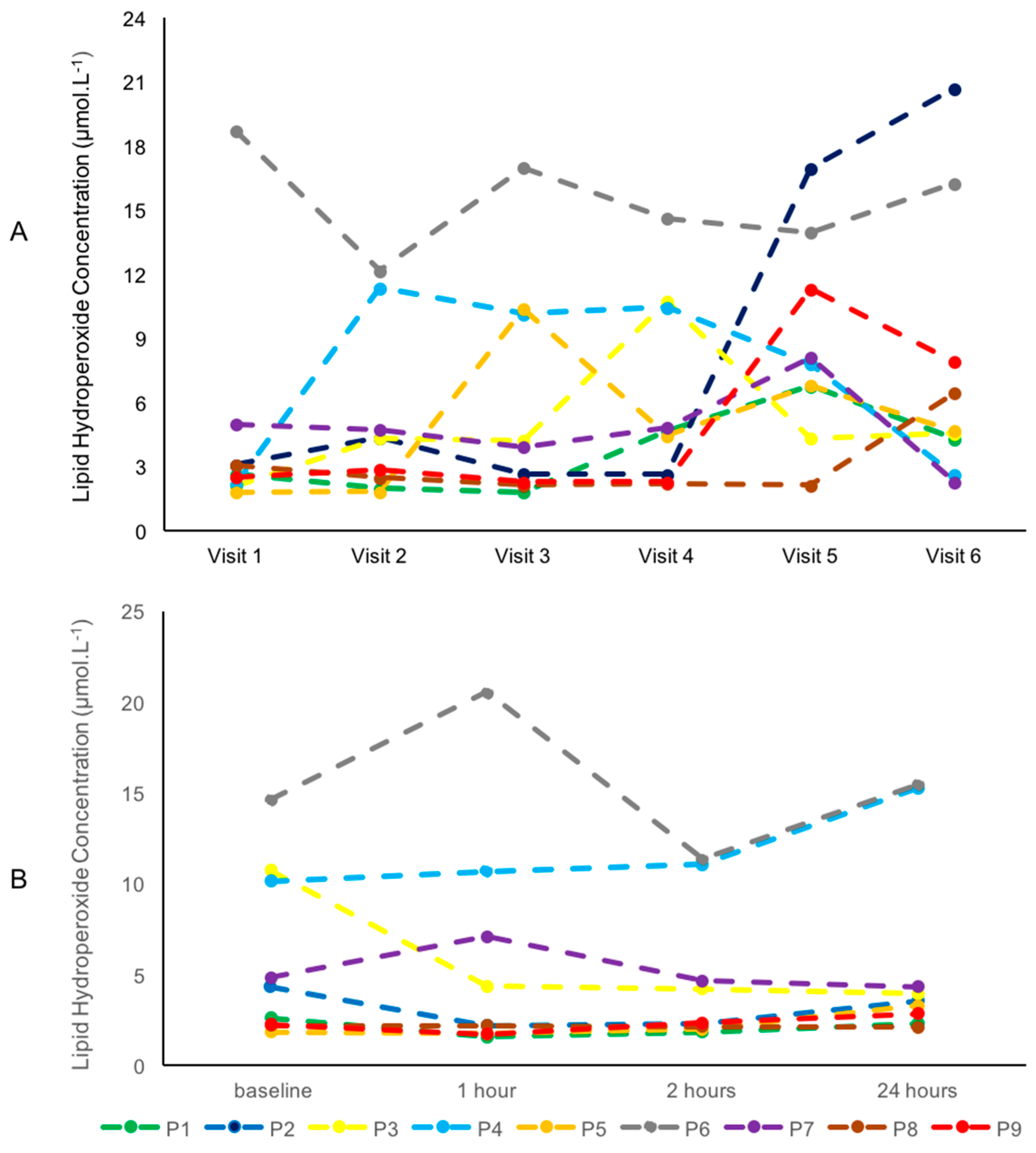
The visit-to-visit as well as the day-to-day variations in baseline lipid hydroperoxides for individual participants, following the six visits, and the diurnal variations in each participant during the control trial are presented in Figure 2A,B.

4. Discussion

Since most studies investigating oxidative stress have used exhaustive protocols, the current study aimed to examine oxidative stress under more real case scenarios of performing physical activity, imitating more of a daily exercise routine for the general population. Thus, the HIIT protocols implemented in the current study provided an average v V ˙ O2max of 75%, which has been suggested to be an optimal training intensity for moderately trained individuals for health purposes [21,35]. No significant differences among the six different exercise protocols, in any of the oxidative stress indices evaluated, were observed.

4.1. Impact of Intermittency

To our knowledge, this is the first study which examines the effect of various exercise intermittency levels, including acceleration/deceleration patterns, on oxidative stress biomarkers. Interestingly, the current results show that various intermittencies and accelerations/decelerations patterns of the HIIE do not significantly influence the biochemical markers of the oxidative stress levels evaluated. These results are in agreement with previous studies, which also found that short bouts of HIIE did not influence oxidative stress levels [36]. Bloomer et al. [37], in particular, used a submaximal intensity, similar to the present study, with results also indicating an absence of oxidative stress (8-Oxo-dG and malondialdehyde) at 1 h and 24 h post exercise, suggesting that higher intensity protocols may be required to elicit oxidative stress. Consequently, based on the current results, submaximal or maximal HIIE—including also plyometric exercise, since plyometric muscle contraction is produced due to acceleration/deceleration patterns—does not negatively impact oxidative stress levels or improve antioxidant status.

4.2. Impact of Acceleration/Deceleration

The current study was also the first which examined the role of plyometric activities (acceleration/deceleration) during HIIT in determining oxidative stress parameters. The results of the current research indicate that oxidative stress was not significantly modified due to the acceleration and deceleration components of the activity performed. However, since no direct comparison (i.e., HIIT including plyometric exercise patterns) to the existing literature could be made, comparisons with eccentric exercise (which imitates, as much as possible, the acceleration/deceleration during running pattern) research investigating oxidative stress are discussed. Interestingly, Margaritelis et al. [38] showed that individuals performing acute eccentric exercise aiming to induce oxidative stress had varied mean responses. It was observed that some individuals experience negative responses, while others experience no increases in the oxidative stress biomarkers tested (F2-isoprostanes, protein carbonyls, and glutathione). Thus, due to great heterogeneity, basal levels of the biomarkers of oxidative stress are considered important when these parameters are examined [38]. Although in the study by Margaritelis et al. [38], as well as in the current study, there were no samples from muscle biopsies, it is possible that the structural damages caused to the muscle due to the eccentric nature of the exercise were more prominent in the muscle compared to blood biomarkers of oxidative stress.
However, previous findings support that eccentric exercise does not increase glutathione status [39]. This is possibly due to insufficient exercise stimulus, which is also supported by the current study, as total glutathione levels in the current study were not also increased. Similarly, Bloomer et al. [40] investigated blood oxidative stress biomarkers and conducted muscle biopsy analysis to determine the responses to squat resistance exercise versus sprints. They found minimal modifications in oxidative stress levels between these exercise conditions, suggesting that this is evident when the participants are relatively well trained. In addition, a systematic review found that engaging in running exercises does not trigger a reaction in specific biomarkers associated with oxidative stress [4]. Instead, the expression of oxidative damage markers in lipids, proteins, and various enzymatic and non-enzymatic antioxidants varies based on the individual’s training status [4]. In the current study, the participants were relatively well-trained recreational athletes. Consequently, taking the current and the above-mentioned results collectively could imply that relatively well-trained individuals possibly have experienced adaptations due to their training status, leading to non-significant modifications in their oxidative stress levels following submaximal HIIT and plyometric exercise.
Supporting this outcome, research examining blood biomarkers and muscle biopsies showed that even exhaustive resistance exercise in trained men showed no upregulation of oxidative stress, as indicated by F2-isoprostanes [41]. According to McAnulty et al. [41], this result could be due to different modes of exercise causing different mechanical stresses that may affect the levels of oxidative stress. Quindry et al. [42], for example, investigated the role of eccentric exercise in participants with homogenous muscle fibre type compositions and found that the fibre type composition may influence oxidative stress levels, as correlations existed between type II muscle fibres and protein carbonyls. Consequently, it is possible that individuals with a higher percentage of type II muscle fibres could show higher oxidative stress compared to individuals with type I muscle fibres [42]. As the literature shows [38,40,41], the association between eccentric exercise and individual responses, along with the physical performance level of the participants and the physiological stimuli, may significantly influence oxidative stress levels. More research, therefore, is required to determine the biomechanical effect of the acceleration and deceleration on oxidative stress during HIIT and to examine whether the eccentric nature of the running component, accompanied with the increased impact forces, may promote oxidative stress.
It has also been suggested that oxidative stress levels are not affected by the absolute metabolic workload [43] but by the exercise duration [37] and intensity [43,44]. In addition to the above, the current study has shown that when exercise is matched for external workload (speed, duration, and distance) but the intermittency or the acceleration varies, there are no substantial influences on oxidative stress. Thus, it is plausible that the increased rate of intermittency or acceleration and deceleration, as a single factor, is not a sufficient contributor to induce oxidative stress. However, a combination of increased intensity, duration, and intermittency or acceleration is required to produce changes in oxidative stress status during HIIE. Previous findings demonstrated that acute continuous exercise elicits adverse inflammatory responses compared to a similar HIIT session, suggesting that the observed reduction in monocyte chemoattractant protein-1 following HIIT could be due to an effective reduction in oxidative stress [45]. Consequently, it is possible that oxidative stress is reduced by this type of activity as a result of decreased inflammation.

4.3. Intra-Individual Variability in Blood Metabolites

Alternatively, it is also possible that the intra-individual variability concerning blood metabolites that was observed in the current study may contribute to enhancing the possibility of type II statistical error. Plotting, for example (Figure 2A), individual data, it is obvious that individuals exhibited substantial inter-individual differences in lipid hydroperoxides during rest. Considering this, the daily, monthly, and seasonal physiological production of ROS levels is not fully understood, as the observations about increased oxidative stress levels in exercise studies are usually indicated against baseline measures [46,47,48,49,50]. Examining the baseline concentration of lipid hydroperoxides of the current participants following each visit to the laboratory (Figure 2B), the data showed a mean value of 6.42 μmol·L−1 in the range of 1.80–20.69 μmol·L−1. As this is a considerable disparity, the day-to-day variations during baseline measurements might be rather important, since this shows that there may be an 11-fold difference in the daily fluctuations of the concentration of lipid hydroperoxides, which would require further investigation. In addition, individuals showed variability in their rest values between visits and while some participants maintained a relatively steady concentration in lipid hydroperoxides (i.e., ranging from 12.19 to 18.71 μmol·L−1 in six visits), other participants had a higher variability ranging from 3.09 to 20.69 μmol·L−1 between the six visits. This individual variability could suggest the existence of outliers; thus, a pronounced dayto-day variation in blood oxidative biomarkers could require careful control measurements to be taken into consideration in future investigations.
Furthermore, intra-individual diurnal fluctuations were also interesting. Upon a closer examination of the control session, where exercise was not performed, individuals showed considerable variation during a 24 h response (Figure 2A,B), with some individuals showing increases in lipid hydroperoxides from baseline to 1 h (participant 6, Figure 2B), while others showed decreases (participant 3, Figure 2B) and others experienced minimum changes (participant 8, Figure 2B). This observation could further supplement the notion that individuals experience different diurnal changes as part of their daily physiological and metabolic processes [46]. Thus, the observed intra-individual fluctuations might be due to the dual roles that ROS were found to play in humans [51]. Thus, in similar studies, it would be sensible to include control sessions where no exercise is performed, with blood samples obtained at the same time points as they would be during an exercise session to decrease the influence of sampling time and the individuality in the biomarkers tested.
As reference values of oxidative stress biomarkers are currently unknown [52], more research is required in an attempt to study and determine the physiological levels of these markers in a healthy population and to investigate the possibility of an upper or lower threshold. In addition, exercise-induced oxidative stress studies could perhaps provide better understanding, as any increases observed would be determined to be due to the exercise stimulus and not due to the artefact of the physiological fluctuations. One study, for example, proposed that the recruitment of participants could be classified into groups based on baseline measurements, as some individuals may experience extreme (low or high) values [53]. This research also showed that even when this artefact is considered, individuals may still experience extreme (low or high) values with the same exercise stimulus; however, it is proposed that baseline measurements should be obtained, and participants should be categorised into groups prior to any oxidative stress-related studies [53].
This study is subject to some limitations. These include the small sample size due to the difficulty in recruiting participants who could complete all exercise protocols on the NMT. In addition, it is possible that exercise-induced oxidative stress occurred immediately after exercise and potentially returned to baseline after 1 h of recovery. However, as the collection of blood samples had to be kept to the minimum since the aim of this study was to investigate whether redox imbalance continues for a prolonged period of time following the completion of exercise (i.e., 24 h), it was decided to examine the effects of oxidative stress following exercise and not immediately after.

5. Conclusions

This study did not show the presence of oxidative stress following various submaximal or maximal HIIT protocols or when protocols were matched for average speed, duration, and distance but varied in either the intermittency or the acceleration and deceleration components. The day-to-day variation in oxidative stress markers needs to be further examined to better understand the individual variability and comprehend, in more depth, the oxidative stress parameters, including the range that individuals exhibit, which lies within their daily variation and is not a reflection of the exercise stimulus. Recommendations for future work could include the impact of intermittency or acceleration on biomarkers of oxidative stress in real-world scenarios to further investigate how oxidative stress occurs within a population.

Author Contributions

E.C.: conceptualization, experimental design, validation, methodology, investigation, project administration, data collection, data analysis, visualization, writing—original draft preparation, and writing—review and editing. H.S.J.: data analysis and writing—review and editing. M.H.: data re-analysis and writing—review and editing. M.C.F.: experimental design, writing—review and editing, and supervision. All authors have read and agreed to the published version of the manuscript.

Funding

This research received no external funding.

Institutional Review Board Statement

The study was conducted according to the guidelines of the Declaration of Helsinki and approved by the Institutional Review Board of the School of Life Sciences, University of Hull, UK (protocol code 1314061, 5 October 2015).

Informed Consent Statement

Written informed consent has been obtained from all the participants involved in the current study.

Data Availability Statement

The raw data supporting the conclusions of this article will be made available by the first author and the corresponding author.

Acknowledgments

The authors express their appreciation to the recreational athletes who voluntarily participated in the study.

Conflicts of Interest

The authors declare no conflicts of interest.

References

  1. Gomez-Cabrera, M.C.; Vina, J.; Ji, L.L. Interplay of oxidants and antioxidants during exercise: Implications for muscle health. Phys. Sportsmed. 2009, 37, 116–123. [Google Scholar] [CrossRef] [PubMed]
  2. Powers, S.K.; Kavazis, A.N.; McClung, J.M. Oxidative stress and disuse muscle atrophy. J. Appl. Physiol. 2007, 102, 2389–2397. [Google Scholar] [CrossRef] [PubMed]
  3. Powers, S.K.; Jackson, M.J. Exercise-induced oxidative stress: Cellular mechanisms and impact on muscle force production. Physiol. Rev. 2008, 88, 1243–1276. [Google Scholar] [CrossRef] [PubMed]
  4. Thirupathi, A.; Pinho, R.A.; Ugbolue, U.C.; He, Y.; Meng, Y.; Gu, Y. Effect of Running Exercise on Oxidative Stress Biomarkers: A Systematic Review. Front. Physiol. 2020, 11, 610112. [Google Scholar] [CrossRef]
  5. Lian, D.; Chen, M.M.; Wu, H.; Deng, S.; Hu, X. The Role of Oxidative Stress in Skeletal Muscle Myogenesis and Muscle Disease. Antioxidants 2022, 11, 755. [Google Scholar] [CrossRef] [PubMed]
  6. Powers, S.K.; Deminice, R.; Ozdemir, M.; Yoshihara, T.; Bomkamp, M.P.; Hyatt, H. Exercise-induced oxidative stress: Friend or foe? J. Sport Health Sci. 2020, 9, 415–425. [Google Scholar] [CrossRef]
  7. Steinbacher, P.; Eckl, P. Impact of oxidative stress on exercising skeletal muscle. Biomolecules 2015, 5, 356–377. [Google Scholar] [CrossRef]
  8. Fisher-Wellman, K.; Bell, H.K.; Bloomer, R.J. Oxidative stress and antioxidant defense mechanisms linked to exercise during cardiopulmonary and metabolic disorders. Oxid. Med. Cell. Longev. 2009, 2, 43–51. [Google Scholar] [CrossRef]
  9. Powers, S.K.; Goldstein, E.; Schrager, M.; Ji, L.L. Exercise Training and Skeletal Muscle Antioxidant Enzymes: An Update. Antioxidants 2022, 12, 39. [Google Scholar] [CrossRef]
  10. Gomez-Cabrera, M.C.; Borras, C.; Pallardo, F.V.; Sastre, J.; Ji, L.L.; Vina, J. Decreasing xanthine oxidase-mediated oxidative stress prevents useful cellular adaptations to exercise in rats. J. Physiol. 2005, 567, 113–120. [Google Scholar] [CrossRef]
  11. Hamilton, K.L.; Staib, J.L.; Phillips, T.; Hess, A.; Lennon, S.L.; Powers, S.K. Exercise, antioxidants, and HSP72: Protection against myocardial ischemia/reperfusion. Free Radic. Biol. Med. 2003, 34, 800–809. [Google Scholar] [CrossRef] [PubMed]
  12. Merry, T.L.; Ristow, M. Do antioxidant supplements interfere with skeletal muscle adaptation to exercise training? J. Physiol. 2016, 594, 5135–5147. [Google Scholar] [CrossRef] [PubMed]
  13. Paulsen, G.; Cumming, K.T.; Holden, G.; Hallen, J.; Ronnestad, B.R.; Sveen, O.; Skaug, A.; Paur, I.; Bastani, N.E.; Ostgaard, H.N.; et al. Vitamin C and E supplementation hampers cellular adaptation to endurance training in humans: A double-blind, randomised, controlled trial. J. Physiol. 2014, 592, 1887–1901. [Google Scholar] [CrossRef]
  14. Powers, S.K.; Schrager, M. Redox signaling regulates skeletal muscle remodeling in response to exercise and prolonged inactivity. Redox Biol. 2022, 54, 102374. [Google Scholar] [CrossRef]
  15. Ristow, M.; Zarse, K.; Oberbach, A.; Kloting, N.; Birringer, M.; Kiehntopf, M.; Stumvoll, M.; Kahn, C.R.; Bluher, M. Antioxidants prevent health-promoting effects of physical exercise in humans. Proc. Natl. Acad. Sci. USA 2009, 106, 8665–8670. [Google Scholar] [CrossRef]
  16. Andrade, F.H.; Reid, M.B.; Westerblad, H. Contractile response of skeletal muscle to low peroxide concentrations: Myofibrillar calcium sensitivity as a likely target for redox-modulation. FASEB J. 2001, 15, 309–311. [Google Scholar] [CrossRef] [PubMed]
  17. Mollica, J.P.; Dutka, T.L.; Merry, T.L.; Lamboley, C.R.; McConell, G.K.; McKenna, M.J.; Murphy, R.M.; Lamb, G.D. S-glutathionylation of troponin I (fast) increases contractile apparatus Ca2+ sensitivity in fast-twitch muscle fibres of rats and humans. J. Physiol. 2012, 590, 1443–1463. [Google Scholar] [CrossRef]
  18. Powers, S.K.; Ji, L.L.; Kavazis, A.N.; Jackson, M.J. Reactive oxygen species: Impact on skeletal muscle. Compr. Physiol. 2011, 1, 941–969. [Google Scholar] [CrossRef]
  19. Lu, Y.; Wiltshire, H.D.; Baker, J.S.; Wang, Q. Effects of High Intensity Exercise on Oxidative Stress and Antioxidant Status in Untrained Humans: A Systematic Review. Biology 2021, 10, 1272. [Google Scholar] [CrossRef]
  20. Draeger, C.L.; Naves, A.; Marques, N.; Baptistella, A.B.; Carnauba, R.A.; Paschoal, V.; Nicastro, H. Controversies of antioxidant vitamins supplementation in exercise: Ergogenic or ergolytic effects in humans? J. Int. Soc. Sports Nutr. 2014, 11, 4. [Google Scholar] [CrossRef]
  21. Atakan, M.M.; Li, Y.; Kosar, S.N.; Turnagol, H.H.; Yan, X. Evidence-Based Effects of High-Intensity Interval Training on Exercise Capacity and Health: A Review with Historical Perspective. Int. J. Environ. Res. Public Health 2021, 18, 7201. [Google Scholar] [CrossRef]
  22. Thompson, W.R. WORLDWIDE SURVEY OF FITNESS TRENDS FOR 2016: 10th Anniversary Edition. ACSM’s Health Fit. J. 2015, 19, 9–18. [Google Scholar] [CrossRef]
  23. Gibala, M.J.; Little, J.P.; Macdonald, M.J.; Hawley, J.A. Physiological adaptations to low-volume, high-intensity interval training in health and disease. J. Physiol. 2012, 590, 1077–1084. [Google Scholar] [CrossRef] [PubMed]
  24. Burgomaster, K.A.; Howarth, K.R.; Phillips, S.M.; Rakobowchuk, M.; Macdonald, M.J.; McGee, S.L.; Gibala, M.J. Similar metabolic adaptations during exercise after low volume sprint interval and traditional endurance training in humans. J. Physiol. 2008, 586, 151–160. [Google Scholar] [CrossRef] [PubMed]
  25. Kessler, H.S.; Sisson, S.B.; Short, K.R. The potential for high-intensity interval training to reduce cardiometabolic disease risk. Sports Med. 2012, 42, 489–509. [Google Scholar] [CrossRef] [PubMed]
  26. Tschakert, G.; Hofmann, P. High-intensity intermittent exercise: Methodological and physiological aspects. Int. J. Sports Physiol. Perform. 2013, 8, 600–610. [Google Scholar] [CrossRef] [PubMed]
  27. Cardoso, A.M.; Bagatini, M.D.; Roth, M.A.; Martins, C.C.; Rezer, J.F.; Mello, F.F.; Lopes, L.F.; Morsch, V.M.; Schetinger, M.R. Acute effects of resistance exercise and intermittent intense aerobic exercise on blood cell count and oxidative stress in trained middle-aged women. Braz. J. Med. Biol. Res. 2012, 45, 1172–1182. [Google Scholar] [CrossRef]
  28. Kingsley, M.I.; Wadsworth, D.; Kilduff, L.P.; McEneny, J.; Benton, D. Effects of phosphatidylserine on oxidative stress following intermittent running. Med. Sci. Sports Exerc. 2005, 37, 1300–1306. [Google Scholar] [CrossRef]
  29. Thompson, D.; Williams, C.; Kingsley, M.; Nicholas, C.W.; Lakomy, H.K.; McArdle, F.; Jackson, M.J. Muscle soreness and damage parameters after prolonged intermittent shuttle-running following acute vitamin C supplementation. Int. J. Sports Med. 2001, 22, 68–75. [Google Scholar] [CrossRef]
  30. Campbell, W.W.; Kraus, W.E.; Powell, K.E.; Haskell, W.L.; Janz, K.F.; Jakicic, J.M.; Troiano, R.P.; Sprow, K.; Torres, A.; Piercy, K.L.; et al. High-Intensity Interval Training for Cardiometabolic Disease Prevention. Med. Sci. Sports Exerc. 2019, 51, 1220–1226. [Google Scholar] [CrossRef] [PubMed]
  31. Currie, K.D.; Dizonno, V.; Oh, P.I.; Goodman, J.M. Acute physiological responses to high-intensity interval exercise in patients with coronary artery disease. Eur. J. Appl. Physiol. 2023, 123, 737–747. [Google Scholar] [CrossRef]
  32. Levinger, I.; Shaw, C.S.; Stepto, N.K.; Cassar, S.; McAinch, A.J.; Cheetham, C.; Maiorana, A.J. What Doesn’t Kill You Makes You Fitter: A Systematic Review of High-Intensity Interval Exercise for Patients with Cardiovascular and Metabolic Diseases. Clin. Med. Insights Cardiol. 2015, 9, 53–63. [Google Scholar] [CrossRef]
  33. Forster, H.V.; Dempsey, J.A.; Thomson, J.; Vidruk, E.; DoPico, G.A. Estimation of arterial PO2, PCO2, pH, and lactate from arterialized venous blood. J. Appl. Physiol. 1972, 32, 134–137. [Google Scholar] [CrossRef] [PubMed]
  34. Borg, G.A. Psychophysical bases of perceived exertion. Med. Sci. Sports Exerc. 1982, 14, 377–381. [Google Scholar] [CrossRef]
  35. Midgley, A.W.; McNaughton, L.R.; Wilkinson, M. Is there an Optimal Training Intensity for Enhancing the Maximal Oxygen Uptake of Distance Runners? Sports Med. 2006, 36, 117–132. [Google Scholar] [CrossRef]
  36. Vieira-Souza, L.M.; Aidar, F.J.; Matos, D.G.d.; Silva, A.N.d.; Miguel-dos-Santos, R.; Santos, J.L.d.; Costa, R.d.A.; Marçal, A.C.; Lauton-Santos, S.; Cabral, B.G.d.A.T.; et al. Short-term hiit does not promote oxidative stress or muscle damage. Rev. Bras. Med. Esporte 2021, 27, 138–141. [Google Scholar] [CrossRef]
  37. Bloomer, R.J.; Goldfarb, A.H.; Wideman, L.; McKenzie, M.J.; Consitt, L.A. Effects of acute aerobic and anaerobic exercise on blood markers of oxidative stress. J. Strength Cond. Res. 2005, 19, 276–285. [Google Scholar] [CrossRef] [PubMed]
  38. Margaritelis, N.V.; Kyparos, A.; Paschalis, V.; Theodorou, A.A.; Panayiotou, G.; Zafeiridis, A.; Dipla, K.; Nikolaidis, M.G.; Vrabas, I.S. Reductive stress after exercise: The issue of redox individuality. Redox Biol. 2014, 2, 520–528. [Google Scholar] [CrossRef] [PubMed]
  39. Camus, G.; Felekidis, A.; Pincemail, J.; Deby-Dupont, G.; Deby, C.; Juchmes-Ferir, A.; Lejeune, R.; Lamy, M. Blood levels of reduced/oxidized glutathione and plasma concentration of ascorbic acid during eccentric and concentric exercises of similar energy cost. Arch. Int. Physiol. Biochim. Biophys. 1994, 102, 67–70. [Google Scholar] [CrossRef] [PubMed]
  40. Bloomer, R.J.; Falvo, M.J.; Fry, A.C.; Schilling, B.K.; Smith, W.A.; Moore, C.A. Oxidative stress response in trained men following repeated squats or sprints. Med. Sci. Sports Exerc. 2006, 38, 1436–1442. [Google Scholar] [CrossRef]
  41. McAnulty, S.R.; McAnulty, L.S.; Nieman, D.C.; Morrow, J.D.; Utter, A.C.; Dumke, C.L. Effect of resistance exercise and carbohydrate ingestion on oxidative stress. Free Radic. Res. 2005, 39, 1219–1224. [Google Scholar] [CrossRef]
  42. Quindry, J.; Miller, L.; McGinnis, G.; Irwin, M.; Dumke, C.; Magal, M.; Triplett, N.T.; McBride, J.; Urbiztondo, Z. Muscle-fiber type and blood oxidative stress after eccentric exercise. Int. J. Sport. Nutr. Exerc. Metab. 2011, 21, 462–470. [Google Scholar] [CrossRef]
  43. Quindry, J.C.; Stone, W.L.; King, J.; Broeder, C.E. The effects of acute exercise on neutrophils and plasma oxidative stress. Med. Sci. Sports Exerc. 2003, 35, 1139–1145. [Google Scholar] [CrossRef]
  44. Bailey, D.M.; Young, I.S.; McEneny, J.; Lawrenson, L.; Kim, J.; Barden, J.; Richardson, R.S. Regulation of free radical outflow from an isolated muscle bed in exercising humans. Am. J. Physiol. Heart Circ. Physiol. 2004, 287, H1689–H1699. [Google Scholar] [CrossRef] [PubMed]
  45. Kaspar, F.; Jelinek, H.F.; Perkins, S.; Al-Aubaidy, H.A.; de Jong, B.; Butkowski, E. Acute-Phase Inflammatory Response to Single-Bout HIIT and Endurance Training: A Comparative Study. Mediat. Inflamm. 2016, 2016, 5474837. [Google Scholar] [CrossRef] [PubMed]
  46. Budkowska, M.; Cecerska-Heryc, E.; Marcinowska, Z.; Siennicka, A.; Dolegowska, B. The Influence of Circadian Rhythm on the Activity of Oxidative Stress Enzymes. Int. J. Mol. Sci. 2022, 23, 14275. [Google Scholar] [CrossRef] [PubMed]
  47. Kliszczewicz, B.; Quindry, C.J.; Blessing, L.D.; Oliver, D.G.; Esco, R.M.; Taylor, J.K. Acute Exercise and Oxidative Stress: CrossFit vs. Treadmill Bout. J. Hum. Kinet. 2015, 47, 81–90. [Google Scholar] [CrossRef] [PubMed]
  48. Välimäki, I.A.; Vuorimaa, T.; Ahotupa, M.; Vasankari, T.J. Strenuous physical exercise accelerates the lipid peroxide clearing transport by HDL. Eur. J. Appl. Physiol. 2016, 116, 1683–1691. [Google Scholar] [CrossRef] [PubMed]
  49. Wadley, A.J.; Chen, Y.W.; Lip, G.Y.; Fisher, J.P.; Aldred, S. Low volume-high intensity interval exercise elicits antioxidant and anti-inflammatory effects in humans. J. Sports Sci. 2016, 34, 1–9. [Google Scholar] [CrossRef] [PubMed]
  50. Wilking, M.; Ndiaye, M.; Mukhtar, H.; Ahmad, N. Circadian rhythm connections to oxidative stress: Implications for human health. Antioxid. Redox Signal 2013, 19, 192–208. [Google Scholar] [CrossRef] [PubMed]
  51. Bartosz, G. Reactive oxygen species: Destroyers or messengers? Biochem. Pharmacol. 2009, 77, 1303–1315. [Google Scholar] [CrossRef] [PubMed]
  52. Davison, G.W.; Ashton, T.; McEneny, J.; Young, I.S.; Davies, B.; Bailey, D.M. Critical difference applied to exercise-induced oxidative stress: The dilemma of distinguishing biological from statistical change. J. Physiol. Biochem. 2012, 68, 377–384. [Google Scholar] [CrossRef] [PubMed]
  53. Margaritelis, N.V.; Theodorou, A.A.; Paschalis, V.; Veskoukis, A.S.; Dipla, K.; Zafeiridis, A.; Panayiotou, G.; Vrabas, I.S.; Kyparos, A.; Nikolaidis, M.G. Experimental verification of regression to the mean in redox biology: Differential responses to exercise. Free Radic. Res. 2016, 50, 1237–1244. [Google Scholar] [CrossRef] [PubMed]
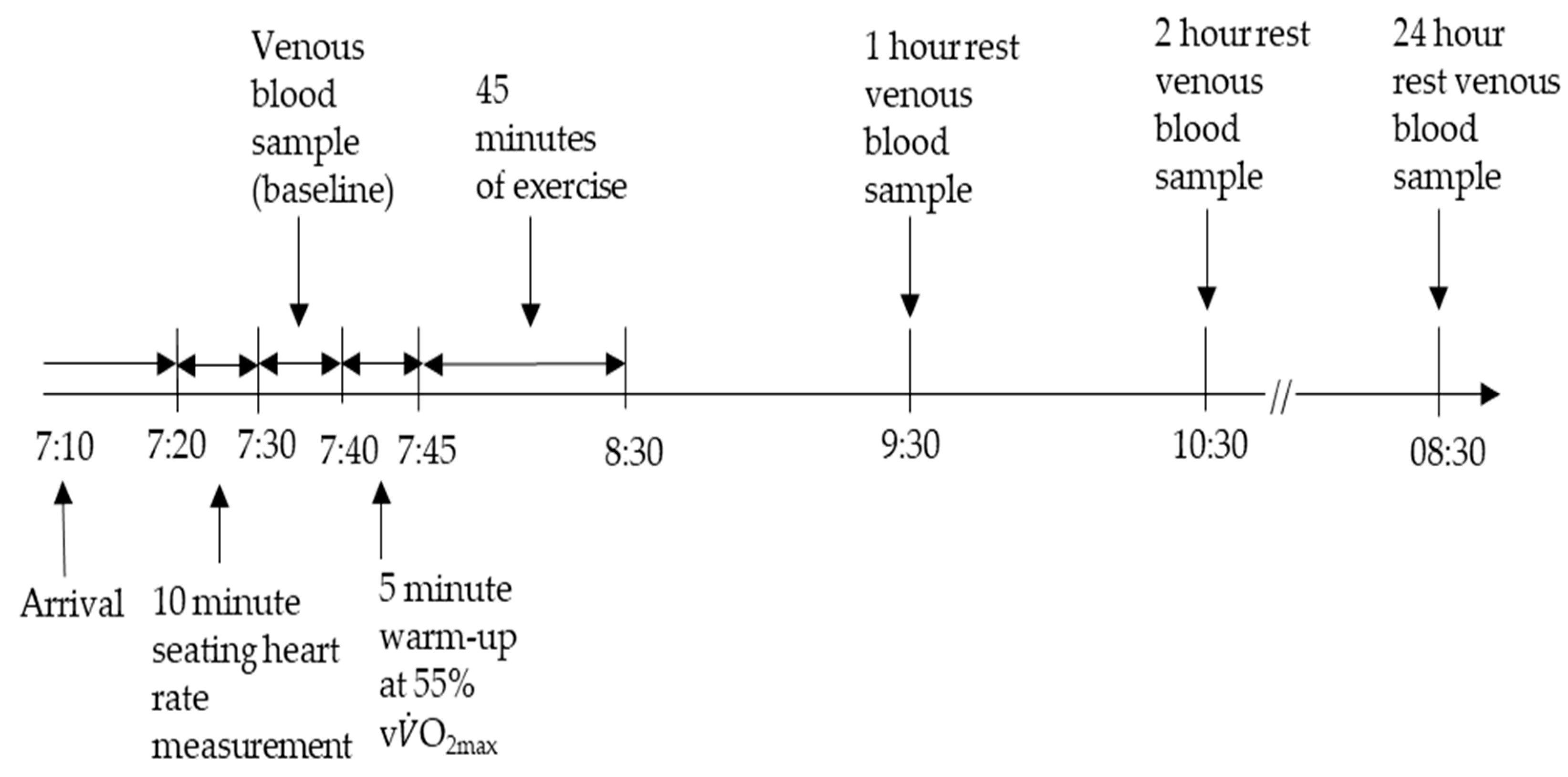
Figure 1. Schematic overview of the experimental protocol.
Applsci 14 02655 g001
Figure 2. (A,B) Day-to-day variation in baseline lipid hydroperoxides for individual participants (n = 9) following 6 visits to the laboratory (A). Diurnal variations in individual participants (n = 9) during control session (B). Blood samples were analysed at baseline, 1 h, 2 h, and 24 h post-exercise to examine lipid hydroperoxides. Each dashed line represents an individual participant (P1–P9).
Applsci 14 02655 g002
Table 1. Participant characteristics.
Participant CharacteristicsValue (±SD)
Age (y) 21 ± 3
Height (cm) 180 ± 4
Body mass (kg) 79.4 ± 7.9
Body fat (%)12 ± 3
Systolic BP (mmHg) 133 ± 10
Diastolic BP (mmHg) 71 ± 6
Resting HR (beats·min−1) 58 ± 13
Estimated HRmax (beats·min−1) 192 ± 2
Peak HR (beats·min−1)183 ± 9
v V ˙ O2max (km·hr−1)14 ± 6
V ˙ O2max (mL·kg−1·min−1)52.0 ± 2
Table 2. (AC) Concentrations of A, lipid hydroperoxides, B, total GSH, and C, superoxide dismutase activity of the 3 HIIT protocols with varying intermittency at baseline and 1 h, 2 h, and 24 h. Median values with 75% upper and 25% lower percentiles.
(A)
Lipid Hydroperoxides (μmol·L−1)
Exercise Protocol Baseline1 h2 h24 h
Control4.35 (2.28–10.16) 2.20 (1.77–7.09)2.36 (2.4–4.67)3.55 (2.85–4.33)
Low intermittent4.22 (2.88–4.68)4.68 (2.41–13.00)4.56 (3.16–10.98)4.39 (2.49–9.74)
Moderately Intermittent2.51 (2.08–4.73)2.57 (2.30–7.62)2.69 (2.53–8.88)2.23 (2.00–7.79)
Highly Intermittent3.92 (2.01–10.39)3.79 (2.01–8.97) 3.94 (2.30–8.78)3.72 (2.23–4.00)
(B)
GSH (μΜ)
Exercise ProtocolBaseline1 h2 h24 h
Control1.44 (0.78–1.91)1.49 (0.95–2.17)1.80 (1.37–2.87)1.64 (1.16–1.98)
Low intermittent0.96 (0.73–1.04)1.76 (1.03–2.61)1.54 (1.37–2.68)1.69 (1.58–1.72)
Moderately Intermittent1.51 (1.11–2.39)1.80 (1.38–2.35)2.04 (1.59–2.89)1.66 (1.38–2.36)
Highly Intermittent1.26 (0.84–1.71)1.79 (1.06–1.94)1.54 (1.35–1.65) 0.99 (0.90–1.86)
(C)
Superoxide Dismutase (U/mL)
Exercise ProtocolBaseline1 h2 h24 h
Control1.15 (0.94–2.45)1.33 (1.17–2.85) 1.32 (1.03–2.65)1.22 (1.07–3.03)
Low intermittent0.95 (0.95–1.29)1.51 (0.83–1.73) 1.02 (1.00–1.19)1.11 (0.94–1.70)
Moderately Intermittent1.01 (0.99–1.83)1.39 (0.99–1.89)1.28 (1.09–2.03)1.41 (0.81–1.90)
Highly Intermittent1.20 (1.07–1.38)1.11 (0.97–1.28) 1.15 (0.85–1.20)1.19 (0.93–1.39)
Table 3. (AC) Concentrations of A, lipid hydroperoxide, B, total GSH, and C, superoxide dismutase activity following 3 different HIIT protocols of varying acceleration at baseline, 1 h, 2 h, and 24 h. Median values with 75% upper and 25% lower percentiles.
(A)
Lipid Hydroperoxides (μmol·L−1)
Exercise ProtocolBaseline1 h2 h24 h
Control4.35 (2.28–10.16)2.20 (1.77–7.09)2.36 (2.14–4.67)3.55 (2.85–4.33)
Low acceleration4.68 (4.31–8.13)4.95 (4.13–7.02)4.65 (4.23–7.84)6.69 (3.85–6.87)
Moderate acceleration6.81 (6.44–11.32)6.22 (4.72–13.27)4.85 (4.58–9.59)4.79 (4.25–9.80)
High acceleration2.51 (2.08–4.73)2.57 (2.30–7.62) 2.69 (2.53–8.88)2.23 (2.00–7.79)
(B)
GSH (μΜ)
Exercise ProtocolBaseline1 h2 h24 h
Control1.44 (0.78–1.91)1.49 (0.95–2.17)1.80 (1.37–2.87)1.64 (1.16–1.98)
Low acceleration1.63 (1.43–2.20)1.44 (0.86–2.11)1.61(1.17–1.93)2.15 (1.30–3.43)
Moderate acceleration2.24 (2.08–2.78)2.01 (1.40–2.59)3.07 (2.16–3.24)1.98 (1.76–2.96)
High acceleration1.51 (1.11–2.39)1.80 (1.38–2.35)2.04 (1.59–2.89)1.66 (1.38–2.36)
(C)
Superoxide Dismutase (U/mL)
Exercise ProtocolBaseline1 h2 h24 h
Control1.15 (0.94–2.45)1.33 (1.17–2.85)1.32 (1.03–2.65)1.22 (1.07–3.03)
Low acceleration1.26 (1.04–3.29)1.27 (1.01–4.28)1.45 (1.14–2.85)1.57 (1.30–3.63)
Moderate acceleration3.05 (1.23–3.59)2.56 (1.44–3.53)2.65 (1.51–3.26)2.76 (1.21–3.38)
High acceleration1.04 (1.04–3.29)1.39 (1.01–4.28)1.28 (1.14–2.85)1.41 (1.30–3.63)
Disclaimer/Publisher’s Note: The statements, opinions and data contained in all publications are solely those of the individual author(s) and contributor(s) and not of MDPI and/or the editor(s). MDPI and/or the editor(s) disclaim responsibility for any injury to people or property resulting from any ideas, methods, instructions or products referred to in the content.