Article

Effects of Potential Prebiotics from Codium fragile on Intestinal Diseases

1
Department of Integrative Biotechnology, Chonnam National University, Yeosu 59626, Republic of Korea
2
Marine Biotechnology Research Center, Jeonnam Bio Foundation, 21-7, Nonggongdanji 4Gil, Wando-eup, Wando-gun 59108, Republic of Korea
3
Bada and Haecho Fishery Corp., 1111-2, Cheonma-ro, Pungyang-myeon, Goheung-gun 59548, Republic of Korea
*
Author to whom correspondence should be addressed.
Appl. Sci. 2024, 14(7), 3037; https://doi.org/10.3390/app14073037
Submission received: 18 February 2024 / Revised: 28 March 2024 / Accepted: 29 March 2024 / Published: 4 April 2024

Abstract

:
This study examined the effects of an extract of the green algae Codium fragile (hereafter referred to as CFE) on dextran sulfate sodium (DSS)-induced colitis. As the administration of CFE increased, the proliferation of Akkermansia muciniphila, which is a key player in metabolic and gastrointestinal disorders, also increased. After CFE administration for 10 weeks, acetic acid was identified as the major metabolite in mouse cecum and β-glucuronidase activity in mouse fecesdecreased. Further, CFE significantly alleviated the acute intestinal injury induced by DSS administration, including DAI score, colon length, and histological score. The experimental group also displayed indications of significantly lower neutrophil activity and inflammation. In conclusion, the protective effect of CFE against DSS colitis suggests its clinical use by IBD patients.

1. Introduction

Recent studies have shown marine algae to possess a wide range of potential health applications, with moderating effects on the immune system being of particular interest in this study [1,2,3,4,5]. Additionally, algal metabolites such as sulfated polysaccharides and polyphenols have demonstrated multistep antiviral capability and provided a new route to develop new therapeutic methods to treat COVID-19 and other viral diseases [6].
Codium fragile is a traditional Asian food ingredient [7] mainly consumed in South Korea, Japan, and China. The main components of green algae are sulfated structural polysaccharides such as ulvans and sulfated cellulose, galactans, pectin, and mannans. These sulfated polysaccharides are not completely fermented by intestinal microbiota [8,9,10]. Some of the unfermented C. fragile polysaccharides have also been observed to have various interactions with biological systems [11,12]. These properties provide opportunities for the potential application of C. fragile polysaccharides as prebiotics.
The gut microbiota are microorganisms that live in the host’s gastrointestinal tract. Human health is greatly influenced by the composition and metabolism of these microorganisms [13]. The microbiota in the human gastrointestinal tract have been studied extensively owing to their role both in pathogenesis and gut health maintenance. An important function of large intestinal microbiota is to break down substrates such as resistant starch and dietary fiber, which are not completely hydrolyzed by host enzymes in the small intestines [14,15,16,17]. Thus, health improvement via regulation of the gut microbiota has become an interesting research field.
Akkermansia muciniphila is a strictly anaerobic bacterium recently isolated from human feces. It uses mucin as the sole source of carbon and nitrogen elements [18]. Ottman et al. reported that A. muciniphila can utilize the mucin-derived monosaccharides fucose, galactose, and N-acetylglucosamin [19,20]. The abundance of A. muciniphila in the feces appears to correlate with general gut health. The presence of A. muciniphila in feces has been associated with a healthy gut, and its abundance has been inversely correlated with several disease states. The abundance of A. muciniphila has been shown to be decreased in patients with ulcerative colitis and Crohn’s disease [21,22]. However, as A. muciniphila is a strict anaerobe with highly limited growth conditions, there are currently no A. muciniphila-containing products in the world. Therefore, consuming prebiotics that can selectively promote A. muciniphila in the intestines is necessary.
Inflammatory bowel diseases (IBDs) such as Crohn’s disease (CD) and ulcerative colitis (UC) are chronic intestinal diseases of unknown etiology [23,24]. Natural compounds have already shown promise as relatively safe therapeutic agents for the treatment and maintenance of IBD symptoms [25,26,27]. Algal polysaccharides are good candidates for the alleviation of intestinal inflammatory diseases due to their potential prebiotic efficacy [28,29].
In this study, we hypothesized that CFE may contain polysaccharides that exhibit therapeutic effects against intestinal inflammatory diseases. Until now, there have been few studies on the protective effects of CFE in mice with DSS-induced colitis. Thus, the present study was conducted to evaluate the protective effect of CFE against dextran sulfate sodium (DSS) colitis along with the proliferation of specific beneficial bacteria associated with a healthy intestine.

2. Materials and Methods

In this section, the methods for studying the effects of CFE on potential prebiotics and intestinal diseases are presented. Section 2.2 presents methods for evaluating prebiotics for growing specific beneficial bacteria important for maintaining intestinal health. In Section 2.3, methods for evaluating improvement in intestinal diseases are presented.

2.1. Preparation of CFE

CFE was prepared according to a previously described method [30]. In brief, previously collected C. fragile were washed, dried, and ground into powder. Boiling water was used to extract CFE from the powder, and the extract was concentrated and ultimately freeze-dried.

2.2. Potential Prebiotics for Improving Intestinal Diseases

2.2.1. Mouse Model

Seven-week-old male BALB/c mice (15–20 g) were obtained from KOSA BIO Inc. (Seongnam, Republic of Korea) and housed in A pathogen-free room (light cycle, 12 h light/dark; temperature, 22 ± 2 °C; humidity, 50 ± 5%). All mice were fed an AIN-93 diet (Research Diets, Inc., New Brunswick, NJ, USA) (Table 1), and sterilized water was provided ad libitum for a week during an adaptation period. Twenty mice were divided into four groups of five: (1) CTRL, fed a normal diet; (2) LCFE group, fed 75 mg of CFE per kg of body weight; (3) MCFE group, fed 150 mg of CFE per kg of body weight; (4) HCFE group, fed 300 mg of CFE per kg of body weight. Each group of mice was administered a daily oral dose of CFE dissolved in sterilized water for 10 weeks. The Mice were sacrificed via cervical dislocation under ether anesthesia. All procedures for this experiment were conducted in accordance with the Institutional Animal Care and Use Committee (IACUC233-041) and the Ethical Committee of Experimental Animals in the Efficacy Evaluation Center of Berry & Biofood Research Institute (BBRI-IACUC-21001).

2.2.2. Real-Time PCR Quantification

Fecal samples from individual mice were collected in sterilized tubes and stored at −80 °C. Genomic DNA was extracted using a QIAamp DNA Stool Mini Kit (Qiagen, Hilden, Germany) according to the manufacturer’s instructions.
The primers used to detect A. muciniphila and S. aureus were based on 16S rRNA gene sequences retrieved from the National Center for Biotechnology Information databases using the Entrez program. Forward primers and reverse primers were designed using the Primer Express 2.0 software (Applied Biosystems, Foster City, CA, USA) (Table 2).
PCR amplification was carried out in a total volume of 25 μL containing 1 × TaqMan Universal PCR Master Mix (Applied Biosystems), both primers (10 pmol each), 50 ng purified target DNA, and final BSA concentration of 0.1 mg/mL (New England Biolabs, Ipswich, MA, USA). A StepOne Plus RT-PCR system (Applied Biosystems) was used for amplification and detection. The amount of genomic DNA extracted was determined using an ultraviolet spectrophotometer at 260 nm. In the PCR assay, we compared different amounts of bacterial DNA extracted from fecal samples to overcome bias due to inhibitory compounds such as bile salts. Each assay was performed in duplicate in the same run. The cycle threshold (CT) was calculated as the cycle number at which the reaction became exponential. The CT of each sample was then compared with a standard curve made by diluting genomic DNA (10-fold dilution) from cultures of the target bacterium.

2.2.3. Short Chain Fatty Acid (SCFA) Analysis

Cecum contents were stored at −80 °C for measurement of SCFA concentration. Acetic acid and butyric acid concentrations were determined by flame ionization detection on an Agilent 7890A GC-MS equipped with a DB-FATWAX Ultra Inert column 30 m × 530 mm × 0.25 μm (Agilent Technologies, Santa Clara, CA, USA).

2.2.4. β-Glucuronidase Activity Analysis

β-glucuronidase activity was analyzed using the method described by Goldin et al. [31]. Fecal samples were incubated at 37 °C in the wells of a microtiter plate, and then 10 mL of sample was added in duplicate. Next, 100 μL of substrate solution was added, and incubated at 37 °C for 60 min. Next, 500 μL of 0.5 N NaOH was introduced to stop the reaction. Protein activity was quantified by measuring the absorbance at 405 nm. Triplicate assays were performed for each effector, and the mean values and standard deviations were reported. The concentration of 4-nitrophenol was determined using a standard curve of 4-nitrophenol in sodium phosphate buffer.

2.3. Potential Prebiotics for Improving Intestinal Diseases

2.3.1. Mouse Model

Mice (C57BL/6J, male, 9 weeks old at the time of purchase, 18 ± 2 g) were acquired from Orient Bio (Sungnam, Republic of Korea). The mice were held in a pathogen-free enclosure for 7 days prior to the exposure of DSS to some mice. For 5 days, 3% (w/v) of DSS was provided in drinking water, followed by 3 days with no DSS (molecular weight 36–50 kDa; MP Biomedicals, Irvine, CA, USA). During the whole experimental period of 8 days, CFE and sulfasalazine (St. Louis, MO, USA) were given to mice daily via oral administration. Thirty mice were divided randomly into six groups of five: (1) normal group fed water (N group), (2) negative control group fed DSS (NC group), (3) positive control group fed DSS + sulfasalazine (150 mg/kg) (PC group), (4) DSS + LCFE group fed DSS + 75 mg of CFE per kg of body weight, (5) DSS + MCFE group fed DSS + 150 mg of CFE per kg of body weight, and (6) DSS + HCFE group fed DSS + 300 mg of CFE per kg of body weight. The Animal Ethics Review Committee of Woojung Bio Inc. (Suwon, Korea) reviewed and approved these animal experiments in line with the Institutional Animal Care and Use Committee guidelines. The approval ID for using the animals at the Animal Facility of Woojung Bio was IACUC2303–041.

2.3.2. Evaluation of the Severity of Colitis

Colon length was measured, as in Zong et al. [32], from the ileocecal junction to the anal verge. We used the colitis DAI scoring system, as in Jeon et al. [33], to evaluate the severity of colitis in the examined mice.

2.3.3. Myeloperoxidase (MPO) Activity

The MPO assay kit (Abcam, Cambridge, MA, USA) was used to measure MPO activity in serum and tissue samples according to the manufacturer’s instructions. Absorbance was read at 412 nm using a multimode microplate reader (BioTek Instruments, Winooski, VT, USA). The results were presented as units per gram of tissue.

2.3.4. Histological Evaluation

Intestinal tissue was fixed with 10% formalin, embedded in paraffin, cut into 3 μm sections, and stained with hematoxylin and eosin (H&E) for microscopic evaluation. The stained slices were subsequently observed under an optical microscope and analyzed using the i-Solution Lite software ver. 8.1 program (Innerview Co., Sungnam, Republic of Korea). Histological evaluations of H&E-stained colonic sections were graded by two blinded investigators (Table 3).

2.3.5. Statistical Analysis

The t-test, one-way ANOVA for comparison of two or more groups, and post-hoc Tukey’s multiple comparison test were conducted using the SPSS 22.0 software (IBM Corp., Armonk, NY, USA). All data are presented as mean ± standard deviation.

3. Results

3.1. Effect of CFE on the Growth of Individual Bacteria

After feeding on CFE for 10 weeks, changes in the DNA log copy number of bacteria in mouse feces were determined (Figure 1). During CFE intake (10 weeks), an increase in beneficial bacteria, A. muciniphila, and Bifidobacterium spp., was observed, while the pathogenic bacteria, S. aureus and Clostridium spp., decreased. Moreover, in the HCEF group, A. muciniphila significantly increased while S. aureus significantly decreased (Figure 1a,c).

3.2. Effect of CFE on Cecum SCFA Production

As shown in Table 4, the main metabolite in mouse cecum contents was acetic acid, followed by butyric acid in small amounts. The contents of SCFAs in CFE-fed groups were higher than in the CTRL group in a CFE concentration-dependent manner. The content of acetic acid in the LCFE group (p < 0.05), the MCFE and HCFE groups (p < 0.001), and butyric acid in the MCFE and HCFE groups (p < 0.05) were higher than in the CTRL group.

3.3. Effect of CFE on Fecal β-Glucuronidase Activities

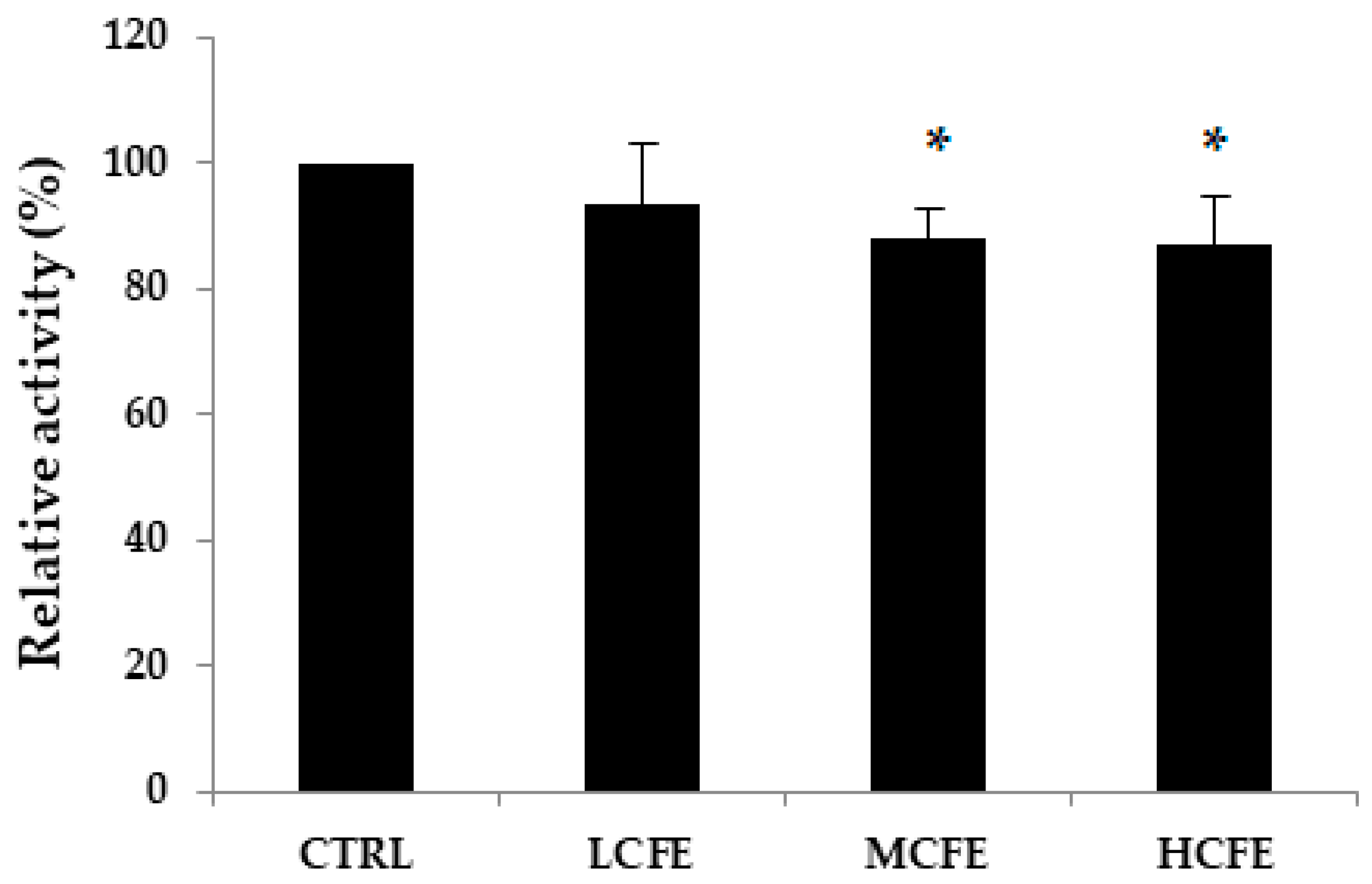
β-Glucuronidase activities in mouse feces were measured at week 10 of CFE feeding (Figure 2). A marked decrease in β-glucuronidase activity was observed in the MCFE and HCFE groups (p < 0.05) compared with the CTRL group, being 88% and 87%, respectively.

3.4. Effect of CFE on Mouse Colitis

To evaluate the effects of CFE on colitis, 9-week-old mice (C57BL/6) were administrated 3% DSS, sulfasalazine (150 mg/kg), and CFE (75 mg, 150 mg, and 300 mg per kg of body weight) separately or in combination, for 8 days (Figure 3a). The DAI score and colon length directly reflect the severity of UC in mouse models and are used to proactively assess the severity of UC. The DAI scores of each mouse group during days 0–7 are presented in Figure 3b. The DAI scores were significantly decreased in the CFE-fed group compared with the NC group. Another indicator that reflects the severity of intestinal inflammation is colon length, which recovers as inflammation improves [34,35]. The colon length of mice in the DSS + HCFE group was significantly longer (p < 0.05) than in mice in the NC group but similar to that of mice in the PC group (Figure 3c,d).

3.5. Effect of CFE on the Histological Injury of Colonic Epithelium Caused by DSS

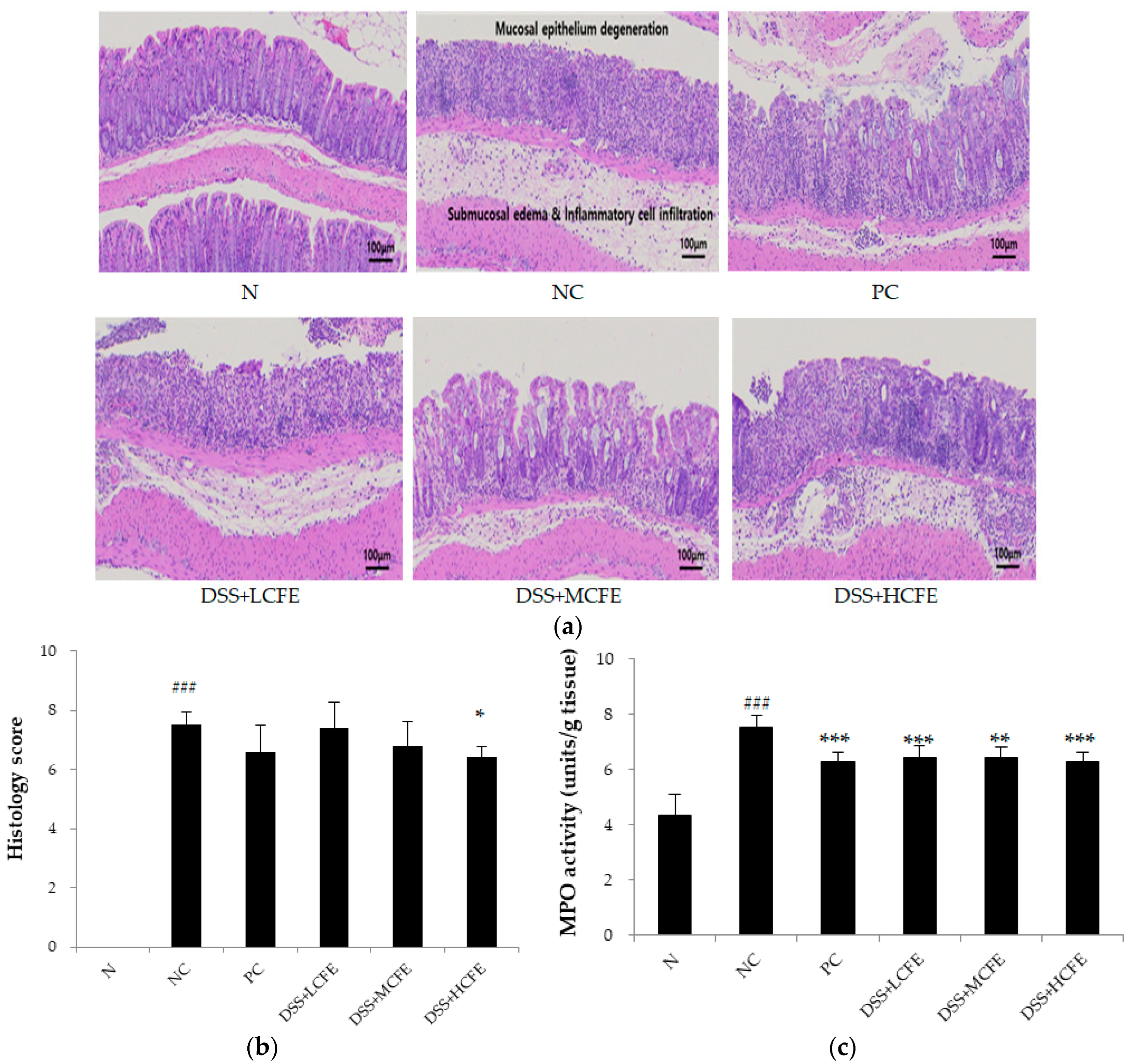
H&E staining was performed to investigate mucosal inflammation. Compared with the N group, the DSS + HCHE (CFE-fed) group or the PC group showed lower microscopic damage. The histological analysis indicated that the histological severity of the colitis was more severe in the NC group compared with the CFE-fed and PC groups (Figure 4b). Compared with the NC group, the DSS + HCHE group showed a significantly decreased histology score. Test results showed that MPO activity in the colon was significantly reduced in the CFE-fed and PC groups compared with the NC group (Figure 4c).

4. Discussion

Sulfated polysaccharides derived from seaweed can be used as prebiotics for gut microorganisms and degraded into other bioactive compounds, such as oligosaccharides, phytochemicals, and SCFAs, which can serve as substrates for these organisms and allow them to grow [36,37,38,39,40]. Sulfated galactan from C. fragile consists of a large amount of galactose residues, with trace arabinose and the presence of pyruvate and sulfate as substituents [7]. Additionally, our previous research reported that CFE comprises many galactose residues, with traces of arabinose and the presence of sulfate as substituents [30].
In the present study, we elucidated the ability of CFE to promote the growth of A. muciniphila. The presence of A. muciniphila in feces has been associated with a healthy intestine, and its abundance has been inversely correlated with several disease states. Further analysis confirmed that A. muciniphila can degrade mucin and exert competitive inhibition on other pathogenic bacteria that degrade the mucin [41]. Therefore, the present study aimed to investigate the efficacy and underlying mechanisms of CFE in alleviating DSS-induced colitis in mice.
The proliferation of bacteria during 10 weeks of CFE feeding was determined using quantitative PCR. As the administration of CFE increased, the proliferation of probiotics increased whereas the proliferation of pathogenic bacteria decreased. Therefore, our results indicate that CFE can be used as a prebiotic material. Moreover, A. muciniphila significantly increased in the HCEF group. A. muciniphila is abundant in the gut microbiota of healthy individuals, and it is beneficial in the prevention and treatment of obesity, type 2 diabetes, and other metabolic dysfunctions [18,19,20,21,42,43,44,45]. Therefore, CFE can be used as a prebiotic that specifically promotes A. muciniphila growth for the treatment of metabolic diseases.
After CFE administration for 10 weeks, acetic acid was identified as the major metabolite in mouse cecum. A similar result was also reported by Li et al. [46], who found that acetic acid was a major metabolite produced by A. muciniphila in static and dynamic cultures. Li et al. [46] reported that acetic acid increased lipolysis and decreased lipid synthesis in BRL-3A cells, thus reducing the accumulation of hepatic fat in BRL-3A cells. Therefore, CFE was thought to reduce lipid synthesis as it led to the production of acetic acid by promoting the growth of A. muciniphila.
β-Glucuronidase activity is a major factor in causing colon cancer [47]. β-Glucuronidase hydrolyzes β-D-glucuronides to glucuronic acid and aglycone, such as alcohol, amine, imine, or a thiol compound. UDP-glucuronosyltransferase catalyzes glucuronide formation. From the liver, where synthesis occurs, it is partially eliminated into the large intestines along with bile. There, it is hydrolyzed to aglycone under the influence of bacterial β-glucuronidase, which is further hydrolyzed to aglycones. High β-glucuronidase activity was observed in patients diagnosed with colonic neoplasia, suggesting that this enzyme plays an important role in promoting colonic neoplasia [48]. During the adaptation period, all mice were fed the AIN-93 diet for 1 week, but the enzymatic activity in each group at week 0 was not the same because the large intestines of all mice form a complex microbial ecosystem. A marked decrease in enzymatic activity was observed in the MCFE and HCFE groups compared with the CTRL group, being 88% and 87%, respectively. These results suggest that β-glucuronidase activity is decreased under CFE feeding of more than 150 mg per kg of body weight.
Dextran sulfate sodium (DSS)–induced colitis is mainly used to evaluate its efficacy against inflammatory bowel disease. Disease activity index (DAI) is increased, and colon length is shortened in DSS-induced colitis [49]. CFE treatment significantly improved the body weight, reduced the overall DAI score, and improved the colon length, suggesting the protective effect of CFE in DSS-induced colitis. Histologic examination consistently revealed an improvement in inflammatory signs, reducing inflammatory infiltrates and restoring intestinal epithelia in CFE-fed groups compared with the NC group, demonstrating that CFE alleviates mouse colitis.
MPO activity is a marker of neutrophil infiltration and is proportional to the number of neutrophils in the inflamed tissue [50]. In our study, MPO activity in the colon was significantly reduced in the CFE-fed group compared with the NC group, indicating that CFE can inhibit neutrophil infiltration and inflammation in mice.

5. Conclusions

In conclusion, the protective effect of CFE against DSS colitis suggests its clinical use by IBD patients. Further detailed studies would be needed to deepen A. muciniphila activity in relation to microecological interventions for IBD, and additional preclinical studies are needed to elucidate the underlying molecular mechanisms regulated by CFE in animal models of DSS-induced colitis. The conventional anti-inflammatory drugs used for treating IBD cause side effects such as allergic responses, diarrhea, vomiting, lymphopenia, raised liver enzymes, and inflammation of the pancreas [51]. It is important to alleviate IBD using natural plants with low toxicity and few side effects. Therefore, further studies on optimal dosage and safety in humans are needed for the development of natural products with enhanced properties for IBD prevention.

Author Contributions

Conceptualization, S.W.O., B.J.A., S.K.K. and S.H.Y.; methodology, S.W.O., B.J.A., S.K.Y. and S.H.Y.; software, S.W.O. and S.K.Y.; validation, S.W.O. and S.H.Y.; formal analysis, S.W.O., B.J.A. and S.H.Y.; investigation, S.W.O., S.K.K., B.J.A. and S.H.Y.; resources, S.W.O., S.K.K., B.J.A. and S.H.Y.; data curation, S.W.O., S.K.Y., B.J.A. and S.H.Y.; writing—original draft preparation, S.W.O.; writing—review and editing, S.W.O., S.K.Y., B.J.A., S.K.K. and S.H.Y.; visualization, S.W.O.; supervision, S.H.Y.; project administration, S.W.O. and S.K.K. All authors have read and agreed to the published version of the manuscript.

Funding

This research was financially supported by the Ministry of Small and Medium-sized Enterprises (SMEs) and Startups (MSS), Korea, under the “Regional Specialized Industry Development Plus Program (R&D, S3365918)” supervised by the Korea Technology and Information Promotion Agency (TIPA) for SMEs.

Institutional Review Board Statement

Animal experiments were approved and performed in accordance with the guidelines of the Berry & Biofood Research Institute (BBRI-IACUC-21001) and Woojung Bio, Inc. (IACUC2303–041).

Informed Consent Statement

Not applicable.

Data Availability Statement

Data supporting the reported results can be provided by the corresponding author at reasonable request.

Acknowledgments

We would like to acknowledge Woojung Bio, Inc. and Berry & Biofood Research Institute for providing technical support.

Conflicts of Interest

The authors declare no conflict of interest.

References

  1. Perez, M.J.; Falque, E.; Dominguez, H. Antimicrobial Action of Compounds from Marine Seaweed. Mar. Drugs 2016, 14, 52. [Google Scholar] [CrossRef] [PubMed]
  2. Su, J.; Guo, K.; Huang, M.; Liu, Y.; Zhang, J.; Sun, L.; Li, D.; Pang, K.L.; Wang, G.; Chen, L. Fucoxanthin, a Marine Xanthophyll Isolated From Conticribra weissflogii ND-8: Preventive Anti-Inflammatory Effect in a Mouse Model of Sepsis. Front. Pharmacol. 2019, 10, 906. [Google Scholar] [CrossRef] [PubMed]
  3. Yayeh, T.; Im, E.J.; Kwon, T.H.; Roh, S.S.; Kim, S.; Kim, J.H.; Hong, S.B.; Cho, J.Y.; Park, N.H.; Rhee, M.H. Hemeoxygenase 1 partly mediates the anti-inflammatory effect of dieckol in lipopolysaccharide stimulated murine macrophages. Int. Immunopharmacol. 2014, 22, 51–58. [Google Scholar] [CrossRef] [PubMed]
  4. Khalifa, S.A.M.; Elias, N.; Farag, M.A.; Chen, L.; Saeed, A.; Hegazy, M.F.; Moustafa, M.S.; Abd El-Wahed, A.; Al-Mousawi, S.M.; Musharraf, S.G. Marine Natural Products: A Source of Novel Anticancer Drugs. Mar. Drugs 2019, 17, 491. [Google Scholar] [CrossRef] [PubMed]
  5. Monmai, C.; Rod-in, W.; Jang, A.-Y.; Lee, S.-M.; Jung, S.-K.; You, S.; Park, W.J. Immune-enhancing effects of anionic macromolecules extracted from Codium fragile coupled with arachidonic acid in RAW264.7 cells. PLoS ONE 2020, 15, e0239422. [Google Scholar] [CrossRef] [PubMed]
  6. Kumar, A.; Singh, R.P.; Kumar, I.; Yadav, P.; Singh, S.K.; Kaushalendra; Singh, P.K.; Gupta, R.K.; Singh, S.M.; Kesawat, M.S.; et al. Algal Metabolites Can Be an Immune Booster against COVID-19 Pandemic. Antioxidants 2022, 11, 452. [Google Scholar] [CrossRef] [PubMed]
  7. Ohta, Y.; Lee, J.B.; Hayashi, K.; Hayashi, T. Isolation of sulfated galactan from Codium fragile and its antiviral effect. Biol. Pharm. Bull. 2009, 32, 892–898. [Google Scholar] [CrossRef] [PubMed]
  8. Wan-Loy, C.; Siew-Moi, P. Marine Algae as a Potential Source for Anti-Obesity Agents. Mar. Drugs 2016, 14, 222. [Google Scholar] [CrossRef] [PubMed]
  9. O’ Sullivan, L.; Murphy, B.; McLoughlin, P.; Duggan, P.; Lawlor, P.G.; Hughes, H.; Gardiner, G.E. Prebiotics from marine macroalgae for human and animal health applications. Mar. Drugs 2010, 8, 2038–2064. [Google Scholar] [CrossRef]
  10. Jiao, G.; Yu, G.; Zhang, J.; Ewart, H.S. Chemical structures and bioactivities of sulfated polysaccharides from marine algae. Mar. Drugs 2011, 9, 196–223. [Google Scholar] [CrossRef]
  11. Yang, Y.; Park, J.; You, S.G.; Hong, S. Immuno-stimulatory effects of sulfated polysaccharides isolated from Codium fragile in olive flounder, Paralichthys olivaceus. Fish Shellfish Immunol. 2019, 87, 609–614. [Google Scholar] [CrossRef] [PubMed]
  12. Park, S.H.; Kim, J.L.; Jeong, S.; Kim, B.R.; Na, Y.J.; Jo, M.J.; Yun, H.K.; Jeong, Y.A.; Kim, D.Y.; Kim, B.G. Codium fragile F2 sensitize colorectal cancer cells to TRAIL-induced apoptosis via c-FLIP ubiquitination. Biochem. Biophys. Res. Commun. 2019, 508, 1–8. [Google Scholar] [CrossRef] [PubMed]
  13. Carmony, R.N.; Gerber, G.K.; Luevano, J.M.; Gatti, D.M.; Somes, L.; Svenson, K.L.; Turnbaugh, P.J. Diet dominates host genotype in shaping the murine gut microbiota. Cell Host Microbes 2015, 17, 72–84. [Google Scholar] [CrossRef] [PubMed]
  14. de Vos, W.M.; de Vos, E.A. Role of the intestinal microbiome in health and disease: From correlation to causation. Nutr. Rev. 2012, 70, S45–S56. [Google Scholar] [CrossRef] [PubMed]
  15. Bird, A.R.; Brown, I.L.; Topping, D.L. Starches, resistant starches, the gut ant starches, the gut microflora and human health. Curr. Issues Intest. Microbiol. 2000, 1, 25–37. [Google Scholar]
  16. Louis, P.; Scott, K.P.; Duncan, S.H.; Flint, H.J. Understanding the effects of diet on bacterial metabolism in the large intestine. J. Appl. Microbiol. 2006, 102, 1197–1208. [Google Scholar] [CrossRef] [PubMed]
  17. Topping, D.L.; Clifton, P.M. Short-chain fatty acids and human colonic function roles of resistant starch and nonstarch polysaccharides. Physiol. Rev. 2001, 81, 1031–1064. [Google Scholar] [CrossRef] [PubMed]
  18. Derrien, M.; Vaughan, E.E.; Plugge, C.M.; de Vos, W.M. Akkermansia muciniphila gen. nov., sp. nov., a human intestinal mucin-degrading bacterium. Int. J. Syst. Evol. Microbiol. 2004, 54, 1469–1476. [Google Scholar] [CrossRef] [PubMed]
  19. Ottman, N.; Reunanen, J.; Meijerink, M.; Pietila, T.E.; Kainulainen, V.; Klievink, J. Pili-like proteins of Akkermansia muciniphila modulate host immune responses and gut barrier function. PLoS ONE 2017, 12, e0173004. [Google Scholar] [CrossRef]
  20. Ottman, N.; Davids, M.; Suarez-Diez, M.; Boeren, S.; Schaap, P.J.; Martins Dos Santos, V.A.P. Genome-scale model and omics analysis of metabolic capacities of Akkermansia muciniphila reveal a preferential mucin-degrading lifestyle. Appl. Environ. Microbiol. 2017, 83, 01014–01017. [Google Scholar] [CrossRef]
  21. Png, C.W.; Linden, S.K.; Gilshenan, K.S.; Zoetendal, E.G.; McSweeney, C.S.; Sly, L.I.; McGuckin, M.A.; Florin, T.H. Mucolytic bacteria with increased prevalence in IBD mucosa augment in vitro utilization of mucin by other bacteria. Am. J. Gastroenterol. 2010, 105, 2420–2428. [Google Scholar] [CrossRef] [PubMed]
  22. Rajilic-Stojanovic, M.; Shanahan, F.; Guarner, F.; de Vos, W.M. Phylogenetic analysis of dysbiosis in ulcerative colitis during remission. Inflamm. Bowel. Dis. 2013, 19, 481–488. [Google Scholar] [CrossRef] [PubMed]
  23. Laura, R.G.; Adam, S.C.; Lauren, F. Ulcerative colitis in adults. JAMA 2020, 324, 1205–1206. [Google Scholar]
  24. Bergemalm, D.; Andersson, E.; Hultdin, J.; Eriksson, C.; Rush, S.T.; D’Amato, M.; Gomollon, F.; Jahnsen, J.; Ricanek, P.; Satsangi, J.; et al. Systemic inflammation in preclinical ulcerative colitis. Gastroenterology 2021, 161, 1526–1539. [Google Scholar] [CrossRef] [PubMed]
  25. Catalan-Serra, I.; Brenna, Ø. Immunotherapy in inflammatory bowel disease: Novel and emerging treatments. Hum. Vaccines Immunother. 2018, 14, 2597–2611. [Google Scholar] [CrossRef] [PubMed]
  26. Dulai, P.S.; Siegel, C.A. The risk of malignancy associated with the use of biological agents in patients with inflammatory bowel disease. Gastroenterol. Clin. 2014, 43, 525–541. [Google Scholar] [CrossRef] [PubMed]
  27. Ferreira, S.S.; Passos, C.P.; Madureira, P.; Vilanova, M.; Coimbra, M.A. Structure–function relationships of immunostimulatory polysaccharides: A review. Carbohydr. Polym. 2015, 132, 378–396. [Google Scholar] [CrossRef] [PubMed]
  28. Okolie, C.L.; CK Rajendran, S.R.; Udenigwe, C.C.; Aryee, A.N.; Mason, B. Prospects of brown seaweed polysaccharides (BSP) as prebiotics and potential immunomodulators. J. Food Biochem. 2017, 41, e12392. [Google Scholar] [CrossRef]
  29. Xie, S.-Z.; Liu, B.; Ye, H.-Y.; Li, Q.-M.; Pan, L.-H.; Zha, X.-Q.; Liu, J.; Duan, J.; Luo, J.-P. Dendrobium huoshanense polysaccharide regionally regulates intestinal mucosal barrier function and intestinal microbiota in mice. Carbohydr. Polym. 2019, 206, 149–162. [Google Scholar] [CrossRef]
  30. Oh, S.; Kim, S.; Jung, K.; Pharm, T.N.A.; Yang, S.; Ahn, B. Potential Prebiotic and Anti-Obesity Effects of Codium fragile Extract. Appl. Sci. 2022, 12, 959. [Google Scholar] [CrossRef]
  31. Goldin, B.R.; Swenson, L.; Dwyer, J.; Sexton, M.; Gorbach, S.L. Effect of diet and Lactobacillus acidophilus supplements on human fecal bacterial enzyme. J. Natl. Cancer. Inst. 1980, 64, 255–261. [Google Scholar] [CrossRef] [PubMed]
  32. Zong, S.; Ye, Z.Y.; Zhang, X.M.; Chen, H.; Ye, M. Protective effect of Lachnum polysaccharide on dextran sulfate sodium-induced colitis in mice. Food Funct. 2020, 11, 846–859. [Google Scholar] [CrossRef] [PubMed]
  33. Jeon, Y.; Lee, J.; Lee, Y.; Kin, D. Puerarin inhibits inflammation and oxidative stress in dextran sulfate sodium-induced colitis mice model. Biomed. Pharmacother. 2020, 124, 109847. [Google Scholar] [CrossRef]
  34. Qiu, X.Y.; Li, X.; Wu, Z.; Zhang, F.; Wang, N.; Wu, N.; Yang, X.; Liu, Y.L. Fungal-bacterial interactions in mice with dextran sulfate sodium (DSS)-induced acute and chronic colitis. RSC Adv. 2016, 6, 65995–66006. [Google Scholar] [CrossRef]
  35. Kim, D.; Lee, M.; Yoo, J.; Park, K.; Ma, J. Fermented herbal formula KIOM-MA-128 protects against acute colitis induced by dextran sodium sulfate in mice. BMC Complement Altern. Med. 2017, 17, 354. [Google Scholar] [CrossRef] [PubMed]
  36. Rose, D.J.; Keshavarzian, A.; Patterson, J.A.; Venkatachalam, M.; Gillevet, P.; Hamaker, B.R. Starch-entrapped microspheres extend in vitro fecal fermentation, increase butyrate production, and influence microbiota pattern. Mol. Nutr. Food Res. 2009, 53, S121–S130. [Google Scholar] [CrossRef] [PubMed]
  37. Timm, D.A.; Stewart, M.L.; Hospattankar, A.; Slavin, J.L. Wheat dextrin, psyllium, and inulin produce distinct fermentation patterns, gas volumes, and short-chain fatty acid profiles in vitro. J. Med. Food 2010, 13, 961–966. [Google Scholar] [CrossRef] [PubMed]
  38. Belenguer, A.; Duncan, S.H.; Calder, A.G.; Holtrop, G.; Louis, P.; Lobley, G.E.; Flint, H.J. Two routes of metabolic cross-feeding between Bifidobacterium adolescentis and butyrate-producing anaerobes from the human gut. Appl. Environ. Microbiol. 2006, 72, 3593–3599. [Google Scholar] [CrossRef] [PubMed]
  39. Macfarlane, G.T.; Macfarlane, S. Bacteria, colonic fermentation, and gastrointestinal health. J. AOAC Int. 2012, 95, 50–60. [Google Scholar] [CrossRef]
  40. Ríos-Covián, D.; Ruas-Madiedo, P.; Margolles, A.; Gueimonde, M.; de los Reyes-Gavilán, C.G.; Salazar, N. Intestinal short chain fatty acids and their link with diet and human health. Front. Microbiol. 2016, 7, 185. [Google Scholar] [CrossRef]
  41. Belzer, C.; de Vos, W.M. Microbes inside–from diversity to function: The case of Akkermansia. ISME J. 2012, 6, 1449–1458. [Google Scholar] [CrossRef] [PubMed]
  42. Karlsson, C.L.; Onnerfalt, J.; Xu, J.; Molin, G.; Ahrne, S.; Thorngren-Jerneck, K. The microbiota of the gut in preschool children with normal and excessive body weight. Obesity 2012, 20, 2257–2261. [Google Scholar] [CrossRef]
  43. Santacruz, A.; Collado, M.C.; Garcia-Valdes, L.; Segura, M.T.; Martin-Lagos, J.A.; Anjos, T. Gut microbiota composition is associated with body weight, weight gain and biochemical parameters in pregnant women. Br. J. Nutr. 2010, 104, 83–92. [Google Scholar] [CrossRef] [PubMed]
  44. Everard, A.; Belzer, C.; Geurts, L.; Ouwerkerk, J.P.; Druart, C.; Bindels, L.B. Cross-talk between Akkermansia muciniphila and intestinal epithelium controls diet-induced obesity. Proc. Natl. Acad. Sci. USA 2013, 110, 9066–9071. [Google Scholar] [CrossRef]
  45. Zhang, X.; Shen, D.; Fang, Z.; Jie, Z.; Qiu, X.; Zhang, C. Human gut microbiota changes reveal the progression of glucose intolerance. PLoS ONE 2013, 8, e71108. [Google Scholar] [CrossRef]
  46. Zhitao, L.; Guoao, H.; Li, Z.; Zhenglong, S.; Yun, J.; Min-jie, G.; Xiaobei, Z. Study of growth, metabolism, and morphology of Akkermansia muciniphila with an in vitro advanced bionic intestinal reactor. BMC Microbiol. 2021, 21, 61. [Google Scholar]
  47. Ouwerkerk, J.P.; van der Ark, K.C.; Davids, M.; Claassens, N.J.; Robert Finestra, T.; de Vos, W.M.; Belzer, C. Adaptation of Akkermansia muciniphila to the oxic-anoxic interface of the mucus layer. Appl. Environ. Microbiol. 2016, 82, 6983–6993. [Google Scholar] [CrossRef]
  48. Kim, D.H.; Jin, Y.H. Intestinal bacterial beta-glucuronidase activity of patients with colon cancer. Arch. Pharm. Res. 2021, 24, 564–567. [Google Scholar] [CrossRef] [PubMed]
  49. Nair, A.B.; Jacob, S. A simple practice guide for dose conversion between animals and human. J. Basic Clin. Pharm. 2016, 7, 27–31. [Google Scholar] [CrossRef]
  50. Wei, W.C.; Ding, M.L.; Zhou, K.; Xie, H.F.; Zhang, M.A.; Zhang, C.F. Protective effects of widelolactone on dextran sodium sulfate induced murine colitis partly through inhibiting the NLRP3 inflammasome activation via AMPK signaling. Biomed. Pharmacother. 2017, 94, 27–36. [Google Scholar] [CrossRef]
  51. Zhang, M.; Viennois, E.; Prasad, M.; Zhang, Y.; Wang, L.; Zhang, Z.; Han, M.K.; Xiao, B.; Xu, C.; Srinivasan, S.; et al. Edible ginger-derived nanoparticles: A novel therapeutic approach for the prevention and treatment of inflammatory bowel disease and colitis-associated cancer. Biomaterials 2016, 101, 321–340. [Google Scholar] [CrossRef] [PubMed]
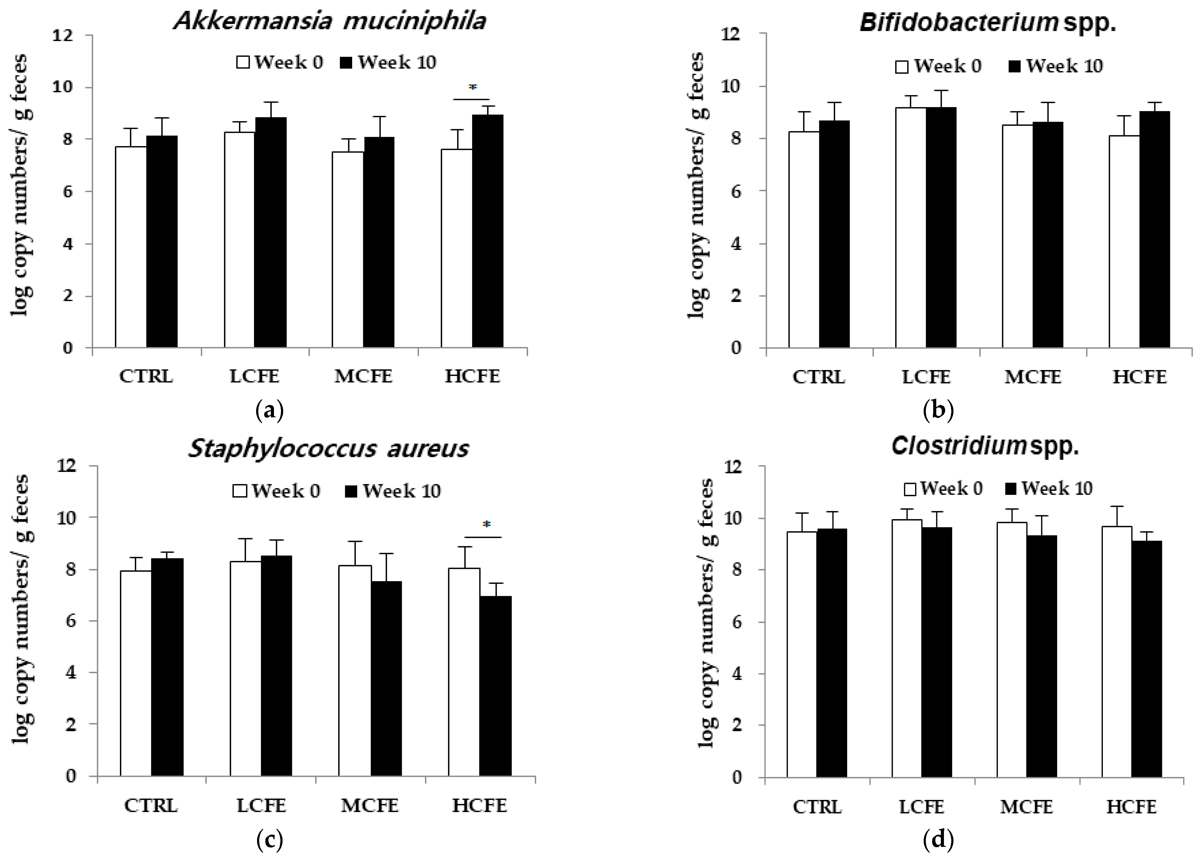
Figure 1. Changes in the number of Akkermansia muciniphila (a), Bifidobacterium spp. (b), Staphylococcus aureus (c), and Clostridium spp. (d) in the feces of mice fed CFE, as determined by quantitative PCR. The data shown are the mean ± SD of five independent experiments. Significant differences are noted as * p < 0.05 compared with week 0.
Applsci 14 03037 g001
Figure 2. Relative fecal β-glucuronidase activity. The data shown are the mean ± SD of five independent experiments. Significant differences are noted as * p < 0.05 compared with the control group.
Applsci 14 03037 g002
Figure 3. Effect of CFE on DSS-induced mouse colitis. (a) Experiment design, (b) DAI (disease activity index) score, (c) Macroscopic appearance, (d) Colon length of mice in each group. The data shown are the mean ± SD of five independent experiments. Significant differences are noted as ### p < 0.05 compared with the normal group and * p < 0.05, ** p < 0.01, and *** p < 0.001 compared with the negative control group.
Applsci 14 03037 g003
Figure 4. CFE prevented DSS-induced colon damage in mice. (a) H&E staining of colon, magnification ×100, (b) Histology score, (c) MPO (myeloperoxidase) activity. The data shown are the mean ± SD of five independent experiments. Significant differences are noted as ### p < 0.05 compared with the normal group and * p < 0.05, ** p < 0.01, and *** p < 0.001 compared with the negative control group.
Applsci 14 03037 g004
Table 1. Detailed dietary composition of AIN-93.
Ingredientg/kg
Casein200.00
Cornstarch397.486
Dextrose132.00
Sucrose100.00
Cellulose50.00
Soybean oil0.014
t-Butylhydroquinone0.014
Salt Mix35.00
Vitamin mix10.00
L-Cystine3.00
Choline Bitartrate2.50
Table 2. 16S rRNA gene-targeted bacteria-specific primers used in this study.
TargetPrimerPrimer Sequence (5′–3′)
ProbioticsBifidobacterium
spp.
Forward
Reverse
CTCCTGGAAACGGGTGG
GGTGTTCTTCCCGATATCTAC
Akkermansia
muciniphila
Forward
Reverse
CAGCACGTGAAGGTGGGGAC
CCTTGCGGTTGGCTTCAGAT
PathogensStaphylococcus
aureus
Forward
Reverse
GCCCCTTAGTGCTGCAGCTA
AGTTTCAACCTTGCGGTCGTA
Clostridium
spp.
Forward
Reverse
TTGAGCGATTTACTTCGGT
CCATCCTGTACTGGCTCAC
Table 3. Histological grading of colitis.
GradeInfiltration LesionEpithelial Lesion
0None None
1Infiltration around crypt basesSome loss of goblet cells
2Infiltration spreading to muscularis mucosaExtensive loss of goblet cells
3Extensive infiltration in the muscularis
Mucosa with abundant edema
Some loss of crypt
4Infiltration spreading to submucosaExtensive loss of crypt
Table 4. Analysis of SCFAs in mouse cecum contents.
GroupsSCFAs (μmol/g)
Acetic AcidButyric Acid
CTRL9.05 ± 2.032.54 ± 0.48
LCFE13.83 ± 1.12 **3.27 ± 0.98
MCFE18.91 ± 2.70 ***3.70 ± 0.53 *
HCFE20.24 ± 1.60 ***3.94 ± 0.69 *
Values are mean ± SD of 5 mice. Significant differences are noted as * p < 0.05, ** p < 0.01, and *** p < 0.001 compared with the control group.
Disclaimer/Publisher’s Note: The statements, opinions and data contained in all publications are solely those of the individual author(s) and contributor(s) and not of MDPI and/or the editor(s). MDPI and/or the editor(s) disclaim responsibility for any injury to people or property resulting from any ideas, methods, instructions or products referred to in the content.