Article

Scapular Motor Control and Upper Limb Movement Quality in Subjects with and without Chronic Shoulder Pain: A Cross-Sectional Study

by
Ana S. C. Melo
1,2,3,4,
Diana C. Guedes
1,
Ricardo Matias
5,6,
Eduardo B. Cruz
7,8,
J. Paulo Vilas-Boas
3,9 and
Andreia S. P. Sousa
1,*
1
Centro de Investigação em Reabilitação (CIR), Escola Superior de Saúde, Politécnico do Porto, Rua Dr. António Bernardino de Almeida, 400, 4200-072 Porto, Portugal
2
Centro de Investigação em Actividade Física, Saúde e Lazer (CIAFEL), Faculdade de Desporto, Universidade do Porto, Rua Dr. Plácido Costa, 91, 4200-450 Porto, Portugal
3
Laboratório de Biomecânica do Porto (LABIOMEP), Universidade do Porto, Rua Dr. Plácido Costa, 91, 4200-450 Porto, Portugal
4
Centro Interdisciplinar de Investigação Aplicada em Saúde (CIIAS), Escola Superior de Saúde, Instituto Politécnico de Setúbal, Campus do IPS Estefanilha, 2914-503 Setúbal, Portugal
5
Physics Department & Institute of Biophysics and Biomedical Engineering (IBEB), Faculty of Sciences, University of Lisbon, 1749-016 Lisbon, Portugal
6
Kinetikos, 3030-199 Coimbra, Portugal
7
Departamento de Fisioterapia, Escola Superior de Saúde, Instituto Politécnico de Setúbal, Campus do IPS Estefanilha, 2914-503 Setúbal, Portugal
8
Centro de Investigação Integrada em Saúde (CHRC), Universidade Nova de Lisboa, 1169-056 Lisboa, Portugal
9
Centro de Investigação, Formação, Inovação e Intervenção em Desporto (CIFI2D), Faculdade de Desporto, Universidade do Porto, Rua Dr. Plácido Costa, 91, 4200-450 Porto, Portugal
*
Author to whom correspondence should be addressed.
Appl. Sci. 2024, 14(8), 3291; https://doi.org/10.3390/app14083291
Submission received: 11 March 2024 / Revised: 5 April 2024 / Accepted: 9 April 2024 / Published: 13 April 2024
(This article belongs to the Special Issue Biomechanics and Motor Control on Human Movement Analysis)

Abstract

:
Despite the existence of several studies about the scapula’s position and motion, in shoulder pain conditions, there are still conflicting findings regarding scapular adaptations and reduced research about the scapula’s role during functional tasks. The present study aimed to compare scapular-related kinematic and electromyographic outcomes during different shoulder movements (with and without load) and the drinking task, between symptomatic and asymptomatic subjects. Forty subjects (divided into two groups) participated in this cross-sectional observational study. Scapulothoracic motion, scapulohumeral rhythm, and movement quality (considering trunk compensation, time-to-peak acceleration, and smoothness), as well as the relative surface electromyographic activity and muscle ratio considering the trapezius, serratus anterior, and levator scapulae (LS), were assessed. The symptomatic group presented the following: (1) changes in scapular upward rotation (p = 0.008) and winging (p = 0.026 and p = 0.005) during backward transport and drink phases; (2) increased muscle activity level of the middle trapezius (MT) in all tasks (p < 0.0001 to p = 0.039), of LS during shoulder elevation with load (p = 0.007), and of LS and LT during most of the drinking task phases (p = 0.007 to p = 0.043 and p < 0.0001 to p = 0.014, respectively); (3) a decreased serratus anterior lower portion activity level (SAlow) during shoulder lowering with load (p = 0.030) and drink phase (p = 0.047); and (4) an increased muscular ratio between scapular abductors/adductors (p = 0.005 to p = 0.036) and elevators/depressors (p = 0.008 to p = 0.028). Compared to asymptomatic subjects, subjects with chronic shoulder pain presented scapular upward rotation and winging adaptations; increased activity levels of MT, LT, and LS; decreased activity levels of SAlow; and increased scapular muscle ratios.

1. Introduction

The crucial role of the scapula in shoulder stability and function [1,2] has motived a large number of studies reporting data about scapula position and/or motion [2,3,4,5,6] and muscular activity [5,7,8,9,10,11]. Apart from the known limitations of assessing scapulothoracic motion [2,12,13], reference values have been established for scapula position [3,4,6] and motion [2,3,4,5,6,14]. Additionally, the roles of the scapular muscles during scapular motion and stability, as well as in shoulder function, have been stated [5].
The mentioned data were mainly reported during shoulder elevation, where the scapula is expected to perform the following: (a) upward rotation [2,3,4,14] through the activity of the serratus anterior (SA) [7,8,9], upper (UT) [9,10], lower (LT) [7,8,10], and middle trapezius (MT) [8,15]; (b) lateral rotation [2,6,14] through the action of SA, MT [7,8], and LT [7,8,10]; (c) posterior tilt [6], through SA [7,8] and LT [10] activity; and (d) a slight elevation [16], through the action of levator scapulae (LS) [11] and UT [10,11]. During the lowering phase, despite similarities with the scapulothoracic kinematics during shoulder elevation (1.1 to 3.3° of maximum difference), less scapular muscular activity was reported for UT, LT, and SA [14]. This difference seems to be related to the type of contraction required, the gravity effect [14], or the action of the antagonist muscles [8,10].
Scapulothoracic motion and muscular changes were described for shoulder pain conditions. These adaptations are generally reported as a reduction in the expected scapular movements [2,6,14,17,18] (except for upward rotation in adhesive capsulitis [2,19]) and as a reduction in muscular activity of the LT, MT, and SA [8,14,20,21] together with an increased activation level of LS, pectoralis minor (Pm) [8,22,23], and UT [19,20,23].
Also, beyond its relevance for shoulder function, the scapula serves as the link between the upper limb and the trunk [14,24], with the primary purpose of maximizing the available degrees of freedom to facilitate the hand’s interaction during its activities [13,25]. Given this fact, it is understandable that one of the major complaints of subjects with shoulder pain is related to its impact on daily activities [26,27]. Therefore, considering functional activities with different degrees of freedom during the patients’ assessment could provide new information for rehabilitation planning.
Scapula outcome data have already been presented for some daily activities, primarily in healthy subjects [26,28,29,30], but few studies have included participants with shoulder pain [26,30]. The use of different tasks across studies seems to be related to the lack of a systematic protocol for upper limb assessment [13], which could limit repeatability [31] and comparison with other studies. However, since the movements of the upper limb could vary according to each task’s purpose [32], studying other daily activities would complement the available knowledge. In particular, studying the drinking task, a common daily activity task associated with basic shoulder movements [33], could not only increase knowledge about a basic daily activity task [33] but also facilitate comparison with other studies. The drinking task has already been considered in the study of other populations [13,32,34,35] and includes a phase, the reaching phase, reported as reliable to assess the upper limb motor performance in subjects with shoulder pain [36].
Despite all the aforementioned facts, discrepancies have arisen during the characterization of subjects with shoulder pain. For instance, there are conflicting findings regarding the activity level of scapular muscles, such as increased activation [37,38] or no changes [26,37] of the LT or increased SA [30,39]. Similarly, there are conflicting reports regarding UT’s activity level, with some studies indicating a decreased activity of this muscle and others recommending promoting higher UT activity levels [22,40,41]. Furthermore, it is unclear whether some of the kinematic and muscular changes identified during the lowering phase of shoulder movements in the scapular plane [42,43] would differ if assessed in a more challenging task, such as movement in the frontal plane, which is more associated with shoulder complex changes like decreased subacromial space [36]. On the other hand, assessing other parameters related to the upper limb function, such as movement quality (goal-directed movements [44]; temporal and performance efficiency; or even movement planning [34,45]), could significantly contribute to evaluating the association between scapular alterations and global upper limb function parameters. Thus, the conflicting results and uncertainties mentioned justify the continuous study of the scapular muscles’ adaptations to enhance knowledge that is useful during rehabilitation planning.
The present cross-sectional study aimed to compare subjects with chronic shoulder pain (symptomatic) and asymptomatic subjects in terms of scapular kinematics (scapulothoracic motion and scapulohumeral rhythm), muscular activity, and upper limb movement quality during shoulder movements in the frontal plane and/or during the drinking task. Given the current state of the art, which mainly reports a reduction in the major movements of the scapula in subjects with shoulder pain, in the present study, it was expected to find a possible decrease in all assessed scapular movements or an increase in scapular winging among symptomatic subjects. Furthermore, considering the previously mentioned changes in scapular muscles’ activity, an increase in the activity of the downward rotators assessed (LS) and possibly the UT, as well as a decreased activation of the other upward rotator muscles, was also expected for the symptomatic subjects. Despite the lack of studies analyzing the parameters of movement quality, it was anticipated that these parameters could reveal differences between symptomatic and asymptomatic subjects, considering the presence of pain.

2. Materials and Methods

2.1. Subjects

Forty volunteers (four males and thirty-six females) participated in this cross-sectional study. Participants were recruited from a higher education institution through a questionnaire sent by e-mail (n = 407). From the 137 answers obtained, individuals were selected if they were between 18 and 65 years old and met the eligibility criteria.
The symptomatic group included participants who reported shoulder pain lasting at least 3 months (duration of the current episode or intermittent complaints over the last 3 months [46]). To be included, participants must have experienced pain (a) in the upper arm, specifically in the shoulder, deltoid, and/or scapular areas; and (b) of moderate or high intensity [≥4 in the numeric pain rating scale (NRS)], either at rest or during active shoulder movements. Additionally, participants in this group had to exhibit type I (posteriorly displaced/prominence of the scapula’s inferior angle [47,48,49]), type II (prominence of the scapula’s medial border [47,48,49]), and/or type III (excessive elevation of the scapula’s superior border and/or excessive/insufficient scapular upward rotation [47,48,49]) on the scapular dyskinesis classification test [47,48]. Both unilateral and bilateral shoulder pain conditions were included; however, in the case of bilateral symptoms, only the most painful shoulder was considered. As for asymptomatic subjects, their sex, age, and dominant upper limb were aligned with the symptomatic participants, to ensure that both groups were matched. Additionally, asymptomatic participants should not have experienced any shoulder pain events in the last 2 years. Subjects were excluded from both groups if they had a history of shoulder fracture, dislocation, tears, infection, or neoplasm; shoulder surgery; cervical and/or thoracic pathologies or pain associated with active movements of these regions; systemic, infectious, and/or neurological disease; or if their body mass index was outside the range 18.5–30 kg/m2.
Each group consisted of two male and eighteen female participants, presented in the demographic characteristics described in Table 1. At baseline, the groups were matched, except for scapular positioning. Changes in scapular positioning were observed in all the subjects from the symptomatic group, whereas only 55% of the asymptomatic subjects exhibited such changes. Symptomatic subjects were further characterized by a specific (n = 4 of tendinopathy, n = 1 adhesive capsulitis, and n = 1 ligament injury) or non-specific (n = 14) shoulder pain, localized around the shoulder (n = 7 in the anterior region, n = 4 in the superior region, n = 3 in the posterior region, and n = 4 all over the shoulder) with a mean intensity of 5.6 in the NRS. These subjects also demonstrated a shoulder function, as measured by the SPADI score, with a mean of 31.48.
Before participation, all subjects read and signed the informed consent form, and the study was approved by the local ethical committee.

2.2. Instrumentation
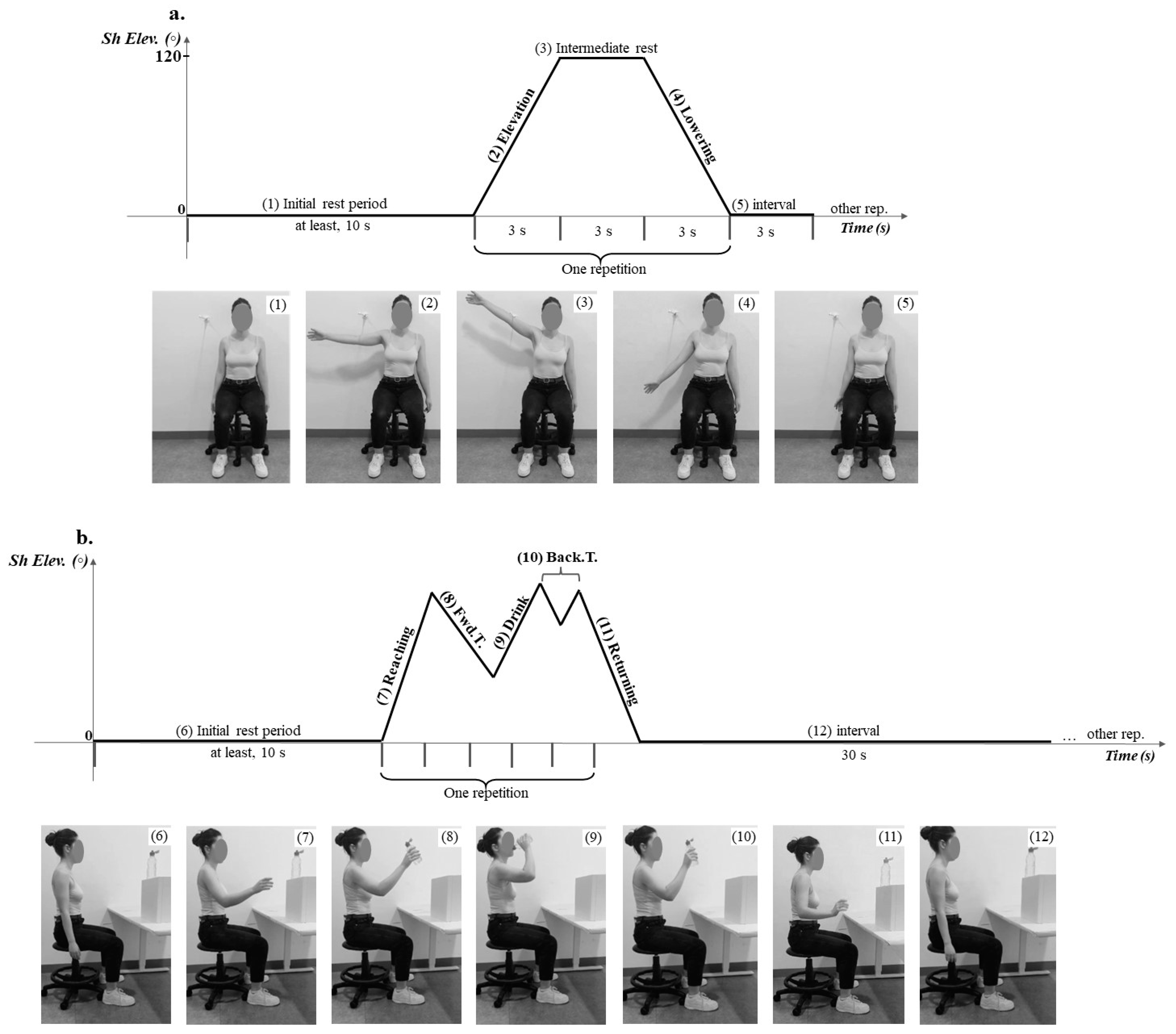
Height (m) was measured using a seca® 222 stadiometer (seca—Medical Scales and Measuring Systems®, Birmingham, UK), with a 1 mm scale. Body mass (kg) was assessed using a seca® 760 scale (seca—Medical Scales and Measuring Systems®, Birmingham, UK), which had a precision of 0.1 kg.
The KINETIKOS CE-marked (Class I medical device) cloud-based platform (KINETIKOS, Coimbra, Portugal) was used to collect and reconstruct, in real time, subjects’ motion using a 3D kinematics model and 5 inertial measurement units (IMUs) (MVN BIOMECH Awinda, Xsens Technologies, Enschede, The Netherlands), according to the ISB recommendations [50]. With an acquisition frequency of 100 Hz, the KINETIKOS platform was also used to define the beginning, the respective phases, and the end of the shoulder movement or task. Four degrees of freedom (DoF) described the scapula’s kinematics utilizing the joint reference frame x-y-z but with joint origin at the centroid of the scapula’s Angulus Acromialis, Trigonum Spinae, and Angulus Inferior [51]. Therefore, scapulothoracic motion was described, relative to the thorax, as abduction (positive) followed by elevation (positive) on an ellipsoidal thoracic surface, winging—the raising of the scapula’s medial border and inferior angle from the thorax—(positive), and upward rotation (positive). The model of the scapulothoracic joint, validated through comparison with bone-pin markers, exhibited an accuracy of 1.85 mm for position (measurement error ranging from 0.40 to 2.4 mm) [51]. Shoulder motions, particularly elevation (positive), were described using three DoFs, considering the humerus relative to the thorax. The model also incorporated three DoFs to describe the trunk movements (to assess possible compensatory movements), considering the thorax sensor’s rotation relative to itself, as lateral flexion (positive towards the side of the assessed upper limb), axial rotation (positive towards the side of the assessed upper limb), and forward flexion (negative). Additionally, the KINETIKOS CE-marked (Class I medical device) cloud-based platform (KINETIKOS, Coimbra, Portugal) was used for data processing and analysis.
The wireless Trigno™ acquisition system (Delsys Inc., Natick, MA, USA) was used to record the surface electromyographic (EMG) signal from the three trapezius portions, SA portions, and LS. For this purpose, 6 pre-amplified sensors (Trigno Avanti Sensor model, Delsys Inc., Natick, MA, USA) were employed, with a 4-bar formation of Ag bar surface electrodes and an inter-electrode distance of 10 mm. These sensors were pre-amplified through a differential amplifier with an adjustable gain (20–450 Hz; common mode rejection coefficient: 95 dB at 50 Hz and gain of 1000). The EMGworks Acquisition software version 4.5.3 (Delsys Inc., Natick, MA, USA) assessed the signal quality and recorded the EMG data, including the submaximal voluntary contractions for normalization, at an acquisition frequency of 2000 Hz. Subsequently, EMGworks Analysis (Delsys Inc., Natick, MA, USA) was used to filter and analyze EMG data. Additionally, to allow for kinematic and EMG synchronization, another Trigno™ sensor was placed on the hand to record accelerometry data. These data were used to identify the beginning of each shoulder movement and drinking task in the electromyographic signal. This time event identification by both the Trigno™ system and the KINETIKOS CE-marked (Class I medical device) cloud-based platform enabled the synchronization between kinematic and electromyographic signals.
To control the cadence of shoulder elevation and lowering movements, a metronome (Metronome Beats, version 6.5.1, Stonekick, London, UK) was used at 20 beats/min.
Shoulder pain intensity was quantified using the self-reported NRS, ranging from 0 (“no pain”) to 10 (“unbearable pain”) [52], which has been demonstrated to be reliable (test–retest intraclass correlation coefficient of 0.84 [53]). The Portuguese version [54,55] of the Shoulder Pain and Disability Index (SPADI), presenting an internal consistency of α = 0.75–0.84, was used to classify shoulder function in symptomatic subjects, with a score ranging from 0 (“no pain/no difficulty”) to 100 (“worst pain imaginable/so difficult required help”) [56].
Scapular presentation from participants of both groups was classified using the scapular dyskinesis classification test (intratester reliability ranging from k = 0.49–0.59 [48]). This test considered the positioning at rest and during shoulder elevation and lowering in the frontal plane [57,58].
To guarantee the correct positioning of the subjects and material and the intended range of shoulder motion, a universal goniometer (BASELINE®, Aurora, IL, USA), with a precision of 1°, was utilized during the assessment moments (intra-observer intraclass correlation coefficient (ICC) of 0.94 for general measurements [59] and 0.91 and 0.93 for shoulder abduction in healthy or shoulder injury subjects, respectively [60]).

2.3. Procedures

Data collection took place in a biomechanical laboratory, where each researcher performed identical tasks to avoid inter-rater errors. Before data collection, anthropometric measurements, such as body mass and height, were taken for each participant.
To evaluate scapulothoracic motion (including scapular abduction, elevation, upward rotation, and winging), scapulohumeral rhythm, and movement quality (such as trunk lateral flexion, rotation or flexion/extension compensation, time-to-peak acceleration, and smoothness), inertial sensors were attached to several body parts. These included the thorax (at the flat anterior aspect of the sternum [3,51], using double-sided tape) and unilaterally, using Velcro straps, to the acromion (positioned superiorly over the flattest posterolateral surface [61]), humerus (laterally, above the elbow), forearm (on its lateral surface), and the back of the hand, following manufacturer recommendations. System calibration, crucial for accurate measures up to 120° of shoulder elevation (a range less affected by skin artifacts) [62], involved participants standing straight with arms close to the body, elbows flexed at 90°, and palms of the hands facing each other, as advised by the software manufacturer.
To record EMG signals, the skin was previously prepared (shaved, abraded, and cleaned with isopropyl alcohol 70%) to reduce impedance to values lower than or equal to 5 KΩ. EMG sensors were positioned parallel to the muscle fiber orientation, at the muscle belly of the trapezius and serratus anterior portions, as well as LS, as detailed in Table 2. These sensors were attached to the skin by a 4-slot sensor adhesive interface (Delsys, Inc.; Natick, MA, USA). Additionally, a sensor aligned with the inertial sensor and with a Velcro strap was placed at the back of the hand to collect acceleration data. Before the recordings, the quality of the raw signals was checked to confirm the correct electrode placement and exclude possible interference or noise.
To allow for the comparison between groups, recordings were conducted during shoulder elevation and lowering in the frontal plane, as well as during a daily activity task (drinking). Participants were seated on a bench with adjustable height to maintain their knees and hips at 90° angles, and their feet were parallel to hip width, flat on the floor. Additionally, the hand of the limb not being tested was supported on the respective thigh [32].
After participants’ familiarization with the movements and task to be evaluated, the upper limb affected by chronic shoulder pain (or of the most painful shoulder in symptomatic subjects) or the upper limb matched with the symptomatic subjects according to side and dominance, in asymptomatic subjects, was assessed. Participants started with the assessed upper limb at the side of the body (with the elbow extended), and the recordings began after an initial rest period of at least 10 s. To prevent fatigue, a rest period of 2 min was implemented between each movement or task.
During the shoulder movements, a wall was used as a reference to guide the upper limb’s motion, and a rod was used to indicate the required maximum elevation range—120°. Each movement was repeated five times, with a 3 s rest interval between repetitions. The 3 s duration of each phase (elevation phase—up to 120° of shoulder elevation; intermediate rest phase—at 120° of shoulder elevation; and lowering phase—returning to the starting position) was controlled by a metronome (Figure 1). Shoulder movements in the frontal plane were conducted under two conditions: with and without load (1 kg dumbbell) [1]. During the assessment of these movements, one participant was unable to complete the entire range of shoulder elevation; however, the data were included in the analysis.
For the drinking task, a 33 mL water bottle was positioned on a box atop a table, with the table’s height adjusted to be aligned with each subject’s olecranon level [32]. The bottle was positioned at a height corresponding to 90° of shoulder elevation [35] in the scapular plane (a position commonly assumed during daily activities [72]) and at a distance equal to the length between the acromion and the trapezium-metacarpal joint of the assessed limb [32]. Participants performed five repetitions of the drinking task at a self-paced speed [32,34,36], with a 30 s rest interval between repetitions, following the verbal instruction “You can drink” [32] (see Figure 1). As the drinking task was the only movement performed at a self-paced rhythm and without a reference to guide the plane of movement, movement quality variables were only assessed for this task. To facilitate the analysis and interpretation of the drinking task, the task was subdivided into different phases: 1. Reach out to the bottle from the starting position; 2. Forward transport of the bottle to the mouth; 3. Drink a sip of water; 4. Backward transport of the bottle to the pickup point (identified by tape); and 5. Return the upper limb to the starting position [32,34].
To normalize the EMG activity level elicited during the requested movements and drinking task, a submaximal voluntary isometric contraction (SVIC) of each muscle was conducted. During these procedures, verbal encouragement was provided as subjects held a dumbbell of 1 kg [73], following the procedures outlined in Table 1. Participants performed three repetitions of 5 s [74], with a minimum rest interval of 30 s between repetitions or 1 min between each new test position [75]. Only data from the three central repetitions of each movement/drinking task [26] were considered for analysis.

2.3.1. Data Processing

The beginning of shoulder elevation movements was defined as the first instant when acceleration recorded by the hand sensor exceeded the mean resting value by ±0.3 m/s2. Thereafter, new phases of the movement were defined every 3 s. For the drinking task, the phases were defined based on the criteria outlined in Table 3.
The EMG signals were digitally filtered using a second-order band-pass Butterworth filter with a pass band from 20 Hz (or 50 Hz, in cases where heart noise was detected) to 450 Hz, to remove noise. The root mean square (RMS) was then computed using a sliding window of 100 samples. Hand acceleration signals were subjected to low-pass filtering using a fourth-order Butterworth filter and a cut-off frequency of 4 Hz [76]. To normalize the data, the mean RMS muscular activity level for each muscle during each phase was divided by the maximum [38] RMS value achieved during each muscle’s SVIC and then multiplied by 100. Then, the mean EMG values for each phase of the movements/drinking task phases were calculated. To assess relative activation of scapular muscles, three muscular ratios [78] were computed, considering muscle involvement in scapulothoracic motions, specially upward and downward rotation (DrvsUr), abduction and adduction (AdvsAb), and elevation and depression (DepvsEl)], according to the following formulas:
D r v s U r   r a t i o = L S   a c t i v i t y   l e v e l L S + U T + M T + L T + S A u p / m e d + S A l o w   a c t i v i t y   l e v e l
A d v s A b   r a t i o = M T + L T   a c t i v i t y   l e v e l M T + L T + S A u p / m e d + S A l o w   a c t i v i t y   l e v e l
D e p v s E l   r a t i o = L T   a c t i v i t y   l e v e l L T + U T + L S   a c t i v i t y   l e v e l
Specifically, for each phase of the movement/drinking task, a value ranging from 0 to 1 was achieved [where 0 indicates no activity of the muscle(s) antagonist(s) for the respective motion and 1 indicates no activity of the muscle(s) agonist(s) for the motion under consideration].
The scapula’s resting position and the range of scapula and shoulder movements of each of phase of the movement/task were calculated using either the mean or the difference between joint angles’ values, namely, (a) the scapula’s resting position, calculated by averaging the resting values recorded during the first 5 s of recording; and (b) the elevation and lowering phases and drinking phases, calculated by the maximum range variation from the starting position of each phase. The scapulohumeral rhythm (SH rht), considering shoulder elevation/lowering (Elev/low) and scapula upward/downward rotation (UR/DR), was calculated using the following formula:
S H   r h t = R a n g e   o f   S h o u l d e r   E l e v / l o w ( ° ) R a n g e   o f   S c a p u l a   U R / D R ( ° )
The trunk compensation during the drinking task was determined by identifying the maximum variation in flexion, same-side (of the painful shoulder) lateral flexion, and same-side (of the painful shoulder) rotation. The time-to-peak acceleration was calculated as the highest instantaneous acceleration recorded by the hand sensor during the reaching movement [79]. This value was then normalized by the task completion time and multiplied by 100. For the global drinking task smoothness, first, the global acceleration was calculated from the x, y, and z components of the hand’s sensor; then, the acceleration data were filtered to remove noise and to smooth the signal using a fourth-order low-pass filter with a cutoff frequency of 20 Hz; and following the identification of the drinking task phases and, consequently, the beginning and end of each drinking task repetition, the total smoothness of each repetition was calculated using the dimensionless jerk2 function [80,81].

2.3.2. Statistical Analysis

The appropriate effect size (d = 0.833) was determined based on a prior study comparing scapulothoracic upward/downward rotation [43] between asymptomatic subjects and symptomatic subjects with shoulder pain. Utilizing G*Power software version 3.1 (Kiel University, Germany), it was found that at least 19 individuals per group (asymptomatic and symptomatic) were required to detect differences with a power of 0.8 and an alpha of 0.05.
Statistical Package for Social Science (IBM, Inc., Chicago, IL, USA) version 27 was used for statistical analysis, with a 95% confidence interval. Data normality was assessed using the Shapiro–Wilk test and histogram analysis.
Sample homogeneity and group comparisons were evaluated through an independent sample t-test, Mann–Whitney, or Fisher Exact Test. Data were presented as means and standard deviation (SD), median and interquartile range (IR), or frequencies, respectively. Effect size (Cohen’s d) was also presented for significant results, except for the variables analyzed using a non-parametric test. Values greater than 0.8 were considered a large effect size, approximately 0.5 represented a moderate effect size, and less than 0.2 indicated a small effect size [65].

3. Results

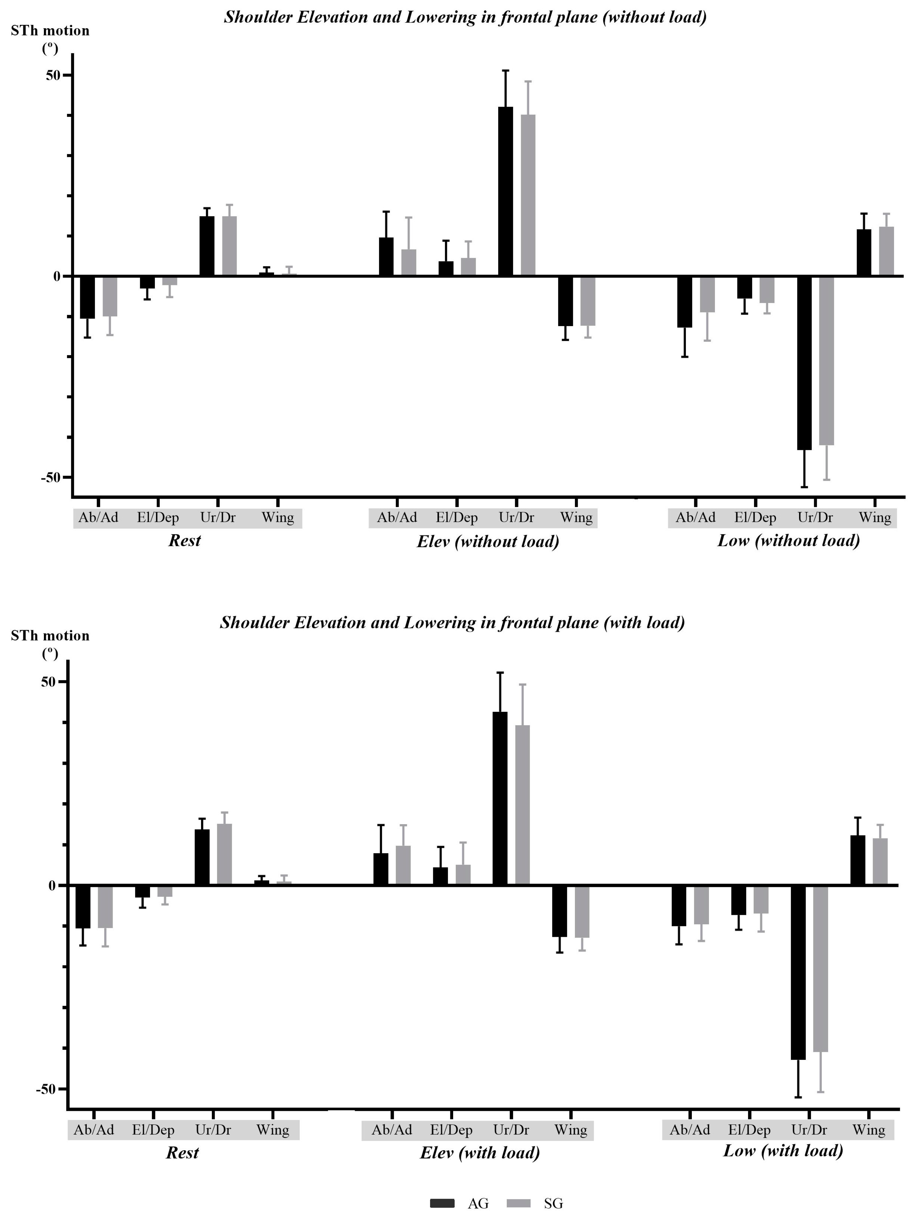
3.1. Shoulder Elevation and Lowering in the Frontal Plane—Comparison between Groups

3.1.1. Scapulothoracic Motion and Scapulohumeral Rhythm Data

When considering both conditions of shoulder elevation/lowering in the frontal plane, no statistically significant differences were found for any of the assessed parameters, including scapulothoracic motions and scapulohumeral rhythm. These findings are detailed in Figure 2 and Table 4.

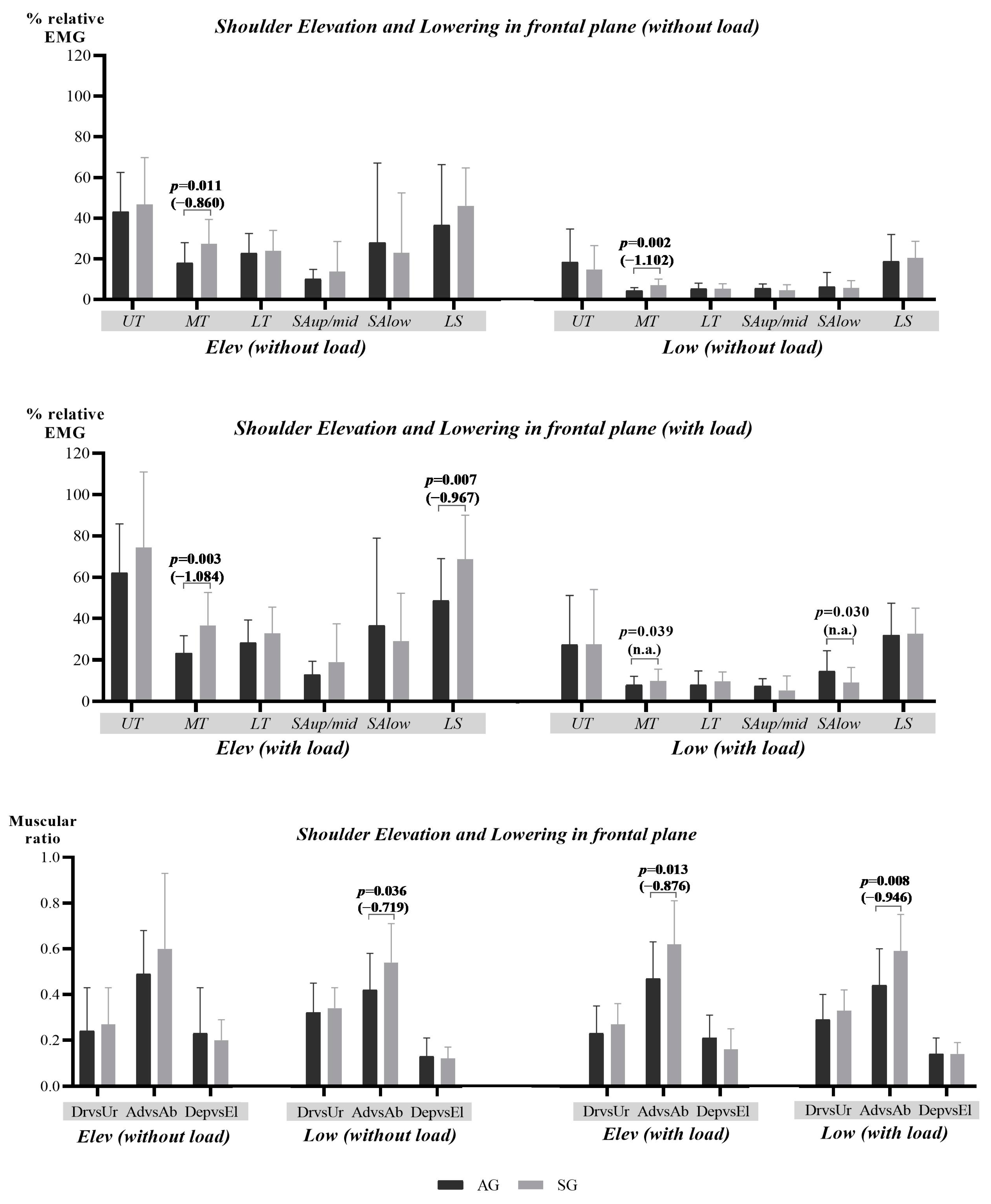
3.1.2. Scapular Muscular Activity and Ratio Data

During shoulder movements, both with and without load, the symptomatic group presented a statistically significant increase in the muscular activity level of the MT during elevation and lowering, together with a higher antagonist relative activation as indicated by the AdvsAb ratio (Figure 3). Furthermore, when the shoulder movements were performed with load, the symptomatic group also presented a significant decrease in the muscular activity level of SAlow during lowering and a significant increase in the muscular activity level of LS during elevation. Whenever calculation was possible, the effect size for these observations ranged from moderate to large, providing additional support to the findings (Figure 3).

3.2. Drinking Task—Comparation between Groups

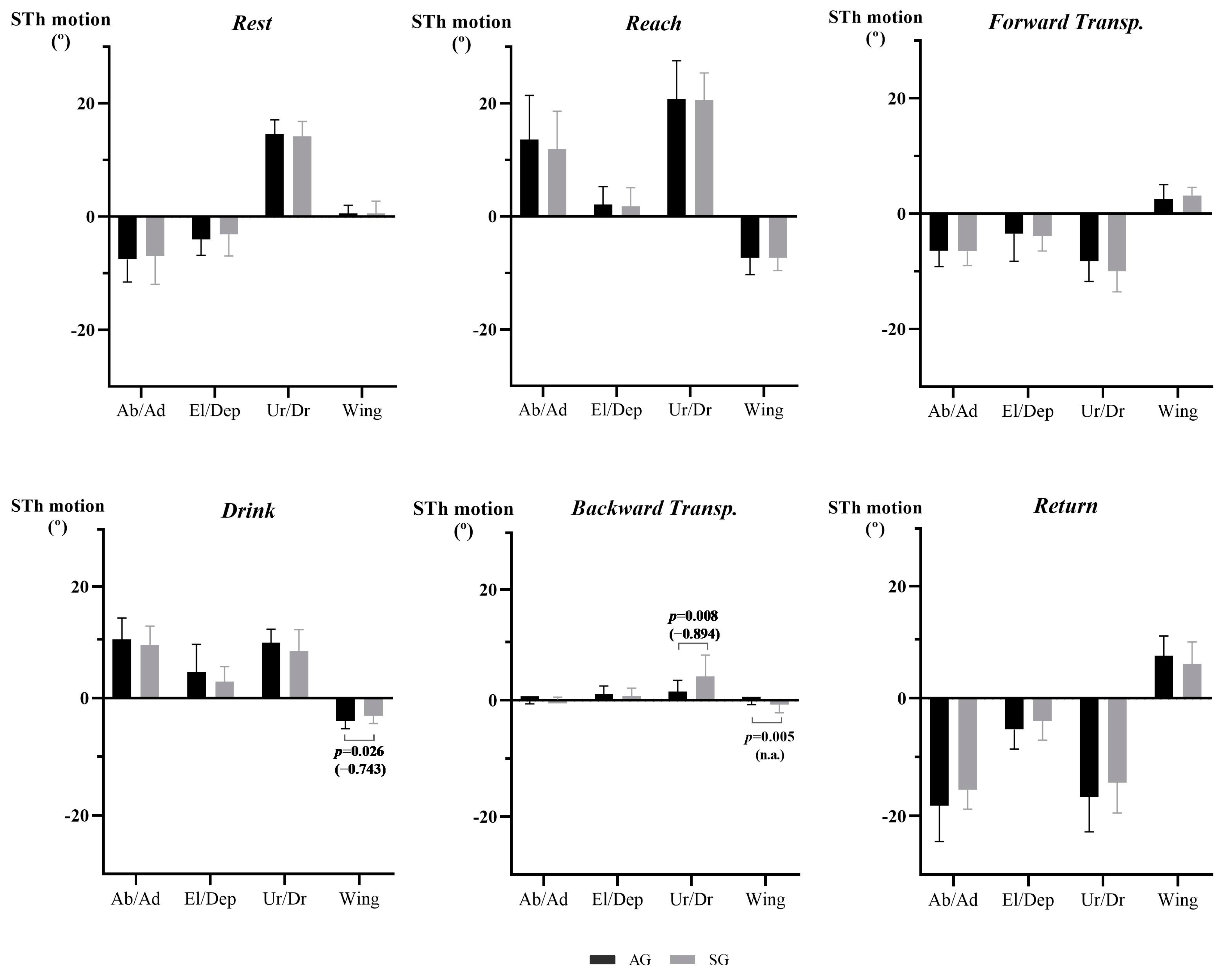
3.2.1. Scapulothoracic Motion, Scapulohumeral Rhythm, and Movement Quality Data

Significant differences were observed during the drinking task, particularly in upward rotation (with a large effect size) and/or winging motions in the drinking and the backward transport phases (with a moderate effect size). More specifically, the symptomatic group presented a higher range of upward rotation motion during the backward transport phase. Then, considering the winging motion, the symptomatic group presented a lower range of motion during the backward transport phase and a higher range of motion during the drinking phase (Figure 4).
No other significant differences were found for the remaining scapulothoracic motions, considering neither scapulohumeral rhythm nor movement quality variables (Table 5).

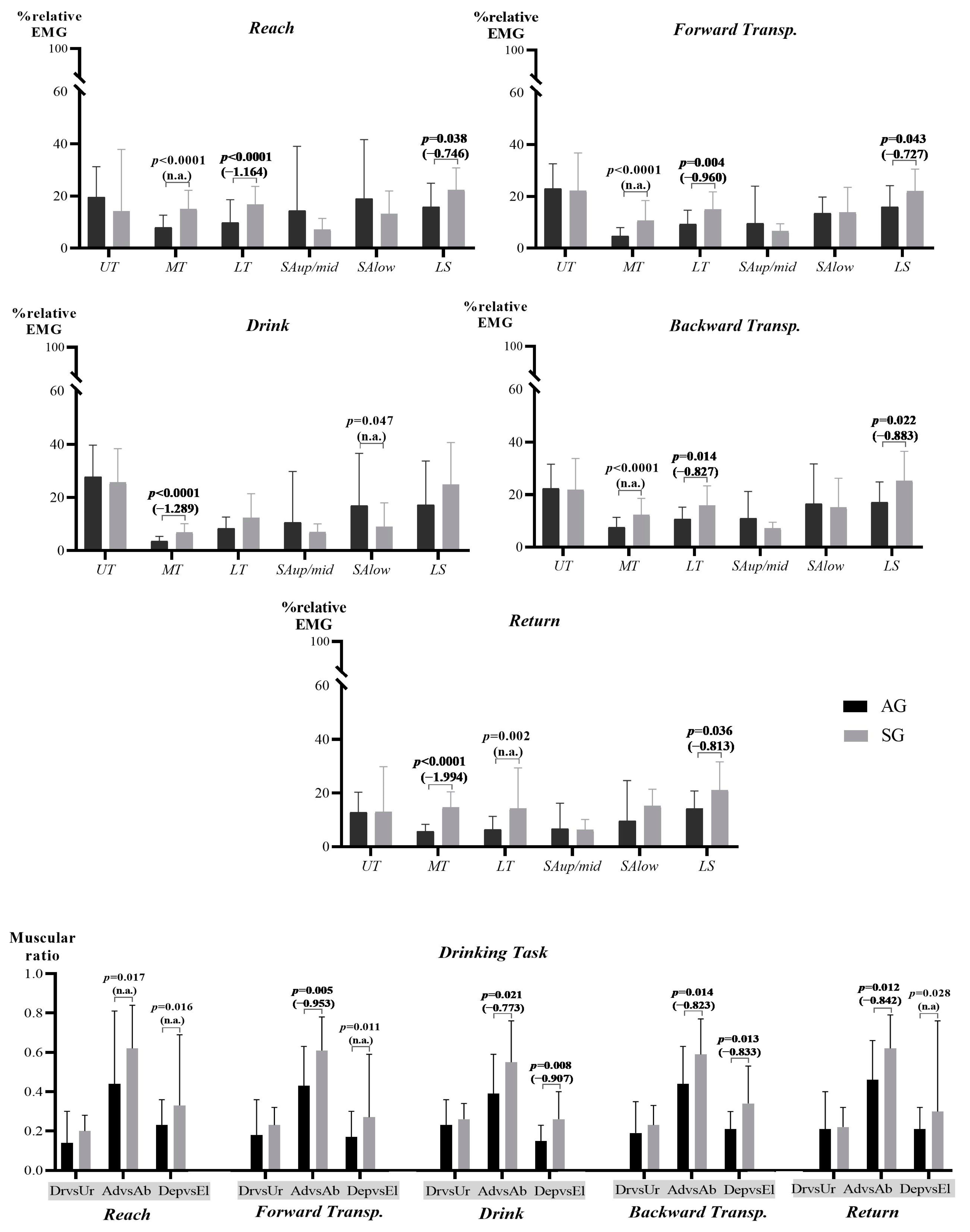
3.2.2. Scapular Muscular Activity and Ratio Data

During the drinking task, the symptomatic group presented a statistically significant increase in the muscular activity of the MT in all the phases, as well as of the LT and LS in the reach, forward transport, backward transport, and return phases. Additionally, during the drinking phase, the symptomatic group presented a significant decrease in the muscular activity level of SAlow. When considering the muscular ratios, the symptomatic group showed a higher antagonist relative activation in both the AdvsAb and DepvsEl ratios across all phases (see Figure 5). These findings were supported by moderate-to-large values of the effect size (Figure 5).

4. Discussion

The present study aimed to compare scapulothoracic motion, scapulohumeral rhythm, and muscular activity (including muscular ratios) during shoulder elevation and lowering in the frontal plane and during a drinking task, between subjects with chronic shoulder pain and asymptomatic subjects. Furthermore, it aimed to compare upper limb movement quality assessed during the drinking task between the same subjects. Several differences were found during the comparison between groups, including changes in scapular motion during the drinking task and changes in muscular activity level during both shoulder movements and the drinking task. Given the chronic nature of the pain experienced by the symptomatic group, such differences are no longer expected as a protective mechanism but rather as a maladaptation [8,82] or a compensatory response [8].

4.1. Scapulothoracic Motion, Scapulohumeral Rhythm, and Movement Quality Data

During shoulder elevation and lowering in the frontal plane, no significant differences were found in scapular motion and scapulohumeral rhythm, despite the identified scapular muscular adaptations and the reported scapular dyskinesis types within the study sample. While changes in muscular coordination/balance were previously suggested as potential contributors to alterations in scapular kinematics [83], the fact that all the symptomatic subjects presented scapular dyskinesis compared to less than half of the asymptomatic group also led to the anticipation of differences between groups. Therefore, in the present study, the variable used to express the entire range of scapula motion in each movement/task phase does not seem to detect differences that could be restricted to a specific range of motion or that the moderate pain intensity (≈5.6 out of 10) [52] and low impact on shoulder function (≈31.5 out of 100) [84,85] experienced by the symptomatic group may not have had a significant impact on overall scapular kinematics [43]. Thus, the results of the present study suggested a similar range of scapular motion between both groups. When these results are considered alongside changes in the muscular activity and ratios, which appear to indicate a negative neuromuscular adaptation [86], they suggested that the symptomatic group expends more energy [87] to achieve the same range of motion [86]. Previous studies comparing the scapular kinematics between asymptomatic and symptomatic subjects with shoulder pain have also report a lack of differences in some scapular movements, such as upward rotation and internal rotation [88].
During the drinking task, changes in scapular motion were found. Specifically, during the drinking phase, when subjects took a sip of water, a higher degree of scapular winging was observed in the symptomatic group. This finding seemed to be supported by the decreased activity of the SAlow during this phase and a compensatory increased activity of the MT. Conversely, during the backward transport phase, increased scapular upward rotation and decreased winging were observed. During this phase, the symptomatic group appeared to perform the expected scapular movement during shoulder elevation [2,3,4,5,6,14] but to an excessive degree compared to asymptomatic subjects. Consequently, as mentioned before, the symptomatic group seemed to expend more energy during the task. Particularly, considering the winging motion, the decrease in its range observed in this phase could be a compensatory mechanism for its increase during the drinking phase. In turn, the higher upward rotation could be a strategy to achieve the required shoulder range of motion [26] and/or to manage pain. Previous studies have often highlighted a decrease in upward rotation during shoulder elevation [17,38]; however, other authors also reported an increase in this motion, proposing it as a compensation for shoulder weakness, joint stiffness, and pain avoidance [89]. Although the findings of the present study were statistically significant, they appear small when compared to the intra-rater minimal detectable change values reported by a previous study during the assessment of scapular protraction/retraction, tilt, or medial/lateral rotation with inertial sensors [90]. However, it is noteworthy that the mentioned study did not include scapular winging or upward rotation, which may limit the comparison.
Previous studies have reported changes in movement quality between healthy subjects and those with neurological conditions involving the upper limb [34,91,92]. However, in the present study, which, to our knowledge, is the first to assess these variables in chronic shoulder pain resulting from musculoskeletal conditions, differences between groups were not found for any of the assessed parameters related to goal-directed/coordination of movements [44], temporal and performance efficiency, or even movement planning [34,45,91].

4.2. Scapular Muscular Activity and Ratio Data

Considering the results presented above, the changes exhibited by the symptomatic group in muscular activity level, which are expectable in pain conditions and given scapular dyskinesis [93], could have primarily arisen to facilitate the entire shoulder motion necessary to ensure the upper limb function [94]. More specifically, in the present study, the symptomatic group an demonstrated increased muscular activity level for the MT, LT, and LS, which was both statistically significant and higher than the intra-session minimal detectable change values found for some scapulothoracic muscles when normalized by maximal voluntary isometric contraction [95] (with the exception of MT during shoulder lowering with load).
Although presenting a muscular activity level below 50% of the SVIC [96] (from 3.67% ± 1.69 to 36.65% ± 16.01), the observed increased activity of the MT was consistent across all the movements and drinking task phases. Similar results were also found for LT during the drinking task, except during the drinking phase. Additionally, the LS also presented an increased activity level in the symptomatic subjects during the shoulder elevation performed with load (the most demanding shoulder elevation) and during various phases of the drinking task phases, except the drinking phase. All these mentioned changes could potentially be compensatory strategies. MT, LT, and SA collaborate in maintaining scapular stability against the thorax [8,15,83,97], with MT and LT acting as retractors to offset protraction forces produced by SA activation [9,97,98]. Furthermore, these muscles contribute to external rotation [8,98], upward rotation [8,15], and, in the case of LT, even to posterior tilt [9] and depression [5,97,98]. Considering these facts and the presence of scapular dyskinesis in all symptomatic subjects (17 of 20 subjects presenting a prominent scapular medial border), it seems that the increased activity of the MT and LT could be an attempt to enhance scapular stability [15] and, in certain situations, counterbalance SA muscle activation [93]. Furthermore, although it was not possible to evaluate the Pm in the present study, the increased activity of MT and LT could also compensate for changes in this muscle that, when changed in shoulder pain conditions [8,99], could favor scapular adaptations such as winging or internal rotation [100]. A previous study comparing overhead athletes with shoulder impingement and healthy athletes during push-up exercises on an unstable surface also reported increased activation of the MT muscle of symptomatic subjects, suggesting that MT may be providing more stabilization in this group [15]. On the other hand, previous studies have reported a decreased MT activity level [37,98] (particularly related to its timing activation) or no differences in this muscle’s activity [30,83] during other tasks. Although some of these findings were reported for subjects with shoulder pain without reference to the assessment of scapular dyskinesis [30,37,83], a study comparing subacromial impingement with and without scapular dyskinesis also did not find differences between groups during shoulder flexion [101]. Moreover, the findings of the present study considering LT muscle activity were consistent with the results of previous studies [38], contradicting what is typically expected in subjects with shoulder pain [8,14,20,21]. So, it seems that shoulder pain conditions may lead to different adaptation strategies, perhaps depending on the presence of scapular dyskinesia or factors such as the muscles affected by pain [102] or those closer to the symptomatic area [103]. Considering the role of LS as a scapular elevator [11,23,104], downward rotator [8,11,23,104], and possibly retractor [104], its increased activity could also be a strategy to allow for symptomatic subjects to perform the entire movement/task and/or to minimize pain during motion [94]. This hypothesis is supported by a previous study that reported scapula elevation as a compensatory mechanism used by subjects with shoulder pain conditions, particularly to reduce possible impingement [16]. However, such compensation may compromise shoulder functional status [16], and other research has suggested a possible increase in scapular elevation as a consequence of scapular dyskinesis [105]. In fact, in the present study, more than half of the symptomatic group presented a scapular dyskinesis type associated with excessive elevation of the scapula’s superior border and/or excessive/insufficient scapular upward rotation. Nevertheless, it seems that scapular adaptations could be both a cause or a consequence of the shoulder pain [2,18,22], which raises questions about the reason for the increased activity of LS. Although explored in fewer studies, LS has been previously mentioned as a muscle that possibly presents an increased activity [23], as observed in the present study, or even shortened length [23,37]. Considering these facts and LT’s crucial role in counteracting scapular elevation [97], the increased activity of this muscle could also be a way to resist excessive shoulder elevation.
Contrary to the significant differences mentioned above, a statistically significant reduction of the SAlow muscle activity level, which was also higher than the intra-session minimal detectable change values [95], was observed in the symptomatic group. The presence of differences only for SAlow could be attributed to the different functions of each portion of the SA. While the upper/middle portion of SA [69,70] is related to stabilizing the scapula’s superior angle [106] and promoting scapular protraction [69], SAlow is more involved in scapular upward [69] and lateral [106] rotation, stabilizing the scapula’s inferior angle, and preventing scapular winging [42,106]. Thus, the decreased activity of SAlow observed during the shoulder lowering in the frontal plane with load (a movement requiring eccentric activity and, consequently, higher scapula and shoulder stabilization and muscular force [107]) and during the drinking phase (which may also require greater control, given the increased shoulder range of motion [91,92] and the need to maintain the bottle in a correct position for drinking [32,34]) seems to reinforce the consequences of shoulder pain and the necessity for compensation to maintain the scapula against the thorax. Similar compensatory needs were also mentioned in a study documenting fatigue of the SA [108]. The findings of the present study align with the commonly reported reduction in SA activity level in subjects with shoulder conditions compared to asymptomatic subjects, both in shoulder movements within a restricted plane [8,15,20,26,38,42] and during daily activity tasks [26].
Although differences in isolated muscular activity were not consistently found, changes in muscular ratio were more consistent. Apart from the shoulder elevation phase without load, symptomatic subjects presented an increased AdvsAb ratio during shoulder movements and drinking tasks. A previous study [83] reported no significant differences between groups for isolated muscular activity, but when the relationship between agonist and antagonist muscles was considered (by a ratio), muscular imbalances were highlighted. In the mentioned study, symptomatic subjects presented a decreased ratio of MT/SA, which was related to the role of these muscles as scapular stabilizers [83]. Conversely, in another study, the ratio of LS/SA changed but in different directions according to elevation or lowering movements [109]. In the present study, the increased AdvsAb ratio suggested that, for the symptomatic group, the relative activity of MT and LT is increased in relation to SA portions. Such findings appear to agree with the hypothesis expressed earlier regarding the necessity of muscular compensation to achieve better scapular stabilization. However, it is also known that changes in muscular coordination could lead to changes in scapular kinematics and function [83], justifying a rehabilitation approach that promotes motor control reorganization. When considering the relationship between scapular elevators and depressors, an increased ratio of DepvsEl was observed during all phases of the drinking task for the symptomatic group (although maintaining values lower than 0.5), indicating an increase in antagonist activity (LT) against scapular elevators (UT and LS). Since LT activity increased only when LS did, this could represent the LT’s attempt to avoid excessive scapular elevation, even if the increased LS activity is associated with a need for greater scapular motion to accomplish the movement/task’s final purpose [94]. Despite a previous hypothesis suggesting that UT could also compensate for changes resulting from shoulder pain [26], the differences between groups were not confirmed in the present study. A previous study testing shoulder elevation in a scapular plane with several loads only found differences between groups when the subjects held a higher load (4.6 kg). Additionally, the location of pain [102,103] could also influence the muscular changes.

4.3. Limitations and Future Studies

The present study has some limitations that should be acknowledged. First, the inability to assess the pectoralis minor (due to the use of surface electromyography) may have restricted data interpretation, given its role in scapular protraction, anterior tilt, and downward and internal rotation [99,104]. Thus, future studies including this muscle are suggested. Then, scapular kinematics were only assessed for shoulder range of motions lower than 120°. While this was necessary to reduce tool error and avoid excessive soft tissue artifacts [110], it may limit the generalizability of the findings. Moreover, the variable used to represent the scapular motion in the present study considered the entire range of motion. This approach could potentially limit the ability to detect differences between groups. Future studies may consider utilizing variables that describe scapular motion across different arcs of motion. Furthermore, the use of surface electromyography, particularly during dynamic movements, may be susceptible to movement artifacts and crosstalk [30,42]. To mitigate these limitations, recommended practices such as skin preparation, electrode positioning, signal filters, and normalization were applied. Finally, although symptomatic subjects were selected based on the presence of chronic shoulder pain (lasting at least for 3 months), data regarding the total duration of shoulder symptoms were not collected. While some aforementioned facts may have limited the observation of more differences between groups in the present study, it is also possible that differences in the chronicity of each subject’s shoulder symptoms could have contributed to the lack of differences between the groups.

5. Conclusions

The findings of the present study indicated that subjects with chronic shoulder pain exhibited increased scapular upward rotation and adaptations in scapular winging during certain phases of the drinking task. Additionally, there was an increased activity level of the middle and lower trapezius and levator scapulae, along with changes in the relative activation of scapular muscles, as evidenced by an increase in the ratio of adductors versus abductors and depressors versus elevators. Moreover, there was a decreased activity level of the lower portion of the serratus anterior during specific shoulder movements in the frontal plane and phases of the drinking task. However, considering movement quality in the selected daily activity task, no differences were observed between the groups. Overall, the results suggested that individuals with chronic shoulder pain present kinematic and muscular adaptations in the scapula, which may represent maladaptation or compensation mechanisms. Therefore, these findings should be considered during the assessment and rehabilitation of shoulder pain.

Author Contributions

Conceptualization, A.S.C.M., E.B.C., J.P.V.-B. and A.S.P.S., Data curation—A.S.C.M. and D.C.G.; Formal analysis, A.S.C.M. and D.C.G.; Funding acquisition A.S.C.M. and A.S.P.S.; Investigation, A.S.C.M., D.C.G. and A.S.P.S.; Methodology, A.S.C.M., E.B.C., J.P.V.-B., R.M., and A.S.P.S.; Project administration—A.S.C.M. and A.S.P.S.; Resources—A.S.C.M. and A.S.P.S.; Software, A.S.C.M., A.S.P.S. and R.M.; Supervision—E.B.C., J.P.V.-B. and A.S.P.S.; Validation—E.B.C., J.P.V.-B., R.M. and A.S.P.S.; Visualization—A.S.C.M. and A.S.P.S.; Writing—original draft preparation, A.S.C.M. and A.S.P.S., Writing—review and editing, A.S.C.M., D.C.G., R.M., E.B.C., J.P.V.-B. and A.S.P.S. All authors have read and agreed to the published version of the manuscript.

Funding

This work was supported by the under Grant SFRH/BD/140874/2018 and through R&D Units funding (UIDB/05210/2020), Fundação para a Ciência e Tecnologia (FCT), Portugal.

Institutional Review Board Statement

The study was conducted in accordance with the Declaration of Helsinki and approved by the Ethics Committee of Escola Superior de Saúde, Instituto Politécnico do Porto (protocol code CE 0071 A and 25 May 2022).

Informed Consent Statement

Informed consent was obtained from all subjects involved in the study.

Data Availability Statement

The data presented in this study are available on request from the corresponding author. The data are not publicly available due to the related project being finished yet.

Acknowledgments

We would like to thank Marco Carvalho, Matilde Moreira, and Rui Rego for their assistance in part of the data processing.

Conflicts of Interest

R.M. is a shareholder of a Kinetikos affiliated company. The other authors have no conflicts of interest to declare.

References

  1. Lempereur, M.; Brochard, S.; Leboeuf, F.; Remy-Neris, O. Validity and reliability of 3D marker based scapular motion analysis: A systematic review. J. Biomech. 2014, 47, 2219–2230. [Google Scholar] [CrossRef] [PubMed]
  2. Ludewig, P.M.; Reynolds, J.F. The association of scapular kinematics and glenohumeral joint pathologies. J. Orthop. Sports Phys. Ther. 2009, 39, 90–104. [Google Scholar] [CrossRef] [PubMed]
  3. Ludewig, P.M.; Phadke, V.; Braman, J.P.; Hassett, D.R.; Cieminski, C.J.; LaPrade, R.F. Motion of the shoulder complex during multiplanar humeral elevation. J. Bone Joint Surg. Am. 2009, 91, 378–389. [Google Scholar] [CrossRef] [PubMed]
  4. Umehara, J.; Yagi, M.; Hirono, T.; Komamura, T.; Nishishita, S.; Ichihashi, N. Relationship between scapular initial position and scapular movement during dynamic motions. PLoS ONE 2019, 14, e0227313. [Google Scholar] [CrossRef] [PubMed]
  5. Harput, G.; Guney, H.; Duzgun, İ. Upper to Middle Trapezius Muscle Activation Ratio During Scapular Retraction Exercise at Different Shoulder Abduction Angles. Türk Fiz. Rehabil. Derg. 2017, 28, 111–117. [Google Scholar] [CrossRef]
  6. Struyf, F.; Nijs, J.; Baeyens, J.P.; Mottram, S.; Meeusen, R. Scapular positioning and movement in unimpaired shoulders, shoulder impingement syndrome, and glenohumeral instability. Scand. J. Med. Sci. Sports 2011, 21, 352–358. [Google Scholar] [CrossRef] [PubMed]
  7. Bateman, M.; Smith, B.E.; Osborne, S.E.; Wilkes, S.R. Physiotherapy treatment for atraumatic recurrent shoulder instability: Early results of a specific exercise protocol using pathology-specific outcome measures. Shoulder Elb. 2015, 7, 282–288. [Google Scholar] [CrossRef] [PubMed]
  8. Phadke, V.; Camargo, P.; Ludewig, P. Scapular and rotator cuff muscle activity during arm elevation: A review of normal function and alterations with shoulder impingement. Rev. Bras. Fisioter. 2009, 13, 1–9. [Google Scholar] [CrossRef]
  9. Yamauchi, T.; Hasegawa, S.; Matsumura, A.; Nakamura, M.; Ibuki, S.; Ichihashi, N. The effect of trunk rotation during shoulder exercises on the activity of the scapular muscle and scapular kinematics. J. Shoulder Elb. Surg. 2015, 24, 955–964. [Google Scholar] [CrossRef]
  10. Castelein, B.; Cools, A.; Parlevliet, T.; Cagnie, B. Modifying the shoulder joint position during shrugging and retraction exercises alters the activation of the medial scapular muscles. Man. Ther. 2016, 21, 250–255. [Google Scholar] [CrossRef]
  11. Paine, R.; Voight, M.L. The role of the scapula. Int. J. Sports Phys. Ther. 2013, 8, 617–629. [Google Scholar] [CrossRef] [PubMed]
  12. Kibler, W.B.; Ludewig, P.M.; McClure, P.W.; Michener, L.A.; Bak, K.; Sciascia, A.D. Clinical implications of scapular dyskinesis in shoulder injury: The 2013 consensus statement from the ‘scapular summit’. Br. J. Sports Med. 2013, 47, 877. [Google Scholar] [CrossRef] [PubMed]
  13. van Andel, C.J.; Wolterbeek, N.; Doorenbosch, C.A.; Veeger, D.H.; Harlaar, J. Complete 3D kinematics of upper extremity functional tasks. Gait Posture 2008, 27, 120–127. [Google Scholar] [CrossRef] [PubMed]
  14. Ebaugh, D.D.; Spinelli, B.A. Scapulothoracic motion and muscle activity during the raising and lowering phases of an overhead reaching task. J. Electromyogr. Kinesiol. 2010, 20, 199–205. [Google Scholar] [CrossRef] [PubMed]
  15. Tucker, W.S.; Armstrong, C.W.; Gribble, P.A.; Timmons, M.K.; Yeasting, R.A. Scapular muscle activity in overhead athletes with symptoms of secondary shoulder impingement during closed chain exercises. Arch. Phys. Med. Rehabil. 2010, 91, 550–556. [Google Scholar] [CrossRef]
  16. Lin, J.j.; Hsieh, S.C.; Cheng, W.C.; Chen, W.C.; Lai, Y. Adaptive patterns of movement during arm elevation test in patients with shoulder impingement syndrome. J. Orthop. Res. 2011, 29, 653–657. [Google Scholar] [CrossRef]
  17. Lawrence, R.L.; Braman, J.P.; Laprade, R.F.; Ludewig, P.M. Comparison of 3-dimensional shoulder complex kinematics in individuals with and without shoulder pain, part 1: Sternoclavicular, acromioclavicular, and scapulothoracic joints. J. Orthop. Sports Phys. Ther. 2014, 44, 636–645, A1–A8. [Google Scholar] [CrossRef]
  18. Lefèvre-Colau, M.M.; Nguyen, C.; Palazzo, C.; Srour, F.; Paris, G.; Vuillemin, V.; Poiraudeau, S.; Roby-Brami, A.; Roren, A. Kinematic patterns in normal and degenerative shoulders. Part II: Review of 3-D scapular kinematic patterns in patients with shoulder pain, and clinical implications. Ann. Phys. Rehabil. Med. 2018, 61, 46–53. [Google Scholar] [CrossRef]
  19. Page, P.; Labbe, A. Adhesive capsulitis: Use the evidence to integrate your interventions. N. Am. J. Sports Phys. Ther. 2010, 5, 266–273. [Google Scholar]
  20. Cole, A.K.; McGrath, M.L.; Harrington, S.E.; Padua, D.A.; Rucinski, T.J.; Prentice, W.E. Scapular bracing and alteration of posture and muscle activity in overhead athletes with poor posture. J. Athl. Train. 2013, 48, 12–24. [Google Scholar] [CrossRef]
  21. Kibler, W.B.; McMULLEN, J.; Uhl, T. Shoulder rehabilitation strategies, guidelines, and practice. Orthop. Clin. 2001, 32, 527–538. [Google Scholar] [CrossRef] [PubMed]
  22. Castelein, B.; Cagnie, B.; Parlevliet, T.; Cools, A. Superficial and Deep Scapulothoracic Muscle Electromyographic Activity During Elevation Exercises in the Scapular Plane. J. Orthop. Sports Phys. Ther. 2016, 46, 184–193. [Google Scholar] [CrossRef] [PubMed]
  23. Castelein, B.; Cagnie, B.; Cools, A. Scapular muscle dysfunction associated with subacromial pain syndrome. J. Hand Ther. 2017, 30, 136–146. [Google Scholar] [CrossRef] [PubMed]
  24. McQuade, K.J.; Borstad, J.; de Oliveira, A.S. Critical and Theoretical Perspective on Scapular Stabilization: What Does It Really Mean, and Are We on the Right Track? Phys. Ther. 2016, 96, 1162–1169. [Google Scholar] [CrossRef] [PubMed]
  25. Yoshizaki, K.; Hamada, J.; Tamai, K.; Sahara, R.; Fujiwara, T.; Fujimoto, T. Analysis of the scapulohumeral rhythm and electromyography of the shoulder muscles during elevation and lowering: Comparison of dominant and nondominant shoulders. J. Shoulder Elb. Surg. 2009, 18, 756–763. [Google Scholar] [CrossRef] [PubMed]
  26. Lin, J.-j.; Hanten, W.P.; Olson, S.L.; Roddey, T.S.; Soto-quijano, D.A.; Lim, H.K.; Sherwood, A.M. Functional activity characteristics of individuals with shoulder dysfunctions. J. Electromyogr. Kinesiol. 2005, 15, 576–586. [Google Scholar] [CrossRef]
  27. Page, M.J.; O’Connor, D.A.; Malek, M.; Haas, R.; Beaton, D.; Huang, H.; Ramiro, S.; Richards, P.; Voshaar, M.J.H.; Shea, B.; et al. Patients’ experience of shoulder disorders: A systematic review of qualitative studies for the OMERACT Shoulder Core Domain Set. Rheumatology 2019, 58, 1410–1421. [Google Scholar] [CrossRef] [PubMed]
  28. Sheikhzadeh, A.; Yoon, J.; Pinto, V.J.; Kwon, Y.W. Three-dimensional motion of the scapula and shoulder during activities of daily living. J. Shoulder Elb. Surg. 2008, 17, 936–942. [Google Scholar] [CrossRef]
  29. Magermans, D.J.; Chadwick, E.K.J.; Veeger, H.E.J.; van der Helm, F.C.T. Requirements for upper extremity motions during activities of daily living. Clin. Biomech. 2005, 20, 591–599. [Google Scholar] [CrossRef]
  30. Sabzehparvar, E.; Khaiyat, O.A.; Ganji Namin, B.; Minoonejad, H. Electromyographic analysis in elite swimmers with shoulder pain during a functional task. Sports Biomech. 2021, 20, 639–649. [Google Scholar] [CrossRef]
  31. Valevicius, A.M.; Jun, P.Y.; Hebert, J.S.; Vette, A.H. Use of optical motion capture for the analysis of normative upper body kinematics during functional upper limb tasks: A systematic review. J. Electromyogr. Kinesiol. 2018, 40, 1–15. [Google Scholar] [CrossRef] [PubMed]
  32. Mesquita, I.A.; da Fonseca, P.F.P.; Borgonovo-Santos, M.; Ribeiro, E.; Pinheiro, A.R.V.; Correia, M.V.; Silva, C. Comparison of upper limb kinematics in two activities of daily living with different handling requirements. Hum. Mov. Sci. 2020, 72, 102632. [Google Scholar] [CrossRef] [PubMed]
  33. Jiang, Y.; Chen, C.; Zhang, X.; Chen, C.; Zhou, Y.; Ni, G.; Muh, S.; Lemos, S. Shoulder muscle activation pattern recognition based on sEMG and machine learning algorithms. Comput. Methods Programs Biomed. 2020, 197, 105721. [Google Scholar] [CrossRef] [PubMed]
  34. Murphy, M.A.; Murphy, S.; Persson, H.c.; Bergström, U.-B.; Sunnerhagen, K. Kinematic Analysis Using 3D Motion Capture of Drinking Task in People With and Without Upper-extremity Impairments. J. Vis. Exp. 2018, 2018, 57228. [Google Scholar] [CrossRef]
  35. Aizawa, J.; Masuda, T.; Koyama, T.; Nakamaru, K.; Isozaki, K.; Okawa, A.; Morita, S. Three-dimensional motion of the upper extremity joints during various activities of daily living. J. Biomech. 2010, 43, 2915–2922. [Google Scholar] [CrossRef] [PubMed]
  36. Roy, J.-S.; Moffet, H.; McFadyen, B.J.; Macdermid, J.C. The kinematics of upper extremity reaching: A reliability study on people with and without shoulder impingement syndrome. Sports Med. Arthrosc. Rehabil. Ther. Technol. 2010, 2, 8. [Google Scholar] [CrossRef] [PubMed]
  37. Page, P. Shoulder muscle imbalance and subacromial impingement syndrome in overhead athletes. Int. J. Sports Phys. Ther. 2011, 6, 51–58. [Google Scholar] [PubMed]
  38. Ludewig, P.M.; Cook, T.M. Alterations in shoulder kinematics and associated muscle activity in people with symptoms of shoulder impingement. Phys. Ther. 2000, 80, 276–291. [Google Scholar] [CrossRef] [PubMed]
  39. Tedla, J.S.; Sangadala, D.R. Proprioceptive neuromuscular facilitation techniques in adhesive capsulitis: A systematic review and meta-analysis. J. Musculoskelet. Neuronal Interact. 2019, 19, 482–491. [Google Scholar]
  40. Pizzari, T.; Wickham, J.; Balster, S.; Ganderton, C.; Watson, L. Modifying a shrug exercise can facilitate the upward rotator muscles of the scapula. Clin. Biomech. 2014, 29, 201–205. [Google Scholar] [CrossRef]
  41. Watson, L.A.; Pizzari, T.; Balster, S. Thoracic outlet syndrome part 2: Conservative management of thoracic outlet. Man. Ther. 2010, 15, 305–314. [Google Scholar] [CrossRef] [PubMed]
  42. Huang, T.S.; Ou, H.L.; Huang, C.Y.; Lin, J.J. Specific kinematics and associated muscle activation in individuals with scapular dyskinesis. J. Shoulder Elb. Surg. 2015, 24, 1227–1234. [Google Scholar] [CrossRef] [PubMed]
  43. Borstad, J.D.; Ludewig, P.M. Comparison of scapular kinematics between elevation and lowering of the arm in the scapular plane. Clin. Biomech. 2002, 17, 650–659. [Google Scholar] [CrossRef] [PubMed]
  44. Gulde, P.; Hermsdörfer, J. Smoothness Metrics in Complex Movement Tasks. Front. Neurol. 2018, 9, 615. [Google Scholar] [CrossRef] [PubMed]
  45. Nordin, N.; Xie, S.Q.; Wünsche, B. Assessment of movement quality in robot- assisted upper limb rehabilitation after stroke: A review. J. NeuroEng. Rehabil. 2014, 11, 137. [Google Scholar] [CrossRef] [PubMed]
  46. Treede, R.D.; Rief, W.; Barke, A.; Aziz, Q.; Bennett, M.I.; Benoliel, R.; Cohen, M.; Evers, S.; Finnerup, N.B.; First, M.B.; et al. Chronic pain as a symptom or a disease: The IASP Classification of Chronic Pain for the International Classification of Diseases (ICD-11). Pain 2019, 160, 19–27. [Google Scholar] [CrossRef] [PubMed]
  47. Huang, T.-S.; Huang, H.-Y.; Wang, T.-G.; Tsai, Y.-S.; Lin, J.-J. Comprehensive classification test of scapular dyskinesis: A reliability study. Man. Ther. 2015, 20, 427–432. [Google Scholar] [CrossRef] [PubMed]
  48. Kibler, W.B.; Uhl, T.L.; Maddux, J.W.; Brooks, P.V.; Zeller, B.; McMullen, J. Qualitative clinical evaluation of scapular dysfunction: A reliability study. J. Shoulder Elb. Surg. 2002, 11, 550–556. [Google Scholar] [CrossRef] [PubMed]
  49. Struyf, F.; Nijs, J.; Mottram, S.; Roussel, N.A.; Cools, A.M.; Meeusen, R. Clinical assessment of the scapula: A review of the literature. Br. J. Sports Med. 2014, 48, 883–890. [Google Scholar] [CrossRef]
  50. Wu, G.; van der Helm, F.C.; Veeger, H.E.; Makhsous, M.; Van Roy, P.; Anglin, C.; Nagels, J.; Karduna, A.R.; McQuade, K.; Wang, X.; et al. ISB recommendation on definitions of joint coordinate systems of various joints for the reporting of human joint motion--Part II: Shoulder, elbow, wrist and hand. J. Biomech. 2005, 38, 981–992. [Google Scholar] [CrossRef]
  51. Seth, A.; Matias, R.; Veloso, A.P.; Delp, S.L. A Biomechanical Model of the Scapulothoracic Joint to Accurately Capture Scapular Kinematics during Shoulder Movements. PLoS ONE 2016, 11, e0141028. [Google Scholar] [CrossRef] [PubMed]
  52. Boonstra, A.M.; Stewart, R.E.; Köke, A.J.A.; Oosterwijk, R.F.A.; Swaan, J.L.; Schreurs, K.M.G.; Schiphorst Preuper, H.R. Cut-Off Points for Mild, Moderate, and Severe Pain on the Numeric Rating Scale for Pain in Patients with Chronic Musculoskeletal Pain: Variability and Influence of Sex and Catastrophizing. Front. Psychol. 2016, 7, 1466. [Google Scholar] [CrossRef] [PubMed]
  53. Kamonseki, D.H.; Haik, M.N.; Ribeiro, L.P.; Almeida, R.F.d.; Almeida, L.A.d.; Fonseca, C.L.; Camargo, P.R. Measurement properties of the Brazilian versions of Fear-Avoidance Beliefs Questionnaire and Tampa Scale of Kinesiophobia in individuals with shoulder pain. PLoS ONE 2021, 16, e0260452. [Google Scholar] [CrossRef] [PubMed]
  54. Duarte, A. Validação intercultural do Shoulder Pain and Disability Index—SPADI; Escola Superior de Tecnologia da Saúde de Coimbra: Coimbra, Portugal, 2002. [Google Scholar]
  55. Leal, S. Constant Score e Shoulder Pain and Disability Index (SPADI)—Adaptação cultural e linguística; Escola Superior de Tecnologia da Saúde de Coimbra: Coimbra, Portugal, 2001. [Google Scholar]
  56. Roy, J.S.; MacDermid, J.C.; Woodhouse, L.J. Measuring shoulder function: A systematic review of four questionnaires. Arthritis Rheum. 2009, 61, 623–632. [Google Scholar] [CrossRef] [PubMed]
  57. Kamonseki, D.H.; Haik, M.N.; Camargo, P.R. Scapular movement training versus standardized exercises for individuals with chronic shoulder pain: Protocol for a randomized controlled trial. Braz. J. Phys. Ther. 2021, 25, 221–229. [Google Scholar] [CrossRef] [PubMed]
  58. Sciascia, A.; Kibler, W.B. Current Views of Scapular Dyskinesis and its Possible Clinical Relevance. Int. J. Sports Phys. Ther. 2022, 17, 117–130. [Google Scholar] [CrossRef] [PubMed]
  59. Norkin, C.C.; White, D.J. Measurement of Joint Motion: A Guide to Goniometry; FA Davis: Philadelphia, PA, USA, 2016. [Google Scholar]
  60. Muir, S.W.; Corea, C.L.; Beaupre, L. Evaluating change in clinical status: Reliability and measures of agreement for the assessment of glenohumeral range of motion. N. Am. J. Sports Phys. Ther. 2010, 5, 98–110. [Google Scholar] [PubMed]
  61. Seth, A.; Dong, M.; Matias, R.; Delp, S. Muscle Contributions to Upper-Extremity Movement and Work From a Musculoskeletal Model of the Human Shoulder. Front. Neurorobot. 2019, 13, 90. [Google Scholar] [CrossRef] [PubMed]
  62. Karduna, A.R.; McClure, P.W.; Michener, L.A.; Sennett, B. Dynamic measurements of three-dimensional scapular kinematics: A validation study. J. Biomech. Eng. 2001, 123, 184–190. [Google Scholar] [CrossRef]
  63. Cools, A.M.; Dewitte, V.; Lanszweert, F.; Notebaert, D.; Roets, A.; Soetens, B.; Cagnie, B.; Witvrouw, E.E. Rehabilitation of scapular muscle balance: Which exercises to prescribe? Am. J. Sports Med. 2007, 35, 1744–1751. [Google Scholar] [CrossRef]
  64. Hermens, H.J.; Freriks, B.; Merletti, R.; Stegeman, D.; Blok, J.; Rau, G.; Disselhorst-Klug, C.; Hägg, G. European recommendations for surface electromyography. Roessingh Res. Dev. 1999, 8, 13–54. [Google Scholar]
  65. Ekstrom, R.A.; Soderberg, G.L.; Donatelli, R.A. Normalization procedures using maximum voluntary isometric contractions for the serratus anterior and trapezius muscles during surface EMG analysis. J. Electromyogr. Kinesiol. 2005, 15, 418–428. [Google Scholar] [CrossRef] [PubMed]
  66. Sommerich, C.M.; Joines, S.M.; Hermans, V.; Moon, S.D. Use of surface electromyography to estimate neck muscle activity. J. Electromyogr. Kinesiol. 2000, 10, 377–398. [Google Scholar] [CrossRef] [PubMed]
  67. Ludewig, P.M.; Cook, T.M. The effect of head position on scapular orientation and muscle activity during shoulder elevation. J. Occup. Rehabil. 1996, 6, 147–158. [Google Scholar] [CrossRef] [PubMed]
  68. Alizadeh, M.; Knapik, G.G.; Marras, W.S. Application of MR-derived cross-sectional guideline of cervical spine muscles to validate neck surface electromyography placement. J. Electromyogr. Kinesiol. 2018, 43, 127–139. [Google Scholar] [CrossRef] [PubMed]
  69. Ekstrom, R.A.; Bifulco, K.M.; Lopau, C.J.; Andersen, C.F.; Gough, J.R. Comparing the function of the upper and lower parts of the serratus anterior muscle using surface electromyography. J. Orthop. Sports Phys. Ther. 2004, 34, 235–243. [Google Scholar] [CrossRef] [PubMed]
  70. Park, S.Y.; Yoo, W.G. Differential activation of parts of the serratus anterior muscle during push-up variations on stable and unstable bases of support. J. Electromyogr. Kinesiol. 2011, 21, 861–867. [Google Scholar] [CrossRef]
  71. Ijspeert, J.; Kerstens, H.C.J.W.; Janssen, R.M.J.; Geurts, A.C.H.; van Alfen, N.; Groothuis, J.T. Validity and reliability of serratus anterior hand held dynamometry. BMC Musculoskelet. Disord. 2019, 20, 360. [Google Scholar] [CrossRef] [PubMed]
  72. Rufo, J.B.; Callegari Ferreira, M.E.; Camargo, B.L.; Rodrigues Martinho Fernandes, L.F. Changes in electromyographic activity of deltoid muscles in women with shoulder pain during a functional task. J. Bodyw. Mov. Ther. 2021, 27, 420–425. [Google Scholar] [CrossRef]
  73. Cid, M.M.; Januario, L.B.; Zanca, G.G.; Mattiello, S.M.; Oliveira, A.B. Normalization of the trapezius sEMG signal—A reliability study on women with and without neck-shoulder pain. Braz. J. Phys. Ther. 2018, 22, 110–119. [Google Scholar] [CrossRef]
  74. Donatelli, R.A. Physical Therapy of the Shoulder, 5th ed.; Elsevier Health Sciences: St. Louis, MI, USA, 2011. [Google Scholar]
  75. Boettcher, C.E.; Ginn, K.A.; Cathers, I. Standard maximum isometric voluntary contraction tests for normalizing shoulder muscle EMG. J. Orthop. Res. 2008, 26, 1591–1597. [Google Scholar] [CrossRef] [PubMed]
  76. Thies, S.B.; Tresadern, P.A.; Kenney, L.P.; Smith, J.; Howard, D.; Goulermas, J.Y.; Smith, C.; Rigby, J. Movement variability in stroke patients and controls performing two upper limb functional tasks: A new assessment methodology. J. Neuroeng. Rehabil. 2009, 6, 2. [Google Scholar] [CrossRef] [PubMed]
  77. Murphy, M.A.; Sunnerhagen, K.S.; Johnels, B.; Willén, C. Three-dimensional kinematic motion analysis of a daily activity drinking from a glass: A pilot study. J. Neuroeng. Rehabil. 2006, 3, 18. [Google Scholar] [CrossRef] [PubMed]
  78. Silva, C.C.; Silva, A.; Sousa, A.; Pinheiro, A.R.; Bourlinova, C.; Silva, A.; Salazar, A.; Borges, C.; Crasto, C.; Correia, M.V.; et al. Co-activation of upper limb muscles during reaching in post-stroke subjects: An analysis of the contralesional and ipsilesional limbs. J. Electromyogr. Kinesiol. 2014, 24, 731–738. [Google Scholar] [CrossRef] [PubMed]
  79. Chang, J.J.; Tung, W.L.; Wu, W.L.; Huang, M.H.; Su, F.C. Effects of robot-aided bilateral force-induced isokinetic arm training combined with conventional rehabilitation on arm motor function in patients with chronic stroke. Arch. Phys. Med. Rehabil. 2007, 88, 1332–1338. [Google Scholar] [CrossRef] [PubMed]
  80. Balasubramanian, S.; Melendez-Calderon, A.; Roby-Brami, A.; Burdet, E. On the analysis of movement smoothness. J. Neuroeng. Rehabil. 2015, 12, 112. [Google Scholar] [CrossRef] [PubMed]
  81. GitHub. Smoothness.py Code. Available online: https://github.com/siva82kb/SPARC/blob/master/scripts/smoothness.py (accessed on 25 January 2023).
  82. Dupuis, F.; Sole, G.; Wassinger, C.A.; Osborne, H.; Beilmann, M.; Mercier, C.; Campeau-Lecours, A.; Bouyer, L.J.; Roy, J.S. The impact of experimental pain on shoulder movement during an arm elevated reaching task in a virtual reality environment. Physiol. Rep. 2021, 9, e15025. [Google Scholar] [CrossRef] [PubMed]
  83. de Morais Faria, C.D.; Teixeira-Salmela, L.F.; de Paula Goulart, F.R.; de Souza Moraes, G.F. Scapular muscular activity with shoulder impingement syndrome during lowering of the arms. Clin. J. Sport. Med. 2008, 18, 130–136. [Google Scholar] [CrossRef] [PubMed]
  84. Tran, G.; Dube, B.; Kingsbury, S.; Tennant, A.; Conaghan, P.; Hensor, E. Investigating the Patient Acceptable Symptom State cut-offs: Longitudinal data from a community cohort using the Shoulder Pain and Disability Index. Rheumatol. Int. 2019, 40, 599–605. [Google Scholar] [CrossRef]
  85. Chester, R.; Khondoker, M.; Shepstone, L.; Lewis, J.S.; Jerosch-Herold, C. Self-efficacy and risk of persistent shoulder pain: Results of a Classification and Regression Tree (CART) analysis. Br. J. Sports Med. 2019, 53, 825–834. [Google Scholar] [CrossRef]
  86. Kibler, B.W.; Ellenbecker, T.; Sciascia, A. Neuromuscular adaptations in shoulder function and dysfunction. Handb. Clin. Neurol. 2018, 158, 385–400. [Google Scholar] [CrossRef] [PubMed]
  87. Lay, B.S.; Sparrow, W.A.; Hughes, K.M.; O’Dwyer, N.J. Practice effects on coordination and control, metabolic energy expenditure, and muscle activation. Hum. Mov. Sci. 2002, 21, 807–830. [Google Scholar] [CrossRef]
  88. Worsley, P.; Warner, M.; Mottram, S.; Gadola, S.; Veeger, H.E.; Hermens, H.; Morrissey, D.; Little, P.; Cooper, C.; Carr, A.; et al. Motor control retraining exercises for shoulder impingement: Effects on function, muscle activation, and biomechanics in young adults. J. Shoulder Elb. Surg. 2013, 22, e11–e19. [Google Scholar] [CrossRef] [PubMed]
  89. McClure, P.W.; Michener, L.A.; Karduna, A.R. Shoulder function and 3-dimensional scapular kinematics in people with and without shoulder impingement syndrome. Phys. Ther. 2006, 86, 1075–1090. [Google Scholar] [CrossRef] [PubMed]
  90. De Baets, L.; Vanbrabant, S.; Dierickx, C.; van der Straaten, R.; Timmermans, A. Assessment of Scapulothoracic, Glenohumeral, and Elbow Motion in Adhesive Capsulitis by Means of Inertial Sensor Technology: A Within-Session, Intra-Operator and Inter-Operator Reliability and Agreement Study. Sensors 2020, 20, 876. [Google Scholar] [CrossRef] [PubMed]
  91. Murphy, M.A.; Willén, C.; Sunnerhagen, K.S. Kinematic variables quantifying upper-extremity performance after stroke during reaching and drinking from a glass. Neurorehabilit. Neural Repair 2011, 25, 71–80. [Google Scholar] [CrossRef] [PubMed]
  92. Kim, K.; Song, W.K.; Lee, J.; Lee, H.Y.; Park, D.S.; Ko, B.W.; Kim, J. Kinematic analysis of upper extremity movement during drinking in hemiplegic subjects. Clin. Biomech. 2014, 29, 248–256. [Google Scholar] [CrossRef]
  93. Longo, U.G.; Risi Ambrogioni, L.; Candela, V.; Berton, A.; Lo Presti, D.; Denaro, V. Scapular Kinematics and Patterns of Scapular Dyskinesis in Rotator Cuff Tears: A Prospective Cohort Study. J. Clin. Med. 2023, 12, 3841. [Google Scholar] [CrossRef]
  94. Lomond, K.V.; Cote, J.N. Movement timing and reach to reach variability during a repetitive reaching task in persons with chronic neck/shoulder pain and healthy subjects. Exp. Brain Res. 2010, 206, 271–282. [Google Scholar] [CrossRef]
  95. Seitz, A.L.; Uhl, T.L. Reliability and minimal detectable change in scapulothoracic neuromuscular activity. J. Electromyogr. Kinesiol. 2012, 22, 968–974. [Google Scholar] [CrossRef]
  96. Escamilla, R.F.; Yamashiro, K.; Paulos, L.; Andrews, J.R. Shoulder muscle activity and function in common shoulder rehabilitation exercises. Sports Med. 2009, 39, 663–685. [Google Scholar] [CrossRef]
  97. Contemori, S.; Panichi, R.; Biscarini, A. Effects of scapular retraction/protraction position and scapular elevation on shoulder girdle muscle activity during glenohumeral abduction. Hum. Mov. Sci. 2019, 64, 55–66. [Google Scholar] [CrossRef] [PubMed]
  98. Camargo, P.R.; Neumann, D.A. Kinesiologic considerations for targeting activation of scapulothoracic muscles—Part 2: Trapezius. Braz. J. Phys. Ther. 2019, 23, 467–475. [Google Scholar] [CrossRef]
  99. Rosa, D.P.; Borstad, J.D.; Pogetti, L.S.; Camargo, P.R. Effects of a stretching protocol for the pectoralis minor on muscle length, function, and scapular kinematics in individuals with and without shoulder pain. J. Hand Ther. 2017, 30, 20–29. [Google Scholar] [CrossRef] [PubMed]
  100. Neumann, D.A.; Camargo, P.R. Kinesiologic considerations for targeting activation of scapulothoracic muscles—Part 1: Serratus anterior. Braz. J. Phys. Ther. 2019, 23, 459–466. [Google Scholar] [CrossRef]
  101. Lopes, A.D.; Timmons, M.K.; Grover, M.; Ciconelli, R.M.; Michener, L.A. Visual scapular dyskinesis: Kinematics and muscle activity alterations in patients with subacromial impingement syndrome. Arch. Phys. Med. Rehabil. 2015, 96, 298–306. [Google Scholar] [CrossRef] [PubMed]
  102. Muceli, S.; Falla, D.; Farina, D. Reorganization of muscle synergies during multidirectional reaching in the horizontal plane with experimental muscle pain. J. Neurophysiol. 2014, 111, 1615–1630. [Google Scholar] [CrossRef]
  103. Struyf, F.; Lluch, E.; Falla, D.; Meeus, M.; Noten, S.; Nijs, J. Influence of shoulder pain on muscle function: Implications for the assessment and therapy of shoulder disorders. Eur. J. Appl. Physiol. 2015, 115, 225–234. [Google Scholar] [CrossRef]
  104. Castelein, B.; Cagnie, B.; Parlevliet, T.; Danneels, L.; Cools, A. Optimal Normalization Tests for Muscle Activation of the Levator Scapulae, Pectoralis Minor, and Rhomboid Major: An Electromyography Study Using Maximum Voluntary Isometric Contractions. Arch. Phys. Med. Rehabil. 2015, 96, 1820–1827. [Google Scholar] [CrossRef]
  105. Mohamed, A.A.; Jan, Y.-K.; El Sayed, W.H.; Wanis, M.E.A.; Yamany, A.A. Dynamic scapular recognition exercise improves scapular upward rotation and shoulder pain and disability in patients with adhesive capsulitis: A randomized controlled trial. J. Man. Manip. Ther. 2020, 28, 146–158. [Google Scholar] [CrossRef]
  106. Hamada, J.; Igarashi, E.; Akita, K.; Mochizuki, T. A cadaveric study of the serratus anterior muscle and the long thoracic nerve. J. Shoulder Elb. Surg. 2008, 17, 790–794. [Google Scholar] [CrossRef] [PubMed]
  107. Hody, S.; Croisier, J.L.; Bury, T.; Rogister, B.; Leprince, P. Eccentric Muscle Contractions: Risks and Benefits. Front. Physiol. 2019, 10, 536. [Google Scholar] [CrossRef] [PubMed]
  108. Szucs, K.; Navalgund, A.; Borstad, J.D. Scapular muscle activation and co-activation following a fatigue task. Med. Biol. Eng. Comput. 2009, 47, 487–495. [Google Scholar] [CrossRef] [PubMed]
  109. Michener, L.A.; Sharma, S.; Cools, A.M.; Timmons, M.K. Relative scapular muscle activity ratios are altered in subacromial pain syndrome. J. Shoulder Elb. Surg. 2016, 25, 1861–1867. [Google Scholar] [CrossRef]
  110. Warner, M.B.; Chappell, P.H.; Stokes, M.J. Measuring scapular kinematics during arm lowering using the acromion marker cluster. Hum. Mov. Sci. 2012, 31, 386–396. [Google Scholar] [CrossRef]
Figure 1. Schematic representation of the duration of the phases of shoulder elevation/lowering in the frontal plane with or without load (a) and of the phases of the drinking task (b) Back. T.—backward transport phase; Fwd. T.—forward transport phase; other rep.—other repetition; Sh. Elev.—range of shoulder (glenohumeral) elevation.
Applsci 14 03291 g001
Figure 2. Scapulothoracic rest position and motion, during shoulder elevation and lowering in the frontal plane (with and without load), according to asymptomatic and symptomatic groups. Ab/Ad—abduction (+) or adduction (−); AG—asymptomatic group; Elev—Shoulder elevation in frontal plane; El/Dep—elevation (+) or depression (−); Low—Shoulder lowering in frontal plane; SG—symptomatic group; Ur/Dr—upward rotation (+) or downward rotation (−); Wing—winging.
Applsci 14 03291 g002
Figure 3. Scapular muscular activity level and ratio, during shoulder elevation and lowering in the frontal plane (with and without load), according to asymptomatic and symptomatic groups. AdvsAb—adductors vs. abductors ratio; AG—asymptomatic group; Elev—shoulder elevation in frontal plane; DepvsEl—depressors vs. elevators ratio; DrvsUr—downward vs. upward rotators ratio; Low—shoulder lowering in frontal plane; LS—levator scapulae; LT—lower trapezius; MT—middle trapezius; n.a.—not applicable (effect size values are not presented in the Figure once the mentioned variables was analyzed using a non-parametric tests); SAlow—serratus anterior lower portion; SAup/mid—serratus anterior upper/middle portion; SG—symptomatic group; UT—upper trapezius. Only significant results, represented by the p-value and, in parentheses, by effect size, were presented in the figure. Muscular ratio was defined as AdvsAb, to standardize the reference to scapulothoracic motion. However, it should be noted that the muscles considered in this ratio also contribute to scapular protraction and retraction, respectively.
Applsci 14 03291 g003
Figure 4. Scapulothoracic rest position and motion, during drinking task phases, according to asymptomatic and symptomatic groups. Ab/Ad—abduction (+) or adduction (−); AG—asymptomatic group; El/Dep—elevation (+) or depression (−); n.a.—not applicable (effect size values are not presented in the Figure once the mentioned variables was analyzed using a non-parametric tests); SG—symptomatic group; Transp.—transport; Ur/Dr—upward rotation (+) or downward rotation (−); Wing—winging. Only significant results, represented by the p-value and, in parentheses, by effect size, were presented in the figure.
Applsci 14 03291 g004
Figure 5. Scapular muscular activity level and ratio, during drinking task phases, according to asymptomatic and symptomatic groups. AbvsAd—abductors vs. adductors ratio; AG—asymptomatic group; DepvsEl—elevators vs. depressors ratio; DrvsUr—downward vs. upward rotators ratio; LS—levator scapulae; LT—lower trapezius; n.a.—not applicable (effect size values are not presented in the Figure once the mentioned variables was analyzed using a non-parametric tests); MT—middle trapezius; SAlow—serratus anterior lower portion; SAup/mid—serratus anterior upper/middle portion; SG—symptomatic group; Transp.—transport; UT—upper trapezius. Only significant results, represented by the p-value and, in parentheses, by effect size, were presented in the FIGURE.
Applsci 14 03291 g005
Table 1. Groups’ demographic characteristics.
CharacteristicsAG (n = 20)SG (n = 20)Comparison between Groups
Md ± IRMd ± IRS-Value (U)p-Value
Body mass (kg)57.500 ± 12.75062.500 ± 15.500U = 248.000p = 0.201
mean ± SDmean ± SDS-value (t)p-value
Height (m)1.651 ± 0.0691.651 ± 0.058t = −0.025p = 0.980
BMI (kg/m2)22.313 ± 3.15823.467 ± 3.105t = −1.166p = 0.251
Age (years)42.050 ± 12.80844.000 ± 13.294t = −0.472p = 0.639
frequency (n)frequency (n)Fisher Exact Testp-value
Scapular
positioning
Without changes45% (n = 9)0% (n = 0)14.381p = 0.001 *
Presenting a
dyskinesis type
55% (n = 11):
type II, n = 7
type II + III, n = 4
100% (n = 20):
type II, n = 8
type III, n = 3
type II + III, n = 9
GenderFemale90% (n = 18)90% (n = 18)-p = 1.000
Male10% (n = 2)10% (n = 2)
Legend: AG—asymptomatic group; IR—interquartile range; Md—median; SD—standard deviation; SG—symptomatic group; S-value—statistical value; Significant results were signed with a *.
Table 2. References for electromyography’s electrode placement and submaximal voluntary isometric contraction’s positioning of the scapular muscles assessed.
MuscleElectrodes’ PlacementSVIC Description and Representation
UT2 cm laterally to the midpoint of the line between the spinous process of C7 and the posterior tip of the acromion [63,64].Shoulder abducted at 90° with the neck at a same-side inclination, opposite-side rotation and extension, in siting position [65].Applsci 14 03291 i001
LTObliquely, at 2/3 of the distance along the line from the scapula’s root of the spine to the T8 spinous process [64].Shoulder abducted (diagonally at 135°), in prone position [63,65].Applsci 14 03291 i002
MTMidway on a horizontal line between the scapula’s root of the spine and the T3 spinous process [20,63].Shoulder horizontal abducted and laterally rotated, in prone position [65]Applsci 14 03291 i003
LSBetween the posterior margin of sternocleidomastoid and anterior margin of the upper trapezius [66,67], at level of C4/5 [68].
SAup/midOver the fourth rib, at the midpoint between the latissimus dorsi and the pectoralis major [69,70].Shoulder flexed, adducted and laterally rotated in a diagonal pattern [69], in supine position.Applsci 14 03291 i004
SAlowOver the seventh rib, in the midline of the axilla [69].Shoulder at 125° of forward flexion [69], in supine position.Applsci 14 03291 i005
Legend: LS—levator scapulae; LT—lower trapezius; MT—middle trapezius; SAlow—serratus anterior lower portion; SAup/mid—serratus anterior upper/middle portion; SVIC—submaximal voluntary isometric contraction; UT—upper trapezius. The SAup/mid and SAlow position tests were modified, as already made by a previous study [71], from seated to supine, to guarantee the existence of two points of resistance as in the original muscular test [69].
Table 3. Definition parameters from the phases of the drinking task.
Phase’s NameDescriptionStartEnd
ReachingBeginning from the starting position with the upper limb at the side of the body, until reaching the bottleHand sensor’s acceleration exceeded the mean resting value by ±0.3 m/s2 [76]Elbow angle is in maximal extension [77]
Forward transportBringing the bottle towards the mouthElbow angle is in maximal extension [77]When shoulder elevation starts
DrinkDrinking a sip of waterAfter forward transport, when shoulder elevation startsWhen shoulder reaches maximum elevation
Backward transportPut the bottle back on the box on the tableWhen shoulder reaches maximum elevationElbow angle is in maximal extension [77]
ReturningMoving back to the upper limb starting positionElbow angle is in maximal extension [77]Hand sensor’s acceleration below ±0.3 m/s2 of the mean resting value [76]
Table 4. Scapulohumeral rhythm, during shoulder elevation/lowering in the frontal plane (with and without load—1 kg dumbbell), according to asymptomatic and symptomatic groups.
Shoulder Elevation in FP without LoadShoulder Lowering in FP without LoadShoulder Elevation in FP with LoadShoulder Lowering in FP with Load
SH Rht Mean ± SDS-value (t)
p-value
Ef.SMean ± SDS-value (t)
p-value
Ef.SMean ± SDS-value (t)
p-value
Ef.SMean ± SDS-value (t)
p-value
Ef.S
AG1.640 ± 0.585t = 0.831
p = 0.411
0.2631.708 ± 0.589t = 0.965
p = 0.341
0.3051.500 ± 0.495t = 0.498
p = 0.622
0.1641.554 ± 0.512t = 0.836
p = 0.409
0.276
SG1.493 ± 0.5281.541 ± 0.5031.417 ± 0.5201.414 ± 0.502
Legend: AG—asymptomatic group; Ef.S—Effect size; SD—standard deviation; SG—symptomatic group; SH Rht—scapulohumeral rhythm; S-value (statistical value).
Table 5. Movement quality assessment, during drinking task, according to asymptomatic and symptomatic groups.
Rest before Drinking TaskEntire Drinking Task
Mean ± SDS-Value (t)
p-Value
Ef.SMean ± SDS-Value (t)
p-Value
Ef.S
SH RhtAG---1.591 ± 0.450t = 0.107
p = 0.916
0.034
SG1.575 ± 0.488
Movement QualitySmoothAG---2.471 ± 0.642t = 1.429
p = 0.161
0.452
SG2.142 ± 0.808
% TPA AG---55.920 ± 13.648t = 0.471
p = 0.641
0.149
SG53.787 ± 14.971
Tr. Lat.Fl.AG−0.054 ± 0.347t = −0.466
p = 0.644
−0.1600.466 ± 2.545t = 0.264
p = 0.793
0.089
SG0.005 ± 0.3810.255 ± 2.149
Md ± IRS-value (U)
p-value
-
Tr.
FwFl.
AG0.102 ± 0.435t = −1.695
p = 0.099
−0.558−6.948 ± 7.570U = 166.000
p = 0.916
-
SG0.343 ± 0.431−5.767 ± 4.630
Md ± IRS-value (U)
p-value
-
Tr. AxRot.AG0.022 ± 0.320U = 189.000
p = 0.989
-6.928 ± 2.960U = 94.000
p = 0.318
-
SG0.043 ± 0.4005.257 ± 4.080
Legend: % TPA—% of time-to-peak acceleration; AG—asymptomatic group; Md—median; Ef.S—effect size; SD—standard deviation; SG—symptomatic group; Smooth.—smoothness; SH Rht—scapulohumeral rhythm; S-value (statistical value); Tr. LatFl.—trunk lateral flexion [(+), to the side of the assessed upper limb; (−), to the opposite side of the assessed upper limb]; Tr. AxRot.—trunk axial rotation [(+), to the side of the assessed upper limb; (−), to the opposite side of the assessed upper limb]; Tr. FwFl.—trunk forward flexion (−), or extension (+).
Disclaimer/Publisher’s Note: The statements, opinions and data contained in all publications are solely those of the individual author(s) and contributor(s) and not of MDPI and/or the editor(s). MDPI and/or the editor(s) disclaim responsibility for any injury to people or property resulting from any ideas, methods, instructions or products referred to in the content.

