Article

Surface Chemistry Study of Normal and Diseased Human Meibum Films Prior to and after Supplementation with Tear Mimetic Eyedrop Formulation

1
Department of Cell and Developmental Biology, Faculty of Biology, University of Sofia “St. Kliment Ohridski”, 1164 Sofia, Bulgaria
2
Department of Ophthalmology, Kyoto Prefectural University of Medicine, Kyoto 602-0841, Japan
3
Research and Development Department, Rohto Pharmaceutical Co., Ltd., Osaka 544-0012, Japan
4
Department of Optics and Spectroscopy, Faculty of Physics, University of Sofia “St. Kliment Ohridski”, 1164 Sofia, Bulgaria
*
Author to whom correspondence should be addressed.
Appl. Sci. 2024, 14(8), 3339; https://doi.org/10.3390/app14083339
Submission received: 14 February 2024 / Revised: 30 March 2024 / Accepted: 10 April 2024 / Published: 15 April 2024
(This article belongs to the Section Surface Sciences and Technology)

Abstract

:
Ophthalmic nanoemulsions that can treat the deficiencies of meibum (MGS) in Meibomian gland disease and restore its functionality in the tear film are greatly sought. The Rohto Dry Aid (RDA) formulation employs TEARSHIELD TECHNOLOGYTM, which uses a multicomponent oil phase of polar and non-polar lipid-like molecules selected to mimic the profiles of healthy meibum. Thus, the interactions of RDA with “diseased” Meibomian (dMGS) films merit deeper analysis, as these interactions might offer important clues for both the development of new ocular formulations and the processes behind the therapeutic action of the nanoemulsions. Pseudobinary dMGS/RDA films were spread at the air–water surface of the Langmuir trough. Surface pressure-area isocycles and stress relaxations were used to access the layer’s response to blink-like cycling and dilatational viscoelasticity, respectively, while film morphology was recorded via Brewster angle microscopy. It was found that RDA is able to reverse the brittleness and to restore the stability of “diseased” MGS films and thus to revert the layer’s properties to the functionality of healthy Meibomian lipids. Therefore, in order to effectively treat dry eyes with MGS-oriented therapy, ophthalmic nanoemulsions warrant more research.

1. Introduction

Тhe air/aqueous tear interface is stabilized by a viscoelastic tear film lipid layer (TFLL). The TFLL consists of a 2.5–3 nm thin monolayer of amphiphilic polar lipids (PL; mainly (O-acyl)-ω-hydroxy fatty acids) at the aqueous interface, overlaid by a thick (>70 nm) stratum of non-polar lipids (NPL; wax and sterol esters, triacylglycerides) facing the air. Both PL and NPL are delivered primarily by the Meibomian gland secretion (MGS or meibum) and therefore Meibomian gland disease (MGD) results in a deficient or dysfunctional TFLL [1,2,3,4,5,6]. Currently MGD is considered to be the main reason for dry eye syndrome (DES), which affects the vast majority of dry eye sufferers across the globe [7,8].
Thus, it is important to study how the surface properties of “normal” Meibomian gland secretion (nMGS) collected from healthy individuals, and meibum from patients with Meibomian gland dysfunction (dMGS) differ between each other. Multiple in vitro and in vivo reports have found that dMGS displays deteriorated interfacial and viscoelastic properties compared to nMGS, and one goal of this study was to further deepen this knowledge via detailed biophysical characterization of the samples [1,2,3]. Langmuir surface balance measurements have proven to be an effective approach for assessing the interfacial properties of MGS films during blink-like area changes that are relevant to physiological conditions [2]. These measurements have also been successfully applied in the development and characterization of ophthalmic formulations [9,10,11,12]. The reorganization of the layers upon blink-like cycling and their dilatational elasticity were investigated using surface pressure (π)-area (A) isocycles and stress relaxations, respectively. Additionally, Brewster angle microscopy (BAM) was employed to access the structure of the films. Furthermore, the brittleness of healthy and diseased MGS films, i.e., their capability to maintain constant surface area at a fixed surface pressure, was also probed.
Another goal was to see whether the supplementation of dMGS with Rohto Dry Aid (RDA), a novel ophthalmic nanoemulsion that uses TEARSHIELD TECHNOLOGYTM to simulate normal tears and restore moistness, can recover the structure and functionality of the diseased Meibomian films, since the formulation was recently shown to facilitate the performance of “healthy” Meibomian lipids [12]. Sesame oil (SO) is implemented in the RDA oil phase as an NPL substitute, while a mixture of polyoxyethylene castor oil 10 (CO-10), polyoxyl 40 stearate (MYS-40), and menthol was chosen to emulate the PL. Sesame oil’s composition of monounsaturated, polyunsaturated, and saturated fats has been previously shown to properly supplement the NPL stratum of human meibum layers [9].

2. Materials and Methods

Surface pressure/area (π-A) isotherms were recorded [9] via the Wilhelmy wire probe method (instrumental accuracy 0.01 mN/m) by µTrough XS Langmuir surface balance, area 135 cm2, volume 100 mL (Kibron, Helsinki, Finland). As a trough subphase, phosphate-buffered saline (PBS, pH 7.4) was used. Meibomian samples were collected by the soft squeeze method [3,4] with permission from the institutional review board of Kyoto Prefectural University of Medicine, in agreement with the tenets of the Declaration of Helsinki and after the donors signed informed-consent documents. The nMGS was provided from five healthy volunteers (4 males, 29.5 ± 3.1 years old; 1 female, 54 years old), while dMGS was collected from four MGD patients (2 males, 59 and 79 years old; 2 females, 86 and 90 years old). The difference in age between the sets of sample donors is related to the prevalence profile of dry eye disease; i.e., MGD and dry eye in general prevail in the population of elderly persons, while optimum tear film stability and lack of signs of dry eye and ocular discomfort is typical for the younger (typically less than fifty years old) age groups [1,2,3,4,7,8]. Until its use in experiments, the MGS solutions (1 mg MGS/mL CHCl3) were stored at −80 °C. A mean molecular weight of Mw = 700 (an average of 650 for wax esters and 750 for cholesteryl esters and (O-acyl)-omega-hydroxy fatty acids) was assumed for MGS [4,13].
Rohto Dry Aid ophthalmic nanoemulsion (Table 1) (Rohto Pharmaceutical, Osaka, Japan) was used to supplement the dMGS in pseudobinary films [12].
To prevent dust contamination and PBS subphase evaporation, an acrylic cover was installed over the apparatus. Under the cover, relative humidity was kept at over 90% via evaporation of water from open vessels fitted into the acrylic cover chamber. Human MGS was evenly deposited onto the air/water surface using a Hamilton microsyringe. After fifteen minutes were allowed to elapse for the evaporation of chloroform, two symmetrically moving barriers began the dynamic compression–expansion isocycling of the film area at the barrier’s maximum rate (70 mm/min), at which no film leakage occurs. Following the third isocycle, the π(A) curves took on a constant form, and those “stationary” π(A) isotherms were used for further analysis. All experiments were repeated in triplicate, and there was a less than 2% difference between the isotherms, which ensures the reproducibility and the reliability of the results. The studies were carried out at the physiological temperature of the ocular surface, 35 °C. A Brewster angle microscope (MC-BAM, Imperx, Boca Raton, FL, USA) was used to observe the films’ morphologies.
To probe the layer’s dilatational viscoelasticity a small compression step, ∆A/Ao = 5% ± 1% (where Ao—initial film area, ∆A—area change) was applied to the film equilibrated at initial surface pressure, π0 = 10 mN/m [14,15,16,17,18,19]. The observed ∆π relaxation transients were analyzed further.

3. Results

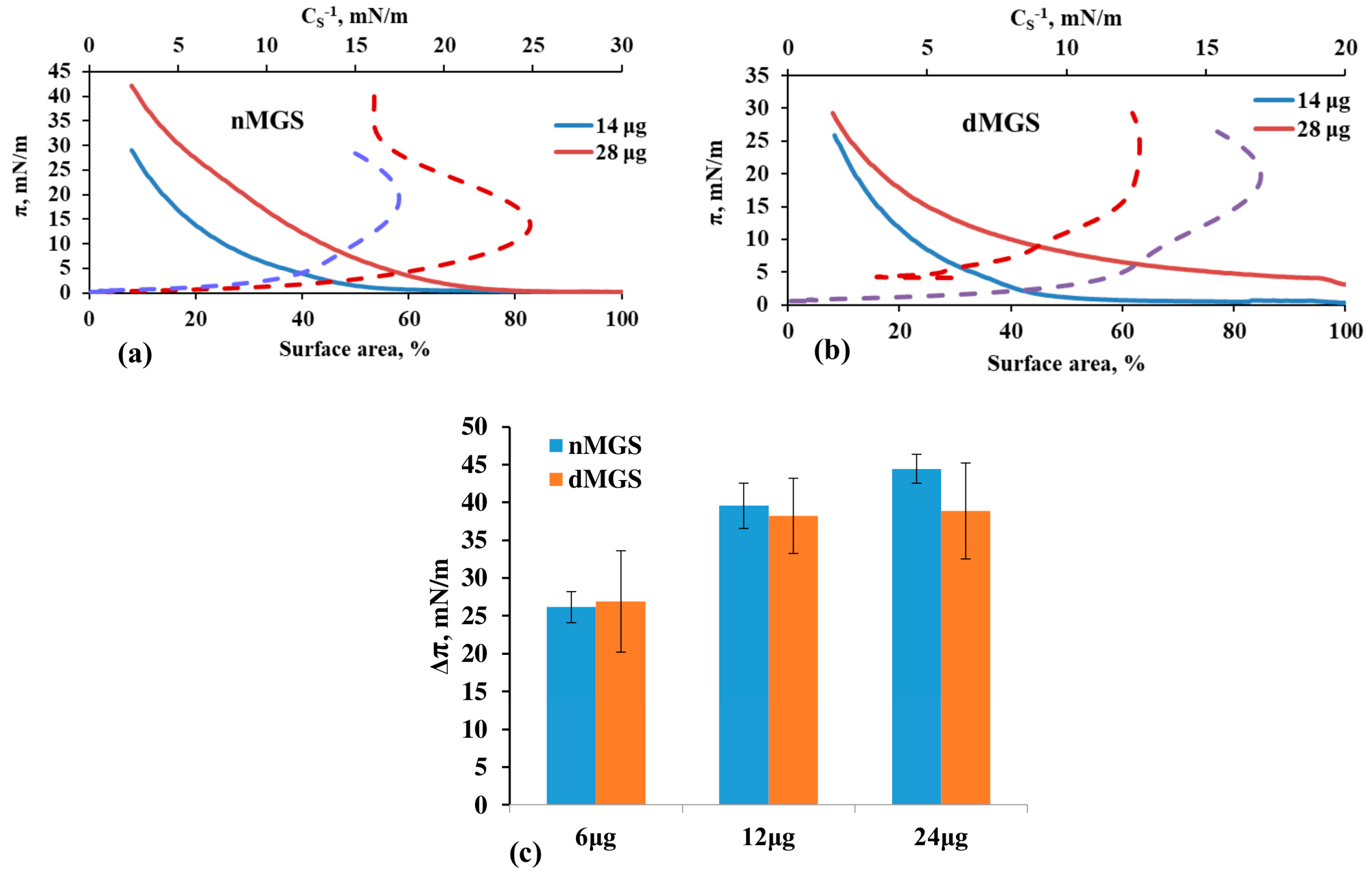
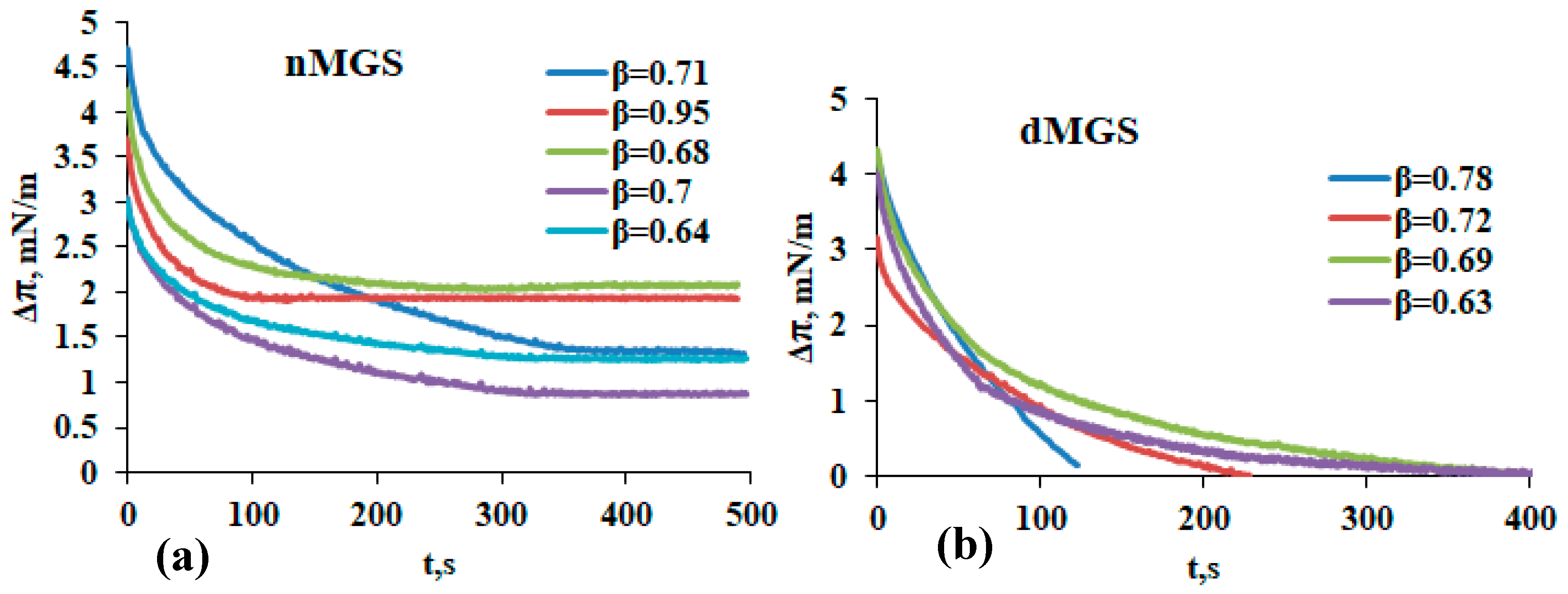
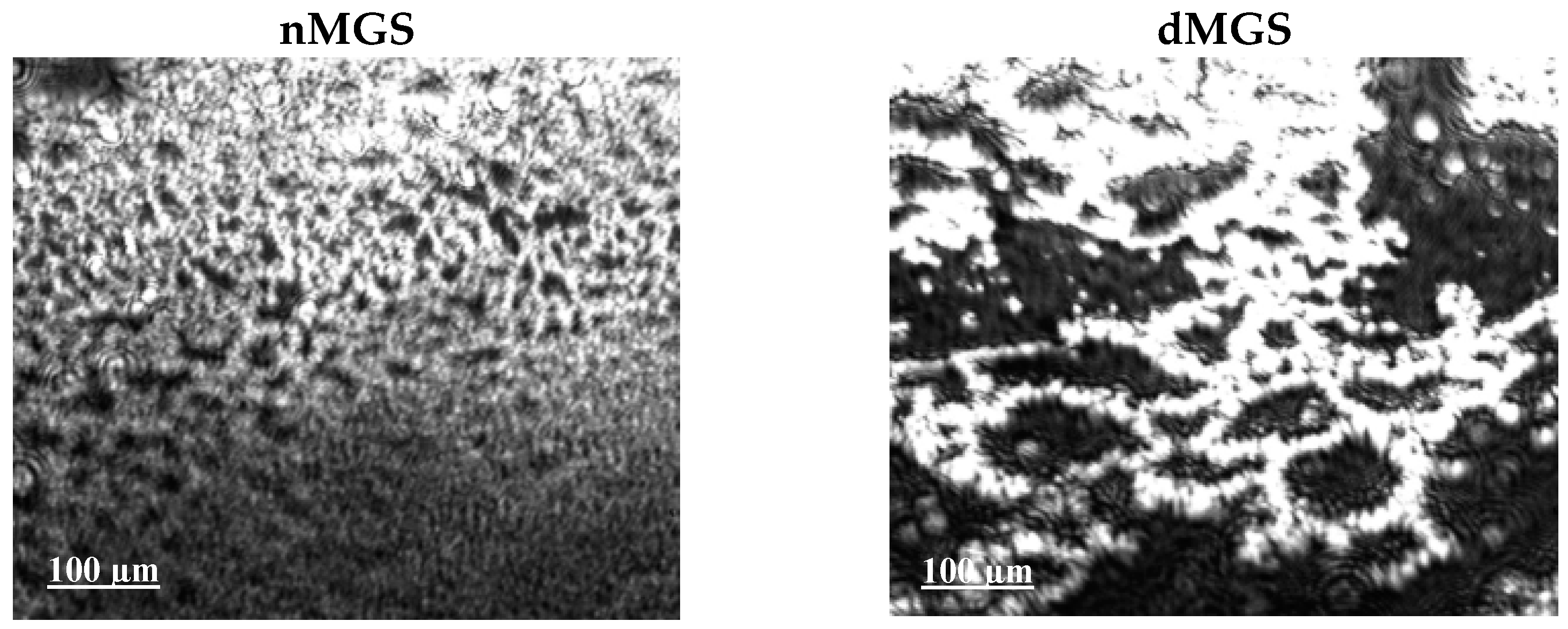
3.1. Surface Pressure Area/Isocycles of nMGS and dMGS Films and Their Morphology as Visualized by Brewster Angle Microscopy

It can be seen (Figure 1) that both nMGS and dMGS formed surface films capable of increasing the surface pressure upon area compression. As previously reported, the effect is proportional to the amount of meibum deposited at the air–water interface, both for healthy and for diseased MGS (Figure 1, upper row).
Although there is a tendency observed that at the same amount of deposited meibum, healthy samples can reach higher maximum π upon completion of film contraction (Figure 1, bottom row), there is great variability between the specimens and due to the small sample size no statistically significant conclusion can be reached.
The films’ in-plane reciprocal compressibility (or rigidity) modulus, or Cs−1 (Figure 1, dotted lines) was calculated as follows (Equation (1)):
Cs−1 = Aπ (−dπ/dA),
where Aπ is the area at the indicated π. The inflexions in the π/Cs−1 curves denote surface pressures at which notable modifications in the layers texture occur at compression. It can be seen that both nMGS and dMGS had Cs−1 < 50 mN/m, which corresponds to the liquid expanded state of the film at the meibum/water interface [14].
Figure 2 summarizes the dependences of the transient elasticity modulus on time which were analyzed via the Kohlrausch–Williams–Watts (KWW) equation (Equation (2)):
Δπ = Am. exp (−(t/τ)β) + ΔπEQ,
Typically the “stretched” exponent β ≤ 1 lowers relative to unity because of augmentation of (i) the film domains interaction and/or (ii) the film heterogeneity (i.e., increased amount of interacting arrays) [15]. Here Am represents the relaxation amplitude and ΔπEQ is the stationary plateau value.
It can be seen (Figure 2) that while β values varied greatly among both “healthy” and “diseased” Meibomian films, the plateau values of Δπ achieved at the end of the relaxation transients were ≥0.8 mN/m for nMGS, while the transients of dMGS layers cannot stabilize to a plateau value and Δπ increment dropped to zero. The latter feature is known to be an indication of mechanically unstable “brittle” layers [16,17,18,19,20].
This was indeed confirmed when experiments were performed to test the ability of Meibomian layers to sustain their surface area at 10 mN/m surface pressure. This value was chosen as it provides a reasonable estimate for the TFLL film pressure in terms of difference between the surface tension of delipidated (i.e., the aqueous tear surface tension) and intact human tears [21]. It can be seen (Figure 3) that, while nMGS films maintained constant surface pressure and surface area values, dMGS layers were indeed mechanically unstable and brittle and displayed constant contraction of film area, probably due to the redistribution of lipid molecules from the aqueous interface to the nonpolar lipid stratum of the layers [2]. In turn, such behavior is expected to result in facilitated instability of the tear film at interblink periods [2,22]. Such distinction in the performance of nMGS and dMGS layers was confirmed between all samples from healthy individuals and from MGD patients, respectively.
BAM visualization (Figure 4) revealed that while nMGS formed continuous films of multilayer thickness (estimated at around 60–100 nm, as explained previously) [9,10,11,12], in contrast dMGS layers were patchy and consisted of darker regions, probably of monolayer thickness, and thick bright regions which cannot enclose to a continuous structure and instead form a sparse bright mesh at the aqueous interface. For such estimations a simple semi-quantitative model relates the intensity, I, of the film regions in the BAM image with its thickness, d (di/dj = [Ii/Ij]1/2, where the subscripts i and j denote the different images) [3]. These observations agree with previous in vitro and in vivo studies which showed that in MGD the lipid layer structure loses its uniformity, which in turn correlates with loss of TF stability.

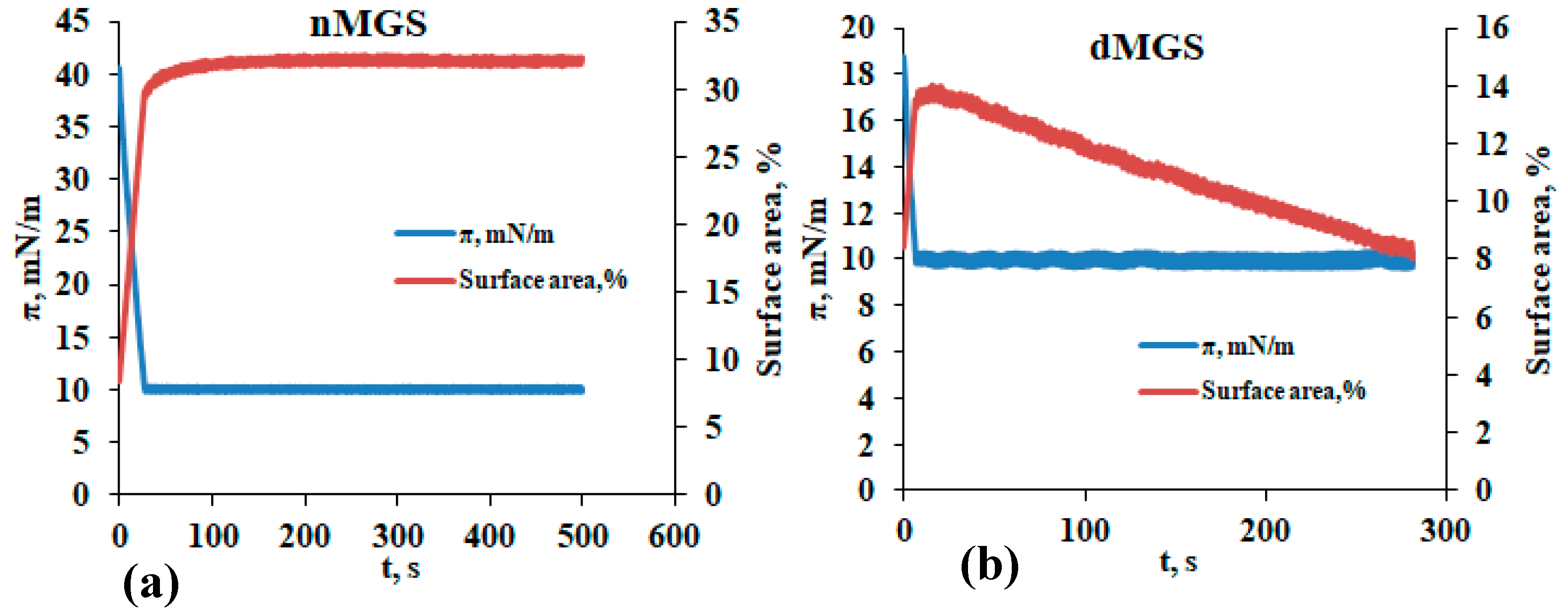
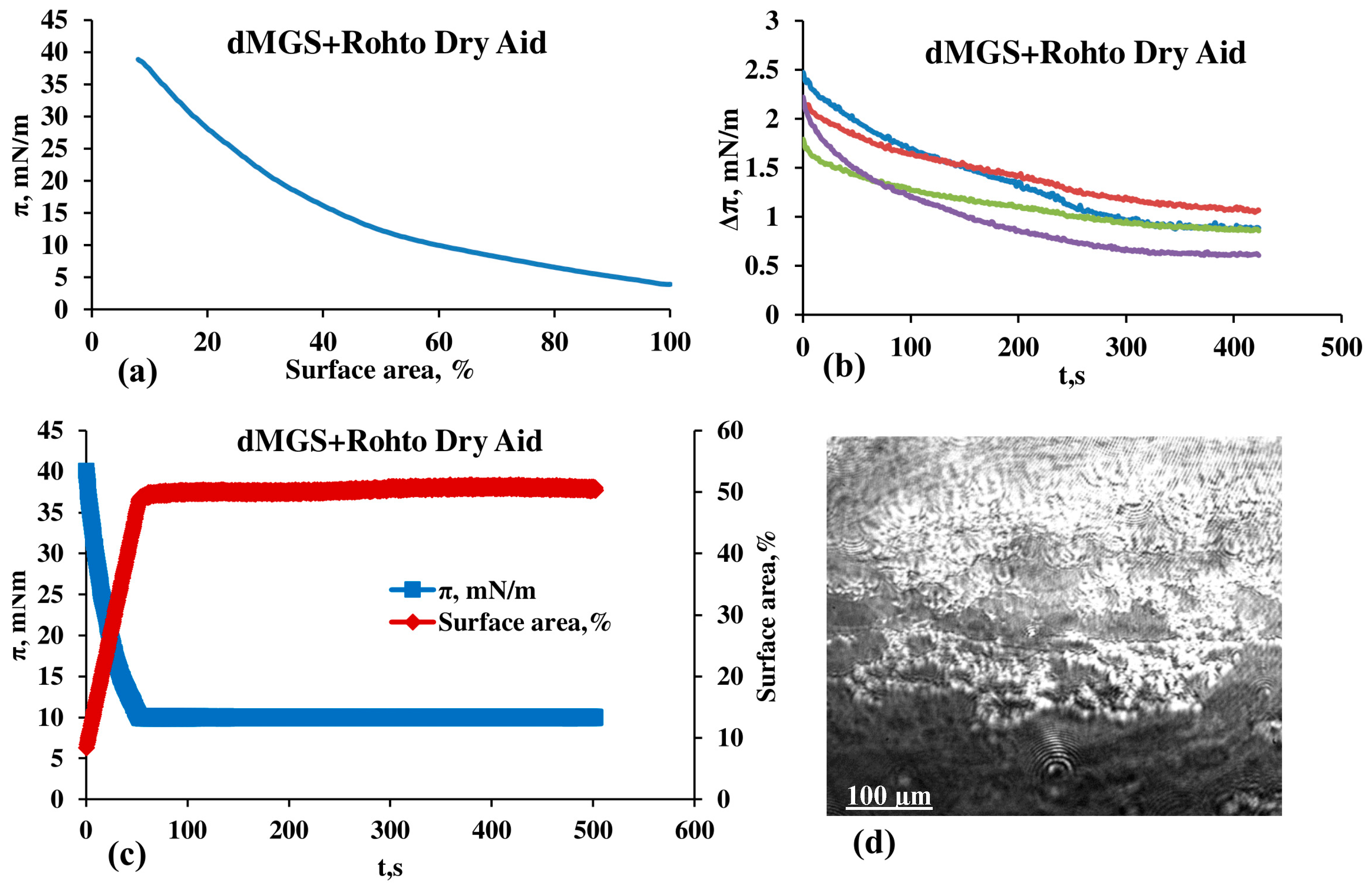
3.2. Effect of Rohto Dry Aid on dMGS Films

As shown by the representative data in Figure 5, the supplementation of dMGS with RDA (at ≤2/1 ratio) universally, for all MGD samples, resulted in the recovery of the ability of the dMGS to attain high surface pressures upon compression. Furthermore, the relaxation transients showed that dMGS reached a finite plateau value of ≥0.6 mN/m within 350 s after the step deformation was seized. This is an indication that the mixed RDA/dMGS layers were mechanically stable, which indeed was confirmed by the ability of these mixed layers to maintain stable value of the film area at 10 mN/m. Also, the morphology of the RDA/dMGS mixed films showed a continuous texture partially similar to the one seen with nMGS. As the continuous structure of the TFLL is an important indication for its ability to stabilize the tear film in vivo, this is an interesting result that hints at the clinical promise of opththalmic nanoemulsion applications to treat lipid-deficiency-related forms of dry eye.
The in vitro results are in excellent agreement with in vivo specular microscopy [2] observations of the impact of RDA on the TFLLs of human volunteers. These will be discussed in detail in a separate publication, but a representative case is shown at Figure 6 (see also the videos TFLL prior RDA. mov and TFLL with RDA.mov provided as Supplementary Materials).
As can be seen, prior to instillation of RDS the tear film lipid layer has a gray color, which corresponds to 70 nm thickness, while 30 min after the instillation of RDA, grey/yellow, yellow, and blue regions appeared, which indicate an increase of TFLL thickness to the 90–120 nm range [23]. Thus, the ability of RDA oil phase to increase the overall thickness of human meibum films observed in vitro shows excellent agreement with the finding as to the capability of the nanoemulsion to thicken the TFLL in vivo at the ocular surface.

4. Discussion

As demonstrated via Brewster Angle Microscopy here and in other studies, because of the extreme hydrophobicity of its lipids, MGS does not exist as a monomolecular film at the air–water interface, even at surface pressures as low as 0.5–10 mN/m. This is also reaffirmed by the inspection of the apparent area per molecule at which the MGS surface pressure/area isotherms take place, i.e., from a lift-off area of ~40–50 Å2/molecule to up to 10 Å2/molecule, which also clearly proves the multilayer structure of the Meibomian films [5,6]. Instead, “healthy” MGS forms rough heterogeneous layers that become more homogeneous with increases in π and maintain a liquid extended-like reciprocal compressibility modulus [5,6,24]. The behavior at the air–water interface of mixtures of triglycerides and cholesterol esters which compositionally resemble meibum is similar, as they also form films of multilayer thickness that display non-collapsibility and reversibility [24,25]. An inherent characteristic of such thick multilayers enriched with NPL is that the PL molecules (situated at the oil/aqueous interface) evade the formation of tightly packed and collapsed domains upon area contraction and instead PL relocate to the upper NPL stratum of the films. During the subsequent area expansion (i.e., a stage similar to the impact of eye opening at blink), this upper stratum of the layer acts as a reservoir, allowing polar lipids to quickly reappear at the interface, thus maintaining the stability of the films upon continuous blink-like area changes. The so-called duplex film structure, which consists of a continuous liquid-like oily suspension placed on top of a polar lipid monolayer [5,6], renders the MGS layers’ capability to evade collapse and to resist material loss for many consecutive blinks in the course of the day. Due to these distinctive characteristics, it is shown that in vivo at the ocular surface, the TFLL is able to maintain stable composition and structure for hours [26]. This latter effect decreases the need for the Meibomian glands to supply novel lipid material in the TFLL and ensures a TFLL turnover rate of 0.93 ± 0.36%/min (i.e., a notably lower rate, compared to 10.3 ± 3.7%/min AT turnover).
These inherent features of “healthy” MGS layers constitute the capacity of the TFLL to sustain its structure between blinks and to contribute to the strength of the tear film. It was recently found that MGS films’ elasticity plays important role in ensuring the structural recovery of the TFLL (revealed by image cross-correlation assay of lipid layer dynamics in vivo) in successive blinks, which in turn aligns with the general stability of the TF in healthy and in dry eyes [5,6,24,27,28]. Indeed, the ability of TFLL in vivo to spread quickly and uniformly while maintaining its texture (i.e., a display of elasticity) upon blinks has been strongly correlated with TF overall stability in numerous clinical studies [2,27,28]. In particular, it was found that the lipid layer spread took much longer in MGD patients (3.54 s in MGD vs. 0.36 s in healthy individuals) [22] and the spatial heterogeneity of TFLL thickness distribution was notably higher (lipid map uniformity = 125 nm2), compared to healthy eyes (lipid map uniformity = 14 nm2) [29]. The clinical studies and in vitro laboratory results that found that MGS and tear specimens from dry-eye patients had diminished surfactant potency and were unable to establish a continuous duplex film in the course of compression, contrary to samples from healthy eyes, also show excellent agreement [2]. Similar trends are also revealed in the current study, where the inability of MGD samples to form continuous layers (as revealed by BAM) is accompanied by inferior performance of MGD samples in stress relaxation transients and in formation of brittle layers that fail to maintain constant surface area at 10 mN/m. The exact reason behind the deteriorated performance of the MGD samples remains uncertain, but it is clear that compositional changes take place in both the PL and NPL lipids. Furthermore, dMGS may consist of up to 22 wt % of non-lipid components (i.e., proteins, salts, and polysaccharides) and these in turn might disturb the normal functionality of tear lipids [3,4,30]. Therefore, the ability of ophthalmic nanoemulsions to improve the spread and organization of MGS layers in vitro via supplementation of polar and nonpolar lipid molecules serves as a trustworthy indicator of the formulations’ potential for use in clinical settings in vivo [10,31].
As shown in a previous study [12], similarly to other oil-in-water ophthalmic nanoemulsions with comparable compositions, RDA films exhibited typical behavior [24,25]. Highly reversible, non-collapsible surface films are formed by nanoemulsion. As demonstrated previously, sesame oil can enhance the nonpolar lipids in MGS films and promote the development of a more homogeneous and well-organized NPL stratum [9]. In contrast to the analysis of pure sesame oil, it is important to talk about how the eyedrops’ amphiphilic ingredients (CO-10, MYS-40, Poloxamer 407, and menthol) may affect how RDA interacts with MGS films and how they may help the eyedrop interact with the meibum films. According to a recent study [31], surfactants like MYS-40, which have a high hydrophilic–lipophilic balance, can make up for a moderate deficiency of polar lipids by incorporating into the TFLL at the interface with the aqueous tear. Consequently, it improves the pseudo-binary MGS/RDA films’ oil-phase spreading and distribution, as well as the non-polar lipid stratum of the films as a whole. The formulation is optimally miscible with the TFLL when high and low HLB surfactants (CO-10 and MYS-40, respectively) are combined [13,29]. Poloxamer 407 has been observed to incorporate into lipid films; however, because of its hydrophilic polyethylene oxide moiety, it displays a strong attraction to water and is easily squeezed out in PBS subphase at π ≤ 20 mN/m in controlled experiments involving both MGS layers and phospholipid monolayers [32]. It has been discovered that menthol can enhance the PL-coated MGS/aqueous interface through its interaction with lipid films. The spread of MGS layers and their dilatational viscoelasticity are improved by the interaction. The outcomes are consistent with recent studies that show that patients with dry eye syndrome had their tear films become more stable after using warm compresses infused with menthol [33].
Earlier studies showed that RDA and its constituents exert a number of potentially clinically beneficial effects on “healthy” tear lipids in vitro: (i) sesame oil locates on top of the thinner (darker as observed by BAM) monolayer patches in the SO/nMGS layers and increases the overall uniformity and thickness of “healthy” meibum films [9]; and (ii) supplementation with RDA accelerates and stabilizes the spread of nMGS at the model tear surface, as seen by the displacement of talcum particles used as visualizer [12]. It is indicative that the latter outcome cannot be accomplished by the supplementation of an extra amount of nMGS to the layers. This observation is consistent with the classical finding of Brown and Dervichian [34] that MGS functions as a “tracer oil” with limited and nonuniform spread, and that it may require either tear-intrinsic or exogenous (i.e., supplemented via eyedrops) surfactants to improve the performance of Meibomian oil at the air/tear interface. In this study, the challenge to RDA was extended, as here the RDA capability was tested as to its ability to restore the functionality of “diseased” meibum, dMGS, collected from MGD patients. The inherent limitations in the surfactant properties of meibum are even more pronounced in the “diseased” meibum as compared to “healthy” MGS samples because dMGS shows a patchier and rougher film morphology, which in turn is accompanied by worsened viscoelasticity and by mechanical instability of dMGS layers [5]. The brittleness (i.e., inability to maintain constant surface area at π = 10 mN/m) of the MGD samples is a distinct feature of dMGS films that is not observed in “healthy” meibum, which always formed stable layers at a surface pressure of 10 mN/m. It can be seen that, with its ability to simultaneously supplement the NPL and PL compartments of the Meibomian films, RDA was able to soothe the heterogeneity of the surface layers and to ensure enhanced spread and uniformity of the surface texture of dMGS layers. These structural changes in turn resulted in improved viscoelasticity and recovered mechanical stability (i.e., no brittleness) of the RDA-supplemented dMGS films. Furthermore, the very same effects can be seen in vivo at the ocular surface, where typically the instillation of RDA resulted in faster upward movement and increased mean thickness of the TFLL, i.e., features well known to correlate with overall improvements in TF stability and ocular surface health [22,23,28]. Hence RDA functions as an integral formulation that can therefore improve the TFLL’s structure, spreading, and elasticity while also supplementing the NPL and PL strata of the Meibomian layers. As discussed earlier the spread and elastic ability are essential characteristics of the “healthy” TFLL. Therefore, the ability of ophthalmic nanoemulsions to improve the structure and spread of MGS films in vitro serves as a trustworthy indicator of these formulations’ potential for clinical use in vivo [10,31].

5. Conclusions

At physiologically relevant conditions RDA balances the NPL stratum of the “diseased” meibum films with sesame oil and the polar lipid stratum of the duplex film with the diverse PL-like molecules present in the eyedrop composition. Consequently, RDA maintains the structure and viscoelastic properties of the MGS duplex multilayer, which are believed to be crucial for the TFLL’s ability to restore and maintain the tear film at the ocular surface in vivo during and between blinks [4,10,31]. The formulation additionally endeavors to enhance the secretory-mucin-rich gel layer of human tears by incorporating polyvinylpyrrolidone, a polymer that is believed to augment the tear film’s shear thinning properties [35,36,37,38,39,40]. These properties are crucial for the lubricating action of TF during blinking, as well as for its tensile strength when at interblink [41,42,43,44].
Overall, the findings support the notion that ophthalmic nanoemulsions are a potentially useful tool for the complex task of soothing the tear film, which is a crucial stage in the treatment of dry eye and other ocular surface diseases [45,46,47].

Supplementary Materials

Supplementary Materials can be found at: https://zenodo.org/records/10657564 (accessed on 29 March 2024).

Author Contributions

Conceptualization, G.A.G. and N.Y.; methodology, G.A.G., P.E. and K.T. (Kazuhiro Tsuji); validation, G.A.G., P.E. and N.P.; formal analysis, G.A.G. and P.E.; investigation, P.E., A.S.K. and N.P.; data curation, P.E., A.S.K. and N.P.; writing—original draft preparation, G.A.G., N.Y., K.T. (Kazuhiro Tsuji), K.T. (Kyoko Takahashi), M.N. and P.E.; writing—review and editing, G.A.G., N.Y., K.T. (Kazuhiro Tsuji), K.T. (Kyoko Takahashi), M.N. and P.E.; visualization, P.E. and N.P.; supervision, G.A.G. and N.Y. All authors have read and agreed to the published version of the manuscript.

Funding

The support of Rohto Pharmaceutical (Osaka, Japan) via a collaborative research grant is gratefully acknowledged. This research was also supported in part by JSPS KAKENHI for Scientific Research from the Japanese Ministry of Education, Culture, Sports, Science and Technology (Grant number: JP20K09794). The research is also partially supported by the Operational Programme “Science and Education for Smart Growth” 2014–2020, co-funded by the European Union through the European structural and investment funds, Project BG05M2OP001-1.002-0019 “Clean technologies for sustainable environment—water, waste, energy for circular economy” (Clean&Circle CoC) by the provision of equipment. Nikola Peev acknowledges the support of contract РД 22-1648/ 07.10.2022 by the Bulgarian Ministry of Education and Science. Ani S. Koeva acknowledges the support of grant by Science Investigations Fund of Sofia University for 2024 year. Kazuhiro Tsuji, Kyoko Takahashi, and Miho Nishiyama аrе employees of the company. The funding organizations had no role in the design or conduct of this research.

Institutional Review Board Statement

Human meibum samples were collected by Prof. Norihiko Yokoi from Kyoto Prefectural University of Medicine in accordance with the tenets of the Declaration of Helsinki and with the permission of the Ethics Committee (IRB approval number ERB-C-1233-4; date: 23 May 2022).

Informed Consent Statement

Informed consent was obtained from all volunteers who donated meibum for biophysical studies.

Data Availability Statement

The raw data supporting the conclusions of this article will be made available by the authors on reasonable request.

Acknowledgments

The support of Rohto Pharmaceutical (Osaka, Japan) via a collaborative research grant, is gratefully acknowledged.

Conflicts of Interest

Authors Kazuhiro Tsuji, Kyoko Takahashi and Miho Nishiyama were employed by the company Rohto Pharmaceutical Co., Ltd. The remaining authors declare that the re-search was conducted in the absence of any commercial or financial relationships that could be construed as a potential conflict of interest.

Abbreviations

ATAqueous tears
BAMBrewster angle microscopy
MGSMeibomian gland secretion (or simply meibum)
nMGSMeibum from healthy individuals
dMGSMeibum from MGD individuals
MGDMeibomian gland disfunction (or disease)
CO-10Polyoxyethylene Castor Oil 10
MGDMeibomian gland disease
MNTMenthol
MYS-40 Polyoxyl 40 stearate
NPLNonpolar lipids
OPOil phase
PLPolar lipids
RDARohto Dry Aid
SOSesame oil
TFTear film
TFLLTear film lipid layer
πSurface pressure
Cs−1Reciprocal compressibility (or rigidity) modulus
ASurface film area
ΔπTransient increase of surface pressure
AmAmplitude of the stress relaxation transient
tTime
τCharacteristic relaxation time
βStretched exponential
ΔπEQEquilibrium increase of surface pressure after completion of the relaxation

References

  1. Willcox, M.D.P.; Argüeso, P.; Georgiev, G.A.; Holopainen, J.M.; Laurie, G.W.; Millar, T.J.; Papas, E.B.; Rolland, J.P.; Schmidt, T.A.; Stahl, U.; et al. TFOS DEWS II Tear Film Report. Ocul. Surf. 2017, 15, 366–403. [Google Scholar] [CrossRef] [PubMed]
  2. Georgiev, G.A.; Eftimov, P.; Yokoi, N. Structure-function relationship of tear film lipid layer: A contemporary perspective. Exp. Eye Res. 2017, 163, 17–28. [Google Scholar] [CrossRef] [PubMed]
  3. Borchman, D.; Foulks, G.N.; Yappert, M.C.; Bell, J.; Wells, E.; Neravetla, S.; Greenstone, V. Human Meibum Lipid Conformation and Thermodynamic Changes with Meibomian-Gland Dysfunction. Investig. Ophthalmol. Vis. Sci. 2011, 52, 3805–3817. [Google Scholar] [CrossRef] [PubMed]
  4. Butovich, I.A. Tear film lipids. Exp. Eye Res. 2013, 117, 4–27. [Google Scholar] [CrossRef] [PubMed]
  5. Georgiev, G.A.; Yokoi, N.; Ivanova, S.; Tonchev, V.; Nencheva, Y.; Krastev, R. Surface relaxations as a tool to distinguish the dynamic interfacial properties of films formed by normal and diseased meibomian lipids. Soft Matter 2014, 10, 5579–5588. [Google Scholar] [CrossRef] [PubMed]
  6. Rosenfeld, L.; Cerretani, C.; Leiske, D.L.; Toney, M.F.; Radke, C.J.; Fuller, G.G. Structural and Rheological Properties of Meibomian Lipid. Investig. Ophthalmol. Vis. Sci. 2013, 54, 2720–2732. [Google Scholar] [CrossRef] [PubMed]
  7. McDonald, M.; Patel, D.A.; Keith, M.S.; Snedecor, S.J. Economic and Humanistic Burden of Dry Eye Disease in Europe, North America, and Asia: A Systematic Literature Review. Ocul. Surf. 2016, 14, 144–167. [Google Scholar] [CrossRef] [PubMed]
  8. Donthineni, P.R.; Kammari, P.; Shanbhag, S.S.; Singh, V.; Das, A.V.; Basu, S. Incidence, demographics, types and risk factors of dry eye disease in India: Electronic medical records driven big data analytics report I. Ocul. Surf. 2019, 17, 250–256. [Google Scholar] [CrossRef] [PubMed]
  9. Eftimov, P.; Yokoi, N.; Georgiev, G.A. Surface Chemistry Study of the Interactions of Sesame Oil with Meibomian Films. Molecules 2022, 27, 464. [Google Scholar] [CrossRef]
  10. Torrent-Burgués, J. Tear Film Constituents and Medicines for Eyes Investigated as Langmuir Films. BioNanoScience 2023, 13, 1324–1338. [Google Scholar] [CrossRef]
  11. Torrent-Burgués, J. Langmuir films study on lipid-containing artificial tears. Colloids Surf. B Biointerfaces 2016, 140, 185–188. [Google Scholar] [CrossRef] [PubMed]
  12. Eftimov, P.; Yokoi, N.; Tsuji, K.; Peev, N.; Georgiev, G.A. Langmuir Trough Study of the Interactions of Tear Mimetic Eyedrop Formulation with Human Meibum Films. Appl. Sci. 2022, 12, 12095. [Google Scholar] [CrossRef]
  13. Mudgil, P.; Millar, T.J. Surfactant Properties of Human Meibomian Lipids. Investig. Ophthalmol. Vis. Sci. 2011, 52, 1661–1670. [Google Scholar] [CrossRef] [PubMed]
  14. Davies, J.T.; Rideal, E.K. Interfacial Phenomena; Academic Press: Cambridge, MA, USA, 1961; p. 64. [Google Scholar]
  15. Tsang, K.Y.; Ngai, K.L. Relaxation in interacting arrays of oscillators. Phys. Rev. E 1996, 54, R3067–R3070. [Google Scholar] [CrossRef] [PubMed]
  16. Ivanova, T.; Minkov, I.; Panaiotov, I.; Saulnier, P.; Proust, J.E. Dilatational properties and morphology of surface films spread from clinically used lung surfactants. Colloid Polym. Sci. 2004, 282, 1258–1267. [Google Scholar] [CrossRef]
  17. Wüstneck, R.; Enders, P.; Wüstneck, N.; Pison, U.; Miller, R.; Vollhardt, D. Surface dilational behaviour of spread dipalmitoyl phosphatidyl glycerol monolayers. PhysChemComm 1999, 2, 50–61. [Google Scholar] [CrossRef]
  18. Wüstneck, R.; Wüstneck, N.; Grigoriev, D.O.; Pison, U.; Miller, R. Stress relaxation behaviour of dipalmitoyl phosphatidylcholine monolayers spread on the surface of a pendant drop. Colloids Surf. B Biointerfaces 1999, 15, 275–288. [Google Scholar] [CrossRef]
  19. Dimitrov, D.S.; Panaiotov, I.; Richmond, P.; Ter-Minassian-Saraga, L. Dynamics of insoluble monolayers: I. Dilatational or elastic modulus, friction coefficient, and Marangoni effect for dipalmitoyl lecithin monolayers. J. Colloid Interface Sci. 1978, 65, 483–494. [Google Scholar] [CrossRef]
  20. Panaiotov, I.; Dimitrov, D.S.; Ter-Minassian-Saraga, L. Dynamics of insoluble monolayers: II. Viscoelastic behavior and marangoni effect for mixed protein phospholipid films. J. Colloid Interface Sci. 1979, 72, 49–53. [Google Scholar] [CrossRef]
  21. Tiffany, J.M.; Winter, N.; Bliss, G. Tear film stability and tear surface tension. Curr. Eye Res. 1989, 8, 507–515. [Google Scholar] [CrossRef]
  22. Goto, E.; Tseng, S.C. Differentiation of lipid tear deficiency dry eye by kinetic analysis of tear interference images. Arch. Ophthalmol. 2003, 121, 173–180. [Google Scholar] [CrossRef] [PubMed]
  23. Korb, D.R.; Baron, D.F.; Herman, J.P.; Finnemore, V.M.; Exford, J.M.; Hermosa, J.L.; Leahy, C.D.; Glonek, T.; Greiner, J.V. Tear film lipid layer thickness as a function of blinking. Cornea 1994, 13, 354–359. [Google Scholar] [CrossRef] [PubMed]
  24. Petrov, P.G.; Thompson, J.M.; Rahman, I.B.; Ellis, R.E.; Green, E.M.; Miano, F.; Winlove, C.P. Two-dimensional order in mammalian pre-ocular tear film. Exp. Eye Res. 2007, 84, 1140–1146. [Google Scholar] [CrossRef] [PubMed]
  25. Smaby, J.M.; Brockman, H.L. Novel surface phase containing cholesteryl esters. 2. Nonequivalence of cholesteryl arachidonate and those with 18-carbon, cis-unsaturated acyl groups. Biochemistry 1981, 20, 724–730. [Google Scholar] [CrossRef] [PubMed]
  26. Mochizuki, H.; Yamada, M.; Hatou, S.; Tsubota, K. Turnover rate of tear-film lipid layer determined by fluorophotometry. Br. J. Ophthalmol. 2009, 93, 1535–1538. [Google Scholar] [CrossRef] [PubMed]
  27. Yokoi, N.; Bron, A.J.; Georgiev, G.A. The Precorneal Tear Film as a Fluid Shell: The Effect of Blinking and Saccades on Tear Film Distribution and Dynamics. Ocul. Surf. 2014, 12, 252–266. [Google Scholar] [CrossRef] [PubMed]
  28. Yokoi, N.; Yamada, H.; Mizukusa, Y.; Bron, A.J.; Tiffany, J.M.; Kato, T.; Kinoshita, S. Rheology of tear film lipid layer spread in normal and aqueous tear-deficient dry eyes. Investig. Ophthalmol. Vis. Sci. 2008, 49, 5319–5324. [Google Scholar] [CrossRef] [PubMed]
  29. Cohen, Y.; Trokel, S.; Arieli, Y.; Epshtien, S.; Gefen, R.; Harris, A. Mapping the Lipid Layer of the Human Tear Film. Cornea 2020, 39, 132–135. [Google Scholar] [CrossRef] [PubMed]
  30. McMahon, A.; Lu, H.; Butovich, I.A. The spectrophotometric sulfo-phospho-vanillin assessment of total lipids in human meibomian gland secretions. Lipids 2013, 48, 513–525. [Google Scholar] [CrossRef]
  31. Weisenberger, K.; Fogt, N.; Fogt, J.S. Comparison of nanoemulsion and non-emollient artificial tears on tear lipid layer thickness and symptoms. J. Optom. 2021, 14, 20–27. [Google Scholar] [CrossRef]
  32. Frey, S.L.; Lee, K.Y. Temperature dependence of poloxamer insertion into and squeeze-out from lipid monolayers. Langmuir 2007, 23, 2631–2637. [Google Scholar] [CrossRef] [PubMed]
  33. Arita, R.; Morishige, N.; Sakamoto, I.; Imai, N.; Shimada, Y.; Igaki, M.; Suzuki, A.; Itoh, K.; Tsubota, K. Effects of a warm compress containing menthol on the tear film in healthy subjects and dry eye patients. Sci. Rep. 2017, 7, 45848. [Google Scholar] [CrossRef] [PubMed]
  34. Brown, S.I.; Dervichian, D.G. The Oils of the Meibomian Glands: Physical and Surface Characteristics. Arch. Ophthalmol. 1969, 82, 537–540. [Google Scholar] [CrossRef] [PubMed]
  35. Tiffany, J.M. The viscosity of human tears. Int. Ophthalmol. 1991, 15, 371–376. [Google Scholar] [CrossRef] [PubMed]
  36. Arshinoff, S.A.; Hofmann, I.; Nae, H. Role of rheology in tears and artificial tears. J. Cataract. Refract. Surg. 2021, 47, 655–661. [Google Scholar] [CrossRef] [PubMed]
  37. Arshinoff, S.; Hofmann, I.; Nae, H. Rheological behavior of commercial artificial tear solutions. J. Cataract. Refract. Surg. 2021, 47, 649–654. [Google Scholar] [CrossRef] [PubMed]
  38. Mehdaoui, H.; Ait Abderrahmane, H.; de Loubens, C.; Nait Bouda, F.; Hamani, S. Dynamics of a Gel-Based Artificial Tear Film with an Emphasis on Dry Disease Treatment Applications. Gels 2021, 7, 215. [Google Scholar] [CrossRef] [PubMed]
  39. Blasco-Martinez, A.; Mateo-Orobia, A.; Blasco-Alberto, J.; Pablo-Julvez, L. Rheological Behavior Patterns in Artificial Tears. Optom. Vis. Sci. 2022, 99, 455–462. [Google Scholar] [CrossRef] [PubMed]
  40. Recchioni, A.; Mocciardini, E.; Ponzini, E.; Tavazzi, S. Viscoelastic properties of the human tear film. Exp Eye Res. 2022, 219, 109083. [Google Scholar] [CrossRef]
  41. Fatt, I. Observations of tear film break up on model eyes. CLAO J. Off. Publ. Contact Lens Assoc. Ophthalmol. Inc. 1991, 17, 267–281. [Google Scholar]
  42. McDonnell, A.; Lee, J.H.; Makrai, E.; Yeo, L.Y.; Downie, L.E. Tear Film Extensional Viscosity Is a Novel Potential Biomarker of Dry Eye Disease. Ophthalmology 2019, 126, 1196–1198. [Google Scholar] [CrossRef] [PubMed]
  43. Lai, H.Y.; Chen, A.; Fang, P.C.; Yu, H.J.; Kuo, M.T. Comparing Tear Film Viscosity between Sjögren and Non-Sjögren Dry Eye Disease. Life 2023, 13, 1484. [Google Scholar] [CrossRef] [PubMed]
  44. Phan, C.M.; Ross, M.; Fahmy, K.; McEwen, B.; Hofmann, I.; Chan, V.W.Y.; Clark-Baba, C.; Jones, L. Evaluating Viscosity and Tear Breakup Time of Contemporary Commercial Ocular Lubricants on an In Vitro Eye Model. Transl. Vis. Sci. Technol. 2023, 12, 29. [Google Scholar] [CrossRef] [PubMed]
  45. Li, S.; Chen, L.; Fu, Y. Nanotechnology-based ocular drug delivery systems: Recent advances and future prospects. J. Nanobiotechnology 2023, 21, 232. [Google Scholar] [CrossRef] [PubMed]
  46. Dhahir, R.K.; Al-Nima, A.M.; Al-Bazzaz, F.Y. Nanoemulsions as Ophthalmic Drug Delivery Systems. Turk. J. Pharm. Sci. 2021, 18, 652–664. [Google Scholar] [CrossRef]
  47. Lv, Z.; Li, S.; Zeng, Z.; Yao, K.; Han, H. Recent progress of nanomedicine in managing dry eye disease. Adv. Ophthalmol. Pract. Res. 2024, 4, 24–33. [Google Scholar] [CrossRef]
Figure 1. Surface pressure/area (solid lines) and surface pressure/reciprocal compressibility (dotted lines) isotherms of nMGS (a) and dMGS (b) at 14 µg (blue lines) and 28 µg meibum (red lines) deposited at the trough surface, as well as (c) dependence of the maximum surface pressure on the total amount of meibum and the health status of MGS samples. The data of panel (c) represent mean ± standard deviation of samples from five healthy individuals and four MGD patients, respectively.
Applsci 14 03339 g001
Figure 2. Stress relaxation transients of nMGS (a) and dMGS (b) films. The values of the exponent β (see Equation (2) in the main text) are also denoted. Each curve corresponds to a sample from an individual healthy donor (panel (a)) or an individual MGD patient (panel (b)).
Applsci 14 03339 g002
Figure 3. Typical transients, which show that (a) nMGS films maintain constant surface area when kept at constant surface pressure of 10 mN/m while (b) dMGS films register continuous film area loss with time.
Applsci 14 03339 g003
Figure 4. Typical BAM micrographs (500 µm × 300 µm) of nMGS and dMGS films at 10 mN/m.
Applsci 14 03339 g004
Figure 5. Impact of RDA supplementation (dMGS/RDA = 2/1) on (a) the surface pressure-area isotherms, (b) stress relaxation transients, (c) area stability, and (d) surface morphology (500 µm × 300 µm BAM micrograph) of dMGS films.
Applsci 14 03339 g005
Figure 6. Typical tear film lipid layer morphology in vivo as visualized with specular microscopy (a) prior to and (b) 30 min after instillation of RDA in the tear film.
Applsci 14 03339 g006
Table 1. RDA composition as provided by the manufacturer. The active-ingredient concentration is specified in brackets. The oil phase and the active-ingredient aqueous phase are part of patent protected TEARSHIELD TECHNOLOGYTM. All of the components are dissolved in a pH 7.4 saline solution.
Active Ingredients *Oil Phase ** (9.12 × 10⁻³ M)
Povidone (3.96 × 10⁻⁵ M),
Propylene Glycol (3.9 × 10⁻² M)
Sesame Oil/Menthol/Polyoxyethylene 10 Castor Oil/Polyoxyl 40 Stearate/Poloxamer 407 = 1.15/0.12/3.2/4.6/0.04
* Here, the term “active ingredients” is used as specified in the OTC monograph of USA. The mean molecular weight of povidone is 1000 KDa. ** Oil phase has a mean molecular weight of 661.5. The molar ratios between the individual compounds are specified.
Disclaimer/Publisher’s Note: The statements, opinions and data contained in all publications are solely those of the individual author(s) and contributor(s) and not of MDPI and/or the editor(s). MDPI and/or the editor(s) disclaim responsibility for any injury to people or property resulting from any ideas, methods, instructions or products referred to in the content.

