Abstract
The high demand for aesthetic treatments among the population has resulted in a wide array of bleaching products available on the market. It is imperative to investigate the potential adverse effects these products may have on dental health. The objective of this systematic review is to assess, based on available experimental in vitro studies in the scientific literature, whether bleaching products exhibit cytotoxic properties against pulp stem cells and fibroblasts. A comprehensive literature search was conducted across the Medline, Scopus, and Lilacs databases using the search formula ((Tooth whitening) OR (bleaching agent)) AND cytotoxicity AND ((stem cell*) OR fibroblast). Following the application of inclusion and exclusion criteria, 14 articles were deemed suitable for full analysis. The most utilized assay in the evaluated studies was the MTT cell viability assay. Fibroblasts emerged as the most scrutinized cell type due to the potential adverse effects of bleaching, such as invasive cervical resorption, which primarily affects the periodontal ligament where fibroblasts are located. It was observed that as the concentration of hydrogen peroxide increases, so does the cytotoxicity of the product. Additionally, other factors such as application time, activation methods, and the type of peroxide used also play a significant role. Bleaching products have been shown to exert cytotoxic effects on fibroblasts. Further exploration of the impact of bleaching agents on dental pulp stem cells is warranted to better understand their implications for these cells.
1. Introduction
In recent years, there has been a trend in dentistry towards aesthetic treatments like dental bleaching, aimed at lightening tooth color to enhance smiles. This procedure involves applying chemical agents that oxidize the organic pigmentation of the tooth, resulting in a lighter shade [1,2]. The mechanism of action involves the breakdown of oxidizing agents into free radicals, which then attack pigmented molecules, reducing their light reflection and producing a whitening effect [3]. The efficacy of this technique varies depending on the extent of tooth discoloration [4,5].
Tooth color, a critical aspect of smile attractiveness, is defined by three dimensions: hue, chroma (saturation), and value. Miller and colleagues have proposed adding a fourth dimension, opacity/translucency. Tooth color is a critical aspect that determines the attractiveness of an individual’s smile [6].
Traditionally, we can speak of intrinsic staining, which occurs after a change in the composition or thickness of the dental tissue [7]. On the other hand, extrinsic discoloration can be direct and exhibit a multifactorial etiology. It is produced by chromogens derived from food ingested in the diet or placed habitually in the mouth, such as tobacco, coffee, or tea. Or it can also be indirect, in which the compounds do not have the color of the resulting stain, such as antiseptics or metallic salts [8]. Understanding these physiological and pathological processes involved in tooth staining allows for individualized treatment approaches.
There are several bleaching agents on the market, such as peroxides. Hydrogen peroxide (HP) is the active agent and the main component which is responsible for the chemical action. It is a strong oxidizing agent that produces free radicals, reactive molecules, and HP ions [4]. In contrast, carbamide peroxide (CP) is a stable complex formed by urea, which stabilizes the pH, and releases HP by decomposition when it encounters water. The former has a longer active permanence and is used in the office when staining is moderate or severe. The latter is used in home treatment, in cases of milder discolorations [9].
Treatment methods include in-office procedures, home treatments prescribed by dentists, or a combination of both. Home treatment involves using custom trays with CP gel for a set duration, following an initial in-office application. In-office procedures often employ higher bleaching agent concentrations, such as 35–40% HP, to achieve faster or more intense results [10,11].
To minimize sensitivity, various materials like remineralizers or desensitizers, e.g., sodium fluoride or casein phosphopeptide amorphous calcium phosphate (CPP-ACP), are used, along with gingival protection barriers like antioxidants (vitamin E or N-acetylcysteine) [12,13,14,15]. Sensitivity and gingival irritation are common adverse effects of bleaching, influenced by the agent’s penetration ability, concentration, and application time [16,17].
In addition to the sensitivity and/or gingival irritation, bleaching can produce relevant iatrogenicity at the cellular level, known as cytotoxicity [18,19]. The oxidizing agent can affect the pulp, producing pathological stress which may lead to necrosis or direct damage to cell membranes. On the other hand, it can cause damage to fibroblasts, generating oxidative deterioration in them [20]. This deterioration can lead to cell death and mutations [21].
This cytotoxicity is evaluated through cell viability studies in fibroblasts and dental pulp stem cells [22]. According to ISO 10993-5, if a material has cell viability values below 70%, it is considered cytotoxic [23]. The most used assay is the MTT assay (3-[4,5-dimethylthiazol-2-yl]-2,5 diphenyl tetrazolium bromide). Its main objective is to calculate cells that are viable in a high yield. A reduction in the number of mitotic cells provides evidence of cell growth inhibition. Some of its advantages are its quick measurement and analysis and the incorporation of several samples [24].
The objective of this systematic review is to perform a qualitative synthesis of in vitro experimental studies available in the scientific literature on the cytotoxic potential against pulp stem cells and fibroblasts of bleaching agents on the market and the factors that modulate it.
2. Materials and Methods
2.1. Subsection
An advanced bibliographic search was carried out in the Medline, Scopus, and Lilacs databases on 29 November 2022 using the following search string: ((Tooth whitening) OR (bleaching agent)) AND cytotoxicity AND ((stem cell*) OR fibroblast). The keyword selection was based on previous evidence and their most used descriptors (Table 1).

Table 1.
Results of database searches.
2.2. Eligibility Criteria and Study Selection
In vitro studies on the cytotoxicity of bleaching agents on human fibroblasts or dental pulp stem cells were considered eligible. Both products with chemical agents and abrasive agents were accepted. Studies on other cell lines and/or other dental products exclusively were discarded.
Study selection was performed aided by reference management software (Mendeley; Elsevier, AMS, The Netherlands). References were imported to Mendeley, and duplicate records were manually discarded. A first screening of the titles and abstracts of the references was performed. Eligible studies were then submitted to a second screening of their full text.
2.3. Data Extraction
For the analysis of the selected articles, the following variables were recorded for each study: author, year of publication, bleaching material and its characteristics (percentage of peroxide, commercial brand, and manufacturer), type of cell, type of test used, and cytotoxicity shown.
2.4. Quality Assessment
The quality of the studies was assessed using the modified CONSORT checklist for the assessment of the risk of bias of in vitro studies on dental materials by Faggion et al. (2012), consisting of 14 items [25].
3. Results
3.1. Study Selection and Flowchart
After the study search with the aforementioned formula, a total of 79 results were found: 35 in Medline, 41 in Scopus, and 4 in Lilacs. After the elimination of duplicates, 59 articles remained. Subsequently, after the title and abstract screening, 45 were excluded, as they did not meet the inclusion and exclusion criteria. Finally, the remaining 14 articles were eligible for the full text analysis and all of them were included in the qualitative synthesis. The study selection flowchart is illustrated in Figure 1.
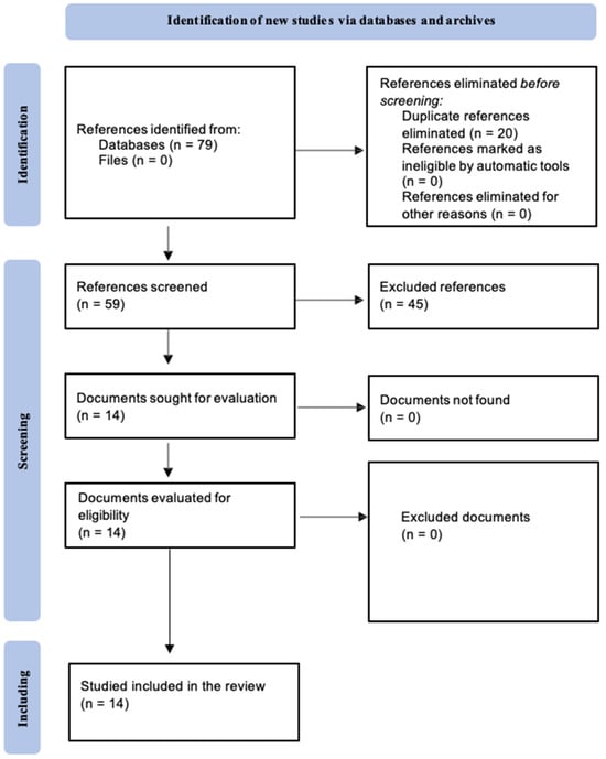
Figure 1.
PRISMA 2020 Flowchart.
3.2. Quality Assessment
In vitro studies were performed using the modified CONSORT checklist by Faggion et al. (2012) [25] consisting of 14 items assessing the quality of the studies (Table 2). All articles present a correctly structured abstract (item 1), as well as an introduction (item 2a) that provides information about the bleaching treatment and the main products to carry it out. The hypothesis and objectives (item 2b) are also clearly stated, as is the description of a specific methodology with the variables studied (items 3 and 4), which allows replication in all the studies analyzed. However, none of them provide an accurate report of the sample size calculation or allocation sequence (items 5–9). It should be noted that the items related to the randomization process are considered “not applicable” in in vitro studies on cell samples of this type. On the other hand, all studies reflect the statistical method used (item 10), as well as the level of significance through confidence intervals and/or p values (item 11). In the discussion section, all the studies include a summary of the most relevant results, as well as a series of comparisons with other published articles. However, only less than half refer to the limitations encountered in the research process (item 12) [26,27,28,29,30,31]. Sources of funding are indicated in most of the studies, except in three (item 13) [21,26,32]. However, information on the complete trial protocols (item 14) is not mentioned in any of the studies.
Table 2.
Modified CONSORT quality analysis of in vitro studies.
3.3. Materials
The 14 studies analyzed mention the use of a wide variety of bleaching products with different compositions, commercial brands, and manufacturers (Table 3).
Table 3.
Materials used in the studies.
3.4. Study Methodology and Results
The data collected from the selected studies are shown in Table 4. From the 14 selected articles for the qualitative synthesis, the vast majority used fibroblasts as cell lines and performed the MTT cell viability assay; only three of them evaluate cytotoxicity with pulp cells [15,18,31].
Table 4.
Data and results obtained from the included studies.
Studies reported that a higher concentration of peroxide resulted in an increased cytotoxicity. A difference between commercial brands was also observed which, using the same concentration of product, resulted in different levels of cytotoxicity, probably due to the additives incorporated in their composition [18,33,34].
Other compounds were also tested among the included studies to search for formulations with less adverse effects on teeth. These compounds include N-TiO2 [27], which is also associated with CaO2 [35]; platinum; or enzymes such as bromelain, ficin, and papain incorporated into whitening products [29]. All of them have been found to be significantly less cytotoxic than traditional peroxides [28].
In addition, authors such as Furukawa [14] and Chen [15] use antioxidants such as vitamin E and N-acetylcysteine respectively, which can reduce the cytotoxicity, especially in their use at the gingival barrier level. However, if it were associated with the bleaching agent, it would mean less oxidation, and therefore a reduction in the tooth whitening effect.
Finally, it is worth mentioning the comparison of bleaching pastes, which, although they do not contain oxidants such as peroxides, may induce a certain cytotoxicity due to the abrasives they contain.
4. Materials and Methods
Due to the high demand for dental bleaching, many products have emerged for this purpose. Iatrogenic problems can appear with the use of some of them, among which is the affectation of pulp tissue. Cytotoxicity is an adverse effect that is increasingly studied, since it can cause irreversible damage at the dental level [2]. The MTT assay is used in studies to verify cell viability [36].
Most bleaching products incorporate peroxides, which can be hydrogen or carbamide. Soares et al. [31] studied the effect of various concentrations of HP (35%, 17.5%, 10%, and 8%) on dental pulp cells. It was determined that cytotoxicity is concentration dependent, assuming a significant increase in the application of HP at 35% compared to the rest. The same concentration was used in another study [33] applied to fibroblasts, where activation by phototherapy with a low intensity laser emitting in the visible red and near infrared was also used. Apart from the decrease in cell viability shown in the application of 35% HP, a compensatory effect was observed in the group that incorporated the low intensity laser. This is attributed to the fact that this type of therapy promotes the regeneration of injured tissues and under stress conditions can increase cell metabolism. On the other hand, light activation involves a degree of heating of the gel, an increase in molecular movement and greater kinetic energy. It leads to an acceleration in diffusion since the chances of reactions are increased [37]. However, the type of light used does not influence the cytotoxicity of the dental pulp [38].
Therefore, whether to activate the materials in the procedure with light remains a subject of debate today; this procedure offers a range of potential benefits and the ability to promote recovery after the inevitable cell damage. Marto et al. [28] established in their study that there is an inhibition of metabolic activity with peroxide-induced oxidative stress whenever photoactivation is present. However, irradiation with LED light has been shown to favor the restoration of activity as well as cell regeneration.
Carbamide peroxide (PC) is another commonly used peroxide, typically applied at home in concentrations of 10–16%. Evidence suggests that it can also be cytotoxic. One study determined how the use of this agent at 22% compared to more typical percentages such as 16% or 10%, leads to a greater inhibition of cellular activity [32]. Sodium perborate (SP), despite its historical use, is currently in disuse [39]. It has been shown to be more harmful to teeth, resulting in erosions. Its cellular toxicity was found to be greater than that of HP [21].
However, the brand selected can have significant relevance despite using similar concentrations. There is evidence that, when comparing different brands, the results in terms of the cytotoxicity produced differ without being a consequence of a higher percentage of one of them [18,34]. Examples of this are Norblanc and Opalescence, both used at concentrations of 37.5–40% HP, with the latter being more harmful [18]. This is likely due to some additives incorporated in bleaching products, which increase the existing adverse effects. Additionally, it is worth mentioning the common incorporation of thickeners such as carbopol (carboxypolymethylene polymer), found in most household bleaching products such as carbamide peroxide. Its main function is to slow down the release process, thus extending the reaction time [40,41].
Whitening can be achieved through various methods. In addition to the application of whitening products in the dental office, there are also home-use products such as whitening toothpastes, aimed at lightening tooth color. These pastes contain abrasive cleaning materials, moisturizers, thickening agents, and fluorides for health benefits. Some whitening pastes also include chemical agents such as HP, sodium citrate, or phosphate salt [42].
Abrasives are insoluble components incorporated into toothpaste to physically remove stains and dental plaque. Significant tooth color changes can be achieved with their use over a certain period and frequency of brushing. Some of these compounds include sodium lauryl sulfate, sodium monofluorophosphate, hydrated silica, sodium benzoate, silicon dioxide, calcium carbonate, dicalcium phosphate dihydrate, calcium pyrophosphate, alumina, perlite, and sodium bicarbonate [43].
However, the excessive incorporation of certain abrasives in toothpaste can damage dental structure, despite their necessity in preventing stains. Additionally, studies show differences in cytotoxicity generated depending on the brand name, which is again important depending on the ingredients it contains [30]. This effective cleaning action must be balanced with remineralization at the dental level to ensure the toothpaste formula is adequate and does not damage tooth enamel [44].
To avoid treatment side effects, ongoing research focuses on new formulas, methods, and barriers. Preventive options such as the use of antioxidants are being explored. Vitamin E, for example, is an essential nutrient with various health benefits, including its antioxidant properties. It contains tocopherols and tocotrienols, potent antioxidants capable of eliminating peroxyl radicals [45,46]. Therefore, studying their interactions with oxidative stress and reactive oxygen species (ROS) is of interest since these are the main mechanisms of action of bleaching.
Different concentrations of vitamin E associated with HP application have been evaluated, with evidence of lower cytotoxicity observed at concentrations from 250 μM [14].
Another studied antioxidant compound is N-acetylcysteine, a metabolite of the amino acid cysteine with antioxidant potential. It acts as a bioprotective agent against oxidative stress by eliminating reactive oxygen species [47,48]. Observations indicate a reduction in HP-associated cytotoxicity with N-acetylcysteine at various concentrations and application times [15]. A recent systematic review indicates how the use of topical otosporin reduces pulpal inflammation when applied after bleaching treatment, as well as the use of remineralizing agents such as bioactive glass-ceramic or MI Paste Plus (a paste made up of casein phosphopeptide and amorphous calcium phosphate) help maintain an alkaline environment, thereby reducing the acidification caused by HP [49].
It is worth noting that the antioxidant potential of such compounds may have a lesser effect on whitening, reducing tooth lightening as they prevent the oxidative reaction involved in treatment. Therefore, their primary recommended use is at a preventive level, as a protector of the gingival barrier, applied to soft tissues to prevent the ROS from damaging periodontal cells.
Research not only focuses on preventive materials but also on discovering agents that achieve whitening with less damage. Titanium oxide (TiO2) is among the most studied compounds. It is a semiconductor that has photocatalytic and photoconductive properties, which make it an oxide with great degradation potential. Its viability is due to its low toxicity, low price, and photostability. Its modification with nitrogen, giving rise to N-TiO2, aims to avoid the collapse of the porous structure that forms the material [50]. This has traditionally been activated with ultraviolet light, but a photocatalyst that activates visible light has recently been introduced. For bleaching, photocatalytic nanoparticles can be used, which when activated by light, produce oxidation. It is shown that the addition of N-TiO2 results in lower cytotoxicity compared to the sole use of HP. However, it shows a lower cell viability when associated with Ag (TiO2/Ag) [27]. It should be noted that this product is a masking agent and therefore does not cause a real change in the color of the dental tissue.
CaO2 is a possible substitute for traditional peroxides since it releases HP in a controlled manner when reacting with water. The release rate can be modified according to factors such as pH and temperature. Therefore, it is one of the most stable solid peroxides [51]. Thus, its incorporation as an active ingredient to N-TiO2 activated with visible light as a photocatalyst has been evaluated. The result not only shows that it is effective in the aspect of efficient tooth whitening, but also that it is less cytotoxic. However, further evidence is still needed to determine its use in clinical practice [36].
Platinum (Pt) is another compound shown to produce fewer adverse effects, particularly when applied to two types of fibroblasts, BHK21/C13 and L929, with varying sensitivity to its action despite both showing some affectation with oxidative action [26].
Proteolytic enzymes such as bromelain, papain, or cysteine are examples of molecules that can be considered active agents with bleaching potential due to their enzymatic activity. Their main feature is the enhancement of HP-dependent oxidation–reduction, as well as decreasing the toxicity produced by electron-donor compounds [52]. Other properties include anti- inflammatory, fibrinolytic, and immunomodulatory effects. Likewise, its use in toothpaste has been demonstrated to eliminate dental stains with reduced roughness thanks to its protease action of protein degradation [53]. This may be relevant for the future use of products that do not contain peroxides, provided that more studies are conducted to provide scientific evidence to support the change in clinical practice.
The limitation of this systematic review lies in the variability of the included studies, both in their methodology and in the products used. It is necessary to establish protocols for the performance of these in vitro studies that allow an adequate comparison between them and the performance of a meta-analysis. On the other hand, most studies used fibroblasts because one of the most common adverse effects is invasive cervical resorption. More studies with dental pulp stem cells are needed to know exactly how bleaching products can affect the vitality and physiological functions of the pulp tissue.
5. Conclusions
All the bleaching products assessed in the included studies can cause pulp damage. The factors that most modulate the cytotoxic potential of these products are concentration, application time, and reaction speed. However, the use of antioxidant and proteolytic enzymes is also a factor to be considered. The product with the highest cytotoxic potential is HP. It is the professional, knowing the characteristics of the products available, who should personalize the treatment for each patient; they should be aware that there are agents with great bleaching power, but which present greater risks of cellular involvement.
Author Contributions
Conceptualization, J.G. and C.L.; methodology, M.M. (Mireia Montaner); software, M.M. (María Melo); validation, M.M. (María Melo), J.G. and C.P.-H.; formal analysis, C.L.; investigation, M.M. (Mireia Montaner) and J.G.; resources, M.M. (María Melo); data curation, M.M. (María Melo) and C.L.; writing—original draft preparation, M.M. (Mireia Montaner); writing—review and editing, J.L.S., J.G. and C.P.-H.; visualization, C.L.; supervision, C.L. and J.G.; project administration, J.G. and J.L.S.; funding acquisition, not applicable. All authors have read and agreed to the published version of the manuscript.
Funding
This research received no external funding.
Institutional Review Board Statement
Not applicable.
Informed Consent Statement
Not applicable.
Data Availability Statement
Not applicable.
Conflicts of Interest
The authors declare no conflicts of interest.
References
- Herrera, A.; Martin, J.; Perez, F.; Bonafe, E.; Reis, A.; Dourado, A.L.; Fernandez, E. Is personality relevant in the choice of bleaching? Clin. Oral Investig. 2016, 20, 2105–2111. [Google Scholar] [CrossRef] [PubMed]
- Bersezio, C.; Martín, J.; Mayer, C.; Rivera, O.; Estay, J.; Vernal, R.; Haidar, Z.S.; Angel, P.; Oliveira, O.B.; Fernández, E. Quality of life and stability of tooth color change at three months after dental bleaching. Qual. Life Res. 2018, 27, 3199–3207. [Google Scholar] [CrossRef] [PubMed]
- Alkahtani, R.; Stone, S.; German, M.; Waterhouse, P. A review on dental whitening. J. Dent. 2020, 100, 103423. [Google Scholar] [CrossRef] [PubMed]
- Carey, C.M. Tooth whitening: What we now know. J. Evid. Based Dent. Pract. 2014, 14, 70–76. [Google Scholar] [CrossRef] [PubMed]
- Mounika, A.; Mandava, J.; Roopesh, B.; Karri, G. Clinical evaluation of color change and tooth sensitivity with in-office and home bleaching treatments. Indian J. Dent. Res. 2018, 29, 423–427. [Google Scholar] [CrossRef] [PubMed]
- Watts, A.; Addy, M. Tooth discolouration and staining: A review of the literature. Br. Dent. J. 2001, 190, 309–316. [Google Scholar] [CrossRef] [PubMed]
- Aka, B.; Celik, E.U. Evaluation of the Efficacy and Color Stability of Two Different At- Home Bleaching Systems on Teeth of Different Shades: A Randomized Controlled Clinical Trial. J. Esthet. Restor. Dent. 2017, 29, 325–338. [Google Scholar] [CrossRef] [PubMed]
- Farawati, F.A.L.; Hsu, S.M.; O’Neill, E.; Neal, D.; Clark, A.; Esquivel-Upshaw, J. Effect of carbamide peroxide bleaching on enamel characteristics and susceptibility to further discoloration. J. Prosthet. Dent. 2019, 121, 340–346. [Google Scholar] [CrossRef] [PubMed]
- Luque-Martinez, I.; Reis, A.; Schroeder, M.; Muñoz, M.A.; Loguercio, A.D.; Masterson, D.; Maia, L.C. Comparison of efficacy of tray-delivered carbamide and hydrogen peroxide for at- home bleaching: A systematic review and meta-analysis. Clin. Oral Investig. 2016, 20, 1419–1433. [Google Scholar] [CrossRef]
- He, L.B.; Shao, M.Y.; Tan, K.; Xu, X.; Li, J.Y. The effects of light on bleaching and tooth sensitivity during in-office vital bleaching: A systematic review and meta-analysis. J. Dent. 2012, 40, 644–653. [Google Scholar] [CrossRef]
- Pereira, R.; Silveira, J.; Dias, S.; Cardoso, A.; Mata, A.; Marques, D. Bleaching efficacy and quality of life of different bleaching techniques—Randomized controlled trial. Clin. Oral Investig. 2022, 26, 7167–7177. [Google Scholar] [CrossRef] [PubMed]
- Maran, B.M.; Vochikovski, L.; de Andrade Hortkoff, D.R.; Stanislawczuk, R.; Loguercio, A.D.; Reis, A. Tooth sensitivity with a desensitizing-containing at-home bleaching gel-a randomized triple-blind clinical trial. J. Dent. 2018, 72, 64–70. [Google Scholar] [CrossRef] [PubMed]
- Gümüştaş, B.; Dikmen, B. Effectiveness of remineralization agents on the prevention of dental bleaching induced sensitivity: A randomized clinical trial. Int. J. Dent. Hyg. 2022, 20, 650–657. [Google Scholar] [CrossRef] [PubMed]
- Furukawa, M.; K-Kaneyama, J.R.; Yamada, M.; Senda, A.; Manabe, A.; Miyazaki, A. Cytotoxic effects of hydrogen peroxide on human gingival fibroblasts in vitro. Oper. Dent. 2015, 40, 430–439. [Google Scholar] [CrossRef] [PubMed]
- Chen, C.; Huang, X.; Zhu, W.; Ding, C.; Huang, P.; Li, R. H2O2 gel bleaching induces cytotoxicity and pain conduction in dental pulp stem cells via intracellular reactive oxygen species on enamel/dentin disc. PLoS ONE 2021, 16, e0257221. [Google Scholar] [CrossRef] [PubMed]
- Faria-E-Silva, A.L.; Nahsan, F.P.S.; Fernandes, M.T.G.; Martins-Filho, P.R.S. Effect of preventive use of nonsteroidal anti-inflammatory drugs on sensitivity after dental bleaching: A systematic review and meta-analysis. J. Am. Dent. Assoc. 2015, 146, 87–93.e1. [Google Scholar] [CrossRef] [PubMed]
- Moncada, G.; Sepúlveda, D.; Elphick, K.; Contente, M.; Estay, J.; Bahamondes, V.; Fernandez, E.; Oliveira, O.; Martin, J.; Martin, G.M.; et al. Effects of light activation, agent concentration, and tooth thickness on dental sensitivity after bleaching. Oper. Dent. 2013, 38, 467–476. [Google Scholar] [CrossRef] [PubMed]
- Llena, C.; Collado-González, M.; García-Bernal, D.; Oñate-Sánchez, R.E.; Martínez, C.M.; Moraleda, J.M.; Rodríguez-Lozano, F.J.; Forner, L. Comparison of diffusion, cytotoxicity and tissue inflammatory reactions of four commercial bleaching products against human dental pulp stem cells. Sci. Rep. 2019, 9, 7743. [Google Scholar] [CrossRef] [PubMed]
- Diomede, F.; Fonticoli, L.; Marconi, G.D.; Della Rocca, Y.; Rajan, T.S.; Trubiani, O.; Murmura, G.; Pizzicannella, J. Decellularized Dental Pulp, Extracellular Vesicles, and 5-Azacytidine: A New Tool for Endodontic Regeneration. Biomedicines 2022, 10, 403. [Google Scholar] [CrossRef]
- Soares, D.G.; Marcomini, N.; Duque, C.C.D.O.; Bordini, E.A.F.; Zuta, U.O.; Basso, F.G.; Hebling, J.; Costa, C.A.D.S. Increased whitening efficacy and reduced cytotoxicity are achieved by the chemical activation of a highly concentrated hydrogen peroxide bleaching gel. J. Appl. Oral Sci. 2019, 27, e20180453. [Google Scholar] [CrossRef]
- Fernandes, A.M.; Marques, M.M.; Camargo, S.E.A.; Cardoso, P.E.; Camargo, C.H.R.; Valera, M.C. Cytotoxicity of non-vital dental bleaching agents in human gingival fibroblasts TT—Avaliação da citotoxicidade de agentes clareadores de uso interno sobre fibroblastos gengivais humanos. Braz. Dent. Sci. 2013, 16, 59–65. [Google Scholar] [CrossRef][Green Version]
- Marconi, G.D.; Fonticoli, L.; Della Rocca, Y.; Rajan, T.S.; Piattelli, A.; Trubiani, O.; Pizzicannella, J.; Diomede, F. Human Periodontal Ligament Stem Cells Response to Titanium Implant Surface: Extracellular Matrix Deposition. Biology 2021, 10, 931. [Google Scholar] [CrossRef] [PubMed]
- ISO 10993-5:2009; Biological Evaluation of Medical Devices—Part 5: Tests for In Vitro Cytotoxicity. ISO: Geneve, Switzerland, 2009; p. 34.
- van Meerloo, J.; Kaspers, G.J.L.; Cloos, J. Cell sensitivity assays: The MTT assay. Methods Mol. Biol. 2011, 731, 237–245. [Google Scholar] [PubMed]
- Faggion, C.M.J. Guidelines for reporting pre-clinical in vitro studies on dental materials. J. Evid. Based Dent. Pract. 2012, 12, 182–189. [Google Scholar] [CrossRef] [PubMed]
- Koulaouzidou, E.; Lambrianidis, T.; Konstantinidis, A.; Kortsaris, A.H. In vitro evaluation of the cytotoxicity of a bleaching agent. Endod. Dent. Traumatol. 1998, 14, 21–25. [Google Scholar] [CrossRef]
- Kurzmann, C.; Verheyen, J.; Coto, M.; Kumar, R.V.; Divitini, G.; Shokoohi-Tabrizi, H.A.; Verheyen, P.; Gentil De Moor, R.J.; Moritz, A.; Agis, H. In vitro evaluation of experimental light activated gels for tooth bleaching. Photochem. Photobiol. Sci. 2019, 18, 1009–1019. [Google Scholar] [CrossRef] [PubMed]
- Marto, C.M.; Laranjo, M.; Paula, A.; Coelho, A.S.; Abrantes, A.M.; Casalta-Lopes, J.; Gonçalves, A.C.; Sarmento-Ribeiro, A.B.; Ferreira, M.M.; Cabrita, A.; et al. Cytotoxic effects of zoom® whitening product in human fibroblasts. Materials 2020, 13, 1491. [Google Scholar] [CrossRef] [PubMed]
- Ribeiro, J.S.; Barboza, A.S.; Cuevas-Suárez, C.E.; da Silva, A.F.; Piva, E.; Lund, R.G. Novel in-office peroxide-free tooth-whitening gels: Bleaching effectiveness, enamel surface alterations, and cell viability. Sci. Rep. 2020, 10, 10016. [Google Scholar] [CrossRef] [PubMed]
- Rode, S.M.; Sato, T.D.P.; Matos, F.S.; Correia, A.M.O.; Camargo, S.E.A. Toxicity and effect of whitening toothpastes on enamel surface. Braz. Oral Res. 2021, 35, e025. [Google Scholar] [CrossRef]
- Soares, D.G.; Basso, F.G.; Hebling, J.; de Souza Costa, C.A. Immediate and late analysis of dental pulp stem cells viability after indirect exposition to alternative in-office bleaching strategies. Clin. Oral Investig. 2015, 19, 1013–1020. [Google Scholar] [CrossRef]
- dos Santos, R.L.; Pithon, M.M.; Martins, F.O.; Romanos, M.T.V. Cytotoxicity of carbamide peroxide bleaching gel on L929 cells TT—Citotoxicidade do gel clareador peróxido de carbamida em células L929. Rev. Odonto Ciência 2010, 25, 271–275. [Google Scholar]
- Dantas, C.M.G.; Vivan, C.L.; Ferreira, L.S.; Freitas, P.M.; Marques, M.M. In vitro effect of low intensity laser on the cytotoxicity produced by substances released by bleaching gel. Braz. Oral Res. 2010, 24, 460–466. [Google Scholar] [CrossRef] [PubMed]
- Hanks, C.T.; Fat, J.C.; Corcoran, J.F.; Wataha, J.C. Cytotoxicity and Dentin Permeability of Carbamide Peroxide and Hydrogen Peroxide Vital Bleaching Materials, in vitro. J. Dent. Res. 1993, 72, 931–938. [Google Scholar] [CrossRef] [PubMed]
- Thacker, M.; Chen, Y.N.; Lin, C.P.; Lin, F.H. Nitrogen-Doped Titanium Dioxide Mixed with Calcium Peroxide and Methylcellulose for Dental Bleaching under Visible Light Activation. Int. J. Mol. Sci. 2021, 22, 3759. [Google Scholar] [CrossRef] [PubMed]
- De Colli, M.; Tortorella, P.; Marconi, G.D.; Agamennone, M.; Campestre, C.; Tauro, M.; Cataldi, A.; Zara, S. In vitro comparison of new bisphosphonic acids and zoledronate effects on human gingival fibroblasts viability, inflammation and matrix turnover. Clin. Oral Investig. 2016, 20, 2013–2021. [Google Scholar] [CrossRef] [PubMed]
- Llena, C.; Martínez-Galdón, O.; Forner, L.; Gimeno-Mallench, L.; Rodríguez-Lozano, F.J.; Gambini, J. Hydrogen Peroxide Diffusion through Enamel and Dentin. Materials 2018, 11, 1694. [Google Scholar] [CrossRef] [PubMed]
- Benetti, F.; Lemos, C.A.A.; de Oliveira Gallinari, M.; Terayama, A.M.; Briso, A.L.F.; de Castilho Jacinto, R.; Sivieri-Araújo, G.; Cintra, L.T.A. Influence of different types of light on the response of the pulp tissue in dental bleaching: A systematic review. Clin. Oral Investig. 2018, 22, 1825–1837. [Google Scholar] [CrossRef]
- Tran, L.; Orth, R.; Parashos, P.; Tao, Y.; Tee, C.W.; Thomas, V.T.; Towers, G.; Truong, D.T.; Vinen, C.; Reynolds, E.C. Depletion Rate of Hydrogen Peroxide from Sodium Perborate Bleaching Agent. J. Endod. 2017, 43, 472–476. [Google Scholar] [CrossRef] [PubMed]
- Sobral-Souza, D.F.; Gouveia, T.H.N.; Condeles, A.L.; Junior, J.C.T.; Muniz, B.V.; Franz-Montan, M.; Pauli, M.C.; Leonardi, G.R.; Lima, D.A.N.L. Effect of accelerated stability on the physical, chemical, and mechanical properties of experimental bleaching gels containing different bioadhesive polymers. Clin. Oral Investig. 2022, 26, 3261–3271. [Google Scholar] [CrossRef]
- Basting, R.T.; Rodrigues, A.L.J.; Serra, M.C. The effect of 10% carbamide peroxide, carbopol and/or glycerin on enamel and dentin microhardness. Oper. Dent. 2005, 30, 608–616. [Google Scholar]
- Ghajari, M.F.; Shamsaei, M.; Basandeh, K.; Galouyak, M.S. Abrasiveness and whitening effect of charcoal-containing whitening toothpastes in permanent teeth. Dent. Res. J. 2021, 18, 51. [Google Scholar]
- Casado, B.G.S.; Moraes, S.L.D.; Souza, G.F.M.; Guerra, C.M.F.; Souto-Maior, J.R.; Lemos, C.A.A.; Vasconcelos, B.C.E.; Pellizzer, E.P. Efficacy of Dental Bleaching with Whitening Dentifrices: A Systematic Review. Int. J. Dent. 2018, 2018, 7868531. [Google Scholar] [CrossRef] [PubMed]
- Joiner, A. Whitening toothpastes: A review of the literature. J. Dent. 2010, 38 (Suppl. 2), e17–e24. [Google Scholar] [CrossRef] [PubMed]
- Jiang, Q. Natural forms of vitamin E: Metabolism, antioxidant, and anti-inflammatory activities and their role in disease prevention and therapy. Free Radic. Biol. Med. 2014, 72, 76–90. [Google Scholar] [CrossRef] [PubMed]
- Miyazawa, T.; Burdeos, G.C.; Itaya, M.; Nakagawa, K.; Miyazawa, T. Vitamin E: Regulatory Redox Interactions. IUBMB Life 2019, 71, 430–441. [Google Scholar] [CrossRef] [PubMed]
- Dodd, S.; Dean, O.; Copolov, D.L.; Malhi, G.S.; Berk, M. N-acetylcysteine for antioxidant therapy: Pharmacology and clinical utility. Expert. Opin. Biol. Ther. 2008, 8, 1955–1962. [Google Scholar] [CrossRef] [PubMed]
- Aldini, G.; Altomare, A.; Baron, G.; Vistoli, G.; Carini, M.; Borsani, L.; Sergio, F. N-Acetylcysteine as an antioxidant and disulphide breaking agent: The reasons why. Free Radic. Res. 2018, 52, 751–762. [Google Scholar] [CrossRef]
- Batista, L.A.S.; Dos Reis-Prado, A.H.; Chaves, H.G.D.S.; de Arantes, L.C.; Morgan, L.F.S.A.; André, C.B.; Suzuki, T.Y.; Benetti, F. Can different agents reduce the damage caused by bleaching gel to pulp tissue? A systematic review of basic research. Restor. Dent. Endod. 2023, 48, e39. [Google Scholar] [CrossRef] [PubMed]
- Hernández, J.M.; García, L.A.; Zeifert, B.H.; García, R.; Zermeño, B.B.; Del Angel, T.; Cueto, A. Síntesis y Caracterización de Nanopartículas de N-TiO2—Anatasa. Superf. Vacío 2008, 21, 1–5. [Google Scholar]
- Northup, A.; Cassidy, D. Calcium peroxide (CaO2) for use in modified Fenton chemistry. J. Hazard. Mater. 2008, 152, 1164–1170. [Google Scholar] [CrossRef]
- Hikisz, P.; Bernasinska-Slomczewska, J. Beneficial Properties of Bromelain. Nutrients 2021, 13, 4313. [Google Scholar] [CrossRef] [PubMed]
- Mazilu, A.; Popescu, V.; Sarosi, C.; Dumitrescu, R.S.; Chisnoiu, A.M.; Moldovan, M.; Dumitrescu, L.S.; Prodan, D.; Carpa, R.; Gheorghe, G.F.; et al. Preparation and In Vitro Characterization of Gels Based on Bromelain, Whey and Quince Extract. Gels 2021, 7, 191. [Google Scholar] [CrossRef] [PubMed]

Disclaimer/Publisher’s Note: The statements, opinions and data contained in all publications are solely those of the individual author(s) and contributor(s) and not of MDPI and/or the editor(s). MDPI and/or the editor(s) disclaim responsibility for any injury to people or property resulting from any ideas, methods, instructions or products referred to in the content. |
© 2024 by the authors. Licensee MDPI, Basel, Switzerland. This article is an open access article distributed under the terms and conditions of the Creative Commons Attribution (CC BY) license (https://creativecommons.org/licenses/by/4.0/).