Article

Analysis of Operated Ankle Fractures in Elderly Patients: Are They All Osteoporotic?

1
Orthopedic and Traumatology Unit, University of Perugia, “Santa Maria della Misericordia” Hospital, P.le G. Menghini 3, 06156 Perugia, Italy
2
Research Unit of Orthopaedic and Trauma Surgery, Department of Medicine and Surgery, Università Campus Bio-Medico di Roma, Via Alvaro del Portillo, 21, 00128 Roma, Italy
*
Author to whom correspondence should be addressed.
Appl. Sci. 2024, 14(9), 3787; https://doi.org/10.3390/app14093787
Submission received: 30 March 2024 / Revised: 20 April 2024 / Accepted: 28 April 2024 / Published: 29 April 2024
(This article belongs to the Section Applied Biosciences and Bioengineering)

Abstract

:
Background: Osteoporosis represents a global problem, which involves high risks of disability and death due to the consequences of fragility fractures. The aim of our study is to verify what type of relationship there is between ankle fractures and osteoporosis. Specifically, we aim to understand how the clinical and anamnestic characteristics of patients, along with the radiographic features of these fractures, may contribute to considering them as osteoporotic fractures. Methods: The study group includes 51 consecutive patients aged 60 years or older operated for ankle fracture from May to October 2022. The fractures were divided into uni-, bi-, or trimalleolar based on the plain X-rays. All patients underwent femoral and vertebral bone mineralometry by DXA associated with the FRAX questionnaire. Results: Ankle fractures in the elderly were associated with reduced BMD (76.5% of the total patients examined, 83.8% considering only women) and low-energy traumas (82%). Furthermore, these fractures present features of increased complexity as bone mass decreases. Conclusions: Ankle fractures in the elderly exhibit characteristics that suggest a relationship with bone fragility. For these reasons, it might be advisable to initiate a comprehensive fracture risk assessment in elderly patients who experience an ankle fracture. These fractures should not be underrated, and antiresorptive therapy must always be taken into consideration when selecting patients at risk.

1. Introduction

Osteoporosis is a systemic skeletal disorder characterized by reduced bone mineral density (BMD) and microarchitectural deterioration. This alteration of normal bone physiology in the end results in increased fracture risk, so-called “fragility fractures” [1]. Osteoporosis is a growing global problem, which involves high risks of disability and death due to the consequences of fragility fractures [2,3] both in men [4] and women.

1.1. Diagnosis of Osteoporosis

The diagnosis of osteoporosis relies on a comprehensive approach, including a detailed patient history, a thorough clinical examination, spinal X-rays, bone mineral density (BMD) measurements, and laboratory investigations [5]. The patient’s history should focus on identifying risk factors such as family history, previous fractures, dietary habits, lifestyle factors (e.g., alcohol consumption), use of medications affecting bone metabolism, and age at menopause onset [6].
Bone mass, expressed as bone mineral density (BMD), can be assessed using various techniques generally indicated as bone densitometry or computerized bone mineralometry (CBM). The most reliable procedure is the so-called DXA (dual-energy X-ray absorptiometry) which should be always measured at two levels: hip and spine.
BMD values are essentially expressed with the T-score the value 0 is the average BMD value of healthy young adults (peak bone mass). The Z-score (0 is the average value of individuals of the same age and gender) can be a useful parameter, even if less used for defining the degree of bone loss. According to the WHO criteria, four categories of subjects are identified based on the measured T-score [7]:
  • T-score from +2.5 to −1.0 SD (normal);
  • T-score between −1.0 and −2.5 SD (osteopenia);
  • T-score < −2.5 SD (osteoporosis);
  • T-score < −2.5 SD in the presence of at least one fragility fracture (severe osteoporosis).
It has been observed that the risk of fracture begins to increase exponentially with densitometric values of T-score < −2.5 standard deviations (SD), which, according to WHO, represents the threshold for diagnosing osteoporosis. Bone mass is directly related to the mechanical strength of the bone and inversely related to its fragility.
BMD measurements of the lumbar spine, femoral neck, total hip, and distal third of the radius are predictive of fragility fractures. Measurements on the distal radius are performed only when lumbar and/or femoral evaluation is imprecise or inaccurate, in severely obese patients, and in patients with hyperparathyroidism.

1.2. Risk Factors

Various risk factors contribute to the development of osteoporosis, including age, previous fractures, family history, comorbidities, and pharmacological therapies. Age is a primary determinant, with an exponential increase in the incidence of osteoporotic fractures in the elderly. Previous fractures, irrespective of the site, serve as strong predictors for subsequent fractures, and a positive family history of fractures independently influences fracture risk [8]. Certain comorbidities, such as rheumatoid arthritis, diabetes, and renal diseases, can elevate the risk of fractures, as can specific pharmacological therapies, notably prolonged use of glucocorticoids.
However, it should be emphasized that up to 30% of patients with fragility fractures have normal T-score values or values in the osteopenia range [6].
A comprehensive risk assessment, integrating BMD and clinical risk factors [9], is essential for identifying individuals at risk of fractures and guiding therapeutic decisions. Algorithms like FRAX® [10] and DeFRA are valuable tools for this integrated assessment.
The role of BMI as a risk factor is still debated and needs to be clarified [11]. The current literature suggests that obesity is correlated with an elevated risk of fractures at certain sites, such as non-hip inferior limb fractures and the proximal humerus. However, contrasting findings suggest instead a potentially protective effect at other sites, including hip fractures and possibly the wrist [12].

1.3. Fragility Fractures

Historically, osteoporosis has been associated with hip and vertebral fractures [13], primarily because these types of fractures occur following low or no-energy trauma almost exclusively in patients with pathologically low bone mineral density. A hip fracture is almost always due to osteoporosis [14] and the same can be said for a vertebral fracture [15] due to minimal or no trauma, so much so that, regardless of BMD, such fractures are considered an incontrovertible sign of severe osteoporosis and, in the vast majority of countries around the world, patients who experience such fractures, are entitled to reimbursement of osteoporosis drugs by the government [16].
It is widely believed that some other fractures, extravertebral and extrafemoral, are to be considered mainly due to bone fragility. These are wrist, proximal humerus, and ankle fractures [17]. However, since these fractures can also occur in young patients and have a traumatic mechanism that is not always well definable as low impact, they are not automatically considered due to severe osteoporosis, unlike hip and vertebral fractures.
Recent epidemiological studies have underscored the rising incidence of ankle fractures in conjunction with osteoporosis, suggesting a possible nexus between the two [18].

1.4. Objective of the Study

This research endeavors to contribute to the existing literature by investigating the debated association between osteoporosis and ankle fractures, with a particular emphasis on discerning whether such fractures can be classified as fragility fractures.
The importance of understanding this association lies in the potential implications for clinical practice, preventive strategies, and therapeutic interventions aimed at improving patient care and reducing the societal burden linked with osteoporotic fractures [19].
The purpose of this study is also to evaluate whether ankle fractures are connected to some individual characteristics and whether the radiographic feature of the fracture can address a definite diagnosis of osteoporosis.

2. Materials and Methods

The authors performed an observational prospective study with a cohort of patients that was extracted from the surgical database of the Orthopedic and Traumatology Clinic of the “Santa Maria della Misericordia” Hospital in Perugia.
All the patients were divided into 5 groups based on their age and each group was further divided by gender.
Inclusion criteria:
  • Patients aged ≥60 years;
  • Ankle fracture treated surgically in our Hospital “Santa Maria della Misericordia” in Perugia from 1 May 2022 to 31 October 2022;
  • DXA performed between 6 months and 1 year from the date of the surgery at the contralateral side relative to the fracture.
Exclusion criteria:
  • Previous ankle fractures;
  • Severe ankle osteoarthritis previous to surgery;
  • Irreducible ankle dislocation;
  • Patients with complications after surgery.
All subjects gave their informed consent for inclusion before they participated in the study. This study was conducted in accordance with the Declaration of Helsinki.

2.1. Clinical and Anamnestic Evaluation

Clinical and anamnestic evaluation of the patients occurred by recording BMI and through a specific questionnaire. In this questionnaire were collected information regarding:
  • The fracture traumatic mechanism description is to divide it into high- or low-energy traumas. Low-energy traumas were defined in agreement with P. Kannus [20,21], who included in the minor sprain type traumas affecting the ankle, i.e., slips, jumps, and falls from a standing position or from a height in any case lower than the patient’s height. The category of high-energy traumas includes those that occurred due to falls from a height greater than one’s own height, car and motorcycle accidents, and others.
  • The menstrual history, considering as a risk factor the presence of amenorrhea periods in fertile age exceeding 9 months, early menopause (before age 45), bilateral ovariectomy during fertile age (iatrogenic early menopause).
  • Diabetes mellitus, alcohol consumption greater than 3 units/day, smoking habit, corticosteroid oral therapy (5 mg/day or more) at the time of evaluation or in the previous 3 months.
  • Protracted period of bed rest, immobilization, or total absence of load on both lower limbs (at least one month) in the immediate period of time preceding the execution of the DXA. This was decided as under these conditions it would not have been possible to discern whether any reduction in BMD was linked to this period of no weight bearing or to an underlying condition preceding the fracture.

2.2. Postoperative Radiographic Evaluation of BMD

The fractures were divided into uni-, bi-, or trimalleolar based on plain X-rays.
The definition of bone mineral density (BMD) was defined through femoral and vertebral bone mineralometry with DXA (HOLOGIC® model Discovery Wi S/N 87061, Perugia, Italy). All patients underwent DXA between 6 months and 1 year from the date of the surgery at the contralateral side relative to the fracture. Mineralometric findings were excluded if not performed with the same radiological device (HOLOGIC® model Discovery Wi S/N 87061) used in our hospital.

2.3. Risk of Future Fractures

In order to assess the risk of future fragility fractures and complete the diagnosis of osteoporosis, the DXA was associated with the FRAX questionnaire [22]. The FRAX® [10] allows the calculation of the percentage probability for a patient to experience a hip or other fracture over the next 10 years. It has been adopted by numerous scientific societies and more recently by health authorities, notably the UK’s National Institute for Health and Care Excellence (NICE) [23]. The input variables required for the calculation include age, gender, weight, height, history of previous fracture, presence of a consanguineous family member with history of fragility fracture, smoking habit, corticosteroid therapy, rheumatoid arthritis, secondary osteoporosis, alcohol consumption greater than or equal to 3 units per day, and the value of BMD determined at the femoral neck expressed in T-score.

2.4. Postoperative Clinical Evaluation

The postoperative ankle clinical functionality was recorded at the final follow-up, i.e., at 6 months from the date of the surgery.
The tool employed for this evaluation was the FADI (foot and ankle disability index) score [24]. FADI score is a scale that has the purpose of collecting a global opinion of the patient about the condition of his ankle, the difficulties encountered in carrying out specific activities, as well as obtaining information about ankle pain.

2.5. Statistics

For all data analyses and graphical representations, Microsoft Excel (16.43 version) was used as the software for calculation. The mean values of the variables were expressed as the mean value ± standard deviation.
Two statistical tools were employed for data analysis: Student’s t-test for paired data and linear regression for the other variables.
The significance of the tested hypotheses was assessed using the p-value, with a statistical significance threshold set at p < 0.05.

3. Results

3.1. Demographic and Clinical Evaluation

Our study included 51 patients, consisting of 14 males and 37 females with a minimum age at the time of surgery of 60 years and a maximum of 82 years (mean value 69.2 ± 6.3 SD).
All the patients were divided by sex in 5 groups based on their age (Table 1).
The average BMI of the patients was 26.94, for men was 26.91 and for women was 28.1. Only one female patient was found to be underweight (BMI < 18.5), 29.5% of the patients were normal weight (BMI between 18.5 and 25), 50% of the patients were overweight (BMI between 25 and 30), and 18.2% were obese.

3.2. BMD

3.2.1. BMD and Type of Fracture

Unimalleolar fractures were observed in 10 patients (19.6%), bimalleolar fractures were found in 20 patients (39.2%) and trimalleolar fractures in 21 (41.2%).
The bone mineral density (BMD), expressed as a T-score for each patient, revealed that 12 patients (23.5%) were categorized as normal, 29 (56.9%) as osteopenic, and 10 (19.6%) as osteoporotic. Among the normal patients, 50% were women and 50% men; among patients classified as osteopenic, women represented 72% of cases (21 patients) while men 27% (8 patients). All 10 patients with osteoporosis were women (Figure 1).
Considering only the women in the study (37), only 16.2% were classified as normal BMD; 56.8% were osteopenic, while 27% were affected by osteoporosis. Overall, 83.8% of women in the study were found to have reduced BMD compared to normal.
The average T-score for men is −1.1 SD, while for women, it is −2.0 SD.
The results of DXA were correlated with the type of fracture. In particular, the percentage of each type of fracture was calculated after dividing the patients into three groups based on BMD: normal, osteopenic, and osteoporotic (Table 2).
The most significant results concern trimalleolar fractures: these occur in 25% of patients with BMD classified as normal, 37% of fractures in osteopenic individuals, and increase to 70% in osteoporotic patients.

3.2.2. BMD and Type of Trauma

In the present study, 42 patients (82.4%) reported ankle fractures due to low-energy trauma, while 9 (17.6%) resulted from high-energy trauma. The correlation between the type of trauma and the DXA result was investigated. Among patients with a normal T-score, the fracture resulted from low-energy trauma in 66.7% of cases, in osteopenic patients in 82.8%, while in osteoporotic patients, low-energy trauma was observed in 100% of the cases (Table 3).
The association of fractures with low-energy trauma seems to be correlated with a reduction in BMD. The group of patients whose ankle fracture occurred due to low-energy trauma had a mean T-score (−1.87 SD) lower than the group reporting high-energy trauma (−1.09 SD), with a difference that proved to be statistically significant (p < 0.05).

3.3. FADI

3.3.1. FADI and BMD

The mean FADI score for the 51 patients included in the study was 79.04. Patients were divided into two groups based on the T-score obtained from the DEXA: the first group consisting of patients with osteoporosis and the second group of non-osteoporotic patients (normal or with osteopenia).
The average FADI score reported by patients with osteoporosis was 63.64, while that of the second group was 81.18. The score difference between the two groups was 17.54 and this difference resulted in being statistically different (p < 0.05).

3.3.2. FADI and Type of Fracture

Patients with a unimalleolar fracture reported an average FADI score of 82.9, those with a bimalleolar fracture had a mean FADI score of 83.2, while patients with a trimalleolar fracture had a mean FADI score of 71.2. The group of patients with trimalleolar fractures showed a mean score of more than 10 points lower than the other two groups, but this difference proved not to be statistically significant (p = 0.09).

3.3.3. FADI and Type of Trauma

The mean FADI score for patients whose ankle fracture resulted from low-energy trauma was 76.75 ± 17.8, while that of patients with high-energy trauma was 88.53 ± 11.3.
The difference between the two scores was 11.78 points and it resulted to be statistically different (p < 0.05).

3.4. FRAX®

The risk of experiencing additional future fractures at 10 years was calculated using the FRAX® algorithm. The patients included in the study have an estimated risk of experiencing a new fracture at 10 years of 8.8%, with a specific risk for the hip of 2.0%. When dividing the patients based on the type of trauma they experienced, whether low or high energy, it emerges that the former have an average fracture risk of 10.8%, while the latter have a risk of 6.2%. These differences align with the association identified between fragility fractures and the type of trauma with a significant difference (p < 0.05).

3.5. BMI

3.5.1. BMI and BMD

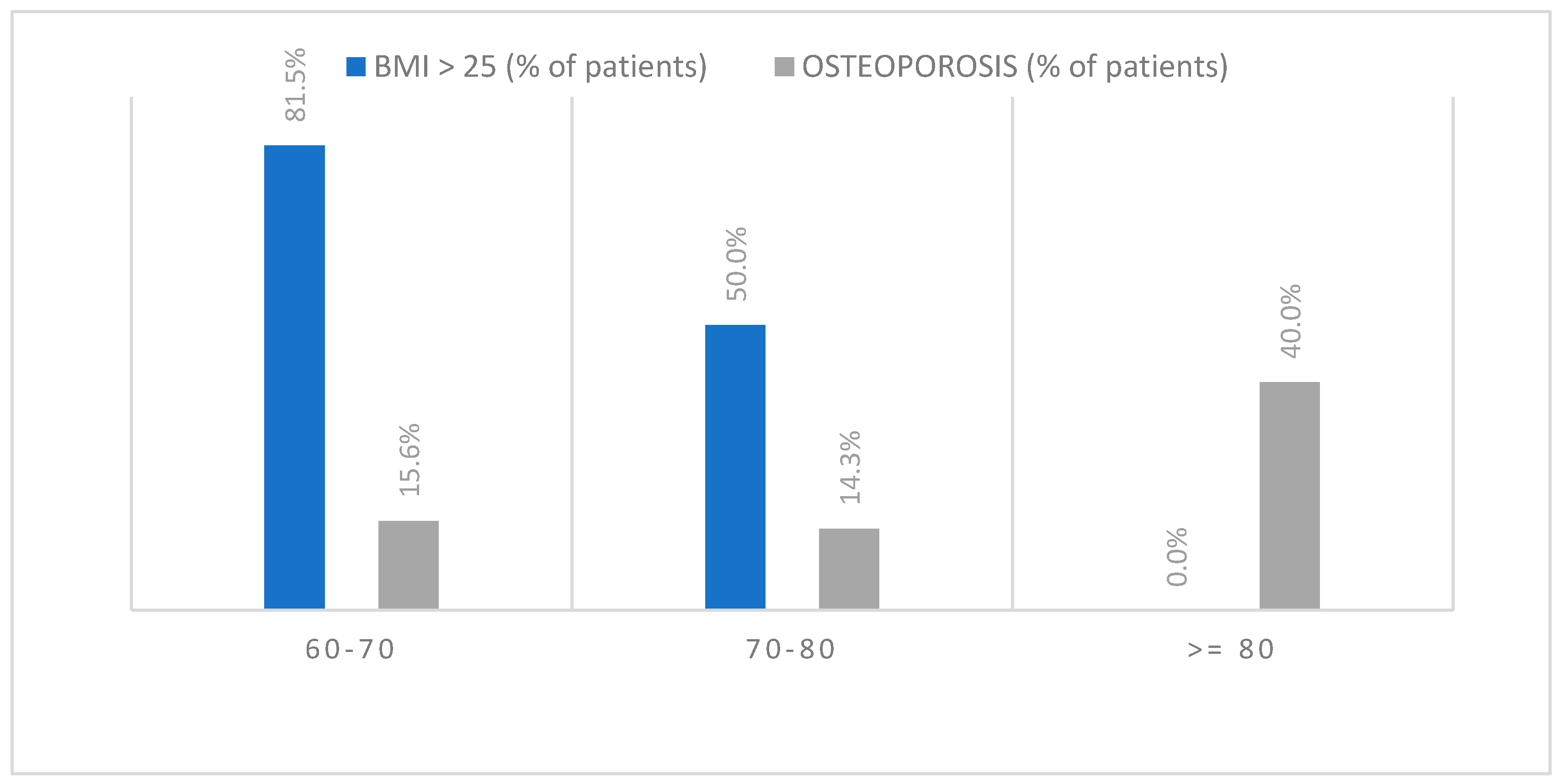
Lacking a control group composed of individuals without ankle fractures, necessary for comparison to identify the impact of risk factors associated with the examined injury, we analyzed the association of two presumed risk factors, osteoporosis, and excessive BMI. Dividing the patients into three age groups, this analysis revealed that, among younger subjects (60–70 years), 81.5% were overweight, and only 15.6% were osteoporotic. In the age group 70–80 years, the prevalence of overweight individuals decreased (50%), while the prevalence of osteoporosis remained stable (14.3%).
Among patients aged over 80 years, there were no overweight individuals observed, and the prevalence of osteoporosis increased to 40% (Figure 2).

3.5.2. BMI and Type of Fracture

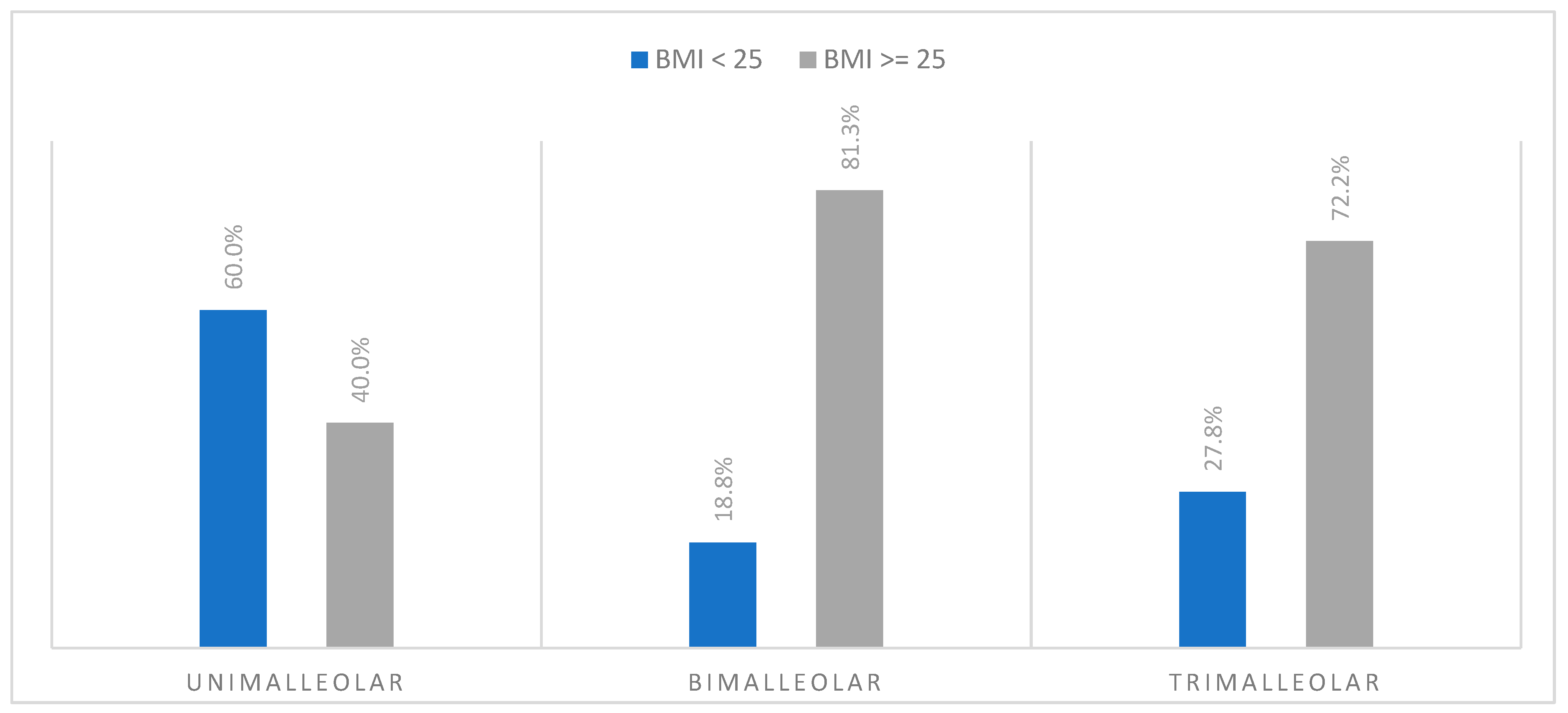
It appears that, in the bi- and trimalleolar fracture patients, 81.3% and 72.2%, respectively, the BMI was greater than 25, indicating overweight (Figure 3). A high BMI seems to correlate with anatomically more complex ankle fractures, but this difference did not result to be statistically significant (p > 0.05).

3.5.3. BMI and Type of Trauma

Among patients with low-energy trauma, 69.4% were found to be obese; among those with high-energy trauma, 62.5% of the patients were obese. Therefore, it does not seem that excessive BMI could influence the traumatic mechanism underlying ankle fractures (p > 0.05).

3.5.4. BMI and FADI

The patients were then divided into two groups based on BMI: the first with BMI ≤ 25 (normal-weight individuals) and the second with a BMI > 25 (overweight and obese individuals). The first group shows better clinical outcomes (mean FADI = 86) compared to the second group (mean FADI = 74.8). Overweight or obese individuals exhibited worse clinical outcomes than the normal-weight patient group, with a statistically significant difference (p < 0.05).

4. Discussion

Ankle fractures constitute the third most common musculoskeletal injury in the elderly, following hip and distal radius fractures [25]. Recent studies demonstrate a significant increase in the incidence and severity of ankle fractures with age [20,26]. Among the factors influencing the orthopedic management of ankle fractures in the elderly, a prominent role is played by bone fragility associated with an underlying condition of osteoporosis [27]. This is due to the acknowledged difficulty in achieving stable fixation of osteoporotic bone, necessitating surgical measures to ensure an optimal outcome.
Ankle fractures in the elderly represent a substantial healthcare burden due to the higher risk of morbidity and mortality [28,29,30]. For this reason, it is debated whether this fracture type, akin to the classic triad of proximal femur, vertebrae, and distal radius, can be considered osteoporotic fragility fractures. The hypothesis of this correlation arises primarily due to the fact that ankle fractures are a common low-energy fracture in the elderly and secondly from the observation that these injuries seem to increase, especially in perimenopausal women [31]. This hypothesis is supported also by the anatomical fact that the bony sites involved in these fractures (medial, lateral, and posterior malleoli) consist of a significant proportion of trabecular bone, the bone tissue most early, frequently, and severely affected by osteoporosis.
Several studies in the literature have sought to evaluate the relationship between these fractures and bone fragility, but their results are conflicting, highlighting a general difficulty in categorizing ankle fractures as osteoporosis-related: some authors support such a correlation reporting a lower BMD and alterations in bone quality in ankle fracture patients [32,33], while others argue that there is no significant link [34,35].
In Italy, Note 79 [36] defines the criteria for prescription and reimbursement by the NHS as limited to conditions of risk of osteoporotic fracture. Fractures eligible for reimbursement for osteoporosis therapy include hip and vertebral fractures, but not other fractures, such as wrist and ankle fractures. This is probably due to the fact that, while hip and vertebral fractures resulting from no or low trauma are easily includable among fragility fractures, for the others the matter is more complex, both because it is not easy to distinguish low- and high-energy traumas (for example, ankle fractures are almost always due to an ankle sprain), and because they are not exclusive fractures of the elderly. From the Swedish register [37], it emerges that hip-fractured patients have an average age of 81.1 years, and the percentage of patients over 65 years is 92.3%; those who undergo a vertebral fracture have an average age of 63, while those over 65 are 53%. Those who undergo an ankle fracture have a much lower average age (52.8), and only 30.5% are over 65. This leads the government system to be cautious on the reimbursement of ankle fractures due to the difficulty in discerning which of these fractures are truly fragility fractures and which are not. The aim of the present study is also to correlate these fractures, based on various parameters, to the actual risk of new fractures, identifying when an ankle fracture can be considered due to fragility.
As well as for purely health economic reasons, the prevention of osteoporotic fractures is essential to reduce the risks of complications and disability [38].
This correlation would also influence decisions regarding fracture management, particularly concerning surgical treatment aimed at ensuring stable fixation and an optimal functional outcome.

4.1. Ankle Fractures and Osteoporosis

The risk of future fragility fractures can be weighed based on BMD, measured with the DXA method, or with risk assessment tools, such as the FRAX® algorithm.
In the literature, the relationship between reduced BMD and ankle fractures is contrasting [39]. Patients in our study show that ankle fractures in the elderly are most frequent with reduced BMD (76.5% of the total patients examined, 83.8% considering only women). These results find confirmation, among others, in the comprehensive and extensive study conducted by Pekka Kannus between 1970 and 2014 in the Finnish population [40], which enhances this association in particular for postmenopausal women.
In our osteoporotic patients, we further observed that fracture complexity was heightened, with 70% of their fractures classified as trimalleolar with only 20% and 10% mono- and bimalleolar, respectively. These fractures thus appear to exhibit greater complexity as bone mass decreases, as suggested also by Lee et al. [41] in their study based on CT scan images of such fractures.
In our experience, a correlation also emerged between patients with lower values of BMD and those with low-energy traumas (82%). Similarly, Seely et al. observed that the majority of ankle fractures in women over 65 years of age are attributed to minor or low-energy trauma [42].
Regarding the calculation of the risk of future fragility fractures at 10 years calculated by the FRAX® algorithm, we found that the low-energy trauma group had a significantly higher average risk compared to the high-energy trauma group (10.8% vs. 6.2%). To support our data, Karlsson et al. found that the likelihood of experiencing additional fractures following an ankle fracture is increased when the fracture results from low-energy trauma rather than severe trauma [43]. These results seem to suggest that these fractures exhibit typical characteristics of fragility fractures, which are indeed associated with a higher risk of refracture [31,44].
To summarize, the results of our study suggest a correlation between ankle fractures in the elderly and osteoporosis. Specifically, suspicions of a connection with osteoporosis should arise, especially in older women with trimalleolar fractures resulting from low-energy traumas. This suspicion should increase further in the case of previous fragility fractures as confirmed by FRAX® results, based also on the investigation of previous osteoporotic fractures.
However, based on the results obtained, it is not possible to definitively categorize ankle fractures in the elderly as typical fragility fractures within the examined sample, and the debate is still open. Based on our results, we suggest that it might be advisable to initiate a comprehensive fracture risk assessment in elderly patients who experience an ankle fracture [45]. This could potentially lead to the implementation of both pharmacological and non-pharmacological secondary prevention measures to reduce the incidence of falls and mitigate their consequences [46,47]. From an orthopedic perspective, in the case of surgical treatment, strategies aimed at ensuring stable fixation should be considered [48], given the often more fragile nature of the bones involved [49].

4.2. FADI and Ankle Fracture

The mean FADI score reported by patients diagnosed with osteoporosis was 63.64, contrasting with the score of 81.18 reported by non-osteoporotic patients. This difference of 17.54 points was found to be statistically significant (p > 0.05). The group of patients with trimalleolar fractures showed a FADI mean score more than 10 points lower than the other two groups (bi- and mono-malleolar fractures), though this discrepancy did not reach statistical significance (p = 0.09). Furthermore, the difference in mean FADI scores between the two types of trauma amounted to 11.78 points, with higher scores for patients with high-energy traumas. At first glance, this difference may seem paradoxical, patients with fractures stemming from high-energy traumatic events demonstrated superior functional outcomes. This relationship can be explained by the higher prevalence of low-energy traumas among osteoporosis patients, who consequently displayed lower mean FADI scores compared to their non-osteoporotic counterparts.
From these results, it emerges how postoperative outcomes reflect the relationship between ankle fractures and osteoporosis. All patients in our study underwent a surgical operation, and it can be observed that, regardless of bone mineral density (BMD), the results were satisfactory. Consistent with findings in the literature [50,51,52], FADI scores were lower in patients with osteoporosis, those with fractures related to low-energy trauma, and those with more complex fractures. This suggests the possibility that these patient categories may benefit from a more aggressive postoperative protocol regarding early mobilization and physiotherapy, as well as potential pharmacological interventions.

4.3. BMI and Ankle Fractures

In our study, among younger subjects (60–70 years), 81.5% were overweight, and only 15.6% had osteoporosis. This result is in accordance with the fact that patients sustaining an ankle fracture are usually younger [17] and have a higher body mass index (BMI) [31] compared to other fragility fractures. In the age group of 70 to 80 years, the prevalence of overweight individuals decreased (50%), while the prevalence of osteoporosis remained stable (14.3%). Among patients aged over 80 years, there were no overweight individuals observed, and the prevalence of osteoporosis increased to 40%. These results suggest that excess body weight may have a greater impact as a risk factor in younger individuals compared to bone fragility; as age increases, the contribution of excess weight may decrease in the face of an increase in fragility-related factors.
In our patient cohort, there appears to be a correlation between a high BMI and anatomically more complex ankle fractures. This relationship may be attributed to the observation that overweight and obese individuals exhibit impaired protective responses during falls, predisposing them to sideways or backward falls [53]. It is demonstrated that, in the obese, the distribution of osteoporotic fractures is different from the typical distribution. Differently from the normal-weight osteoporotic people, whose most frequent fragility fractures are located at the hip or spine, in obese osteoporotic individuals other sites of fractures are prevalent, like ankle fractures [54,55,56]. Rinonapoli et al. [11] called this particular phenomenon “obese’s fracture site paradox”. Additionally, patients with higher BMIs generate greater forces upon impact during a fall, which correlates with an increased likelihood of sustaining more complex ankle fractures [57].
Regarding the relationship with the type of trauma underlying the fracture, no BMI-related differences were observed. This result likely reflects the fact that low-energy traumas are more common among patients aged over 80 years in which there were no overweight individuals observed.
In accordance with the literature, patients with high BMI exhibited significantly worse postoperative outcomes in terms of FADI scores. This result correlates with the fact that these patients often have poor lifestyle habits such as smoking, sedentary behavior, and alcohol consumption. Another aspect recently highlighted is that, especially in obese patients and to a lesser extent in overweight ones, the perception of postoperative pain is significantly greater, possibly due to alterations in cellular pain signaling [58].

4.4. Study Limitations

A limitation of our study is the recruitment of only ankle fractures who underwent surgery, but we are working to expand our work to include and compare those treated conservatively. Another limitation of our study is the small sample size, the imbalance in terms of patients’ sex distribution, and the lack of both negative (drawn from the general population without ankle fractures) and positive (individuals with previous typical osteoporotic fractures) control groups.

5. Conclusions

In conclusion, ankle fractures in the elderly exhibit characteristics that reasonably suggest a relationship with bone fragility. They show an increasing prevalence in females with advancing age, are associated with low-energy traumas (82%) and demonstrate a reduction in bone mineral density (83.8% considering women). Furthermore, these fractures present features of increased complexity as bone mass decreases. However, pending further expansion and completion of the work, they cannot be conclusively labeled as osteoporotic fractures but rather as fractures that can occur in osteoporotic bones.

Author Contributions

G.R., M.D., P.C. and A.C. contributed to the study conceptualization and design. Material preparation and data collection and analysis were performed by M.D. and P.C.; statistics and software were performed by G.R.; the first draft of the manuscript was written by M.D. and G.R.; writing—review and editing: M.D., P.C., G.R. and A.C. All authors have read and agreed to the published version of the manuscript.

Funding

This research received no external funding.

Institutional Review Board Statement

All subjects gave their informed consent for inclusion before they participated in the study. The study was conducted in accordance with the Declaration of Helsinki.

Informed Consent Statement

Informed consent was obtained from all subjects involved in the study(Approval date: 15 January 2022, Approval code: 4725/22.).

Data Availability Statement

The datasets used and analyzed during the current study are available from the corresponding author (Marco Donantoni) upon reasonable request. In order to ensure the confidentiality of data and to avoid data loss or manipulation, precautionary measures have been taken: the data are restricted to authorized members only. Authorized members are Donantoni, Ceccarini, Rinonapoli, and Caraffa. All information about the patients enrolled in the study is stored on a secure server, and database access is protected through a password that periodically changes. Only authorized members have the password. Paper material about the clinical evaluation is kept in a cabinet in the office of Ceccarini.

Conflicts of Interest

The authors declare no conflicts of interest.

References

  1. Wa, P. Consensus development conference: Diagnosis, prophylaxis, and treatment of osteoporosis. Am. J. Med. 1993, 94, 646–650. [Google Scholar] [CrossRef] [PubMed]
  2. Therdyothin, A.; Phiphopthatsanee, N.; Wajanavisit, W.; Woratanarat, P.; Laohajaroensombat, S.; Tawonsawatruk, T. Is ankle fracture related to low bone mineral density and subsequent fracture? A systematic review. Osteoporos. Sarcopenia 2020, 6, 151–159. [Google Scholar] [CrossRef] [PubMed]
  3. Bliuc, D.; Nguyen, N.D.; Milch, V.E.; Nguyen, T.V.; Eisman, J.A.; Center, J.R. Mortality risk associated with low-trauma osteoporotic fracture and subsequent fracture in men and women. JAMA 2009, 301, 513–521. [Google Scholar] [CrossRef] [PubMed]
  4. Rinonapoli, G.; Ruggiero, C.; Meccariello, L.; Bisaccia, M.; Ceccarini, P.; Caraffa, A. Osteoporosis in men: A review of an underestimated bone condition. Int. J. Mol. Sci. 2021, 22, 2105. [Google Scholar] [CrossRef] [PubMed]
  5. Kanis, J.A. Assessment of fracture risk and its application to screening for postmenopausal osteoporosis: Synopsis of a WHO report. Osteoporos. Int. 1994, 4, 368–381. [Google Scholar] [CrossRef] [PubMed]
  6. Tarantino, U.; Iolascon, G.; Cianferotti, L.; Masi, L.; Marcucci, G.; Giusti, F.; Marini, F.; Parri, S.; Feola, M.; Rao, C.; et al. Clinical Guidelines for the Prevention and Treatment of Osteoporosis: Summary Statements and Recommendations from the Italian Society for Orthopaedics and Traumatology; Springer International Publishing: Berlin/Heidelberg, Germany, 2017; Volume 18. [Google Scholar]
  7. Lu, Y.; Genant, H.K.; Shepherd, J.; Zhao, S.; Mathur, A.; Fuerst, T.P.; Cummings, S.R. Classification of osteoporosis based on bone mineral densities. J. bone Miner. Res. Off. J. Am. Soc. Bone Miner. Res. 2001, 16, 901–910. [Google Scholar] [CrossRef]
  8. Morris, J.; Karkenny, A.J.; Toro, J.B. The management of osteoporosis after fragility fracture: The orthopaedic perspective. JBJS Rev. 2017, 5, e4. [Google Scholar] [CrossRef] [PubMed]
  9. Kanis, J.A.; Oden, A.; Johnell, O.; Johansson, H.; De Laet, C.; Brown, J.; Burckhardt, P.; Cooper, C.; Christiansen, C.; Cummings, S.; et al. The use of clinical risk factors enhances the performance of BMD in the prediction of hip and osteoporotic fractures in men and women. Osteoporos. Int. 2007, 18, 1033–1046. [Google Scholar] [CrossRef] [PubMed]
  10. Kanis, J.A.; Hans, D.; Cooper, C.; Baim, S.; Bilezikian, J.P.; Binkley, N.; Cauley, J.A.; Compston, J.E.; Dawson-Hughes, B.; El-Hajj Fuleihan, G.; et al. Interpretation and use of FRAX in clinical practice. Osteoporos. Int. 2011, 22, 2395–2411. [Google Scholar] [CrossRef]
  11. Rinonapoli, G.; Pace, V.; Ruggiero, C.; Ceccarini, P.; Bisaccia, M.; Meccariello, L.; Caraffa, A. Obesity and Bone: A Complex Relationship. Int. J. Mol. Sci. 2021, 22, 13662. [Google Scholar] [CrossRef]
  12. Fassio, A.; Idolazzi, L.; Rossini, M.; Gatti, D.; Adami, G.; Giollo, A.; Viapiana, O. The obesity paradox and osteoporosis. Eat. Weight Disord. 2018, 23, 293–302. [Google Scholar] [CrossRef] [PubMed]
  13. Felsenberg, D.; Silman, A.J.; Lunt, M.; Armbrecht, G.; Ismail, A.A.; Finn, J.D.; Cockerill, W.C.; Banzer, D.; Benevolenskaya, L.I.; Bhalla, A.; et al. Incidence of vertebral fracture in europe: Results from the european prospective osteoporosis study (EPOS). J. Bone Miner. Res. 2002, 17, 716–724. [Google Scholar] [CrossRef] [PubMed]
  14. Cooper, C.; Campion, G.; Melton, L.J. Hip fractures in the elderly: A world-wide projection. Osteoporos. Int. 1992, 2, 285–289. [Google Scholar] [CrossRef] [PubMed]
  15. Waterloo, S.; Ahmed, L.A.; Center, J.R.; Eisman, J.A.; Morseth, B.; Nguyen, N.D.; Nguyen, T.; Sogaard, A.J.; Emaus, N. Prevalence of vertebral fractures in women and men in the population-based Tromsø study. BMC Musculoskelet. Disord. 2012, 13, 19–21. [Google Scholar] [CrossRef] [PubMed]
  16. Clynes, M.A.; Harvey, N.C.; Curtis, E.M.; Fuggle, N.R.; Elaine, M.; Cooper, C. The Epidemiology of Osteoporosis. Br. Med. Bull. 2020, 133, 105–117. [Google Scholar] [CrossRef] [PubMed]
  17. Hasselman, C.T.; Vogt, M.T.; Stone, K.L.; Cauley, J.A.; Conti, S.F. Foot and ankle fractures in elderly white women. Incidence and risk factors. J. Bone Jt. Surg. Am. 2003, 85, 820–824. [Google Scholar] [CrossRef] [PubMed]
  18. So, E.; Juels, C.; Scott, R.T.; Sietsema, D.L. A Comparison of Ankle Fractures Relative to Other Fragility Fractures: A Review and Analysis of the American Orthopaedic Association’s Own the Bone Database. Foot Ankle Int. 2023, 44, 879–887. [Google Scholar] [CrossRef] [PubMed]
  19. Ruggiero, C.; Zampi, E.; Rinonapoli, G.; Baroni, M.; Serra, R.; Zengarini, E.; Baglioni, G.; Duranti, G.; Ercolani, S.; Conti, F.; et al. Fracture prevention service to bridge the osteoporosis care gap. Clin. Interv. Aging 2015, 10, 1035–1042. [Google Scholar] [CrossRef] [PubMed]
  20. Kannus, P.; Palvanen, M.; Niemi, S.; Parkkari, J.; Jrvinen, M. Increasing number and incidence of low-trauma ankle fractures in elderly people: Finnish statistics during 1970-2000 and projections for the future. Bone 2002, 31, 430–433. [Google Scholar] [CrossRef]
  21. Kannus, P.; Parkkari, J.; Niemi, S.; Palvanen, M. Epidemiology of osteoporotic ankle fractures in elderly persons in Finland. Ann. Intern. Med. 1996, 125, 975–978. [Google Scholar] [CrossRef]
  22. Compston, J.E.; McClung, M.R.; Leslie, W.D. Osteoporosis seminar 2019. Lancet 2019, 393, 364–376. [Google Scholar] [CrossRef] [PubMed]
  23. Gregson, C.L.; Armstrong, D.J.; Bowden, J.; Cooper, C.; Edwards, J.; Gittoes, N.J.L.; Harvey, N.; Kanis, J.; Leyland, S.; Low, R.; et al. UK Clinical Guideline for the Prevention and Treatment of Osteoporosis. Arch. Osteoporos. 2022, 17, 58. [Google Scholar] [CrossRef] [PubMed]
  24. Gogate, N.; Satpute, K.; Hall, T. The effectiveness of mobilization with movement on pain, balance and function following acute and sub acute inversion ankle sprain—A randomized, placebo controlled trial. Phys. Ther. Sport 2021, 48, 91–100. [Google Scholar] [CrossRef] [PubMed]
  25. Kadakia, R.J.; Ahearn, B.M.; Schwartz, A.M.; Tenenbaum, S.; Bariteau, J.T. Ankle fractures in the elderly: Risks and management challenges. Orthop. Res. Rev. 2017, 9, 45–50. [Google Scholar] [CrossRef] [PubMed]
  26. Koval, K.J.; Lurie, J.; Zhou, W.; Sparks, M.B.; Cantu, R.V.; Sporer, S.M.; Weinstein, J. Ankle fractures in the elderly: What you get depends on where you live and who you see. J. Orthop. Trauma 2005, 19, 635–639. [Google Scholar] [CrossRef] [PubMed]
  27. Anderson, S.A.; Li, X.; Franklin, P.; Wixted, J.J. Ankle fractures in the elderly: Initial and long-term outcomes. Foot Ankle Int. 2008, 29, 1184–1188. [Google Scholar] [CrossRef] [PubMed]
  28. Toole, W.P.; Elliott, M.; Hankins, D.; Rosenbaum, C.; Harris, A.; Perkins, C. Are low-energy open ankle fractures in the elderly the new geriatric hip fracture? J. Foot Ankle Surg. 2015, 54, 203–206. [Google Scholar] [CrossRef] [PubMed]
  29. Kadakia, R.J.; Hsu, R.Y.; Hayda, R.; Lee, Y.; Bariteau, J.T. Evaluation of one-year mortality after geriatric ankle fractures in patients admitted to nursing homes. Injury 2015, 46, 2010–2015. [Google Scholar] [CrossRef] [PubMed]
  30. Hsu, R.Y.; Lee, Y.; Hayda, R.; DiGiovanni, C.W.; Mor, V.; Bariteau, J.T. Morbidity and mortality associated with geriatric ankle fractures. J. Bone Jt. Surg. Am. Vol. 2014, 97, 1748–1755. [Google Scholar] [CrossRef] [PubMed]
  31. Valtola, A.; Honkanen, R.; Kröger, H.; Tuppurainen, M.; Saarikoski, S.; Alhava, E. Lifestyle and other factors predict ankle fractures in perimenopausal women: A population-based prospective cohort study. Bone 2002, 30, 238–242. [Google Scholar] [CrossRef] [PubMed]
  32. Biver, E.; Durosier, C.; Chevalley, T.; Herrmann, F.R.; Ferrari, S.; Rizzoli, R. Prior ankle fractures in postmenopausal women are associated with low areal bone mineral density and bone microstructure alterations. Osteoporos. Int. 2015, 26, 2147–2155. [Google Scholar] [CrossRef] [PubMed]
  33. Honkanen, R.; Kröger, H.; Tuppurainen, M.; Alhava, E.; Saarikoski, S. Fractures and low axial bone density in perimenopausal women. J. Clin. Epidemiol. 1995, 48, 881–888. [Google Scholar] [CrossRef] [PubMed]
  34. Stein, E.M.; Liu, X.S.; Nickolas, T.L.; Cohen, A.; Thomas, V.; McMahon, D.J.; Zhang, C.; Cosman, F.; Nieves, J.; Greisberg, J.; et al. Abnormal microarchitecture and stiffness in postmenopausal women with ankle fractures. J. Clin. Endocrinol. Metab. 2011, 96, 2041–2048. [Google Scholar] [CrossRef] [PubMed]
  35. Lee, D.-O.; Kim, J.-H.; Yoo, B.-C.; Yoo, J.-H. Is osteoporosis a risk factor for ankle fracture?: Comparison of bone mineral density between ankle fracture and control groups. Osteoporos. Sarcopenia 2017, 3, 192–194. [Google Scholar] [CrossRef] [PubMed]
  36. AIFA. Nota 79. 2015; pp. 4–12. Available online: https://www.aifa.gov.it/documents/20142/1728074/nota-79.pdf (accessed on 29 March 2024).
  37. Bergh, C.; Wennergren, D.; Möller, M.; Brisby, H. Fracture incidence in adults in relation to age and gender: A study of 27,169 fractures in the Swedish Fracture Register in a well-defined catchment area. PLoS ONE 2020, 15, e0244291. [Google Scholar] [CrossRef] [PubMed]
  38. Ruggiero, C.; Baroni, M.; Talesa, G.R.; Cirimbilli, A.; Prenni, V.; Bubba, V.; Parretti, L.; Bogini, R.; Duranti, G.; Caraffa, A.; et al. The interdisciplinary fracture liaison service improves health-related outcomes and survival of older adults after hip fracture surgical repair. Arch. Osteoporos. 2022, 17, 135. [Google Scholar] [CrossRef] [PubMed]
  39. Kuru, P.; Akyüz, G.; Cerşit, H.P.; Çelenlioğlu, A.E.; Cumhur, A.; Biricik, Ş.; Kozan, S.; Gökşen, A.; Özdemir, M.; Lüleci, E. Fracture history in osteoporosis: Risk factors and its effect on quality of life. Balk. Med. J. 2014, 31, 295–301. [Google Scholar] [CrossRef] [PubMed]
  40. Kannus, P.; Niemi, S.; Parkkari, J.; Sievänen, H. Declining incidence of fall-induced ankle fractures in elderly adults: Finnish statistics between 1970 and 2014. Arch. Orthop. Trauma Surg. 2016, 136, 1243–1246. [Google Scholar] [CrossRef] [PubMed]
  41. Lee, K.M.; Chung, C.Y.; Kwon, S.S.; Won, S.H.; Lee, S.Y.; Chung, M.K.; Park, M.S. Ankle fractures have features of an osteoporotic fracture. Osteoporos. Int. 2013, 24, 2819–2825. [Google Scholar] [CrossRef] [PubMed]
  42. Seeley, D.G.; Browner, W.S.; Nevitt, M.C.; Genant, H.K.; Scott, J.C.; Cummings, S.R. Which fractures are associated with low appendicular bone mass in elderly women? The Study of Osteoporotic Fractures Research Group. Ann. Intern. Med. 1991, 115, 837–842. [Google Scholar] [CrossRef] [PubMed]
  43. Karlsson, M.K.; Hasserius, R.; Obrant, K.J. The ankle fracture as an index of future fracture risk. A 25–40 year follow-up of 1063 cases. Acta Orthop. Scand. 1993, 64, 482–484. [Google Scholar] [CrossRef] [PubMed]
  44. Taylor, A.J.; Gary, L.C.; Arora, T.; Becker, D.J.; Curtis, J.R.; Kilgore, M.L.; Morrisey, M.A.; Saag, K.G.; Matthews, R.; Yun, H.; et al. Clinical and demographic factors associated with fractures among older Americans. Osteoporos. Int. 2011, 22, 1263–1274. [Google Scholar] [CrossRef] [PubMed]
  45. Charde, S.H.; Joshi, A.; Raut, J. A Comprehensive Review on Postmenopausal Osteoporosis in Women. Cureus 2023, 15, e48582. [Google Scholar] [CrossRef] [PubMed]
  46. Aguado, P.; Garcés, M.V.; Casaús, M.L.G.; del Campo, M.T.; Richi, P.; Coya, J.; Torrijos, A.; Gijón, J.; Mola, E.M.; Martínez, M.E. Alta prevalencia de deficiencia de vitamina D en mujeres posmenopáusicas de una consulta reumatológica en Madrid. Evaluación de dos pautas de prescripción de vitamina D. Med. Clínica 2000, 114, 326–330. [Google Scholar] [CrossRef] [PubMed]
  47. Hodsman, A.B.; Hanley, D.A.; Josse, R. Do bisphosphonates reduce the risk of osteoporotic fractures? An evaluation of the evidence to date. CMAJ 2002, 166, 1426–1430. [Google Scholar] [PubMed]
  48. Ziegler, P.; Bahrs, C.; Konrads, C.; Hemmann, P.; Ahrend, M.D. Ankle fractures of the geriatric patient: A narrative review. EFORT Open Rev. 2023, 8, 1–10. [Google Scholar] [CrossRef] [PubMed]
  49. Olsen, J.R.; Hunter, J.; Baumhauer, J.F. Osteoporotic ankle fractures. Orthop. Clin. N. Am. 2013, 44, 225–241. [Google Scholar] [CrossRef] [PubMed]
  50. Warner, S.J.; Garner, M.R.; Fabricant, P.D.; Lorich, D.G. Bone density correlates with clinical outcomes after ankle fracture fixation. Arch. Orthop. Trauma Surg. 2018, 138, 1653–1657. [Google Scholar] [CrossRef] [PubMed]
  51. Strauss, E.J.; Egol, K.A. The management of ankle fractures in the elderly. Injury 2007, 38, 2–9. [Google Scholar] [CrossRef]
  52. Hoogervorst, P.; Bergen, C.; Van den Bekerom, M. Management of Osteoporotic and Neuropathic Ankle Fractures in the Elderly. Curr. Geriatr. Rep. 2017, 6, 9–14. [Google Scholar] [CrossRef]
  53. Ong, T.; Sahota, O.; Tan, W.; Marshall, L. A United Kingdom perspective on the relationship between body mass index (BMI) and bone health: A cross sectional analysis of data from the Nottingham Fracture Liaison Service. Bone 2014, 59, 207–210. [Google Scholar] [CrossRef] [PubMed]
  54. Compston, J.E.; Watts, N.B.; Chapurlat, R.; Cooper, C.; Boonen, S.; Greenspan, S.; Pfeilschifter, J.; Silverman, S.; Díez-Pérez, A.; Lindsay, R.; et al. Obesity is not protective against fracture in postmenopausal women: GLOW. Am. J. Med. 2011, 124, 1043–1050. [Google Scholar] [CrossRef] [PubMed]
  55. Prieto-Alhambra, D.; Premaor, M.O.; Fina Avilés, F.; Hermosilla, E.; Martinez-Laguna, D.; Carbonell-Abella, C.; Nogués, X.; Compston, J.E.; Díez-Pérez, A. The association between fracture and obesity is site-dependent: A population-based study in postmenopausal women. J. bone Miner. Res. Off. J. Am. Soc. Bone Miner. Res. 2012, 27, 294–300. [Google Scholar] [CrossRef] [PubMed]
  56. King, C.M.; Hamilton, G.A.; Cobb, M.; Carpenter, D.; Ford, L.A. Association between ankle fractures and obesity. J. Foot Ankle Surg. Off. Publ. Am. Coll. Foot Ankle Surg. 2012, 51, 543–547. [Google Scholar] [CrossRef] [PubMed]
  57. Lacombe, J.; Cairns, B.J.; Green, J.; Reeves, G.K.; Beral, V.; Armstrong, M.E.G. The Effects of Age, Adiposity, and Physical Activity on the Risk of Seven Site-Specific Fractures in Postmenopausal Women. J. Bone Miner. Res. 2016, 31, 1559–1568. [Google Scholar] [CrossRef] [PubMed]
  58. Cardoso, D.V.; Paccaud, J.; Dubois-Ferrière, V.; Barea, C.; Hannouche, D.; Veljkovic, A.; Lübbeke, A. The effect of BMI on long-term outcomes after operatively treated ankle fractures: A study with up to 16 years of follow-up. BMC Musculoskelet. Disord. 2022, 23, 317. [Google Scholar] [CrossRef] [PubMed]
Figure 1. Classification of patients based on BMD T-scores stratified by gender.
Applsci 14 03787 g001
Figure 2. Percentage of patients with BMI > 25 and osteoporosis by age ranges.
Applsci 14 03787 g002
Figure 3. Percentage of patients with BMI < and >25 by type of fracture.
Applsci 14 03787 g003
Table 1. Data distribution of the patients by sex and age.
Age RangesSexTot (N°)Tot (%)
60–65M + F1835.3%
M633.3%
F1266.7%
65–70M + F1427.5%
M535.7%
F964.3%
70–75M + F1019.6%
M220.0%
F880.0%
75–80M + F47.8%
M00%
F4100%
80–85M + F59.8%
M120.0%
F480.0%
Table 2. BMD correlation with type of fracture: the percentage of each type of fracture was calculated after dividing the patients into normal, osteopenic, and osteoporotic.
Total Fractures
Type of FractureN%
Unimalleolar1019.6
Bimalleolar2039.2
Trimalleolar 2141.2
Non-specificated00.0
Total51100.0
Normal BMD (T-Score > −1 DS)
Type of FractureN%
Unimalleolar216.7
Bimalleolar758.3
Trimalleolar 325.0
Non-specificated00.0
Total12100.0
Osteopenia (−2.5 DS < T-Score < −1 DS)
Type of FractureN%
Unimalleolar620.7
Bimalleolar1241.4
Trimalleolar 1137.9
Non-specificated00.0
Total29100.0
Osteoporosis (T-Score < −2.5 DS)
Type of FractureN%
Unimalleolar220.0
Bimalleolar110.0
Trimalleolar 770.0
Non-specificated00.0
Total10100.0
Table 3. Correlation of BMD and mechanism of trauma: from the data collected, it appears that the less trauma that causes the fracture, the lower the BMD.
NormalOsteopeniaOsteoporosis
Low-energy trauma866.7%2482.8%10100.0%
High-energy trauma433.3%517.2%00.0%
TOTAL12100.0%29100.0%10100.0%
Disclaimer/Publisher’s Note: The statements, opinions and data contained in all publications are solely those of the individual author(s) and contributor(s) and not of MDPI and/or the editor(s). MDPI and/or the editor(s) disclaim responsibility for any injury to people or property resulting from any ideas, methods, instructions or products referred to in the content.