Article

Lactic Acid Bacteria from Kiwi: Antifungal and Biofilm-Inhibitory Activities Against Candida albicans

1
Department of Dermatology, Graduate School, Kyung Hee University, Seoul 02447, Republic of Korea
2
Graduate School of Biotechnology, Kyung Hee University, Yongin 17104, Republic of Korea
3
Department of Biopharmaceutical Biotechnology, Graduate School, Kyung Hee University, Yongin 17104, Republic of Korea
*
Author to whom correspondence should be addressed.
These authors contributed equally to this work.
Appl. Sci. 2025, 15(3), 1647; https://doi.org/10.3390/app15031647
Submission received: 7 January 2025 / Revised: 29 January 2025 / Accepted: 4 February 2025 / Published: 6 February 2025

Abstract

:
Urogenital infections impact millions of individuals globally each year, with vulvovaginal candidiasis (VVC) being one of the most prevalent conditions affecting women. Candida albicans is the primary pathogen responsible for VVC. The utilization of probiotics as an alternative therapeutic approach to antibiotics in managing such infections has gained increasing attention. This study aimed to evaluate the potential of THY-F51, a lactic acid bacterium isolated from kiwi, as a probiotic to support vaginal health through its antifungal, anti-biofilm, and anti-inflammatory properties against C. albicans. The identification of THY-F51 was confirmed through 16S rRNA gene sequencing. A series of evaluations were performed to determine its antifungal efficacy against C. albicans, biofilm-inhibitory activity, antioxidant properties, and effects on inflammatory cytokines. Cytotoxicity assays and assessments of bacterial survival under vaginal pH conditions (pH 3.8–4.5) were also conducted. The results demonstrated that THY-F51, identified as Leuconostoc citreum, exhibited potent antifungal activity against C. albicans, with an MIC of 1.25 mg/mL and an MFC of 2.5 mg/mL. Furthermore, THY-F51 displayed a strong inhibition of C. albicans biofilm formation, as well as notable antioxidant activity in the supernatant. Additionally, THY-F51 demonstrated high survival rates under vaginal pH conditions, an absence of cytotoxic effects, and a significant reduction in C. albicans adhesion to HeLa cells. Moreover, THY-F51 effectively suppressed C. albicans-induced inflammatory cytokines, including TNF-α, IL-1β, IL-6, and IL-8. These findings suggest that THY-F51, isolated from kiwi, holds substantial promise as a safe and effective probiotic for reducing vaginal inflammation and promoting vaginal health.

1. Introduction

Vulvovaginal candidiasis (VVC) is a common fungal infection affecting the vulva and vagina, primarily caused by Candida species [1]. It affects a significant proportion of women during their lifetime, with an estimated 75% of women experiencing at least one episode of VVC [2]. C. albicans, a yeast that is part of the normal vaginal microbiota, can overgrow and cause infection when the vaginal environment is disrupted, leading to an imbalance in microbial populations [3]. VVC interferes with a woman’s daily life by causing severe itching, burning, pain during urination, and dyspareunia, which can decrease productivity and lead to psychological distress, frustration, and depression [4]. During pregnancy, it increases the risk of preterm birth and low birth weight, while in immunocompromised women, it can raise the risk of systemic infections [5].
C. albicans adheres to vaginal epithelial cells using adhesion proteins and transitions from a yeast to a hyphal form, enhancing its tissue penetration ability [6]. It secretes hydrolytic enzymes such as aspartyl proteases (SAPs) and phospholipases that break down the epithelial barrier, inhibit complement activation, and impair macrophage and neutrophil activity, facilitating immune evasion [7]. Moreover, Candida forms biofilms to escape antifungal treatment and immune defenses, and hyphal invasion activates inflammasomes, leading to the excessive release of pro-inflammatory cytokines such as IL-1β, thereby exacerbating inflammation and tissue damage [8]. Elevated estrogen levels promote Candida adhesion and hyphal growth, while the reduction in lactobacilli due to antibiotic use and immune suppression further facilitates infection [9].
To treat C. albicans infections, antifungal agents are commonly employed, each with distinct mechanisms: azoles inhibit ergosterol production, a crucial component of the fungal cell membrane [10]; polyenes bind to ergosterol, forming pores in the membrane [11]; echinocandins prevent the synthesis of essential cell wall components [12]; and flucytosine disrupts DNA and RNA synthesis [13]. Although these treatments typically take several days to weeks to show effectiveness, they can cause side effects such as kidney or liver damage, and prolonged or excessive use may contribute to the development of resistance [14]. In response to these concerns, there is growing interest in exploring alternatives with fewer side effects and enhanced safety profiles [15].
In recent years, there has been growing interest in using probiotics as a complementary or alternative approach for treating or preventing VVC, with a focus on LAB due to their potential to restore the natural vaginal microbiota and inhibit Candida overgrowth [16]. For instance, a study by Mastromarino et al. demonstrated that the administration of the Lactobacillus rhamnosus GR1 and Lactobacillus reuteri RC-14 strains significantly reduced the colonization of C. albicans in the vaginal tract of women with recurrent VVC [17]. The authors showed that these probiotics produced lactic acid and hydrogen peroxide, which created an acidic environment hostile to Candida growth. Moreover, Lactobacillus strains were observed to adhere to vaginal epithelial cells, forming a protective biofilm that limited Candida’s ability to colonize and infect the vaginal mucosa [18]. These findings underscore the role of probiotics in preventing VVC recurrence by balancing the vaginal microbiota and competing with Candida for essential nutrients and binding sites [19]. Additionally, a study by Kovachev et al. also confirmed that Lactobacillus supplementation led to a significant reduction in the recurrence of VVC by restoring the vaginal microbiome and limiting Candida overgrowth, further supporting the potential of probiotics as a therapeutic strategy for VVC [20].
Therefore, our study aims to demonstrate the potential of lactic acid bacteria isolated from kiwi as promising candidates for the prevention and treatment of vulvovaginal candidiasis (VVC) by evaluating their antifungal activity against C. albicans, biofilm inhibition, and the suppression of inflammatory cytokine production.

2. Materials and Methods

2.1. Isolation and Identification of Bacteria

To isolate lactic acid bacteria, fresh kiwi fruits were chopped into small pieces and inoculated into MRS broth, followed by incubation at 30 °C for 48 h. The resulting culture was serially diluted in 0.85% NaCl solution to obtain concentrations ranging from 10−6 to 10−9. The diluted samples were plated onto Bromocresol Purple (BCP) agar (Eiken Chemical Co., Ltd., Tokyo, Japan) and incubated at 37 °C for 24 h. Colonies that turned yellow were selected and subcultured onto Man, Rogosa, and Sharpe (MRS) agar (Difco, Detroit, MI, USA) for standard cultivation at 37 °C. To confirm strain identity, the 16S rRNA gene was amplified using primers 27F and 1492R. The obtained sequences were analyzed using the EzBioCloud database (https://www.ezbiocloud.net/identify) (accessed on 23 November 2024) for further identification.

2.2. Bacterial Cultivation Conditions and Preparation of Metabolites

To evaluate antifungal activity, the strains Candida albicans KCTC 7270, Candida albicans ATCC 10231, and Candida tropicalis KCTC 17762 were obtained from the Korean Collection for Type Cultures (KCTC) and the American Type Culture Collection (ATCC). These strains were cultured in Yeast Malt (YM) broth under aerobic conditions at 37 °C prior to use in experiments. The selected lactic acid bacteria (LAB) were cultured in MRS broth for 24 h, and the cell-free supernatant (CFS) was obtained by centrifugation and filtration through a 0.22 μm membrane filter for experimental use. Additionally, live bacterial cells were harvested by centrifugation to collect the pellet, which was washed with phosphate-buffered saline (PBS) and subsequently heat-killed at 121 °C using an autoclave before inclusion in experiments.

2.3. Broth Microdilution Method

The minimum inhibitory concentration (MIC) and minimum fungicidal concentration (MFC) of the selected LAB were determined using the broth microdilution method, following the guidelines recommended by the Clinical and Laboratory Standards Institute (CLSI) [21]. The CFS of the selected LAB was serially diluted by half, and 100 μL of each dilution was added to the wells of a 96-well microtiter plate (Thermo Fisher Scientific, Waltham, MA, USA). Subsequently, 100 μL of YM broth containing 1 × 10⁵ CFU/mL of C. albicans pathogens was added to each well, and the plates were incubated at 37 °C for 24 h. Optical density (OD) values were measured to assess microbial growth, with MRS broth serving as the control. Following this, 1 μL from each well was streaked onto YM agar plates using an inoculation loop, and the plates were incubated at 37 °C for 24 h to enumerate the colony-forming units (CFUs). The lowest concentration of LAB that completely inhibited colony formation on YM agar plates was identified as the MFC.

2.4. Scanning Electron Microscope (SEM)

In this study, a dilution of 108 CFU/mL of C. albicans KCTC 7270, C. albicans ATCC 10231, and C. tropicalis KCTC 17762 was exposed to the MIC of the LAB CFS at 37 °C for 24 h. After incubation, the fungal cells were fixed in PBS containing 2.5% glutaraldehyde at 4 °C for 2 h. Subsequently, the samples were post-fixed in 1% osmium tetroxide solution (w/v) at the same temperature for 1 h. The fixed samples were dehydrated through a graded ethanol series (30%, 50%, 70%, 80%, 90%, and 100%) and then dried using 100 μL of hexamethyldisilazane (Sigma-Aldrich, St. Louis, MO, USA) for 24 h. Finally, the dried cells were mounted on carbon tape adhered to stubs and visualized using a scanning electron microscope (SU8010, Hitachi, Tokyo, Japan).

2.5. Biofilm Formation Crystal Violet Assay

To evaluate the effects on biofilm formation, 100 μL of candida pathogenic strain dilution (1 × 10⁵ CFU/mL) was added to a 96-well microtiter plate containing LAB supernatants diluted by half. The plate was incubated at 37 °C for 24 h. Following incubation, the biofilms were stained with 0.01% crystal violet solution (0.1% acetic acid) and then fixed by releasing the bound crystal violet with 33% acetic acid. Absorbance was measured at 595 nm using a microplate reader (Molecular Devices, San Francisco, CA, USA) to assess biofilm formation [22]. MRS broth was used as the control.

2.6. Antioxidant Activity

The antioxidant activity of the selected LAB was determined using the 1,1-diphenyl-2-picrylhydrazyl (DPPH) and 2,2′-azino-bis-(3-ethylbenzothiazoline)-6-sulfonic acid (ABTS) assays. After the reaction of the CFS with the DPPH and ABTS solutions, absorbance was measured at 595 nm and 405 nm, respectively, using a microplate reader. Superoxide dismutase (SOD)-like activity was assessed using the method described by Marklund and Marklund [23]. Similarly, the SOD-like activity of the CFS was measured against pyrogallol (Sigma-Aldrich, Co., St. Louis, MO, USA). Each well of a 96-well microplate was filled with 10 μL of CFS, 130 μL of Tris–HCl buffer, and 10 μL of 7.2 mM pyrogallol (prepared immediately before use). Absorbance was measured at 420 nm using a microplate reader. Radical scavenging activity and SOD-like activity were calculated using the following formula:
% of radical inhibition = (OD0 − ODx) OD0 × 100
where OD0 is the absorbance of the negative control; ODx is absorbance of various tested CFS and ascorbic acid concentrations.
In addition to the above-mentioned assays, the Oxygen Radical Absorbance Capacity (ORAC) assay was performed using the OxiSelect ORAC Activity Assay Kit (Cell Biolabs, San Diego, CA, USA) following the manufacturer’s instructions, with Trolox as the control standard. The fluorescence readings were taken over 11 cycles, and ORAC values were calculated from the net fluorescence area under the curve, expressed as micromoles of Trolox equivalents (TE) per gram of dry weight (μ·mol TE/g DW). Furthermore, the total antioxidant capacity (T-AOC) assay was conducted using the T-AOC Colorimetric Assay Kit (Elabscience, Wuhan, China). Absorbance at 520 nm was measured after incubating the sample with chromogenic and ferric salt solutions. The total antioxidant capacity was calculated based on the absorbance difference between the sample and control wells, expressed as units per milliliter (U/mL).

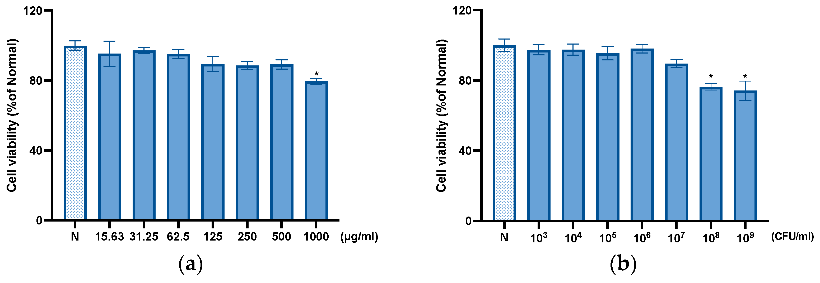
2.7. Cell Culture and Viability

HeLa cells derived from human keratinocytes were obtained from the Korean Cell Line Bank (Seoul, Republic of Korea). The cells were cultured in Dulbecco’s Modified Eagle Medium (DMEM; Hyclone Laboratories Inc., Logan, UT, USA) supplemented with 10% heat-inactivated fetal bovine serum (FBS) and 1% penicillin/streptomycin solution (Gibco BRL, Grand Island, NY, USA) in an incubator at 37 °C with 5% CO2. The effect of the sample on HeLa cell viability was evaluated using an MTT assay [24]. After 24 h of exposure, the supernatant in each well was completely removed, and 100 µL of MTT solution (0.1 mg/mL) was added. Following 10 h of incubation, the supernatant was aspirated, and 500 µL of dimethyl sulfoxide (DMSO) was added to each well [24]. Absorbance was measured at 595 nm using a microplate reader.

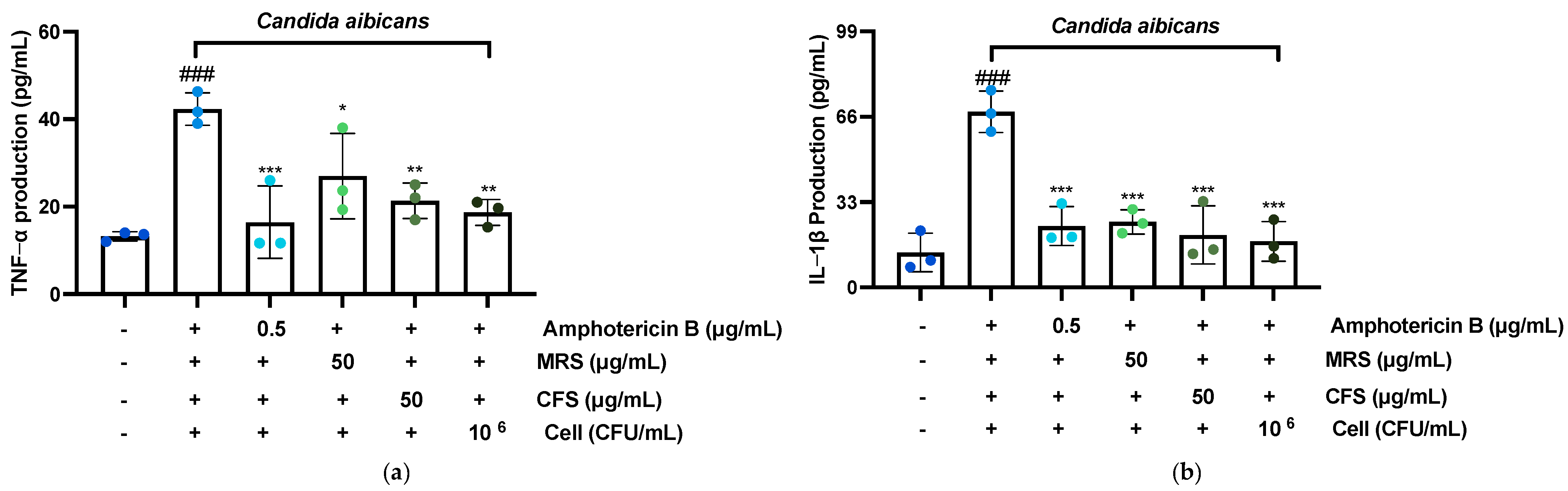
2.8. Enzyme-Linked Immunosorbent Assay
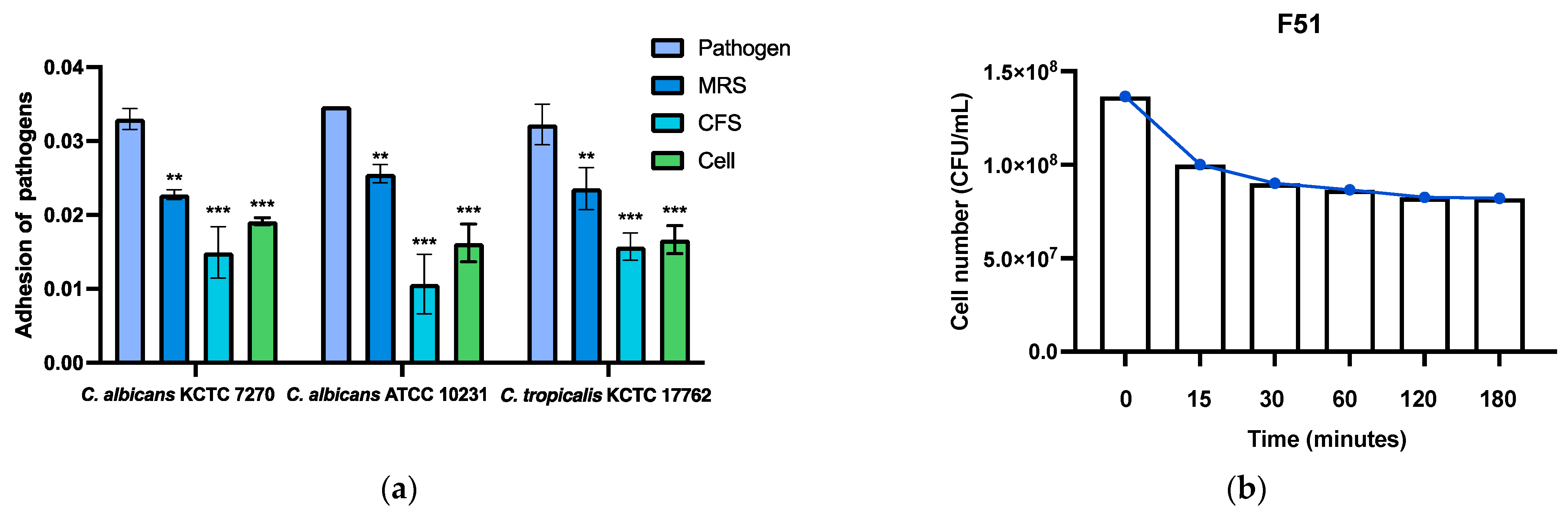
We performed a quantitative assessment of TNF-α, IL-1β, IL-6, and IL-8 levels in the supernatant of the photoaging model using ELISA kits from a commercial supplier. The experimental protocol was followed meticulously, adhering to the manufacturer’s instructions for the reagents.

2.9. Adhesion Assay

The adhesion assay was performed by seeding HeLa cells in 96-well plates at a density of 1 × 10⁵ cells per well. After the cells reached 70–80% confluence, they were adapted with either lactic acid bacteria supernatants or live cells for 2 h. Following the adaptation, C. albicans KCTC 7270 was added, and the cells were incubated for an additional 2 h. Non-adherent cells were removed by washing with PBS, and the remaining cells were stained with crystal violet and fixed with 33% acetic acid. Adhesion was quantified by measuring absorbance at 595 nm [25].

2.10. Acid Tolerance

The selected LAB were adjusted to pH 4.0 using HCl based on the vaginal pH and incubated at 37 °C for a specified duration [26]. After the incubation period, 1 mL of the bacterial suspension was collected at predetermined time points and serially diluted using 85% NaCl solution. The diluted samples were then spread onto MRS agar plates, which were incubated at 37 °C for 24 h. Acid resistance was determined by comparing the final plate count at 3 h to the initial plate count at 0 h. The experiment was performed in duplicate to ensure reproducibility.

2.11. Statistical Analysis

Statistical analysis was conducted using GraphPad Prism 9.0 software (GraphPad Software Inc., La Jolla, CA, USA), employing both one-way ANOVA and two-way ANOVA approaches. Data obtained from three independent experiments are presented as the mean ± standard deviation. Statistical significance was determined using p-values of <0.05, <0.01, and <0.001 as thresholds.

3. Results

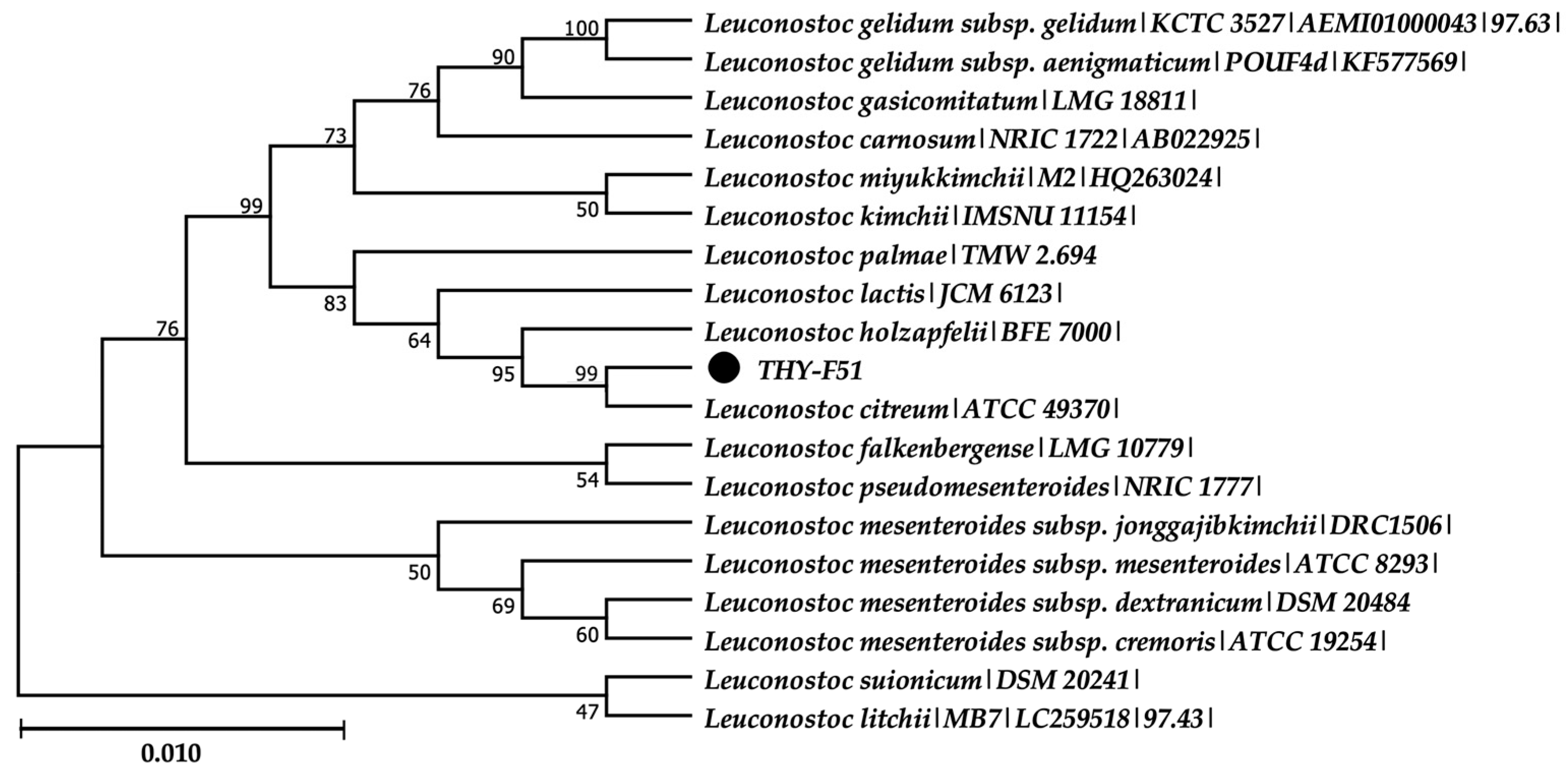
3.1. Isolation and Identification of Leuconostoc citreum THY-F51

The phylogenetic analysis of the 16S rRNA gene of the THY-F51 strain revealed that its closest relative was Leuconostoc citreum (ATCC 49370T), with a similarity of 99.65%. This relationship was further confirmed by the phylogenetic tree shown in Figure 1.

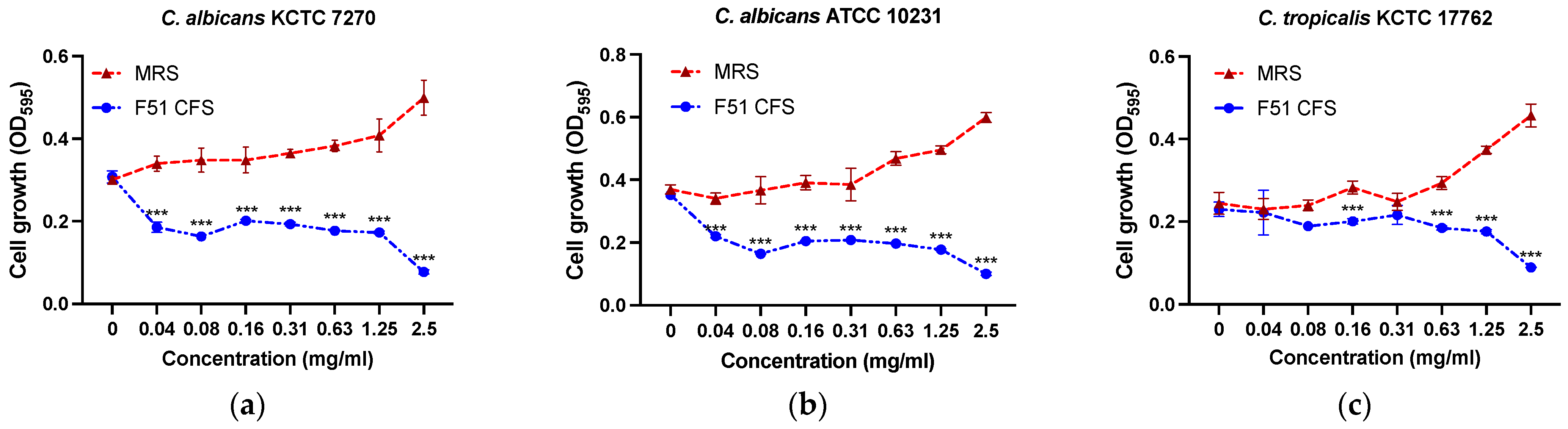
3.2. Antibacterial Activity

The THY-F51 strain exhibited significant antimicrobial activity against Candida after the MIC and MFC values were screened. The results indicated that the THY-F51 CFS demonstrated MIC and MFC values of 1.25 mg/mL and 2.5 mg/mL, respectively, against C. albicans KCTC 7270, C. albicans ATCC 10231, and C. tropicalis KCTC 17762. These results are shown in Figure 2 and Table 1 below.

3.3. SEM Result of Pathogens with THY-F51 Treatment

As shown in Figure 3, the changes in the cell surface induced by the MIC (1.25 mg/mL) of THY-F51 were investigated using scanning electron microscopy. In the untreated condition, C. albicans yeast exhibited a uniform spherical shape with an intact cell wall. However, after exposure to the THY-F51 CFS for 24 h, the C. albicans cells exhibited significant damage, including the distortion of the cell membrane, leading to a marked disruption in the normal yeast morphology. These changes suggest that THY-F51 induces damage to the cell wall and membrane, compromising the structural integrity of C. albicans.

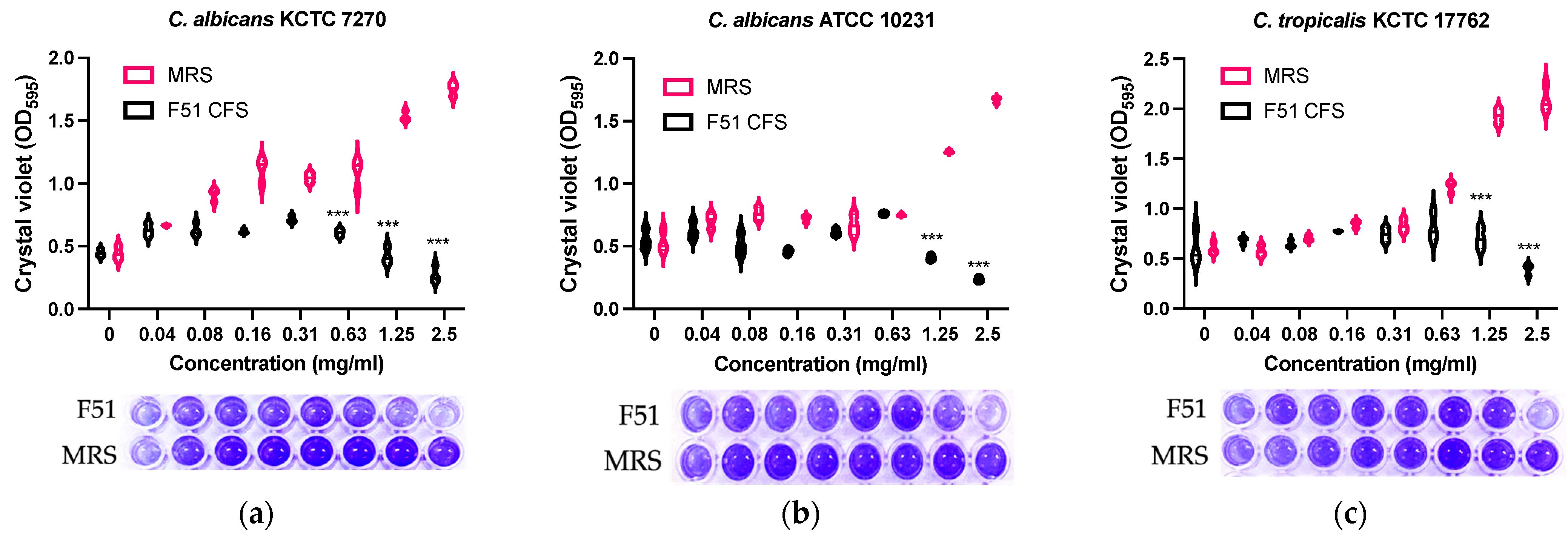
3.4. Inhibition of Biofilm Formation by E. faecalis HM20

As shown in Figure 4, the THY-F51 CFS exhibited an inhibitory effect on the biofilm formation of Candida pathogens, including C. albicans KCTC 7270, C. albicans ATCC 10231, and C. tropicalis KCTC 17762. Compared to the control group (MRS), the biofilm formation of Candida increased in a dose-dependent manner, while the THY-F51 treatment group showed a significant inhibitory effect. Moreover, at a concentration of 2.5 mg/mL, biofilm formation was inhibited for all three pathogens.

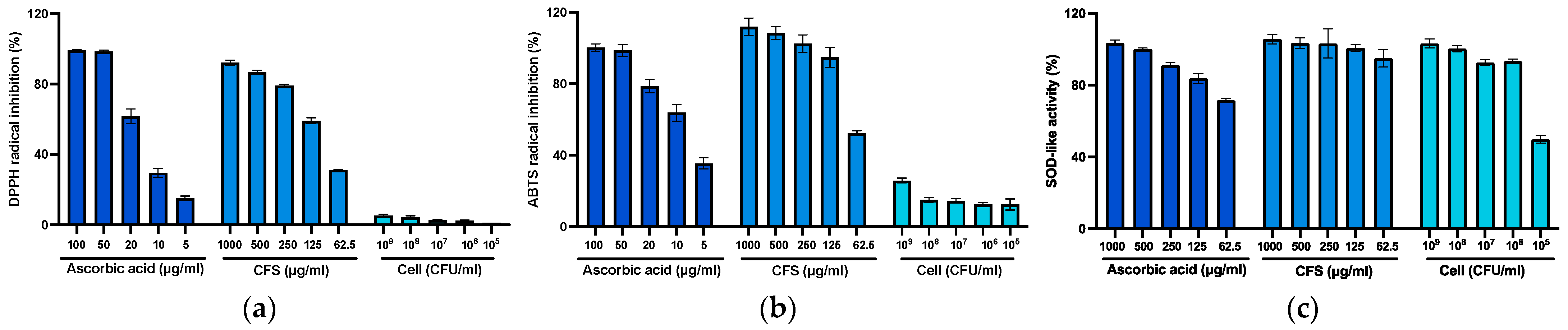
3.5. Effect of Radical Scavenging Activity and Superoxide Dismutase-Like Assay

The DPPH and ABTS assays are commonly used to evaluate the radical scavenging ability of probiotics, measuring their ability to neutralize stable free radicals (DPPH•) and radical cations (ABTS•+), respectively. These assays provide a rapid and sensitive assessment of antioxidant activity against both hydrophilic and lipophilic substances [27,28]. ORAC analysis complements these methods by quantifying the ability of probiotics to neutralize oxygen free radicals, offering biologically relevant insights into ROS protection [29]. Additionally, SOD activity analysis evaluates the enzymatic antioxidant potential of probiotics, determining their role in mitigating oxidative stress by enhancing endogenous superoxide dismutase activity [30]. Finally, T-AOC analysis measures the total antioxidant capacity of probiotics, reflecting their combined ability to neutralize various reactive species and provide comprehensive protection against oxidative stress [31]. As shown in Figure 5, the THY-F51 CFS demonstrated a significant dose-dependent reduction in DPPH and ABTS free radical formation, while live cells did not show a significant decrease in radical formation. However, both the CFS and live cells exhibited antioxidant effects in the SOD-like activity assay. And the ORAC values for the cell-free supernatant and live cells were 120.45 μmol TE/g and 63.65 μmol TE/g, respectively, while the T-AOC values were 5.27 (U/mL) for the cell-free supernatant and 2.34 (U/mL) for the live cells (Table 2). Studies have shown a notable distinction in antioxidant capacity between the cell-free supernatant and live cells of probiotics, with the supernatant exhibiting higher ORAC and T-AOC values compared to live cells. These findings indicate that the antioxidant compounds present in the cell-free supernatant are likely pivotal in the scavenging of ROS.

3.6. Cytotoxic Effect of THY-F51

No significant toxicity was observed in the toxicity tests on HeLa cells when treated with different concentrations of the THY-F51 CFS and cells, as shown in Figure 6. Among the CFS, all concentrations were safe except for the highest concentration (1000 μg/mL), and in the case of live cells, no significant toxicity was observed except for at concentrations of 109 and 108 CFU/mL. Based on these results, the cell experiments described below were conducted with the supernatant at 50 μg/mL and live cells at 106 CFU/mL.

3.7. Effect of F51 on TNF-α, IL-1β, IL-6, and IL-8 Production

TNF-α, IL-1β, IL-6, and IL-8 are cytokines that play crucial roles in the inflammatory response. During Candida vaginitis, the concentrations of these cytokines increase, with TNF-α and IL-1β promoting inflammation, while IL-6 and IL-8 assist in leukocyte recruitment and cell migration to the site of inflammation [31]. Based on these phenomena, as shown in Figure 7, THY-F51 exhibited inhibition effects almost comparable to the control group, Amphotericin B. Interestingly, the inhibitory effects of THY-F51 live cells were stronger than those of the CFS. One reason for this is that live cells can directly impact Candida and play an important role in suppressing the inflammatory response. Live cells release various bioactive substances through their cell walls, and these substances can exert direct inhibitory effects on Candida interactions. Additionally, live cells may produce molecules that regulate immune responses, potentially modulating the expression of inflammatory cytokines such as TNF-α, IL-1β, IL-6, and IL-8 or alleviating the inflammatory response.

3.8. Anti-Adhesion Ability and Acid Tolerance Assays of THY-F51

As shown in Figure 8a, an inhibitory effect on Candida albicans attached to HeLa cells was observed, and unlike in the inflammation experiment, the THY-F51 CFS showed better inhibition than the live cells. The inhibition rates were 54.69%, 69.30%, and 51.31%, demonstrating more than 50% inhibition. Additionally, Figure 8b shows the effect of THY-F51 on survival under vaginal pH conditions. Although the survival rate decreased over time compared to the initial rate at 0 min, THY-F51 remained viable even after 180 min. This suggests that it can survive in the vaginal environment with a pH range of 4.0.

4. Discussion

Our study demonstrated that Leuconostoc citreum THY-F51, a lactic acid bacterium isolated from kiwi fruit, exhibited significant antifungal activity against Candida species, the major causative agents of VVC. Additionally, it was shown to effectively suppress Candida-induced inflammatory cytokine production and inhibit biofilm formation, which is a key factor in Candida pathogenicity. These findings suggest that L. citreum THY-F51 may serve as a next-generation, safe, and promising alternative for patients facing antibiotic resistance, highlighting its potential as a preventive and therapeutic agent for managing VVC, a prevalent infection of the female reproductive tract.
Noteworthy findings include the antifungal activity of THY-F51’s CFS against C. albicans, with MIC and MFC values of 1.25 mg/mL (250 µL/mL) and 2.5 mg/mL (500 µL/mL), respectively (Figure 2, Table 1). These values are comparable to those from antifungal studies using a Lactobacillus plantarum CFS, which reported MIC and MFC values of 200 µL/mL [32]. While L. plantarum is a well-established probiotic strain with confirmed antifungal properties, the antifungal activity of THY-F51 has not been previously studied, making these findings a novel contribution to the field. Furthermore, SEM analysis (Figure 3) revealed significant morphological changes in the C. albicans cell wall, such as deformation and shrinkage, suggesting that the antifungal activity of THY-F51 may be due to metabolites like bacteriocins, organic acids, and pH-altering compounds. These metabolites likely disrupted lipid–protein interactions in the fungal membrane, leading to destabilization and deformation [33]. While various antifungal compounds were evaluated, the exact active agents remain unidentified, highlighting the need for further mechanistic studies and genomic profiling to fully characterize their antifungal potential.
The current study demonstrated that the CFS of THY-F51 significantly inhibits fungal biofilm formation in C. albicans KCTC 7270, C. albicans ATCC 10231, and C. tropicalis KCTC 17762, with biofilm formation significantly reduced at 1× MIC (1.25 mg/mL) and nearly completely abolished at 2× MIC (2 mg/mL), while the control group treated with an MRS medium exhibited enhanced biofilm formation at higher concentrations (Figure 4), indicating the potent anti-biofilm activity of L. citreum THY-F51 and its potential as a therapeutic candidate for biofilm-associated fungal infections, in line with a previous study by Morici et al. (2016) [34].
VVC is a condition where inhibiting not only the Candida species but also inflammation plays a crucial therapeutic role [35]. C. albicans adheres to the vaginal epithelium, and the hyphal form invades tissues, causing damage to epithelial cells and triggering immune responses [36]. The host immune system recognizes Candida via pattern recognition receptors (PRRs) such as Toll-like receptors (TLRs) and Dectin-1, which are expressed on the surface of immune cells like dendritic cells and macrophages [37]. Upon recognition of Candida, immune cells initiate the production of inflammatory cytokines such as IL-1β, IL-6, IL-8, and TNF-α, as well as chemotactic factors [38]. During biofilm formation, Candida produces a toxin called candidalysin, which disrupts cell membranes and causes direct damage to host tissues [39]. This leads to the release of damage-associated molecular patterns (DAMPs), which further stimulate the immune response and amplify the inflammatory process, contributing to the release of inflammatory mediators, including TNF-α, IL-1β, IL-6, IL-8, prostaglandins, histamine, chemokines, MMPs, nitric oxide, and leukotrienes [40].
In this study, we evaluated the inhibitory effects of the THY-F51 CFS and live cells on the production of four key inflammatory mediators (TNF-α, IL-1β, IL-6, and IL-8) induced by C. albicans in HeLa cells. As shown in Figure 7, the inhibition rates for TNF-α were found to be 49.60% for the CFS and 55.90% for live cells, while IL-1β inhibition rates were 70.06% for the CFS and 73.74% for live cells, both surpassing the positive control, Amphotericin B, which showed a 65.03% inhibition rate. For IL-6, the inhibition rates were 14.98% for the CFS and 21.62% for live cells, with live cells demonstrating a significantly stronger inhibitory effect. In the case of IL-8, the inhibition rates were 48.73% for the CFS and 66.37% for live cells, with live cells showing a higher inhibitory effect than Amphotericin B, which exhibited a 53.38% inhibition rate. These findings suggest that THY-F51 live cells exert more potent anti-inflammatory effects compared to the CFS. This difference may be due to two main mechanisms: first, live cells may interact directly with immune cells by binding to pattern recognition receptors (PRRs), such as Toll-like receptors (TLRs), on host cells, which induces the suppression of inflammatory mediators and activates intracellular signaling pathways to inhibit inflammation [41]. Second, live bacteria can modulate immune responses through the secretion of metabolites (e.g., lactic acid, acetic acid) or cell wall components (e.g., peptidoglycans, teichoic acids); although the CFS contains metabolites secreted by live cells, live cells are more effective due to their involvement in intercellular interactions and their continuous production of immune-modulating metabolites [42].
Antioxidants play a critical role in alleviating Candida-induced vaginitis by neutralizing excessive ROS, which contribute to oxidative stress, promote Candida overgrowth, and compromise the integrity of the vaginal mucosa. These compounds suppress inflammatory pathways, such as NF-κB, thereby reducing the production of pro-inflammatory cytokines and promoting the growth of lactic acid bacteria, which maintain the acidic vaginal pH required to inhibit Candida proliferation [43]. Moreover, antioxidants enhance immune responses by modulating T-cell and macrophage activity and activating the Nrf2-Keap1 signaling pathway, which upregulates antioxidant-related gene expression and supports tissue regeneration. Collectively, these mechanisms mitigate infection, reduce inflammation, and promote mucosal healing [44].
Through these mechanisms, we also validated the antioxidant efficacy of THY-F51 in our study. As detailed in Table 2, the CFS of THY-F51 exhibited superior antioxidant properties compared to live cells. This is likely due to the presence of metabolic byproducts and secondary metabolites in the CFS, such as peptides, organic acids, and phenolic compounds, which are known to have strong antioxidant activity [45]. Furthermore, the ORAC value of the THY-F51 CFS was measured to be 220.45 μmol TE/g, which is comparable to the ORAC value of Lactobacillus brevis 603, reported as 237.96 ± 49 μmol TE/g by Persichetti et al. [46]. Similarly, the T-AOC value of THY-F51 CFS was 5.27 U/mL, significantly higher than the reported T-AOC value of L. plantarum of 0.78 U/mL [47]. These findings underscore the substantial antioxidant potential of the THY-F51 CFS and its relevance in addressing oxidative stress-related conditions, such as Candida-induced vaginitis.
Cell adhesion inhibition plays a crucial role in preventing pathogenic microorganisms, such as Candida, from adhering to host cells, which helps avoid infection, biofilm formation, and associated inflammatory responses [48]. In this study, the THY-F51 CFS demonstrated a significant reduction in Candida adhesion to HeLa cells, with a more substantial inhibitory effect compared to live THY-F51 cells (Figure 8a). This enhanced inhibition can be attributed to the presence of antimicrobial compounds in the CFS, such as organic acids, hydrogen peroxide, and bacteriocins, which collectively lower the environmental pH and hinder Candida adhesion. Furthermore, survival assays conducted in an acidic environment mimicking the vaginal pH of 4.0 revealed that although the initial bacterial concentration decreased from 13.6 × 10⁶ CFU/mL to 1 × 10⁶ CFU/mL after 15 min, the viable cell count remained stable for up to 180 min, indicating the THY-F51 strain’s ability to survive and function in acidic conditions (Figure 8b). This resilience to low-pH environments, similar to those found in the vaginal tract, suggests that THY-F51 can maintain its viability and antimicrobial function under physiological conditions. Additionally, cytotoxicity assays performed on HeLa cells demonstrated no adverse effects at relevant concentrations, confirming the safety profile of THY-F51 at therapeutic dosages (Figure 6).
L. citreum THY-F51 exhibits significant antifungal, anti-inflammatory, and anti-biofilm properties; however, several limitations must be addressed before its broader clinical application. A primary concern is the inter-individual variability in microbiota responses, which may influence the strain’s efficacy. Differences in immune and microbial profiles among individuals, as discussed in previous studies [49], can lead to variable therapeutic outcomes. Therefore, clinical trials should incorporate diverse patient populations to assess the strain’s effectiveness across different microbiota compositions and health conditions. Furthermore, while the promising in vitro results provide initial insights, the absence of key physiological factors, such as immune responses and host–microbe interactions, in these models limits their translatability to real-world clinical settings. In vivo studies, including animal models and human clinical trials, are essential to confirm the observed antifungal effects and establish a comprehensive safety profile, especially with regard to the potential disruption of the host microbiota or modulation of immune functions [50].
Additionally, challenges in large-scale production, including issues related to cost-effectiveness, strain stability, and regulatory approval, must be addressed. Optimizing production processes and exploring cost-effective manufacturing strategies are essential steps in overcoming these barriers [51]. Previous clinical studies examining probiotics in the context of chronic VVC have reported mixed outcomes, with some studies showing enhanced clinical outcomes when probiotics are co-administered with antifungal agents [52], while others report limited efficacy [53]. These discrepancies highlight the need for well-designed clinical trials to determine the optimal dosing, duration, and patient selection for THY-F51 in the treatment and prevention of VVC. Addressing these limitations through comprehensive clinical studies will facilitate a better understanding of the strain’s therapeutic potential and safety profile.

5. Conclusions

L. citreum THY-F51, isolated from kiwi fruit, demonstrates considerable promise as a probiotic candidate for the management of VVC. This strain exhibited potent antifungal activity against C. albicans, inhibited biofilm formation, and significantly reduced the production of inflammatory cytokines, including TNF-α, IL-1β, IL-6, and IL-8. Additionally, THY-F51 showed high viability under vaginal pH conditions and no cytotoxic effects. These results highlight the potential of THY-F51 as a safe and effective probiotic for promoting vaginal health and mitigating VVC, warranting further clinical exploration.

Author Contributions

Conceptualization, X.J. and T.-H.Y.; methodology, X.J., Q.Z. and T.-H.Y.; software, T.T.M.N.; validation, X.J., S.-J.P. and G.-S.Y.; formal analysis, X.J. and G.-S.Y.; investigation, S.-J.Y.; resources, T.-H.Y.; data curation, X.J. and Q.Z.; writing—original draft preparation, X.J., Q.Z., T.T.M.N. and T.-H.Y.; writing—review and editing, X.J. and Q.Z.; visualization, T.-H.Y.; supervision, T.-H.Y.; project administration, T.-H.Y.; funding acquisition, T.-H.Y. All authors have read and agreed to the published version of the manuscript.

Funding

This research received no external funding.

Institutional Review Board Statement

Not applicable.

Informed Consent Statement

Not applicable.

Data Availability Statement

Data is contained within the article.

Conflicts of Interest

The authors declare no conflicts of interest.

References

  1. Neal, C.M.; Martens, M.G. Clinical Challenges in Diagnosis and Treatment of Recurrent Vulvovaginal Candidiasis. SAGE Open Med. 2022, 10, 20503121221115201. [Google Scholar] [CrossRef]
  2. Centers for Disease Control and Prevention (CDC). Vulvovaginal Candidiasis. Available online: https://www.cdc.gov/std/treatment-guidelines/candidiasis.htm (accessed on 7 January 2025).
  3. Sun, Z.; Ge, X.; Qiu, B.; Xiang, Z.; Jiang, C.; Wu, J.; Li, Y. Vulvovaginal Candidiasis and Vaginal Microflora Interaction: Microflora Changes and Probiotic Therapy. Front. Cell. Infect. Microbiol. 2023, 13, 1123026. [Google Scholar] [CrossRef] [PubMed] [PubMed Central]
  4. Adolfsson, A.; Hagander, A.; Mahjoubipour, F.; Larsson, P.G. How Vaginal Infections Impact Women’s Everyday Life: Women’s Lived Experiences of Bacterial Vaginosis and Recurrent Vulvovaginal Candidiasis. Adv. Sex. Med. 2017, 7, 1–19. [Google Scholar] [CrossRef]
  5. Gigi, R.M.S.; Buitrago-Garcia, D.; Taghavi, K.; Dunaiski, C.M.; van de Wijgert, J.H.H.M.; Peters, R.P.H.; Low, N. Vulvovaginal Yeast Infections during Pregnancy and Perinatal Outcomes: Systematic Review and Meta-Analysis. BMC Womens Health 2023, 23, 116. [Google Scholar] [CrossRef] [PubMed] [PubMed Central]
  6. Zhu, W.; Filler, S.G. Interactions of Candida Albicans with Epithelial Cells. Cell Microbiol. 2010, 12, 273–282. [Google Scholar] [CrossRef] [PubMed] [PubMed Central]
  7. Mayer, F.L.; Wilson, D.; Hube, B. Candida Albicans Pathogenicity Mechanisms. Virulence 2013, 4, 119–128. [Google Scholar] [CrossRef] [PubMed] [PubMed Central]
  8. Fan, F.; Liu, Y.; Liu, Y.; Lv, R.; Sun, W.; Ding, W.; Cai, Y.; Li, W.; Liu, X.; Qu, W. Candida Albicans Biofilms: Antifungal Resistance, Immune Evasion, and Emerging Therapeutic Strategies. Int. J. Antimicrob. Agents 2022, 60, 106673. [Google Scholar] [CrossRef] [PubMed]
  9. Kumwenda, P.; Cottier, F.; Hendry, A.C.; Kneafsey, D.; Keevan, B.; Gallagher, H.; Tsai, H.J.; Hall, R.A. Estrogen Promotes Innate Immune Evasion of Candida Albicans through Inactivation of the Alternative Complement System. Cell Rep. 2022, 38, 110183. [Google Scholar] [CrossRef] [PubMed] [PubMed Central]
  10. Herrick, E.J.; Patel, P.; Hashmi, M.F. Antifungal Ergosterol Synthesis Inhibitors. 2024 Mar 1. In StatPearls; StatPearls Publishing: Treasure Island, FL, USA, 2025. [Google Scholar]
  11. Te Welscher, Y.M.; van Leeuwen, M.R.; de Kruijff, B.; Dijksterhuis, J.; Breukink, E. Polyene Antibiotic That Inhibits Membrane Transport Proteins. Proc. Natl. Acad. Sci. USA 2012, 109, 11156–11159. [Google Scholar] [CrossRef] [PubMed] [PubMed Central]
  12. Liu, E.H.; Qi, L.W.; Li, B.; Peng, Y.B.; Li, P.; Li, C.Y.; Cao, J. High-Speed Separation and Characterization of Major Constituents in Radix Paeoniae Rubra by Fast High-Performance Liquid Chromatography Coupled with Diode-Array Detection and Time-of-Flight Mass Spectrometry. Rapid Commun. Mass Spectrom. 2009, 23, 119–130. [Google Scholar] [CrossRef] [PubMed]
  13. Lavallée, P.; Deschênes, M. Dendroarchitecture and Lateral Inhibition in Thalamic Barreloids. J. Neurosci. 2004, 24, 6098–6105. [Google Scholar] [CrossRef] [PubMed] [PubMed Central]
  14. Hudson, B.S.; Kohler, B.E.; Schulten, K. Linear Polyene Electronic Structure and Potential Surfaces. In Excited States; Elsevier: Amsterdam, The Netherlands, 1982; Volume 6, pp. 1–95. [Google Scholar] [CrossRef]
  15. Gupta, A.K.; Tomas, E. New Antifungal Agents. Dermatol. Clin. 2003, 21, 565–576. [Google Scholar] [CrossRef]
  16. Oerlemans, E.F.; Bellen, G.; Claes, I.; Henkens, T.; Allonsius, C.N.; Wittouck, S.; van den Broek, M.F.L.; Wuyts, S.; Kiekens, F.; Donders, G.G.G.; et al. Impact of a Lactobacilli-Containing Gel on Vulvovaginal Candidosis and the Vaginal Microbiome. Sci. Rep. 2020, 10, 7976. [Google Scholar] [CrossRef]
  17. Mastromarino, P.; Cassetta, M.I.; Capobianco, D.; Trinchieri, V.; Ristori, M. Lactobacillus Rhamnosus GR1 and Lactobacillus Reuteri RC-14 in the Prevention and Treatment of Recurrent Vulvovaginal Candidiasis: A Randomized, Double-Blind, Placebo-Controlled Study. J. Clin. Microbiol. 2013, 51, 1245–1251. [Google Scholar]
  18. O’Hanlon, D.E.; Moench, T.R.; Cone, R.A. In Vitro Model of the Genital Tract to Study the Interaction of Lactobacillus Strains with Candida Albicans in the Vaginal Environment. FEMS Immunol. Med. Microbiol. 2013, 39, 33–41. [Google Scholar] [CrossRef]
  19. Rizzo, J.; Caggiano, G. Role of Probiotics in the Prevention and Treatment of Recurrent Vulvovaginal Candidiasis. J. Appl. Microbiol. 2018, 125, 940–951. [Google Scholar]
  20. Kovachev, S.; Todorova, I.; Yordanov, D. Effectiveness of Lactobacillus Supplementation in Reducing the Recurrence of Vulvovaginal Candidiasis by Restoring the Vaginal Microbiome and Inhibiting Candida Overgrowth. J. Clin. Microbiol. 2020, 58, e01720-19. [Google Scholar]
  21. Clinical and Laboratory Standards Institute (CLSI). M07-A10: Methods for Dilution Antimicrobial Susceptibility Tests for Bacteria That Grow Aerobically; Approved Standard—Tenth Edition; Clinical and Laboratory Standards Institute: Malvern, PA, USA, 2018. [Google Scholar] [CrossRef]
  22. Stewart, P.S.; William, M.R. The Use of Crystal Violet to Assess Biofilm Formation in the Laboratory. J. Microbiol. Methods 2001, 44, 369–373. [Google Scholar] [CrossRef]
  23. Marklund, S.; Marklund, G. Involvement of the Superoxide Anion Radical in the Autoxidation of Pyrogallol and a Convenient Assay for Superoxide Dismutase. Eur. J. Biochem. 1974, 47, 469–474. [Google Scholar] [CrossRef] [PubMed]
  24. Mosmann, T. Rapid Colorimetric Assay for Cellular Growth and Survival: Application to Proliferation and Cytotoxicity Assays. J. Immunol. Methods 1983, 65, 55–63. [Google Scholar] [CrossRef]
  25. Gao, X.; Zhang, S.; Li, Y. Evaluation of the Effect of Lactobacillus Strains on the Adhesion of Candida Albicans to Vaginal Epithelial Cells. J. Appl. Microbiol. 2017, 123, 1041–1050. [Google Scholar] [CrossRef]
  26. Bizzini, A. Acid Tolerance of Lactic Acid Bacteria: Methods and Applications in the Food Industry. Int. J. Food Microbiol. 2010, 137, 145–152. [Google Scholar] [CrossRef]
  27. Re, R.; Pellegrini, N.; Proteggente, A.; Pannala, A.; Yang, M.; Rice-Evans, C. Antioxidant Activity Applying an Improved ABTS Radical Cation Decolorization Assay. Free Radic. Biol. Med. 1999, 26, 1231–1237. [Google Scholar] [CrossRef] [PubMed]
  28. Rahman, M.M.; Islam, M.B.; Biswas, M.; Khurshid Alam, A.H.M. In Vitro Antioxidant and Free Radical Scavenging Activity of Different Parts of Tabebuia Pallida Growing in Bangladesh. BMC Res. Notes 2015, 8, 621. [Google Scholar] [CrossRef]
  29. Asma, U.; Bertotti, M.L.; Zamai, S.; Arnold, M.; Amorati, R.; Scampicchio, M. A Kinetic Approach to Oxygen Radical Absorbance Capacity (ORAC): Restoring Order to the Antioxidant Activity of Hydroxycinnamic Acids and Fruit Juices. Antioxidants 2024, 13, 222. [Google Scholar] [CrossRef]
  30. Prior, R.L.; Cao, G. In Vivo Total Antioxidant Capacity: Comparison of Different Analytical Methods. Free Radic. Biol. Med. 1999, 27, 1173–1181. [Google Scholar] [CrossRef] [PubMed]
  31. Netea, M.G. Role of Cytokines in the Host Defense against Candida Species. Clin. Microbiol. Rev. 2006, 19, 27–54. [Google Scholar] [CrossRef]
  32. Salari, S.; Ghasemi Nejad Almani, P. Antifungal Effects of Lactobacillus Acidophilus and Lactobacillus Plantarum against Different Oral Candida Species Isolated from HIV/AIDS Patients: An in Vitro Study. J. Oral Microbiol. 2020, 12, 1769386. [Google Scholar] [CrossRef] [PubMed] [PubMed Central]
  33. Jiang, Y.; Thienpont, B.; Sapuru, V.; Hite, R.K.; Dittman, J.S.; Sturgis, J.N.; Scheuring, S. Membrane-Mediated Protein Interactions Drive Membrane Protein Organization. Nat. Commun. 2022, 13, 7373. [Google Scholar] [CrossRef]
  34. Morici, P.; Fais, R.; Rizzato, C.; Tavanti, A.; Lupetti, A. Inhibition of Candida albicans biofilm formation by the synthetic lactoferricin derived peptide hLF1-11. PLoS ONE 2016, 11, e0167470. [Google Scholar] [CrossRef]
  35. Sobel, J.D. Vulvovaginal Candidosis. Lancet 2007, 369, 1961–1971. [Google Scholar] [CrossRef]
  36. Noverr, M.C.; Huffnagle, G.B. Pathogenesis of Candida Albicans Biofilms. Curr. Opin. Microbiol. 2004, 7, 309–314. [Google Scholar] [CrossRef]
  37. Netea, M.G.; Kullberg, B.J.; Van Der Meer, J.W. Cytokine Response to Candida Albicans in Health and Disease. FEMS Immunol. Med. Microbiol. 2006, 47, 1–12. [Google Scholar] [CrossRef]
  38. Gaffen, S.L. T-Helper Cell Differentiation in the Presence of Candida Albicans. Curr. Opin. Microbiol. 2005, 8, 334–338. [Google Scholar]
  39. Koh, A.Y.; Pier, G.B. The Impact of Candida Albicans on Host Immune Responses. Microbes Infect. 2009, 11, 1371–1378. [Google Scholar]
  40. Jiang, Y.; Zhao, Y. Candidalysin Triggers a Cascade of Immune Responses That Promote Inflammation in Candida Albicans Infection. Front. Immunol. 2014, 5, 537. [Google Scholar] [CrossRef]
  41. Liu, Y.; Wang, H. Bacterial Modulation of Host Immune Responses through Pattern Recognition Receptors. Front. Immunol. 2017, 8, 117. [Google Scholar] [CrossRef]
  42. Turovskiy, Y.; Talarico, S. The Role of Live Bacteria in Host Immune Modulation. Front. Microbiol. 2018, 9, 1006. [Google Scholar] [CrossRef]
  43. Sobel, J.D.; Faro, S. Vaginitis: Candida, Bacterial Vaginosis, and Other Pathogens. Clin. Obstet. Gynecol. 2019, 62, 91–98. [Google Scholar] [CrossRef]
  44. Muthusamy, V.P.; Sivakumar, V. Role of Nrf2-Keap1 Pathway in Inflammation and Tissue Repair. Biochem. Pharmacol. 2018, 154, 9–24. [Google Scholar] [CrossRef]
  45. Rabelo, A.P.; Silva, J.P.; Nunes, J.R. Antioxidant Properties of Microbial Metabolites and Their Role in Probiotic Therapy. Antioxidants 2020, 9, 574. [Google Scholar] [CrossRef]
  46. Persichetti, M.; Coccetti, P.; Cotugno, G. Antioxidant Properties of Lactobacillus Brevis 603 and Its Potential Therapeutic Applications. Microorganisms 2018, 6, 112. [Google Scholar] [CrossRef]
  47. Vinderola, G.; Ouwehand, A.; von Wright, M. Lactobacillus Plantarum: A Review of Its Antioxidant Activity. Antioxidants 2019, 8, 260. [Google Scholar] [CrossRef]
  48. Basso, M. Inhibition of Candida Albicans Adhesion and Biofilm Formation by Curcumin: Potential for the Treatment of Candida Infections. Mycoses 2017, 60, 97–106. [Google Scholar] [CrossRef]
  49. Hsieh, C. Differences in Immune Responses and Microbial Profiles in Individuals with Recurrent Vulvovaginal Candidiasis. Infect. Dis. Obstet. Gynecol. 2014, 2014, 395458. [Google Scholar]
  50. Cummings, J.H. The Role of the Gut Microbiota in Human Health and Disease: An Overview. Clin. Microbiol. Rev. 2019, 32, e00025-19. [Google Scholar]
  51. Roberfroid, M.B. Prebiotics and Health: An Overview. In Prebiotics and Probiotics Science and Technology; Springer: Dordrecht, The Netherlands, 2010. [Google Scholar]
  52. Sobel, J.D. Fungal Infections in Women: Clinical Considerations and the Role of Probiotics. Infect. Dis. Clin. N. Am. 2007, 21, 563–577. [Google Scholar]
  53. Hemalatha, S. Efficacy of Probiotics in the Management of Vaginal Candidiasis: A Systematic Review. J. Clin. Diagn. Res. 2014, 8, 157–161. [Google Scholar]
Figure 1. A comparative analysis of the 16S rRNA gene sequences of the THY-F51 strain and closely related reference strains was conducted to construct a neighbor-joining phylogenetic tree. The bootstrap values (expressed as percentages from 1000 iterations) are shown at the branching points. The scale bar represents 0.001 substitutions per nucleotide position.
Applsci 15 01647 g001
Figure 2. Antibacterial activity of the THY-F51 CFS (a) against C. albicans KCTC 7270, (b) C. albicans ATCC 10231, and (c) C. tropicalis KCTC 17762 of THY-F51. The observed statistical significance at *** p < 0.001 is in relation to the MRS control group.
Applsci 15 01647 g002
Figure 3. Scanning electron microscopy images (magnification: 100,000×, 500,000×; scale bar: 0.5 µm, 0.1 µm) confirming the antimicrobial effects of the THY-F51 CFS on C. albicans.
Applsci 15 01647 g003
Figure 4. Anti-biofilm-formation activities of strain THY-F51 against associated (a) C. albicans KCTC 7270, (b) C. albicans ATCC 10231, and (c) C. tropicalis KCTC 17762. Data are presented as mean ± SD of the result in three replicates. ***, p < 0.001 compared to control.
Applsci 15 01647 g004
Figure 5. Assessment of antioxidant activities using (a) DPPH free radical scavenging activity, (b) ABTS radical cation decolorization activity, and (c) SOD-like activities in the cell-free supernatant from THY-F51.
Applsci 15 01647 g005
Figure 6. Cytotoxic effect of strain THY-F51. (a) CFS and (b) cells from THY-F51 on HeLa cells. The results are expressed as a percentage of the control in three replicate cultures, and the values of cell proliferation are presented as the mean ± SD. *, p < 0.05.
Applsci 15 01647 g006
Figure 7. THY-F51 inhibits (a) TNF−α, (b) IL−1β, (c) IL−6, and (d) IL−8 in the C. albicans inflammation model. The results are presented as the mean ± SD from three replicate experiments. ### p < 0.001 compared with the unirradiated group. *, p < 0.05; **, p < 0.01; ***, p < 0.001 compared with the group treated only with UVB irradiation.
Applsci 15 01647 g007aApplsci 15 01647 g007b
Figure 8. (a) The inhibitory effect of THY-F51 on the adhesion of Candida to HeLa cells. (b) Acid tolerance test: the survival rate of THY-F51 under vaginal pH (4.0) conditions was measured by colony-forming units (CFUs). **, p < 0.01; ***, p < 0.001.
Applsci 15 01647 g008
Table 1. MIC and MFC analysis of THY-F51 against Candida.
THY-F51Minimum Inhibitory Concentration (mg/mL)
C. albicans KCTC 7270C. albicans ATCC 10231C. tropicalis KCTC 17762
1.251.251.25
Minimal Fungicidal Concentration (mg/mL)
C. albicans KCTC 7270C. albicans ATCC 10231C. tropicalis KCTC 17762
2.52.52.5
Table 2. Antioxidant properties of THY-F51.
Ascorbic AcidTHY-F51 CFSTHY-F51 Cell
DPPH IC50 value (μg/mL)0.222.009.11
ABTS IC50 value (μg/mL)9.1261.24297.01
SOD-like IC50 value (μg/mL)31.5555.5162.60
ORAC value (μmol TE/g DW)898.5220.45163.65
T-AOC value (U/mL)223.25.272.34
Disclaimer/Publisher’s Note: The statements, opinions and data contained in all publications are solely those of the individual author(s) and contributor(s) and not of MDPI and/or the editor(s). MDPI and/or the editor(s) disclaim responsibility for any injury to people or property resulting from any ideas, methods, instructions or products referred to in the content.

