
The Effects of Skill Level on Lower-Limb Injury Risk During the Serve Landing Phase in Male Tennis Players


 , , , ,
  , and
, and 
Abstract
:1. Introduction
2. Materials and Methods
2.1. Participants
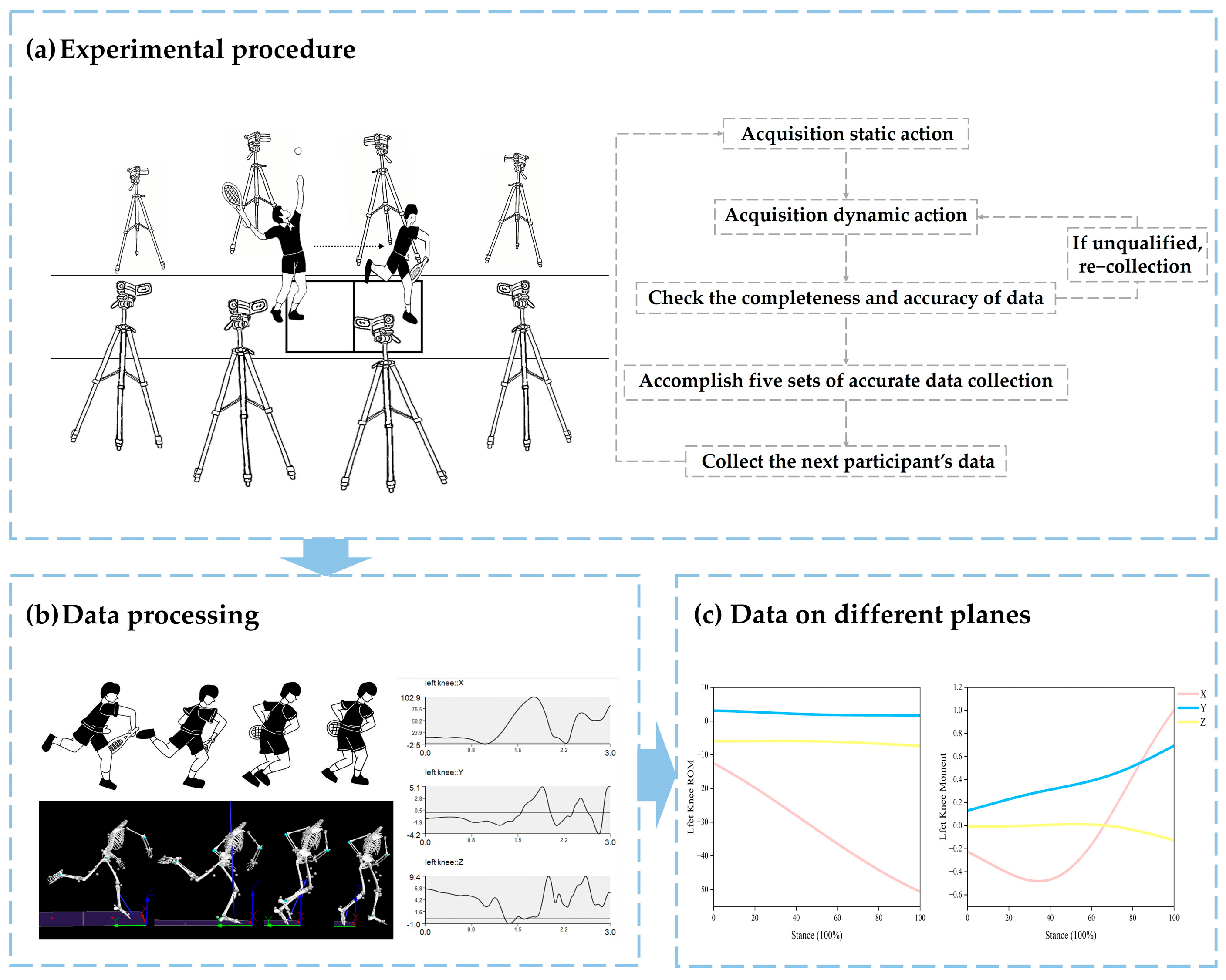
2.2. Experimental Procedure
2.3. Data Processing
2.4. Statistical Analysis
3. Results
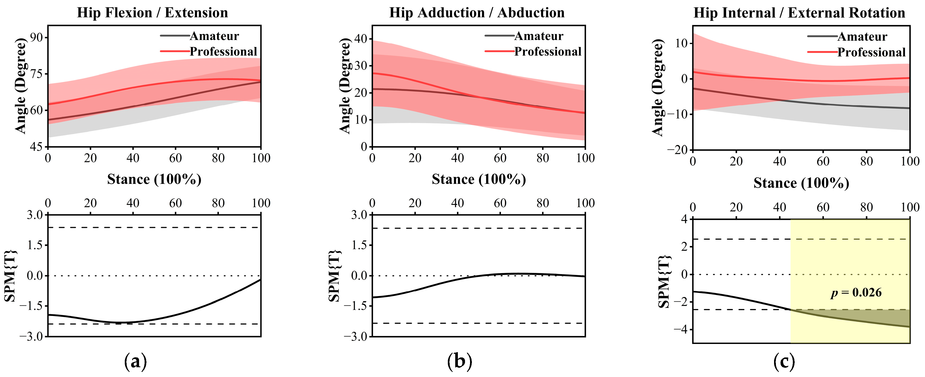
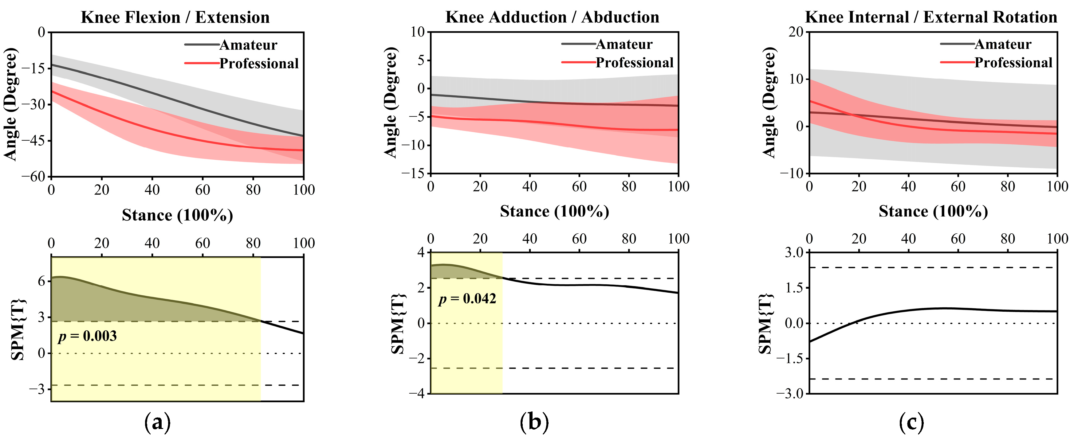
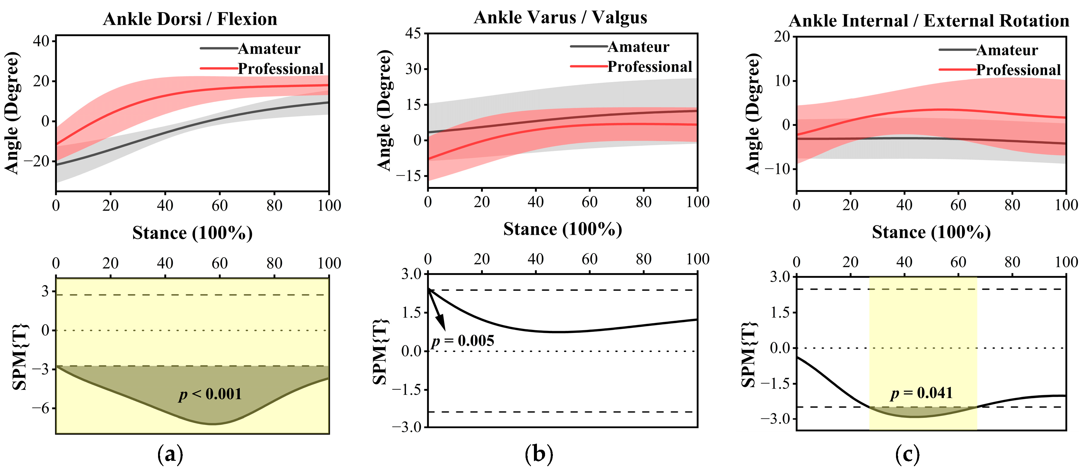
3.1. ROM
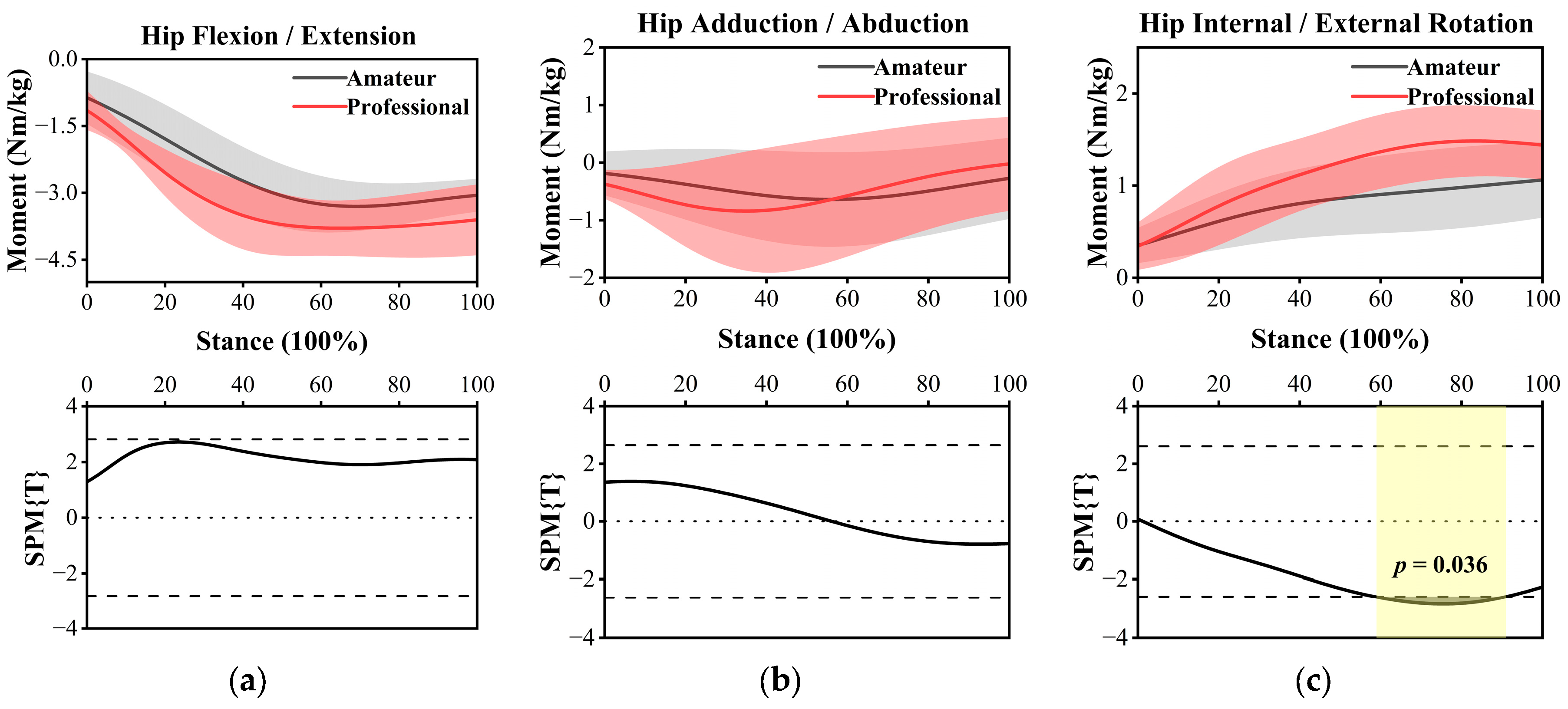
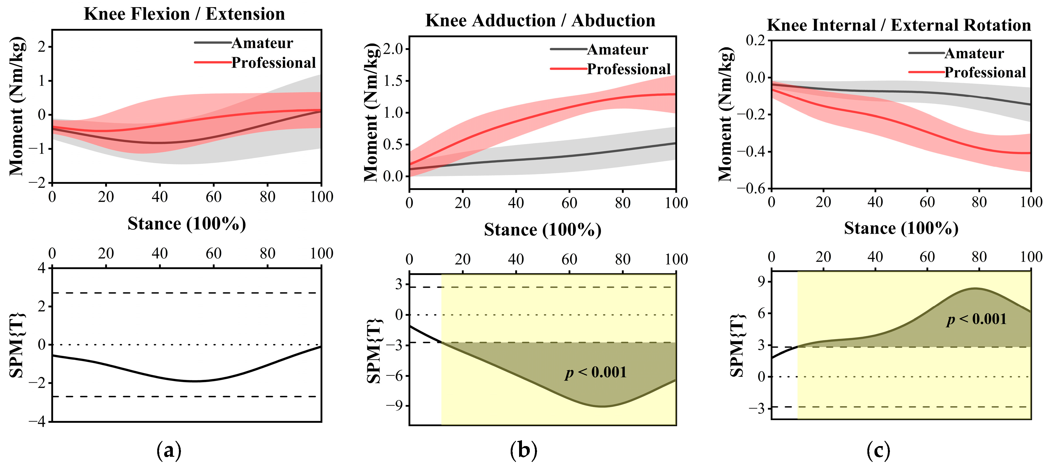
3.2. Joint Moment
3.3. Stiffness of Lower Extremities
4. Discussion
5. Conclusions
Author Contributions
Funding
Institutional Review Board Statement
Informed Consent Statement
Data Availability Statement
Conflicts of Interest
References
- Llewellyn, M.P.; Lake, R.J. Tennis and the Olympics: An Historical Examination of Their On-Off Relationship Since 1896; Routledge International Handbooks; First Issued in Paperback 2020; Routledge: London, UK; New York, NY, USA, 2020; ISBN 978-1-315-53357-5. [Google Scholar]
- Chow, J.; Carlton, L.; Lim, Y.-T.; Chae, W.-S.; Shim, J.-H.; Kuenster, A.; Kokubun, K. Comparing the Pre- and Post-Impact Ball and Racquet Kinematics of Elite Tennis Players’ First and Second Serves: A Preliminary Study. J. Sports Sci. 2003, 21, 529–537. [Google Scholar] [CrossRef] [PubMed]
- Abrams, G.D.; Sheets, A.L.; Andriacchi, T.P.; Safran, M.R. Review of Tennis Serve Motion Analysis and the Biomechanics of Three Serve Types with Implications for Injury. Sports Biomech. 2011, 10, 378–390. [Google Scholar] [CrossRef] [PubMed]
- Pluim, B.M.; Staal, J.B.; Windler, G.E.; Jayanthi, N. Tennis Injuries: Occurrence, Aetiology, and Prevention. Br. J. Sports Med. 2006, 40, 415–423. [Google Scholar] [CrossRef] [PubMed]
- Sell, K.; Hainline, B.; Yorio, M.; Kovacs, M. Injury Trend Analysis from the US Open Tennis Championships between 1994 and 2009. Br. J. Sports Med. 2014, 48, 546–551. [Google Scholar] [CrossRef]
- Renström, P.A.F.H. Knee Pain in Tennis Players. Clin. Sports Med. 1995, 14, 163–175. [Google Scholar] [CrossRef]
- Pluim, B.M.; Loeffen, F.G.J.; Clarsen, B.; Bahr, R.; Verhagen, E.A.L.M. A One-season Prospective Study of Injuries and Illness in Elite Junior Tennis. Scand. Med. Sci. Sports 2016, 26, 564–571. [Google Scholar] [CrossRef]
- Hjelm, N.; Werner, S.; Renstrom, P. Injury Profile in Junior Tennis Players: A Prospective Two Year Study. Knee Surg. Sports Traumatol. Arthrosc. 2010, 18, 845–850. [Google Scholar] [CrossRef]
- Delgado, G.J.; Chung, C.B.; Lektrakul, N.; Azocar, P.; Botte, M.J.; Coria, D.; Bosch, E.; Resnick, D. Tennis Leg: Clinical US Study of 141 Patients and Anatomic Investigation of Four Cadavers with MR Imaging and US. Radiology 2002, 224, 112–119. [Google Scholar] [CrossRef]
- Perkins, R.H.; Davis, D. Musculoskeletal Injuries in Tennis. Phys. Med. Rehabil. Clin. 2006, 17, 609–631. [Google Scholar] [CrossRef]
- Abrams, G.D.; Renstrom, P.A.; Safran, M.R. Epidemiology of Musculoskeletal Injury in the Tennis Player. Br. J. Sports Med. 2012, 46, 492–498. [Google Scholar] [CrossRef]
- van der Does, H.T.D.; Brink, M.S.; Benjaminse, A.; Visscher, C.; Lemmink, K.A.P.M. Jump Landing Characteristics Predict Lower Extremity Injuries in Indoor Team Sports. Int. J. Sports Med. 2016, 37, 251–256. [Google Scholar] [CrossRef] [PubMed]
- Getgood, A.; Hoshino, Y.; Roessler, P.P.; Kuroroda, R.; Piedade, S.R. Biomechanics of Musculoskeletal Injuries. In The Sports Medicine Physician; Rocha Piedade, S., Imhoff, A.B., Clatworthy, M., Cohen, M., Espregueira-Mendes, J., Eds.; Springer International Publishing: Cham, Switzerland, 2019; pp. 27–35. ISBN 978-3-030-10432-0. [Google Scholar]
- Aicale, R.; Tarantino, D.; Maffulli, N. Overuse Injuries in Sport: A Comprehensive Overview. J. Orthop. Surg. Res. 2018, 13, 309–319. [Google Scholar] [CrossRef] [PubMed]
- van Mechelen, W.; Hlobil, H.; Zijlstra, W.P.; de Ridder, M.; Kemper, H.C.G. Is Range of Motion of the Hip and Ankle Joint Related to Running Injuries? Int. J. Sports Med. 1992, 13, 605–610. [Google Scholar] [CrossRef] [PubMed]
- Bradley, P.S.; Portas, M.D. The Relationship Between Preseason Range of Motion and Muscle Strain Injury in Elite Soccer Players. J. Strength Cond. Res. 2007, 21, 1155–1159. [Google Scholar]
- Dufek, J.S.; Bates, B.T. Biomechanical Factors Associated with Injury During Landing in Jump Sports. Sports Med. 1991, 12, 326–337. [Google Scholar] [CrossRef]
- Nigg, B.M. Biomechanics, Load Analysis and Sports Injuries in the Lower Extremities. Sports Med. 1985, 2, 367–379. [Google Scholar] [CrossRef]
- Lorimer, A.V.; Keogh, J.W.L.; Hume, P.A. Using Stiffness to Assess Injury Risk: Comparison of Methods for Quantifying Stiffness and Their Reliability in Triathletes. PeerJ 2018, 6, e5845. [Google Scholar] [CrossRef]
- Butler, R.J.; Crowell, H.P.; Davis, I.M. Lower Extremity Stiffness: Implications for Performance and Injury. Clin. Biomech. 2003, 18, 511–517. [Google Scholar] [CrossRef]
- Söğüt, M. A Comparison of Serve Speed and Motor Coordination between Elite and Club Level Tennis Players. J. Hum. Kinet. 2017, 55, 171–176. [Google Scholar] [CrossRef]
- Reid, M.; Elliott, B.; Alderson, J. Lower-Limb Coordination and Shoulder Joint Mechanics in the Tennis Serve. Med. Sci. Sports Exerc. 2008, 40, 308–315. [Google Scholar] [CrossRef]
- Colomar, J.; Corbi, F.; Brich, Q.; Baiget, E. Determinant Physical Factors of Tennis Serve Velocity: A Brief Review. Int. J. Sports Physiol. Perform. 2022, 17, 1159–1169. [Google Scholar] [CrossRef] [PubMed]
- Pluim, B.M.; Jansen, M.G.T.; Williamson, S.; Berry, C.; Camporesi, S.; Fagher, K.; Heron, N.; Van Rensburg, D.C.J.; Moreno-Pérez, V.; Murray, A.; et al. Physical Demands of Tennis Across the Different Court Surfaces, Performance Levels and Sexes: A Systematic Review with Meta-Analysis. Sports Med. 2023, 53, 807–836. [Google Scholar] [CrossRef]
- Chen, Y.; Wang, T.; Zhao, Y.; Zhan, G.; Tang, Y.; Wang, Z. Kinematic Differences between Female National and Provincial Athletes in the Tennis Serve. PeerJ 2024, 12, e18410. [Google Scholar] [CrossRef] [PubMed]
- Martin, C.; Bideau, B.; Ropars, M.; Delamarche, P.; Kulpa, R. Upper Limb Joint Kinetic Analysis during Tennis Serve: Assessment of Competitive Level on Efficiency and Injury Risks. Scand. Med. Sci. Sports 2014, 24, 700–707. [Google Scholar] [CrossRef] [PubMed]
- Hernández-Davó, J.L.; Moreno, F.J.; Sanz-Rivas, D.; Hernández-Davó, H.; Coves, Á.; Caballero, C. Variations in Kinematic Variables and Performance in the Tennis Serve According to Age and Skill Level. Int. J. Perform. Anal. Sport 2019, 19, 749–762. [Google Scholar] [CrossRef]
- Liang, Z.; Wu, J.; Yu, J.; Ying, S.; Liu, Z.; Zhang, Y.; Gu, Y.; Li, J. Comparison and Analysis of the Biomechanics of the Lower Limbs of Female Tennis Players of Different Levels in Foot-up Serve. Front. Physiol. 2023, 14, 1125240. [Google Scholar] [CrossRef]
- Olmedilla Zafra, A.; Prieto Andreu, J.M.; Blas Redondo, A. Relaciones entre estrés psicosocial y lesiones deportivas en tenistas. Univ. Psychol. 2010, 10, 909–922. [Google Scholar] [CrossRef]
- Guevara, S.; Sheehy, D.; Waddington, G.; Drew, M.; Keegan, R.; Toohey, L. Injuries Impacted a Third of Athletes Who Left the High-Performance Sport Pathway. A 4-Year Retrospective Study. J. Sci. Med. Sport 2024, 27, S40. [Google Scholar] [CrossRef]
- Ayala, F.; Moreno-Pérez, V.; Vera-Garcia, F.J.; Moya, M.; Sanz-Rivas, D.; Fernandez-Fernandez, J. Acute and Time-Course Effects of Traditional and Dynamic Warm-Up Routines in Young Elite Junior Tennis Players. PLoS ONE 2016, 11, e0152790. [Google Scholar] [CrossRef]
- Fong, C.-M.; Blackburn, J.T.; Norcross, M.F.; McGrath, M.; Padua, D.A. Ankle-Dorsiflexion Range of Motion and Landing Biomechanics. J. Athl. Train. 2011, 46, 5–10. [Google Scholar] [CrossRef]
- Blackburn, J.T.; Padua, D.A. Sagittal-Plane Trunk Position, Landing Forces, and Quadriceps Electromyographic Activity. J. Athl. Train. 2009, 44, 174–179. [Google Scholar] [CrossRef] [PubMed]
- Pan, J.; Ho, M.M.Y.; Loh, R.B.C.; Iskandar, M.N.S.; Kong, P.W. Foot morphology and running gait pattern between the left and right limbs in recreational runners. Phys. Act. Health 2023, 7, 43–52. [Google Scholar] [CrossRef]
- Johnson, C.D. Performance Demands of Professional Male Tennis Players. Br. J. Sports Med. 2006, 40, 696–699. [Google Scholar] [CrossRef] [PubMed]
- Kovalchik, S.A.; Reid, M. Comparing Matchplay Characteristics and Physical Demands of Junior and Professional Tennis Athletes in the Era of Big Data. J. Sports Sci. Med. 2017, 16, 489–497. [Google Scholar]
- Chang, H.; Cen, X. Can running technique modification benefit patellofemoral pain improvement in runners? A systematic review and meta-analysis. Int. J. Biomed. Eng. Technol. 2024, 45, 83–101. [Google Scholar] [CrossRef]
- Kovacs, M.; Ellenbecker, T. An 8-Stage Model for Evaluating the Tennis Serve: Implications for Performance Enhancement and Injury Prevention. Sports Health A Multidiscip. Approach 2011, 3, 504–513. [Google Scholar] [CrossRef]
- Lambrich, J.; Muehlbauer, T. Biomechanical Analyses of Different Serve and Groundstroke Techniques in Tennis: A Systematic Scoping Review. PLoS ONE 2023, 18, e0290320. [Google Scholar] [CrossRef]
- Girard, O.; Micallef, J.-P.; Millet, G.P. Lower-Limb Activity during the Power Serve in Tennis: Effects of Performance Level. Med. Sci. Sports Exerc. 2005, 37, 1021. [Google Scholar]
- Muaidi, Q.I.; Nicholson, L.L.; Refshauge, K.M. Do Elite Athletes Exhibit Enhanced Proprioceptive Acuity, Range and Strength of Knee Rotation Compared with Non-Athletes? Scand. J. Med. Sci. Sports 2009, 19, 103–112. [Google Scholar] [CrossRef]
- Powers, C.M. The Influence of Abnormal Hip Mechanics on Knee Injury: A Biomechanical Perspective. J. Orthop. Sports Phys. Ther. 2010, 40, 42–51. [Google Scholar] [CrossRef]
- Durand, S.; Ripamonti, M.; Beaune, B.; Rahmani, A. Leg Ability Factors in Tennis Players. Int. J. Sports Med. 2010, 31, 882–886. [Google Scholar] [CrossRef] [PubMed]
- Devita, P.; Skelly, W.A. Effect of Landing Stiffness on Joint Kinetics and Energetics in the Lower Extremity. Med. Sci. Sports Exerc. 1992, 24, 108–115. [Google Scholar] [CrossRef] [PubMed]
- Pruyn, E.C.; Watsford, M.L.; Murphy, A.J.; Pine, M.J.; Spurrs, R.W.; Cameron, M.L.; Johnston, R.J. Relationship between Leg Stiffness and Lower Body Injuries in Professional Australian Football. J. Sports Sci. 2012, 30, 71–78. [Google Scholar] [CrossRef] [PubMed]
- Rice, R.P.; Roach, K.; Kirk-Sanchez, N.; Waltz, B.; Ellenbecker, T.S.; Jayanthi, N.; Raya, M. Age and Gender Differences in Injuries and Risk Factors in Elite Junior and Professional Tennis Players. Sports Health A Multidiscip. Approach 2022, 14, 466–477. [Google Scholar] [CrossRef]
- Hewett, T.E.; Myer, G.D.; Ford, K.R.; Heidt, R.S.; Colosimo, A.J.; McLean, S.G.; van den Bogert, A.J.; Paterno, M.V.; Succop, P. Biomechanical Measures of Neuromuscular Control and Valgus Loading of the Knee Predict Anterior Cruciate Ligament Injury Risk in Female Athletes: A Prospective Study. Am. J. Sports Med. 2005, 33, 492–501. [Google Scholar] [CrossRef]
- Ueno, R.; Navacchia, A.; DiCesare, C.A.; Ford, K.R.; Myer, G.D.; Ishida, T.; Tohyama, H.; Hewett, T.E. Knee Abduction Moment Is Predicted by Lower Gluteus Medius Force and Larger Vertical and Lateral Ground Reaction Forces during Drop Vertical Jump in Female Athletes. J. Biomech. 2020, 103, 109669. [Google Scholar] [CrossRef]
- Csintalan, R.P.; Ehsan, A.; McGarry, M.H.; Fithian, D.F.; Lee, T.Q. Biomechanical and Anatomical Effects of an External Rotational Torque Applied to the Knee: A Cadaveric Study. Am. J. Sports Med. 2006, 34, 1623–1629. [Google Scholar] [CrossRef]
- van den Bekerom, M.P.J.; Oostra, R.J.; Alvarez, P.G.; van Dijk, C.N. The Anatomy in Relation to Injury of the Lateral Collateral Ligaments of the Ankle: A Current Concepts Review. Clin. Anat. 2008, 21, 619–626. [Google Scholar] [CrossRef]
- Windt, J.; Gabbett, T.J. How Do Training and Competition Workloads Relate to Injury? The Workload—Injury Aetiology Model. Br. J. Sports Med. 2017, 51, 428–435. [Google Scholar] [CrossRef]
- Sugimoto, D.; McCartney, R.E.; Parisien, R.L.; Dashe, J.; Borg, D.R.; Meehan, W.P. Range of Motion and Ankle Injury History Association with Sex in Pediatric and Adolescent Athletes. Physician Sportsmed. 2018, 46, 24–29. [Google Scholar] [CrossRef]
- Elliott, B. Biomechanics and Tennis. Br. J. Sports Med. 2006, 40, 392–396. [Google Scholar] [CrossRef] [PubMed]
- Reid, M.; Whiteside, D.; Gilbin, G.; Elliott, B. Effect of a Common Task Constraint on the Body, Racket, and Ball Kinematics of the Elite Junior Tennis Serve. Sports Biomech. 2013, 12, 15–22. [Google Scholar] [CrossRef] [PubMed]
- Kaya, M.; Soyal, M.; Karakuş, M. The Effect of the Leg and Back Strength of the Serve and Tennis Players to the Serve Throwing Speed and Agility. Phys. Educ. Stud. 2018, 22, 237–242. [Google Scholar] [CrossRef]
- Leteneur, S.; Gillet, C.; Sadeghi, H.; Allard, P.; Barbier, F. Effect of Trunk Inclination on Lower Limb Joint and Lumbar Moments in Able Men during the Stance Phase of Gait. Clin. Biomech. 2009, 24, 190–195. [Google Scholar] [CrossRef]
- Müller, R.; Ertelt, T.; Blickhan, R. Low Back Pain Affects Trunk as Well as Lower Limb Movements during Walking and Running. J. Biomech. 2015, 48, 1009–1014. [Google Scholar] [CrossRef]
- Sciascia, A.; Cromwell, R. Kinetic Chain Rehabilitation: A Theoretical Framework. Rehabil. Res. Pract. 2012, 2012, 853037. [Google Scholar] [CrossRef]
- Sidiropoulos, A.; Magill, R.; Gordon, A. Coordination of the Upper and Lower Extremities during Walking in Children with Cerebral Palsy. Gait Posture 2021, 86, 251–255. [Google Scholar] [CrossRef]
- Chéron, C.; Le Scanff, C.; Leboeuf-Yde, C. Association between Sports Type and Overuse Injuries of Extremities in Adults: A Systematic Review. Chiropr. Man. Ther. 2017, 25, 4–13. [Google Scholar] [CrossRef]
- Hopper, D.M.; Hopper, J.L.; Elliott, B.C. Do Selected Kinanthropometric and Performance Variables Predict Injuries in Female Netball Players? J. Sports Sci. 1995, 13, 213–222. [Google Scholar] [CrossRef]
- Pas, H.I.M.F.L.; Bodde, S.; Kerkhoffs, G.M.M.J.; Pluim, B.; Tiemessen, I.J.H.; Tol, J.L.; Verhagen, E.; Gouttebarge, V. Systematic Development of a Tennis Injury Prevention Programme. BMJ Open Sport Exerc. Med. 2018, 4, e000350. [Google Scholar] [CrossRef]
- Fleck, S.J.; Falkel, J.E. Value of Resistance Training for the Reduction of Sports Injuries. Sports Med. 1986, 3, 61–68. [Google Scholar] [CrossRef] [PubMed]
- Zheng, Z.; Xia, Q. Effcets of Flexibility Exercises to Prevent Knee Injuries in Tennis Players. Rev. Bras. Med. Esporte 2023, 29, e2023_0040. [Google Scholar] [CrossRef]
- Pluim, B.M.; Drew, M.K. It’s Not the Destination, It’s the ‘Road to Load’ That Matters: A Tennis Injury Prevention Perspective. Br. J. Sports Med. 2016, 50, 641–642. [Google Scholar] [CrossRef] [PubMed]
- Hewett, T.E.; Lindenfeld, T.N.; Riccobene, J.V.; Noyes, F.R. The Effect of Neuromuscular Training on the Incidence of Knee Injury in Female Athletes. A Prospective Study. Am. J. Sports Med. 1999, 27, 699–706. [Google Scholar] [CrossRef]
- McHugh, M.P.; Cosgrave, C.H. To Stretch or Not to Stretch: The Role of Stretching in Injury Prevention and Performance. Scand. J. Med. Sci. Sports 2010, 20, 169–181. [Google Scholar] [CrossRef]
- Ayala, F.; Calderón-López, A.; Delgado-Gosálbez, J.C.; Parra-Sánchez, S.; Pomares-Noguera, C.; Hernández-Sánchez, S.; López-Valenciano, A.; Croix, M.D.S. Acute Effects of Three Neuromuscular Warm-Up Strategies on Several Physical Performance Measures in Football Players. PLoS ONE 2017, 12, e0169660. [Google Scholar] [CrossRef]
- Dines, J.S.; Bedi, A.; Williams, P.N.; Dodson, C.C.; Ellenbecker, T.S.; Altchek, D.W.; Windler, G.; Dines, D.M. Tennis Injuries: Epidemiology, Pathophysiology, and Treatment. J. Am. Acad. Orthop. Surg. 2015, 23, 181–189. [Google Scholar] [CrossRef]
- Kovacs, M.S. Applied Physiology of Tennis Performance. Br. J. Sports Med. 2006, 40, 381–386. [Google Scholar] [CrossRef]
- Sun, D.; Song, Y.; Cen, X.; Wang, M.; Baker, J.S.; Gu, Y. Workflow Assessing the Effect of Achilles Tendon Rupture on Gait Function and Metatarsal Stress: Combined Musculoskeletal Modeling and Finite Element Analysis. Proc. Inst. Mech. Eng. H 2022, 236, 676–685. [Google Scholar] [CrossRef]
- Song, Y.; Cen, X.; Sun, D.; Bálint, K.; Wang, Y.; Chen, H.; Gao, G.; Bíró, I.; Zhang, M.; Gu, Y. Curved carbon-plated shoe may further reduce forefoot loads compared to flat plate during running. Sci. Rep. 2024, 14, 13215. [Google Scholar] [CrossRef]
- Cen, X.; Song, Y.; Yu, P.; Sun, D.; Simon, J.; Bíró, I.; Gu, Y. Effects of Plantar Fascia Stiffness on the Internal Mechanics of Idiopathic Pes Cavus by Finite Element Analysis: Implications for Metatarsalgia. Comput. Methods Biomech. Biomed. Eng. 2024, 27, 1961–1969. [Google Scholar] [CrossRef] [PubMed]
- Song, Y.; Cen, X.; Wang, M.; Bálint, K.; Tan, Q.; Sun, D.; Gao, S.; Li, F.; Gu, Y.; Wang, Y.; et al. The Influence of Simulated Worn Shoe and Foot Inversion on Heel Internal Biomechanics during Running Impact: A Subject-Specific Finite Element Analysis. J. Biomech. 2025, 180, 112517. [Google Scholar] [CrossRef] [PubMed]
- Shu, L.; Yamamoto, K.; Yoshizaki, R.; Yao, J.; Sato, T.; Sugita, N. Multiscale Finite Element Musculoskeletal Model for Intact Knee Dynamics. Comput. Biol. Med. 2022, 141, 105023. [Google Scholar] [CrossRef] [PubMed]
- Bulat, M.; Korkmaz Can, N.; Arslan, Y.Z.; Herzog, W. Musculoskeletal Simulation Tools for Understanding Mechanisms of Lower-Limb Sports Injuries. Curr. Sports Med. Rep. 2019, 18, 210–216. [Google Scholar] [CrossRef]


| Variable | n | Age (years) | Height (cm) | Weight (kg) | Years of Training |
|---|---|---|---|---|---|
| Amateur | 15 | 21 ± 1.94 | 176 ± 2.67 | 70 ± 3.65 | 6.67 ± 1.29 |
| Professional | 15 | 20 ± 1.05 | 177 ± 2.69 | 71 ± 4.15 | 11.73 ± 0.96 |
| Joint | Plane | Amateur | Professional | p |
|---|---|---|---|---|
| Hip | X | −0.866 ± 0.589 | −1.151 ± 0.438 | 0.212 |
| Y | −0.069 ± 0.506 | 0.110 ± 0.661 | 0.482 | |
| Z | 1.094 ± 0.417 | 1.511 ± 0.417 | 0.029 | |
| Knee | X | 0.235 ± 0.955 | 0.337 ± 0.608 | 0.764 |
| Y | 0.521 ± 0.259 | 1.354 ± 0.244 | <0.001 | |
| Z | −0.034 ± 0.257 | −0.063 ± 0.468 | 0.087 | |
| Ankle | X | −0.569 ± 0.296 | −0.755 ± 0.211 | 0.104 |
| Y | 0.217 ± 0.215 | 0.173 ± 0.287 | 0.690 | |
| Z | 0.360 ± 0.175 | 0.678 ± 0.137 | <0.001 |
| Joint | Plane | Amateur | Professional | p |
|---|---|---|---|---|
| Hip | X | 0.166 ± 0.049 | 0.278 ± 0.079 | <0.001 |
| Y | 0.088 ± 0.054 | 0.075 ± 0.022 | 0.478 | |
| Z | 0.161 ± 0.112 | 0.162 ± 0.065 | 0.987 | |
| Knee | X | 0.036 ± 0.009 | 0.037 ± 0.008 | 0.761 |
| Y | 0.253 ± 0.177 | 0.249 ± 0.123 | 0.954 | |
| Z | 0.063 ± 0.066 | 0.076 ± 0.095 | 0.732 | |
| Ankle | X | 0.094 ± 0.129 | 0.076 ± 0.011 | 0.658 |
| Y | 0.027 ± 0.013 | 0.036 ± 0.046 | 0.540 | |
| Z | 0.265 ± 0.278 | 0.429 ± 1.010 | 0.609 |
| Joint | Plane | ROM | Joint Moment | Peak Joint Moment | Joint Stiffness |
|---|---|---|---|---|---|
| Hip | X | - | - | - | <0.001 |
| Y | - | - | - | - | |
| Z | 0.026 (45~100%) | 0.036 (59~91%) | 0.029 | - | |
| Knee | X | 0.003 (0~82%) | - | - | - |
| Y | 0.042 (0~29%) | <0.001 (12~100%) | <0.001 | - | |
| Z | - | <0.001 (19~100%) | - | - | |
| Ankle | X | <0.001 | 0.004 (6~74%) | - | - |
| Y | - | - | - | - | |
| Z | 0.041 (27~67%) | - | <0.001 | - |
Disclaimer/Publisher’s Note: The statements, opinions and data contained in all publications are solely those of the individual author(s) and contributor(s) and not of MDPI and/or the editor(s). MDPI and/or the editor(s) disclaim responsibility for any injury to people or property resulting from any ideas, methods, instructions or products referred to in the content. |
© 2025 by the authors. Licensee MDPI, Basel, Switzerland. This article is an open access article distributed under the terms and conditions of the Creative Commons Attribution (CC BY) license (https://creativecommons.org/licenses/by/4.0/).
Share and Cite
Pan, J.; Sun, D.; Li, F.; Zhou, Z.; Wang, Y.; Cen, X.; Song, Y.; Fekete, G. The Effects of Skill Level on Lower-Limb Injury Risk During the Serve Landing Phase in Male Tennis Players. Appl. Sci. 2025, 15, 2681. https://doi.org/10.3390/app15052681
Pan J, Sun D, Li F, Zhou Z, Wang Y, Cen X, Song Y, Fekete G. The Effects of Skill Level on Lower-Limb Injury Risk During the Serve Landing Phase in Male Tennis Players. Applied Sciences. 2025; 15(5):2681. https://doi.org/10.3390/app15052681
Chicago/Turabian StylePan, Jianqi, Dong Sun, Fengping Li, Zhanyi Zhou, Yucheng Wang, Xuanzhen Cen, Yang Song, and Gusztáv Fekete. 2025. "The Effects of Skill Level on Lower-Limb Injury Risk During the Serve Landing Phase in Male Tennis Players" Applied Sciences 15, no. 5: 2681. https://doi.org/10.3390/app15052681
APA StylePan, J., Sun, D., Li, F., Zhou, Z., Wang, Y., Cen, X., Song, Y., & Fekete, G. (2025). The Effects of Skill Level on Lower-Limb Injury Risk During the Serve Landing Phase in Male Tennis Players. Applied Sciences, 15(5), 2681. https://doi.org/10.3390/app15052681