Review

Enhancement of EPR Effect for Passive Tumor Targeting: Current Status and Future Perspectives

by
Ioanna-Aglaia Vagena
1,†,
Christina Malapani
2,†,
Maria-Anna Gatou
2,
Nefeli Lagopati
1,3 and
Evangelia A. Pavlatou
2,*
1
Laboratory of Biology, Department of Basic Medical Sciences, Medical School, National and Kapodistrian University of Athens, 11527 Athens, Greece
2
Laboratory of General Chemistry, School of Chemical Engineering, National Technical University of Athens, Zografou Campus, 15772 Athens, Greece
3
Biomedical Research Foundation, Academy of Athens, 11527 Athens, Greece
*
Author to whom correspondence should be addressed.
These authors contributed equally to this work.
Appl. Sci. 2025, 15(6), 3189; https://doi.org/10.3390/app15063189
Submission received: 29 December 2024 / Revised: 18 February 2025 / Accepted: 12 March 2025 / Published: 14 March 2025
(This article belongs to the Section Materials Science and Engineering)

Abstract

:
The Enhanced Permeability and Retention (EPR) effect is a key mechanism for passive tumor targeting, which involves the selective accumulation of therapeutic nanoparticles in tumors due to their unique vascular characteristics. While previous reviews have explored this phenomenon, the present review offers a comprehensive, multidisciplinary approach, highlighting recent advancements in strategies to enhance the EPR effect, as well as novel insights into the role of tumor microenvironment heterogeneity and the multifaceted approaches to overcome EPR-related challenges. This review provides a detailed analysis of the latest developments in nanocarriers’ design, including size, shape, and surface modifications, as well as cutting-edge multi-stage drug delivery systems. Furthermore, the integration of physical, pharmacological, and combinatory therapies to optimize the EPR effect is also discussed, aiming to improve the clinical translation of nanomedicines. Unlike other reviews, this work emphasizes the dynamic interaction between the tumor microenvironment and the vascular network, which remains underexplored in the current literature. In addition, specific clinical trials’ outcomes are highlighted and future directions to address existing limitations are proposed, offering a clearer roadmap regarding clinical applications in cancer therapy.

1. Introduction

The phenomenon of enhanced permeability and retention (EPR) is a pathophysiological phenomenon first observed by Konno and his team in 1984 [1]. This phenomenon occurs in solid tumors and refers to the selective accumulation of macromolecules, such as proteins, liposomes, micelles, and other soluble particles larger than 40 kDa, within the tumor interstitium passively, as well as their prolonged retention in the tissues [2,3].
Solid tumors exhibit various characteristics that can serve as significant evidence for the functioning of the EPR phenomenon and its exploitation for the effective targeting of tumors with nanomedicines [3]. Generally, the phenomenon under study represents the functional outcome of abnormalities including the hyperpermeability of blood vessels, reduced lymphatic drainage, increased stiffness of the extracellular matrix (ECM), and high interstitial fluid pressure (IFP) in tumors [2].
Specifically, solid tumors exhibit increased angiogenesis driven by cancer cells, resulting in the mass development of abnormal, structurally, and functionally impaired blood vessels [3]. This angiogenesis leads to the hyperproliferation, migration, and rearrangement of endothelial cells to meet their energy demands for oxygen and nutrients [4,5]. Consequently, endothelial cells can exhibit low density, creating large gaps between them and increased vascular permeability to macromolecules and nanoparticles, with pore sizes in the interstitial spaces ranging from 100 to 780 nm [3,5,6]. Additionally, abnormal lymphangiogenesis is observed, leading to ineffective lymphatic drainage in the tissues of solid tumors, which further contributes to the prolonged retention of extracellular macromolecules [5,6]. Finally, tissues express high levels of inflammatory factors and mediators that sustain the EPR phenomenon, such as prostaglandins, bradykinin, nitric oxide, proteases, interferon gamma, angiotensin-converting enzyme (ACE) inhibitors, and vascular permeability factor (VPF) or vascular endothelial growth factor (VEGF), among others [6,7,8].
Theoretically, enhanced permeability and retention (EPR) allows for the development of various nanomedicines, which reduce systemic toxicity and remain in the target area to exert their therapeutic effect [7]. However, targeting using the EPR phenomenon requires assumptions that are not guaranteed in real conditions, such as mass transfer exclusively towards cancer cells regardless of concentration gradients, interstitial fluid pressure (IFP) variations due to leakage from blood vessels, transport resistance, and selective application of IFP to lymphatics rather than blood vessels [9,10,11]. Moreover, some drugs do not distribute effectively, and controlling the mechanism remains challenging [12]. Over the past decades, EPR has been a recognized mechanism for the selective entry of nanoparticles, successful in preclinical models. However, few nanomedicines have transitioned to clinical therapy, as most achieve low toxicity but do not improve the clinical outcomes for patients [3,7].
In this context, both the characteristics of nanomedicine itself and those of the target site play crucial roles. The shape and size of the nanomedicine are critical factors determining its retention in the tumor site, as they dictate its circulation time in the bloodstream, distribution across various tissues and organs, and recognition and clearance through the reticuloendothelial system (RES), which includes macrophages in the liver, spleen, and bone marrow [3,7,10,13,14]. Additionally, the lack of control over essential parameters affecting the effective delivery of nanomedicines to the target, such as their release rate, internalization, or surface modification with immune-evasive components like polyethylene glycol (PEG), and their biocompatibility has led to disappointing results in exploiting the EPR phenomenon for therapeutic purposes [3,7,10].
Significant heterogeneity encountered both among different tumors and within different regions of the same tumor also impacts the ineffectiveness of the EPR phenomenon at the clinical level [1,3,7]. For instance, tumors with increased vascular density tend to exhibit higher EPR efficacy, unlike irregularities in blood vessel structures that cause heterogeneous vascular permeability in EPR [3,7,8]. Furthermore, the diverse microenvironments of tumors, particularly high pericellular coverage and dense extracellular matrix (ECM), pose significant barriers for nanoparticles to approach tumors, reducing vascular permeability and effective drug distribution [3,7]. Moreover, due to the heterogeneity in tumor perfusion, access to drugs and immune cells to the bloodstream is hindered, leading to conditions like hypoxia that promote tumor progression while conferring resistance to oxygen-dependent therapies [5]. Another factor is the increased interstitial fluid pressure (IFP) within tumor tissues, which drives nanomedicines back into circulation or even increases IFP peripherally compared to the tumor center, hindering transport from the circulatory system to the tissue [3,7].
To strengthen the EPR phenomenon and facilitate the transition of nanomedicine to clinical application, it is deemed necessary to consider the aforementioned variables by developing methods aimed at creating effective multi-stage drug delivery nano-systems for therapeutic purposes, as well as modifying the biological characteristics of solid tumors [7,15,16].
This review presents a comprehensive examination of the EPR phenomenon, focusing on recent advancements and innovative strategies aimed at enhancing its effectiveness in cancer therapy. While many reviews have addressed the fundamentals of EPR, this work highlights the unique challenges posed by tumor heterogeneity and explores cutting-edge approaches to overcoming these barriers. Specifically, we discuss the role of the TME in influencing EPR, including the impact of tumor-associated cells, ECM components, and extracellular signaling, which have been underexplored in earlier reviews. Furthermore, we emphasize novel techniques for enhancing nanocarrier permeability and retention, including physical, pharmacological, and biologically targeted strategies.
In the development of drug delivery strategies and the improvement in the EPR phenomenon, inorganic, polymeric, lipid, and hybrid nanoparticles play a key role [17].
Inorganic nanoparticles (NPs), such as gold (AuNPs), silver (AgNPs), platinum (PtNPs), and zinc oxide (ZnO) nanoparticles, have proven to be particularly capable in the application of anti-cancer therapies, mainly due to their ability to affect angiogenesis and selectively target cancer cells. AuNPs, for example, have shown significant activity in inhibiting angiogenesis through interaction with VEGF-165, leading to a reduction in VEGFR-2 signaling and thus limiting cell proliferation and migration [18]. Similarly, research by Barui et al. revealed that biosynthesized AuNPs enhance angiogenesis through ROS production and Akt pathway activation, promoting the formation of new blood vessels. AgNPs, likewise, have been studied for their ability to inhibit neovascularization through the disruption of VEGF signaling and ROS production, making them suitable for applications in cancer therapy. Zinc oxide nanoparticles and their nanophysical forms (ZONFs) have also shown pro-angiogenic properties. Activation of the MAPK–Akt–eNOS pathway and the production of ROS and NO promotes neovascularization, making them useful for applications in ischemic diseases and tissue engineering [19].
Polymeric nanoparticles, such as PEG-, PLGA-, and PAMAM-based nanoparticles, are being investigated for their potential to enhance targeted drug delivery [20,21,22]. PEG polymeric nanoparticles have been used for delayed drug release due to their ability to prolong drug release, enhancing the efficacy of treatments [23]. PLGA nanoparticles, on the other hand, have been developed for the local targeted delivery of anticancer drugs, such as paclitaxel, with the aim of prolonged action and enhancing therapeutic efficacy [23]. Moreover, polymeric dendrimers such as PAMAM, with their ability to incorporate multiple drug molecules, allow optimized targeted therapy in cancer [23].
Lipid nanoparticles, such as liposomes and solid lipid nanoparticles (SLNs), find applications in chemotherapy and hyperthermia, while nanostructured lipid nanoparticles (NLCs) improve drug bioavailability [24]. SLNs enhance the efficacy of chemotherapy through improved targeted drug release and bypassing resistance mechanisms, such as drug export from cancer cells via P-glycoprotein [25]. SLNs have also been found to be effective in inhibiting angiogenesis, limiting blood vessel growth and nutrient delivery to tumors [25]. The study by Zhao et al. demonstrated the combined administration of drugs, such as doxorubicin and curcumin via SLNs, which improved cytotoxicity and apoptosis in cancer cells while reducing toxicity in normal cells [26]. SLN/NLC drug delivery systems, through passive and active targeting, offer improved permeability and drug delivery to tumors. In a study by Lin et al., SLNs/NLCs showed significant improvement in drug delivery to tumors through these mechanisms. Also, SLNs/NLCs improve permeability in the brain for central nervous system therapies, allowing for effective targeted therapy [27].
Finally, hybrid nanoparticles, such as polymer-structured nanoparticles (NCPs), nanoscale metal–organic frameworks (NMOFs), polysiloxanes (PSQs), porous-core nanoparticles (MSNs), and iron oxide nanoparticles (IONPs), have been developed to incorporate diagnostic and therapeutic functions. These hybrid systems enhance the application of nanomedicine through the EPR effect and the enhancement of cancer therapy via EPR [28]. A typical example of these hybrid nanoparticles is AGuIX (Activation and Guiding of Irradiation by X-ray) nanoparticles, which have a hydrodynamic diameter of 4 ± 2 nm and are based on polysiloxane. They incorporate both diagnostic and therapeutic activities by using X-irradiation for their activation. These nanoparticles have the potential to exploit the EPR effect for the passive targeting of tumors, as their small size allows them to easily penetrate tumor vessels. The passive accumulation of nanoparticles in the tumor leads to an increased local concentration, while their elimination through the kidneys reduces the risk of toxicity. Furthermore, AGuIX NPs are activated by X-irradiation, enhancing tumor toxicity and providing effective radiotherapy even with low radiation doses. This approach was also verified in clinical studies, where the accumulation and persistence of AGuIX NPs in tumors in humans with brain metastases were monitored by MRI [29] (Figure 1).
By integrating recent advancements in nanomedicine design, TME-targeting approaches, and clinical trial data, this review offers fresh perspectives on optimizing the EPR effect for improved therapeutic outcomes. The review also examines the development of multi-stage drug delivery systems and the potential of combinatory therapies to address the limitations of EPR in clinical settings. Ultimately, this work aims to provide insights into the future of EPR-based cancer therapies, focusing on strategies that could bridge the gap between preclinical success and clinical efficacy.

2. Distinctive Characteristics of Tumor Microenvironment and Heterogeneity of EPR

2.1. Components of Tumor Microenvironment

Cancer constitutes a dynamic system of neoplastic cells interacting with a complex array of non-neoplastic cells and non-cellular components from local or distant host tissues, promoting the growth of complex tissues: tumors [30,31,32,33,34]. The tumor microenvironment (TME) consists of tumor cells, stromal tissue (immune cells, fibroblasts, myofibroblasts, cytokines, and vascular tissue), and the extracellular matrix (ECM) [35]. These elements form a complex and constantly evolving entity where tumor and noncancer cells maintain mutual interactions throughout tumor progression [32,36,37]. Basal lamina plays a critical role in these interactions, as it partially separates the parenchyma (the tumor core) from the stromal region (part of the TME). This separation is imperfect, with poorly defined boundaries, thus facilitating TME interactions that support survival, local invasion, metastatic spread, and tumor cell response to therapeutic drugs, as well as angiogenesis and extracellular signaling [31,32,36,37].
Cancer cells arise from genetic and epigenetic alterations, acquiring characteristics such as autonomous proliferation, resistance to cell death, invasion, immune evasion, reproductive immortality, and the ability to metastasize [38]. They invade healthy tissues and spread to different parts of the body through the lymphatic and circulatory systems [36,39]. In addition, tumors can eliminate diverse cancer cells and microenvironments, contributing to their heterogeneity [31,36]. A typical example is cancer stem cells that contribute to self-renewal (leading to carcinogenesis) and immune tolerance [36]. Tumor heterogeneity is intensified as the cancer progresses, in parallel with the maturation of cellular and non-cellular components of the TME [40].
The TME varies according to the type of cancer, showing heterogeneity; however, the consistent features include non-malignant cells and extracellular components of the host feeding on a vascular network [36,41]. These components stimulate uncontrolled cell proliferation and play a vital role in all stages of carcinogenesis and tumor progression, such as in blood vessel development, by supporting tumor metabolism [30,36]. Furthermore, the primary function of stromal factors is to structure and remodel functional ones [42]. Specifically, the key stromal cells of the TME include fibroblasts, cancer-associated fibroblasts (CAFs), pericytes, vascular cells (endothelial and wall cells), and immune cells, including T and B lymphocytes, natural killer cells, and tumor-associated macrophages [33,40,43]. Additional cell types include mesenchymal stem cells, peripheral adipocytes, and neurons [43]. Some investigators consider specialized cell types, such as endothelial cells, pericytes, adipocytes, and immune cells, to be non-stromal but still contribute significantly to tumor growth, metastasis, and therapeutic resistance [42].
Another categorization of noncancerous cells and components of the TME includes immune cells, blood cells, endothelial cells, adipocytes, and the stroma, which consists of specialized connective tissue cells such as fibroblasts, mesenchymal stromal cells, osteoblasts, and chondrocytes, as well as the extracellular matrix (ECM), which supports functional tissues [42]. Non-cellular chemical–physical elements such as the extracellular matrix (ECM) and soluble factors such as chemokines, cytokines, growth factors, extracellular vesicles, signaling molecules, and antibodies also play a critical role [32,38,40,44]. Interventional studies involving animals or humans and other studies requiring ethical approval should indicate the authority that granted the approval and the corresponding ethical approval code. Table 1 summarizes the primary components of TME and their respective roles.
The tumor microenvironment is influenced by factors such as the immune response and intracellular bacteria, such as Fusobacterium nucleatum. These bacteria can enhance the immune response, with T cells recognizing both infected and non-infected cancer cells. Treatment with LipoAgTNZ (an antibiotic silver-tinidazole complex encapsulated in liposomes) enhances tumor recognition by activating CD8+ T cells that produce IFN-γ (a cytokine that enhances cellular immunity against infections and cancer). Additionally, infection with F. nucleatum reduces the interaction of chaperone proteins with the cell membrane, indicating immunosuppressive action in the tumor [51].

2.2. Cell Communication Within Tumor Microenvironment

Intercellular communication is essential for the proper function and movement of cells, as well as for their response to external stimuli. Furthermore, it plays an important role in the dynamic and reciprocal exchange of information between cancer and normal cells, which is often mediated by the extracellular matrix (ECM) [44,52].
Intercellular communication is a complex system in which cells transmit signals either through direct contact with other cells, via electrical coupling, gap junctions (GJs), receptor–ligand interactions, or nanotubes (e.g., membrane nanotubes, MNTs), or indirectly through endocrine or paracrine signaling. Non-cancerous stromal cells communicate through the transport of healthy mitochondria and other organelles via tunnel or horizontal transport nanotubes. Soluble factors such as cytokines, chemokines, and growth factors, as well as membrane vesicles, which do not require specific receptors, facilitate indirect signaling between neighboring or distant cells [44].

2.2.1. Direct Intercellular Communication

Gap Junctions

Gap junctions (GJs) are specialized membrane structures that facilitate direct intercellular communication through the formation of intercellular channels or pores. These pores bridge the 2–4 nm gap between adjacent cells, enabling the direct passage of various molecules and ions (up to 1.2 kDa in size) as well as electrical signals between cells [44,53,54]. These channels consist of two connexons, each formed by hexameric connexin oligomers (CXs) [44,54]. Connexins are synthesized in the endoplasmic reticulum, forming hexameric rings with four transmembrane regions and extracellular loops responsible for cell recognition and adhesion [53,54]. Regulation occurs through the phosphorylation of connexins, determining the assembly or disassembly of gap junctions [44].
In cancer cells, the loss of functional gap junctions is observed, as various carcinogens, including toxins, organic solvents, metals, and pharmaceuticals, inhibit their function. Furthermore, connexins themselves can inhibit cell proliferation or induce cell migration and invasion outside of the tumor nucleus [44,55].

Ligand–Receptor Pairs

Intercellular communication through signaling requires direct physical contact between neighboring cells, with the ligand of one cell binding to homologous membrane receptors on the neighboring cell, creating specific signaling and response pathways in the target cell [44,56]. Depending on the ligand, a different adhesion molecule, such as cadherins, selectins, and members of the immunoglobulin superfamily, binds and becomes part of an adhesion network [44,57].
At epithelial migration barriers, intercellular adhesion complexes such as tight junctions (TJs), adherens junctions (AJs), and desmosomes are formed. Tight junctions regulate barrier permeability, facilitating the transport of ions and molecules, while contributing to osmosis, cell differentiation, and the regulation of cell polarity [44,58,59,60].
Structural and genetic changes or the altered expression of receptors and adhesion molecules contribute to the development of diseases such as breast cancer, for example, through the dysfunction of the estrogen receptor [56]. In cancer, the loss of cellular connections due to the altered regulation of tight junctions (TJs) or E-cadherin affects disease development, metastasis, and prognosis [44,60]. However, during metastasis, the loss of cell–cell contact alone is insufficient; the absence of this protein is also required [61]. Alterations in the expression of both E-cadherin and other components of desmosomes have been observed in various types of cancer, such as gastric and prostate cancer [44].

Tunnel Nanotubes (TNT) and Tumor Microtubules (TM)

The transport of chemical or biological materials as well as electrical coupling between cells over long distances is facilitated by the formation of tunnel nanotubes (TNTs) and tumor microtubules (TMs).
Tunnel nanotubes (TNTs) are long and thin (50–200 nm) cytoplasmic extensions formed by F-actin filaments [44,62,63]. The open end of the channel connects the cytoplasm of cells, transporting cellular vesicles, mitochondria, miRNAs, viral particles, and proteins, and is involved in cellular homeostasis, tissue repair, cancer invasion, and resistance to therapies [44,62,63,64].
Tumor microtubules (TMs) are larger, richer in F-actin, and have a longer lifespan. They contain microcapsules and mitochondria, aiding in vesicle transport and local ATP production. Tumor microtubules contribute to the aggression, invasion, and therapeutic resistance of tumor cells, as observed in glioma cells [44,65].

2.2.2. Indirect Cell Communication

Signaling by Extracellular Vesicles

Extracellular vesicles (EVs) are lipid bilayer particles, naturally released from cells carrying signaling-active molecules such as DNA/RNA, proteins, lipids, and metabolites. Different types of extracellular vesicles are released under both normal and pathological conditions (e.g., cancer) and alter the metabolism of the recipient cell [44,66].
EVs are categorized based on size as apoptotic bodies (500–2000 nm), microvesicles (50–1000 nm), and exosomes (40–120 nm). Apoptotic bodies are released during apoptosis and may carry fragments of genomic DNA. Microvesicles are excreted from the plasma membrane, while exosomes are released from the multivesicular bodies (MVBs) [44,67,68].
Tumor-associated EVs (TEVs) released by tumor cells and tumor microenvironment cells circulate in body fluids and induce phenotypic changes in target cells locally and distantly [66]. They allow communication between tumor cells and the microenvironment, influencing processes involved in tumor initiation, progression, and metastasis, such as angiogenesis and resistance to cell death [66,68]. For example, fibroblasts secrete exosomes to deliver nutrients to malignant cells, whereas cancer cells use EVs to communicate changes in their microenvironment [44,68].

Signaling by Cytokines, Chemokines, and Growth Factors

Cytokines, chemokines, and growth factors contribute to cross-communication between cancer and non-cancer cells [44].
Cytokines are usually polypeptides or glycoproteins with relatively low molecular weight (6–70 kDa), and they regulate the functions, differentiation, proliferation, and apoptosis of target cells. They act through receptors to activate various intracellular signaling pathways for growth, metastasis, homeostasis, and the inhibition of tumor progression [44,69]. Cytokines include interferons (IFNs), the tumor necrosis factor (TNF) superfamily, chemokines, and growth factors [69].
Chemokines, a large family of 45–50 proteins (8–12 kDa), activate intracellular signaling pathways through receptors (GPCRs) that interact with G-proteins. They are classified into four classes (CXC, CC, XC, CX3C) based on the location of cysteine residues, with corresponding receptors CCR, CXCR, XCR, and CX3CR [70,71]. Chemokines modulate tumor immune infiltration and act directly on neoplastic, fibroblast, or endothelial cells to regulate processes that contribute to tumor growth through various mechanisms, such as growth activity, angiogenesis, and tumor spread and metastasis. They also play a critical role in leukocyte migration, cancer-associated inflammation, and macrophage recruitment to provide key growth, trophic, and angiogenic factors [44,70,71].

3. Morphological Characteristics of the Vascular Network

Morphological and functional abnormalities of the tumor vasculature are crucial for the enhanced permeability and retention (EPR) phenomenon, facilitating the targeted delivery of nanomedicines. The extracellular matrix (ECM) acts as a structural scaffold for cellular repair and consists of a random and disorganized complex of collagen fibers, glycosaminoglycans (GAGs), solutes, proteins, and residues [72,73]. It regulates cell behavior and tissue homeostasis by providing biochemical signals and determines the diffusion and convection forces within the vascular wall for molecular movement, which are influenced by the degree of collagen organization. Enzymes such as collagenase restore mobility by enhancing the diffusion of molecules, while the covalent binding of GAGs to proteins such as collagen alters the viscosity of the environment and fluid diffusion directions [72]. Cancer and stromal cells secrete angiogenic and growth factors, as well as cytokines, stimulating the formation of blood vessels with abnormal architecture (Figure 2) [74,75].

3.1. Vascular Network Morphology and Hypervascularity

The microvascular network of normal tissues appears as an organized structure of mature blood vessels (arteries, arterioles, capillaries, venules, and veins) [74,76,77]. In contrast, the tumor vasculature, due to its aggressive growth and overexpression of angiogenic factors, forms a disorganized network of immature vessels with structural and functional abnormalities. While blood flow follows a similar course to normal vessels, tumor vessels exhibit irregular branching and spiral pathways, forming dendritic structures and creating a disorganized maze [74,76,78,79]. Cancer vessels vary in diameter and often appear larger, with discontinuities such as bumps and blind ends, disrupting tissue nutrition [74,76]. Their shape varies from serpentine to compressed channels with small lumens and includes microvascular outgrowths, vascular complexes, garland formations, and glomerular proliferation [80,81,82,83].
The tumor vascular network shows significant spatial heterogeneity and hypervascularity, i.e., zones of increased microvascular density (hot spots) in which the likelihood of metastasis is increased [8,84,85]. Hypervascularity is associated with EPR, usually in smaller tumors, although it varies depending on the type of cancer [3,8].

3.2. Intravascular Environment

The intravascular environment is lined by endothelial cells (ECs), which bind to adhesion molecules, such as claudins, forming structures depending on the properties of the organ [79,80,81]. The endothelium of veins and arteries forms a continuous monolayer, whereas that of capillaries appears continuous, separated, or discontinuous, depending on the function of the tissue [81]. ECs are supported by wall cells, attaching their basal side to a basement membrane (BM), limiting vascular leakage and hyperpermeability, thus acting as a dynamic barrier between blood and tissue [75,81]. In tumors, ECs are poorly connected and, in some regions, multilayered, and the basement membrane shows irregular thickness and composition. Wall cells are fewer and loosely connected to ECs around tumor vessels, with the extent of these changes depending on the type of tumor [80]. In addition, tumor blood vessels may incorporate tumor cells, forming mosaic vessels [86]. The contractile potential of ECs contributes to the regulation of vessel diameter and blood pressure [81,87]. Vascular smooth muscle cells support the high-pressure arterial system in the form of concentric layers and the lower-pressure venous system with thinner layers. Pericytes stabilize the smaller blood vessels, aiding in their maturation and EC proliferation through direct contact or tight junctions [81,88].

3.3. Types of Blood Vessels

In normal tissues, arterial blood flows through arterioles, capillaries, post-capillary venules, and veins, except in certain tissues (e.g., skin), where arteriovenous shunts bypass capillaries. Cancerous tissues follow the same route. Each type of vessel has a characteristic structure and function. Arteries, large blood vessels lined by endothelium, are surrounded by elastic tissue and multiple layers of smooth muscle cells. Arterioles (~10–20 μm in diameter) transition to capillaries, the main sites of nutrient and waste exchange due to their thin walls and small diameter (4–9 μm). Capillaries have endothelial junctions spaced 100–200 nm apart, facilitating molecular exchange via paracellular and transcellular pathways, while larger proteins are transported via vesicles. Their continuous endothelial lining is supported by a basement membrane and pericytes. In specialized tissues, such as the kidney, the endothelium shows fenestrations, i.e., zones of the extreme thinning of endothelial cells (50–150 nm in diameter). Venules, larger than capillaries (approximately 20 μm in diameter), are lined by cuboidal monolayer endothelium, basement membrane, and pericytes, but lack smooth muscle cells [75]. Another region considered favorable for metastasis is these post-capillary venules [76].

3.4. Functional Characteristics of the Tumor Vascular System

The cancer microvasculature exhibits unique functional features, such as angiogenesis, hyperangiogenesis, irregular blood flow, extensor vascular permeability, and abnormal lymphatic drainage, which collectively describe the enhanced permeability and retention (EPR) phenomenon [85]. Factors such as cancer type, stage, and location affect blood vessel flow and pressure, endothelial leakage rate, vascular permeability, and molecular retention, contributing to tumor heterogeneity [72].

3.4.1. Angiogenesis

The vasculature, in both normal and cancerous tissues, is required to supply and regulate oxygen, nutrients, growth factors, and cells, as well as to remove waste, supporting tissue nutrition and tumor growth [74,89]. Tumors exploit the host vasculature to meet their increased nutritional requirements due to their high rate of proliferation [80]. During tumor growth, reduced blood supply, due to heterogeneity in blood vessel size and arteriovenous drainage, leads to nutrient deprivation, the accumulation of acidic metabolic by-products, and high oxygen consumption, creating a hypoxic microenvironment [74,90,91]. Hypoxia triggers a vascular switch by promoting growth and facilitating tumor metastasis, allowing cancer cells to migrate through newly formed blood vessels, creating secondary tumors [74,90,91,92]. Without vascular support, tumors fail to grow and become necrotic or apoptotic.
Angiogenesis, the formation of new capillaries, is essential for tumors to grow beyond their minimum size (diameter up to 0.8–1.0 mm), providing oxygen, nutrients, and waste disposal and facilitating their migration [74,77,89,93,94]. It is carried out through three processes: (1) the sprouting of new capillaries (neovascularization) through the remodeling of existing daughter vessels, namely endothelial cell proliferation, migration, and stabilization by pericytes and smooth muscle cells; (2) de novo angiogenesis, where new blood vessels are formed from endothelial progenitor cells (EPCs) from bone marrow or vascular walls; (3) the division of large “mother” vessels into smaller daughter vessels [78,80,81,87,92,93,95].
Angiogenesis is a fundamental process during embryonic development and in various physiological and pathological conditions in adults [89]. It consists of four stages: (1) tumor cells release cytokines and growth factors that activate neighboring normal cells; (2) angiogenesis is stimulated, with the removal of pericytes and degradation of the basement membrane and extracellular matrix (ECM); (3) endothelial cells proliferate and migrate with the help of soluble factors; (4) consolidation with pericytes and the maturation of the initial microvessels into complete tubular structures and eventually into capillaries, where blood flow is established [77,92,93,96]. In the revised model of angiogenesis, endothelial cells form filopodia and migrate through the ECM, followed by proliferating stem cells that form lumen and recruit pericytes for stabilization. The apical cells from two migrating endothelial cells fuse to form a blood vessel [96,97]. The tumor microenvironment and normal tissue play a crucial role in these stages [92].
Angiogenesis is tightly regulated by a dynamic balance between the stimulators and inhibitors of angiogenesis (pro- and anti-angiogenic factors, respectively) [94,95]. Under physiological conditions, this balance tightly controls endothelial cell activation when necessary [95]. During pre-tumor development, genomic instability of the endothelial cells leads to the expression of multiple angiogenic factors at high levels. Angiogenesis inhibitors, either direct or indirect, inhibit tumor growth and cause tumor regression by targeting individual angiogenic factors [87,94]. Most angiogenic factors involved in normal and pathological angiogenesis are classical peptide growth factors that bind to specific tyrosine kinase receptors on the surface of endothelial cells [94]. Some factors involved in the regulation of angiogenesis in rapidly growing tumors are discussed below.

Positive Regulators of Angiogenesis

Stimulators of angiogenesis include the vascular endothelial growth factor family (VEGF-A, -B, -C, and -D) and placental growth factor (PlGF), which bind with different affinities to VEGF receptors (VEGFRs) on endothelial cells [77,94,98]. VEGF-A, the main mediator of angiogenesis, interacts with VEGFR-1 and VEGFR-2, inducing angiogenesis and vascular permeability, with VEGFR-2 dominating [77,87,98]. PlGF and VEGF-B bind to VEGFR-1, while VEGF-C and VEGF-D bind to VEGFR-2 and VEGFR-3, promoting angiogenesis and lymphangiogenesis [87]. Angiogenic factors are secreted by various host cells, including platelets, muscle cells, and tumor-associated stromal cells. VEGFRs are extensively expressed in endothelial cells [98].
Angiopoietins, also a family of angiogenesis stimulators, are proteins that act as ligands for the Tie receptor family in endothelial cells, playing a critical role in vascular remodeling and stabilization. The Tie-2 receptor facilitates budding and branching, whereas both Tie-1 and Tie-2 maintain vascular integrity. Angiopoietin-1 (Ang-1) activates Tie-2, recruits pericytes to stabilize blood vessels, reduces vascular permeability, and supports endothelial cell survival. In contrast, Ang-2 functions as an antagonist [77,94]. Among chemokines, CXC chemokines with the Glu-Leu-Arg amino acid motif activate endothelium, while others inhibit angiogenesis. Other growth factors, such as transforming growth factor-alpha (TGF-alpha) and platelet-derived growth factor-BB (PDGF-BB), and protein factors, such as pleiotrophin, angiogenin, platelet-activating factor (PAF), and the HIV tat gene product, promote endothelial cell proliferation and migration [94].
Transforming growth factor-β (TGF-β), whose receptors are expressed by endothelial cells and pericytes, at low concentrations, upregulates angiogenic factors and proteases, whereas, at high concentrations, it inhibits endothelial cell growth, promotes basement membrane remodeling, and stimulates smooth muscle cell differentiation [77]. Other promoters of angiogenesis include fibroblast growth factors (FGFs), platelet-derived growth factors (PDGFs), interferons (IFNs), prostaglandins E1 and E2, nicotinamide, and heparin [78,94].

Negative Regulators of Angiogenesis

The genomic instability of cancer cells leads to the overexpression of multiple angiogenic factors. The inhibition of angiogenesis can prevent tumor growth and cause tumor regression. Inhibitors target specific angiogenic factors and are classified as direct or indirect. Direct inhibitors, such as angiostatin, act directly on endothelial cells to suppress angiogenesis, whereas indirect inhibitors affect other cell types [87,94].
Angiostatin, for example, an anti-angiogenic protein derived from plasminogen, reduces the growth of distant metastases by enhancing apoptosis in metastatic tumors. Specifically, it attacks the energy system of metastatic tissue by inhibiting ATP synthase F1F0. Another example is interleukins (ILs), a group of cytokines released by leukocytes, in which the presence or absence of the Glu-Leu-Arg amino acid motif at the NH2 end of their structure (as in IL-8 and IL-4 respectively) enhances or inhibits angiogenesis [77].

3.4.2. Extensive Vascular Permeability

Vascular permeability refers to the passage of substances through the endothelial barrier. Small molecules (less than 40 kDa) are extruded spontaneously, whereas larger molecules require the disruption of the vascular barrier [75]. There are two pathways that control the passage of solutes across the endothelial barrier: the intercellular (or transcellular) pathway, i.e., the vesicle-mediated transport of small molecules (less than 3 nm), and the paracellular pathway, which limits the diffusion of macromolecules and is controlled by endothelial cell–cell junctions [99,100]. The permeability of the paracellular pathway is influenced by the opening and closing of endothelial junctions, which are the final barrier to the passage of solutes [85,99,100].
The endothelial cells lining the inner lumen of blood vessels are connected in three ways: through tight junctions (TJs), adhesion junctions (AJs), and gap junctions (GJs). TJs and AJs regulate the permeability of the vascular barrier, whereas GJs connect adjacent endothelial cells, transporting water and ions [99,100]. The former are composed of transmembrane proteins such as claudins and nectins, which bind to peripheral proteins such as ZO family kinases and catenins, regulating vascular permeability and cell adhesion [99,100]. Vascular endothelial cadherin (VE-cadherin) is expressed in blood vessels and its dysfunction leads to a hyperpermeability phenotype. Other adhesion proteins are N-cadherin and platelet–endothelial cell adhesion molecules (PECAM-1) and junctional adhesion molecules (JAMs) [100].
Vascular permeability is regulated by several physiological signals, such as VEGF [99]. In cancer, tumor size, high protein content, increased interstitial fluid pressure, large vessel diameter, and reduced blood flow slow down the extrusion of molecules from blood vessels. However, changes in endothelial cell properties lead to increased plasma and plasma protein extraction compared to normal vessels [85]. The tumor vasculature is characterized by extensive angiogenesis and the overexpression of angiogenic factors that increase endothelial cell proliferation and permeability. This results in the disruption of the endothelial barrier, creating pores (0.1–3 μm), and allows the diffusion and extrusion of membrane proteins and nutrients, especially macromolecules, into the interstitial space [75,100,101]. Vascular permeability varies depending on the type of tumor and during tumor growth, regression, and recurrence [46].

3.4.3. Irregular Blood Flow

Blood flow within a tumor affects growth, metastasis, and treatment and is regulated by the difference between arterial and venous pressure, as well as by flow resistance, which depends on vessel architecture and viscosity [85,102,103].
Within the tumor, microvascular pressures are increased due to venous compression, whereas drainage vein pressures are reduced [85]. Morphological abnormalities of blood vessels, such as weak connections between endothelial cells and pericytes, the absence of smooth muscle cells, basement membrane heterogeneity, and high blood viscosity due to vascular hyperpermeability, increase resistance to blood flow [85,100,102]. As a result, the total perfusion rate (blood flow per unit volume) decreases despite increased vascular density, leading to uneven blood flow or even reverse flow in some vessels [100,102].
Tumors are classified into regions according to perfusion rate: the vascular necrotic region (few or no blood vessels and dead tissue), the intermediate semi-necrotic region (some viable tissue), the region of stabilized microcirculation (stable blood flow), and the advanced front (slow growth) [104]. Perfusion heterogeneity exacerbates hypoxia, while reduced flow and increased permeability promote the accumulation of metabolic wastes, such as lactic and carbonic acid, lowering extracellular pH but maintaining intracellular pH at 7.4 [100,102,105].

3.4.4. Decreased Lymphatic Drainage and Increased Interstitial Fluid Pressure (IFP)

Transport of Dissolved Substances Through Vascular Walls

An ideal vascular network enables sequential blood flow from arteries to veins via arterioles, capillaries, and venules, with capillaries and post-capillary venules serving as primary sites for fluid and solute exchange due to their thin walls and large surface area [106]. No molecule or cell reaches cancer cells without traversing the vascular and interstitial compartments. In normal tissues, solute transport occurs via diffusion, driven by concentration gradients, and convection, driven by pressure gradients. Small hydrophilic and lipophilic molecules primarily diffuse, whereas large molecules utilize both mechanisms. According to Starling’s hypothesis, capillary hydrostatic and osmotic pressure gradients regulate movement through the vascular wall. While some interstitial fluid is reabsorbed by the microvasculature, the rest is cleared by the lymphatic system [106].

High Interstitial Fluid Pressure (IFP)

The tumor tissue exhibits increased stiffness due to increased interstitial fluid pressure (IFP), which remains high in the tumor core and decreases sharply at the periphery, driving the interstitial fluid flow (IFF) outwards. IFP in solid tumors ranges from 5 to 40 mm Hg, compared to 0–3 mm Hg in normal tissue [106,107]. Elevated IFP prevents the penetration of therapeutic agents through the EPR effect, as drugs reaching the tumor periphery cannot diffuse centrally [106,107,108,109]. However, macromolecular agents can be extruded and accumulate on the tumor surface. IFP at the tumor site is like the microvascular pressures of the surrounding capillaries, contributing to tumor cell migration and promoting tumor invasion and proliferation [106].
The exact mechanisms underlying the increase in IFP remain unclear but likely include increased vascular permeability, ECM stiffness, increased blood influx, and inadequate lymphatic drainage [107,109].

Decreased Lymphatic Drainage

The lymphatic system is vital for tissue fluid balance and immunity [110]. It parallels the vascular network, extends throughout the body, and drains the components of the interstitial fluid, especially lipids and plasma proteins [75,111]. Under normal conditions, the initial lymphatic vessels collect extracellular fluids, which carry tissue waste, proteins, and antigens, which together with immune cells form the lymph. It is transported from the interstitial space into the bloodstream through a network of lymphatic vessels and lymph nodes, which filter and regulate immune responses. The movement of lymph is controlled by the contraction of smooth muscle cells and the direction of flow is determined by valves that prevent backflow. Because of the large gaps in endothelial cells and the absence of a basement membrane and pericytes, lymphatic vessels have increased permeability.
In cancer, the obstruction or dysfunction of lymphatic vessels due to increased interstitial fluid pressure (IFP) leads to the accumulation of fluids in tissues, i.e., extracellular lipids and macromolecules, reduced immunity, and the migration of cancer cells into lymph nodes, causing immunosuppression [75,101,110,111,112]. Lymphangiogenesis, induced by the binding of VEGF-C and VEGF-D to VEGFR-3 receptors, facilitates tumor drainage and cancer spread through the lymphatic system [75,112].
Increased vascular permeability in solid tumors allows the passage of liposomes, nanoparticles, and macromolecular drugs with pore sizes smaller than the pores, enhancing the phenomenon of increased permeability and retention. The lack of lymphatic drainage leads to the trapping and retention of anticancer agents in the tumor site. In addition to the unique characteristics of the cancer vascular network, the physicochemical properties of the administered nanomedicines play a crucial role in the effective targeting of tumors using the EPR effect [101].

3.4.5. Vascular Mediators Related to the EPR Phenomenon

The enhanced permeability and retention (EPR) phenomenon in solid tumors is influenced by the increased production of vascular mediators that aid in the extravasation of macromolecules [113,114]. Specifically, vascular mediators facilitate the opening of gaps between endothelial cells from 20 nm to 2 μm, compared to gaps below 5 nm in normal vessels, allowing the extravasation of macromolecules or nanoparticles larger than 10 nm in size [115]. Vascular mediators involved in the EPR phenomenon include: (a) bradykinin, produced through the activation of the kallikrein–kinin system via a proteolytic cascade; (b) nitric oxide (NO), produced by the inducible form of NO synthase (iNOS) in leukocytes and cancer cells, as well as by the oxidative by-product of NO, peroxynitrite (ONOO-); (c) prostaglandins (PGs); (d) angiotensin-converting enzyme (ACE) inhibitors; and (e) vascular permeability factor (VPF)/VEGF and other cytokines [8,116].
Bradykinin (quinine), a major inflammatory mediator, is present at high levels in tumors, causing extravasation, increased vascular permeability, and fluid accumulation in inflammatory or cancerous tissues through vasodilatation, resulting in swelling and pain [8,116,117]. Due to its very short half-life and pain-causing action, ACE inhibitors are used. ACE inhibitors block the conversion of angiotensin I to angiotensin II and enhance the vascular actions of bradykinin, increasing permeability and enhancing the EPR phenomenon [8,118]. Increased levels of bradykinin lead to the accumulation of anticancer drugs in tumors with more selective delivery and reduced side effects in normal tissues, especially under angiotensin II-induced hypertensive conditions [116]. In addition, bradykinin stimulates nitric oxide (NO) synthesis, increasing tumor permeability in parallel with prostaglandins, which contributes to the accumulation of macromolecules in tumors [8,117,119]. High concentrations of bradykinin derivatives are found in the plasma and biological fluids of patients with advanced stages of cancer [8,120,121].
Nitric oxide (NO), a critical signaling molecule in the body, is produced in large quantities by infiltrating leukocytes via inducible NO synthase (iNOS) [116]. Increased levels of NO correlate with tumor weight and increase vascular permeability, facilitating better drug delivery [116,117,118]. NO derivatives, such as peroxynitrite (ONOO-), resulting from the reaction of NO with carbon dioxide, specifically target tumors and enhance the EPR effect by activating matrix metalloproteinases (MMPs), such as collagenase, leading to extracellular matrix disruption and increased vascular permeability [8,122]. MMP inhibitors can reduce fluid accumulation in tumors [116].
Prostaglandins (PGs), lipid molecules derived from arachidonic acid via cyclooxygenases (COXs), are important inflammatory mediators and can be increased by inflammatory cytokines. PGE1 and PGI2 enhance blood vessel permeability and the EPR effect, like nitric oxide (NO) [6,73,74]. Administration of the PGI2 analog, beraprost, increases extravasation in tumors without affecting blood pressure or flow in normal tissues, while inhibiting tumor growth [8]. COX inhibitors again reduce both permeability and tumor growth [116,117,118].
Vascular endothelial growth factor (VEGF), as previously mentioned, is a key angiogenesis factor critical for the growth of solid tumors and is significantly increased in most tumors in hypoxic environments. VEGF action leads to increased extravasation and vascular permeability and involves increased nitric oxide (NO) production, thus contributing to the EPR effect, like bradykinin [8,116,117]. VEGF inhibitors have been developed as therapeutic tools, targeting the reduction in angiogenesis and tumor growth [8].
The enhanced permeability and retention (EPR) phenomenon results from multifactorial interactions between vascular factors, which influence each other through cross-mediated interactions. The inhibition of one factor can significantly reduce the EPR effect but not eliminate it completely. Conversely, the activation of one factor can trigger the activation of many other steps, rapidly leading to an intense EPR effect [8]. Table 2 summarizes all the information related to the morphological characteristics of the vascular network as mentioned in Section 3.

4. The Use of EPR in Passive Tumor Targeting with Nanomedicines

Nanomedicine is broadly defined as the biomedical and clinical applications of rationally designed nanoscale materials, typically ranging from 1 to 100 nm in size. These nanoparticles exhibit unique optical, magnetic, and biological properties [123]. The application of nanoparticles as carriers for therapeutic agents in the medical field requires strategies for encapsulation, conjugation, or surface modification. Macromolecular carriers, such as proteins, liposomes, polymers, and micelles, can be loaded with various small-molecule therapeutic agents, including chemotherapeutic drugs, peptides, antibodies, and nucleic acid-based drugs, to achieve selective tumor delivery and toxicity of anticancer drugs [110,123]. Nanocarriers are used to improve the solubility and bioavailability of drugs within living organisms, potentially leading to greater therapeutic efficacy in diseased cells compared to the administration of free drugs [123].
Long-circulating pharmaceutical nanocarriers, such as liposomes, micelles, and polymeric nanoparticles, can accumulate in various pathological areas with vascular abnormalities via the enhanced permeability and retention (EPR) phenomenon during passive tumor targeting [124]. This phenomenon has also been observed in various molecules, including natural and synthetic polymers with molecular sizes greater than 7–8 nm [110]. The accumulation of nanocarriers in the human body, and subsequently within solid tumors, certainly occurs due to the pathophysiological characteristics of the tumor’s vascular network, but the extent of this accumulation varies significantly between patients and tumor types [125].

4.1. Active and Passive Tumor Targeting

Nanoparticles have been extensively studied as diagnostic and therapeutic agents for tumor targeting, either passively through the EPR phenomenon or actively through receptor targeting, or with a strategy combining both to enhance accumulation and retention [125,126]. The goal of these strategies is the effective delivery of nanoparticles and their therapeutic payloads, preferably to diseased tissues, while minimizing their accumulation in healthy organs and cells [123].
Nanoparticles designed for active tumor targeting carry specific surface ligands (targeting ligands), which are biomolecules that recognize and bind to specific cell surface receptors on cancer cells with high affinity. The rationale for using these strategies over passive targeting is twofold: (a) improving the retention of passively accumulated nanoparticles in diseased areas due to the specific interaction between surface ligands and cell surface receptors, and (b) increasing the specific interaction of nanoparticles with the targeted diseased cells while minimizing interactions of the nanoparticle with other non-target cells [123].
Passive tumor targeting is closely associated with the enhanced permeability and retention phenomenon [123]. Passive targeting deposits the drug or drug–carrier complex at a specific site because of physicochemical or pharmacological factors [127]. Specifically, the intravenous administration of nanoscale materials, first reported by Matsumura and Maeda in 1986, leads to the spontaneous accumulation of administered macromolecular drugs in areas of solid tumors with leaky vessels and the retention of nanoparticles within the tumor due to reduced lymphatic drainage [123,127]. The synthesis of therapeutic molecules in nanocarriers has been significantly developed to produce anticancer nanomedicines and their targeted delivery to solid tumors and hematologic cancers, exploiting the EPR phenomenon [125].

4.2. Biological Barriers Encountered by Nanoparticles

Due to the unique biological characteristics of the vascular system in rapidly growing solid tumors, the accumulation of nanocarriers occurs to a satisfactory extent due to enhanced vascular permeability and defective lymphatic drainage; however, the extent of this accumulation varies between patients and tumor types [125,128]. Additionally, depending on their route of administration, nanocarriers encounter various biological barriers within the body during their journey from the bloodstream to their extravasation into target tissues, as they interact with the body’s biological components and are recognized as foreign materials by the body [129]. These barriers include the interaction of nanocarriers with blood cells and proteins; immunoclearance, primarily in the liver and spleen; extravasation through the endothelium of blood vessels; penetration into tissue; internalization by target cells; and, when necessary, diffusion through the cytoplasm to enter organelles [128,129]. For example, a key barrier that a nanocarrier encounters in the tumor microenvironment is the extracellular matrix, which determines the plasticity of the tumor and, consequently, its progression, metastasis, and invasion; the richer the matrix components, the more difficult it is to deliver nanomedicines to the tissues [5,130]. Even if nanomedicines approach the tumor’s blood vessels, their penetration into the interior is slow due to poor blood flow in the blood vessels, which results from irregular angiogenesis [5,8,82]. Additionally, the tumor microenvironment includes immune cells such as macrophages, which rapidly detect and remove nanoparticles through phagocytosis [5,131].
As a result of the obstacles that nanocarriers encounter during their journey within the body, their pharmacokinetics are affected, leading to the ineffective delivery of their payloads to target tissues [128,129].
To achieve the region-specific delivery of nanocarriers, it is important to exploit both the pathophysiological conditions of the body and the unique characteristics of tumors, as well as the biophysical and chemical interactions between the carrier and the nanoparticle, including the surface and physicochemical properties of the nanocarrier [129].

4.2.1. Nanocarrier Specifications

Nanoparticles used in the administration of nanomedicines to tumors via the EPR phenomenon should possess certain physicochemical characteristics, the most important of which are size, shape, and surface charge, to achieve prolonged circulation time, high penetration rate, intracellular internalization, and, consequently, high extravasation and accumulation within the tumor [72,127]. Furthermore, nanoparticles should have a high drug-loading capacity and exhibit a very low probability of the immediate release of the therapeutic agent. They should also have the potential to be combined with ligands for targeted drug delivery. The therapeutic agents should be fully released from the nanoparticles at an optimal rate based on the design of the formulation. Additionally, nanoparticles should be biocompatible, biodegradable, and non-immunogenic, while organic solvents and toxic components should be excluded from the manufacturing process. All formulation components of the formulation should be safe, affordable, and commercially available, and the manufacturing process should be characterized by simplicity, cost-effectiveness, and ease of scaling up. The nanoparticle system should be able to participate in various processes during production, such as lyophilization, sterilization, drying, mixing, granulation, compression, capsule filling, and packaging [132,133].
Another parameter that should be considered is the concentration of nanomedicines in the bloodstream, as this affects the forces of diffusion and convection, which are necessary for their extravasation into the interstitium. Specifically, higher concentrations in the bloodstream can impede the outflow of nanoparticles from the tumor [72]. Finally, nanoparticles should be stable both in the bloodstream and during storage. The stability of nanoparticles is crucial to ensure they can traverse biological barriers, considering their physicochemical properties, such as size, size distribution, and zeta potential [132,133].

Utilized Materials

The materials used as drug carriers in cancer therapy must be biocompatible and biodegradable to avoid toxicity and adverse side effects, stable in blood circulation, have appropriate size and shape, controllable surface properties, and achieve targeted delivery. Below are the main categories of these materials and their key characteristics.
One type of nanoparticles used in cancer therapy is polymeric nanoparticles (PNPs), which offer controlled drug diffusion at neutral pH and high stability. PNPs can passively target tumors through the enhanced permeability and retention (EPR) effect but also actively through functionalized surfaces with specific target molecules. Their structure and characteristics affect their interaction with serum proteins, body distribution, and tumor penetration. They offer drugs protection from hepatic inactivation, enzymatic degradation, and rapid clearance, improving their pharmacological properties without requiring modifications to the drug molecule [20,134,135,136,137]. Additionally, monoclonal antibody nanoparticles are utilized for targeted therapy, as they use monoclonal antibodies (mAbs), which are specifically designed to recognize and bind to specific antigens on the surface of cancer cells [134,137]. A fundamental material in the construction of lipid-based nanocarriers is liposomes, which exhibit prolonged drug release in acidic environments while being stable, biocompatible, and capable of carrying both hydrophobic and hydrophilic drugs [132,134,138]. Furthermore, nanogels and dendrimers are classes of carriers characterized by pulsatile drug distribution in acidic and basic environments, respectively [134]. Other inorganic materials, such as metals, metal oxides, and carbon-based materials, include gold nanoparticles, which release the drug through a photo-thermally activated mechanism at neutral pH, as well as mesoporous silica nanoparticles, which possess high drug-loading capacity and controlled release. However, the toxicity of metallic nanoparticles remains a serious concern due to the generated reactive oxygen species and their effects on cellular structure [132,134,139,140,141]. Finally, hybrid nanoparticles combine the properties of both organic and inorganic nanoparticles, offering improved biocompatibility and stability [134,142].
Almost all nanoparticle delivery systems approved by the FDA or currently in clinical trials are based on liposomes or polymers [21]. The nanoparticles used also present defined physicochemical properties, including size, shape, and surface chemistry, which are further analyzed below [21,123].

Dependence of the Phenomenon on Nanoparticles’ Size and Shape

Nanoparticles intended for drug delivery are manufactured by various methods, which determine their size and shape, endowing them with unique properties [21]. The size or molecular weight of the macromolecule of interest is a key parameter that determines the effectiveness of the EPR effect for the targeted delivery of anticancer drugs to solid tumors [8,126,127]. The EPR effect depends on the size of the molecules, as it influences various biological phenomena such as circulation half-life, the ability to pass through the endothelial gaps of blood vessels, diffusion, retention within tumor tissues, and uptake by macrophages [5,20].
Molecules with sizes ranging from 10 to 200 nm or 40 to 800 kDa in mass, such as nanoparticles and plasma proteins, exhibit a strong EPR effect, as they can permeate the tumor’s blood vessels [101,126,127]. Specifically, nanoparticles smaller than 70 nm tend to accumulate in the tumor if it is highly permeable due to the endothelial pores, which are about 0.1–3 μm in size [72,101]. For effective drug accumulation using the EPR effect, long-circulating nanoparticles must not only circulate but also be capable of extravasation from the blood vessels to be transported into the tumor’s interstitial tissue [110,125]. Therefore, they should exhibit a hydrodynamic diameter that exceeds the renal clearance threshold, considering the size of the renal glomerular pores, approximately 6 nm [110,125]. Nanoparticles, liposomes, and macromolecular drugs larger than 6–8 nm or 40 kDa, which is also the lower limit of renal clearance, penetrate the vascular system and accumulate in tumor tissues [110]. They tend to diffuse in and out of the blood vessels, exhibiting prolonged circulation time, i.e., half-life (t1/2), as well as very slow clearance from the body, resulting in selective penetration and retention in tumor tissues and high AUC (a pharmacokinetic indicator that demonstrates drug absorption and retention in the body) [101,110,126,143,144]. However, low-molecular-weight molecules do not exhibit significant accumulation due to their removal through renal excretion [110,126,143]. Moreover, these molecules easily pass through the normal vascular system into healthy tissues and organs, causing unwanted effects in these areas [110]. Furthermore, smaller drugs, up to 1–2 μm, exhibit easy extrusion from the IFP, while very large molecules have low tissue penetration, making the size of drugs critical [101,110,144].
Typically, nanodrugs used have a diameter ranging from 10 to 100 nm, providing satisfactory accumulation through the EPR effect [143]. Another study observed that the optimal delivery efficiency using the EPR effect is with nanoparticles sized 100–200 nm [110].
The impact of nanoparticle shape on their effective delivery and accumulation within solid tumors is not fully defined, as it is influenced by the material properties of the nanoparticles and the type of cancer [5]. However, shape plays a very important role in circulation time in the blood, the ability to remain in blood vessels, and absorption by cancer cells [127].
Dendrimers exhibit a three-dimensional structure with a highly branched architecture, offering high drug loading capacity, and can be used for the delivery of genes, drugs, and anticancer agents. On the other hand, micelles are ideal for the delivery of water-soluble drugs with their hydrophobic core and hydrophilic surface. Additionally, nanospheres consist of a matrix in which the drug is distributed through encapsulation or adsorption and can be surface-modified for targeted delivery by adding polymers or biological materials. Nanocapsules also offer a spherical structure but have a central core that encapsulates the drug and an external polymeric membrane for the attachment of targeting ligands or antibodies. Finally, fullerenes and nanotubes provide an impressive carbon-based formation, ideal for attaching antibodies or ligands for targeted delivery [145]. The nanoparticle shape that has been observed to accumulate the most in tumors is elongated, such as carbon nanotubes with a high aspect ratio and nanorods. Porous materials, additionally, aid in the filtration process [72]. In another study, it was reported that nanospheres are taken up at a rate five times greater than nanorods [127].
The variety of these shapes allows for the customization of nanoparticles according to the needs of each medical application, thus enhancing the efficiency and effectiveness of the therapy [145].
For example, in a study involving the preparation of gold nanostructures of similar size but different shapes, specifically nanospheres, nanodisks, nanorods, and nanocages, it was found that nanospheres had the longest circulation time in the blood and the highest overall uptake within the tumor for breast cancer in mice. It was also observed that nanospheres and nanodisks were located at the surface of the tumor, while nanorods and nanocages approached the tumor cores, providing the potential for the application of this shape for photothermal therapy [5,140]. In another case, where the goal was drug encapsulation and release, worm-shaped micelles were preferred over spherical ones [5,146]. Thus, it can be concluded that the optimal shape of nanodrugs is determined by considering the application for which it is intended [5].

Hardness Dependence

The rigidity of nanoparticles, defined as their ability to resist deformation, plays a crucial role in the accumulation of nanomedicines within tumors [5,147]. Specifically, softer and more flexible nanoparticles exhibit greater surface contact with cancer cells without necessarily being internalized, making them useful for effective targeting [5]. Moreover, these nanoparticles are less readily absorbed by macrophages of the immune system and, due to their deformability, are not easily removed by the body’s biological filtration systems.
Consequently, nanoparticles made from soft materials demonstrate longer circulation times in the bloodstream and higher accumulation in tumor tissues compared to their harder counterparts [147].

Nanoparticles’ Biocompatibility and Stability

The bioavailability of a material refers to its ability to elicit an appropriate host response in a specific situation, impacting either locally or systemically within the organism. This means that the presence of a material in tissue should perform predicted functions, with the induced response being appropriate for the intended application. Additionally, the nature of the response to a specific material and its suitability may vary depending on the context [148].
A high degree of bioavailability is achieved when a material interacts with the organism without causing undesirable toxic, immunogenic, thrombogenic, or carcinogenic reactions. Biocompatibility is heavily influenced by anatomy, meaning that the same biomaterial can elicit different reactions depending on the tissue it appears in or the application it is used for. Moreover, the inherent characteristics of biomaterials, such as their stable physicochemical properties, regardless of external conditions, do not alone determine their biocompatibility [148]. Therefore, the selection of a biomaterial for optimal bioavailability should consider both its structure and composition as well as the intended application and target tissue [148].
Biodegradable nanoparticles, often used in the targeted delivery of drugs and biomolecules, can degrade internally and subsequently be excreted from the body. In contrast, non-biodegradable nanoparticles tend to accumulate in the body for extended periods and can remain in the mononuclear phagocyte system (MPS), such as in the liver and spleen, leading to potentially irreversible toxic side effects. Biodegradable nanoparticles are generally preferred over non-biodegradable ones because they do not require future removal and are made from proteins, polysaccharides, and composite biodegradable polymers [148].
Another key factor influencing the behavior of nanomedical formulations in living organisms, especially concerning the enhanced permeability and retention (EPR) effect, is biocompatibility. The nanocarrier encapsulating the anticancer drug should be degradable and removable from the organism via filtration after releasing the therapeutic agent to be considered biodegradable and safe [144]. The reticuloendothelial system (RES) or mononuclear phagocyte system includes monocytes, macrophages, and other phagocytic cells in the liver and spleen [149]. This system filters and destroys non-natural or foreign particles, which are ultimately removed from the immune system via opsonization and phagocytosis. During intravenous infusion, nanoparticles are taken up by the RES and macrophages that are prominent in the liver and spleen. Non-biocompatible materials accumulate in the tissues of these organs, forming granulomas from macrophage fusion, which leads to tissue damage and malignancy [110,143,144]. Furthermore, their concentration in the plasma decreases rapidly (short half-life) [110,143]. To avoid the removal of macromolecular drugs or nanoparticles by the RES, they must exhibit good biocompatibility. To achieve this, various biocompatible polymers, such as polyethylene glycol (PEG), are used. PEG is linked to the surface of nanoparticles, imparting both hydrophilicity and biocompatibility. This surface modification increases plasma half-life and consequently enhances drug retention in circulation and tumor accumulation [110,144].
The absorption of nanoparticles by proteins of the reticuloendothelial system depends on particle size, surface charge, and surface hydrophobicity. Particles around 100 nm in size with hydrophilic surfaces undergo relatively less opsonization and clearance, extending their circulation time in the blood [21].

Nanoparticles’ Surface Charge Requirement

The surface charge of nanoparticles plays a crucial role in their clearance and, consequently, their retention time within the body [72]. Specifically, because the luminal side of blood vessels, i.e., the surface of the vascular endothelium, carries carboxyl, phosphate, and sulfate groups that create a negative charge, positively charged drug molecules or cationic nanocarriers easily bind to the surface of endothelial cells. They are then removed from systemic circulation, reducing their plasma concentration (and the area under the curve, AUC) and thus their tumor accumulation [110,143]. However, positively charged nanodrugs exhibit favorable binding to cellular membranes and can enter cells [143]. In contrast, negatively charged nanodrugs do not show this behavior, as they have an increased plasma half-life, but are quickly removed by the RES [110,143]. Additionally, both positively and negatively charged nanoparticles have an affinity for binding to the extracellular matrix, thereby reducing their interstitial diffusion [72]. Therefore, considering all of the above, the ideal surface charge for nanomedicines is neutral or slightly negative [143].

Conjugation with Polymers—Example of Polyethylene Glycol (PEG)

The surface characteristics of nanoparticles and their pharmaceutical complexes are a primary factor in determining their biocompatibility within the organism [148]. These characteristics can be modified by the surface functionalization of nanocarriers with polymers to avoid detection by the patient’s immune system and prevent inactivation by the reticuloendothelial system (RES) [127,143,148]. This system consists of a group of 35 proteins that are inactive and found in the blood either dissolved or attached to the surface of blood cells. The activation of this system involves the recognition, opsonization, and removal of pathogens or foreign materials, significantly influencing the immune response to a material [148].
The importance of polymeric drug delivery systems for parenteral administration, which bypasses the digestive system, has grown exponentially. For polymers to be suitable carriers for drugs, they must meet several requirements. Specifically, they should have a neutral or slightly negative surface charge, possess appropriate functional groups for covalent bonding with the drug, and be compatible with it. They should also be inert, non-toxic, biocompatible, biodegradable, and have a molecular weight below the renal excretion threshold. Additionally, they should have a long circulation half-life to maintain therapeutic levels in the blood, appropriate biodegradation kinetics, mechanical properties, and ease of processing, as well as be abundantly available and cost-effective. Finally, they should be designed to encapsulate either hydrophilic or hydrophobic drug molecules and provide increased solubility for the latter [21,124,143,150]. Proteins or nanoparticles conjugated with biocompatible polymers are referred to as “stealth” particles. Their main feature is that they are not recognized by the immune system and interact with the environment similarly to endothelial cells and plasma proteins, affecting the stability, distribution, efficacy, and toxicity of nanoparticles [143].
Synthetic biodegradable polymers are increasingly used for drug delivery because they are free from most of the problems associated with natural polymers (e.g., albumin, chitosan, heparin, etc.) [21].
A characteristic example of the surface modification of nanodrugs with polymers is the conjugation with polyethylene glycol (PEG) polymer chains, which protect nanocarriers from the reticuloendothelial system, preventing their removal. Due to the high molecular weight of PEG chains, the half-life of a drug in the body is prolonged, extending its activity and reducing its administration frequency for passive tumor targeting [127,148]. Moreover, PEG-coated nanocarriers used in drug delivery applications reduce macrophage uptake, thereby increasing their circulation time. The increase in the half-life of nanocarriers with PEG coating is attributed to its physical properties, which can also reduce or prevent the adsorption of RES proteins on their surface and, consequently, their opsonization [21,148]. Therefore, the use of PEG dramatically reduces the immune response to a substance’s surface, including reduced protein adsorption, platelet aggregation, neutrophil activation, hemolytic activity, and coagulation [148].
PEG is commonly used for the design of biodegradable and biocompatible nanoparticles. The repeating ethylene glycol units form tight associations with water molecules, creating a hydration layer. As a result, it increases the solubility of water-soluble compounds, reduces drug toxicity, creates the desired pharmacokinetic profile, and enhances specific uptake at the target site [20]. Additionally, this polymer exhibits solubilization capability in both aqueous and organic environments, making it ideal for constructing drug carriers under mild physiological conditions [124].
A characteristic example is the PEG coating on liposomes containing the anticancer drug doxorubicin (DOX). These are successfully used to treat solid tumors in patients with metastatic breast carcinoma, showing high efficacy and reduced side effects [151]. Additionally, to predict therapeutic efficacy through the EPR effect, the mineral magnetite, with the chemical formula Fe₃O₄, was conjugated with PEG, improving biocompatibility while reducing non-specific phagocytosis [152].
The strategies used for conjugating drugs or biomolecules with PEG involve the use of various coupling agents, such as dicyclohexylcarbodiimide (DCC) or N-hydroxysuccinimide (NHS) esters. During the conjugation process, stable covalent bonds, such as esters, amides, and disulfides, are formed to prevent the release of the drug during its transport to the target. The PEG–prodrug conjugate is expected to be stable during circulation and to degrade or hydrolyze upon arrival at the target site. Therefore, PEG conjugation assists in the release of the parent drug through activation by external or intracellular enzymes or by a change in pH [153].
Another class of biodegradable polymers used as drug carriers includes poly(amides), poly(amino acids), poly(alkyl α-cyanoacrylates), poly(esters), poly(ortho esters), poly(urethanes), poly(acrylamides), N-(2-hydroxypropyl)methacrylamide (HPMA), soluble collagen, polylactic acid (PLA), polyglycolic acid (PGA), and poly(lactic-co-glycolic acid) (PLGA) [21,144,149]. However, nanoparticles modified with such polymers have been observed to remain in the bloodstream for extended periods but do not achieve satisfactory accumulation in tumors, with approximately 80% of the initial drug dose being removed within a few hours of administration [133,154].

Significant Parameters

The half-life (t1/2) of a nanomedicine in the bloodstream is crucial for the success of targeted drug delivery to tumors through the enhanced permeability and retention (EPR) effect. The half-life affects the stability of the nanomedicine and its ability to release the active pharmaceutical ingredients (APIs) appropriately.
To achieve targeted drug delivery through the EPR effect, it is essential that the nanomedicine remains stable in the bloodstream with a long half-life, ideally over 8 h. A long half-life allows nanoparticles to remain in the plasma for a sufficient period, increasing the likelihood of accumulation in tumors via the EPR effect. The EPR effect exploits the increased permeability of tumor blood vessels and the reduced removal of nanoparticles, facilitating focused drug delivery [123,143].
Furthermore, nanomedicines that persist in circulation for extended periods, such as liposomes and micelles, exhibit dose-independent, non-saturated, logarithmic linear kinetics, which contributes to increased bioavailability and efficiency in passive accumulation in tumors. This characteristic behavior of nanocarriers enhances their ability to accumulate in pathological areas with impaired vasculature via the EPR effect [151].
Effective internalization of nanoparticles (NPs) can improve their retention, the impact of the EPR effect, and therapeutic efficacy, as many nanotherapies target intracellular sites [145]. Nanoparticles approach the components of the plasma membrane or the extracellular matrix to enter cells through endocytosis [155]. To enhance cellular uptake, NPs can be decorated with targeting ligands that recognize specific receptors on the surface of cancer cells [145].
Endocytosis involves the passage of nanoparticles through membrane receptors, leading to the formation of intracellular vesicles which are then detached from the membrane. These vesicles are subsequently transported to their intended cellular compartments. Depending on the type of cell being targeted and the proteins, lipids, and molecules involved in endocytosis, the type of endocytosis is determined [156,157]. Physical and chemical characteristics of nanoparticles selected for endocytosis play a significant role in effectiveness, reduced toxicity, and cellular uptake, with the most important being size, shape, material, and surface characteristics such as chemistry and charge [158]. For drugs conjugated with hydrophilic polymers, such as PEG, cellular uptake and cytotoxicity are lower. In contrast, the use of hydrophobic (or amphiphilic) polymers provides higher affinity with cellular membranes and improves drug cellular uptake [143].
As mentioned, the EPR effect is used in passive targeting due to the accumulation of the prodrug in the tumor resulting from reduced lymphatic drainage [130]. To achieve the desired therapeutic outcome, active pharmaceutical ingredients (APIs) need to enter cancer cells [146]. Therefore, after internalization, NPs must either release their therapeutic payload for diffusion through cellular compartments to reach the target or be directed through intracellular transport pathways for release at specific subcellular locations. In addition to cytoplasmic delivery, targeting may involve intracellular organelles such as the nucleus, mitochondria, endoplasmic reticulum, and Golgi apparatus. Nanoparticles (NPs) may release their therapeutic payload gradually during circulation in the human body, resulting in nanoparticles that circulate for an extended period, reaching the tumor with a reduced payload. Consequently, careful attention to drug release, NPs’ pharmacokinetics, and NPs’ extravasation is required for optimal outcomes [145].
The stability of the nanomedicine alone is not sufficient. If the nanomedicine is excessively stable and does not release the active substances within a reasonable time after reaching the tumor, its therapeutic efficacy may be limited. Low-molecular-weight prodrugs release drug molecules slowly from the high-molecular-weight carriers to which they are covalently bonded, providing high bioavailability and low systemic toxicity [153]. Active substances that are non-covalently encapsulated may be separated from nanoparticles due to the presence of counterions, such as plasma proteins, before reaching the tumor, thus reducing therapeutic effectiveness and making their results like those of free low-molecular-weight drugs. For drugs conjugated with hydrophilic polymers, such as PEG, cellular uptake and cytotoxicity are lower. Conversely, the use of hydrophobic (or amphiphilic) polymers provides higher affinity with cellular membranes and improves drug cellular uptake [143]. Drugs formulated in polymeric platforms are released either through dilution via the polymeric barrier, the erosion of the polymer material, or a combination of these mechanisms [21].
In summary, the ideal balance between drug stability and release is critical for the success of nanomedicines. A long half-life helps maintain high drug concentrations in the blood, while the effective release of APIs is essential for achieving adequate therapeutic results [123,143,151].
For precise controlled drug release, various stimuli-sensitive nanoparticles have been developed that recognize changes in the tumor environment and are activated by external stimuli, such as heat, light, magnetic fields, or ultrasound. Thermal-sensitive nanoparticles are designed to release APIs when exposed to heat through mechanisms such as the thermal degradation of heat-sensitive linkers, phase transitions in lipid-based nanoparticles, or thermal expansion that alters the nanoparticle structure. For example, liposomes with gel-to-liquid crystalline transition temperatures release drugs more efficiently when heated. Light-sensitive nanoparticles, on the other hand, employ photothermal or photochemical processes to facilitate drug release. These include the photothermal effect, where light-absorbing materials like gold nanorods convert light into heat, and photoinduced degradation, where light activates photosensitive linkers or coatings. Reactive oxygen species (ROS) generation through photosensitizers also contributes to nanoparticle disruption and drug release. Magnetic nanoparticles respond to magnetic fields through hyperthermia induced by alternating magnetic fields, controlled movement via static fields, or magnetostriction effects, which result in deformation and drug release. For instance, iron oxide nanoparticles coated with temperature-sensitive polymers can release drugs under magnetic hyperthermia. Finally, ultrasound waves trigger drug release through cavitation, where microbubbles collapse to disrupt nanoparticles, acoustic streaming that enhances drug transport, and thermal effects due to ultrasound-induced localized heating. An example is the enhanced drug release from polymeric nanoparticles through cavitation effects. These mechanisms highlight the versatility of external stimuli in controlling drug delivery systems and improving targeted therapy [145,158,159,160,161,162,163].
Another promising strategy to improve targeted drug delivery and their therapeutic efficacy is the use of pH-responsive chemical bonds, such as hydrazone bonds, to link polymers with APIs. Tumors often exhibit a hypoxic environment with low pH, which induces anaerobic glycolysis. By using bonds that break down in acidic pH, APIs can be released inside tumors, while these bonds must remain stable in neutral pH to ensure stability during circulation. Additionally, the use of peptide linkers that are degraded by proteases abundant in tumor tissues represents an alternative strategy to enhance targeted therapy [143].

Cellular Barriers for Nucleic-Acid Therapies

Nucleic-acid therapies, including small interfering RNA (siRNA), messenger RNA (mRNA), and plasmid DNA (pDNA), face multiple cellular barriers that impact their delivery, uptake, and therapeutic efficacy. The negatively charged and hydrophilic nature of nucleic acids prevents passive diffusion across the cell membrane, making them effectively rely on carrier systems, such as lipid NPs, polymeric NPs, or viral vectors, which facilitate cellular entry via endocytosis [164]. Once internalized, nucleic acids are often trapped in endosomes and degraded before reaching their intracellular targets. Strategies such as pH-responsive polymers, ionizable lipids, and fusogenic peptides are employed to enhance endosomal escape, improving cytosolic delivery [165].
Additionally, nucleic acids are susceptible to enzymatic degradation by extracellular and intracellular nucleases, which can hinder their therapeutic potential. To address this, chemical modifications like 2’-O-methylation and phosphorothioate backbones are used to enhance stability and prolong circulation half-life, ensuring better therapeutic outcomes [166].

5. Enhancing the EPR Effect

Strategies to enhance the EPR effect have been widely studied in both preclinical and clinical settings. These settings can be broadly divided into two main categories: physical and pharmacological approaches. Physical approaches involve using external stimuli like radiation or heat to temporarily increase the permeability of tumor tissues. On the other hand, pharmacological approaches involve administering drugs that alter the tumor microenvironment to promote better nanoparticle accumulation.

5.1. Enhancing the EPR Effect by Targeting the Tumor Microenvironment (TME)

The EPR effect is a cornerstone of nanodrug delivery, leveraging the leaky vasculature and impaired lymphatic drainage of tumors to facilitate the accumulation of therapeutic agents. However, the variability of the EPR effect across different tumor types and individuals poses a significant challenge to the consistent efficacy of nanodrugs. To address this limitation, innovative strategies targeting the tumor microenvironment (TME) have been developed. These strategies include the molecular targeting of specific TME markers, the use of external physical methods, and physiological modifications of the TME to enhance drug delivery and therapeutic outcomes [167].

5.1.1. Targeting Molecular Markers in the TME

One promising approach to overcoming tumor heterogeneity and enhancing the EPR effect involves equipping nanodrugs with ligands that specifically target components of the TME. For instance, targeting extracellular matrix (ECM) components and specific molecular markers such as ανβ3 integrin has shown potential in improving nanodrug delivery. Arginyl-glycylaspartic acid (RGD)-modified nanoparticles, which bind to ανβ3 integrin, have demonstrated enhanced drug accumulation and therapeutic efficacy in tumors. However, while RGD-modified nanoparticles show promise, they may also carry risks of immune activation, which must be carefully managed to avoid adverse effects [168,169].

5.1.2. Enzyme-Activated Drug Delivery Systems

Another strategy involves the use of TME-specific enzymes to activate drugs within delivery systems, thereby enhancing EPR-based drug delivery. This approach offers several advantages, including increased tumor drug accumulation, reduced systemic side effects, and improved treatment outcomes. For example, peptide-based doxorubicin (DOX) prodrugs have been designed to self-assemble into nanoparticles (NPs) that target tumors via the EPR effect. Once localized in the tumor, enzymes such as cathepsin B, which are overexpressed in the TME, activate the prodrug, releasing DOX directly into the tumor tissue. This targeted release mechanism minimizes off-target effects and maximizes therapeutic efficacy [170].

5.1.3. Hyaluronic Acid (HA)-Based Nanodrugs

Hyaluronic acid (HA)-based nanodrugs have also been extensively explored for their ability to enhance drug accumulation in tumors through the EPR effect. HA is a natural polysaccharide that binds to CD44 receptors, which are overexpressed in many cancer cells. This binding facilitates the targeted delivery of therapeutic agents to the TME. For example, a thermoresponsive self-assembled system has been developed to deliver both a matrix metalloproteinase (MMP) inhibitor and paclitaxel conjugated with HA. This dual-targeting approach effectively addresses both the TME and tumor cells, demonstrating significant potential for inhibiting tumor growth and metastasis [171].

5.1.4. Targeting EGFR and CD44 in Combination

The Epidermal Growth Factor Receptor (EGFR) is another critical target in cancer therapy due to its overexpression in various cancer types. Strategies to exploit EGFR for enhanced drug delivery include the use of monoclonal antibodies, EGF ligands, and EGF-conjugated liposomes, all of which improve the specificity of drug delivery to EGFR-overexpressing tumors. Combining EGFR targeting with CD44 targeting has been shown to further enhance the EPR effect. Additionally, PEGylated recombinant human hyaluronidase (PEGPH20) has emerged as a promising therapeutic agent. By degrading HA in the TME, PEGPH20 improves the efficacy of chemotherapeutic agents such as gemcitabine, leading to enhanced tumor growth inhibition in preclinical models [172,173].

5.2. Physical Strategies

Physical strategies, such as radiation therapy (RT), hyperthermia (HT), and photodynamic therapy (PDT), have emerged as promising approaches to enhance the enhanced permeability and retention (EPR) effect in cancer treatment. These methods leverage external stimuli to modify the tumor microenvironment (TME), improving the delivery and efficacy of nanodrugs. Below, we discuss these strategies in detail, supported by recent research findings.

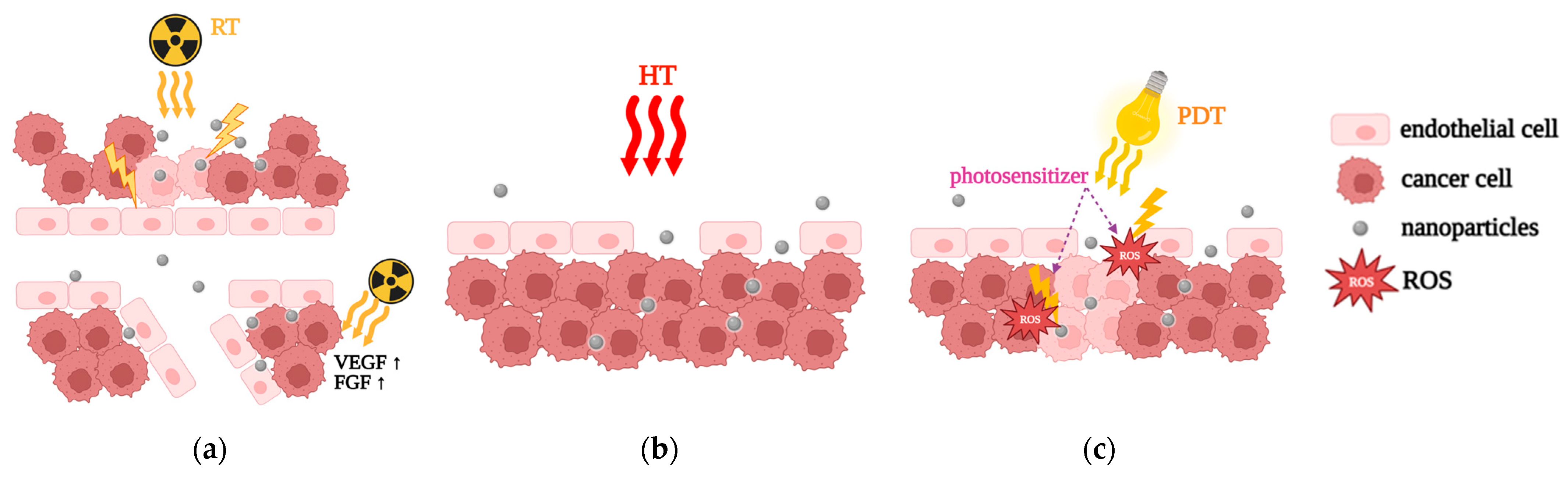
5.2.1. Radiation Therapy (RT)

Radiation therapy (RT) (Figure 3a) is a well-established physical strategy that enhances the EPR effect by increasing vascular permeability and reducing interstitial fluid pressure within tumors. RT achieves this by inducing endothelial cell apoptosis and upregulating the expression of pro-angiogenic factors such as vascular endothelial growth factor (VEGF) and fibroblast growth factor (FGF) [174]. These changes facilitate the penetration and distribution of nanoparticles (NPs) within the tumor, thereby improving drug delivery [175].
RT is often combined with liposomal chemotherapy to maximize therapeutic outcomes. For instance, De Lange Davies et al. demonstrated that combining RT with Caelyx® (liposomal doxorubicin) significantly improved drug distribution throughout osteosarcoma xenograft models. This combination not only delayed tumor growth but also extended the tumor doubling time compared to control conditions, highlighting the synergistic effects of RT and liposomal chemotherapy [176].
Further studies by Lammers et al. explored the combination of carrier-based radiochemotherapy for solid tumors. They found that administering RT 24 h after the intravenous (IV) injection of 31 and 65 kDa hydroxypropyl methacrylamide (HPMA) copolymer NPs significantly increased NP accumulation in various tumor types. This approach consistently enhanced the EPR effect across different tumor models, demonstrating its potential for improving nanoparticle-based cancer treatment [177].
However, RT faces challenges related to non-specific tissue targeting, which can result in damage to healthy cells. To address this, researchers have explored the use of radiosensitizers, such as NPs loaded with radioenhancers or chemotherapy agents [178]. These radiosensitizers aim to intensify tumor treatment efficacy while sparing healthy tissue. For example, gold nanoparticles have been studied for their ability to enhance the EPR effect during RT. Ashton et al. investigated vascular-targeted AuNPs combined with RT and liposomal iodine delivery. They found that AuNPs significantly increased the accumulation of liposomal iodine in tumors, particularly at lower radiation doses, demonstrating their potential as effective radiosensitizers compared to non-functionalized nanomedicine combined with RT [179].

5.2.2. Hyperthermia (HT)

Hyperthermia (HT) (Figure 3b) is another widely studied physical strategy that enhances the EPR effect by heating tumors to mild temperatures (typically 40–43 °C). This mild heat induces vasodilation, increasing blood flow to the tumor and improving the delivery of nanodrugs. The enhanced blood flow is the principal mechanism through which HT boosts the EPR effect [180,181].
Recent studies have explored the combination of HT with ultrasound to enhance the effectiveness of nanomedicine. For example, Fu et al. developed a nanomedicine system combining ferrate and doxorubicin NPs, which exploit the EPR effect for tumor accumulation. Ultrasound-induced hyperthermia triggered the simultaneous release of ferrate and doxorubicin, boosting tumor oxygenation and enhancing doxorubicin’s ability to induce tumor cell death. Their study demonstrated a significant reduction in tumor volumes in mice with osteosarcoma, particularly when using NPs containing both ferrate and doxorubicin in combination with ultrasound [182].
Thermosensitive liposomes have also been extensively researched for their potential applications in oncology, particularly in breast cancer treatment. Vujaskovic et al. demonstrated that combining paclitaxel and liposomal doxorubicin with hyperthermia as neoadjuvant therapy is a viable and well-tolerated treatment approach for patients with locally advanced breast cancer (LABC) [183]. Similarly, Zagar et al. found that combining liposomal doxorubicin with mild local hyperthermia was safe and resulted in objective responses in heavily pretreated patients with chest wall recurrences of breast cancer [184].
Despite its potential, HT is not widely adopted in clinical practice due to challenges such as temperature control and inadequate monitoring. Addressing these issues is critical for the broader application of HT in enhancing the EPR effect [5].

5.2.3. Photodynamic Therapy (PDT)

Photodynamic therapy (PDT) (Figure 3c) is a physical strategy that uses reactive oxygen species (ROS) generated by photosensitizers to induce tumor cell death. Kim et al. developed a nanoparticle system designed for PDT that responds to ROS. These NPs encapsulate both doxorubicin and a photosensitizer, accumulating in tumors due to the EPR effect. During PDT, ROS trigger the release of doxorubicin and the photosensitizer by degrading the linker between the polyethylene glycol (PEG) coating and doxorubicin. This release mechanism enhances ROS production within the tumor, improving the therapeutic effectiveness of the treatment [185].
However, the effectiveness of PDT is hindered by the hypoxic conditions commonly found within tumors, which limit the therapy’s reliance on the consumption of singlet oxygen. Overcoming this limitation remains a key area of research for improving PDT-based cancer treatments [186].

5.3. Pharmacological Strategies

5.3.1. Vascular Normalization

In the realm of cancer treatment, one classical approach involves the use of antiangiogenic agents to disrupt the blood supply to tumors, thereby cutting off oxygen and nutrients. While this strategy aims to starve tumors, its effectiveness as a standalone treatment has been limited in clinical practice. Recent studies have explored the concept of vascular normalization, which seeks to restore the structure and function of tumor vasculature, thereby improving drug delivery and therapeutic outcomes.
Tong et al. demonstrated that vascular normalization could be achieved using moderate doses of anti-VEGF receptor antibodies. This approach reduced necrotic and hypoxic areas within tumors, leading to a more uniform distribution of therapeutic agents. By normalizing the tumor vasculature, the study highlighted the potential for improved drug penetration and efficacy [187].
Another promising strategy involves the use of tyrosine kinase inhibitors (TKIs), a class of small molecules that target specific signaling pathways involved in angiogenesis. For instance, erlotinib, a TKI targeting the epidermal growth factor receptor (EGFR), has been shown to enhance the accumulation of human serum albumin-bound paclitaxel in various tumor models. This improvement in drug delivery is attributed to the normalization of tumor vasculature, which facilitates the enhanced permeability and retention (EPR) effect [188].
Imatinib, another TKI that inhibits VEGF-independent angiogenesis, has also been investigated for its ability to promote vascular normalization. In studies involving A549 lung carcinoma, imatinib was found to increase the accumulation of nanomedicine within tumors, further underscoring the potential of TKIs in enhancing EPR-mediated drug delivery and improving therapeutic outcomes [188].

5.3.2. Fibrinolytic Co-Therapy

Cancer patients often experience a hypercoagulative state, which exacerbates mortality and morbidity. This condition can lead to vascular occlusion within tumors, driven by the overexpression of proinflammatory cytokines, platelet activation, and leukocytes overexpressing tissue factors. These factors not only cause vascular blockages but also contribute to tumor heterogeneity in the EPR effect, resulting in poor drug delivery [189].
Fibrinolytic therapy, which involves the dissolution of fibrin clots in blocked vessels, has emerged as a potential strategy to improve tumor vasculature and enhance drug delivery [190]. For example, Zhang et al. pretreated A549 tumor xenograft mice with tissue plasminogen activator (tPA), a thrombolytic drug, before administering paclitaxel-loaded nanoparticles. The pretreatment with tPA reduced fibrin deposits at tumor vessel walls, leading to higher accumulation and deeper penetration of 115 nm nanoparticles compared to saline-treated groups. This study demonstrated the potential of fibrinolytic therapy to enhance the EPR effect and improve nanomedicine delivery [191].
Another study investigated the impact of tPA pretreatment on the biodistribution and penetration of Doxil® (liposomal doxorubicin) in tumors. While tPA pretreatment did not significantly affect the biodistribution of Doxil®, it enhanced the penetration of the drug into tumors, further supporting the role of fibrinolytic therapy in improving drug delivery [192].
Despite its potential, fibrinolytic co-therapy faces challenges such as the short half-life and low specificity of tPA. Addressing these limitations is critical for the broader application of fibrinolytic therapy in cancer treatment [190].

5.3.3. Bradykinin (BK), BK Mediators, Nitric Oxide (NO), and NO Mediators

Bradykinin (BK) and its receptors, B1 and B2, play a significant role in regulating vascular permeability. The activation of these receptors promotes vasodilation, disrupts endothelial cell junctions, and contracts the endothelial cell cytoskeleton, thereby increasing vascular permeability. Many tumors overexpress BK receptors and contain high levels of bradykinin, which enhances tumor vascular permeability and facilitates the EPR effect [193].
Researchers have explored the use of angiotensin-converting enzyme (ACE) inhibitors, such as enalapril and captopril, to prevent the degradation of bradykinin. By inhibiting bradykinin degradation, these ACE inhibitors enhance tumor vascular permeability and improve the accumulation of nanomedicine. For instance, enalapril has been shown to increase the delivery of bacterial agents to tumors, while captopril improves nanoparticle accumulation in glioma tumors. These findings highlight the potential of ACE inhibitors in enhancing the EPR effect and improving therapeutic outcomes [194,195].
Nitric oxide (NO) is another key mediator of vascular permeability. NO enhances tumor vessel permeability by binding to soluble guanylate cyclase in smooth muscle cells, leading to vasodilation. In cancer, the overexpression of endothelial nitric oxide synthases (eNOS) and inducible nitric oxide synthases (iNOS) in tumors allows for selective NO production, which can be exploited to improve drug delivery [196].
Studies have investigated the use of NO-generating agents, such as L-arginine and hydroxyurea, to increase NO levels in tumors. These agents have been shown to enhance the accumulation of therapeutic agents in tumors, inhibit tumor growth, and reduce metastasis. By leveraging the role of NO in vascular permeability, researchers aim to develop more effective strategies for enhancing the EPR effect and improving cancer treatment outcomes [197].

6. Clinical Trials and Limitations

Nanomedicine has made significant strides in cancer treatment by leveraging the enhanced permeability and retention (EPR) effect of the tumor microenvironment to facilitate drug delivery. The EPR effect allows nanoparticles to accumulate in tumors due to the leaky nature of tumor vasculature, which is particularly advantageous for nanocarriers like liposomes and micelles. These nanoparticles passively target tumors, enhancing the delivery and effectiveness of chemotherapeutic agents. However, translating the EPR effect from preclinical models to clinical applications remains challenging [198]. A major limitation is tumor heterogeneity, which refers to variations in vascular permeability and interstitial pressure between tumor types. Such differences restrict the effectiveness of the EPR effect across various cancer types. Research by Jain et al. emphasizes that the tumor microenvironment, including factors like blood flow and pressure, significantly influences nanoparticle delivery [11]. Thus, modulating these factors is crucial to optimizing nanoparticle distribution and therapeutic outcomes.
Moreover, human and animal models differ considerably in tumor architecture and immune responses, further complicating clinical translation [199]. These disparities in tumor biology lead to variability in how nanoparticles accumulate and function within different individuals. Several strategies have been proposed to enhance the EPR effect in cancer therapy. One approach involves the use of nitric oxide donors to improve blood flow, while another uses enzyme-based methods, such as matrix metalloproteinases, to increase nanoparticle penetration through the tumor stroma [200,201]. Additionally, functionalized nanoparticles targeting specific tumor receptors, alongside external stimuli like hyperthermia and ultrasound, have shown promise in boosting drug delivery and therapeutic efficacy [202,203].
Despite these advances, clinical translation remains inconsistent. Variability in tumor vascularization, immune infiltration, and extracellular matrix (ECM) composition affects the accumulation of nanoparticles in tumors. Even in cases where EPR conditions appear favorable, therapeutic efficacy can be unpredictable. Furthermore, factors such as nanoparticle size, surface properties, and circulation time impact biodistribution and clearance, introducing challenges like off-target delivery and rapid clearance by the immune system [180]. To address these issues, several clinical trials are underway to optimize the EPR effect in cancer therapy. Notable trials include the use of ThermoDox®, a thermosensitive liposomal doxorubicin formulation, in combination with radiofrequency ablation (RFA) to enhance vascular permeability and improve drug delivery in hepatocellular carcinoma (HCC) [204]. Another trial explores sonoporation, using ultrasound and microbubbles to create temporary pores in tumor vasculature, enhancing nanoparticle delivery in pancreatic cancer [205]. In glioblastoma multiforme, magnetic nanoparticles are directed by external magnetic fields to improve tumor targeting and accumulation [206]. Other trials are investigating doxorubicin-loaded nanoparticles combined with vascular-modulating agents in breast cancer, as well as liposomal irinotecan (Onivyde®) in metastatic pancreatic cancer, which aim to increase drug retention through the EPR effect [207,208]. Table 3 summarizes the primary information on the clinical trials referred to above.
In conclusion, while the EPR effect remains a cornerstone of cancer nanomedicine, its clinical application faces limitations due to tumor heterogeneity and the variability of patient responses. Current strategies, such as tumor microenvironment modulation, ligand-targeted nanoparticles, and external physical stimuli, hold promise for enhancing therapeutic outcomes. Nevertheless, continued refinement and better patient stratification are essential for the successful clinical translation of EPR-based therapies.

7. Conclusions and Future Perspectives

Significant progress has been made in developing and refining strategies to enhance the EPR effect, a key mechanism designed to improve the delivery of therapeutic agents, specifically to tumor tissues. Despite these advancements, the clinical application of EPR-based strategies remains fraught with challenges. These difficulties arise largely from the complex and heterogeneous nature of the tumor microenvironment, which varies widely across different tumor types and between species, complicating the effective translation of preclinical successes into clinical treatments.
One of the major issues is the intrinsic variability within the TME. Tumors exhibit a high degree of heterogeneity in their cellular composition, extracellular matrix, blood vessel architecture, and metabolic activity. This variability can significantly influence how drugs are distributed and retained within the tumor tissue. Strategies that have shown promise in preclinical animal models often encounter unexpected failures in human clinical trials. These failures highlight the substantial differences between the TME in animal models and that in human tumors. For instance, the vascular architecture in animal tumors may not accurately represent the complexity found in human tumors, leading to discrepancies in how drugs are delivered and absorbed.
Addressing these challenges requires a multifaceted approach that involves a deeper and more comprehensive understanding of the fundamental mechanisms underpinning the EPR effect. This understanding must be complemented by advances in tools and methodologies for studying and manipulating the TME. Researchers need to develop and refine experimental models and techniques that more accurately reflect human tumor conditions. This includes improving imaging technologies to visualize the TME in greater detail, developing molecular profiling techniques to better understand tumor biology, and creating more sophisticated preclinical models that closely mimic human disease.
Recent advancements in the field offer renewed hope for overcoming these obstacles. Innovations in imaging and diagnostic technologies, such as high-resolution imaging, multiplexed molecular assays, and advanced biomarker analysis, are providing new insights into the TME at both cellular and molecular levels. These advancements reveal previously unrecognized aspects of tumor vasculature and tumor–stroma interactions, which may identify new therapeutic targets. For example, advances in nanoparticle technology have enhanced the ability to target specific components of the TME, potentially improving drug delivery and efficacy.
Moreover, integrating EPR-enhancing strategies with other therapeutic modalities, such as targeted therapies, immunotherapies, and combination therapies, may offer a more effective approach to cancer treatment. By combining EPR-enhancing strategies with these complementary approaches, researchers can exploit synergies between different modalities, potentially overcoming the limitations of each individual approach. For instance, combining EPR-enhanced drug delivery with immune checkpoint inhibitors or targeted therapies may improve therapeutic outcomes by not only increasing drug concentration at the tumor site but also by modulating the immune response.
To accelerate progress, concerted efforts are required from researchers, clinicians, and the pharmaceutical industry to develop and validate new strategies for optimizing the EPR effect and enhancing the TME. This includes collaborative research to generate large, well-characterized datasets that can provide insights into the variability of the TME and how it affects drug delivery and efficacy. Additionally, there is a need for continuous innovation in therapeutic formulations and delivery systems to address the specific challenges posed by different tumor types and individual patient characteristics.
In summary, while the clinical application of EPR-enhancing strategies faces significant challenges due to the heterogeneous nature of the TME and the limitations of existing preclinical models, ongoing research and technological advancements hold promise for overcoming these obstacles. By deepening our understanding of the TME and integrating EPR-enhancing strategies with complementary therapeutic approaches, we can move closer to developing more effective and personalized cancer therapies. This progress will be critical in advancing cancer treatment and improving patient outcomes in clinical settings.

Author Contributions

Conceptualization, E.A.P. and N.L.; methodology, E.A.P., N.L., I.-A.V., C.M. and M.-A.G.; validation, E.A.P., N.L., I.-A.V., C.M. and M.-A.G.; formal analysis, I.-A.V., C.M. and M.-A.G.; investigation, I.-A.V. and C.M.; resources, E.A.P. and N.L.; writing—original draft preparation, I.-A.V. and C.M.; writing—review and editing, E.A.P., N.L., I.-A.V., C.M. and M.-A.G.; visualization, I.-A.V., C.M. and M.-A.G.; supervision, E.A.P. and N.L. All authors have read and agreed to the published version of the manuscript.

Funding

This research received no external funding.

Institutional Review Board Statement

Not applicable.

Data Availability Statement

Data sharing is not applicable.

Conflicts of Interest

The authors declare no conflicts of interest.

References

  1. Konno, T.; Maeda, H.; Iwai, K.; Maki, S.; Tashiro, S.; Uchida, M.; Miyauchi, Y. Selective targeting of anti-cancer drug and simultaneous image enhancement in solid tumors by arterially administered lipid contrast medium. Cancer 1984, 54, 2367–2374. [Google Scholar] [CrossRef] [PubMed]
  2. Lahooti, B.; Akwii, R.G.; Zahra, F.T.; Sajib, M.S.; Lamprou, M.; Alobaida, A.; Lionakis, M.S.; Mattheolabakis, G.; Mikelis, C.M. Targeting endothelial permeability in the EPR effect. J. Control. Release 2023, 361, 212–235. [Google Scholar] [CrossRef] [PubMed]
  3. Sun, R.; Xiang, J.; Zhou, Q.; Piao, Y.; Tang, J.; Shao, S.; Zhou, Z.; Bae, Y.H.; Shen, Y. The tumor EPR effect for cancer drug delivery: Current status, limitations, and alternatives. Adv. Drug Deliv. Rev. 2022, 191, 114614. [Google Scholar] [CrossRef]
  4. Folkman, J. Role of angiogenesis in tumor growth and metastasis. Semin. Oncol. 2002, 29, 15–18. [Google Scholar] [CrossRef]
  5. Ikeda-Imafuku, M.; Wang, L.L.W.; Rodrigues, D.; Shaha, S.; Zhao, Z.; Mitragotri, S. Strategies to improve the EPR effect: A mechanistic perspective and clinical translation. J. Control. Release 2022, 345, 512–536. [Google Scholar] [CrossRef]
  6. Wu, J. The enhanced permeability and retention (EPR) effect: The significance of the concept and methods to enhance its application. J. Pers. Med. 2021, 11, 771. [Google Scholar] [CrossRef]
  7. Yhee, J.Y.; Son, S.; Son, S.; Joo, M.K.; Kwon, I.C. The EPR Effect in Cancer Therapy. Cancer Targeted Drug Delivery. In Cancer Targeted Drug Delivery; Bae, Y., Mrsny, R., Park, K., Eds.; Springer: New York, NY, USA, 2013; pp. 621–632. [Google Scholar] [CrossRef]
  8. Fang, J.; Nakamura, H.; Maeda, H. The EPR effect: Unique features of tumor blood vessels for drug delivery, factors involved, and limitations and augmentation of the effect. Adv. Drug Deliv. Rev. 2011, 63, 136–151. [Google Scholar] [CrossRef]
  9. Youn, Y.S.; Bae, Y.H. Perspectives on the past, present, and future of cancer nanomedicine. Adv. Drug Deliv. Rev. 2018, 130, 3–11. [Google Scholar] [CrossRef]
  10. Blanco, E.; Shen, H.; Ferrari, M. Principles of nanoparticle design for overcoming biological barriers to drug delivery. Nat. Biotechnol. 2015, 33, 941–951. [Google Scholar] [CrossRef]
  11. Jain, R.K. Normalizing tumor microenvironment to treat cancer: Bench to bedside to biomarkers. J. Clin. Oncol. 2013, 31, 2205–2218. [Google Scholar] [CrossRef]
  12. Peer, D.; Karp, J.M.; Hong, S.; Farokhzad, O.C.; Margalit, R.; Langer, R. Nanocarriers as an emerging platform for cancer therapy. Nat. Nanotechnol. 2007, 2, 751–760. [Google Scholar] [CrossRef] [PubMed]
  13. Geng, Y.; Dalhaimer, P.; Cai, S.; Tsai, R.; Tewari, M.; Minko, T.; Discher, D.E. Shape effects of filaments versus spherical particles in flow and drug delivery. Nat. Nanotechnol. 2007, 2, 249–255. [Google Scholar] [CrossRef]
  14. Yu, W.; Liu, R.; Zhou, Y.; Gao, H. Size-tunable strategies for a tumor targeted drug delivery system. ACS Cent. Sci. 2020, 6, 100–116. [Google Scholar] [CrossRef] [PubMed]
  15. Tredan, O.; Galmarini, C.M.; Patel, K.; Tannock, I.F. Drug resistance and the solid tumor microenvironment. J. Natl. Cancer Inst. 2007, 99, 1441–1454. [Google Scholar] [CrossRef] [PubMed]
  16. Stylianopoulos, T.; Jain, R.K. Design considerations for nanotherapeutics in oncology. Nanomedicine 2015, 10, 1461–1474. [Google Scholar] [CrossRef]
  17. Kim, T.; Hyeon, T. Applications of inorganic nanoparticles as therapeutic agents. Nanotechnology 2014, 25, 012001. [Google Scholar] [CrossRef]
  18. Mukherjee, P.; Bhattacharya, R.; Wang, P.; Wang, L.; Basu, S.; Nagy, J.A.; Atala, A.; Mukhopadhyay, D.; Soker, S. Antiangiogenic properties of gold nanoparticles. Clin. Cancer Res. 2005, 11, 3530–3534. [Google Scholar] [CrossRef]
  19. Barui, A.K.; Nethi, S.K.; Haque, S.; Basuthakur, P.; Patra, C.R. Recent development of metal nanoparticles for angiogenesis study and their therapeutic applications. ACS Appl. Bio Mater. 2019, 2, 5492–5511. [Google Scholar] [CrossRef]
  20. Kang, H.; Rho, S.; Stiles, W.R.; Hu, S.; Baek, Y.; Hwang, D.W.; Kashiwagi, S.; Kim, M.S.; Choi, H.S. Size-dependent EPR effect of polymeric nanoparticles on tumor targeting. Adv. Healthc. Mater. 2020, 9, e1901223. [Google Scholar] [CrossRef]
  21. Acharya, S.; Sahoo, S.K. PLGA nanoparticles containing various anticancer agents and tumour delivery by EPR effect. Adv. Drug Deliv. Rev. 2011, 63, 170–183. [Google Scholar] [CrossRef]
  22. Ray, S.; Li, Z.; Hsu, C.H.; Hwang, L.P.; Lin, Y.C.; Chou, P.T.; Lin, Y.Y. Dendrimer- and copolymer-based nanoparticles for magnetic resonance cancer theranostics. Theranostics 2018, 8, 6322–6349. [Google Scholar] [CrossRef] [PubMed]
  23. Doppalapudi, S.; Jain, A.; Domb, A.J.; Khan, W. Biodegradable polymers for targeted delivery of anti-cancer drugs. Expert Opin. Drug Deliv. 2016, 13, 891–909. [Google Scholar] [CrossRef] [PubMed]
  24. García-Pinel, B.; Porras-Alcalá, C.; Ortega-Rodríguez, A.; Sarabia, F.; Prados, J.; Melguizo, C.; López-Romero, J.M. Lipid-based nanoparticles: Application and recent advances in cancer treatment. Nanomaterials 2019, 9, 638. [Google Scholar] [CrossRef] [PubMed]
  25. Wong, H.L.; Bendayan, R.; Rauth, A.M.; Li, Y.; Wu, X.Y. Chemotherapy with anticancer drugs encapsulated in solid lipid nanoparticles. Adv. Drug Deliv. Rev. 2007, 59, 491–504. [Google Scholar] [CrossRef]
  26. Zhao, X.; Chen, Q.; Liu, W.; Li, Y.; Tang, H.; Liu, X.; Yang, X. Codelivery of doxorubicin and curcumin with lipid nanoparticles results in improved efficacy of chemotherapy in liver cancer. Int. J. Nanomed. 2014, 10, 257–270. [Google Scholar] [CrossRef]
  27. Lin, X.; Gao, R.; Zhang, Y.; Qi, N.; Zhang, Y.; Zhang, K.; He, H.; Tang, X. Lipid nanoparticles for chemotherapeutic applications: Strategies to improve anticancer efficacy. Expert Opin. Drug Deliv. 2012, 9, 767–781. [Google Scholar] [CrossRef]
  28. Sailor, M.J.; Park, J.-H. Hybrid nanoparticles for detection and treatment of cancer. Adv. Mater. 2012, 24, 3779–3802. [Google Scholar] [CrossRef]
  29. Bort, G.; Lux, F.; Dufort, S.; Crémillieux, Y.; Verry, C.; Tillement, O. EPR-mediated tumor targeting using ultrasmall-hybrid nanoparticles: From animal to human with theranostic AGuIX nanoparticles. Theranostics 2020, 10, 1319–1331. [Google Scholar] [CrossRef]
  30. Upreti, M.; Jyoti, A.; Sethi, P. Tumor microenvironment and nanotherapeutics. Transl. Cancer Res. 2013, 4, 309–319. [Google Scholar] [CrossRef]
  31. Wang, J.J.; Lei, K.F.; Han, F. Tumor microenvironment: Recent advances in various cancer treatments. Eur. Rev. Med. Pharmacol. Sci. 2018, 22, 3855–3864. [Google Scholar] [CrossRef]
  32. Anderson, N.M.; Simon, M.C. The tumor microenvironment. Curr. Biol. 2020, 30, R921–R925. [Google Scholar] [CrossRef] [PubMed]
  33. Xiao, Y.; Yu, D. Tumor microenvironment as a therapeutic target in cancer. Pharmacol. Ther. 2020, 221, 107753. [Google Scholar] [CrossRef] [PubMed]
  34. Gulzar, A.; Xu, J.; Wang, C.; He, F.; Yang, D.; Gai, S.; Yang, P.; Lin, J.; Jin, D.; Xing, B. Tumour microenvironment responsive nanoconstructs for cancer theranostic. Nano Today 2019, 26, 16–56. [Google Scholar] [CrossRef]
  35. Weber, C.E.; Kuo, P.C. The tumor microenvironment. Surg. Oncol. 2012, 21, 172–177. [Google Scholar] [CrossRef]
  36. Arneth, B. Tumor microenvironment. Medicina 2020, 56, 15. [Google Scholar] [CrossRef]
  37. Turley, S.J.; Cremasco, V.; Astarita, J.L. Immunological hallmarks of stromal cells in the tumour microenvironment. Nat. Rev. Immunol. 2015, 15, 669–682. [Google Scholar] [CrossRef]
  38. Ribeiro Franco, P.I.; Rodrigues, A.P.; de Menezes, L.B.; Pacheco Miguel, M. Tumor microenvironment components: Allies of cancer progression. Pathol. Res. Pract. 2019, 216, 152729. [Google Scholar] [CrossRef]
  39. Junttila, M.R.; de Sauvage, F.J. Influence of tumour micro-environment heterogeneity on therapeutic response. Nature 2013, 501, 346–354. [Google Scholar] [CrossRef]
  40. Roma-Rodrigues, C.; Mendes, R.; Baptista, P.V.; Fernandes, A.R. Targeting tumor microenvironment for cancer therapy. Int. J. Mol. Sci. 2019, 20, 840. [Google Scholar] [CrossRef]
  41. Wu, T.; Dai, Y. Tumor microenvironment and therapeutic response. Cancer Lett. 2017, 387, 61–68. [Google Scholar] [CrossRef]
  42. Valkenburg, K.C.; de Groot, A.E.; Pienta, K.J. Targeting the tumour stroma to improve cancer therapy. Nat. Rev. Clin. Oncol. 2018, 15, 366–381. [Google Scholar] [CrossRef] [PubMed]
  43. Nikolopoulou, P.A.; Koufaki, M.A.; Kostourou, V. The adhesome network: Key components shaping the tumour stroma. Cancers 2021, 13, 525. [Google Scholar] [CrossRef]
  44. Petrova, V.; Annicchiarico-Petruzzelli, M.; Melino, G.; Amelio, I. The hypoxic tumour microenvironment. Oncogenesis 2018, 7, 10. [Google Scholar] [CrossRef]
  45. Farc, O.; Cristea, V. An overview of the tumor microenvironment, from cells to complex networks. Exp. Ther. Med. 2021, 21, 96. [Google Scholar] [CrossRef]
  46. Lei, X.; Lei, Y.; Li, J.K.; Du, W.X.; Li, R.G.; Yang, J.; Li, J.; Li, F.; Tan, H.B. Immune cells within the tumor microenvironment: Biological functions and roles in cancer immunotherapy. Cancer Lett. 2020, 470, 126–133. [Google Scholar] [CrossRef]
  47. Atiya, H.; Frisbie, L.; Pressimone, C.; Coffman, L. Mesenchymal stem cells in the tumor microenvironment. In Tumor Microenvironment: Advances in Experimental Medicine and Biology; Birbrair, A., Ed.; Springer: Cham, Switzerland, 2020; Volume 1234, pp. 31–42. [Google Scholar] [CrossRef]
  48. Jiang, Z.; Zhou, J.; Li, L.; Liao, S.; He, J.; Zhou, S.; Zhou, Y. Pericytes in the tumor microenvironment. Cancer Lett. 2023, 556, 216074. [Google Scholar] [CrossRef] [PubMed]
  49. Zlotnik, A. Chemokines and cancer. Int. J. Cancer 2006, 119, 2026–2029. [Google Scholar] [CrossRef] [PubMed]
  50. Goustin, A.S.; Leof, E.B.; Shipley, G.D.; Moses, H.L. Growth factors and cancer. Cancer Res. 1986, 46, 1015–1029. [Google Scholar]
  51. Wang, M.; Rousseau, B.; Qiu, K.; Huang, G.; Zhang, Y.; Su, H.; Le Bihan-Benjamin, C.; Khati, I.; Artz, O.; Foote, M.B.; et al. Killing tumor-associated bacteria with a liposomal antibiotic generates neoantigens that induce anti-tumor immune responses. Nat. Biotechnol. 2024, 42, 1263–1274. [Google Scholar] [CrossRef]
  52. Walker, C.; Mojares, E.; Del Río Hernández, A. Role of extracellular matrix in development and cancer progression. Int. J. Mol. Sci. 2018, 19, 3028. [Google Scholar] [CrossRef]
  53. Meşe, G.; Richard, G.; White, T.W. Gap junctions: Basic structure and function. J. Investig. Dermatol. 2007, 127, 2516–2524. [Google Scholar] [CrossRef]
  54. Schmidt, K.; Windler, R.; De Wit, C. Communication through gap junctions in the endothelium. Adv. Pharmacol. 2016, 77, 209–240. [Google Scholar] [CrossRef] [PubMed]
  55. Beckmann, A.; Hainz, N.; Tschernig, T.; Meier, C. Facets of communication: Gap junction ultrastructure and function in cancer stem cells and tumor cells. Cancers 2019, 11, 288. [Google Scholar] [CrossRef] [PubMed]
  56. Ma, F.; Zhang, S.; Song, L.; Wang, B.; Wei, L.; Zhang, F. Applications and analytical tools of cell communication based on ligand-receptor interactions at single cell level. Cell Biosci. 2021, 11, 121. [Google Scholar] [CrossRef] [PubMed]
  57. De Palma, M.; Biziato, D.; Petrova, T.V. Microenvironmental regulation of tumour angiogenesis. Nat. Rev. Cancer 2017, 17, 457–474. [Google Scholar] [CrossRef]
  58. Anderson, J.M.; Van Itallie, C.M. Physiology and function of the tight junction. Cold Spring Harb. Perspect. Biol. 2009, 1, a002584. [Google Scholar] [CrossRef]
  59. Tsukita, S.; Furuse, M. Claudin-based barrier in simple and stratified cellular sheets. Curr. Opin. Cell Biol. 2002, 14, 531–536. [Google Scholar] [CrossRef]
  60. Martin, T.A.; Jiang, W.G. Loss of tight junction barrier function and its role in cancer metastasis. Biochim. Biophys. Acta Biomembr. 2009, 1788, 872–891. [Google Scholar] [CrossRef]
  61. Onder, T.T.; Gupta, P.B.; Mani, S.A.; Yang, J.; Lander, E.S.; Weinberg, R.A. Loss of E-cadherin promotes metastasis via multiple downstream transcriptional pathways. Nature 2008, 453, 1000–1004. [Google Scholar] [CrossRef]
  62. Mittal, R.; Karhu, E.; Wang, J.S.; Delgado, S.; Zukerman, R.; Mittal, J.; Jhaveri, V.M. Cell communication by tunneling nanotubes: Implications in disease and therapeutic applications. J. Cell. Physiol. 2019, 234, 1130–1146. [Google Scholar] [CrossRef]
  63. Dagar, S.; Pathak, D.; Oza, H.V.; Mylavarapu, S.V. Tunneling nanotubes and related structures: Molecular mechanisms of formation and function. Biochem. J. 2021, 478, 3977–3998. [Google Scholar] [CrossRef] [PubMed]
  64. Gurke, S.; Barroso, J.F.; Gerdes, H.H. The art of cellular communication: Tunneling nanotubes bridge the divide. Histochem. Cell Biol. 2008, 129, 539–550. [Google Scholar] [CrossRef] [PubMed]
  65. Roehlecke, C.; Schmidt, M.H.H. Tunneling nanotubes and tumor microtubes in cancer. Cancers 2020, 12, 857. [Google Scholar] [CrossRef]
  66. Schubert, A.; Boutros, M. Extracellular vesicles and oncogenic signaling. Mol. Oncol. 2021, 15, 3–26. [Google Scholar] [CrossRef]
  67. Fonseca, P.; Vardaki, I.; Occhionero, A.; Panaretakis, T. Metabolic and signaling functions of cancer cell-derived extracellular vesicles. Int. Rev. Cell Mol. Biol. 2016, 326, 175–199. [Google Scholar] [CrossRef]
  68. Kanada, M.; Bachmann, M.H.; Contag, C.H. Signaling by extracellular vesicles advances cancer hallmarks. Trends Cancer 2016, 2, 84–94. [Google Scholar] [CrossRef]
  69. Yi, M.; Li, T.; Niu, M.; Zhang, H.; Wu, Y.; Wu, K.; Dai, Z. Targeting cytokine and chemokine signaling pathways for cancer therapy. Signal Transduct. Target. Ther. 2024, 9, 176. [Google Scholar] [CrossRef]
  70. Vicari, A.P.; Caux, C. Chemokines in cancer. Cytokine Growth Factor Rev. 2002, 13, 143–154. [Google Scholar] [CrossRef]
  71. Lacalle, R.A.; Blanco, R.; Carmona-Rodríguez, L.; Martín-Leal, A.; Mira, E.; Mañes, S. Chemokine receptor signaling and the hallmarks of cancer. Int. Rev. Cell Mol. Biol. 2017, 331, 181–244. [Google Scholar] [CrossRef]
  72. Subhan, M.A.; Yalamarty, S.S.K.; Filipczak, N.; Parveen, F.; Torchilin, V.P. Recent advances in tumor targeting via EPR effect for cancer treatment. J. Pers. Med. 2021, 11, 571. [Google Scholar] [CrossRef]
  73. Theocharis, A.D.; Skandalis, S.S.; Gialeli, C.; Karamanos, N.K. Extracellular matrix structure. Adv. Drug Deliv. Rev. 2016, 97, 4–27. [Google Scholar] [CrossRef] [PubMed]
  74. Nagy, J.A.; Chang, S.H.; Shih, S.C.; Dvorak, A.M.; Dvorak, H.F. Heterogeneity of the tumor vasculature. Semin. Thromb. Hemost. 2010, 36, 321–331. [Google Scholar] [CrossRef] [PubMed]
  75. Claesson-Welsh, L. Vascular permeability-the essentials. Ups. J. Med. Sci. 2015, 120, 135–143. [Google Scholar] [CrossRef]
  76. Siemann, D.W. The unique characteristics of tumor vasculature and preclinical evidence for its selective disruption by tumor-vascular disrupting agents. Cancer Treat. Rev. 2011, 37, 63–74. [Google Scholar] [CrossRef]
  77. Nussenbaum, F.; Herman, I.M. Tumor angiogenesis: Insights and innovations. J. Oncol. 2010, 2010, 132641. [Google Scholar] [CrossRef]
  78. Hiratsuka, S. Vasculogenensis, angiogenesis and special features of tumor blood vessels. Front. Biosci. 2011, 16, 1413–1427. [Google Scholar] [CrossRef]
  79. Eelen, G.; Treps, L.; Li, X.; Carmeliet, P. Basic and therapeutic aspects of angiogenesis updated. Circ. Res. 2020, 127, 310–329. [Google Scholar] [CrossRef]
  80. Potente, M.; Gerhardt, H.; Carmeliet, P. Basic and therapeutic aspects of angiogenesis. Cell 2011, 146, 873–887. [Google Scholar] [CrossRef]
  81. Viallard, C.; Larrivée, B. Tumor angiogenesis and vascular normalization: Alternative therapeutic targets. Angiogenesis 2017, 20, 409–426. [Google Scholar] [CrossRef]
  82. Nakamura, Y.; Mochida, A.; Choyke, P.L.; Kobayashi, H. Nanodrug delivery: Is the enhanced permeability and retention effect sufficient for curing cancer? Bioconjug. Chem. 2016, 27, 2225–2238. [Google Scholar] [CrossRef] [PubMed]
  83. Orasanu, C.I.; Aschie, M.; Deacu, M.; Bosoteanu, M.; Vamesu, S.; Enciu, M.; Bălţătescu, G.I.; Cozaru, G.C.; Mitroi, A.F.; Voda, R.I. Implications of cellular immaturity in necrosis and microvascularization in glioblastomas IDH-wild-type. Clin. Pract. 2022, 12, 1054–1068. [Google Scholar] [CrossRef] [PubMed]
  84. Nagy, J.A.; Dvorak, H.F. Heterogeneity of the tumor vasculature: The need for new tumor blood vessel type-specific targets. Clin. Exp. Metastasis 2012, 29, 657–662. [Google Scholar] [CrossRef] [PubMed]
  85. Fu, Y.; Nagy, J.A.; Dvorak, A.M.; Dvorak, H.F. Tumor blood vessels: Structure, function and classification. In Antiangiogenic Agents in Cancer Therapy. Cancer Drug Discovery and Development; Teicher, B.A., Ellis, L.M., Eds.; Humana Press: Totowa, NJ, USA, 2008; pp. 205–224. [Google Scholar] [CrossRef]
  86. Armulik, A.; Genové, G.; Betsholtz, C. Pericytes: Developmental, physiological, and pathological perspectives, problems, and promises. Dev. Cell 2011, 21, 193–215. [Google Scholar] [CrossRef]
  87. Cao, Y. Tumor angiogenesis and therapy. Biomed. Pharmacother. 2005, 59, S340–S343. [Google Scholar] [CrossRef] [PubMed]
  88. Sandoo, A.; van Zanten, J.J.; Metsios, G.S.; Carroll, D.; Kitas, G.D. The endothelium and its role in regulating vascular tone. Open Cardiovasc. Med. J. 2010, 4, 302–312. [Google Scholar] [CrossRef]
  89. Folkman, J. Tumor angiogenesis. Adv. Cancer Res. 1985, 43, 175–203. [Google Scholar] [CrossRef]
  90. Weis, S.M.; Cheresh, D.A. Tumor angiogenesis: Molecular pathways and therapeutic targets. Nat. Med. 2011, 17, 1359–1370. [Google Scholar] [CrossRef]
  91. Jiang, X.; Wang, J.; Deng, X.; Xiong, F.; Zhang, S.; Gong, Z.; Li, X.; Cao, K.; Deng, H.; He, Y.; et al. The role of microenvironment in tumor angiogenesis. J. Exp. Clin. Cancer Res. 2020, 39, 204. [Google Scholar] [CrossRef]
  92. Al-Ostoot, F.H.; Salah, S.; Khamees, H.A.; Khanum, S.A. Tumor angiogenesis: Current challenges and therapeutic opportunities. Cancer Treat. Res. Commun. 2021, 28, 100422. [Google Scholar] [CrossRef]
  93. Detmar, M. Tumor angiogenesis. J. Investig. Dermatol. Symp. Proc. 2000, 5, 20–23. [Google Scholar] [CrossRef]
  94. Cavallaro, U.; Christofori, G. Molecular mechanisms of tumor angiogenesis and tumor progression. J. Neurooncol. 2000, 50, 63–70. [Google Scholar] [CrossRef] [PubMed]
  95. Nikitenko, L.L. Vascular endothelium in cancer. Cell Tissue Res. 2009, 335, 223–240. [Google Scholar] [CrossRef] [PubMed]
  96. Ronca, R.; Benkheil, M.; Mitola, S.; Struyf, S.; Liekens, S. Tumor angiogenesis revisited: Regulators and clinical implications. Med. Res. Rev. 2017, 37, 1231–1274. [Google Scholar] [CrossRef] [PubMed]
  97. Lugano, R.; Ramachandran, M.; Dimberg, A. Tumor angiogenesis: Causes, consequences, challenges and opportunities. Cell. Mol. Life Sci. 2020, 77, 1745–1770. [Google Scholar] [CrossRef]
  98. Kerbel, R.S. Tumor Angiogenesis. N. Engl. J. Med. 2008, 358, 2039–2049. [Google Scholar] [CrossRef]
  99. García-Román, J.; Zentella-Dehesa, A. Vascular permeability changes involved in tumor metastasis. Cancer Lett. 2013, 335, 259–269. [Google Scholar] [CrossRef]
  100. Azzi, S.; Hebda, J.K.; Gavard, J. Vascular permeability and drug delivery in cancers. Front. Oncol. 2013, 3, 211. [Google Scholar] [CrossRef]
  101. Kim, J.; Cho, H.; Lim, D.-K.; Joo, M.K.; Kim, K. Perspectives for improving the tumor targeting of nanomedicine via the EPR effect in clinical tumors. Int. J. Mol. Sci. 2023, 24, 10082. [Google Scholar] [CrossRef]
  102. Fukumura, D.; Jain, R.K. Tumor microenvironment abnormalities: Causes, consequences, and strategies to normalize. J. Cell Biochem. 2007, 101, 937–949. [Google Scholar] [CrossRef]
  103. Netti, P.A.; Roberge, S.; Boucher, Y.; Baxter, L.T.; Jain, R.K. Effect of transvascular fluid exchange on pressure-flow relationship in tumors: A proposed mechanism for tumor blood flow heterogeneity. Microvasc. Res. 1996, 52, 27–46. [Google Scholar] [CrossRef]
  104. Raghunand, N.; Gatenby, R.A.; Gillies, R.J. Microenvironmental and cellular consequences of altered blood flow in tumours. Br. J. Radiol. 2003, 76, S11–S22. [Google Scholar] [CrossRef] [PubMed]
  105. Tannock, I.F.; Rotin, D. Acid pH in tumors and its potential for therapeutic exploitation. Cancer Res. 1989, 49, 4373–4384. [Google Scholar] [PubMed]
  106. Jain, R.K. Transport of molecules across tumor vasculature. Cancer Metastasis Rev. 1987, 6, 559–593. [Google Scholar] [CrossRef]
  107. Salavati, H.; Debbaut, C.; Pullens, P.; Ceelen, W. Interstitial fluid pressure as an emerging biomarker in solid tumors. Biochim. Biophys. Acta Rev. Cancer 2022, 1877, 188792. [Google Scholar] [CrossRef] [PubMed]
  108. Boucher, Y.; Leunig, M.; Jain, R.K. Tumor angiogenesis and interstitial hypertension. Cancer Res. 1996, 56, 4264–4266. [Google Scholar]
  109. Rofstad, E.K.; Galappathi, K.; Mathiesen, B.S. Tumor interstitial fluid pressure—A link between tumor hypoxia, microvascular density, and lymph node metastasis. Neoplasia 2014, 16, 586–594. [Google Scholar] [CrossRef]
  110. Zhou, H.; Lei, P.-j.; Padera, T.P. Progression of metastasis through lymphatic system. Cells 2021, 10, 627. [Google Scholar] [CrossRef]
  111. Nakamura, H.; Jun, F.; Maeda, H. Development of next-generation macromolecular drugs based on the EPR effect: Challenges and pitfalls. Expert Opin. Drug Deliv. 2014, 12, 53–64. [Google Scholar] [CrossRef]
  112. Karpanen, T.; Alitalo, K. Lymphatic vessels as targets of tumor therapy? J. Exp. Med. 2001, 194, F37–F42. [Google Scholar] [CrossRef]
  113. Maeda, H. The enhanced permeability and retention (EPR) effect in tumor vasculature: The key role of tumor-selective macromolecular drug targeting. Adv. Enzym. Regul. 2001, 41, 189–207. [Google Scholar] [CrossRef]
  114. Maeda, H. The link between infection and cancer: Tumor vasculature, free radicals, and drug delivery to tumors via the EPR effect. Cancer Sci. 2013, 104, 779–789. [Google Scholar] [CrossRef]
  115. Taurin, S.; Greish, K. Enhanced Vascular Permeability in Solid Tumors: A Promise for Anticancer Nanomedicine. In Tight Junctions in Cancer Metastasis. Cancer Metastasis—Biology and Treatment; Martin, T., Jiang, W., Eds.; Springer: Dordrecht, The Netherlands, 2013; Volume 19, pp. 81–118. [Google Scholar] [CrossRef]
  116. Maeda, H.; Kabanov, A.; Kataoka, K.; Okano, T. (Eds.) Polymer Drugs in the Clinical Stage: Advantages and Prospects; Springer: New York, NY, USA, 2003; Volume 519, p. 226. [Google Scholar] [CrossRef]
  117. Bazak, R.; Houri, M.; El Achy, S.; Hussein, W.; Refaat, T. Passive targeting of nanoparticles to cancer: A comprehensive review of the literature. Mol. Clin. Oncol. 2014, 2, 904–908. [Google Scholar] [CrossRef] [PubMed]
  118. Islam, R.; Maeda, H.; Fang, J. Factors affecting the dynamics and heterogeneity of the EPR effect: Pathophysiological and pathoanatomic features, drug formulations and physicochemical factors. Expert Opin. Drug Deliv. 2021, 19, 199–212. [Google Scholar] [CrossRef] [PubMed]
  119. Appiah, E.; Nakamura, H.; Assumang, A.; Etrych, T.; Haratake, M. Chemical modification of bradykinin-polymer conjugates for optimum delivery of nanomedicines to tumors. Nanomed. Nanotechnol. Biol. Med. 2024, 57, 102744. [Google Scholar] [CrossRef] [PubMed]
  120. Matsumura, Y.; Kimura, M.; Yamamoto, T.; Maeda, H. Involvement of the kinin-generating cascade in enhanced vascular permeability in tumor tissue. Jpn. J. Cancer Res. 1988, 79, 1327–1334. [Google Scholar] [CrossRef]
  121. Maeda, H.; Matsumura, Y.; Kato, H. Purification and identification of [hydroxyprolyl3] bradykinin in ascitic fluid from a patient with gastric cancer. J. Biol. Chem. 1988, 263, 16051–16054. [Google Scholar] [CrossRef]
  122. Yoshikawa, T.; Mori, Y.; Feng, H.; Phan, K.Q.; Kishimura, A.; Kang, J.H.; Mori, T.; Katayama, Y. Rapid and continuous accumulation of nitric oxide-releasing liposomes in tumors to augment the enhanced permeability and retention (EPR) effect. Int. J. Pharm. 2019, 565, 481–487. [Google Scholar] [CrossRef]
  123. Narum, S.M.; Le, T.; Le, D.P.; Lee, J.C.; Donahue, N.D.; Yang, W.; Wilhelm, S. Passive targeting in nanomedicine: Fundamental concepts, body interactions, and clinical potential. In Micro and Nano Technologies, Nanoparticles for Biomedical Applications; Chung, E.J., Leon, L., Rinaldi, C., Eds.; Elsevier: Amsterdam, The Netherlands, 2020; pp. 37–53. [Google Scholar] [CrossRef]
  124. Parveen, S.; Sahoo, S.K. Nanomedicine: Clinical applications of polyethylene glycol conjugated proteins and drugs. Clin. Pharmacokinet. 2006, 45, 965–988. [Google Scholar] [CrossRef]
  125. Shi, Y.; van der Meel, R.; Chen, X.; Lammers, T. The EPR effect and beyond: Strategies to improve tumor targeting and cancer nanomedicine treatment efficacy. Theranostics 2020, 10, 7921–7924. [Google Scholar] [CrossRef]
  126. Li, R.; Zheng, K.; Yuan, C.; Chen, Z.; Huang, M. Be active or not: The relative contribution of active and passive tumor targeting of nanomaterials. Nanotheranostics 2017, 1, 346–357. [Google Scholar] [CrossRef]
  127. Pathak, Y.V. Surface Modification of Nanoparticles for Targeted Drug Delivery; Springer: Cham, Switzerland, 2020; 499p. [Google Scholar] [CrossRef]
  128. Patel, P.; Hanini, A.; Shah, A.; Patel, D.; Patel, S.; Bhatt, P.; Pathak, Y.V. Surface modification of nanoparticles for targeted drug delivery. In Surface Modification of Nanoparticles for Targeted Drug Delivery; Pathak, Y., Ed.; Springer: Cham, Switzerland, 2019; pp. 19–31. [Google Scholar] [CrossRef]
  129. Onzi, G.; Guterres, S.S.; Pohlmann, A.R.; Frank, L.A. Passive Targeting and the Enhanced Permeability and Retention (EPR) Effect. In The ADME Encyclopedia; Talevi, A., Ed.; Springer: Cham, Switzerland, 2020; pp. 1–13. [Google Scholar] [CrossRef]
  130. Poltavets, V.; Kochetkova, M.; Pitson, S.M.; Samuel, M.S. The role of the extracellular matrix and its molecular and cellular regulators in cancer cell plasticity. Front. Oncol. 2018, 8, 431. [Google Scholar] [CrossRef] [PubMed]
  131. Rodriguez, P.L.; Harada, T.; Christian, D.A.; Pantano, D.A.; Tsai, R.K.; Discher, D.E. Minimal “self” peptides that inhibit phagocytic clearance and enhance delivery of nanoparticles. Science 2013, 339, 971–975. [Google Scholar] [CrossRef] [PubMed]
  132. Halwani, A.A. Development of pharmaceutical nanomedicines: From the bench to the market. Pharmaceutics 2022, 14, 106. [Google Scholar] [CrossRef]
  133. Bae, Y.H.; Park, K. Targeted drug delivery to tumors: Myths, reality and possibility. J. Control. Release 2011, 153, 198–205. [Google Scholar] [CrossRef]
  134. Chehelgerdi, M.; Chehelgerdi, M.; Allela, O.Q.B.; Pecho, R.D.C.; Jayasankar, N.; Rao, D.P.; Thamaraikani, T.; Vasanthan, M.; Viktor, P.; Lakshmaiya, N.; et al. Progressing nanotechnology to improve targeted cancer treatment: Overcoming hurdles in its clinical implementation. Mol. Cancer 2023, 22, 169. [Google Scholar] [CrossRef]
  135. Parveen, S.; Sahoo, S.K. Polymeric nanoparticles for cancer therapy. J. Drug Target. 2008, 16, 108–123. [Google Scholar] [CrossRef]
  136. Sun, L.; Wu, Q.; Peng, F.; Liu, L.; Gong, C. Strategies of polymeric nanoparticles for enhanced internalization in cancer therapy. Colloids Surf. B 2015, 135, 56–72. [Google Scholar] [CrossRef]
  137. Goodall, S.; Jones, M.L.; Mahler, S. Monoclonal antibody-targeted polymeric nanoparticles for cancer therapy—Future pro-spects. J. Chem. Technol. Biotechnol. 2014, 90, 1169–1176. [Google Scholar] [CrossRef]
  138. Maruyama, K. Intracellular targeting delivery of liposomal drugs to solid tumors based on EPR effects. Adv. Drug Deliv. Rev. 2011, 63, 161–169. [Google Scholar] [CrossRef]
  139. Black, K.C.; Wang, Y.; Luehmann, H.P.; Cai, X.; Xing, W.; Pang, B.; Zhao, Y.; Cutler, C.S.; Wang, L.V.; Liu, Y.; et al. Radio-active 198Au-doped nanostructures with different shapes for in vivo analyses of their biodistribution, tumor uptake, and intratumoral distribution. ACS Nano 2014, 8, 4385–4394. [Google Scholar] [CrossRef]
  140. Xu, L.; Wang, X.; Xu, M.; Liu, S. Single-particle hyperspectral imaging for monitoring of gold nanoparticle aggregates in macrophages. J. Phys. Chem. B 2023, 127, 3231–3240. [Google Scholar] [CrossRef] [PubMed]
  141. Baeza, A.; Manzano, M.; Colilla, M.; Vallet-Regí, M. Recent advances in mesoporous silica nanoparticles for antitumor therapy: Our contribution. Biomater. Sci. 2016, 4, 803–813. [Google Scholar] [CrossRef]
  142. He, C.; Lu, J.; Lin, W. Hybrid nanoparticles for combination therapy of cancer. J. Control. Release 2015, 219, 224–236. [Google Scholar] [CrossRef] [PubMed]
  143. Fang, J.; Islam, W.; Maeda, H. Exploiting the dynamics of the EPR effect and strategies to improve the therapeutic effects of nanomedicines by using EPR effect enhancers. Adv. Drug Deliv. Rev. 2020, 157, 142–160. [Google Scholar] [CrossRef]
  144. Taurin, S.; Nehoff, H.; Greish, K. Anticancer nanomedicine and tumor vascular permeability; Where is the missing link? J. Control. Release 2012, 164, 265–275. [Google Scholar] [CrossRef]
  145. Haley, B.; Frenkel, E. Nanoparticles for drug delivery in cancer treatment. Urol. Oncol. 2008, 26, 57–64. [Google Scholar] [CrossRef]
  146. Loverde, S.M.; Klein, M.L.; Discher, D.E. Nanoparticle shape improves delivery: Rational coarse grain molecular dynamics (rCG-MD) of taxol in worm-like PEG-PCL micelles. Adv. Mater. 2012, 24, 3823–3830. [Google Scholar] [CrossRef]
  147. Hui, Y.; Yi, X.; Hou, F.; Wibowo, D.; Zhang, F.; Zhao, D.; Gao, H.; Zhao, C.X. Role of nanoparticle mechanical properties in cancer drug delivery. ACS Nano 2019, 13, 7410–7424. [Google Scholar] [CrossRef]
  148. Naahidi, S.; Jafari, M.; Edalat, F.; Raymond, K.; Khademhosseini, A.; Chen, P. Biocompatibility of engineered nanoparticles for drug delivery. J. Control. Release 2013, 166, 182–194. [Google Scholar] [CrossRef]
  149. Assani, K.; Neidhard-Doll, A.; Goswami, T. Mechanical properties of nanoparticles in the drug delivery kinetics. J. Pharm. Biopharm. Res. 2022, 4, 248–255. [Google Scholar] [CrossRef]
  150. Maeda, H.; Bharate, G.Y.; Daruwalla, J. Polymeric drugs for efficient tumor-targeted drug delivery based on EPR-effect. Eur. J. Pharm. Biopharm. 2009, 71, 409–419. [Google Scholar] [CrossRef] [PubMed]
  151. Torchilin, V. Tumor delivery of macromolecular drugs based on the EPR effect. Adv. Drug Deliv. Rev. 2011, 63, 131–135. [Google Scholar] [CrossRef] [PubMed]
  152. Chen, L.; Zang, F.; Wu, H.; Li, J.; Xie, J.; Ma, M.; Gu, N.; Zhang, Y. Using PEGylated magnetic nanoparticles to describe the EPR effect in tumor for predicting therapeutic efficacy of micelle drugs. Nanoscale 2018, 10, 1788–1797. [Google Scholar] [CrossRef]
  153. Banerjee, S.S.; Aher, N.; Patil, R.; Khandare, J. Poly(ethylene glycol)-prodrug conjugates: Concept, design, and applications. J. Drug Deliv. 2012, 2012, 103973. [Google Scholar] [CrossRef]
  154. Hong, M.; Zhu, S.; Jiang, Y.; Tang, G.; Pei, Y. Efficient tumor targeting of hydroxycamptothecin loaded PEGylated niosomes modified with transferrin. J. Control. Release 2009, 133, 96–102. [Google Scholar] [CrossRef]
  155. Zhao, F.; Zhao, Y.; Liu, Y.; Chang, X.; Chen, C.; Zhao, Y. Cellular uptake, intracellular trafficking, and cytotoxicity of nano-materials. Small 2011, 7, 1322–1337. [Google Scholar] [CrossRef]
  156. Behzadi, S.; Serpooshan, V.; Tao, W.; Hamaly, M.A.; Alkawareek, M.Y.; Dreaden, E.C.; Brown, D.; Alkilany, A.M.; Farokhzad, O.C.; Mahmoudi, M. Cellular uptake of nanoparticles: Journey inside the cell. Chem. Soc. Rev. 2017, 46, 4218–4244. [Google Scholar] [CrossRef]
  157. Kumari, S.; Mg, S.; Mayor, S. Endocytosis unplugged: Multiple ways to enter the cell. Cell Res. 2010, 20, 256–275. [Google Scholar] [CrossRef]
  158. Lammers, T.; Kiessling, F.; Hennink, W.E.; Storm, G. Drug targeting to tumors: Principles, pitfalls and (pre-) clinical progress. J. Control. Release 2012, 161, 175–187. [Google Scholar] [CrossRef]
  159. Torchilin, V.P. Multifunctional, stimuli-sensitive nanoparticulate systems for drug delivery. Nat. Rev. Drug Discov. 2014, 13, 813–827. [Google Scholar] [CrossRef]
  160. Dutta, S.; Ganguly, B.N. Characterization of ZnO nanoparticles grown in presence of folic acid template. J. Nanobiotechnol. 2012, 10, 29. [Google Scholar] [CrossRef]
  161. Kneidl, B.; Peller, M.; Winter, G.; Lindner, L.H.; Hossann, M. Thermosensitive liposomal drug delivery systems: State of the art review. Int. J. Nanomed. 2014, 9, 4387–4398. [Google Scholar] [CrossRef]
  162. Linsley, C.S.; Wu, B.M. Recent advances in light-responsive on-demand drug-delivery systems. Ther. Deliv. 2017, 8, 89–107. [Google Scholar] [CrossRef]
  163. Arruebo, M.; Fernandez-Pacheco, R.; Ibarra, M.R.; Santamaría, J. Magnetic nanoparticles for drug delivery. Nano Today 2007, 2, 22–32. [Google Scholar] [CrossRef]
  164. Jung, H.N.; Lee, S.Y.; Lee, S.; Youn, H.; Im, H.J. Lipid nanoparticles for delivery of RNA therapeutics: Current status and the role of in vivo imaging. Theranostics 2022, 12, 7509–7531. [Google Scholar] [CrossRef] [PubMed]
  165. Qiu, C.; Xia, F.; Zhang, J.; Shi, Q.; Meng, Y.; Wang, C.; Pang, H.; Gu, L.; Xu, C.; Guo, Q.; et al. Advanced strategies for overcoming endosomal/lysosomal barrier in nanodrug delivery. Research 2023, 6, 0148. [Google Scholar] [CrossRef]
  166. Kaczmarek, J.C.; Kowalski, P.S.; Anderson, D.G. Advances in the delivery of RNA therapeutics: From concept to clinical reality. Genome Med. 2017, 9, 60. [Google Scholar] [CrossRef]
  167. Park, J.; Choi, Y.; Chang, H.; Um, W.; Ryu, J.H.; Kwon, I.C. Alliance with EPR effect: Combined strategies to improve the EPR effect in the tumor microenvironment. Theranostics 2019, 9, 8073–8090. [Google Scholar] [CrossRef]
  168. Fu, S.; Xu, X.; Ma, Y.; Zhang, S.; Zhang, S. RGD peptide-based non-viral gene delivery vectors targeting integrin αvβ3 for cancer therapy. J. Drug Target. 2019, 27, 1–11. [Google Scholar] [CrossRef]
  169. Wang, X.; Wang, H.; Jiang, K.; Zhang, Y.; Zhan, C.; Ying, M.; Zhang, M.; Lu, L.; Wang, R.; Wang, S.; et al. Liposomes with cyclic RGD peptide motif triggers acute immune response in mice. J. Control. Release 2019, 293, 201–214. [Google Scholar] [CrossRef]
  170. Shim, M.K.; Park, J.; Yoon, H.Y.; Lee, S.; Um, W.; Kim, J.H.; Kang, S.W.; Seo, J.W.; Hyun, S.W.; Park, J.H.; et al. Carrier-free nanoparticles of cathepsin B-cleavable peptide-conjugated doxorubicin prodrug for cancer targeting therapy. J. Control. Release 2019, 294, 376–389. [Google Scholar] [CrossRef] [PubMed]
  171. Lv, Y.; Xu, C.; Zhao, X.; Lin, C.; Yang, X.; Xin, X.; Zhang, L.; Qin, C.; Han, X.; Yang, L.; et al. Nanoplatform assembled from a CD44-targeted prodrug and smart liposomes for dual targeting of tumor microenvironment and cancer cells. ACS Nano 2018, 12, 1519–1536. [Google Scholar] [CrossRef] [PubMed]
  172. Zalba, S.; Contreras, A.M.; Merino, M.; Navarro, I.; de Ilarduya, C.T.; Trocóniz, I.F.; Koning, G.; Garrido, M.J. EGF-liposomes promote efficient EGFR targeting in xenograft colocarcinoma model. Nanomedicine 2016, 11, 465–477. [Google Scholar] [CrossRef] [PubMed]
  173. Infante, J.R.; Korn, R.L.; Rosen, L.S.; LoRusso, P.; Dychter, S.S.; Zhu, J.; Maneval, D.C.; Jiang, P.; Shepard, H.M.; Frost, G.; et al. Phase 1 trials of PEGylated recombinant human hyaluronidase PH20 in patients with advanced solid tumours. Br. J. Cancer 2018, 118, 153–161. [Google Scholar] [CrossRef]
  174. Park, J.S.; Qiao, L.; Su, Z.Z.; Hinman, D.; Willoughby, K.; McKinstry, R.; Yacoub, A.; Duigou, G.J.; Young, C.S.; Grant, S.; et al. Ionizing radiation modulates vascular endothelial growth factor (VEGF) expression through multiple mitogen activated protein kinase dependent pathways. Oncogene 2001, 20, 3266–3280. [Google Scholar] [CrossRef]
  175. Dhaliwal, A.; Zheng, G. Improving accessibility of EPR-insensitive tumor phenotypes using EPR-adaptive strategies: Designing a new perspective in nanomedicine delivery. Theranostics 2019, 9, 8091–8108. [Google Scholar] [CrossRef]
  176. Davies Cde, L.; Lundstrøm, L.M.; Frengen, J.; Eikenes, L.; Bruland, S.Ø.S.; Kaalhus, O.; Hjelstuen, M.H.; Brekken, C. Radiation improves the distribution and uptake of liposomal doxorubicin (caelyx) in human osteosarcoma xenografts. Cancer Res. 2004, 64, 547–553. [Google Scholar] [CrossRef]
  177. Lammers, T.; Peschke, P.; Kühnlein, R.; Subr, V.; Ulbrich, K.; Debus, J.; Huber, P.; Hennink, W.; Storm, G. Effect of radiotherapy and hyperthermia on the tumor accumulation of HPMA copolymer-based drug delivery systems. J. Control. Release 2007, 117, 333–341. [Google Scholar] [CrossRef]
  178. Liu, Y.; Zhang, P.; Li, F.; Jin, X.; Li, J.; Chen, W.; Li, Q. Metal-based nanoenhancers for future radiotherapy: Radiosensitizing and synergistic effects on tumor cells. Theranostics 2018, 8, 1824–1849. [Google Scholar] [CrossRef]
  179. Ashton, J.R.; Castle, K.D.; Qi, Y.; Kirsch, D.G.; West, J.L.; Badea, C.T. Dual-energy CT imaging of tumor liposome delivery after gold nanoparticle-augmented radiation therapy. Theranostics 2018, 8, 1782–1797. [Google Scholar] [CrossRef]
  180. Golombek, S.K.; May, J.N.; Theek, B.; Appold, L.; Drude, N.; Kiessling, F.; Lammers, T. Tumor targeting via EPR: Strategies to enhance patient responses. Adv. Drug Deliv. Rev. 2018, 130, 17–38. [Google Scholar] [CrossRef] [PubMed]
  181. Dunne, M.; Regenold, M.; Allen, C. Hyperthermia can alter tumor physiology and improve chemo- and radio-therapy efficacy. Adv. Drug Deliv. Rev. 2020, 163–164, 98–124. [Google Scholar] [CrossRef] [PubMed]
  182. Fu, J.; Li, T.; Yang, Y.; Jiang, L.; Wang, W.; Fu, L.; Zhu, Y.; Hao, Y. Activatable nanomedicine for overcoming hypoxia-induced resistance to chemotherapy and inhibiting tumor growth by inducing collaborative apoptosis and ferroptosis in solid tumors. Biomaterials 2021, 268, 120537. [Google Scholar] [CrossRef]
  183. Vujaskovic, Z.; Kim, D.W.; Jones, E.; Lan, L.; McCall, L.; Dewhirst, M.W.; Craciunescu, O.; Stauffer, P.; Liotcheva, V.; Betof, A.; et al. A phase I/II study of neoadjuvant liposomal doxorubicin, paclitaxel, and hyperthermia in locally advanced breast cancer. Int. J. Hyperth. 2010, 26, 514–521. [Google Scholar] [CrossRef]
  184. Zagar, T.M.; Vujaskovic, Z.; Formenti, S.; Rugo, H.; Muggia, F.; O’Connor, B.; Myerson, R.; Stauffer, P.; Hsu, I.C.; Diederich, C.; et al. Two phase I dose-escalation/pharmacokinetics studies of low temperature liposomal doxorubicin (LTLD) and mild local hyperthermia in heavily pretreated patients with local regionally recurrent breast cancer. Int. J. Hyperth. 2014, 30, 285–294. [Google Scholar] [CrossRef]
  185. Kim, Y.; Uthaman, S.; Pillarisetti, S.; Noh, K.; Huh, K.M.; Park, I.K. Bioactivatable reactive oxygen species-sensitive nanopar-ticulate system for chemo-photodynamic therapy. Acta Biomater. 2020, 108, 273–284. [Google Scholar] [CrossRef]
  186. Huang, T.; Zhao, M.; Yu, Q.; Feng, Z.; Xie, M.; Liu, S.; Zhang, K.Y.; Zhao, Q.; Huang, W. De novo design of polymeric carrier to photothermally release singlet oxygen for hypoxic tumor treatment. Research 2019, 2019, 9269081. [Google Scholar] [CrossRef]
  187. Tong, R.T.; Boucher, Y.; Kozin, S.V.; Winkler, F.; Hicklin, D.J.; Jain, R.K. Vascular normalization by vascular endothelial growth factor receptor 2 blockade induces a pressure gradient across the vasculature and improves drug penetration in tumors. Cancer Res. 2004, 64, 3731–3736. [Google Scholar] [CrossRef]
  188. Chen, Q.; Xu, L.; Chen, J.; Yang, Z.; Liang, C.; Yang, Y.; Liu, Z. Tumor vasculature normalization by orally fed erlotinib to modulate the tumor microenvironment for enhanced cancer nanomedicine and immunotherapy. Biomaterials 2017, 148, 69–80. [Google Scholar] [CrossRef]
  189. Caine, G.J.; Stonelake, P.S.; Lip, G.Y.; Kehoe, S.T. The hypercoagulable state of malignancy: Pathogenesis and current debate. Neoplasia 2002, 4, 465–473. [Google Scholar] [CrossRef]
  190. Navi, B.B.; Reiner, A.S.; Kamel, H.; Iadecola, C.; Okin, P.M.; Tagawa, S.T.; Panageas, K.S.; DeAngelis, L.M. Arterial throm-boembolic events preceding the diagnosis of cancer in older persons. Blood 2019, 133, 781–789. [Google Scholar] [CrossRef] [PubMed]
  191. Zhang, B.; Jiang, T.; She, X.; Shen, S.; Wang, S.; Deng, J.; Shi, W.; Mei, H.; Hu, Y.; Pang, Z.; et al. Fibrin degradation by rtPA enhances the delivery of nanotherapeutics to A549 tumors in nude mice. Biomaterials 2016, 96, 63–71. [Google Scholar] [CrossRef]
  192. Kirtane, A.R.; Sadhukha, T.; Kim, H.; Khanna, V.; Koniar, B.; Panyam, J. Fibrinolytic enzyme cotherapy improves tumor perfusion and therapeutic efficacy of anticancer nanomedicine. Cancer Res. 2017, 77, 1465–1475. [Google Scholar] [CrossRef]
  193. Zuraw, B.L.; Christiansen, S.C. HAE pathophysiology and underlying mechanisms. Clin. Rev. Allergy Immunol. 2016, 51, 216–229. [Google Scholar] [CrossRef]
  194. Fang, J.; Liao, L.; Yin, H.; Nakamura, H.; Shin, T.; Maeda, H. Enhanced bacterial tumor delivery by modulating the EPR effect and therapeutic potential of Lactobacillus casei. J. Pharm. Sci. 2014, 103, 3235–3243. [Google Scholar] [CrossRef]
  195. Zhang, B.; Jiang, T.; Tuo, Y.; Jin, K.; Luo, Z.; Shi, W.; Mei, H.; Hu, Y.; Pang, Z.; Jiang, X. Captopril improves tumor nanomedicine delivery by increasing tumor blood perfusion and enlarging endothelial gaps in tumor blood vessels. Cancer Lett. 2017, 410, 12–19. [Google Scholar] [CrossRef]
  196. Yang, B.; Cai, B.; Deng, P.; Wu, X.; Guan, Y.; Zhang, B.; Cai, W.; Schaper, J.; Schaper, W. Nitric oxide increases arterial endotheial permeability through mediating VE-cadherin expression during arteriogenesis. PLoS ONE 2015, 10, e0127931. [Google Scholar] [CrossRef] [PubMed]
  197. Islam, W.; Fang, J.; Imamura, T.; Etrych, T.; Subr, V.; Ulbrich, K.; Maeda, H. Augmentation of the Enhanced Permeability and Retention effect with nitric oxide-generating agents improves the therapeutic effects of nanomedicines. Mol. Cancer Ther. 2018, 17, 2643–2653. [Google Scholar] [CrossRef]
  198. Andrew, Z.; Wang, A.Z. EPR or no EPR? The billion-dollar question. Sci. Transl. Med. 2015, 7, 294ec112. [Google Scholar] [CrossRef]
  199. Schiering, C.; Guarnerio, J.; Basso, V.; Muzio, L.; Mondino, A. Antigen-experienced CD4(+) T cells limit naïve T-cell priming in response to therapeutic vaccination in vivo. Cancer Res. 2010, 70, 6161–6170. [Google Scholar] [CrossRef]
  200. Griffon-Etienne, G.; Boucher, Y.; Brekken, C.; Suit, H.D.; Jain, R.K. Taxane-induced apoptosis decompresses blood vessels and lowers interstitial fluid pressure in solid tumors: Clinical implications. Cancer Res. 1999, 59, 3776–3782. [Google Scholar] [PubMed]
  201. Almutairi, S.; Kalloush, H.M.; Manoon, N.A.; Bardaweel, S.K. Matrix Metalloproteinases Inhibitors in Cancer Treatment: An Updated Review (2013–2023). Molecules 2023, 28, 5567. [Google Scholar] [CrossRef] [PubMed]
  202. Look, J.; Wilhelm, N.; von Briesen, H.; Noske, N.; Günther, C.; Langer, K.; Gorjup, E. Ligand-modified human serum albumin nanoparticles for enhanced gene delivery. Mol. Pharm. 2015, 12, 3202–3213. [Google Scholar] [CrossRef]
  203. Malam, Y.; Loizidou, M.; Seifalian, A.M. Liposomes and nanoparticles: Nanosized vehicles for drug delivery in cancer. Trends Pharmacol. Sci. 2009, 30, 592–599. [Google Scholar] [CrossRef]
  204. Phase 3 Study of ThermoDox with Radiofrequency Ablation (RFA) in Treatment of Hepatocellular Carcinoma (HCC). Available online: https://clinicaltrials.gov/search?term=NCT00617981 (accessed on 13 September 2024).
  205. COVID-OUT: Early Outpatient Treatment for SARS-CoV-2 Infection (COVID-19). Available online: https://clinicaltrials.gov/search?term=NCT04510194 (accessed on 13 September 2024).
  206. Magnetic Nanoparticle Thermoablation-Retention and Maintenance in the Prostate:A Phase 0 Study in Men (MAGNABLATE I). Available online: https://clinicaltrials.gov/search?term=NCT02033447 (accessed on 13 September 2024).
  207. Disulfiram in Recurrent Glioblastoma. Available online: https://clinicaltrials.gov/search?term=NCT02678975 (accessed on 13 September 2024).
  208. Study of MM-398 with or Without 5-FU/LV, Versus 5-FU/LV in Patients with Metastatic Pancreatic Cancer (NAPOLI-1). Available online: https://clinicaltrials.gov/search?term=NCT01494506 (accessed on 13 September 2024).
Figure 1. Schematic illustration of the different types of nanoparticles used for passive and active tumor targeting through the EPR effect (created with BioRender.com).
Applsci 15 03189 g001
Figure 2. Comparative imaging of the vascular endothelium of normal and cancerous tissue. The vascular system of the normal tissue is well-organized and differentiated, in contrast to cancerous tissue, which is a complex of abnormal and branched blood vessels. In normal tissue, the endothelial cells are organized in a monolayer that adheres to the homogeneous basement membrane, which in turn is covered with mature pericytes. In cancerous tissue, the vascular system appears with abnormal structure and function. The basement membrane is heterogeneous, the immature pericytes detach, and the endothelial cells are irregularly stacked, resulting in the creation of interspaces. These gaps allow for the enhanced permeability of macromolecules and their retention within the tumor. The abnormal tumor vasculature also creates a hypoxic and acidic microenvironment with high interstitial fluid pressure (IFP) (created with BioRender.com).
Applsci 15 03189 g002
Figure 3. Schematic illustration of (a) radiation therapy (RT), (b) hyperthermia (HT), and (c) photodynamic therapy (PDT) approaches (created with BioRender.com).
Applsci 15 03189 g003
Table 1. Summary of the key components of a tumor’s microenvironment and their roles.
Components of the Tumor Microenvironment
Cellular Components
ComponentCharacteristics
Cancerous
[31]
  • Autonomous proliferation.
  • Metastatic capability in healthy tissues (formation of cancerous sites).
  • Immunological tolerance.
  • Resistance to cell death.
Fibroblasts
[31,45]
  • Key component of the tissue.
  • Normal fibroblasts: Secretion and remodeling of the ECM, regulation of cellular growth and survival.
  • Cancer-associated fibroblasts (CAFs): Secretion of various factors (growth factors, angiogenesis factors, etc.) for tumor growth, angiogenesis, ECM remodeling, metastasis, and immune suppression.
Endothelial cells
[31,45]
  • Under hypoxic conditions, tumors activate hypoxia-inducible factors (HIFs), which promote the secretion of VEGF, leading to the formation of new blood vessels from endothelial cells.
  • Immature vessels in tumors exhibit leakage and irregular blood flow.
  • Endothelial cells are converted into CAFs under the influence of TGF-β and BMP, promoting cancerous infiltration.
  • Endothelial cells assist the entry of cancer cells into the bloodstream by disrupting the endothelial barrier for cancer cell dissemination.
  • Lymphatic endothelial cells are responsible for the collection and transport of lymph from tissues back into the bloodstream. They participate in angiogenesis and influence the management of lymphatic flow.
Pericytes
[31]
  • Pericytes surround blood vessels and provide structural support, contributing to their stability.
  • Their alteration impairs the vascular barrier and facilitates the proliferation of endothelial cells and tumor angiogenesis.
  • Promote immunological tolerance and affect the therapeutic efficacy of chemotherapeutic drugs.
  • Increase ECM synthesis to create a pre-metastatic niche for cancer cells.
  • Paracrine communication with cancer cells for angiogenesis and immunological tolerance.
  • Lymphatic pericytes have a similar role.
Immune cells
[31,46]
  • Tumor-associated immune cells:
    • CD8+ T cells (CTLs): Destroy cancer cells through apoptosis.
    • NK cells: Destroy cancer cells and enhance immunity.
    • Dendritic cells (DCs): Activate T cells through antigen presentation.
    • M1 macrophages: Produce pro-inflammatory cytokines against tumors.
    • N1 neutrophils: Kill cancer cells and induce an immune response.
  • Immune cells that promote tumor growth:
    • Tregs: Suppress immune response and reduce the effectiveness of CTLs.
    • MDSCs: Enhance angiogenesis and suppress T cells.
  • B cells: May either enhance the immune response or promote tumor growth.
Adipocytes
[31]
  • Secrete substances that support tumor growth.
  • Cancer cells activate lipolysis, releasing energy.
  • Leptin from adipocytes promotes tumor growth.
  • Adipocytes secrete MMPs that modify the extracellular matrix.
Mesenchymal stem cells (MSCs)
[47]
  • Originate from bone marrow and other sites.
  • Differentiate into various types of cells.
  • Affect cancer cell behavior through the secretion of growth factors and cytokines and remodel the ECM.
Non-cellular components
ComponentCharacteristics
Extracellular matrix
(ECM)
[31]
  • Structural support: Functions as a natural scaffold for cancer cells, providing a framework for their growth.
  • Metastasis: High levels of collagen and fibroblasts are associated with the development of dysplasia.
  • Remodeling: Proteases such as MMPs degrade the ECM, promoting tumor metastasis.
  • Storage and release of cytokines and growth factors (e.g., VEGF, FGF, PDGF, TGF-β) for angiogenesis and development.
Extracellular
vesicles
[48]
  • Facilitate communication between cancerous and stromal cells.
  • Promote inflammation, tumor progression, angiogenesis, and metastasis.
  • Hypoxia increases extracellular vesicle production and promotes the conversion of stromal cells to cancer-associated fibroblasts.
Chemokines
[49]
  • Determine metastatic targets, influencing which organs metastatic tumors preferentially target.
  • Support the interaction of cancer cells with the stromal environment.
  • Enhance migration and angiogenesis.
  • Influence of hypoxia: Increases the expression of chemokine receptors, thereby enhancing metastasis.
  • Affect the function and distribution of immune cells.
Growth
factors
[50]
  • Promote tumor cell growth and survival through binding to specific receptors.
  • Act locally rather than endocrinically, affecting nearby cellular interactions.
  • Promote metastasis by regulating cell motility and survival.
  • Influence interactions with the stromal environment, enhancing angiogenesis and tissue repair.
  • Certain oncogenes are associated with growth factor receptors, affecting tumor development.
Table 2. Summary of all the information about the morphological characteristics of the vascular network.
SectionDetailsReferences
Vascular Network MorphologyTumor vasculature is disorganized with structural and functional abnormalities due to overexpression of angiogenic factors.[72,73]
Vascular Network HypervascularityTumors exhibit hypervascularity with regions of increased microvascular density (hot spots), increasing the likelihood of metastasis.[8,84,85]
Intravascular EnvironmentEndothelial cells (ECs) are poorly connected in tumors and may form multilayered structures with irregular basement membranes.[74,75,79,80,81]
Tumor Vascularity vs. Normal VascularityTumor vessels are more variable in size and structure, larger than normal, and show branching and convoluted pathways.[74,76,77,78]
Types of Blood Vessels in TumorsTumors include arteries, arterioles, capillaries, venules, and veins, with varying structures based on tissue function.[74,75,76,77,78,79,80,81]
Factors Influencing VascularizationCancer and stromal cells secrete angiogenic and growth factors, which stimulate abnormal blood vessel formation.[74,75]
Vascular Smooth Muscle and PericytesVascular smooth muscle cells support arteries, and pericytes stabilize smaller vessels, contributing to endothelial cell (EC) proliferation.[81,88]
Blood FlowTumor blood flow follows a similar pattern as in normal tissue, with greater structural abnormalities in blood vessels.[74]
Irregular Blood FlowIncreased microvascular pressures, vessel abnormalities, and blood flow resistance contribute to tumor hypoxia and reduced perfusion despite high vascular density.[85,100,102,104]
Blood Flow Regions in TumorsVascular regions in tumors: necrotic (no blood flow), semi-necrotic (some blood flow), stabilized microcirculation, and advanced fronts.[104]
Tumor Vascular System Functional FeaturesUnique features like angiogenesis, hyperangiogenesis, irregular blood flow, vascular permeability, and abnormal lymphatic drainage describe the EPR phenomenon.[72,85]
AngiogenesisEssential for tumor growth, triggered by hypoxia, enables migration, nutrient supply, and waste disposal. Process involves sprouting, de novo angiogenesis, and division of parent vessels.[74,77,80,89,93]
Angiogenesis MechanismA 4-stage process involving cytokine release, endothelial cell proliferation, and pericyte fixation. Endothelial cells form filopodia, proliferate, and recruit pericytes.[77,92,93,96]
Angiogenesis RegulationBalance between pro-angiogenic and anti-angiogenic factors. Overexpression of pro-angiogenic factors leads to cancer growth.[87,94,95]
Positive Regulators of AngiogenesisVEGF family (VEGF-A, B, C, D), PlGF, angiopoietins, CXC chemokines, TGF-alpha, PDGF-BB, and others stimulate endothelial proliferation and migration.[77,94,98]
Negative Regulators of AngiogenesisInhibitors like angiostatin, interleukins, and other cytokines suppress angiogenesis and prevent tumor growth.[77,87,94]
Permeability and Fluid DiffusionTumor permeability is altered due to ECM changes and is regulated by EC junctions (TJs, AJs, GJs), whose disruption in cancer leads to endothelial barrier breakdown. This increases vascular permeability and facilitates molecular diffusion and transport.[72,73,75,85,99,100]
Lymphatic Drainage and Interstitial Fluid PressureDecreased lymphatic drainage leads to increased interstitial fluid pressure (IFP) and cancer tissue stiffness. IFP ranges from 5 to 40 mm Hg in tumors.[85,106]
Table 3. Summary of the primary information on the clinical trials.
Clinical TrialTherapeutic ApproachCancer TypeMechanism to Enhance EPR EffectReference
ThermoDox® + RFAThermosensitive liposomal doxorubicinHepatocellular carcinoma (HCC)Heat-induced vascular permeability increase[204]
Sonoporation
(ultrasound + microbubbles)
Enhanced NP deliveryPancreatic cancerTemporary pore formation in tumor vasculature[205]
Magnetic NPsMagnetic field-directed targetingGlioblastoma multiformeImproved tumor accumulation[206]
Doxorubicin-loaded NPs and vascular-modulating agentsChemotherapy enhancementBreast cancerImproved nanoparticle retention[207]
Liposomal irinotecan(Onivyde®)Nanoparticle-based chemotherapyMetastatic pancreatic cancerEnhanced drug retention via EPR[208]
Disclaimer/Publisher’s Note: The statements, opinions and data contained in all publications are solely those of the individual author(s) and contributor(s) and not of MDPI and/or the editor(s). MDPI and/or the editor(s) disclaim responsibility for any injury to people or property resulting from any ideas, methods, instructions or products referred to in the content.

