Review

Exploring the Clinical Utility of Cardiorespiratory Optimal Point in Heart Failure Patients: Creating a New Research Gap

by
Weder A. Silva
1,
Alberto Souza Sá Filho
1,
Plinio S. Ramos
2,
Adriana M. Güntzel Chiappa
3,
Vicente Aprigliano
4,*,
Iransé Oliveira-Silva
1,
Raphael Martins Cunha
1,
James Oluwagbamigbe Fajemiroye
1,5,
Rodolfo P. Vieira
1,
Gerson Ferrari
6,7 and
Gaspar R. Chiappa
1,8,9
1
Graduate Program, Evangelical University of Goiás (UniEVANGÉLICA), Anápolis 75083-515, GO, Brazil
2
Faculdade de Ciências Médicas de Três Rios, Três Rios 25804-250, RJ, Brazil
3
Hospital de Clínicas de Porto Alegre, Porto Alegre 90035-903, RS, Brazil
4
Escuela de Ingeniería de Construcción y Transporte, Pontificia Universidad Católica de Valparaíso, Avda Brasil 2147, Valparaíso 2362804, Chile
5
Institute of Biological Sciences, Federal University of Goiás, Campus Samambaia, Goiânia 74690-900, GO, Brazil
6
Facultad de Ciencias de la Salud, Universidad Autónoma de Chile, Providencia, Santiago 7500912, Chile
7
Escuela de Ciencias de la Actividad Física, el Deporte y la Salud, Universidad de Santiago de Chile (USACH), Estación Central, Santiago 9170022, Chile
8
Faculty of Health Sciences, Universidad Autónoma de Chile, Providencia, Santiago 7500912, Chile
9
Health Sciences and Technologies Graduate Program, University of Brasilia, Centro Metropolitano, Conjunto A—Lote 01, Ceilândia, Brasília 72220-900, DF, Brazil
*
Author to whom correspondence should be addressed.
Appl. Sci. 2025, 15(7), 3495; https://doi.org/10.3390/app15073495
Submission received: 5 February 2025 / Revised: 18 March 2025 / Accepted: 19 March 2025 / Published: 22 March 2025
(This article belongs to the Special Issue New Insights into Physical Therapy)

Abstract

:
The cardiorespiratory optimal point (COP) is an emerging submaximal parameter from cardiopulmonary exercise testing (CPET) that reflects the optimal balance between cardiac workload and pulmonary ventilation. Recent studies have explored the clinical utility and prognostic value of the COP in various populations, particularly in patients with heart failure (HF). This comprehensive literature review evaluated the current evidence on the COP and its potential as an independent risk factor for cardiovascular disease and mortality. The COP has been identified as a predictor of all-cause and cardiovascular mortality, with elevated values being significantly associated with an increased risk. Studies have demonstrated that higher COP values correlate with greater mortality risk independent of traditional risk factors, with significant sex-based differences. Evidence suggests that COP values greater than 26 significantly influence mortality rates and lead to a worse prognosis in patients with HF. For example, individuals with a COP > 30 had an approximately six-fold higher mortality risk (17.1%) than those with a COP < 22, and the combination of a high COP (>30) + low VO2max leads to a significant increase in the risks of adverse effects (30.9%). This underscores the importance of the COP in the clinical management and risk stratification of HF patients. While the COP shows promise as a valuable submaximal marker with significant prognostic implications, further research is needed to establish its superiority over other established prognostic markers and elucidate the underlying mechanisms linking the COP to health outcomes. Nonetheless, the ability of the COP to predict mortality and enhance risk stratification in diverse populations makes it a promising tool in clinical practice.

1. Introduction

Cardiopulmonary exercise testing (CPET) has gained recognition as a valuable tool for assessing functional capacity based on various factors and on athletic performance. Various studies have shown that the utilization of CPET underscores the importance of identifying non-invasive prognostic markers to enhance the detection of cardiovascular risk (CVR) [1]. Previous studies have specifically highlighted the significance of key markers, including maximal oxygen consumption (V′O2max) [2,3,4], the V′E-V′CO2 slope ratio (ventilatory efficiency index) [5,6], periodic breathing [7,8], ΔVO2/ΔWR (work rate) [9], kinetics of oxygen consumption [10,11], circulatory power [12,13], and oxygen-uptake-efficiency slope (OUES) [14,15]. These markers provided valuable insights into patients’ physiological status and overall cardiovascular health (Table 1).
Recent developments in this field have included the introduction of a novel marker known as the cardiorespiratory optimal point (COP) (first described by Hollmann) [16], which can be easily identified during submaximal tests (without the need to predispose patients to strenuous physical activity) [17]. The COP represents the exercise intensity that maximizes cardiorespiratory efficiency and is characterized by the lowest value of the ventilatory equivalent of oxygen (V′E/V′O2) within a specific minute before reaching the first ventilatory threshold [17,18]. This marker is interpreted as the minimum number of liters of air required to extract one liter of oxygen through the bloodstream, thereby reflecting the efficiency of the respiratory response to oxygen consumption during exercise [17,18,19,20].
A lower COP implies better cardiovascular conditioning, and the subject exerts less effort to transport the same amount of oxygen, demonstrating a more efficient interaction between the heart and the lungs [17,19,20]. It is important to note that further research and scientific inquiry are necessary to better understand the implications and potential applications of the COP in CPET.
The COP is influenced by factors, such as fitness level, sex, and age, that are important in clinical practice. For instance, research has shown variations in the COP between men and women, and it may also vary across age brackets [17,18]. Understanding and identifying this marker is crucial because of its potential prognostic value [21]. Nevertheless, we acknowledge the existence of several other non-invasive prognostic markers, some of which have already been consolidated in the literature due to their predictive capacity for mortality, such as V′O2, V′E-V′CO2 slope, OUES, and others. Recently, Kroesen et al. [22] demonstrated that the COP proved to be an excellent predictor of adverse outcomes in patients with heart failure (HF), similar to the relative peak of VO2peak and superior to other classic maximal, submaximal, and multicomponent variables, and can be used as an alternative to V′O2peak.
Despite these findings, the extent to which the COP may be superior to these markers remains unknown. Additionally, there is a gap in knowledge regarding the behavior of the COP in patients with inspiratory muscle weakness, such as patients with HF. Therefore, our objective was to review studies with the highest quality of evidence regarding these topics and that evaluated and/or compared the COP with other variables acquired during CPET. Furthermore, we explore this new gap in the population with HF, in order to encourage new research on this relevant marker.
Table 1. Cut-point values for prognostic variables derived from cardiopulmonary exercise testing.
Prognostic
Variable
Number of
Patients
Cut-Point Values for Poor Prognostic
Peak V′O2Mancini et al. [3]114<14 mL/kg.min−1
Myers et al. [4]644<10 mL/kg.min−1
Stelken et al. [2]181<50% of predicted
V′E-V′CO2 slopeChua et al. [5]173>34
Corra et al. [7]600>34
Periodic breathingCorra et al. [6]323PB > 60% of time, amplitude > 14%
Leite et al. [8]84More than 2 regular oscillations, amplitude > 5 L
ΔV′O2/ΔWRKoike et al. [9]385<7
V′O2 kinetics in the recoveryGroote et al. [10]153Peak V′O2 > 39% of predicted and R V′O2 < 2.5
Scrutinio et al. [11]196T1/2 V′O2 peak > 200 s
Circulatory powerCohen-Solal et al. [12]175<3.047 mL/(kg.min−1) mm Hg
Scharf et al. [13]154<5.000% mm Hg
COPSilva et al. [20]2205>23.3
Laukkanen et al. [21]3160>24.3
Laukkanen et al. [23]2190>29
Peterman et al. [24]3160>24
Ferreira et al. [25]2190>23.3
Wernhart et al. [26]2205>26
Ramos et al. [17]277Low: <26.0; Moderate: 26.0–30.7; high: >30.7
Charitonidis et al. [27]30≥36
ΔV′O2/ΔWR indicates delta oxygen-uptake/delta-work-rate slope; peak V′O2, peak oxygen uptake; R V′O2, the ratio between total V′O2 during and after exercise; T1/2 V′O2 peak, time to reach 50% of peak oxygen uptake during recovery; V′E-V′CO2 slope, the ratio of the increase in minute ventilation to the increase in carbon dioxide output expressed as a slope; and COP, cardiorespiratory optimal point.

2. Methods

Our research was conducted independently, focusing on trials published in English within the last 20 years to ensure a comprehensive body of evidence for discussion. Beginning in February 2024, two independent researchers (W.A.S. and G.C.) systematically searched multiple databases, including PubMed, Bireme, Web of Science, and Scopus. To enhance the thoroughness of our review, we also examined the reference lists of retrieved articles for additional relevant studies. Each researcher conducted an individual search, after which they compared their results to screen and select the most pertinent articles. The search strategy incorporated Boolean operators and keywords, including “cardiorespiratory optimal point” or “COP”, “cardiopulmonary exercise testing” or “CPET”, “submaximal exercise testing”, “ventilatory efficiency” or “V′E/V′CO2 slope”, “oxygen uptake” or “VO2” or “VO2 max”, “heart failure” or “congestive heart failure” or “HFrEF” or “HFpEF”, and “prognostic marker” or “predictive value” or “mortality risk”. However, it is necessary to emphasize that we did not significantly restrict the selection of studies given the scarcity of research involving the topic as this could have compromised our discussion.

3. Is the COP a New and Reliable Marker?

The COP is a relatively recent marker of cardiovascular risk. It was initially documented in 2012 by Ramos et al. [17] and subsequently corroborated by other studies as an additional non-invasive prognostic marker of interest in the field of cardiology [17,19,20,21,23,28]. Despite the limited number of publications on this topic that relate to healthy individuals, preliminary studies have indicated its potential utility as a predictive tool for assessing mortality and cardiovascular risk [16,17,18,19], especially in patients with a previous diagnosis of HF [22,25,26]. As mentioned above, one of the main advantages of the COP is that it can be measured relatively easily during submaximal exercise tests on a treadmill or stationary bicycle, as opposed to other available markers such as oxygen consumption [21]. Recent studies have suggested that the COP may better indicate cardiovascular disease risk than other traditional markers [21,23]. In addition, research has shown that lower COP values are associated with a reduced risk of coronary artery disease, stroke, and HF [25].
The concept of the COP is important in exercise physiology [17,19,20,22,23,25,26,28]. The literature defines the COP as the highest level of interaction between the efficiencies of the cardiovascular and respiratory systems [17,19,23]. Studies have reported average values ranging from 24 to 26 in healthy individuals [17,19,24]. However, when analyzing the average values of patients with HF, an increase in these values was observed from 29 to 36 [18,22,23,25,26,29]. Previous studies have shown that higher COP values are associated with a higher risk of death (see Table 2).
Significantly higher COP values represent an imbalance between the cardiac and respiratory systems, directly impacting the efficiency of these mechanisms, compromising the ventilatory response during exercise, and, consequently, culminating in an increase in the V″E-V′CO2 intercept, as well as ventilation variability, which may be associated with a higher incidence of periodic breathing [30,31].
Additionally, changes in central mechanisms, such as cardiac output (SV × HR), and peripheral mechanisms, such as peripheral extraction rate, can compromise cardiopulmonary interactions. This has been observed in patients with HF and chronic obstructive pulmonary disease [32,33,34]. These adaptations could lead to changes in the COP values. Kroesen et al. [22] demonstrated that the COP is associated with classic risk factors and that a cardiac rehabilitation program can significantly reduce COP values and improve the clinical prognosis of patients with HF.
Several studies have investigated differences in the COP between men and women [19,23,24]. Ramos et al. [17] analyzed COP behavior in 624 middle-aged individuals (387 men and 237 women) and observed differences between the sexes, which were mainly attributed to lower V′O2 max, V′E, and body dimensions. Additionally, age and sex significantly predicted the COP variable based on linear regression analysis (p < 0.001), explaining 18% and 13% of COP variations in healthy men and women, respectively. Oyarzo-Aravena et al. [19] added that, in trained individuals, the COP also differed significantly by sex, being lower in male athletes (COP between 22 and 30; p < 0.001). The authors also highlighted that such differences seem to be independent of the training regime, which corroborates the previously described points [19]. We believe that other factors may also influence the COP and should be investigated, such as general health status, the type of ergometer used, and the force generation capacity of the inspiratory muscles.
Regarding the force generation capacity of inspiratory muscles, no studies have examined the relationship between inspiratory muscle weakness (maximal inspiratory pressure [PImax] < 70 cmH2O) [35] and the COP response in patients with HF. However, in order to broaden the perspective on the subject, we will present information derived from secondary data from our databases (not published, but carried out from the population available in the article by Ramalho et al. [36]). Our group observed this phenomenon in 256 patients with HF with preserved inspiratory muscle strength (n = 54 and COP = 24.27 ± 3.29). These patients had lower COP values than those with inspiratory muscle weakness (IMW) (IMW, n = 202; COP = 42.91 ± 9.98; and p < 0.001) (Figure 1). Furthermore, IMW was negatively correlated with the COP (r = −0.51; p < 0.001), suggesting that a decrease in PImax may increase the COP. In this cohort, we found that HF patients with IMW showed a higher mortality rate than those with preserved inspiratory muscle strength (χ2 = 6.988 and p = 0.03; Figure 1). Illustrative examples of HF patients with and without IMW are shown in Figure 2 and Figure 3. It is worth noting that HF patients with IMW experience significant changes in functional capacity, ventilation efficiency, and exercise tolerance. The differences between these two conditions, which are primarily related to the IMW component, were noteworthy.
The presence of inspiratory muscle weakness in HF has been considered an independent predictor of mortality that is more accurate than the 6 min walk distance and left ventricular ejection fraction, as referenced previously [36]. However, the effect of the combination of the COP and IMW on mortality in patients with HF remains unknown.

4. Does COP Have Independent Prognostic Value?

Several studies have evaluated the prognostic relevance of the COP for adverse cardiovascular disease outcomes and mortality [17,18,21,23,24,25,29]. A study of healthy individuals found that the COP was associated with fatal mortality outcomes and that high COP values were associated with a higher risk of mortality [29]. The authors demonstrated a strong and independent association between the COP and coronary disease, general cardiovascular disease, and all-cause mortality when the COP was greater than 25, following a dose–response relationship. Laukkanen et al. [23], in a prospective study with a mean follow-up of 28 years, demonstrated the existence of a linear relationship between the COP and the risk of sudden cardiac death following a dose–response relationship in middle-aged men. The authors further stated that the COP could potentially be used to predict the established cardiovascular disease risk factors.
Consequently, these studies posited that the COP is a variable capable of detecting cardiovascular risk and mortality in both healthy individuals and those with HF, as previously noted. However, they did not compare its predictive capacity for mortality with those of other variables commonly used in the literature. This constitutes a critical consideration because, while the COP may demonstrate significant values in a univariate analysis model, its performance may differ in a multivariate logistic regression model, implying that it may not independently influence mortality. In contrast, Ramos et al. [18] have incorporated several variables into the multivariate model, such as age, smoking status, history of type 2 diabetes, systolic blood pressure, total cholesterol, high-density lipoprotein cholesterol, low-density lipoprotein cholesterol, body mass index, fasting plasma glucose, alcohol consumption, prevalent coronary heart disease, family history of coronary heart disease, use of cholesterol medication, total physical activity, and socioeconomic status, highlighting the crucial role of the COP as a predictor of mortality.
We analyzed the data collected by our group on patients with HF [36], examining the ROC curve behavior of various non-invasive prognostic variables such as the COP, V′O2, PImax, V′E-V′CO2 slope, 6 min walk test, OUES, and circulatory power (Table 3). The COP had the lowest area under the ROC curve, demonstrating a lower sensitivity than the other variables (Figure 4). The OUES significantly outperformed the reference line, exhibiting a larger area than all other variables. This appears to be a unique finding, suggesting that the OUES variable may be a better predictor of mortality than the COP. However, further studies are required to confirm these results.
Consequently, the COP may represent an essential index for the risk of mortality, demonstrating an inverse and dose-dependent relationship. The higher the COP value, the greater the risk of mortality from cardiovascular disease and all-cause mortality. Finally, the determination of the COP could also have prognostic utility, being relevant when a test of maximum effort is not feasible or desirable. However, further research is needed to confirm its prognostic relevance, with more comprehensive studies in different populations, particularly in patients with HF.

5. Is the COP Concept Comparable to That of the Oxygen-Uptake-Efficiency Slope?

The concept of the COP is not similar to that of an OUES. These are distinct concepts in the field of cardiopulmonary physiology. On the one hand, the COP refers to the ideal balance between the cardiovascular and pulmonary systems during exercise, representing the optimal level of oxygen uptake, ventilation, and other physiological variables that maximize exercise efficiency.
In contrast, the OUES is a measurement of functional cardiopulmonary reserve derived from the submaximal values of oxygen uptake obtained during CPX [37,38]. It involves plotting oxygen uptake (V′O2) against the logarithm of minute ventilation (V′E) at submaximal work rates and calculating the slope of the line using the following equation:
[V′O2 = a × log V′E + b]
where “a” denotes the OUES value. A higher “a” value indicates a steeper slope and superior oxygen-uptake efficiency during exercise [39]. The OUES value represents the steepness of the slope, which reflects oxygen-uptake efficiency during exercise.
A general interpretation is that a higher OUES is associated with greater efficiency in oxygen uptake during exercise. This means that an individual can achieve a higher level of V′O2 relative to ventilation, indicating better cardiopulmonary performance. Furthermore, the OUES has been used as an indicator of cardiopulmonary function in diverse populations [40,41,42], such as patients with HF [38,43,44] and coronary artery disease [45,46]. A lower OUES value has been associated with worse clinical outcomes and prognoses for patients with these conditions [47,48], whereas a higher OUES value is associated with a better prognosis and response to therapeutic interventions.
However, it is important to note that the specific interpretation of the OUES may vary depending on the clinical context, study population, and other variables assessed. In this context, it is crucial to comprehensively consider the clinical information and results of other cardiopulmonary function tests in conjunction with the OUES to understand an individual’s functional capacity. We evaluated 256 patients with HF and categorized them into two groups: COP < 36 (n = 105) and COP ≥ 36 (n = 151). We assessed the OUES (2775 ± 574 vs. 1760 ± 664 and p < 0.001), V’O2 (19.70 ± 5.6 vs. 16.93 ± 6.0 mL/kg.min−1 and p < 0.001), and PImax (68 ± 16 vs. 53 ± 11 cmH2O and p < 0.001) [36]. These data suggest that low OUES values result in higher COP values, further supporting the notion that the COP may have a significant clinical utility as an independent prognostic marker.

6. Clinical and Prognostic Applications

The COP holds significant promise as a prognostic and clinical marker, offering non-invasive insights into cardiorespiratory efficiency that can be applied across various patient populations [19,22,49]. Its submaximal nature makes it particularly advantageous for evaluating individuals who may not tolerate maximal exercise testing, including older adults, patients with HF, and individuals with respiratory limitations [50].
As a measure that reflects the efficiency of oxygen uptake at low intensity, the COP can provide valuable information about a patient’s cardiorespiratory health without imposing the physical demands associated with maximal exertion. For instance, evidence suggests that COP values exceeding 26 significantly influence mortality rates and lead to a worse prognosis in patients with HF due to a pronounced imbalance between ventilatory demand and cardiac function, following a dose-dependent relationship [24]. That is, individuals with a COP > 30 had approximately six times greater risk of mortality (17.1%) than those with a COP < 22 [18]. This characteristic has spurred growing interest in its application as a prognostic marker for assessing and monitoring patients at risk of cardiovascular and respiratory conditions. In populations undergoing rehabilitation, such as post-myocardial infarction patients or those recovering from HF, a COP reduction may demonstrate improvements in response to rehabilitation interventions. Therefore, it is important to emphasize that submaximal exercise testing is less expensive, easier to perform or repeat to observe changes in clinical status or treatment options, and less burdensome for patients compared with maximal exercise testing [51,52].
However, despite its potential, we realize that, to date, the COP should not be seen as a marker that will replace CPET (VO2peak), but rather as a complementary variable, with greater applicability when it comes to clinical monitoring. Finally, longitudinal follow-up studies in different populations and across different levels of clinical severity are needed to identify the effects of different types of cardiovascular rehabilitation on COP behavior.

7. Future Studies

There is still a lack of information in the literature regarding the effects of interventions on the COP. However, some studies have shown associations between the COP and those cardiopulmonary variables that can be used to determine athletic performance [19]. Nevertheless, Silva et al. [20] did not find the same association when evaluating 198 professional soccer players according to their different positions on the field, demonstrating that the COP is a distinct variable, although complementary to the analyzed variables (V′O2max and anaerobic threshold). Similarly, more recently, Charitonidis et al. [27] found no association between the COP and V′O2max or ventilatory threshold in adolescent male and female volleyball players, corroborating the findings of Silva et al. [20].
Additionally, there is still a lack of studies addressing the influence of the type of ergometer used, type of exercise, type of protocol, body position (prone, supine, reclined, sitting, or standing), diet (high in carbohydrates versus high fat), menstrual cycle, and medications on the COP, and/or the influence of ergogenic resources, and mechanistic and physiological studies.
Finally, a clear advantage of the COP is that it does not require elevated levels of exertion during CPET, making it beneficial for individuals who are exercise intolerant or unable to perform the test. As a result, the COP serves as a valuable and applicable index, particularly for groups that require specialized expertise. In this regard, it is important to consider that individuals with panic disorder may experience compromised results during CPET because of phobic anxiety, which can consequently affect their breathing capacity [28,53,54]. Ramos et al. [28] sought to test the hypothesis that exercise-related respiratory symptoms in patients with panic disorder are not abnormalities in cardiorespiratory interactions. To this end, the authors compared the COPs of patients with panic disorder with those of healthy individuals matched by sex and age. The results showed no significant difference in the COP between the patients with panic disorder and healthy individuals (21.9 ± 0.47 versus 23.4 ± 0.65 and p = 0.067).
Ferreira Reis et al. [25] tested 442 individuals with HF and reduced ejection fraction to evaluate and compare the prognostic power of several parameters during submaximal CPET for risk stratification and found that the COP had the greatest prognostic power among all analyzed parameters, even being superior to V′O2max. Furthermore, a COP value ≥ 36 achieved 100% sensitivity and 89% specificity for predicting death and heart transplantation outcomes. Another interesting point was that the COP maintained its high predictive power regardless of sex, age, disease etiology (ischemic or non-ischemic), baseline heart rate (sinus rhythm or atrial fibrillation), and the presence of chronic kidney disease.
In older adults, Cofre-Bolados et al. [49] found that after 18 months of detraining due to the limitations inherent to the SARS-CoV-2 pandemic, octogenarian older adults demonstrated changes in different submaximal parameters obtained from CPET when compared to the cutoff values existing in the literature, including the COP; 32% of the evaluated cases had values greater than 30, indicating a worse prognosis, according to a previously published study [25]. In summary, few studies have evaluated the COP in vulnerable populations, and there is an emerging need for new studies in special populations, particularly given that the COP is an index with prognostic potential.
Recently, Kroesen et al. [22] demonstrated that the COP is a robust and promising tool for risk stratification in patients with HF. Through detailed analyses, the COP exhibited high sensitivity (100%) and specificity (89%) in predicting critical outcomes, such as death and heart transplantation, in patients with HF and reduced ejection fraction. Moreover, the COP stood out as the parameter with the greatest prognostic power during submaximal testing, surpassing VO2max in predictive efficacy. These findings highlight the COP’s potential as a practical and reliable alternative for assessment in vulnerable populations, particularly those with exercise intolerance or an inability to perform maximal tests. Thus, the COP has emerged as a valuable tool for guiding clinical decisions in specialized settings while underscoring the need for further studies in specific populations and diverse clinical scenarios.

8. Perspectives and Limitations

This literature review focuses on the COP as an emerging prognostic marker that has garnered increasing attention in diverse populations. Recent research suggests that the COP may serve as a greater indicator of mortality risk than conventional measures, such as V′O2max and maximum heart rate, because this marker considers cardiovascular and pulmonary capacity and the interplay between these systems.
With advances in technology, novel tools and devices may be developed to measure and monitor the COP more accurately and efficiently, facilitating a deeper understanding of how the COP is related to mortality in different populations. Furthermore, investigating this marker could enhance our understanding of the underlying mechanisms linking cardiorespiratory fitness and health, thereby aiding in the identification of strategies for preventing and treating chronic diseases associated with physical inactivity and sedentary lifestyles.
So far, it is worth noting that the COP does not have a scientifically robust basis; there is a lack of mechanistic studies that demonstrate the potential and originality of the COP as an independent index, despite some existing work illustrating some possible prognostic value, especially in clinically impaired individuals. For instance, individuals with cardiorespiratory complications commonly present high basal levels of ventilation at rest, which, in turn, predisposes these individuals to a high probability of demonstrating an elevated COP. In addition, it is unclear what additional clinical and prognostic information is gained by examining the COP metric because elevations in V′E-V′CO2 at the VT1 point and V′E-V′CO2 slope would provide obvious prognostic information that is sought when ordering a CPET. In this way, this substantial oversight is concerning because a large number of studies published by the Wasserman and Whipp group clearly detail the significance of gas exchange and ventilatory response during the part of the exercise that is being proposed in these more modern studies, such as the COP. Another point that requires attention concerns the heterogeneity of the studies reviewed, especially in terms of the subtypes of HF, sex, age group, type of ergometer used, and level of physical conditioning. Additionally, it is worth noting that the COP has poor performance in multivariate models, some of the reasons being the complexity of the data and the interaction between multiple variables, as demonstrated by a study that used different adjustment models for conventional risk factors and found important reductions in COP prediction after multivariate analyses [23,24]. For example, collinearity can lead to instability problems in the model coefficients and make it difficult to identify the real impact of the COP on predictions or relationships between variables. Furthermore, in some cases, the COP alone may not be sufficient to represent all aspects of cardiorespiratory health or disease severity. Subgroup analyses can provide a more detailed and accurate view, adjusting the interpretation of the COP according to the specific characteristics of the patients.
Finally, additional studies are required to gain a deeper understanding of the potential of the COP as an independent marker in both healthy and cardiovascular patient populations.

9. Conclusions

The COP is a dimensionless and easily obtainable variable that requires only the V′E and V′O2max data extracted during CPET at a reasonably low intensity before the first ventilatory threshold. The strong association between the COP and classic ergospirometric variables, such as maximum oxygen consumption (VO2max) and ventilatory efficiency (V′E-V′CO2 slope), indicates that its application can add value to clinical practice. Studies highlight the COP as a prognostic marker, offering a new perspective on physical fitness, chronic disease risk, and mortality. The COP can signal declining ventilatory efficiency in patients with inspiratory muscle weakness, with values above 26 being associated with higher mortality and leading to a worse prognosis in patients with HF due to a ventilatory–cardiac imbalance. Studies are required to fill gaps in the scientific literature and provide an understanding of the potential uses of the COP, mainly in patients with HF.

Author Contributions

W.A.S., A.S.S.F. and G.R.C. designed this study. G.R.C., G.F. and V.A. wrote the manuscript’s first draft, and all authors provided critical feedback. W.A.S., A.M.G.C., I.O.-S., P.S.R. and R.M.C. performed the search. W.A.S. and G.R.C. conducted the screening and data extraction for the study. J.O.F., P.S.R. and R.P.V. performed the data analysis and synthesis. All authors have read and confirmed that they met the ICMJE criteria for authorship. W.A.S. is the guarantor of the manuscript. All authors have read and agreed to the published version of the manuscript.

Funding

This research received no external funding.

Institutional Review Board Statement

Not applicable.

Informed Consent Statement

Not applicable.

Data Availability Statement

Data are contained within the article.

Conflicts of Interest

The authors declare no conflicts of interest.

References

  1. Petrovszki, Z.; Czimbalmos, O.; Gal, V.; Korosi, G.; Nagy, E.; Mikulan, R.; Horvath, G. Age- and Post-Based Complex Analyses of Heart Rate Variability in Young Male Handball Players for Potential Prevention of Overload-induced Injuries. Phys. Act. Health 2025, 9, 67–82. [Google Scholar] [CrossRef]
  2. Stelken, A.M.; Younis, L.T.; Jennison, S.H.; Miller, D.D.; Miller, L.W.; Shaw, L.J.; Kargl, D.; Chaitman, B.R. Prognostic value of cardiopulmonary exercise testing using percent achieved of predicted peak oxygen uptake for patients with ischemic and dilated cardiomyopathy. J. Am. Coll. Cardiol. 1996, 27, 345–352. [Google Scholar] [CrossRef] [PubMed]
  3. Mancini, D.M.; Eisen, H.; Kussmaul, W.; Mull, R.; Edmunds, L.H., Jr.; Wilson, J.R. Value of peak exercise oxygen consumption for optimal timing of cardiac transplantation in ambulatory patients with heart failure. Circulation 1991, 83, 778–786. [Google Scholar] [CrossRef] [PubMed]
  4. Myers, J.; Gullestad, L.; Vagelos, R.; Do, D.; Bellin, D.; Ross, H.; Fowler, M.B. Cardiopulmonary exercise testing and prognosis in severe heart failure: 14 mL/kg/min revisited. Am. Heart J. 2000, 139, 78–84. [Google Scholar] [CrossRef]
  5. Chua, T.P.; Ponikowski, P.; Harrington, D.; Anker, S.D.; Webb-Peploe, K.; Clark, A.L.; Poole-Wilson, P.A.; Coats, A.J. Clinical correlates and prognostic significance of the ventilatory response to exercise in chronic heart failure. J. Am. Coll. Cardiol. 1997, 29, 1585–1590. [Google Scholar] [CrossRef]
  6. Corra, U.; Giordano, A.; Bosimini, E.; Mezzani, A.; Piepoli, M.; Coats, A.J.; Giannuzzi, P. Oscillatory ventilation during exercise in patients with chronic heart failure: Clinical correlates and prognostic implications. Chest 2002, 121, 1572–1580. [Google Scholar] [CrossRef]
  7. Corra, U.; Mezzani, A.; Bosimini, E.; Scapellato, F.; Imparato, A.; Giannuzzi, P. Ventilatory response to exercise improves risk stratification in patients with chronic heart failure and intermediate functional capacity. Am. Heart J. 2002, 143, 418–426. [Google Scholar] [CrossRef]
  8. Leite, J.J.; Mansur, A.J.; de Freitas, H.F.; Chizola, P.R.; Bocchi, E.A.; Terra-Filho, M.; Neder, J.A.; Lorenzi-Filho, G. Periodic breathing during incremental exercise predicts mortality in patients with chronic heart failure evaluated for cardiac transplantation. J. Am. Coll. Cardiol. 2003, 41, 2175–2181. [Google Scholar] [CrossRef]
  9. Koike, A.; Itoh, H.; Kato, M.; Sawada, H.; Aizawa, T.; Fu, L.T.; Watanabe, H. Prognostic power of ventilatory responses during submaximal exercise in patients with chronic heart disease. Chest 2002, 121, 1581–1588. [Google Scholar] [CrossRef]
  10. de Groote, P.; Millaire, A.; Decoulx, E.; Nugue, O.; Guimier, P.; Ducloux, G. Kinetics of oxygen consumption during and after exercise in patients with dilated cardiomyopathy. New markers of exercise intolerance with clinical implications. J. Am. Coll. Cardiol. 1996, 28, 168–175. [Google Scholar] [CrossRef]
  11. Scrutinio, D.; Passantino, A.; Lagioia, R.; Napoli, F.; Ricci, A.; Rizzon, P. Percent achieved of predicted peak exercise oxygen uptake and kinetics of recovery of oxygen uptake after exercise for risk stratification in chronic heart failure. Int. J. Cardiol. 1998, 64, 117–124. [Google Scholar] [CrossRef] [PubMed]
  12. Cohen-Solal, A.; Tabet, J.Y.; Logeart, D.; Bourgoin, P.; Tokmakova, M.; Dahan, M. A non-invasively determined surrogate of cardiac power (’circulatory power’) at peak exercise is a powerful prognostic factor in chronic heart failure. Eur. Heart J. 2002, 23, 806–814. [Google Scholar] [CrossRef]
  13. Scharf, C.; Merz, T.; Kiowski, W.; Oechslin, E.; Schalcher, C.; Brunner-La Rocca, H.P. Noninvasive assessment of cardiac pumping capacity during exercise predicts prognosis in patients with congestive heart failure. Chest 2002, 122, 1333–1339. [Google Scholar] [CrossRef] [PubMed]
  14. Arena, R.; Myers, J.; Abella, J.; Pinkstaff, S.; Brubaker, P.; Kitzman, D.; Peberdy, M.A.; Bensimhon, D.; Chase, P.; Guazzi, M. Prognostic significance of the oxygen uptake efficiency slope: Percent-predicted versus actual value. Am. J. Cardiol. 2010, 105, 757–758. [Google Scholar] [CrossRef]
  15. Ribeiro, J.P.; Stein, R.; Chiappa, G.R. Beyond peak oxygen uptake: New prognostic markers from gas exchange exercise tests in chronic heart failure. J. Cardiopulm. Rehabil. 2006, 26, 63–71. [Google Scholar] [CrossRef] [PubMed]
  16. Hollmann, W. Historical remarks on the development of the aerobic-anaerobic threshold up to 1966. Int. J. Sports Med. 1985, 6, 109–116. [Google Scholar] [CrossRef]
  17. Ramos, P.S.; Ricardo, D.R.; Araujo, C.G. Cardiorespiratory optimal point: A submaximal variable of the cardiopulmonary exercise testing. Arq. Bras. Cardiol. 2012, 99, 988–996. [Google Scholar] [CrossRef]
  18. Ramos, P.S.; Araujo, C.G. Cardiorespiratory optimal point during exercise testing as a predictor of all-cause mortality. Rev. Port. Cardiol. 2017, 36, 261–269. [Google Scholar] [CrossRef]
  19. Oyarzo-Aravena, A.; Arce-Alvarez, A.; Salazar-Ardiles, C.; Ramirez-Campillo, R.; Alvarez, C.; Toledo, C.; Izquierdo, M.; Andrade, D.C. Cardiorespiratory optimal point as a submaximal evaluation tool in endurance athletes: An exploratory study. Front. Physiol. 2023, 14, 1087829. [Google Scholar] [CrossRef]
  20. Silva, C.G.D.S.E.; Castro, C.L.B.D.; Franca, J.F.; Bottino, A.; Myers, J.; Araújo, C.G.S.D. Cardiorespiratory Optimal Point in Professional Soccer Players: A Novel Submaximal Variable During Exercise. Int. J. Cardiovasc. Sci. 2018, 31, 323–332. [Google Scholar]
  21. Laukkanen, J.A. Is Cardiorespiratory Optimal Point Measured During the Maximal Cardiopulmonary Exercise Test a Relevant Indicator of Sports Performance? Int. J. Cardiovasc. Sci. 2018, 31, 320–322. [Google Scholar]
  22. Kroesen, S.H.; Snoek, J.A.; RRJ, V.A.N.K.; Molinger, J.; Araujo, C.G.; Hopman, M.T.E.; Eijsvogels, T.M.H.; Bakker, E.A. Comparison of Cardiopulmonary Exercise Test Variables to Predict Adverse Events in Patients with Heart Failure. Med. Sci. Sports Exerc. 2024, 56, 2394–2403. [Google Scholar] [CrossRef] [PubMed]
  23. Laukkanen, J.A.; Savonen, K.; Hupin, D.; Araujo, C.G.S.; Kunutsor, S.K. Cardiorespiratory optimal point during exercise testing and sudden cardiac death: A prospective cohort study. Prog. Cardiovasc. Dis. 2021, 68, 12–18. [Google Scholar] [CrossRef]
  24. Peterman, J.E.; Harber, M.P.; Fleenor, B.S.; Whaley, M.H.; Araujo, C.G.; Kaminsky, L.A. Cardiorespiratory Optimal Point Is a Submaximal Exercise Test Variable and a Predictor of Mortality Risk: THE BALL STATE ADULT FITNESS LONGITUDINAL LIFESTYLE STUDY (BALL ST). J. Cardiopulm. Rehabil. Prev. 2022, 42, E90–E96. [Google Scholar] [CrossRef]
  25. Ferreira Reis, J.; Goncalves, A.; Bras, P.; Moreira, R.; Pereira-da-Silva, T.; Timoteo, A.T.; Soares, R.; Cruz Ferreira, R. The prognostic value of the cardiorespiratory optimal point during submaximal exercise testing in heart failure. Rev. Port. Cardiol. 2022, 41, 751–758. [Google Scholar] [CrossRef] [PubMed]
  26. Wernhart, S.; Mincu, R.; Balcer, B.; Rammos, C.; Muentjes, C.; Rassaf, T. The cardiorespiratory optimal point as a discriminator of lesion severity in adults with congenital heart disease. J. Sports Med. Phys. Fitness 2023, 63, 941–948. [Google Scholar] [CrossRef] [PubMed]
  27. Charitonidis, K.; Koutlianos, N.; Anagnostaras, K.; Anifanti, M.; Kouidi, E.; Deligiannis, A. Combination of novel and traditional cardiorespiratory indices for the evaluation of adolescent volleyball players. Hippokratia 2019, 23, 70–74. [Google Scholar]
  28. Ramos, P.S.; Sardinha, A.; Nardi, A.E.; de Araujo, C.G. Cardiorespiratory optimal point: A submaximal exercise variable to assess panic disorder patients. PLoS ONE 2014, 9, e104932. [Google Scholar] [CrossRef]
  29. Laukkanen, J.A.; Kunutsor, S.K.; Araujo, C.G.; Savonen, K. Cardiorespiratory optimal point during exercise testing is related to cardiovascular and all-cause mortality. Scand. J. Med. Sci. Sports 2021, 31, 1949–1961. [Google Scholar] [CrossRef]
  30. Agostoni, P.; Corra, U.; Emdin, M. Periodic Breathing during Incremental Exercise. Ann. Am. Thorac. Soc. 2017, 14, S116–S122. [Google Scholar] [CrossRef]
  31. Karsten, M.; Salvioni, E.; Palermo, P.; Mattavelli, I.; Scatigna, M.; Mapelli, M.; Grilli, G.; Pezzuto, B.; Apostolo, A.; Ribeiro, G.D.S.; et al. Periodic breathing during exercise in heart failure: Beyond the classic risk factors. Eur. Heart J. Suppl. 2025, 27, i103–i108. [Google Scholar] [CrossRef]
  32. Chiappa, G.R.; Borghi-Silva, A.; Ferreira, L.F.; Carrascosa, C.; Oliveira, C.C.; Maia, J.; Gimenes, A.C.; Queiroga, F., Jr.; Berton, D.; Ferreira, E.M.; et al. Kinetics of muscle deoxygenation are accelerated at the onset of heavy-intensity exercise in patients with COPD: Relationship to central cardiovascular dynamics. J. Appl. Physiol. 2008, 104, 1341–1350. [Google Scholar] [CrossRef]
  33. Oliveira, M.F.; Alencar, M.C.; Arbex, F.; Souza, A.; Sperandio, P.; Medina, L.; Medeiros, W.M.; Hirai, D.M.; O’Donnell, D.E.; Neder, J.A. Effects of heart failure on cerebral blood flow in COPD: Rest and exercise. Respir. Physiol. Neurobiol. 2016, 221, 41–48. [Google Scholar] [CrossRef] [PubMed]
  34. Sperandio, P.A.; Borghi-Silva, A.; Barroco, A.; Nery, L.E.; Almeida, D.R.; Neder, J.A. Microvascular oxygen delivery-to-utilization mismatch at the onset of heavy-intensity exercise in optimally treated patients with CHF. Am. J. Physiol. Heart Circ. Physiol. 2009, 297, H1720–H1728. [Google Scholar] [CrossRef]
  35. Polkey, M.I. Respiratory Muscle Assessment in Clinical Practice. Clin. Chest Med. 2019, 40, 307–315. [Google Scholar] [CrossRef] [PubMed]
  36. Ramalho, S.H.R.; Cipriano Junior, G.; Vieira, P.J.C.; Nakano, E.Y.; Winkelmann, E.R.; Callegaro, C.C.; Chiappa, G.R. Inspiratory muscle strength and six-minute walking distance in heart failure: Prognostic utility in a 10 years follow up cohort study. PLoS ONE 2019, 14, e0220638. [Google Scholar] [CrossRef]
  37. Milani, J.; Milani, M.; Cipriano, G.F.B.; de Castro, I.; Hansen, D.; Cipriano Junior, G. Oxygen Uptake Efficiency Slope in South American Healthy Adults: COMPREHENSIVE REFERENCE VALUES AND INTERNATIONAL COMPARISONS. J. Cardiopulm. Rehabil. Prev. 2023, 43, 290–300. [Google Scholar] [CrossRef] [PubMed]
  38. Hollenberg, M.; Tager, I.B. Oxygen uptake efficiency slope: An index of exercise performance and cardiopulmonary reserve requiring only submaximal exercise. J. Am. Coll. Cardiol. 2000, 36, 194–201. [Google Scholar] [CrossRef]
  39. Baba, R.; Nagashima, M.; Goto, M.; Nagano, Y.; Yokota, M.; Tauchi, N.; Nishibata, K. Oxygen uptake efficiency slope: A new index of cardiorespiratory functional reserve derived from the relation between oxygen uptake and minute ventilation during incremental exercise. J. Am. Coll. Cardiol. 1996, 28, 1567–1572. [Google Scholar] [CrossRef]
  40. Baba, R.; Kubo, N.; Morotome, Y.; Iwagaki, S. Reproducibility of the oxygen uptake efficiency slope in normal healthy subjects. J. Sports Med. Phys. Fitness 1999, 39, 202–206. [Google Scholar]
  41. Pogliaghi, S.; Dussin, E.; Tarperi, C.; Cevese, A.; Schena, F. Calculation of oxygen uptake efficiency slope based on heart rate reserve end-points in healthy elderly subjects. Eur. J. Appl. Physiol. 2007, 101, 691–696. [Google Scholar] [CrossRef] [PubMed]
  42. Mourot, L.; Perrey, S.; Tordi, N.; Rouillon, J.D. Evaluation of fitness level by the oxygen uptake efficiency slope after a short-term intermittent endurance training. Int. J. Sports Med. 2004, 25, 85–91. [Google Scholar] [CrossRef]
  43. Baba, R.; Tsuyuki, K.; Kimura, Y.; Ninomiya, K.; Aihara, M.; Ebine, K.; Tauchi, N.; Nishibata, K.; Nagashima, M. Oxygen uptake efficiency slope as a useful measure of cardiorespiratory functional reserve in adult cardiac patients. Eur. J. Appl. Physiol. Occup. Physiol. 1999, 80, 397–401. [Google Scholar] [CrossRef] [PubMed]
  44. Stein, R.; Chiappa, G.R.; Guths, H.; Dall’Ago, P.; Ribeiro, J.P. Inspiratory muscle training improves oxygen uptake efficiency slope in patients with chronic heart failure. J. Cardiopulm. Rehabil. Prev. 2009, 29, 392–395. [Google Scholar] [CrossRef]
  45. Defoor, J.; Schepers, D.; Reybrouck, T.; Fagard, R.; Vanhees, L. Oxygen uptake efficiency slope in coronary artery disease: Clinical use and response to training. Int. J. Sports Med. 2006, 27, 730–737. [Google Scholar] [CrossRef]
  46. Van de Veire, N.R.; Van Laethem, C.; Philippe, J.; De Winter, O.; De Backer, G.; Vanderheyden, M.; De Sutter, J. VE/VCO2 slope and oxygen uptake efficiency slope in patients with coronary artery disease and intermediate peakVO2. Eur. J. Cardiovasc. Prev. Rehabil. 2006, 13, 916–923. [Google Scholar] [CrossRef] [PubMed]
  47. Gordon, J.; Michelis, K.C.; Pandey, A.; Ayers, C.; Thibodeau, J.T.; Grodin, J.L.; Drazner, M.H. Oxygen Uptake Efficiency Slope and Prognosis in Heart Failure With Reduced Ejection Fraction. Am. J. Cardiol. 2023, 201, 273–280. [Google Scholar] [CrossRef]
  48. Peterman, J.E.; Novelli, D.S.; Fleenor, B.S.; Whaley, M.H.; Kaminsky, L.A.; Harber, M.P. Oxygen Uptake Efficiency Slope as a Predictor of Mortality Risk: The ball state adult fitness longitudinal lifestyle study (ball ST). J. Cardiopulm. Rehabil. Prev. 2023, 43, 282–289. [Google Scholar] [CrossRef]
  49. Cofre-Bolados, C.; Ferrari, G.; Valdivia-Moral, P.; Vidal-Diaz, F.; Ramirez-Velez, R.; Izquierdo-Redin, M. Sub Maximal Ergospirometry Parameters in Untrained Non-Frail Octogenarian Subjects. Medicina 2022, 58, 378. [Google Scholar] [CrossRef]
  50. Stickland, M.K.; Butcher, S.J.; Marciniuk, D.D.; Bhutani, M. Assessing exercise limitation using cardiopulmonary exercise testing. Pulm. Med. 2012, 2012, 824091. [Google Scholar] [CrossRef]
  51. Song, J.; Chen, X.; Wang, B.; Cheng, Y.; Wang, Y. Effect of Exercise-Based Cardiac Rehabilitation on Patients With Chronic Heart Failure After Transcatheter Aortic Valve Replacement: A Randomized Controlled Trial. J. Cardiopulm. Rehabil. Prev. 2025, 45, 51–56. [Google Scholar] [CrossRef] [PubMed]
  52. Telford, R.D. Low physical activity and obesity: Causes of chronic disease or simply predictors? Med. Sci. Sports Exerc. 2007, 39, 1233–1240. [Google Scholar] [CrossRef] [PubMed]
  53. Caldirola, D.; Namia, C.; Micieli, W.; Carminati, C.; Bellodi, L.; Perna, G. Cardiorespiratory response to physical exercise and psychological variables in panic disorder. Braz. J. Psychiatry 2011, 33, 385–389. [Google Scholar] [CrossRef] [PubMed]
  54. Sardinha, A.; Freire, R.C.; Zin, W.A.; Nardi, A.E. Respiratory manifestations of panic disorder: Causes, consequences and therapeutic implications. J. Bras. Pneumol. 2009, 35, 698–708. [Google Scholar] [CrossRef]
Figure 1. Kaplan–Meier survival estimates based on pulmonary reserve capacity (COP) and maximal inspiratory pressure (PImax) levels. The curves represent different combinations of COP and PImax: COP < 36 and PImax < 70 cmH2O (blue line), COP < 36 and PImax ≥ 70 cmH2O (red line), and COP ≥ 36 and PImax ≤ 70 cmH2O (green line). The inset boxplot compares COP between IMSP and IMW group, showing a statistically significant difference (*** p < 0.001).
Applsci 15 03495 g001
Figure 2. Illustrative examples of cardiopulmonary exercise test responses in HF patients with IMW. A multi-panel graphic display to represent metabolic (C,E), cardiovascular response (B,E), ventilatory (A,D,F), gas exchange (F,H,I), and breathing pattern (G) variables during an incremental CPET. In a 58-year-old heart failure patient with inspiratory muscle weakness (PImax = 40 cm H2O), with a severely reduced ejection fraction.
Applsci 15 03495 g002
Figure 3. Illustrative examples of cardiopulmonary exercise test responses in patients with HF without IMW. A multi-panel graphic display to represent metabolic (C,E), cardiovascular response (B,E), ventilatory (A,D,F), gas exchange (F,H,I), and breathing pattern (G) variables during an incremental CPET. In a 56-year-old heart failure patient with inspiratory muscle strength preserved (PImax = 98 cmH2O), with a severely reduced ejection fraction.
Applsci 15 03495 g003
Figure 4. The figure presents ROC (receiver operating characteristic) curves comparing the prognostic capacity of different variables obtained from cardiopulmonary tests. The area under the curve (AUC) indicates the predictive accuracy of each parameter. The OUES (oxygen-uptake-efficiency slope) demonstrated the highest accuracy (AUC = 0.976), followed by PImax (maximum inspiratory pressure) with an AUC of 0.769. In contrast, COP (cardiorespiratory optimal point) exhibited lower performance (AUC = 0.371), indicating reduced sensitivity and specificity in predicting adverse clinical outcomes. The reference line represents a random prediction model (AUC = 0.5). This comparison highlights the potential of an OUES as a robust marker for clinical decision-making in HF patients.
Applsci 15 03495 g004
Table 2. Description of mortality studies and cut-off point values of the COP.
Author, YearSampleNumber SizeAge, Years
(Mean ± SD)
VO2Results and Cut-Off Point Values
Ferreira Reis et al. [25]Adult patients with HFrEF442
(80% male)
56.2 ± 12.517.9 ± 6.1 mL/kg.min−1COP had the highest predictive power of all parameters analyzed in submaximal CPET, with a value ≥36 achieving a sensitivity of 100% and a specificity of 89% for the primary outcome (death and heart transplantation).
Mean 29.6 ± 7.4—>36 (Worse prognostic)
20% reduction in survival at 30 months of follow-up
Ramos et al. [18]Healthy (18%) or with chronic disease (81%)333158 ± 11.123.9 ± 10.1
mL/kg.min−1
The all-cause mortality rate increased from 1.4% for COP < 22 to 17.1% in subjects with COP > 30. That is, COP is a good predictor of all-cause mortality, as it indicates that individuals with COP > 30 exhibited approximately six times greater risk of mortality than those with COP < 22. The combination of high COP (>30) + low VO2max showed high mortality (30.9%) and low COP (<22) + VO2max mortality was only 0.5%.
COP: 22–30 HR 2.15 [95% CI 1.15–4.03]
>30—HR 3.72 [1.98–6.98]
Peterman et al. [24]Healthy adults (46% females)316044.0 ± 12.532.8 ± 10.5
mL/kg.min−1
The cardiorespiratory optimum was significantly lower in participants identified as alive compared to those who died at follow-up (24.3 ± 4.7 and 26.0 ± 5.4, respectively). In conclusion, COP is predictive of all-cause mortality in men, regardless of traditional risk factors including VO2. For women, however, COP is unrelated to mortality after adjusting for possible confounders.
Mean 24.6 ± 4.9; Survivor 24.3 ± 4.7; Deceased 26.0 ± 5.4
Laukkanen et al. [29]Men Participants of the Kuopio Ischaemic Heart Disease
risk factor study was used for the current analysis
2.19052.8 ± 5. 12427 ± 623
mL/min
In age-adjusted analysis, the HR (95% CI) per 1 SD increase in COP for SCD was 5.02 (2.85–8.85), which was attenuated to 2.51 (1.36–4.62) after further adjustment for the conventional risk factors (smoking status, history of type 2 diabetes, systolic blood pressure, total cholesterol, high-density lipoprotein-cholesterol, low-density lipoprotein cholesterol, body mass index, fasting plasma glucose, alcohol consumption, prevalent coronary heart disease, family history of coronary heart disease, use of cholesterol medication, total physical activity and socioeconomic status.
Mean: 23.3 ± 4.5;
COP adjusted for age:
<24.6 = HR 1.30 [0.92–1.84] (p = 0.14)
>24.7 = HR 2.11 [1.52–2.94] (p < 0.001)
COPD adjusted for other risk factors
<24.6 = HR 1.23 [0.87–1.75] (p = 0.24)
>24.7 = HR 1.55 [1.10–2.19] (p < 0.001)
Laukkanen et al. [23]Participants of the Kuopio Ischaemic Heart Disease2.20552.8 ± 5.230.5 ± 7.9
mL/kg.min−1
During a median follow-up of 28.8 years, there were 402 CHD deaths, and risk of CHD mortality increased continuously with increasing COP from 26. During the follow-up period, there were 607 CVD deaths and the risk of CVD mortality increased continuously with increasing COP from 25. A total of 1348 all-cause deaths occurred during the follow-up period, and the risk of all-cause mortality increased continuously with increasing COP from 25 onwards. all causes in dose–response modes. COP during exercise may improve long-term risk prediction for CVD mortality.
Mean 23.3; COP to all-cause mortality
1 SD increase HR 3.46 [2.66–4.51]
Quintile (23.97–26.30): HR 1.35 [1.12–1.61]; Quintile (>26.30) HR 1.99 [1.67–2.37]
Kroesen et al. [22]HF patients27767 [58–74] Years13.9 [11.0–17.5]
mL/kg.min−1
Median COP was 28.2 [24.9–32.1] and was reached at 51 ± 15% of VO2peak. Lower age, female sex, higher body mass index, the absence of a pacemaker or the absence of chronic obstructive pulmonary disease and lower NT-pro-BNP concentrations were associated with a lower COP. Participation in cardiac rehabilitation (CR) reduced COP (−0.8, 95% confidence interval (CI): −1.3; −0.3). Low COP had a reduced risk (adjusted hazard ratio 0.53, 95%CI 0.33; 0.84) for adverse clinical outcomes as compared to high COP.
The low COP group (18.0–26.0) had a 0.53 times risk (95% CI: 0.34; 0.85) of clinical outcomes compared to the high COP group (30.7–57.7). HRs of the moderate COP group (26.0–30.7) did not differ from the high COP group
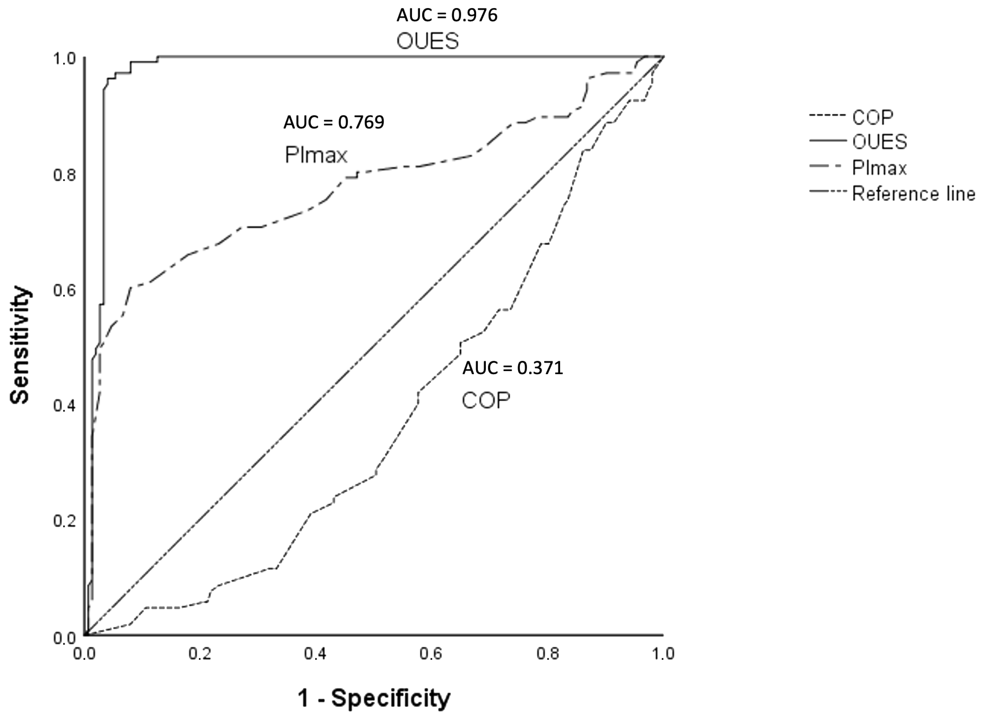
Abbreviations: COP = cardiorespiratory optimal point; SD = standard deviation; V′O2 = oxygen uptake; and HFrEF = heart failure with reduced ejection fraction. In brackets = minimum and maximum values.
Table 3. Receiver operating characteristic area under the curve (AUC).
VariablesAUC
COP0.371
V′O2p0.859
PImax0.769
V′E-V′CO2 slope0.386
6 MWT0.731
OUES0.976
Circulatory power0.511
COP = cardiopulmonary optimal point; V′O2p = oxygen uptake; PImax = inspiratory muscle pressure; V′E-V′CO2 = ventilatory response to carbon dioxide output; 6 MWT = 6 min walk test; and OUES = oxygen-uptake-efficiency slope.
Disclaimer/Publisher’s Note: The statements, opinions and data contained in all publications are solely those of the individual author(s) and contributor(s) and not of MDPI and/or the editor(s). MDPI and/or the editor(s) disclaim responsibility for any injury to people or property resulting from any ideas, methods, instructions or products referred to in the content.

