Review

Stingers—A Review of Current Understanding and Management

by
Teleale F. Gebeyehu
1,
James S. Harrop
1,*,
Joshua A. Dian
1,
Stavros Matsoukas
1 and
Alexander R. Vaccaro
2
1
Department of Neurological Surgery, Thomas Jefferson University, Philadelphia, PA 19107, USA
2
Rothman Orthopedic Institute, Philadelphia, PA 19107, USA
*
Author to whom correspondence should be addressed.
Appl. Sci. 2025, 15(7), 3510; https://doi.org/10.3390/app15073510
Submission received: 10 February 2025 / Revised: 18 March 2025 / Accepted: 21 March 2025 / Published: 23 March 2025
(This article belongs to the Special Issue Recent Advances in Sports Injuries and Physical Rehabilitation)

Simple Summary

Sports injury can be of multiple sorts. One of the various types is compression or stretch injury to a network of nerves in the neck area. This injury is commonly known as ‘Stinger’ or ‘Burner’. In this review, we summarized the recent understanding and management principles of a stinger. The injury happens to be common in contact sports like American Football and Rugby although Mixed Martial Arts (MMA) can also cause it. It is reported up to 50.3% of football players have been affected. It manifests with numbness, pins and needles sensation, and/or weakness of upper extremities right after a sports incident. There can also be neck stiffness. Mostly it is mild and self-limiting, but in certain instances active management with physiotherapy and rehabilitation is necessary after appropriate examinations and diagnostic tests. Cervical x-ray and Magnetic Resonance Imaging (MRI) are the main stays of evaluation even though nerve conduction tests can be utilized. In rare and unlikely instances where the nerve(s) gets severed, surgical intervention is needed and recovery may not be full. However, most athletes return to their regular engagement in sports, often not missing the next practice or game.

Abstract

A stinger is the transient sensory and motor loss of one upper extremity caused by a stretch or compression injury to the brachial plexus or the exiting cervical nerve roots. Athletes from contact sports like American football, rugby, wrestling, and horseback riding are most affected. Given today’s competitive athletic culture and recent advancements in sports medicine, understanding the clinical, diagnostic, and therapeutic aspects of stingers is crucial. Thus, this narrative review highlights current knowledge of pathology, clinical features, diagnostic modalities, radiologic measurements, management, rehabilitation, and return-to-play protocols. Recent and prominent English publications on adult athletes revealed that the brachial plexus upper roots (C5 and C6) are most affected. Stingers accounted for 62.2% of neck injuries in National Football League athletes and affected up to 50.3% of football players. Grading is based on injury severity to the nerves. Most stingers are mild, lasting a few seconds to minutes. Return-to-play protocols remain controversial; however, stingers have promising prognoses and can mostly be resolved with conservative treatment and rehabilitation. Severe injuries require surgical intervention but rarely occur in athletics. In conclusion, many recent developments in the evaluation and treatment of stingers impact current treatment plans, return-to-play decisions, and the future of athletes.

1. Introduction

Stingers, also known as “burners,” are defined as transient sensory and motor losses of one upper extremity due to a stretch or compression injury to the brachial plexus or the exiting cervical nerve roots (Figure 1) [1,2,3,4]. Stingers most commonly occur during contact sports like American football and rugby, but other sports like wrestling, boxing, cycling, and horseback riding have been implicated as causes [1,2,3,4]. The symptoms can persist from a few seconds, quickly resolving on the field, to several hours. In severe instances, they can take up to 2–6 weeks to completely resolve [3,5,6,7]. The athlete typically complains of sharp pain in the supraclavicular area, followed by unilateral arm pain that may sometimes adhere to a dermatomal distribution that reaches the fingertips [6,8,9]. Other symptoms include associated paresthesia, tingling, numbness, decreased cervical range of motion, and extremity weakness resulting in mild to complete loss of strength and a “dead-arm” sensation [3,5,6]. All nerve roots in the brachial plexus are susceptible to injury. However, the upper roots (C5 and C6) are most frequently affected due to underlying anatomical features and susceptibility to hyperextension and direct compression, resulting in stingers [5] (Figure 2). Stingers are a common occurrence in contact sports [1,7]. In today’s highly competitive and career-oriented athletic culture, decisions regarding the health of athletes are critical to both the player and the team. Thus, there is a persistent need to understand the pathology, clinical features, investigative modalities, novel radiologic measurements, management, rehabilitation, and return-to-play decision protocol for stingers, especially among health professionals involved in caring for athletes. This study aims to provide a concise review of our current understanding of the clinical, diagnostic, and therapeutic aspects of stingers, including rehabilitation and prevention measures.

2. Materials and Methods

This study is a narrative review. PubMed and Google Scholar were searched for the terms “Stingers” and “Burners” without using Boolean operators, and the resulting article titles were screened. Exclusion criteria were centered on language, recency, and relevance. Mainly literature published in English within the past five years was reviewed, along with their respective bibliographies. Foundational literature, regardless of the year of publication, was included to provide basic and prominent knowledge and to draw comparisons between older and new developments. Furthermore, we reviewed publications focused primarily on adult athletes. However, the studies had relevant information about adolescents as well. Therefore, reference selection was purely illustrative based on the topic the publication addresses, comprehensiveness, and recency. The types of literature reviewed included case reports, narrative reviews, systematic reviews, retrospective studies, prospective observational studies, expert opinions (including a modified Delphi consensus), and book chapters.
A narrative interpretation of the literature and the clinical knowledge of the authors who have worked with adolescent, collegiate, and professional athletes was compiled and composed for informative purposes. Since we aimed to consolidate the latest information for the management of stingers, a review of current management and rehabilitation protocols was also similarly constructed using recent publications. Most information was repeated across the literature, except for material about recent advances in management protocols. All literature had level IV and V evidence, mainly due to a lack of systematic reviews and randomized control trials, which are difficult to perform because of the complexity and functional consequences of the injury [10].

3. Epidemiology

The true incidence of stingers is difficult to determine since athletes may underreport them. This underreporting can be intentional, as athletes may fear negative career consequences, or unintentional, as athletes may overlook symptoms if they are transient and mild [1,6]. However, considering the anatomy of the nerves involved, the most susceptible athletes are those participating in activities that can potentially involve a violent contralateral neck flexion and ipsilateral shoulder depression or direct compression of the lateral neck and shoulder area [11,12]. The sports reported to be associated with stingers include but are not limited to American football, rugby, wrestling, hockey, gymnastics, and boxing [12,13,14,15]. The highest incidence was noted in American football [7,12,13,14,15], especially in players on the defensive ends (25.8%) and those engaged in tackling (36.7%) [1]. Offensive linemen had a 23.6% stinger rate [1]. The National Collegiate Athletic Association (NCAA) injury surveillance program reported 2.04 exposure injury incidences per 10,000 athletes between 2009 and 2015 [1]. A study by Beaulieu-Jones et al. showed stingers accounted for 62.2% of neck injuries in athletes playing for the National Football League [16]. Starr et al. used a survey and identified that 50.3% of high school, college, and professional football players had a lifetime occurrence of stingers, with the highest incidence in running backs (69%), defensive linemen (60%), linebackers (55%), and defensive secondaries (54%) [17]. Another study in Japan by Kawasaki et al. showed that 33.9% of high school and university rugby players had stingers, with a recurrence rate of 37.3% [2].

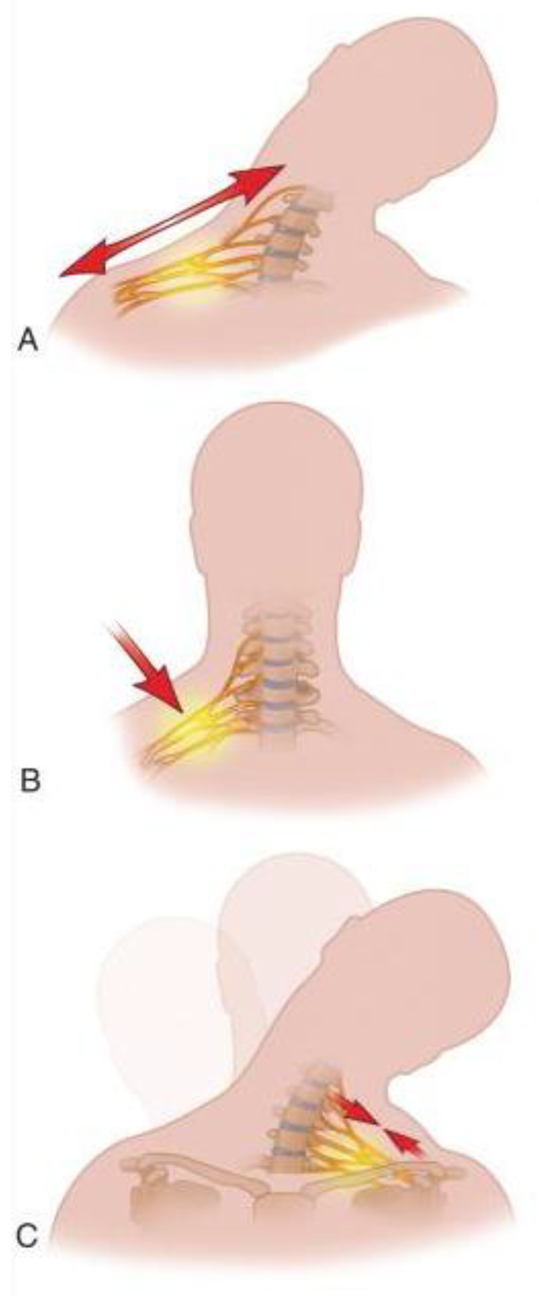
4. Mechanism of Injury

The two main mechanisms implicated in stingers are stretching and compression of the brachial plexus nerves [6,7,18] (Figure 2). Compression of the nerves can happen at the nerve root area by bony elements of a narrowed neural foramina during axial loading or by hyperextension and lateral flexion of the neck. Alternatively, direct impact at Erb’s point can cause compression [6,18]. Compression of the nerve roots is seen in chronic stingers, where repetitive trauma to the neck results in anatomical changes to the cervical vertebra and surrounding structures. One anatomical change is cervical canal and neural foraminal stenosis due to changes in the sub-axial space over time [19]. Injury by compression at the neural roots happens mostly during tackling in football or rugby athletes with these anatomical changes [20,21]. Compression at Erb’s point affects nerves distal to the root area. Erb’s point is located where the C5 and C6 join to form the superior trunk bundle right above the clavicle, where the nerves are most superficial [11]. Markley et al. studied tackle athletes and showed that protective shoulder pads can cause brachial plexus injury by compressing nerves at Erb’s point against the medial scapula during the tackle [22]. Conversely, stretch injury is usually due to ipsilateral shoulder depression and contralateral neck flexion, which is common in specific tackling techniques that use the top of the head or helmet as the point of contact. These techniques, namely clothesline and spear tackling, are now banned [23].
All contact athletes are at risk of injury from any of the mechanisms of injury. However, age and length of involvement in contact sports play a crucial role in determining which type of injury an athlete most experiences. Older and ’veteran’ athletes (usually collegiate and professional league players) who have experienced multiple impacts usually have anatomical changes in the cervical spine [5,19,24,25]. The changes include scalene muscle hypertrophy, cervical canal, and foramen stenosis, degenerative changes like osteophyte formation, and denticulate ligament stiffening that could all expose the nerves to repeated trauma. This leads to the loss of the neural protective tissue, making the nerves prone to trauma and less compliant [4,26]. Therefore, the changes within the cervical spinal column and the resulting neuroforaminal narrowing in ‘veteran’ athletes cause them to suffer injuries closer to the spinal cord area, where the nerves originate. This is because there is high tension and less compliance of the nerves during an impact that would hyperextend the neck in the opposite direction of the nerves’ natural course [4,26]. In contrast, younger athletes (usually high schoolers) experience more distal nerve damage due to the absence of changes within the cervical column, sparing the nerves from chronic damage and preserving neural compliance at the root area [5,6].

5. Grading

Grading for stingers is based on the severity of neuronal injury (Table 1). Neurapraxia, or Grade I stinger, refers to a mild injury where the axon of the nerves is not severed but will temporarily experience an interruption of conduction due to neuronal stretch. This type of injury is usually accompanied by normal neuronal and surrounding tissue (Schwann cells, endoneurium, epineurium, and perineurium) and lasts for a very short time, with complete neurologic recovery expected [5,27]. Axonotmesis, or Grade II Stinger, refers to a more extensive injury where the axon of the nerve is severed and undergoes Wallerian degeneration. This type of injury can have a longer-lasting effect on the neurologic deficit, usually taking up to 2 weeks to resolve and a maximum of a year and a half to recover [5,27]. Grade III stinger refers to a more severe case and is rare in athletics [5,27]. This grade can be further categorized into Grade IIIA, or neurotmesis, where complete, irreversible neural damage occurs. The worst form is a Grade IIIB stinger, characterized by nerve root avulsion and pre-ganglionic damage [5]. Grade III stingers are irrecoverable through the body’s innate healing process and require surgical intervention to facilitate functional recovery. However, complete recovery is rare, and affected individuals are usually left with permanent deficits [5,28,29,30,31,32].

6. Clinical Presentation

Athletes typically present with unilateral upper limb weakness and a dermatomal or non-dermatomal burning sensation (hence the terms “burner” and “stinger”) that begins at the supraclavicular area and radiates down the upper limb after forceful contact between the shoulder and neck [6,33,34,35]. Other sensory symptoms like numbness and a sensation of “dead arm” are also common [35]. The symptoms widely vary, ranging from only sensory abnormality in a dermatomal fashion to complete loss of strength and sensation in the affected limb [36]. A Canadian study of football players by Charbonneau et al. reported tingling in 77%, numbness in 61%, weakness in 44%, and neck pain in 17% of athletes [37]. Depending on the severity of the injury, symptom duration can range from seconds to days [7]. Knowing the mechanism of injury is crucial in evaluating an athlete with stingers since it can inform other possible injuries. The mechanism of injury is usually witnessed by the sideline medical staff or from video recordings of the incident. An alternative diagnosis of vertebral fracture, spinal cord, brain, or vascular injury can be indicated by other clinical symptoms and findings, such as persistent neck pain with limited range of motion, lower extremity involvement, bilateral upper extremity involvement, headache, dizziness, nausea, vomiting, changes in mentation, speech, and vision, and changes in vital signs like blood pressure, brachial-brachial index, heart rate, and temperature [7,38,39,40,41]. The presence of these symptoms warrants further evaluation.

7. On-Field Evaluation and Management

7.1. Basic Support

The evaluation of injured athletes starts on the field or the sidelines. Similar to the evaluation of other traumatic injuries, suspected stingers should be reviewed with an advanced trauma life support (ATLS) structured assessment to rule out other serious and life-threatening injuries involving the airway, breathing, and circulation [35,42].

7.2. Immobilization Protocols

All athletes with neck injuries should be considered to have sustained spinal cord injury until proven otherwise [41]. Therefore, spinal cord and other life-threatening injuries should be thoroughly evaluated and ruled out before other lower-priority examinations are performed [4,42]. If such an injury cannot be ruled out from the assessment, head and neck stabilization should be promptly prioritized, and the athlete should be transferred to an appropriate trauma facility. Any manipulation of the affected area, such as removing helmets and shoulder pads, should be performed with utmost caution, at the appropriate location, using the necessary equipment and assistance from other caregivers. This should be done under the supervision of practitioners who are well versed in the acute management of spinal cord injury to minimize the risk of neurologic deterioration and adverse outcomes [4,19,42]. Some studies also noted that helmets and shoulder pads contribute to spine alignment, further supporting the notion of not rushing to remove them [43,44,45]. Exceptions requiring immediate removal of the helmet or shoulder pads include difficulties in assessing or managing the airway due to the design of the helmet, facemask, or chinstrap, the need for cardiopulmonary resuscitation, the need for chest access for external defibrillation, and the presence of scalp injury requiring visual access [44,45].

7.3. Hospital Transfer Criteria and Further Care

Suspicion of the spinal cord or other life-threatening injury warrants immediate imaging to assess the extent of the injury and, therefore, transfer to a hospital [10]. Symptoms of a stinger lasting more than 5 min and occurring for a second time also require imaging [10], mandating transfer to a facility with such capacity. Once the possibility of a serious injury is ruled out, the athlete should be evaluated with the appropriate physical examination, focusing on inspecting for asymmetry indicating dislocation or fracture, persistent cervical or neck tenderness, range of motion for neck and shoulder, and limb strength [5,6,34,35]. Importantly, muscles innervated by the brachial plexus should be evaluated in detail beyond cursory grip strength testing alone. For the ideal management of stingers, Weinstein [35] emphasizes the need for conducting at least a minimum test of the spinatii, deltoid, biceps, brachioradialis, triceps, serratus anterior, wrist flexors, extensors, and grip strength, with the unaffected side serving as a control. Pain due to nerve injury within the spinal foramina can be reproduced with extension and lateral flexion (Spurling’s test), while brachial plexus injury lacks this feature [6].
Other suspected injuries of vascular origin should be evaluated in a hospital setting to confirm their presence and characterize them for the appropriate management. CT angiography, MR angiography supplemented with digital subtraction angiography, and ultrasonography are all used to verify and plan interventions for vascular injuries [5,42,46].

8. Imaging

Imaging is commonly used for diagnosis and screening and can reveal important findings in athletes with stingers [9,10,47]. However, the absence of definitive recommendations or guidelines makes imaging indications a controversial subject [48]. A study by Standaert and Herring on expert opinion and controversies mentioned that the findings of imaging were not always clear [18]. A recent modified Delphi consensus study with the Cervical Spine Research Society by Schroder et al. showed a super majority (69.9%) of spine surgeons agreed that athletes who experience first-time stingers and symptoms lasting from seconds to less than 5 min need no imaging studies [10]. This contrasts with the old consensus of considering imaging for symptoms that last longer than one hour [18]. Schroder et al. also showed that a super majority (84.5%) of surgeons agreed that further consideration, including imaging, is indicated if the symptoms persist for more than 5 min or if athletes have more than one episode of stingers, regardless of the duration of symptoms [10]. Essential imaging included cervical radiographs with anterior, posterior, lateral flexion/extension, and odontoid views, as well as MRI [5,6,18]. CT scan was reported as an alternative for those who cannot perform MRI [5,6,18]. Single-photon emission computed tomography (SPECT) was also reported to facilitate the diagnosis of an occult cervical vertebra fracture [8].
Chronic stingers are believed to result from degenerative anatomical changes in the cervical spine [5,19,24,25]. Hence, imaging can possibly identify those at risk of sustaining stingers based on changes observed in the imaging. Torg et al. described the Torg–Pavlov ratio to confirm and screen for stingers (Figure 3A) [7]. The Torg–Pavlov ratio is determined by dividing the distance from the midpoint of the posterior aspect of the vertebral body to the nearest point on the corresponding spino-laminar line by the anteroposterior width of the vertebral body on a plain radiograph [7]. According to Torg et al., 95% of all reported cases had a Torg–Pavlov ratio of <0.8 [7]. However, a very low positive predictive value for asymptomatic athletes makes this measurement insufficient for screening [49]. The role of MRI in screening is also a recent novel development. While studying the changes in the sub-axial space for chronic stingers, Presciutti et al. determined the mean sub-axial cervical space available for the cord (MSCSAC) [19]. MSCSAC is the average of the values determined by subtracting the sagittal cord diameter from the disc-level spinal canal diameter between C3 and C6 [19] (Figure 3B) and is a better alternative to the classic Torg–Pavlov ratio [19]. This novel measurement has a sensitivity of 80% and a negative predictive value of 0.23 for stingers when 5.0 mm was used as a cut-off. When a cut-off value of 4.3 mm was used, MSCSAC had a 96% specificity with a 13.25 positive likelihood ratio. Presciutti et al. also found that these measurements are 20% more accurate than the classic Torg ratio [19]. Other novel measures used to objectively identify cervical spinal canal stenosis are cord-to-canal area ratio (Figure 4A), which is the ratio of transverse cord area to transverse canal area, and cord compression ratio (Figure 4B), which is the ratio of sagittal to axial cord diameter (a/b in Figure 4B) [27]. These measures are highly predictive of individuals at risk for cervical spine injury after minor trauma and can indirectly predict those at risk for chronic stingers.

9. Electrodiagnostic Studies

Nerve conduction tests and electromyography (EMG) monitor progress and serve as additional tools to supplement clinical findings and imaging [5,27]. These tests mostly use muscles innervated by C5 and C6 nerves and can inform the clinician about the site, severity, and regeneration of nerves after injury. Results are based on signals generated from normal and involved muscles or nerves [5,27]. For instance, limb muscle involvement can imply plexus or root injury, while paraspinal muscle involvement implies root injury due to the anatomical orientation of the posterior rami nerves supplying the paraspinal muscles [27]. Sensory amplitudes can also be used to localize injury sites. A normal peripheral sensation signal conduction velocity and amplitude is seen in preganglionic injury. However, normal amplitude does not rule out post-ganglionic injury [27]. The severity of injury also correlates with the presence and magnitude of abnormal spontaneous electrical activities that manifest as positive sharp waves and fibrillation. The absence of positive sharp waves and fibrillations, however, does not rule out nerve root injury or rule in brachial plexus injury [27]. Abnormal electrical activity takes 2 weeks to develop and 3–5 weeks to maximize [27]. Complete nerve transection is known for the absence of muscle unit recruitment and has the worst prognosis. On the other hand, discrete motor unit recruitment that occurs despite an attempt to maximally contract muscle indicates a conduction block, which can result from severe stretch injury (neurapraxia) or axonal degeneration. However, after the first few weeks, when neurapraxia is expected to subside, it is a sign of axonal degeneration [27]. Long polyphasic potentials and nascent potentials (axonal regeneration potentials, ARPs) develop over time and are seen as signs of reinnervation and regeneration of axons, respectively [27]. Terminal collateral sprouts polyphasic potentials that identify chronic and recurrent injury [27]. For these various reasons, even though it is possible to use EMG as early as 7 days post-injury, its specificity increases after 4–6 weeks and remains most effective during the follow-up period rather than the acute phase [5,6,7,27].

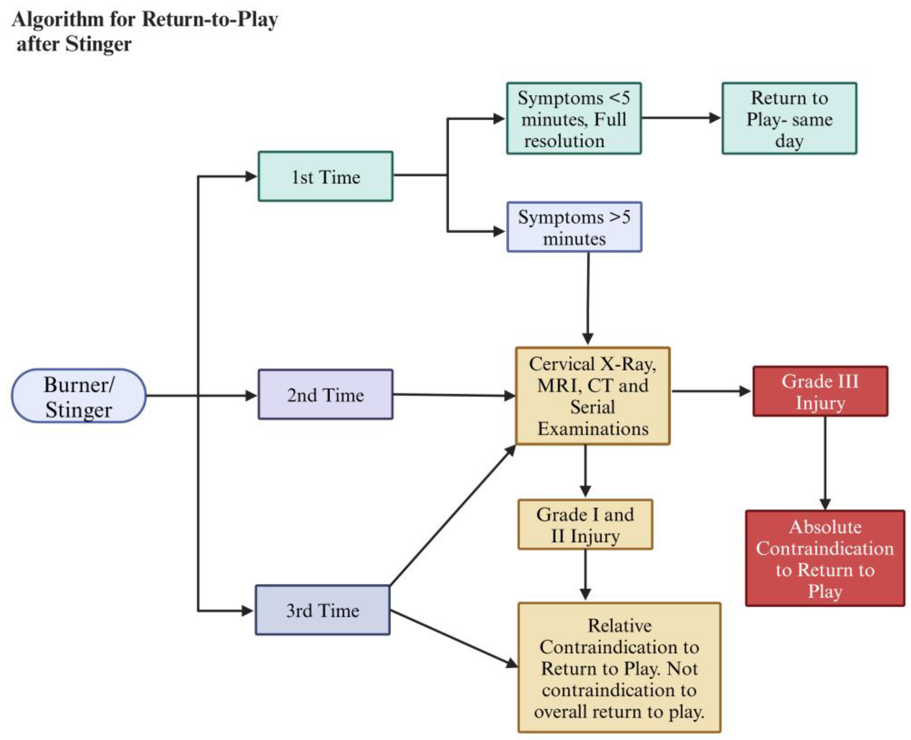
10. Return-to-Play Protocol

Most stingers are mild, lasting a few seconds to minutes, and do not interfere with participating in the next game or practice [7,8,36,37]. The decision to clear athletes to return to play is a controversial issue that relies on expert opinions, as there are no available definitive studies [4,5,6,7,9,10,12,33]. The current expert consensus takes the presence and duration of symptoms, number of stinger episodes, and imaging findings to either give a temporary or permanent withdrawal from contact sports [9,10]. According to the modified Delphi study by Schroder et al., if an athlete experienced the first episode of a stinger and the symptoms fully resolved in less than five minutes (no strength, sensory, and range of motion limitations are seen), the majority (84.5%) of the participants agreed the athlete can return to play the same day without a need for MRI or further imaging [10]. Athletes with more than one episode of stingers or symptoms lasting more than 5 min have a relative contraindication to immediately returning to play. They need to undergo imaging and serial examinations to rule out any underlying cervical abnormalities [9,10]. The most critical parameters to consider are pain- and weakness-free full range of motion in the neck and shoulder joints [4,5,6,7,9,10]. Sustaining a Grade IIIA or IIIB injury is an absolute contraindication to return to play, and further treatment is recommended (Figure 5).
The return-to-play algorithm is a great decision-making tool, but it is not without limitations. As Schroeder et al. [10] pointed out, the algorithm is based primarily on expert opinions, making it suboptimal. Certain decision pathways are vulnerable to subjectivity in clinical assessment, leaning on players’ reports rather than an objective assessment of neurological symptom resolution. Furthermore, the standardized algorithm may not adequately account for inter-individual differences in recovery. Thus, the authors of this study lean toward performing an MRI for all occurrences of stingers regardless of symptom duration. This algorithm would benefit from prospective studies to validate its use and define its limitations further.

11. Treatment

Management of stingers is dependent on the severity or grade of nerve injury [5,6,35]. For athletes who sustain Grade I and II injuries, the mainstays of treatment are pain control, reduction of inflammation, physical rehabilitation, and prevention of recurrence [5,6,35]. For athletes who sustain Grade III injury with nerve root avulsion or neurotmesis, the mainstay of treatment is timely surgical reconstruction of the severed nerve with the addition of non-operative treatment protocols [5,6,28,50].
Pain is controlled with rest, analgesics, and a cervical collar [27]. Cervical region epidural injections can also be used; however, utmost caution should be exercised because of the risks of traumatizing the cord directly during the procedure or indirectly due to the high pressure from administering medication in a narrowed and compromised canal [51].
The main target of physical therapy is to correct postural abnormalities, flexibility, and strength [27]. A prominent postural abnormality noticed after such injury is the reduction or absence of normal cervical lordosis [27,35], which needs to be corrected. Postural imbalance is usually associated with thoracic kyphosis, scapulae protraction, glenohumeral joint internal rotation, hyperflexion of the lower cervical and upper thoracic vertebral segments, and mid-cervical hypermobility. This imbalance is also associated with weakening due to shortening (capital and cervical extensors, sternocleidomastoid, upper trapezius, levator scapula, pectoralis minor and major, anterior deltoid, subscapularis, serratus anterior, and the anterior scalene muscles) or lengthening (capital and cervical flexors, middle and lower trapezius, rhomboids, thoracic extensors, and latissimus dorsi) of various neck, shoulder, and back muscles [27,35]. These abnormalities are all targeted with the appropriate physiotherapy, myofascial strengthening, and stretching with manual therapy to manipulate and mobilize hypomobile joints and regain balance [27,35].
Weinstein [35] suggested that the cervical and thoracic area musculature and joints should be stabilized in a sequence that starts from the head and neck and descends to the shoulder, thoracic, and upper limb musculature. Weinstein also recommended starting with isometric and isotonic techniques at neutral positions and slightly varying the head position against gravity to help strengthen the muscles and avoid neural irritation. After these isometric techniques help in the concentric strengthening of lengthened muscles, a balanced strengthening of the anterior and posterior shoulder stabilizers (shoulder girdle), thoracic extensors, and upper limb strengthening should follow [35]. The sequence finally recommended performing multiplanar movements against incremental resistance of the muscles [35].
Prevention focuses on addressing the biomechanical factors that led to the injury, which includes proper tackling techniques (specifically avoiding clothesline and spear tackling) for the athlete, use of protective equipment like a cowboy collar and shoulder pads, restoration of neck area motion, and strengthening [7,27,52].
Nerve stretch injuries usually cause stingers. Neurotmesis and nerve root avulsion are infrequent for athletes with contact sports injuries. Surgical treatment is, therefore, rarely needed. In the rare instance where surgical treatment is required, it is a salvage procedure for individuals with loss of function due to severe nerve damage (Grades IIIA and IIIB) with no return of function in sight [28,50]. The surgery involves exploring the brachial plexus to identify the injury of the brachial plexus anatomy and strategize repair options. Surgical repair options include direct anastomosis, interposition grafting, and nerve transfer with an associated partial or complete sacrifice of a less-needed healthy nerve. Shoulder abduction and elbow flexion are the two crucial functions that are the focus of surgical repair. Garg et al. and Yang et al. performed a meta-analysis and systematic review independently, comparing the surgical options to determine the superiority of nerve transfer alone versus a combination of nerve transfer with nerve repair. Both concluded from their synthesized data that nerve transfer is a better option for motor outcomes for shoulder abduction and elbow flexion [28,50]. However, the systematic review by Yang et al. showed that neither of the surgeries was superior to the other for shoulder abduction, based on a post hoc statistical analysis that entailed a large number of statistical manipulations to demonstrate a significant benefit from nerve transfer [28]. Therefore, the issue of which type of surgery should be preferred is not yet settled and is an open area for future research.
The timing of surgery is another controversial issue. The current suggestion is to wait 3 to 6 months to allow reinnervation and settle concomitant injuries that make surgery unfeasible [31]. This approach was advocated for in cases of nerve root avulsion or damage where regeneration is not expected [28]. However, Kato et al. documented that outcomes depend on the timing of surgery, and early surgery within 1 month of injury showed excellent outcomes in terms of functional recovery [32]. This improved outcome is also sensible, considering that a motor endplate that becomes atrophied and denucleated diminishes the chances of recovery over time, especially after a year of injury. Moreover, it is important to note the lengthy physiological period required for a nerve to regenerate, reach, and reinnervate a muscle [29,30,31]. In conditions where a motor end plate has been rendered dysfunctional, the microvascular transfer of normal muscle with a motor endplate, in addition to nerve grafting, has been suggested [30].

12. Conclusions

Stingers are injuries to the nerve roots forming the brachial plexus, most commonly to C5 and C6. They commonly occur in athletes involved in contact sports like American football, rugby, wrestling, hockey, gymnastics, and boxing. The mechanism of injury can be stretching or compression of the nerve roots. The grading of stingers is based on the severity of the nerve damage, which can be neural stretching, severed axons, damaged neurons, or nerve root avulsion. After injury, stingers manifest as sensory or motor deficits in the neck region. Stingers are mainly managed conservatively with rest, pain control, physiotherapy, and rehabilitation. However, surgical repair of the nerve roots is needed in certain rare and unlikely instances for athletes. The prognosis is mostly promising except where there is complete nerve damage or nerve root avulsion.

13. Limitations

This study is not without limitations. The main limitation is that this study is not a systematic review, although we believe it provides a comprehensive and current review of the topic. However, a systematic review would provide more detailed information regarding studies published over the years, summarizing the course and evolution of managing stingers. Our aim was not to review all the literature. Therefore, a narrative review was composed to inform the readers of the current understanding of the management of stingers from firsthand clinical experience and a comprehensive literature review.
Another limitation of this study is the lack of epidemiological data for certain sports like mixed martial arts, judo, and weightlifting. Logically, these sports are likely to have a high incidence of stingers. However, due to a lack of studies demonstrating the incidence of stingers, we were not able to include relevant information. We strongly encourage researchers to conduct epidemiological studies in these underrepresented sports.

Author Contributions

T.F.G.: writing—original draft preparation, methodology, visualization, project administration. J.S.H.: conceptualization, review and editing, project administration, supervision. J.A.D.: review and editing, methodology. S.M.: reviewing and editing, methodology. A.R.V.: conceptualization, review and editing, methodology, project administration, supervision. All authors have read and agreed to the published version of the manuscript.

Funding

The authors did not receive any funding for this work.

Institutional Review Board Statement

Not needed since this is a review of publicly available literature.

Informed Consent Statement

Not applicable.

Data Availability Statement

All information is from review of literature cited in this manuscript.

Acknowledgments

Our team would like to thank Hemma Murali at Thomas Jefferson University Library for editing this manuscript thoroughly. A special thanks to Pamela Walter at Thomas Jefferson University for facilitating the editing process.

Conflicts of Interest

James S. Harrop—none. Teleale F. Gebeyehu—none. Joshua A. Dian—none. Stavros Matsoukas—none. Alexander R. Vaccaro receives royalties from Stryker, Globus, Medtronic, Atlas Spine, Alphatech Spine, SpineWave, Spinal Elements, Curiteva, Elsevier, Jaypee, Stout Medical, Taylor Francis/Hodder and Stoughton, Wolters Kluwer, Wheel House Medical, and Thieme; has stock or stock options in Accelus, Advanced Spinal Intellectual Properties, Atlas, Avaz Surgical, AVKN Patient Driven Care, Cytonics, Deep Health, Dimension Orthotics LLC, Electrocore, Flagship Surgical, FlowPharma, Rothman Institute and Related Properties, Globus, Harvard MedTech, Innovative Surgical Design, Jushi (Haywood), Orthobullets, Parvizi Surgical Innovation, Progressive Spinal Technologies, Sentryx, Stout Medical, See All AI, and ViewFi Health; is a consultant for Curiteva, Medcura, Stryker, Globus, Spinal Elements, Accelus, Wheel House Medical, and Ferring Pharmaceutical; serves on Scientific Advisory Board/Board of Directors/Committee for National Spine Health Foundation (NSHF), Sentryx, and Accelus; and is a member in good standing/independent contractor for AO Spine.

References

  1. Green, J.; Zuckerman, S.L.; Dalton, S.L.; Djoko, A.; Folger, D.; Kerr, Z.Y. A 6-year surveillance study of “Stingers” in NCAA American Football. Res. Sports Med. 2017, 25, 26–36. [Google Scholar] [CrossRef] [PubMed]
  2. Kawasaki, T.; Ota, C.; Yoneda, T.; Maki, N.; Urayama, S.; Nagao, M.; Nagayama, M.; Kaketa, T.; Takazawa, Y.; Kaneko, K. Incidence of Stingers in Young Rugby Players. Am. J. Sports Med. 2015, 43, 2809–2815. [Google Scholar] [CrossRef]
  3. Thomas, B.E.; McCullen, G.M.; Yuan, H.A. Cervical spine injuries in football players. J. Am. Acad. Orthop. Surg. 1999, 7, 338–347. [Google Scholar] [CrossRef] [PubMed]
  4. Schroeder, G.D.; Vaccaro, A.R. Cervical Spine Injuries in the Athlete. J. Am. Acad. Orthop. Surg. 2016, 24, e122–e133. [Google Scholar] [CrossRef] [PubMed]
  5. Ahearn, B.M.; Starr, H.M.; Seiler, J.G. Traumatic Brachial Plexopathy in Athletes: Current Concepts for Diagnosis and Management of Stingers. J. Am. Acad. Orthop. Surg. 2019, 27, 677–684. [Google Scholar] [CrossRef]
  6. Bowles, D.R.; Canseco, J.A.; Alexander, T.D.; Schroeder, G.D.; Hecht, A.C.; Vaccaro, A.R. The Prevalence and Management of Stingers in College and Professional Collision Athletes. Curr. Rev. Musculoskelet. Med. 2020, 13, 651–662. [Google Scholar] [CrossRef]
  7. Torg, J.S. Cervical spine injuries and the return to football. Sports Health 2009, 1, 376–383. [Google Scholar] [CrossRef]
  8. Vaccaro, A.R.; Klein, G.R.; Ciccoti, M.; Pfaff, W.L.; Moulton, M.J.; Hilibrand, A.J.; Watkins, B. Return to play criteria for the athlete with cervical spine injuries resulting in stinger and transient quadriplegia/paresis. Spine J. 2002, 2, 351–356. [Google Scholar] [CrossRef]
  9. Kazarian, G.S.; Qureshi, S. Return to Play After Injuries to the Cervical Spine. Clin. Spine Surg. 2024, 37, 425–432. [Google Scholar] [CrossRef]
  10. Schroeder, G.D.; Canseco, J.A.; Patel, P.D.; Hilibrand, A.S.; Kepler, C.K.; Mirkovic, S.M.; Watkins, R.G.I.; Dossett, A.; Hecht, A.C.; Vaccaro, A.R. Updated Return-to-Play Recommendations for Collision Athletes After Cervical Spine Injury: A Modified Delphi Consensus Study With the Cervical Spine Research Society. Neurosurgery 2020, 87, 647–654. [Google Scholar] [CrossRef]
  11. Papadopoulos, E. Brachial Plexus Anatomy A comprehensive Overview. Int. J. Anat. Var. 2024, 17, 669–670. [Google Scholar]
  12. Tosti, R.; Rossy, W.; Sanchez, A.; Lee, S.G. Burners, stingers, and other brachial plexus injuries in the contact athlete. Oper. Tech. Sports Med. 2016, 24, 273–277. [Google Scholar]
  13. Adele, M.; Christopher, M.; Scott, R.L.; Dustin, C.; Comstock, R.D. Epidemiology of Cervical Spine Injuries in High School Athletes Over a Ten-Year Period. Pm&R. 2018, 10, 365–372. [Google Scholar] [CrossRef]
  14. Deckey, D.G.; Makovicka, J.L.; Chung, A.S.; Hassebrock, J.D.; Patel, K.A.; Tummala, S.V.; Pena, A.; Asprey, W.; Chhabra, A. Neck and Cervical Spine Injuries in National College Athletic Association Athletes: A 5-Year Epidemiologic Study. Spine 2020, 45, 55–64. [Google Scholar] [CrossRef]
  15. DePasse, J.M.; Durand, W.; Palumbo, M.A.; Daniels, A.H. Sex- and Sport-Specific Epidemiology of Cervical Spine Injuries Sustained During Sporting Activities. World Neurosurg. 2019, 122, e540–e545. [Google Scholar] [CrossRef] [PubMed]
  16. Beaulieu-Jones, B.R.; Rossy, W.H.; Sanchez, G.; Whalen, J.M.; Lavery, K.P.; McHale, K.J.; Vopat, B.G.; Van Allen, J.J.; Akamefula, R.A.; Provencher, M.T. Epidemiology of Injuries Identified at the NFL Scouting Combine and Their Impact on Performance in the National Football League: Evaluation of 2203 Athletes From 2009 to 2015. Orthop. J. Sports Med. 2017, 5, 2325967117708744. [Google Scholar] [CrossRef]
  17. Starr, H.M., Jr.; Anderson, B.; Courson, R.; Seiler, J.G. Brachial plexus injury: A descriptive study of American football. J. Surg. Orthop. Adv. 2014, 23, 90–97. [Google Scholar] [CrossRef]
  18. Standaert, C.J.; Herring, S.A. Expert opinion and controversies in musculoskeletal and sports medicine: Stingers. Arch. Phys. Med. Rehabil. 2009, 90, 402–406. [Google Scholar] [PubMed]
  19. Presciutti, S.M.; DeLuca, P.; Marchetto, P.; Wilsey, J.T.; Shaffrey, C.; Vaccaro, A.R. Mean subaxial space available for the cord index as a novel method of measuring cervical spine geometry to predict the chronic stinger syndrome in American football players. J. Neurosurg. Spine 2009, 11, 264–271. [Google Scholar] [CrossRef]
  20. Meyer, S.A.; Schulte, K.R.; Callaghan, J.J.; Albright, J.P.; Powell, J.W.; Crowley, E.T.; el-Khoury, G.Y. Cervical spinal stenosis and stingers in collegiate football players. Am. J. Sports Med. 1994, 22, 158–166. [Google Scholar] [CrossRef]
  21. Kelly, J.D.t.; Aliquo, D.; Sitler, M.R.; Odgers, C.; Moyer, R.A. Association of burners with cervical canal and foraminal stenosis. Am. J. Sports Med. 2000, 28, 214–217. [Google Scholar] [CrossRef] [PubMed]
  22. Markey, K.L.; Di Benedetto, M.; Curl, W.W. Upper trunk brachial plexopathy. The stinger syndrome. Am. J. Sports Med. 1993, 21, 650–655. [Google Scholar] [CrossRef] [PubMed]
  23. Torg, J.S.; Vegso, J.J.; O’Neill, M.J.; Sennett, B. The epidemiologic, pathologic, biomechanical, and cinematographic analysis of football-induced cervical spine trauma. Am. J. Sports Med. 1990, 18, 50–57. [Google Scholar] [CrossRef]
  24. Levitz, C.L.; Reilly, P.J.; Torg, J.S. The pathomechanics of chronic, recurrent cervical nerve root neurapraxia. The chronic burner syndrome. Am. J. Sports Med. 1997, 25, 73–76. [Google Scholar] [CrossRef]
  25. Greenberg, J.; Leung, D.; Kendall, J. Predicting chronic stinger syndrome using the mean subaxial space available for the cord index. Sports Health 2011, 3, 264–267. [Google Scholar] [CrossRef]
  26. Hakkaku, T.; Nakazato, K.; Koyama, K.; Kouzaki, K.; Hiranuma, K. Cervical Intervertebral Disc Degeneration and Low Cervical Extension Independently Associated With a History of Stinger Syndrome. Orthop. J. Sports Med. 2017, 5, 2325967117735830. [Google Scholar] [CrossRef]
  27. Feinberg, J.H. Burners and stingers. Phys. Med. Rehabil. Clin. N. Am. 2000, 11, 771–784. [Google Scholar]
  28. Yang, L.J.; Chang, K.W.; Chung, K.C. A systematic review of nerve transfer and nerve repair for the treatment of adult upper brachial plexus injury. Neurosurgery 2012, 71, 417–429. [Google Scholar] [CrossRef] [PubMed]
  29. Birch, R.; Dunkerton, M.; Bonney, G.; Jamieson, A.M. Experience with the free vascularized ulnar nerve graft in repair of supraclavicular lesions of the brachial plexus. Clin. Orthop. Relat. Res. 1988, 237, 96–104. [Google Scholar]
  30. Bishop, A.T. Functioning free-muscle transfer for brachial plexus injury. Hand Clin. 2005, 21, 91–102. [Google Scholar] [CrossRef]
  31. Giuffre, J.L.; Kakar, S.; Bishop, A.T.; Spinner, R.J.; Shin, A.Y. Current concepts of the treatment of adult brachial plexus injuries. J. Hand Surg. Am. 2010, 35, 678–688. [Google Scholar] [CrossRef] [PubMed]
  32. Kato, N.; Htut, M.; Taggart, M.; Carlstedt, T.; Birch, R. The effects of operative delay on the relief of neuropathic pain after injury to the brachial plexus: A review of 148 cases. J. Bone Joint Surg. Br. 2006, 88, 756–759. [Google Scholar] [CrossRef] [PubMed]
  33. Carr, J.B.; Dines, J.S. Transient brachial plexopathy (stingers/burners). In Spinal Conditions in the Athlete: A Clinical Guide to Evaluation, Management and Controversies; Springer Nature: Berlin/Heidelberg, Germany, 2020; pp. 109–121. [Google Scholar]
  34. Bateman, J.E. Nerve Injuries about the Shoulder in Sports. JBJS 1967, 49, 785–792. [Google Scholar]
  35. Weinstein, S.M. Assessment and rehabilitation of the athlete with a “stinger”: A model for the management of noncatastrophic athletic cervical spine injury. Clin. Sports Med. 1998, 17, 127–135. [Google Scholar]
  36. Cantu, R.C.; Li, Y.M.; Abdulhamid, M.; Chin, L.S. Return to play after cervical spine injury in sports. Curr. Sports Med. Rep. 2013, 12, 14–17. [Google Scholar] [CrossRef]
  37. Charbonneau, R.M.E.; McVeigh, S.A.; Thompson, K. Brachial Neuropraxia in Canadian Atlantic University Sport Football Players: What Is the Incidence of “Stingers”? Clin. J. Sport Med. 2012, 22, 472–477. [Google Scholar] [CrossRef]
  38. Ban, V.S.; Botros, J.A.; Madden, C.J.; Batjer, H.H. Neurosurgical Emergencies in Sports Neurology. Curr. Pain. Headache Rep. 2016, 20, 55. [Google Scholar] [CrossRef] [PubMed]
  39. Guterman, A.; Smith, R.W. Neurological sequelae of boxing. Sports Med. 1987, 4, 194–210. [Google Scholar] [CrossRef]
  40. Weinstein, S.M.; Cantu, R.C. Cerebral stroke in a semi-pro football player: A case report. Med. Sci. Sports Exerc. 1991, 23, 1119–1121. [Google Scholar]
  41. Blatz, D.; Ross, B.; Dadabo, J. Cervical spine trauma evaluation. Handb. Clin. Neurol. 2018, 158, 345–351. [Google Scholar]
  42. Bailes, J.E.; Petschauer, M.; Guskiewicz, K.M.; Marano, G. Management of cervical spine injuries in athletes. J. Athl. Train. 2007, 42, 126–134. [Google Scholar] [CrossRef] [PubMed]
  43. Banerjee, R.; Palumbo, M.A.; Fadale, P.D. Catastrophic cervical spine injuries in the collision sport athlete, part 1: Epidemiology, functional anatomy, and diagnosis. Am. J. Sports Med. 2004, 32, 1077–1087. [Google Scholar] [CrossRef] [PubMed]
  44. Patel, P.D.; Divi, S.N.; Canseco, J.A.; Donnally, C.J., 3rd; Galetta, M.; Vaccaro, A., Jr.; Schroeder, G.D.; Hsu, W.K.; Hecht, A.C.; Dossett, A.B.; et al. Management of Acute Subaxial Trauma and Spinal Cord Injury in Professional Collision Athletes. Clin. Spine Surg. 2022, 35, 241–248. [Google Scholar] [CrossRef]
  45. Swartz, E.E.; Boden, B.P.; Courson, R.W.; Decoster, L.C.; Horodyski, M.B.; Norkus, S.A.; Rehberg, R.S.; Waninger, K.N. National Athletic Trainers’ Association position statement: Acute management of the cervical spine–injured athlete. J. Athl. Train. 2009, 44, 306–331. [Google Scholar] [CrossRef] [PubMed]
  46. Evans, C.; Chaplin, T.; Zelt, D. Management of Major Vascular Injuries: Neck, Extremities, and Other Things that Bleed. Emerg. Med. Clin. N. Am. 2018, 36, 181–202. [Google Scholar] [CrossRef]
  47. Torg, J.S.; Naranja, R.J., Jr.; Pavlov, H.; Galinat, B.J.; Warren, R.; Stine, R.A. The relationship of developmental narrowing of the cervical spinal canal to reversible and irreversible injury of the cervical spinal cord in football players. J. Bone Joint Surg. Am. 1996, 78, 1308–1314. [Google Scholar] [CrossRef]
  48. Zaremski, J.L.; Horodyski, M.; Herman, D.C. Recurrent stingers in an adolescent American football player: Dilemmas of return to play. A case report and review of the literature. Res. Sports Med. 2017, 25, 384–390. [Google Scholar] [CrossRef]
  49. Herzog, R.J.; Wiens, J.J.; Dillingham, M.F.; Sontag, M.J. Normal Cervical Spine Morphometry and Cervical Spinal Stenosis in Asymptomatic Professional Football Players: Plain Film Radiography, Multiplanar Computed Tomography, and Magnetic Resonance Imaging. Spine 1991, 16, S178–S186. [Google Scholar] [CrossRef]
  50. Garg, R.; Merrell, G.A.; Hillstrom, H.J.; Wolfe, S.W. Comparison of nerve transfers and nerve grafting for traumatic upper plexus palsy: A systematic review and analysis. JBJS 2011, 93, 819–829. [Google Scholar] [CrossRef]
  51. Epstein, N.E. Major risks and complications of cervical epidural steroid injections: An updated review. Surg. Neurol. Int. 2018, 9, 86. [Google Scholar] [CrossRef]
  52. Gorden, J.A.; Straub, S.J.; Swanik, C.B.; Swanik, K.A. Effects of Football Collars on Cervical Hyperextension and Lateral Flexion. J. Athl. Train. 2003, 38, 209–215. [Google Scholar] [PubMed]
Figure 1. Anatomic illustration of the brachial plexus. All nerve roots are prone to injury. However, C5 and C6 are most frequently involved. Created in BioRender. Gebeyehu, T. (2024) https://BioRender.com/g33z849, accessed on 22 March 2025.
Applsci 15 03510 g001
Figure 2. Various mechanisms of injury causing stingers: (A) stretch injury with depressed shoulder and laterally hyperextended neck, (B) direct injury at Erb’s point, and (C) lateral flexion and hyperextension of the neck compressing the brachial plexus. Used with permission from Clinical Tree Publications, DeLee, Drez, & Miller’s Orthopaedic Sports Medicine, Orthopedics, Stingers, Online publication, 27 January 2024. Stingers—Clinical Tree.
Applsci 15 03510 g002
Figure 3. (A) Torg–Pavlov ratio, the ratio of canal diameter (distance from the midpoint of the posterior aspect of the vertebral body to the nearest point on the corresponding spino-laminar line ,designated as ‘a’) to vertebral body diameter (anteroposterior width of the vertebral body ,designated as ‘b’) on a sagittal cervical plain x-ray (TPR = a/b). (B) Mean sub-axial cervical space available for the cord is the mean of the values for cord diameter deducted from disk level canal diameter at each level between C3 and C6.
Applsci 15 03510 g003
Figure 4. (A) Illustration of cord to canal area measurement, the ratio of area within the inner circle (orange, [cord]) to area within the outer circle (area of pink and orange combined, [canal]). (B) Cord compression ratio = a/b, the ratio of sagittal [a] to axial [b] cord diameter.
Applsci 15 03510 g004
Figure 5. Algorithm for return-to-play after a stinger injury. Created in BioRender. Gebeyehu, T. (2024) https://BioRender.com/y07q789, accessed on 22 March 2025.
Applsci 15 03510 g005
Table 1. Grades of stinger injury and the mode of treatment.
GradeNerve InjuryMode of Treatment
INeurapraxiaUsually transient with no need for any intervention. Pain control, rest, and physiotherapy as needed if symptoms persist. Consider MRI and other imaging with serial examinations if symptoms persist for >5 min.
IIAxonotmesisPain control, rest and physiotherapy, serial monitoring with electrodiagnostic studies, and imaging (MRI, radiographs).
IIIANeurotmesisImaging, electrodiagnostic studies, nerve repair ± nerve transfer [28,29,30,31], pain control, and physiotherapy.
IIIBNerve root avulsionImaging, electrodiagnostic studies, nerve transfer [28,29,30,31], pain control, and physiotherapy.
Disclaimer/Publisher’s Note: The statements, opinions and data contained in all publications are solely those of the individual author(s) and contributor(s) and not of MDPI and/or the editor(s). MDPI and/or the editor(s) disclaim responsibility for any injury to people or property resulting from any ideas, methods, instructions or products referred to in the content.

