Article

Bioinformatics-Based Management of Vitellogenin-like Protein’s Role in Pathogen Defense in Nicotiana tabacum L.

Department of Management, Bar-Ilan University, Ramat Gan 5290002, Israel
*
Author to whom correspondence should be addressed.
Appl. Sci. 2025, 15(8), 4463; https://doi.org/10.3390/app15084463
Submission received: 9 March 2025 / Revised: 8 April 2025 / Accepted: 14 April 2025 / Published: 18 April 2025
(This article belongs to the Special Issue Research on Computational Biology and Bioinformatics)

Abstract

:
The primary objective of this study was to identify and characterize pathogen defense proteins in the Nicotiana tabacum L. proteome, focusing on their structural, functional, and evolutionary properties, as well as their interactions with pathogen-derived molecules. Specifically, we aimed to comprehensively analyze the proteome to pinpoint potential uncharacterized defense-related protein that has emerging roles in immune responses and antioxidant activity across plants and animals. Through integrated computational approaches, we determined evolutionary relationships, and structural modeling of the selected protein was performed using different modeling software, followed by validation through multiple metrics, including stereochemical checks (Ramachandran plot), MolProbity analysis, and Z-scores. We further investigated the functional binding regions or interaction sites. We performed molecular docking to investigate the molecular interactions between selected proteins and pathogen-associated molecular patterns (PAMPs), specifically β-glucan and peptidoglycan (PGN), to elucidate their defensive mechanisms. Last, normal mode analysis (NMA), molecular dynamics simulation (MDS), and post-simulation analyses were employed to evaluate the stability and mobility of the protein–ligand complexes. Uncharacterized vitellogenin-like protein (VLP: ID A0A1S4CXB2) with the potential defense domain chosen because of its predicted immune-related features, stress response patterns, and unknown pathogen role at new immunity functions. Phylogenetic analysis revealed significant sequence homology with VLPs from other members of the Solanaceae family. Structural modeling showed a high-quality model, with docking studies indicating a stronger affinity for PGN (−10.16 kcal/mol) and β-glucan (−7.19 kcal/mol), highlighting its potential involvement in pathogen defense. NMA, MDS, and post-simulation analyses revealed that PGN exhibits more substantial binding stability and more extensive interactions with VLP than β-glucan. Our findings confirmed that VLPs in N. tabacum may function as pattern recognition receptors (PRRs), capable of recognizing and responding to pathogens by activating immune signaling pathways. Future experimental validation of these interactions could further elucidate the role of VLPs in plant defense and their potential application in biotechnological approaches for sustainable agriculture.

1. Introduction

Plants are constantly attacked by various pathogens, including fungi, bacteria, and viruses, which can cause significant damage to plant health and reduce crop yields [1]. Consequently, plants have evolved multilayered defense mechanisms, including physical barriers (e.g., bark, waxy cuticles, and cell walls), chemical defenses via secondary metabolites, and inducible responses, such as producing antimicrobial proteins and enzymes [2,3]. These effective pathogen defenses are crucial for the survival and productivity of plants, particularly in agricultural settings where crop losses can have substantial economic and food security implications [4,5,6,7].
Nicotiana tabacum L., commonly known as cultivated tobacco, is an herbaceous annual plant in the genus Nicotiana. It is the most widely cultivated species in the genus and is commercially harvested for its leaves, which are processed into tobacco [8,9]. Due to genetic tractability and a well-characterized genome, it is an effective model organism for elucidating plant defense mechanisms. Beyond its economic value, N. tabacum is known to produce a variety of phytochemicals—such as nicotine, polyphenols, and solanesol—with demonstrated antimicrobial and antioxidant properties that contribute to pathogen resistance [10,11].
Numerous studies of N. tabacum extracts have reported antibacterial activity against Bacillus subtilis, Corynebacterium pyogenes, and Pseudomonas [12,13,14], antiparasitic properties against parasitic nematodes and other pathogens [15], analgesic and antifungal properties [16]. The phytochemicals in N. tabacum have potential pharmaceutical value in treating neurodegenerative diseases (e.g., Alzheimer’s and Parkinson’s), inflammatory diseases (e.g., colitis, arthritis, sepsis, multiple sclerosis, and myocarditis), and metabolic syndrome (e.g., obesity and fatty liver) [16,17]. As a well-studied and genetically tractable plant, N. tabacum is often a model organism in plant biology research, including studies on plant–pathogen interactions and defense mechanisms [18]. Understanding the defense mechanisms and bioactive compounds of N. tabacum can inform the development of more disease-resistant and pest-tolerant crop varieties, which is crucial for sustainable agriculture [10,18,19].
As a commercially important crop, insights into its pathogen interactions can lead to the development of disease-resistant crop varieties, ultimately benefiting sustainable agriculture. It exhibits versatile defense mechanisms, including systemic and local resistance, providing broader insights into plant defense. Its significance lies in modeling plant defense mechanisms, showing resistance to various pathogens, and mounting systemic resistance, which is vital for understanding plant coordination against pathogens [20,21,22]. Studies on its interactions with pathogens, such as tobacco mosaic virus and Pseudomonas tabaci, reveal complex inductions of defense genes and the production of phytochemicals, including nicotine, which possess antimicrobial properties. N. tabacum’s genetic tractability, well-studied genome, agricultural relevance, and versatile defense make it invaluable for understanding plant defense principles and developing effective disease management strategies [10,18,19].
Computational methods enable the systematic analysis of plant proteins through sequence-based, expression-based, and interaction-based approaches. Sequence-based methods encompass alignment, domain analysis, and phylogenetic profiling, whereas expression-based approaches utilize transcriptomics and co-expression data. Interaction-based methods predict protein–protein interactions and reconstruct pathways. These tools facilitate high-throughput analysis, functional annotation, and integrative studies, advancing research in plant biology, agriculture, and biotechnology [23,24,25,26,27,28,29].
This study aimed to systematically identify and characterize pathogen defense proteins within the N. tabacum L. proteome through structural, evolutionary, and molecular interaction analyses. To achieve this, we (1) conducted a proteome-wide evaluation of structural and functional properties to prioritize uncharacterized defense-related candidates; (2) assessed evolutionary relationships of that protein with homologs across plant species; (3) constructed and validated 3D models using computational approaches; (4) elucidate molecular-level interactions with pathogen-associated molecular patterns (PAMPs). By integrating these strategies, we aimed to elucidate molecular-level defense responses in N. tabacum, offering insights into plant immunity. These efforts establish a framework for understanding N. tabacum’s immune mechanisms, with potential applications in developing disease-resistant crops.

2. Materials and Methods

2.1. Retrieval of Pathogen Defense Protein Sequence

To analyze the pathogen defense protein in N. tabacum, we downloaded the reference proteome of N. tabacum from the UniProt database (https://www.uniprot.org/ (accessed on 12 July 2024)), identified by Proteome ID: UP000084051. Uncharacterized proteins within this dataset were isolated to search for defense-related proteins. We utilized the InterPro online server (https://www.ebi.ac.uk/interpro/ (accessed on 12 July 2024)) for detailed structural annotation of these proteins [30]. InterPro utilizes multimodal and signature forecasting models to categorize proteins into families, predict domains, and pinpoint critical structural and functional elements. It integrates a wide range of databases, including NCBIfam, SFLD, PANTHER, HAMAP, PROSITE profiles, PROSITE patterns, SMART, CDD, PRINTS, Pfam, PIRSF, SUPERFAMILY, and CATH-Gene3D, with tools such as Phobius, SignalP, Coils, MobiDBLite, and TMHMM. Additionally, InterPro incorporates databases such as SignalP_EUK, AntiFam, FunFam, and PIRSR to categorize proteins, creating a robust resource for protein analysis by leveraging the strengths of multiple databases.

2.2. Structural and Functional Annotation Using InterPro

InterPro was also utilized to determine the structural domain of Vitellogenin-like Protein (VLP), integrating multiple databases. The Argot2.5 online server (https://www.medcomp.medicina.unipd.it/Argot2-5/ (accessed on 14 July 2024)) was also employed to extract functional information about VLP. Argot2.5 utilizes Gene Ontology (GO) term clustering based on semantic similarity, employing a weighted scoring system that evaluates biologically relevant hits from complementary methods: BLAST 2.2.26+ (for sequence homology-based matches) and HMMER 3.0 (for profile-based domain detection). This dual-input approach ensures comprehensive feature annotation. To identify relevant VLP proteins, HMMER was used to perform a profile-based similarity search against the Pfam database, which contains curated protein families represented by profile Hidden Markov Models (HMMs). The workflow began with querying the VLP sequence against Pfam using HMMER’s hmmscan tool, which identifies significant matches to known protein families based on sequence conservation and structural domains. Hits with an E-value below 1e−5 were considered significant and retained for further analysis. The identified sequences were then cross-referenced with InterProScan results to confirm domain architecture and ensure consistency in functional annotation. This combined approach enabled us to identify homologous VLP proteins and their associated conserved domains reliably.

2.3. Physiochemical Properties

The ExPASy-ProtParam (https://web.expasy.org/protparam/ (accessed on 24 July 2024)) and EMBOSS-PEPSTATS (https://www.ebi.ac.uk/jdispatcher/seqstats/emboss_pepstats (accessed on 24 July 2024)) online web servers [31,32] calculated the physicochemical properties of VLP to understand their biological functions, stability, structure, and interactions with other molecules. ExPASy-ProtParam calculates various physical and chemical parameters for protein sequences, including the theoretical isoelectric point (pI), molecular weight in Daltons (Da), extinction coefficients, grand average of hydropathicity, aliphatic index, instability index, positively and negatively charged residues, and estimated half-life. SoluProt (https://loschmidt.chemi.muni.cz/soluprot/ (accessed on 25 July 2024)) was employed to predict the soluble protein expression in E. coli, where a solubility score above 0.5 indicates soluble expression, while a score below 0.5 indicates insoluble expression. EMBOSS-PEPSTATS calculates the absorption coefficients of proteins in reduced and cystine-bridge forms, the isoelectric point, and the probability of expression in inclusion bodies.

2.4. Phylogenetic Analysis

Phylogenetic analysis was performed to investigate the evolutionary origin, roles, and relationships of the pathogen defense VLP in N. tabacum in comparison to those in related plant species. Potential VLP homologs were identified using the Basic Local Alignment Search Tool for proteins (BLASTp) (https://blast.ncbi.nlm.nih.gov/ (accessed on 26 July 2024)) [33,34], with a clustered nr database optimized for greater taxonomic depth and a sequence identity threshold of 90% [35] to ensure high-confidence matches. The retrieved homologous sequences were individually aligned using Clustal W (https://www.genome.jp/tools-bin/clustalw (accessed on 26 July 2024)) and then analyzed using the MEGA X program (https://www.megasoftware.net/ (accessed on 26 July 2024)) [36] with default parameters to generate a multiple sequence alignment. The optimal substitution model was selected using the Bayesian Information Criterion (BIC) to maximize phylogenetic accuracy. Finally, a maximum likelihood (ML) tree was constructed using 1000 bootstrap replicates in MEGA X, with the BIC-selected model, to assess node support. This pipeline ensured robust evolutionary inference while separating methodological steps from interpretive results [37,38,39,40].

2.5. Structure Prediction, Refinement, and Validation

2.5.1. Secondary Structure Prediction

The secondary structures of VLP were predicted using the SOPMA tool (https://npsa.lyon.inserm.fr/cgi-bin/npsa_automat.pl?page=/NPSA/npsa_sopma.html (accessed on 28 July 2024)). The analysis employed default settings, which included four conformational states: alpha helix, beta sheet, turn, and coil. A window width of 17 and a similarity threshold of 8 were used for the prediction [41]. The PSI-blast-based secondary structure prediction (PSIPRED) (http://bioinf.cs.ucl.ac.uk/psipred/ (accessed on 28 July 2024)) [42] predicted the secondary structure of VLP in a graphical cartoon format using an artificial neural network and machine learning technologies in its algorithm.

2.5.2. 3D Structure Modeling

The structure of pathogen defense VLP in N. tabacum was modeled ab initio because there is a lack of experimentally validated structure for VLP in the Protein Data Bank (PDB) and the UniProt database (UniProtKB). Afterward, the VLP sequence was submitted to the SWISS-MODEL (https://swissmodel.expasy.org/ (accessed on 28 July 2024)) web server to develop a homology model with sufficient query sequence coverage and identity. The confident match to a protein of known structure was below 40% [43], so comparative modeling of VLP could not be performed.

2.5.3. Ab Initio Structure Prediction Using I-TASSER, RoseTTAFold, and AlphaFold

The structure of pathogen defense VLP in N. tabacum was then modeled on both the I-TASSER server (https://zhanggroup.org/I-TASSER/ (accessed on 28 July 2024)) [44,45,46] and ROBETTA Baker server (http://robetta.bakerlab.org (accessed on 28 July 2024)) using RoseTTAFold [47]. I-TASSER predicts structures by identifying structural templates from the PDB using a threading approach and constructing full-length models through iterative fragment assembly simulations. The final models are refined and functionally annotated based on structural similarities with known proteins in databases [44,45,46]. Meanwhile, the ROBETTA Baker server employs a deep learning-based modeling method that outperforms all other methods for protein structure modeling. The most reliable 3D structure was selected based on the confidence value. The confidence values typically range from 0.00 (poor) to 1.00 (excellent), with higher numbers indicating greater reliability of the predicted structure. The AlphaFold-predicted structure of VLP in N. tabacum was downloaded from the AlphaFold protein structure database (https://alphafold.ebi.ac.uk/ (accessed on 28 July 2024)) and identified by AF-A0A1S4CXB2-F1.

2.5.4. Structure Validation, Refinement, and Quality Assessment

Ramachandran plots were then validated in all the predicted structures [48,49] to find a good-quality model. The Ramachandran plot generated by PROCHECK provides a visual representation of the φ (phi) and ψ (psi) dihedral angles of amino acid residues in the protein backbone, enabling the evaluation of the model’s conformational stability and structural integrity. If any of these predicted structures have more than 90% of their residues in the most favored regions of the Ramachandran plots, then we will select that structure. Otherwise, the 3D-predicted structures of VLP by I-TASSER, RoseTTAFold, and AlphaFold underwent refinement using the GalaxyRefine online web server (https://galaxy.seoklab.org/cgi-bin/submit.cgi?type=REFINE (accessed on 30 July 2024)) [50]. This server conducted repacking and side-chain rebuilding processes to relax the structure through molecular dynamic simulations (MDS). Ramachandran plots, Z-scores, and MolProbity were then used to validate the refined 3D models. The quality of the modeled VLP structures was assessed using the ProSA tool (https://prosa.services.came.sbg.ac.at/prosa.php (accessed on 1 August 2024)), which provided Z-scores to compare the models against experimental protein structures determined by X-ray crystallography and NMR [51]. Further validation was conducted using MolProbity (http://molprobity.biochem.duke.edu/ (accessed on 1 August 2024)), which evaluated the overall model quality [52].

2.6. Molecular Docking

2.6.1. Ligands Selection and Preparation

VLP in N. tabacum was investigated for its interaction with PAMPs, specifically focusing on peptidoglycan (PGN) and β-1,3-glucan as model ligands. This selection was based on the vitellogenin (Vg) domain’s established role as a PRR capable of binding diverse PAMPs: (1) PGN, a conserved bacterial cell wall component (represented by Staphylococcus aureus and B. subtilis); (2) β-1,3-glucan, a fungal cell wall signature (from Candida albicans and Aspergillus fumigatus). These ligands were prioritized because of their (i) broad representation across major pathogen groups (bacteria/fungi), (ii) established roles in triggering plant immune responses, and (iii) structural compatibility with the Vg domain’s multivalent binding pocket, as demonstrated by its additional capacity to recognize LPS, LTA, and phosphatidylserine in other systems. While β-1,3-glucan binding alone may not directly confer resistance, β-glucans are well-established microbe-associated molecular patterns (MAMPs) that trigger plant immune responses, including salicylic acid signaling, the production of reactive oxygen species (ROS), and the deposition of callose. Thus, VLP’s interaction with β-glucan suggests a potential role in fungal recognition and immune modulation, consistent with known PRR-mediated defense mechanisms [53,54,55].
The VLP in N. tabacum, which contains a Vg domain, can recognize PAMPs in bacteria and fungi. To investigate this recognition capability, we selected PGN and β-glucan as representative PAMPs for bacteria and fungi, respectively, to evaluate their binding affinities with VLP. The 3D structures of PGN (PubChem CID: 478104487) and β-glucan (PubChem CID: 46173706) were downloaded from the PubChem database (https://pubchem.ncbi.nlm.nih.gov/ (accessed on 2 August 2024)). These ligand structures were subjected to energy minimization, and their torsion angles were carefully defined to prepare them for the molecular docking procedure.

2.6.2. Protein Preparation

Several steps were performed using MGL tools (https://ccsb.scripps.edu/mgltools/ (accessed on 2 August 2024)) to prepare the 3D-modeled vitellogenin-like protein (VLP) structure for molecular docking [56]. These steps removed water molecules, ligands, nonpolar hydrogens, and nonstandard side chains from each protein structure. Any missing atoms in the amino acids were carefully restored. Polar hydrogens, hydrogen atoms associated with histidine residues, and Kollman charges were then carefully added.

2.6.3. Functional Interaction Site Prediction

After that, we utilized the Computed Atlas of Surface Topography of Proteins (CASTp 3.0) to identify the functional binding regions or interaction sites of VLP. CASTp (http://sts.bioe.uic.edu/castp/calculation.html (accessed on 2 August 2024)) [57] It is an online tool that measures and identifies empty spaces within 3D protein structures.

2.6.4. Molecular Docking

The process of molecular docking of VLP with PGN and β-glucan was facilitated using Molecular Operating Environment (MOE) software v.2022.02 (https://www.chemcomp.com/en/Products.htm (accessed on 5 August 2024)) to ensure reliability and precision (Molecular Operating Environment (MOE), 2022.02 Chemical Computing Group ULC, 910-1010 Sherbrooke St. W., Montreal, QC H3A 2R7, Canada, 2024). Subsequently, 3D protonation and energy minimization were performed using default settings in MOE to address any structural issues within the target proteins. The functional interaction sites of the target proteins were identified, and dummy atoms were introduced using the MOE Site Finder program. The resulting complexes were analyzed to evaluate ligand-protein interactions and characterize the binding site. The ligand conformation with the highest docking score was selected for further analysis of protein–ligand interactions [58,59].

2.7. Internal Coordinates Normal Mode Analysis

Docked complexes of VLP with PGN and β-glucan were selected for further investigation using the iMODS server (https://imods.iqf.csic.es/ (accessed on 10 August 2024)) [60], which employs NMA to examine the collective motion of the complexes in terms of internal coordinates and torsional angles. This essential dynamics approach was employed to assess protein stability and predict motion, taking into account various contributing factors. We uploaded the VLP-PGN, and VLP-β-glucan docked complex PDB files to the iMODS server. Backbone atoms (N, HA, and C) were selected for dihedral angle analysis, with a 10-min execution limit to optimize computational efficiency. This study aimed to gain insights into the dynamic behavior and stability of protein–ligand complexes, focusing on deformability, B-factors, eigenvalues, variance, covariance maps, and the elastic network formed by VLP with PGN and β-glucan [61]. Deformability refers to a molecule’s ability to change shape in response to external forces, measuring flexibility and mechanical characteristics in biological molecules. It is assessed by calculating parameters such as the root mean square deviation (RMSD) or the root mean square fluctuation (RMSF), which indicate the extent of deviation from the initial structure and atomic fluctuations around average positions [62,63]. B-factors gauge atomic mobility within a molecule, identifying regions with varying levels of motion and measuring the extent of atomic displacements from equilibrium [64]. Eigenvalues describe a molecule’s vibrational modes, calculated in NMA to identify low-frequency modes crucial for biological functions [60,65]. This analysis provides a deeper understanding of the interaction mechanisms of these phytochemicals and their potential therapeutic effects on targeted receptors.

2.8. Molecular Dynamic Simulation

The molecular dynamic simulation was performed on the VLP-PGN and VLP-β-glucan docked complexes, calculating the physical movements at the atomic level. The 100 nanoseconds (ns) MDS was performed to predict the movement of the binding of VLP-PGN and VLP-β-glucan under physiological conditions, applying Newton’s classical equation using Desmond software v.2023.2 (https://www.schrodinger.com/platform/products/desmond/ (accessed on 5 September 2024)) [66,67,68,69,70,71]. Maestro’s Protein Preparation Wizard optimized and minimized the docked complexes of VLP-PGN and VLP-β-glucan for one ns to avoid distorted geometries, poor contact, and steric conflicts. We used the TIP3P model to represent the solvent and applied the OPLS_2005 force field in an orthorhombic box [72,73]. During the MDS, we set the physiological conditions to 1 atm pressure and a temperature of 300 K. We added 0.15 M NaCl (matching physiological salinity) and counterions (Na+/Cl) to neutralize the net charge of the VLP-PGN and VLP-β-glucan docked complexes systems, ensuring electrostatically balanced simulations. We captured the trajectories every 100 picoseconds (ps). After the MDS, we assessed the stability of the interactions between VLP-PGN and VLP-β-glucan by calculating the RMSD, RMSF, hydrogen bonds, and changes in secondary structure elements (SSE) over time [66,67,68,69,70,71].

2.9. Post-Simulation Analysis

2.9.1. Principal Components and Dynamic Cross-Correlation Matrix Analysis

The essential dynamics of the protein systems were analyzed using Principal Component Analysis (PCA), which identifies and characterizes the collective motions critical for the biological activity of proteins. This analysis focuses on large-scale motions that dominate the conformational changes during the MDS. The covariance matrix of the atomic displacements was constructed from trajectory data to understand these essential motions. The eigenvectors, representing the direction of motion, and eigenvalues, denoting the magnitude of the motion, were derived from this matrix. The first few PCs, which captured most of the variance, were examined to elucidate the dominant motions in the protein. Furthermore, dynamic cross-correlation matrices (DCCM) were generated to assess the correlation between the movements of residue pairs throughout the simulation. The DCCM highlights cooperative and anti-cooperative motions within the protein complexes. For the VLP-PGN and VLP-β-glucan docked complexes, the PCA and DCCM analyses provided insights into residual displacement patterns and collective motions over the simulation time. These analyses revealed key conformational changes, offering a deeper understanding of the interaction dynamics between the protein and ligand complexes [74,75].

2.9.2. Binding Affinity Calculations Using MM-GBSA

The docking score indicates the binding affinity between a protein and a ligand while ignoring important factors influencing the docking score, such as solvation effects, entropy changes, etc. Since then, the MM-GBSA Prime module approach [76] calculates binding free energy (ΔGbind) based on van der Waals forces, solvation energies, and electrostatic interactions of docked complexes. This approach enables us to understand the docked complex’s behavior in a solvent over the specified time frame. In this study, we initiated the MD trajectory frames at 0 ns and set them at 25 ns intervals during the MDS run for docked complexes of VLP with PGN and β-glucan. The overall ΔGbind of the docked complex was calculated using Equation (1). ΔGbind for molecular mechanics of gas-phase energy (ΔEgas), solvation-free energy (ΔGsol), and entropy (TΔS) was determined using Equation (2).
Δ G b i n d = G c o m p l e x ( G r e c e p t o r + G l i g a n d )
Δ G b i n d = Δ E g a s + Δ G s o l T Δ S
where Gbind = binding-free energy, Gcomplex = docked complex-free energy, Gvaccine = vaccine-free energy, Greceptor = TLR4 receptor-free energy, ΔEgas = van der Waals and electrostatic interactions, excluding internal energy. ΔGsol = nonpolar (solvent-accessible surface area) + polar (generalized Born) components. TΔS = conformational entropy, including rotational, translational, and vibrational entropy changes upon binding.

2.10. Tools Used in This Study

The details of the bioinformatics tools used for structural, evolutionary, and molecular interaction analyses of N. tabacum VLP are summarized in Supplementary Table S1. This table provides information on the software functions, access links, and critical parameters used during the analysis, offering transparency into the methodologies employed in this study.

3. Results

3.1. Retrieval of Pathogen Defense Protein Sequence

The reference proteome of N. tabacum (Proteome ID: UP000084051) comprises 73,619 proteins, including 88 plastid proteins, 150 mitochondrial proteins, and 73,381 chromosomal proteins. Ninety proteins were identified as uncharacterized. After a comprehensive analysis, InterPro identified an uncharacterized Vitellogenin-like Protein (VLP), designated as a plant defense VLP with the UniProt ID A0A1S4CXB2, whose role in plant defense against pathogens remains unexplored. This specific protein was selected based on its predicted structural domain architecture related to lipid transport and immune response combined with high expression levels in stress-responsive tissues, as identified through public transcriptomic datasets. Its annotation as a potential defense-related protein, yet lacking functional characterization in the context of pathogen response, made it a compelling candidate for further investigation into its putative role in plant immunity.

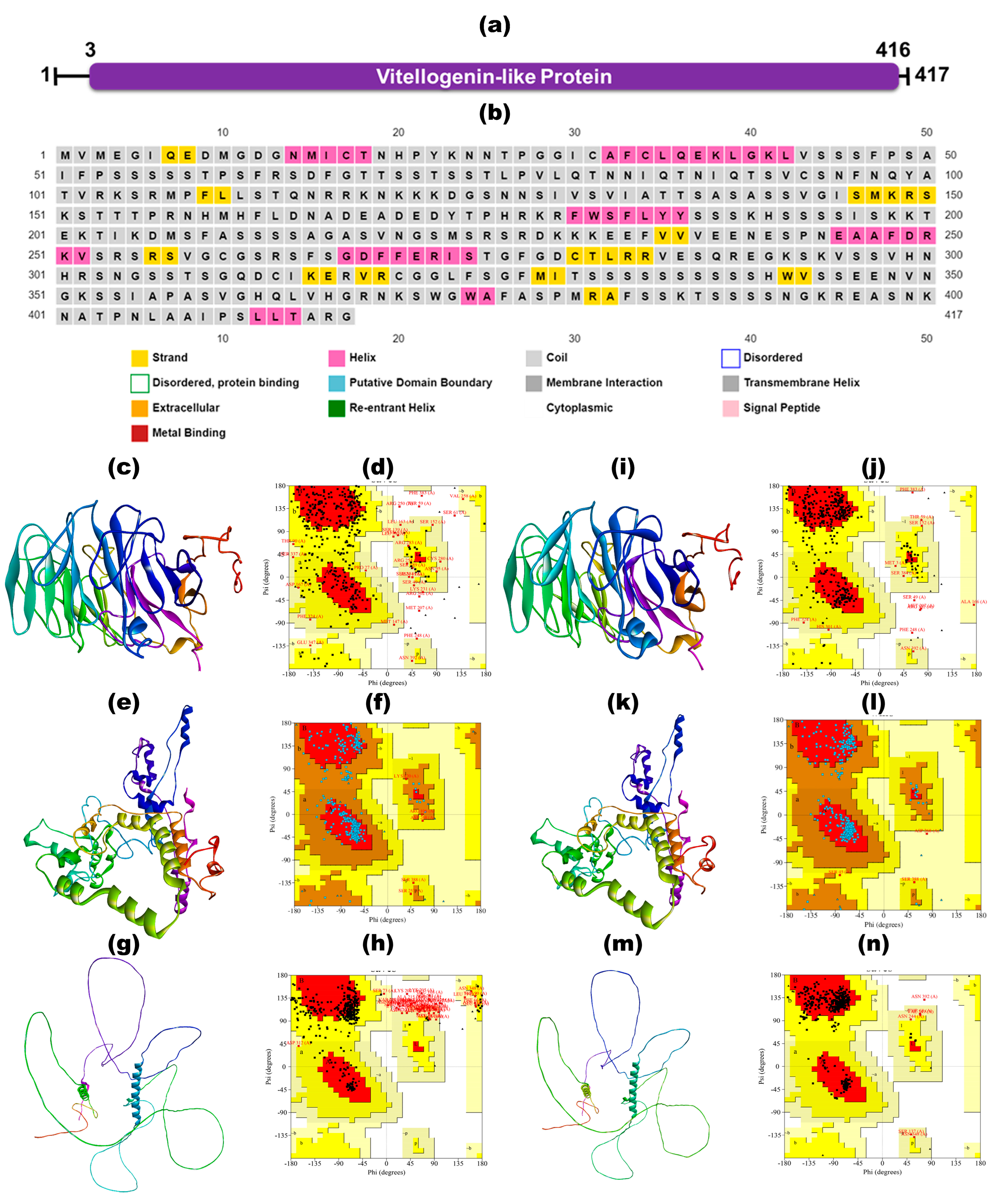
3.2. Structural and Functional Annotation Using InterPro

InterPro identified the VLP domain, spanning amino acids 3 to 416, corresponding to the PANTHER database entry PTHR34460, as illustrated in Figure 1a. The functional characterization of VLP revealed its involvement as an integral component of the cellular membrane, with associated GO terms GO:0016020 and GO:0016021. The systematic examination of the physiochemical properties of plant defense VLP of N. tabacum provides crucial insights into their functionality within biological contexts.

3.3. Physiochemical Properties

The physicochemical properties of VLP reveal an essential nature characterized by an abundance of positively charged amino acid residues, which exhibit higher stability than other proteins. Table 1 presents a comprehensive overview of the physiochemical characteristics of plant defense VLP of N. tabacum.

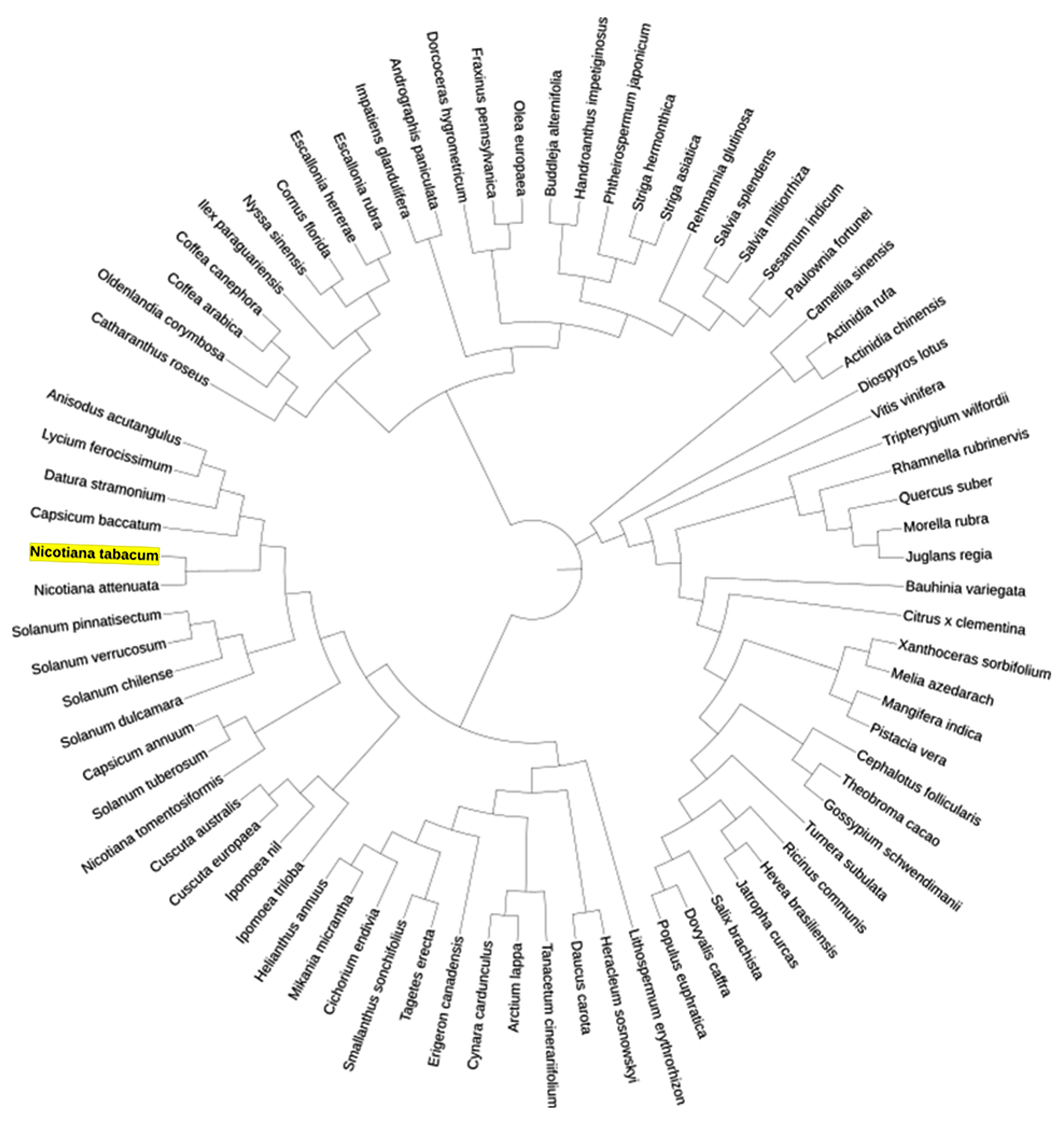
3.4. Phylogenetic Analysis

A phylogenetic analysis was conducted to characterize VLP in N. tabacum, revealing its evolutionary origin, function, and relationships among different plants. The analysis elucidated how VLP has evolved, its connections to other proteins, and its distribution across plant groups, facilitating functional inference based on similarities or dissimilarities with known proteins. The ancestral lineage of VLP in N. tabacum was elucidated using the ML method with the Le Gascuel 2008 model, selected based on its minimal BIC value of 59,785.02594. The phylogenetic tree for VLP in N. tabacum was constructed using protein sequences, achieving the highest log likelihood of −31,553.29. This tree incorporated 417 amino acid sequences, spanning 657 positions in the final dataset.
The analysis revealed that VLP in N. tabacum demonstrated close phylogenetic relationships with VLP homologs from related genera within the Solanaceae family, including Capsicum, Solanum, Datura, Lycium, and Anisodus. These genera share a common ancestor and form distinct clades, reflecting evolutionary diversification driven by ecological adaptations and selective pressures. The close clustering of the N. tabacum VLP with these homologs, as shown in Figure 2, underscores a conserved evolutionary trajectory across Solanaceae. While only a single N. tabacum VLP sequence was available for this analysis, the phylogenetic positioning suggests functional conservation and potential divergence from other Solanaceae members, supporting its unique role in plant defense and adaptation.

3.5. Structure Prediction, Refinement and Validation

3.5.1. Secondary Structure Prediction

The SOPMA server predicted that the secondary structure of VLP consists of 13.67% alpha helices, 10.07% extended strands, and 76.29% random coils. A visual representation of these secondary structures for each amino acid was generated for a complete VLP sequence using PSIPRED, as illustrated in Figure 1b.

3.5.2. Ab Initio Structure Prediction Using I-TASSER, RoseTTAFold, and AlphaFold

After that, the tertiary structure of VLP was generated using two online servers: iTASSER and RoseTTAFold. VLP 3D structure prediction result from the I-TASSER server shows four predicted models. The models were predicted based on C-score, Exp. TM-Score, Exp. RMSD, No. of decoys, and Cluster density. The best score model is shown in Figure 1c. VLP structure prediction results from the ROBETTA Baker server showed five predicted models using RoseTTAFold, and model 1 (Figure 1e) was prioritized based on its highest C-score. The AlphaFold-predicted structure of VLP in N. tabacum (Identifier: AF-A0A1S4CXB2-F1) is shown in Figure 1g.
These three structures, obtained from the I-TASSER server, ROBETTA Baker server, and AlphaFold, were subjected to structure validation using a Ramachandran plot to determine the best-predicted structure. The iTASSER model exhibited 54.5% of residues in the most favored regions (Figure 1d). In contrast, the RoseTTAFold model displayed superior structural quality with 83.2% of residues in these regions (Figure 1f). In comparison, the AlphaFold model showed 50.0% of residues in the most favored areas of the Ramachandran plots (Figure 1h).

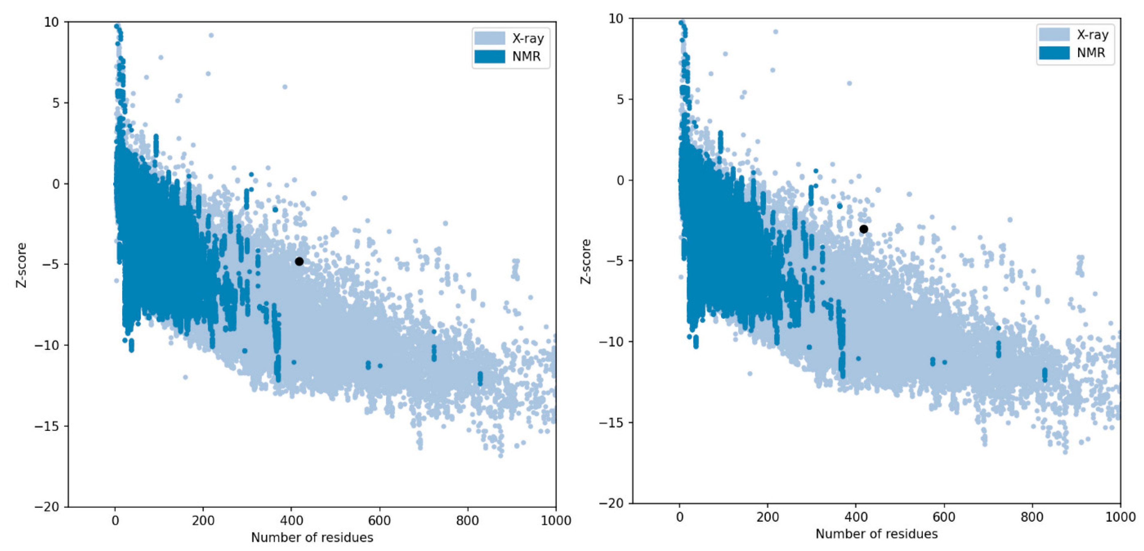
3.5.3. Structure Validation, Refinement, Quality Assessment, and Model Selection

Based on the Ramachandran plots, the RoseTTAFold model (Figure 1e) of VLP was the best-predicted structure, but it was not a high-quality model, as it would have had over 90% of its residues in the most favored regions. Therefore, we refined all the 3D modeled structures using the GalaxyRefine server, which performed repacking and side-chain rebuilding processes to relax the structures through MDS. GalaxyRefine produced five refined models, from which we selected the models with the highest percentage of residues in the Ramachandran-favored regions and the lowest RMSD values. The selected iTASSER refined model exhibited 88.2% of residues in the Ramachandran favored regions and an RMSD value of 0.462 (Figure 1i). For RoseTTAFold, the selected refined model showed superior quality, with 97.8% of residues in the favored regions and an RMSD value of 0.348 (Figure 1k). The chosen refined AlphaFold model demonstrated 96.4% of residues in the favored areas with an RMSD value of 0.856 (Figure 1m).
The refined 3D structures of VLP were evaluated using Ramachandran plot analysis, revealing differences in the quality of the models generated by iTASSER, RoseTTAFold, and AlphaFold. The iTASSER refined model showed 76.7% of residues in the most favored regions, 19.8% in additional allowed regions, 2.1% in generously allowed regions, and 1.3% in disallowed regions (Figure 1j). In contrast, the RoseTTAFold refined model demonstrated a higher quality, with 89.3% of residues in the most favored regions, 9.9% in additional allowed regions, 0.5% in generously allowed regions, and only 0.3% in disallowed regions (Figure 1l). Similarly, the refined AlphaFold model exhibited 90.4% of residues in the most favored regions, 8.0% in additional allowed regions, 1.3% in generously allowed regions, and 0.3% in disallowed regions (Figure 1n).
By comparing these models, the AlphaFold refined structure had the highest percentage of residues in the most favored regions, indicating superior structural quality. RoseTTAFold and AlphaFold outperformed iTASSER in residues in the most favored regions and residues in disallowed regions, with RoseTTAFold slightly trailing AlphaFold. These results suggest that AlphaFold and RoseTTAFold provide more reliable models for VLP, with AlphaFold demonstrating a marginal advantage in overall structural accuracy. Due to this marginal difference, we further validated the structures predicted by AlphaFold and RoseTTAFold using MolProbity metrics, revealing significant differences in the quality of the models (as shown in Table 2).
In terms of all-atom contacts, the RoseTTAFold refined model had a clashscore of 1.93 (99th percentile), indicating very few severe steric overlaps (>0.4 Å per 1000 atoms), compared with the refined AlphaFold model, which had a clashscore of 18.88 (36th percentile). This result suggests that the RoseTTAFold model significantly minimizes steric clashes. Regarding protein geometry, the RoseTTAFold model had 0.27% poor rotamers and 98.91% favored rotamers, both metrics meeting the desired goals, whereas the AlphaFold model fell slightly short, with 0.55% poor rotamers and 97.54% favored rotamers. The RoseTTAFold model also performed better in terms of Ramachandran plot metrics, with 0.24% outliers and 97.83% favored regions, compared with AlphaFold’s 0.72% outliers and 96.39% favored regions. The Rama distribution Z-score for RoseTTAFold was −0.25 ± 0.38, indicating a good fit, whereas AlphaFold had a Z-score of −2.65 ± 0.27, outside the acceptable range (absolute Z-score < 2). Additionally, the MolProbity score for RoseTTAFold was 1.00 (100th percentile), indicating an excellent overall structural quality, while AlphaFold had a MolProbity score of 2.01 (75th percentile).
Last, we utilized the Z-score, as determined by the ProSA tool, to assess the overall quality of the AlphaFold and RoseTTAFold structure models, which assesses model accuracy based on statistical comparisons with experimentally validated structures. The RoseTTAFold model exhibited a more favorable Z-score of −4.79, indicating a higher-quality structure closely resembling native proteins solved by X-ray crystallography. In contrast, the AlphaFold model yielded a Z-score of −3.01, suggesting comparatively lower structural quality, as shown in Figure 3.
Overall, the RoseTTAFold refined model outperformed the refined AlphaFold model in most structural evaluation metrics, indicating a higher quality and greater accuracy in the structure of VLP in N. tabacum. The RoseTTAFold refined 3D structural model of Pathogen Defense VLP in N. tabacum has been deposited in the ModelArchive repository, an open-access platform for computational structure models. The model can be accessed via https://www.modelarchive.org/doi/10.5452/ma-wajr2 (accessed on 13 April 2025).

3.6. Functional Interaction Site Prediction

The binding pockets within the 3D structure of the plant defense VLP of N. tabacum, predicted by RoseTTAFold, were identified using the CASTp 3.0 online server. The analysis revealed a binding pocket with a surface area of 2461.98 Å2 and a surface volume of 2893.06 Å3, as shown in Table 3.

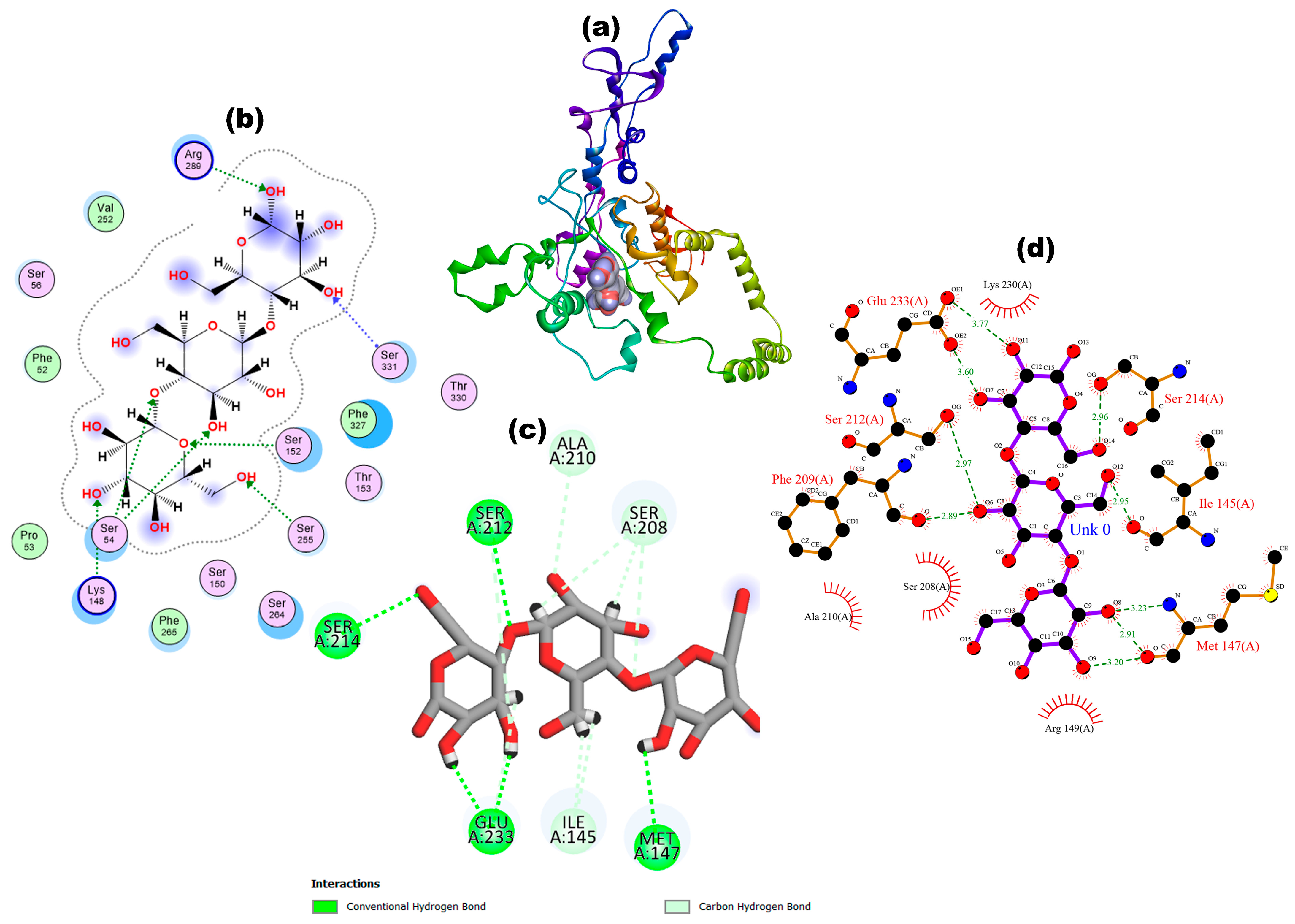
3.7. Molecular Docking

The molecular docking outcomes for the plant defense VLP of N. tabacum with PGN and β-glucan revealed significant differences in binding affinity, RMSD, and interaction profiles (Table 4). The binding affinity of VLP with PGN was −10.16 kcal/mol, with an RMSD of 2.3 Å, indicating a slightly higher deviation from the ideal docking pose, as shown in Figure 4. An RMSD value of 2.3 Å suggests the binding pose is relatively stable. However, it is not as optimal as lower RMSD values. Key amino acid residues involved in the interaction included Ser152, which formed a conventional hydrogen bond and two carbon-hydrogen bonds with bond lengths of 2.99 Å, 3.50 Å, and 3.38 Å, respectively. Additionally, Ser150 formed an unfavorable donor–donor bond (2.38 Å), while Ser262 formed a conventional hydrogen bond (2.69 Å). Ser264 and Ser331 each formed a carbon-hydrogen bond, with bond lengths of 3.26 Å and 3.71 Å, respectively. Last, Phe265 formed a pi-alkyl bond (5.41 Å), and Arg289 formed two unfavorable donor–donor bonds (2.23 Å and 2.02 Å).
In contrast, the binding affinity of VLP with β-glucan was significantly higher at −7.19 kcal/mol, with a much lower RMSD of 1.45 Å, indicating a more stable and precise docking pose compared with PGN, as shown in Figure 5. An RMSD value of 1.45 Å suggests that the interaction with β-glucan is highly stable, reflecting a more optimal docking conformation. The interactions involved Ile145, which formed two carbon-hydrogen bonds (2.51 and 2.57 Å), Met147 with one conventional hydrogen bond (2.03 Å), and Ser208 with three carbon-hydrogen bonds (2.35, 2.86, and 3.48 Å). Additional interactions included Ala210 with one carbon-hydrogen bond (3.42 Å), Ser212 with one conventional hydrogen bond and one carbon-hydrogen bond (2.36 and 3.3 Å, respectively), Ser214 with one conventional hydrogen bond (2.08 Å) and Glu233 with two conventional hydrogen bonds and one carbon-hydrogen bond (2.00, 2.25, and 2.44 Å). These results highlight the more robust and diverse binding interactions of VLP with PGN compared with β-glucan, suggesting a potentially higher affinity and specificity of VLP towards fungal PAMPs.

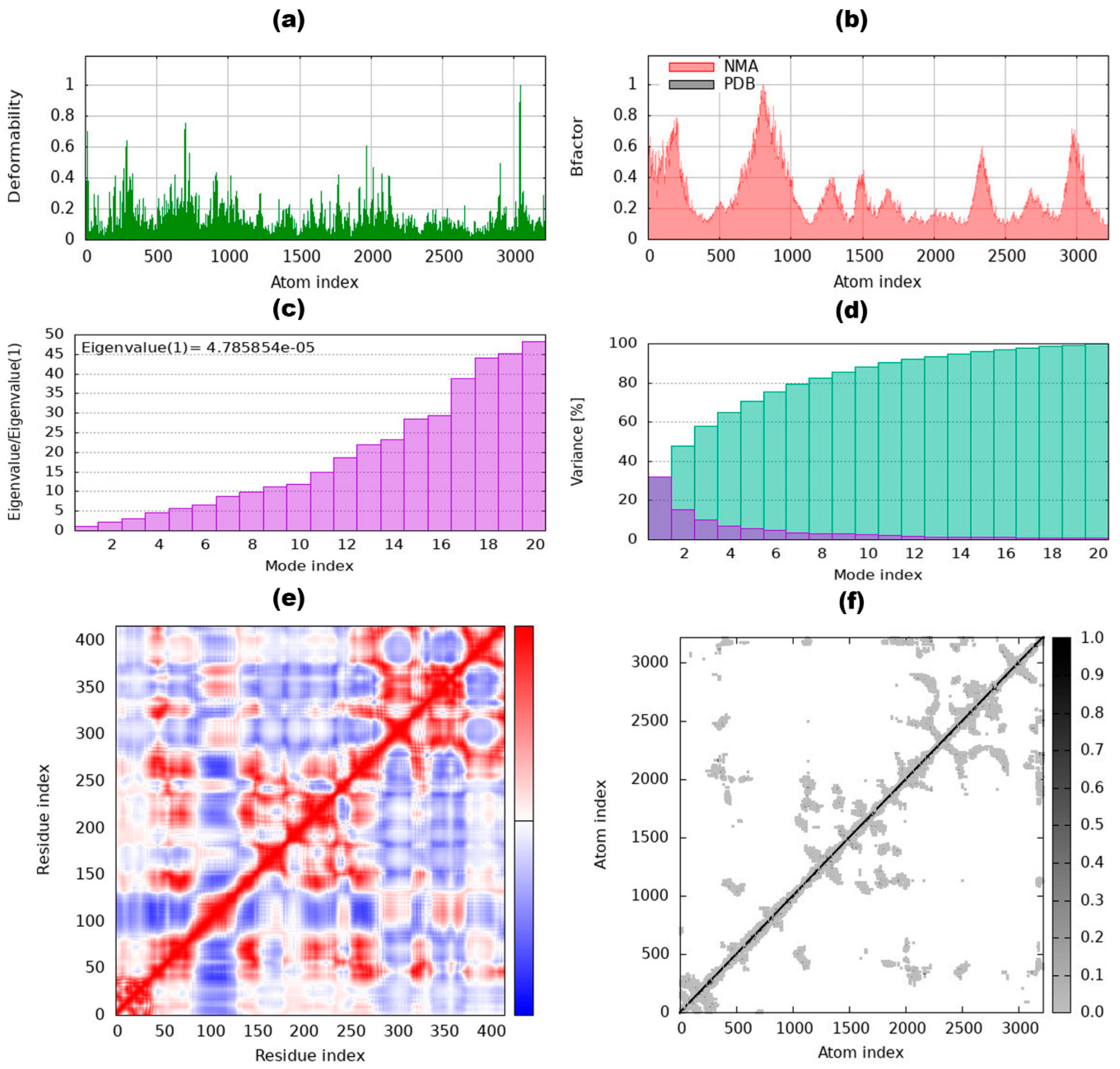
3.8. Internal Coordinates Normal Mode Analysis

The iMODS tool was used to analyze the dynamic behavior and normal modes of the complexes formed between VLP and PGN, as well as between VLP and β-glucan. This analysis evaluated stability, mobility, and deformability attributes, with visual representations of residues and docked complexes shown in Figure 4a and Figure 5a and deformability values in Figure 6a and Figure 7a. For the VLP complexes with PGN and β-glucan, most atom indexes exhibited deformability within the range of 0.2 to 0.8.
The atom index, a unique numerical label assigned to each atom, facilitates the tracking and analysis of the position and movement of individual atoms during NMA. This characteristic aids in identifying specific atoms and understanding their contributions to the molecule’s vibrational modes and collective motions [60,77]. B-factor values obtained through NMA provided insights into the mobility of the docked complexes, revealing atomic displacements that are generally below the experimentally derived B-factor values available in the PDB. These experimental PDB B-factor values reflect atoms’ atomic displacement or flexibility in the crystal structure. The B-factors from NMA simulations are presented for all atomic index complexes in Figure 6b and Figure 7b.
Eigenvalues, which gauge the system’s rigidity and identify variations in VLP dynamics, were determined to be 4.785854 × 10⁻⁵ and 4.10515 × 10⁻⁵, respectively, as shown in Figure 6c and Figure 7c. Variance graphs elucidate the relative contributions of each normal mode to equilibrium motions. Covariance graphs visualize the mobility characteristics of specific molecular regions. Elastic network graphs represent the stiffness of the springs connecting atom pairs, as presented in Figure 6d–f and Figure 7d–f.

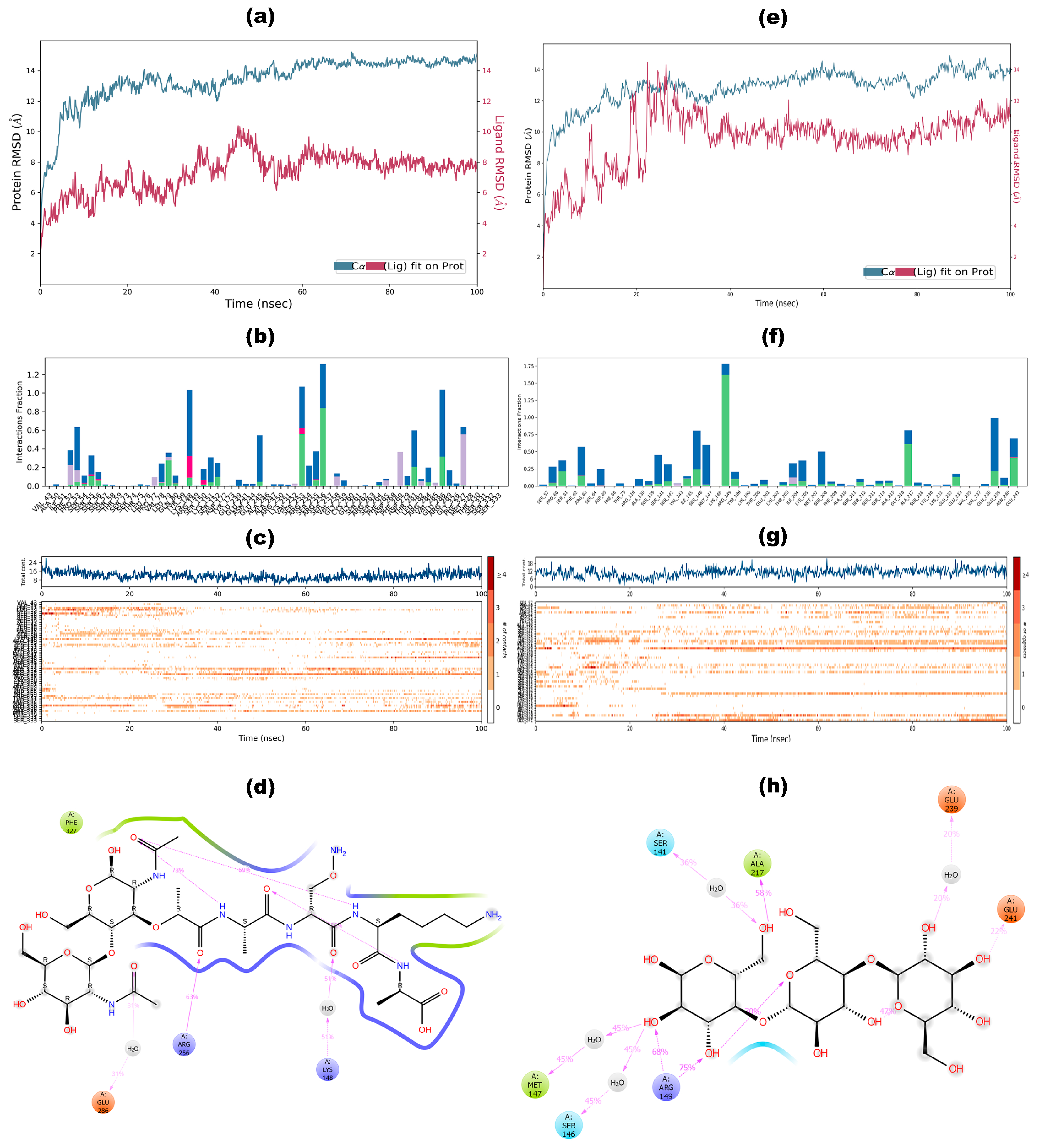
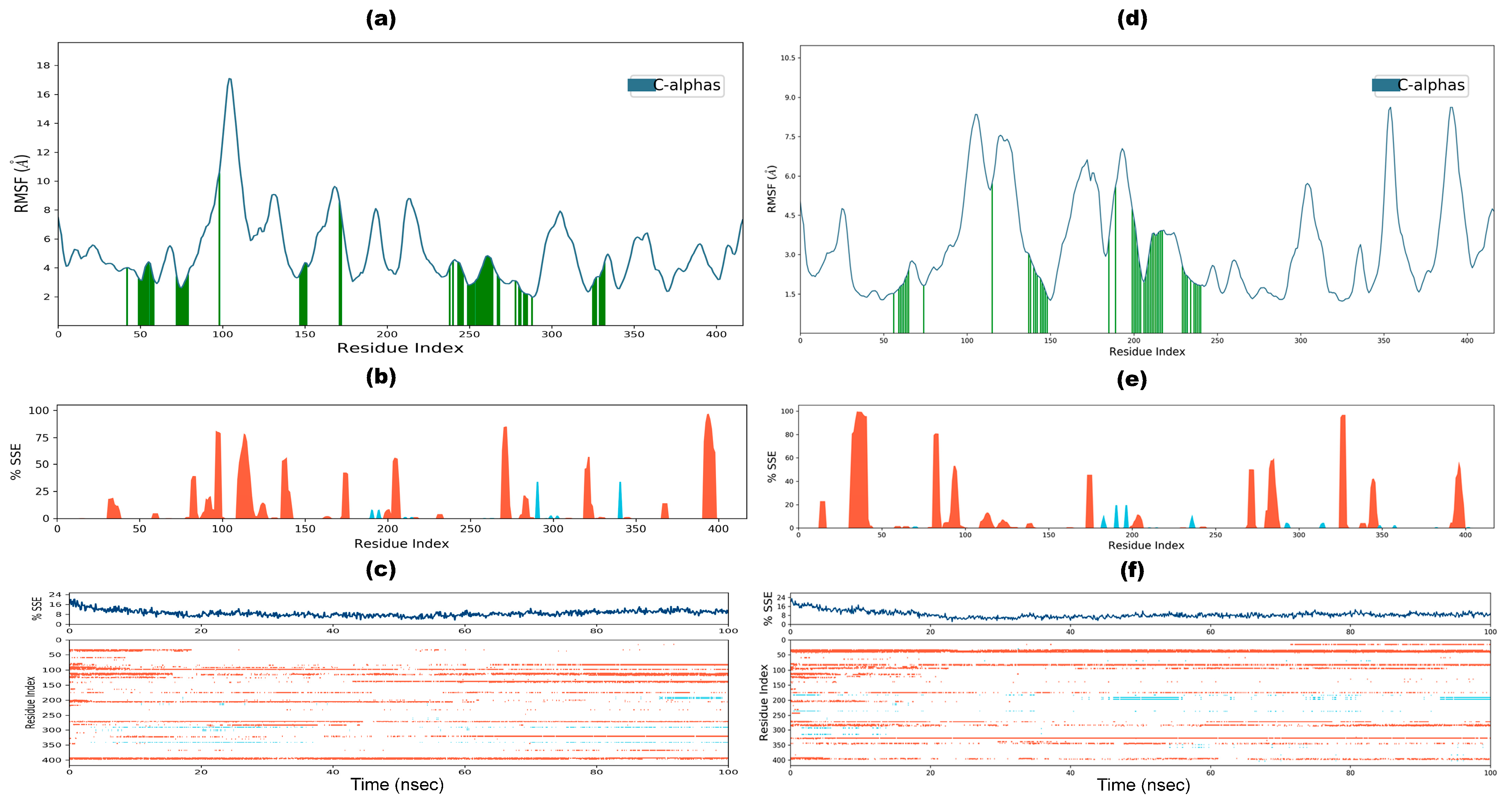
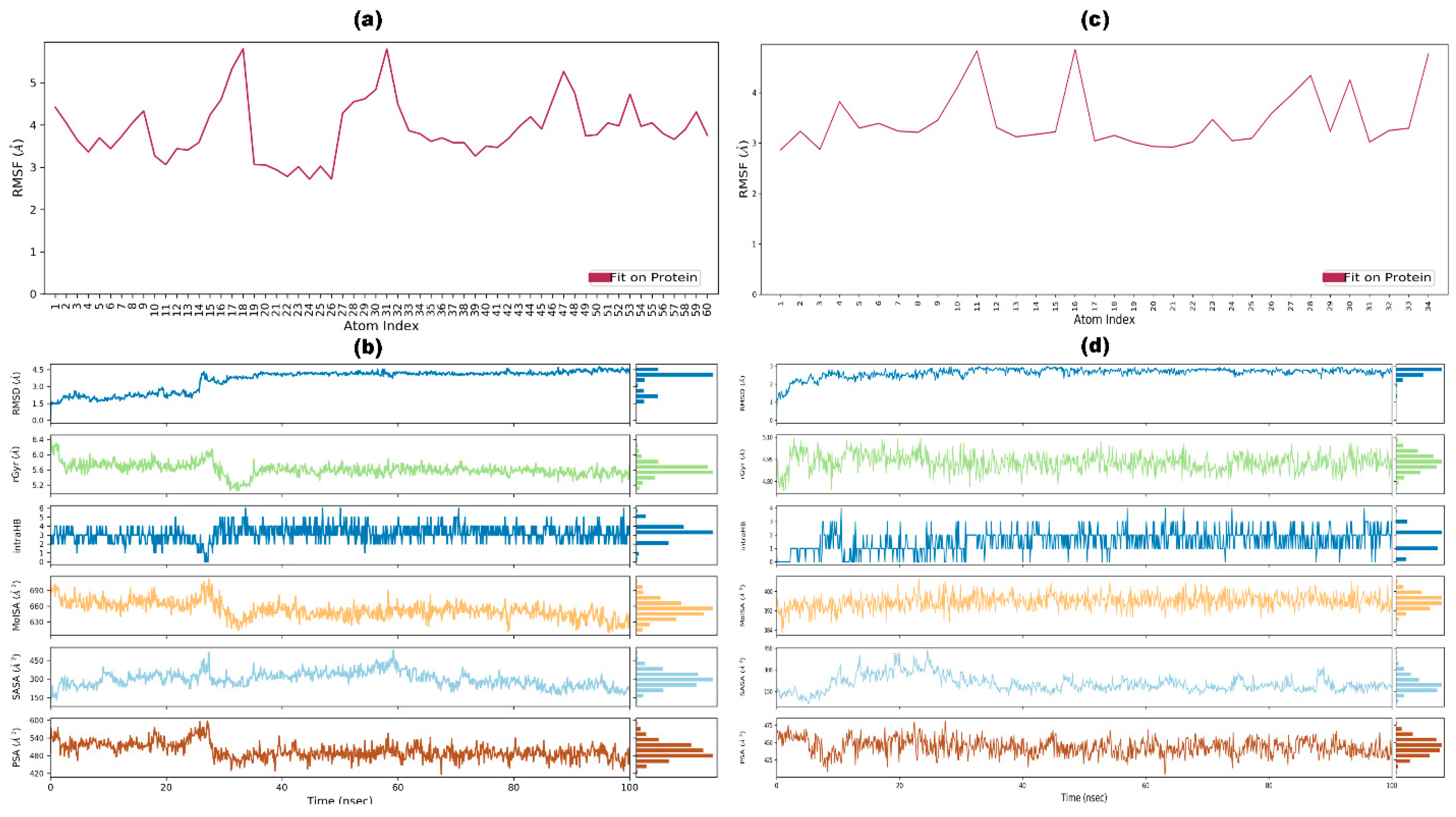
3.9. Molecular Dynamic Simulation

MDS revealed a stable and dynamic docked complex between VLP and PGN. The average RMSD for VLP (protein carbon alpha) was 13.37 Å (Figure 8a, blue), while the RMSD for PGN (ligand) fitting onto VLP was 7.3 Å (Figure 8a, red). Similarly, the MDS results for the VLP-β-glucan complex indicated stability and dynamics, with an average RMSD of 12.8 Å for the VLP (protein carbon alpha) (Figure 8e, blue) and an RMSD of 9.52 Å for β-glucan (ligand) fitting onto VLP (Figure 8e, red). RMSD values below 2 Å generally indicate stability; however, values above this threshold can still reflect meaningful dynamics depending on the context and nature of the proteins involved [78,79]. The observed RMSD values suggest that VLP may undergo significant conformational changes, which are essential for its function or interaction with other molecules. The average RMSD value of 7.3 Å for PGN fitting onto VLP indicates moderate stability during the interaction. This upshot suggests that while PGN maintains a consistent binding pose relative to VLP, it may also experience conformational adjustments as it interacts with the protein. The average RMSD value of 9.52 Å for β-glucan fitting onto VLP indicates moderate stability during their interaction. Both complexes’ relatively high RMSD values indicate flexibility within VLP-PGN and VLP-β-glucan interactions. This flexibility enables dynamic interactions that enhance binding affinity and specificity as proteins adapt their shapes to bind effectively with ligands or other proteins [80,81].
Another critical factor during the MDS is the contact number between VLP (protein) and PGN (ligand). Figure 8b depicts the types of bonds (hydrogen bonds, green; water bridges, blue; hydrophobic interactions, purple) formed with the amino acids of VLP and PGN during the MDS. Figure 8c represents the minimum eight and the maximum of more than 24 total contents between VLP and PGN. Similarly, Figure 8c also shows the number of contacts between PGN and the amino acids of VLP, ranging from 0 (white) to ≥4 (brown). Figure 4d illustrates the percentages of ion donors and acceptors in the PGN, including amino acids from protein and water molecules. Another critical factor during the MDS is the contact number between VLP (protein) and β-glucan (ligand). Figure 8f depicts the types of bonds formed with the amino acids of VLP and β-glucan during the MDS. Figure 8g represents the minimum 0 and maximum 18 total contents between VLP and β-glucan. Similarly, Figure 8g also shows the number of contacts between β-glucan and the amino acids of VLP, ranging from 0 (white) to ≥4 (brown). Figure 5d illustrates the percentages of ion donors and acceptors in the β-glucan, along with the amino acids of protein and water molecules.
The analysis of RMSF and SSE provided insights into the stability, flexibility, and structural behavior of VLP when complexed with PGN and β-glucan. RMSF values, which measure the flexibility and fluctuation of amino acid residues, revealed that most residue indices had lower RMSF values for both complexes, confirming the stability and rigidity of VLP (Figure 9a,d). Similarly, B-factors derived from IC-NMA showed consistent results with RMSF data and experimental B-factors from X-ray crystallography, further supporting the stability of VLP (Figure 6b and Figure 7b). Regarding SSE, the distribution of alpha helices and beta strands was assessed over residue indices and simulation time. For the VLP-PGN complex, the total SSE percentage was 9.0%, with 8.55% alpha helices—indicative of structural rigidity—and 0.45% beta strands, suggesting that minor flexibility aids ligand binding (Figure 9b,c). In comparison, the VLP-β-glucan complex exhibited a slightly lower SSE percentage of 8.75%, with 8.34% alpha helices and 0.41% beta strands, reflecting similar structural stability while maintaining the flexibility required for effective ligand interaction (Figure 9e,f). These results highlight the comparable stability of VLP in both complexes, with subtle differences in flexibility that may influence ligand binding dynamics.
In parallel, the RMSF value of the ligand fluctuations was calculated with an atom index to check the interaction of PGN and β-glucan fragments with VLP and their entropic role during docking. The fit PGN on the VLP graph (Figure 10a,c) exhibited more fluctuation in RMSF at higher atom indices than the fit β-glucan on the VLP graph. The RMSD, intramolecular hydrogen bonds (IntraHBs), radius of gyration (rGyr), molecular surface area (MolSA), polar surface area (PSA), and solvent accessible surface area (SASA) of PGN and β-glucan in complex with VLP was studied further during the 100 ns-long MDS and the resultant images are presented in Figure 10b,d.
The comparative analysis of PGN and β-glucan during MDS highlighted their respective structural stability, conformational spread, and interaction dynamics. The average RMSD value for PGN was 3.61 Å. In comparison, β-glucan exhibited a slightly lower RMSD of 2.62 Å, indicating robust structural stability in both ligands, with β-glucan marginally more stable. The rGyr, indicative of the ligands’ conformational spread, averaged 5.61 Å for PGN, with values ranging from 5.2 to 6.4 Å, suggesting a more extended conformation compared with β-glucan, which had an average rGyr of 4.93 Å and a range of 4.8 to 5.1 Å. PGN formed a maximum of 6 IntraHBs during the simulation, while β-glucan formed up to 4, reflecting differences in their intramolecular interactions. The MolSA, which relates to van der Waals interactions, was significantly larger for PGN (630–700 Å2, average 653.3 Å2) than for β-glucan (384–420 Å2, average 395.89 Å2), indicating a more incredible spatial occupation for PGN. Similarly, the SASA, representing water exposure, was higher for PGN (150–470 Å2, average 297.95 Å2) compared with β-glucan (130–450 Å2, average 203 Å2). This result suggests that PGN is more solvent-exposed than β-glucan. Finally, the PSA, reflecting the ligands’ potential for polar interactions and hydrogen bonding, averaged 495.9 Å2 for PGN and 443.9 Å2 for β-glucan, indicating slightly more significant polar interaction potential for PGN. While both ligands demonstrated strong stability, PGN displayed a more extended conformation, higher spatial occupation, and greater solvent exposure. In contrast, β-glucan exhibited greater compactness and a slightly reduced interaction potential.

3.10. Post-Simulation Analysis

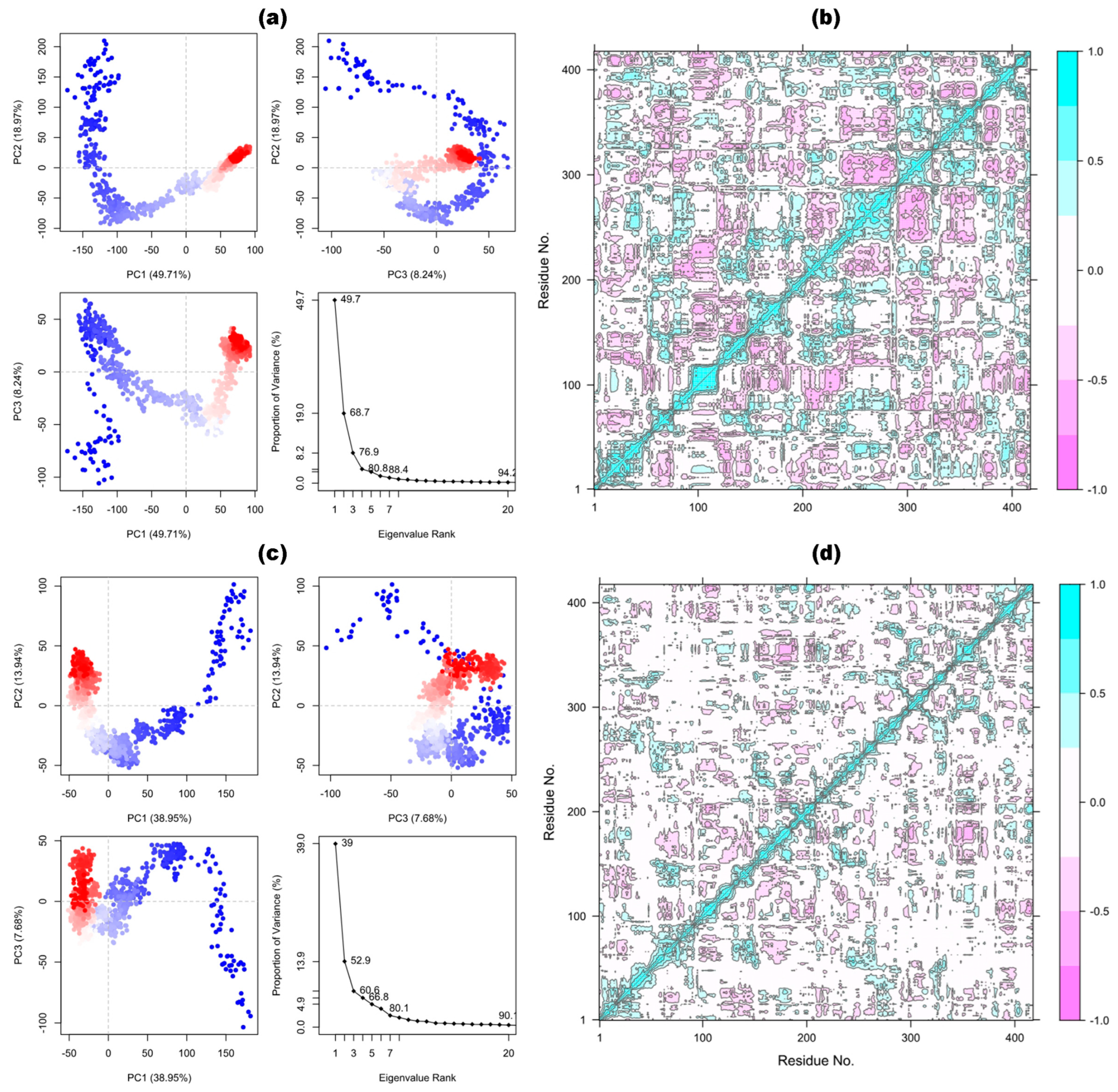
3.10.1. Principal Components and Dynamic Cross-Correlation Matrix Analysis

PCA was used to assess domain dynamics and motion in the VLP-PGN and VLP-β-glucan docked complexes during 100 ns simulations, with results expressed as a fraction of variance (eigen fraction) derived from a covariance matrix of eigenmodes. The VLP-PGN complex exhibited a sharp 80.8% variance drop at the first four eigenmodes (Figure 11a). The VLP-β-glucan complex displayed a similar 80.1% drop but at seven eigenmodes (Figure 11b), indicating significant conformational changes in the VLP active pocket induced by both ligands. After these points, both complexes stabilized, showing no notable variations in eigenvalues from 4 to 20, which supports the stability of the protein–ligand interactions. The initial three eigenfractions revealed clustered motions from the MDS trajectory, with early flexibility transitioning into compact cluster motions as the simulation progressed. Two-dimensional plots illustrated the overall conformational shifts in VLP for both systems, with a color gradient from red to blue reflecting gradual structural changes over time. This comparison highlights that while both ligands induce substantial conformational changes and stabilize the VLP active pocket, their dynamics differ slightly in the number of eigenmodes involved in the initial variance drop. The conformational dynamics of VLP induced by PGN and β-glucan were assessed using DCCM analysis, focusing on the correlated motions of alpha carbon atoms during the simulation (Figure 11c,d). The DCCM analysis revealed significant alterations in the correlated motions and dynamic behavior of the VLP structure when it was docked with PGN compared with β-glucan. These findings align closely with the results observed in the PCA plots, providing consistent evidence of ligand-induced structural changes in VLP.

3.10.2. Binding Affinity Calculations Using MM-GBSA

Table 5 presents the ΔGbind calculations and its components for two systems, VLP-PGN and VLP-β-glucan, across different simulation time points (0 ns, 25 ns, 50 ns, 75 ns, and 100 ns). The ΔGbind for VLP-PGN and VLP-β-glucan exhibited fluctuations over the simulation time, with VLP-PGN generally showing stronger binding than VLP-β-glucan. The ΔGbind analysis revealed that VLP-PGN exhibits more substantial binding stability than VLP-β-glucan across all simulation time points. At 0 ns, ΔGbind for VLP-PGN is −85.41 kcal/mol, which decreases to −64.65 kcal/mol at 100 ns. In contrast, VLP-β-glucan exhibits weaker binding, starting at −66.09 kcal/mol and decreasing to −43.87 kcal/mol. On average, VLP-PGN maintains better stability than VLP-β-glucan. The van der Waals (ΔGbindvdW) interactions are a major contributor to the binding energy for both systems, with VLP-PGN showing consistently stronger interactions. Hydrogen bonding (ΔGbindHbond) is slightly more pronounced in VLP-β-glucan, while ΔGbindSolv GB imposes a higher penalty for VLP-PGN.

4. Discussion

This study uses computational approaches to identify and characterize pathogen defense proteins in N. tabacum. By analyzing the structural and functional characteristics of N. tabacum, we identified an uncharacterized pathogen defense VLP in its proteome. Vg and VLPs, traditionally known for their role in reproduction and egg development in oviparous animals, including insects, are now recognized for their functions beyond serving as nutrient reserves for developing embryos [82,83]. Emerging evidence suggests these proteins exhibit immune-relevant and antioxidant properties in animals and plants. Vg and its derived yolk proteins, such as lipovitellin and phosvitin, possess potent antioxidant properties, scavenging free radicals and protecting cells from oxidative stress [84]. Additionally, VLPs can modulate the immune system by binding to and neutralizing pathogens and regulating the production of immune-related molecules. These properties suggest their involvement in plant pathogen defense mechanisms. For example, insect VLPs can suppress the oxidative burst response in plants, a key defense mechanism against pathogens, which may potentially facilitate pathogen infection and transmission. They may also bind to and neutralize pathogens, preventing disease and modulating plant immune signaling pathways [85,86,87].
The role of VLPs in plant pathogen defense is an emerging research area, with studies on insect-transmitted plant viruses providing initial insights. VLPs, identified in plants and increasingly recognized for their role in plant defense against pathogens such as bacteria and fungi, may act as PRRs. They can induce defense responses, exhibit antimicrobial activity, and interact directly with PAMPs, helping plants recognize and respond to pathogens. As PRRs, they may bind to PAMPs, activate immune signaling pathways, produce reactive oxygen species, and induce systemic acquired resistance. In plants such as rice and Arabidopsis, VLPs have been shown to recognize bacterial and fungal components, activate defense genes, and enhance resistance. These proteins emerge as essential players in plant immunity, potentially developing crops with improved disease resistance [88,89].
The phylogenetic tree of N. tabacum VLP protein sequence clusters closely with sequences from the genera Capsicum, Solanum, Datura, Lycium, and Anisodus, suggesting significant sequence homology among these VLP proteins, all of which belong to the Solanaceae family, indicating a common evolutionary origin and potential conservation because of shared functions or structural requirements (Figure 2). This clustering implies that these VLPs might perform similar roles, possibly related to plant defense mechanisms against viral infections, retaining crucial structural features. Evolutionary pressures, such as horizontal gene transfer, convergent evolution, or shared environmental factors, may have driven this conservation. Understanding the sequence similarity and structural conservation can inform biotechnological applications, such as vaccine development, by allowing data extrapolation across these related species. Molecular studies can reveal critical domains and motifs responsible for VLP function, while ecological interactions might reflect adaptive response mechanisms to pathogens. Comparative genomic studies can elucidate the genetic context of VLP genes, enhancing our understanding of the genetic mechanisms behind these similarities. Thus, the close clustering of the N. tabacum VLP protein sequence with those from other genera suggests potential evolutionary and functional similarities, warranting further investigation into their possible roles and applications.
The physicochemical properties of the N. tabacum VLP protein sequence (Table 1) highlight its molecular weight (45.145 kDa), theoretical pI (~10), and stability across various conditions, as indicated by half-life estimates and an aliphatic index of 49.81. The sequence’s structural predictions, refined using iTASSER, RoseTTAFold, and AlphaFold, were validated through Ramachandran plots and MolProbity metrics, revealing RoseTTAFold as the most accurate initial model. However, after further refinement using GalaxyRefine, the iTASSER model showed 88.2% of residues in favored regions (RMSD 0.462), while RoseTTAFold excelled with 97.8% in the favored areas (RMSD 0.348). The MolProbity analysis further confirmed RoseTTAFold’s superior quality, displaying minimal steric clashes and favorable rotamer conformations. The ProSA tool assessment corroborated these findings, showing that RoseTTAFold (−4.79 Z-score) closely matches experimentally determined structures, suggesting it is the most accurate VLP model for N. tabacum.
The functional interaction site prediction of the N. tabacum VLP protein, using CastP 3.0, identified a significant site with an area of 2461.98 Å2 and volume of 2893.06 Å3, involving numerous amino acids (Table 3). Molecular docking studies revealed that VLP binds more strongly to PGN than β-glucan, with binding affinities of −10.16 kcal/mol and −7.19 kcal/mol, respectively. Meanwhile, the β-glucan interaction exhibited lower RMSD (1.45 Å) than PGN (2.3 Å), indicating a more stable binding. Dynamic behavior and normal mode analysis using iMODS highlighted the stability and mobility of the VLP-β-glucan complex, with B-factor values and eigenvalues supporting its stability. These findings suggest a possible interaction between the N. tabacum VLP and β-glucan, which may be relevant to plant defense mechanisms against fungal pathogens and merits further investigation.
MDS provided valuable insights into the structural stability, flexibility, and interaction dynamics of VLP with PGN and β-glucan. The observed RMSD for both complexes suggested that VLP undergoes significant conformational changes upon docking with PGN and β-glucan, and it maintains a stable dynamic equilibrium. The VLP-PGN complex exhibited an average RMSD of 13.37 Å for the protein, with PGN showing moderate stability, as indicated by an RMSD of 7.3 Å. This outcome suggests that PGN adopts a stable binding pose relative to VLP but undergoes slight conformational adjustments during the simulation. Similarly, the VLP-β-glucan complex exhibited an average RMSD of 12.8 Å for VLP and 9.52 Å for the β-glucan, indicating comparable flexibility and suggesting that both ligands interact with VLP through dynamic binding processes. These RMSD results were consistent with prior research suggesting that RMSD values above 2 Å may still reflect meaningful dynamics, especially in protein–ligand interactions [78,79].
The contact analysis further elucidated the nature of interactions between VLP and the ligands. During the MDS, both PGN and β-glucan formed various interactions with VLP, including hydrogen bonds, water bridges, and hydrophobic interactions, which are essential for ligand binding and stabilization. PGN, in particular, showed a higher frequency of contacts with VLP, with a maximum of 24 total contacts, compared with β-glucan’s 18. The diversity in the types and number of interactions formed by each ligand supports the idea that their distinct structures influence their binding modes and interaction dynamics with VLP. PGN’s larger molecular surface area and higher solvent accessibility further suggest that it occupies more space in the interaction, likely contributing to a more extended conformation during the binding process.
The structural rigidity and flexibility of VLP were also assessed through RMSF and SSE. RMSF values confirmed that most residues of VLP remained stable, with minimal fluctuations, suggesting that the protein backbone does not experience significant changes during the complex formation. The distribution of alpha helices and beta strands revealed a similar pattern in both VLP complexes. The VLP-PGN complex had 9.0% of its structure in SSE, predominantly composed of alpha helices. This result highlights the structural rigidity required for ligand binding, allowing only a small proportion of beta strands to be flexible. Similarly, the VLP-β-glucan complex exhibited a slightly lower SSE value of 8.75%. Still, the distribution of alpha helices and beta strands remained comparable, reinforcing the overall structural stability of the protein in both interactions.
Further analysis of ligand dynamics revealed that PGN demonstrated more fluctuation in its RMSF than β-glucan, suggesting that PGN may exhibit higher flexibility during the binding process. PGN, with its larger MolSA and higher SASA, likely interacts more extensively with its environment than β-glucan, which is more compact. The differences in these properties could indicate PGN’s more extended conformation and more significant potential for multiple interaction sites with VLP.
The PCA and DCCM analysis offered further insights into the conformational dynamics of VLP when docked with PGN and β-glucan. Both ligands induced significant conformational changes in the VLP active pocket, with the PCA results revealing that the VLP-PGN complex experienced a variance drop of 80.8% at the first four eigenmodes. In contrast, the VLP-β-glucan complex showed a similar drop at seven eigenmodes. These findings suggest that both ligands effectively modulate the flexibility and stability of VLP, albeit with slight differences in the number of eigenmodes involved in the initial variance drop. The DCCM analysis confirmed that PGN and β-glucan induce distinct but significant alterations in the correlated motions of the VLP structure, with PGN demonstrating a more pronounced effect on VLP’s dynamic behavior than β-glucan. These results highlight the critical role of ligand-induced conformational changes in modulating the functional interactions between VLP and its ligands.
Last, VLP-PGN demonstrated more substantial binding stability than VLP-β-glucan across all simulation time points, with a ΔGbind of −85.41 kcal/mol at 0 ns and decreasing to −64.65 kcal/mol at 100 ns. In contrast, VLP-β-glucan started at −66.09 kcal/mol and decreased to −43.87 kcal/mol. The ΔGbindvdW contributed significantly to the binding energy, with VLP-PGN exhibiting stronger interactions. ΔGbindHbond was slightly more prominent in VLP-β-glucan, while ΔΔGbindSolv GB imposed a higher penalty for VLP-PGN.

5. Conclusions

This study identifies and characterizes pathogen defense proteins in N. tabacum through computational approaches, focusing on an uncharacterized pathogen defense VLP. Traditionally known for their roles in reproduction, VLPs are now recognized for their immune-relevant and antioxidant activities in animals and plants. The structural and functional analysis of the N. tabacum VLP revealed significant potential in plant defense, with strong binding affinity and stability towards β-glucan, a fungal PAMP. This study highlighted the dynamic interactions between VLPs and PAMP (PGN and β-glucan) in N. tabacum. MDS results showed that PGN exhibits more substantial binding stability and more extensive interactions with VLP than β-glucan. Our findings suggested that N. tabacum VLPs could act as PRRs, recognizing and responding to pathogens by activating immune signaling pathways and exhibiting antimicrobial activity. Future research should focus on experimentally validating the predicted interactions and functional roles of VLPs in plant defense. Investigating the mechanistic pathways of VLPs in vivo will enhance our understanding of their role in immune modulation and pathogen neutralization. Additionally, exploring the evolutionary conservation and structural features of VLPs across different species within the Solanaceae family could provide insights into their universal functions and potential biotechnological applications. By leveraging the structural insights from this study, future work could aim to engineer VLP-based strategies for developing crops with enhanced resistance to a wide range of pathogens, contributing to sustainable agriculture and food security.

Supplementary Materials

The following supporting information can be downloaded at https://www.mdpi.com/article/10.3390/app15084463/s1. Table S1. Bioinformatics tools employed for structural, evolutionary, and molecular interaction analyses of Nicotiana tabacum VLP, including software functions, access links, and critical parameters. Table S2. Accession Numbers, Protein Names, and Plant Species Corresponding to Hypothetical and Uncharacterized Proteins Used in the Phylogenetic Analysis of VLPs from N. tabacum and Its Homologs. The sequences were retrieved from the NCBI Clustered Non-Redundant (nr) database via BLASTp to construct a phylogenetic tree to study the evolutionary relationships among VLPs in Nicotiana tabacum and its homologs. The accession numbers listed provide transparency and reproducibility of the analysis.

Author Contributions

Conceptualization, A.E.; methodology, A.E. and H.M.; validation, A.E., H.M. and A.Y.R.; formal analysis, A.E.; data curation, H.M. and A.Y.R.; writing—original draft preparation, A.E. and H.M.; writing—review and editing, H.M. and A.Y.R.; visualization, A.E., H.M. and A.Y.R.; supervision, A.E. All authors have read and agreed to the published version of the manuscript.

Funding

This research received no external funding.

Institutional Review Board Statement

Not applicable.

Informed Consent Statement

Not applicable.

Data Availability Statement

The original contributions presented in the study are included in the article/Supplementary Materials, further inquiries can be directed to the corresponding author. Meanwhile, the refined 3D structure of Pathogen Defense Vitellogenin-like Protein in Nicotiana tabacum modeled by RoseTTAFold can be accessed via https://www.modelarchive.org/doi/10.5452/ma-wajr2 (accessed on 13 April 2025).

Acknowledgments

While preparing this work, the author(s) used ChatGPT v3.5 (OpenAI) to refine grammar and language. After utilizing this tool, the author(s) thoroughly review and edit the content as needed and take full responsibility for the accuracy and integrity of the publication.

Conflicts of Interest

The authors declare no conflicts of interest.

References

  1. Otávio, O.A.; dos Santos, M.S.; Antunes Filho, S.; Backx, B.P. Nanotechnology for the Control of Plant Pathogens and Pests. Plant Nano Biol. 2024, 8, 100080. [Google Scholar] [CrossRef]
  2. War, A.R.; Paulraj, M.G.; Ahmad, T.; Buhroo, A.A.; Hussain, B.; Ignacimuthu, S.; Sharma, H.C. Mechanisms of Plant Defense against Insect Herbivores. Plant Signal. Behav. 2012, 7, 1306. [Google Scholar] [CrossRef] [PubMed]
  3. Hönig, M.; Roeber, V.M.; Schmülling, T.; Cortleven, A. Chemical Priming of Plant Defense Responses to Pathogen Attacks. Front. Plant Sci. 2023, 14, 1146577. [Google Scholar] [CrossRef] [PubMed]
  4. Peng, H.; Feng, H.; Zhang, T.; Wang, Q. Editorial: Plant Defense Mechanisms in Plant-Pathogen Interactions; Frontiers Media SA: Lausanne, Switzerland, 2023; Volume 14, p. 1292294. [Google Scholar]
  5. Saeed, A. Plant Defense Mechanisms Against Bacterial Pathogens. Genet. Genom. Sci. 2019, 4, 1–6. [Google Scholar] [CrossRef]
  6. Mithöfer, A.; Maffei, M.E. General Mechanisms of Plant Defense and Plant Toxins. In Plant Toxins; Springer: Dordrecht, The Netherlands, 2017; pp. 3–24. ISBN 978-94-007-6464-4. [Google Scholar]
  7. Kaur, S.; Samota, M.K.; Choudhary, M.; Choudhary, M.; Pandey, A.K.; Sharma, A.; Thakur, J. How Do Plants Defend Themselves against Pathogens-Biochemical Mechanisms and Genetic Interventions. Physiol. Mol. Biol. Plants 2022, 28, 485–504. [Google Scholar] [CrossRef]
  8. Drapal, M.; Enfissi, E.M.A.; Fraser, P.D. The Chemotype Core Collection of Genus Nicotiana. Plant J. 2022, 110, 1516–1528. [Google Scholar] [CrossRef]
  9. Zou, X.; Bk, A.; Rauf, A.; Saeed, M.; Al-Awthan, Y.S.; Al-Duais, M.A.; Bahattab, O.; Hamayoon Khan, M.; Suleria, H.A.R. Screening of Polyphenols in Tobacco (Nicotiana tabacum) and Determination of Their Antioxidant Activity in Different Tobacco Varieties. ACS Omega 2021, 6, 25361–25371. [Google Scholar] [CrossRef]
  10. Zhang, W.; Pan, X.; Fu, J.; Cheng, W.; Lin, H.; Zhang, W.; Huang, Z. Phytochemicals Derived from Nicotiana tabacum L. Plant Contribute to Pharmaceutical Development. Front. Pharmacol. 2024, 15, 1372456. [Google Scholar] [CrossRef]
  11. Stewart, G.G. A History of the Medicinal Use of Tobacco 1492–1860. Med. Hist 1967, 11, 228–268. [Google Scholar] [CrossRef]
  12. Zou, X.; BK, A.; Abu-Izneid, T.; Aziz, A.; Devnath, P.; Rauf, A.; Mitra, S.; Emran, T.B.; Mujawah, A.A.H.; Lorenzo, J.M.; et al. Current Advances of Functional Phytochemicals in Nicotiana Plant and Related Potential Value of Tobacco Processing Waste: A Review. Biomed. Pharmacother. 2021, 143, 112191. [Google Scholar] [CrossRef]
  13. Sharma, Y.; Srivastava, N.; Bala, K. Antioxidant and Antimicrobial Activity of Protein-AgNPs from the Stem of Nicotiana tabacum. Curr. Bioact. Compd. 2021, 17, 37–46. [Google Scholar] [CrossRef]
  14. Akinpelu, D.A.; Obuotor, E.M. Antibacterial Activity of Nicotiana tabacum Leaves. Fitoterapia 2000, 71, 199–200. [Google Scholar] [CrossRef] [PubMed]
  15. Upadhyay, R.K. Antiparasitic Potential of Plant Natural Products against Protozoans, Nematodes and Cestodes: A Review. Gen. Med. Clin. Pract. 2024, 7, 1–5. [Google Scholar] [CrossRef] [PubMed]
  16. Prommaban, A.; Kheawfu, K.; Chittasupho, C.; Sirilun, S.; Hemsuwimon, K.; Chaiyana, W. Phytochemical, Antioxidant, Antihyaluronidase, Antityrosinase, and Antimicrobial Properties of Nicotiana tabacum L. Leaf Extracts. Evid. Based Complement. Altern. Med. 2022, 2022, 5761764. [Google Scholar] [CrossRef]
  17. Tariq, M.; Ahmad, Z.; Shah, S.A.; Gul, Z.; Khan, S.A. Phytochemical Analysis and Antibacterial Activity of Nicotiana tabacum and Nicotiana Rustica. RADS J. Biol. Res. Appl. Sci. 2021, 12, 60–65. [Google Scholar] [CrossRef]
  18. Ganapathi, T.R.; Suprasanna, P.; Rao, P.S.; Bapat, V.A. Tobacco (Nicotiana tabacum L.)—A Model System for Tissue Culture Interventions and Genetic Engineering. Indian J. Biotechnol. 2004, 3, 171–184. [Google Scholar]
  19. Al-Snafi, A.E. Pharmacological and Toxicological Effects of Nicotiana tabacum. World J. Adv. Pharm. Med. Res. 2022, 3, 006–018. [Google Scholar] [CrossRef]
  20. Manzoor, S.; Nabi, S.U.; Rather, T.R.; Gani, G.; Mir, Z.A.; Wani, A.W.; Ali, S.; Tyagi, A.; Manzar, N. Advancing Crop Disease Resistance through Genome Editing: A Promising Approach for Enhancing Agricultural Production. Front. Genome Ed. 2024, 6, 1399051. [Google Scholar] [CrossRef]
  21. Yin, Q.; Ren, Z.; Wu, D.; Feng, Z.; Zhu, Z.; Jaisi, A.; Wang, H.; Yang, M. Comparative Effects of Biocontrol Agent and Pathogen on Nicotiana tabacum: Insights into Fungal-Plant Interactions. Plant Signal Behav. 2025, 20, 2453562. [Google Scholar] [CrossRef]
  22. Ahmad, Z.; Niyazi, S.; Firdoos, A.; Wang, C.; Manzoor, M.A.; Ramakrishnan, M.; Upadhyay, A.; Ding, Y. Enhancing Plant Resilience: Nanotech Solutions for Sustainable Agriculture. Heliyon 2024, 10, e40735. [Google Scholar] [CrossRef]
  23. Kęska, P.; Gustaw, W.; Stadnik, J. Trends in in Silico Approaches to the Prediction of Biologically Active Peptides in Meat and Meat Products as an Important Factor for Preventing Food-Related Chronic Diseases. Appl. Sci. 2021, 11, 11236. [Google Scholar] [CrossRef]
  24. Bhat, G.R.; Sethi, I.; Rah, B.; Kumar, R.; Afroze, D. Innovative in Silico Approaches for Characterization of Genes and Proteins. Front. Genet. 2022, 13, 865182. [Google Scholar] [CrossRef]
  25. Kingdon, A.D.H.; Alderwick, L.J. Structure-Based in Silico Approaches for Drug Discovery against Mycobacterium Tuberculosis. Comput. Struct. Biotechnol. J. 2021, 19, 3708–3719. [Google Scholar] [CrossRef] [PubMed]
  26. Batool, M.; Ahmad, B.; Choi, S. A Structure-Based Drug Discovery Paradigm. Int. J. Mol. Sci. 2019, 20, 2783. [Google Scholar] [CrossRef]
  27. Dhiman, V.; Biswas, S.; Shekhawat, R.S.; Sadhukhan, A.; Yadav, P. In Silico Characterization of Five Novel Disease-Resistance Proteins in Oryza sativa Sp. Japonica against Bacterial Leaf Blight and Rice Blast Diseases. bioRxiv 2023. [Google Scholar] [CrossRef]
  28. Hasan, R.; Rony, M.N.H.; Ahmed, R. In Silico Characterization and Structural Modeling of Bacterial Metalloprotease of Family M4. J. Genet. Eng. Biotechnol. 2021, 19, 25. [Google Scholar] [CrossRef]
  29. Agnihotry, S.; Pathak, R.K.; Singh, D.B.; Tiwari, A.; Hussain, I. Protein Structure Prediction. In Bioinformatics: Methods and Applications; Academic Press: Cambridge, MA, USA, 2021; pp. 177–188. ISBN 9780323897754. [Google Scholar]
  30. Paysan-Lafosse, T.; Blum, M.; Chuguransky, S.; Grego, T.; Pinto, B.L.; Salazar, G.A.; Bileschi, M.L.; Bork, P.; Bridge, A.; Colwell, L.; et al. InterPro in 2022. Nucleic Acids Res 2023, 51, D418–D427. [Google Scholar] [CrossRef]
  31. Gasteiger, E.; Hoogland, C.; Gattiker, A.; Duvaud, S.; Wilkins, M.R.; Appel, R.D.; Bairoch, A. Protein Identification and Analysis Tools on the ExPASy Server. In The Proteomics Protocols Handbook; Humana Press: Totowa, NJ, USA, 2005; pp. 571–607. ISBN 978-1-59259-890-8. [Google Scholar]
  32. Madeira, F.; Madhusoodanan, N.; Lee, J.; Eusebi, A.; Niewielska, A.; Tivey, A.R.N.; Lopez, R.; Butcher, S. The EMBL-EBI Job Dispatcher Sequence Analysis Tools Framework in 2024. Nucleic Acids Res. 2024, 52, W521–W525. [Google Scholar] [CrossRef]
  33. Altschul, S.F.; Wootton, J.C.; Gertz, E.M.; Agarwala, R.; Morgulis, A.; Schäffer, A.A.; Yu, Y.K. Protein Database Searches Using Compositionally Adjusted Substitution Matrices. FEBS J. 2005, 272, 5101–5109. [Google Scholar] [CrossRef]
  34. Altschul, S.F.; Madden, T.L.; Schäffer, A.A.; Zhang, J.; Zhang, Z.; Miller, W.; Lipman, D.J. Gapped BLAST and PSI-BLAST: A New Generation of Protein Database Search Programs. Nucleic Acids Res. 1997, 25, 3389–3402. [Google Scholar] [CrossRef]
  35. Steinegger, M.; Söding, J. MMseqs2 Enables Sensitive Protein Sequence Searching for the Analysis of Massive Data Sets. Nat. Biotechnol. 2017, 35, 1026–1028. [Google Scholar] [CrossRef] [PubMed]
  36. Tamura, K.; Stecher, G.; Kumar, S. MEGA11: Molecular Evolutionary Genetics Analysis Version 11. Mol. Biol. Evol. 2021, 38, 3022–3027. [Google Scholar] [CrossRef] [PubMed]
  37. Uddin, M.B.; Sajib, E.H.; Hoque, S.F.; Bappy, M.N.I.; Elahi, F.; Ghosh, A.; Muhit, S.; Hassan, M.M.; Hasan, M.; Chelliah, R.; et al. Genomic Diversity and Molecular Dynamics Interaction on Mutational Variances among RB Domains of SARS-CoV-2 Interplay Drug Inactivation. Infect. Genet. Evol. 2022, 97, 105128. [Google Scholar] [CrossRef] [PubMed]
  38. Zhang, Y.B.; Yuan, Y.; Pang, Y.X.; Yu, F.L.; Yuan, C.; Hu, X.; Wang, D. Phylogenetic Reconstruction and Divergence Time Estimation of Blumea Dc. (Asteraceae: Inuleae) in China Based on Nrdna Its and Cpdna Trnl-f Sequences. Plants 2019, 8, 210. [Google Scholar] [CrossRef]
  39. Tamura, K.; Peterson, D.; Peterson, N.; Stecher, G.; Nei, M.; Kumar, S. MEGA5: Molecular Evolutionary Genetics Analysis Using Maximum Likelihood, Evolutionary Distance, and Maximum Parsimony Methods. Mol. Biol. Evol. 2011, 28, 2731–2739. [Google Scholar] [CrossRef]
  40. Keane, T.M.; Creevey, C.J.; Pentony, M.M.; Naughton, T.J.; McInerney, J.O. Assessment of Methods for Amino Acid Matrix Selection and Their Use on Empirical Data Shows That Ad Hoc Assumptions for Choice of Matrix Are Not Justified. BMC Evol. Biol. 2006, 6, 29. [Google Scholar] [CrossRef]
  41. Geourjon, C.; Deléage, G. Sopma: Significant Improvements in Protein Secondary Structure Prediction by Consensus Prediction from Multiple Alignments. Bioinformatics 1995, 11, 681–684. [Google Scholar] [CrossRef]
  42. McGuffin, L.J.; Bryson, K.; Jones, D.T. The PSIPRED Protein Structure Prediction Server. Bioinformatics 2000, 16, 404–405. [Google Scholar] [CrossRef]
  43. Waterhouse, A.; Bertoni, M.; Bienert, S.; Studer, G.; Tauriello, G.; Gumienny, R.; Heer, F.T.; De Beer, T.A.P.; Rempfer, C.; Bordoli, L.; et al. SWISS-MODEL: Homology Modelling of Protein Structures and Complexes. Nucleic Acids Res. 2018, 46, W296–W303. [Google Scholar] [CrossRef]
  44. Yang, J.; Zhang, Y. I-TASSER Server: New Development for Protein Structure and Function Predictions. Nucleic Acids Res. 2015, 43, W174–W181. [Google Scholar] [CrossRef]
  45. Zhou, X.; Zheng, W.; Li, Y.; Pearce, R.; Zhang, C.; Bell, E.W.; Zhang, G.; Zhang, Y. I-TASSER-MTD: A Deep-Learning-Based Platform for Multi-Domain Protein Structure and Function Prediction. Nat. Protoc. 2022, 17, 2326–2353. [Google Scholar] [CrossRef] [PubMed]
  46. Zheng, W.; Zhang, C.; Li, Y.; Pearce, R.; Bell, E.W.; Zhang, Y. Folding Non-Homologous Proteins by Coupling Deep-Learning Contact Maps with I-TASSER Assembly Simulations. Cell Rep. Methods 2021, 1, 100014. [Google Scholar] [CrossRef]
  47. Kim, D.E.; Chivian, D.; Baker, D. Protein Structure Prediction and Analysis Using the Robetta Server. Nucleic Acids Res. 2004, 32, W526. [Google Scholar] [CrossRef] [PubMed]
  48. Rodriguez, R.; Chinea, G.; Lopez, N.; Pons, T.; Vriend, G. Homology Modeling, Model and Software Evaluation: Three Related Resources. Bioinformatics 1998, 14, 523–528. [Google Scholar] [CrossRef] [PubMed]
  49. Laskowski, R.A.; MacArthur, M.W.; Moss, D.S.; Thornton, J.M. PROCHECK: A Program to Check the Stereochemical Quality of Protein Structures. J. Appl. Crystallogr. 1993, 26, 283–291. [Google Scholar] [CrossRef]
  50. Ko, J.; Park, H.; Heo, L.; Seok, C. GalaxyWEB Server for Protein Structure Prediction and Refinement. Nucleic Acids Res. 2012, 40, W294–W297. [Google Scholar] [CrossRef]
  51. Wiederstein, M.; Sippl, M.J. ProSA-Web: Interactive Web Service for the Recognition of Errors in Three-Dimensional Structures of Proteins. Nucleic Acids Res. 2007, 35, W407–W410. [Google Scholar] [CrossRef]
  52. Williams, C.J.; Headd, J.J.; Moriarty, N.W.; Prisant, M.G.; Videau, L.L.; Deis, L.N.; Verma, V.; Keedy, D.A.; Hintze, B.J.; Chen, V.B.; et al. MolProbity: More and Better Reference Data for Improved All-Atom Structure Validation. Protein Sci. 2018, 27, 293–315. [Google Scholar] [CrossRef]
  53. Ménard, R.; Alban, S.; De Ruffray, P.; Jamois, F.; Franz, G.; Fritig, B.; Yvin, J.C.; Kauffmann, S. β-1,3 Glucan Sulfate, but Not β-1,3 Glucan, Induces the Salicylic Acid Signaling Pathway in Tobacco and Arabidopsis. Plant Cell 2004, 16, 3020. [Google Scholar] [CrossRef]
  54. Goodridge, H.S.; Wolf, A.J.; Underhill, D.M. Beta-Glucan Recognition by the Innate Immune System. Immunol. Rev. 2009, 230, 38–50. [Google Scholar] [CrossRef]
  55. Fesel, P.H.; Zuccaro, A. β-Glucan: Crucial Component of the Fungal Cell Wall and Elusive MAMP in Plants. Fungal Genet. Biol. 2016, 90, 53–60. [Google Scholar] [CrossRef] [PubMed]
  56. Ahmed, M.Z.; Hameed, S.; Ali, M.; Zaheer, A. In Silico Molecular Docking Analysis of Limonene with The Fat Mass and Obesity-Associated Protein by Using Autodock Vina. Sci. J. Inform. 2021, 8, 154–160. [Google Scholar]
  57. Tian, W.; Chen, C.; Lei, X.; Zhao, J.; Liang, J. CASTp 3.0: Computed Atlas of Surface Topography of Proteins. Nucleic Acids Res. 2018, 46, W363–W367. [Google Scholar] [CrossRef]
  58. Katuwal, S.; Upadhyaya, S.R.; Marahatha, R.; Shrestha, A.; Regmi, B.P.; Khadayat, K.; Basnet, S.; Basnyat, R.C.; Parajuli, N. In Silico Study of Coumarins: Wedelolactone as a Potential Inhibitor of the Spike Protein of the SARS-CoV-2 Variants. J. Trop. Med. 2023, 2023, 4771745. [Google Scholar] [CrossRef]
  59. Shah, M.; Yamin, R.; Ahmad, I.; Wu, G.; Jahangir, Z.; Shamim, A.; Nawaz, H.; Nishan, U.; Ullah, R.; Ali, E.A.; et al. In-Silico Evaluation of Natural Alkaloids against the Main Protease and Spike Glycoprotein as Potential Therapeutic Agents for SARS-CoV-2. PLoS ONE 2024, 19, e0294769. [Google Scholar] [CrossRef]
  60. López-Blanco, J.R.; Aliaga, J.I.; Quintana-Ortí, E.S.; Chacón, P. IMODS: Internal Coordinates Normal Mode Analysis Server. Nucleic Acids Res. 2014, 42, W271–W276. [Google Scholar] [CrossRef]
  61. Hossain, A. Molecular Docking, Drug-Likeness and ADMET Analysis, Application of Density Functional Theory (DFT) and Molecular Dynamics (MD) Simulation to the Phytochemicals from Withania Somnifera as Potential Antagonists of Estrogen Receptor Alpha (ER-α). Curr. Comput. Aided Drug Des. 2020, 17, 797–805. [Google Scholar] [CrossRef]
  62. Singh, S.; Singh, V.K. Molecular Dynamics Simulation: Methods and Application. In Frontiers in Protein Structure, Function, and Dynamics; Springer: Singapore, 2020; pp. 213–238. [Google Scholar]
  63. Fujii, S.; Kono, H.; Takenaka, S.; Go, N.; Sarai, A. Sequence-Dependent DNA Deformability Studied Using Molecular Dynamics Simulations. Nucleic Acids Res. 2007, 35, 6063–6074. [Google Scholar] [CrossRef]
  64. Petrov, D.; Margreitter, C.; Grandits, M.; Oostenbrink, C.; Zagrovic, B. A Systematic Framework for Molecular Dynamics Simulations of Protein Post-Translational Modifications. PLoS Comput. Biol. 2013, 9, e1003154. [Google Scholar] [CrossRef]
  65. Lopéz-Blanco, J.R.; Garzón, J.I.; Chacón, P. IMod: Multipurpose Normal Mode Analysis in Internal Coordinates. Bioinformatics 2011, 27, 2843–2850. [Google Scholar] [CrossRef]
  66. Bowers, K.J.; Chow, E.; Xu, H.; Dror, R.O.; Eastwood, M.P.; Gregersen, B.A.; Klepeis, J.L.; Kolossvary, I.; Moraes, M.A.; Sacerdoti, F.D.; et al. Scalable Algorithms for Molecular Dynamics Simulations on Commodity Clusters. In Proceedings of the 2006 ACM/IEEE Conference on Supercomputing, SC’06, Tampa, FL, USA, 11–17 November 2006. [Google Scholar]
  67. Ferreira, L.G.; Oliva, G.; Andricopulo, A.D. Target-Based Molecular Modeling Strategies for Schistosomiasis Drug Discovery. Future Med. Chem. 2015, 7, 753–764. [Google Scholar] [CrossRef] [PubMed]
  68. Ferreira, L.G.; Dos Santos, R.N.; Oliva, G.; Andricopulo, A.D. Molecular Docking and Structure-Based Drug Design Strategies. Molecules 2015, 20, 13384. [Google Scholar] [CrossRef] [PubMed]
  69. Fateen, A.; Zeeshan Ahmed, M.; Mahmood Ashraf, N.; Khurshid, M.; Ali, M.; Mutahir, Z. Computational analysis of action mechanism and evolutionary insight of chitin degrading enzymes in Bacillus cereus. Xi’an Shiyou Daxue Xuebao (Ziran Kexue Ban)/J. Xi’an Shiyou Univ. Nat. Sci. Ed. 2024, 67, 55–101. [Google Scholar]
  70. Hildebrand, P.W.; Rose, A.S.; Tiemann, J.K.S. Bringing Molecular Dynamics Simulation Data into View; Elsevier Current Trends: Amsterdam, The Netherlands, 2019; Volume 44, pp. 902–913. [Google Scholar]
  71. Rasheed, M.A.; Iqbal, M.N.; Saddick, S.; Ali, I.; Khan, F.S.; Kanwal, S.; Ahmed, D.; Ibrahim, M.; Afzal, U.; Awais, M. Identification of Lead Compounds against Scm (Fms10) in Enterococcus Faecium Using Computer Aided Drug Designing. Life 2021, 11, 77. [Google Scholar] [CrossRef]
  72. Shivakumar, D.; Williams, J.; Wu, Y.; Damm, W.; Shelley, J.; Sherman, W. Prediction of Absolute Solvation Free Energies Using Molecular Dynamics Free Energy Perturbation and the Opls Force Field. J. Chem. Theory Comput. 2010, 6, 1509–1519. [Google Scholar] [CrossRef]
  73. Meng, X.-Y.; Zhang, H.-X.; Mezei, M.; Cui, M. Molecular Docking: A Powerful Approach for Structure-Based Drug Discovery. Curr. Comput. Aided Drug Des. 2012, 7, 146–157. [Google Scholar] [CrossRef]
  74. Bahuguna, A.; Khaket, T.P.; Bajpai, V.K.; Shukla, S.; Park, I.W.; Na, M.K.; Huh, Y.S.; Han, Y.K.; Kang, S.C.; Kim, M. N-Acetyldopamine Dimers from Oxya Chinensis Sinuosa Attenuates Lipopolysaccharides Induced Inflammation and Inhibits Cathepsin C Activity. Comput. Struct. Biotechnol. J. 2022, 20, 1177–1188. [Google Scholar] [CrossRef]
  75. Bharadwaj, S.; Azhar, E.I.; Kamal, M.A.; Bajrai, L.H.; Dubey, A.; Jha, K.; Yadava, U.; Kang, S.G.; Dwivedi, V.D. SARS-CoV-2 Mpro Inhibitors: Identification of Anti-SARS-CoV-2 Mpro Compounds from FDA Approved Drugs. J. Biomol. Struct. Dyn. 2022, 40, 2769–2784. [Google Scholar] [CrossRef]
  76. Adelusi, T.I.; Bolaji, O.Q.; Ojo, T.O.; Adegun, I.P.; Adebodun, S. Molecular Mechanics with Generalized Born Surface Area (MMGBSA) Calculations and Docking Studies Unravel Some Antimalarial Compounds Using Heme O Synthase as Therapeutic Target. ChemistrySelect 2023, 8, e202303686. [Google Scholar] [CrossRef]
  77. Ghosh, P.; Bhakta, S.; Bhattacharya, M.; Sharma, A.R.; Sharma, G.; Lee, S.S.; Chakraborty, C. A Novel Multi-Epitopic Peptide Vaccine Candidate Against Helicobacter Pylori: In-Silico Identification, Design, Cloning and Validation Through Molecular Dynamics. Int. J. Pept. Res. Ther. 2021, 27, 1149–1166. [Google Scholar] [CrossRef]
  78. Mirzadeh, A.; Kobakhidze, G.; Vuillemot, R.; Jonic, S.; Rouiller, I. In Silico Prediction, Characterization, Docking Studies and Molecular Dynamics Simulation of Human P97 in Complex with P37 Cofactor. BMC Mol. Cell Biol. 2022, 23, 39. [Google Scholar] [CrossRef] [PubMed]
  79. Al-Karmalawy, A.A.; Dahab, M.A.; Metwaly, A.M.; Elhady, S.S.; Elkaeed, E.B.; Eissa, I.H.; Darwish, K.M. Molecular Docking and Dynamics Simulation Revealed the Potential Inhibitory Activity of ACEIs Against SARS-CoV-2 Targeting the HACE2 Receptor. Front. Chem. 2021, 9, 661230. [Google Scholar] [CrossRef] [PubMed]
  80. El Khatabi, K.; Aanouz, I.; Alaqarbeh, M.; Ajana, M.A.; Lakhlifi, T.; Bouachrine, M. Molecular Docking, Molecular Dynamics Simulation, and ADMET Analysis of Levamisole Derivatives against the SARS-CoV-2 Main Protease (MPro). BioImpacts 2022, 12, 107–113. [Google Scholar] [CrossRef]
  81. Shivanika, C.; Deepak Kumar, S.; Ragunathan, V.; Tiwari, P.; Sumitha, A.; Brindha Devi, P. Molecular Docking, Validation, Dynamics Simulations, and Pharmacokinetic Prediction of Natural Compounds against the SARS-CoV-2 Main-Protease. J. Biomol. Struct. Dyn. 2022, 40, 585–611. [Google Scholar] [CrossRef]
  82. Sparling, D.W. Bioindicators of Contaminant Exposure. In Ecotoxicology Essentials; Academic Press: Cambridge, MA, USA, 2016; pp. 45–66. [Google Scholar] [CrossRef]
  83. Zhang, S.; Wang, S.; Li, H.; Li, L. Vitellogenin, a Multivalent Sensor and an Antimicrobial Effector. Int. J. Biochem. Cell Biol. 2011, 43, 303–305. [Google Scholar] [CrossRef]
  84. Sun, C.; Zhang, S. Immune-Relevant and Antioxidant Activities of Vitellogenin and Yolk Proteins in Fish. Nutrients 2015, 7, 8818. [Google Scholar] [CrossRef]
  85. Sheng, Y.; Chen, J.; Jiang, H.; Lu, Y.; Dong, Z.; Pang, L.; Zhang, J.; Wang, Y.; Chen, X.; Huang, J. The Vitellogenin Receptor Gene Contributes to Mating and Host-Searching Behaviors in Parasitoid Wasps. iScience 2023, 26, 106298. [Google Scholar] [CrossRef]
  86. Li, L.; Li, X.J.; Wu, Y.M.; Yang, L.; Li, W.; Wang, Q. Vitellogenin Regulates Antimicrobial Responses in Chinese Mitten Crab, Eriocheir Sinensis. Fish Shellfish Immunol. 2017, 69, 6–14. [Google Scholar] [CrossRef]
  87. Upadhyay, S.K.; Singh, H.; Dixit, S.; Mendu, V.; Verma, P.C. Molecular Characterization of Vitellogenin and Vitellogenin Receptor of Bemisia Tabaci. PLoS ONE 2016, 11, e0155306. [Google Scholar] [CrossRef]
  88. Huo, Y.; Yu, Y.; Chen, L.; Li, Q.; Zhang, M.; Song, Z.; Chen, X.; Fang, R.; Zhang, L. Insect Tissue-Specific Vitellogenin Facilitates Transmission of Plant Virus. PLoS Pathog. 2018, 14, e1006909. [Google Scholar] [CrossRef]
  89. He, Y.-Z.; Wang, Y.-M.; Yin, T.-Y.; Cuellar, W.J.; Liu, S.-S.; Wang, X.-W. Gut-Expressed Vitellogenin Facilitates the Movement of a Plant Virus across the Midgut Wall in Its Insect Vector. mSystems 2021, 6, e0058121. [Google Scholar] [CrossRef]
Figure 1. Prediction of secondary and tertiary structures, refinement, and validation of the plant defense VLP of N. tabacum. (a) Highlighting domain(s) with their respective location and sequence. (b) Secondary structure parameters prediction. The depiction shows the helix, coils, signal peptide, membrane interaction, strands, disordered, protein binding, and transmembrane helix. (c) The tertiary structure of VLP predicted by Nasser and (d) its Ramachandran plot. (e) The tertiary structure of VLP predicted by RoseTTAFold, and (f) its Ramachandran plot. (g) The tertiary structure of VLP predicted by AlphaFold and (h) its Ramachandran plot. (in) Representation of the refined 3D modeled structures of plant defense VLP of N. tabacum using different servers with GalaxyRefine. (i) Refined tertiary structure of VLP predicted by iTasser, and (j) its Ramachandran plot. (k) Refined tertiary structure of VLP predicted by RoseTTAFold, and (l) its Ramachandran plot. (m) Refined tertiary structure of VLP predicted by AlphaFold and (n) its Ramachandran plot.
Applsci 15 04463 g001
Figure 2. The construction of a phylogenetic tree elucidating the evolutionary relationships among VLPs in N. tabacum and its homologs was performed using a rigorous computational approach. Homologous VLP sequences were retrieved from the NCBI Clustered Non-Redundant (nr) database via BLASTp, and the corresponding accession numbers are provided in Supplementary Table S2 for transparency and reproducibility. The tree was constructed using the p-distance model for substitution with amino acids as the substitution type, uniform rates and patterns, and 1000 bootstrap replications to assess branch reliability. Bootstrap values are labeled on the corresponding branches in the figure. Our analysis demonstrated that the N. tabacum VLP protein clustered closely with those from related genera, including Capsicum, Solanum, Datura, Lycium, and Anisodus, reflecting strong evolutionary conservation among Solanaceae species.
Applsci 15 04463 g002
Figure 3. Comparative validation of 3D structures of plant defense VLP of N. tabacum generated by RoseTTAFold (left) and AlphaFold (right). The RoseTTAFold model (Z-score: −4.79) exhibited better structural quality and greater similarity to native X-ray structures than the AlphaFold model (Z-score: −3.01), indicating a more reliable prediction.
Applsci 15 04463 g003
Figure 4. Molecular docking interactions of plant defense VLP of N. tabacum with PGN. (a) 3D docked complex, 2D ligand interactions are shown by (b) MOE (initial docking and interaction profiles, highlighting key binding interactions), (c) Discovery Studio (detailed 2D interaction diagrams, showing spatial and interaction), and (d) Ligplot (simplified and clear 2D map, emphasizing specific ligand-receptor interactions such as hydrogen bonds and hydrophobic contacts).
Applsci 15 04463 g004
Figure 5. Molecular docking interactions of plant defense VLP of N. tabacum with β-glucan. (a) 3D docked complex, 2D ligand interactions are shown by (b) MOE (initial docking and interaction profiles, highlighting key binding interactions), (c) Discovery Studio (detailed 2D interaction diagrams, showing spatial and interaction), and (d) Ligplot (simplified and clear 2D map, emphasizing specific ligand-receptor interactions such as hydrogen bonds and hydrophobic contacts).
Applsci 15 04463 g005
Figure 6. NMA analysis was performed on the docked complex of VLP with PGN using the iMODS server. The results included a deformability analysis (a), which provided insights into NMA mobility and predicted atomic displacements through NMA-derived B-factors. These B-factors correlated with experimental data, enhancing the understanding of protein atom mobility. The B-factor analysis for PDB entries via NMA provided further insights into structural flexibility and dynamics (b). Eigenvalues were depicted (c), with percentage variance indicating the proportion of total variance each mode accounted for in NMA (d). The covariance map displayed a gradient from dark blue (minimum value) to bright red (maximum value) (e), and the elastic network map of the complexes was shown (f).
Applsci 15 04463 g006
Figure 7. NMA analysis was performed on the docked complex of VLP with β-glucan using the iMODS server. The results included a deformability analysis (a), which provided insights into NMA mobility and predicted atomic displacements through NMA-derived B-factors. These B-factors correlated with experimental data, enhancing the understanding of protein atom mobility. The B-factor analysis for PDB entries via NMA provided further insights into structural flexibility and dynamics (b). Eigenvalues were depicted (c), with percentage variance indicating the proportion of total variance each mode accounted for in NMA (d). The covariance map displayed a gradient from dark blue (minimum value) to bright red (maximum value) (e), and the elastic network map of the complexes was shown (f).
Applsci 15 04463 g007
Figure 8. MDS results of VLP with PGN and β-glucan complexes. (a) RMSD of VLP (blue) and PGN (red) shows stable structural behavior. (b) Types of interactions formed between VLP and PGN, including hydrogen bonds, water bridges, and hydrophobic interactions. (c) Contact analysis reveals interactions between VLP and PGN. (d) Percentages of ion donors and acceptors in PGN, including amino acids from VLP and water molecules. (e) RMSD of VLP (blue) and β-glucan (red), showing stable structural behavior. (f) Types of interactions formed between VLP and β-glucan, including hydrogen bonds, water bridges, and hydrophobic interactions. (g) Contact analysis revealing interactions between VLP and β-glucan. (h) Percentages of ion donors and acceptors in β-glucan, along with amino acids from VLP and water molecules.
Applsci 15 04463 g008
Figure 9. RMSF of VLP residues with PGN, indicating structural rigidity and stability (a). SSE distribution of alpha helices and beta strands of VLP residues with PGN (b). SSE behavior over simulation time, with a total SSE of 9.0%, indicating that structural flexibility facilitates ligand binding of VLP residues to PGN (c). RMSF of VLP residues with β-glucan, indicating structural rigidity and stability (d). SSE distribution of alpha helices and beta strands of VLP residues with β-glucan (e). SSE behavior over simulation time, with a total SSE of 8.75%, indicating structural flexibility that facilitates ligand binding of VLP residues to β-glucan (f).
Applsci 15 04463 g009
Figure 10. RMSF of PGN with atom index showing ligand fluctuations (a). Analysis of PGN during MDS, including RMSD, intramolecular hydrogen bonds, a radius of gyration, molecular surface area, polar surface area, and solvent-accessible surface area, all indicate the stable interaction of PGN with VLP (b). RMSF of β-glucan with atom index showing ligand fluctuations (c). Analysis of β-glucan during MDS, including RMSD, intramolecular hydrogen bonds, radius of gyration, molecular surface area, polar surface area, and solvent accessible surface area, all indicate the stable interaction of β-glucan with VLP (d).
Applsci 15 04463 g010
Figure 11. PCA and DCCM analysis for the MDS trajectories of VLP-PGN and VLP-β-glucan docked complexes. PCA of VLP-PGN (a) and VLP-β-glucan (b) docked complexes, the percentage of overall mean square displacement of residue positional fluctuation noted in each dimension is expressed by the corresponding eigenvalue (PCs). The color values, ranging from blue to white to red, represent the periodic jump observed during the 100 ns simulation—dynamic cross-correlation for VLP-PGN (c) and VLP-β-glucan (d) docked complexes. The positive and negative correlations in the residue movement are represented by cyan and red colors, respectively.
Applsci 15 04463 g011
Table 1. Physicochemical properties of plant defense VLP of N. tabacum. VLP comprises 417 amino acids with a molecular weight of ~45.1 kDa and a basic nature, as indicated by high pI values (9.81–10.43) and a greater number of positively charged residues. The protein shows moderate thermostability (aliphatic index: 49.81), good stability across expression systems, and hydrophilicity (GRAVY: −0.658). Despite limited solubility (0.345), the high improbability of inclusion body formation (0.979) suggests good expression potential for further functional applications.
Physiochemical PropertiesVLP
Number of amino acids417
Molecular weight (kDa)45.145
Average Residue Weight (EMBOSS-PEPSTATS)108.264
Theoretical pI (ExPASy-ProtParam)9.81
Theoretical pI (EMBOSS-PEPSTATS)10.43
Negatively charged residues (Asp + Glu)33
Positively charged residues (Arg + Lys)54
FormulaC1916H3048N584O642S19
Total number of atoms6209
Ext. coefficient (ExPASy-ProtParam)29,450, Abs 0.1% (=1 g/L) 0.652, assuming all Cys residues are reduced, and 29,950, Abs 0.1% (=1 g/L) 0.663, assuming all pairs of Cys residues form cystines
Molar ext. coefficients (EMBOSS-PEPSTATS)0.652 (reduced), 0.663 (cystine bridges)
Estimated Half-life (mammalian reticulocytes, in vitro) (hours)30 h
Estimated Half-life (yeast, in vivo) (hours)>20 h
Estimated Half-life (E. coli, in vivo) (hours)>10 h
Aliphatic index49.81
Grand average of hydropathicity (GRAVY)−0.658
Solubility0.345
Improbability of expression in inclusion bodies0.979
Table 2. Structural Evaluation Metrics Using MolProbity for Plant Defense VLP of N. tabacum Modeled by RoseTTAFold and AlphaFold.
CategoryMetricRefined RoseTTAFoldRefined AlphaFold
All-Atom ContactsClashscore (number of serious steric overlaps (>0.4 Å) per 1000 atoms), all atoms1.93 (99th percentile)18.88 (36th percentile)
Protein GeometryPoor rotamers1 (0.27%)2 (0.55%)Goal: <0.3%
Favored rotamers362 (98.91%)357 (97.54%)Goal: >98%
Ramachandran outliers1 (0.24%)3 (0.72%)Goal: <0.05%
Ramachandran favored406 (97.83%)400 (96.39%)Goal: >98%
Rama distribution Z-score−0.25 ± 0.38−2.65 ± 0.27Goal: abs (Z score) < 2
MolProbity score1.00 (100th percentile)2.01 (75th percentile)
Cβ deviations >0.25Å2 (0.51%)0 (0.00%)Goal: 0
Bad bonds17/3218 (0.53%)21/3218 (0.65%)Goal: 0%
Bad angles25/4324 (0.58%)22/4324 (0.51%)Goal: <0.1%
Peptide OmegasCis Prolines0/14 (0.00%)0/14 (0.00%)Expected: ≤1 per chain, or ≤5%
Low-resolution CriteriaCaBLAM outliers8 (1.9%)10 (2.4%)Goal: <1.0%
CA Geometry outliers2 (0.48%)3 (0.73%)Goal: <0.5%
Table 3. Functional interaction site prediction of the 3D modeled structure of plant defense VLP of N. tabacum, predicted by RoseTTAFold using the CastP 3.0 server.
Functional Interaction SiteArea (SA) Å2Volume (SA) Å3Interaction Amino Acids
Applsci 15 04463 i0012461.982893.06Met1, Met3, Phe33, Gln36, Glu37, Leu39, Gly40, Lys41, Val43, Ser44, Ser49, Ile51, Phe52, Pro53, Ser54, Ser55, Ser56, Ser57, Ser58, Thr59, Ser61, Phe62, Arg63, Ser64, Ser74, Thr75, Leu76, Pro77, Val78, Leu79, Thr81, Asn82, Gln85, Thr86, Ser91, Ser139, Ser142, Val143, Ile145, Ser146, Met147, Lys148, Arg149, Ser150, Lys151, Ser152, Thr153, Thr154, Pro156, Arg157, Phe183, Tyr185, Glu201, Ile204, Lys205, Met207, Ser208, Phe209, Ala210, Ser212, Ala215, Lys230, Glu233, Phe234, Val235, Glu245, Ala246, Ala247, Phe248, Arg250, Val252, Ser253, Arg254, Ser255, Arg256, Gly259, Cys260, Gly261, Ser262, Arg263, Ser264, Phe265, Ser266, Gly267, Phe269, Glu271, Ile273, Asp279, Thr281, Leu282, Arg283, Val285, Glu286, Arg289, Leu323, Phe324, Met328, Thr330, Ser331, His341, Leu364, and His366.
Table 4. Systematic examination of molecular docking outcomes involving plant defense VLP of N. tabacum with PGN and β-glucan. Abbreviations: VLP—Vitellogenin-like Protein; PGN—Peptidoglycan; RMSD—Root Mean Square Deviation; Å—Angstrom.
ProteinLigandBinding Affinity (kcal/mol)RMSDAmino Acid ResiduesNumber and Types of Bond InteractionBond Length (A°)
VLPPGN−10.162.3Ser1501 Unfavourable donor–donor2.38
Ser1521 Conventional hydrogen bond, 2 Carbon hydrogen bond2.99, 3.50, 3.38
Ser2621 Conventional hydrogen bond2.69
Ser2641 Carbon hydrogen bond3.26
Phe2651 Pi Alkyl5.41
Arg2892 Unfavourable donor–donor2.23, 2.02
Ser3311 Carbon hydrogen bond3.71
β-glucan−7.191.45Ile1452 Carbon hydrogen bond2.51, 2.57
Met1471 Conventional hydrogen bond2.03
Ser2083 Carbon hydrogen bond2.35, 2.86, 3.48
Ala2101 Carbon hydrogen bond3.42
Ser2121 Conventional hydrogen bonds, 1 Carbon hydrogen bond2.36, 3.3
Ser2141 Conventional hydrogen bonds2.08
Glu2332 Conventional hydrogen bonds, 1 Carbon hydrogen bond2.00, 2.25, 2.44
Table 5. MM-GBSA ΔGbind calculation of docked complexes of VLP with PGN and β-glucan.
Free Energies (kcal/mol)ΔGbindΔGbindLipoΔGbindCovalentΔGbindvdWΔGbindCoulombΔGbindHbondΔGbindSolv GB
VLP-PGN VLP β-glucan VLP-PGN VLP β-glucan VLP-PGN VLP β-glucan VLP-PGN VLP β-glucan VLP-PGN VLP β-glucan VLP-PGN VLP β-glucan VLP-PGN VLP β-glucan
0 ns−85.4146−66.0888−19.9471−12.833410.939313.71073−84.2806−37.3172−44.2088−50.9057−3.3445−7.4436155.4271438.70038
25 ns−48.0244−28.0909−15.5065−5.354454.1436360.789766−49.1873−26.3458−32.4691−25.0725−3.03638−2.5167448.0312130.40881
50 ns−65.3214−49.0764−18.1111−11.97297.6698215.886676−61.9296−38.4276−32.3607−44.9816−2.56722−4.7674441.9773245.18656
75 ns−76.5874−48.3983−20.4385−12.422912.457356.199441−62.9388−41.6997−39.7965−47.324−3.70663−4.417537.8357351.26634
100 ns−64.6475−43.8721−19.869−11.294−1.80282.74656−77.6791−34.9507−27.0734−35.1664−2.51416−4.0558964.2909438.84835
Mean ± SD−68.00−47.11−18.77−10.786.683.87−67.20−35.75−35.18−40.69−3.03−4.6449.5140.88
SD14.0913.582.033.085.722.2513.895.796.7810.500.511.7910.597.83
Disclaimer/Publisher’s Note: The statements, opinions and data contained in all publications are solely those of the individual author(s) and contributor(s) and not of MDPI and/or the editor(s). MDPI and/or the editor(s) disclaim responsibility for any injury to people or property resulting from any ideas, methods, instructions or products referred to in the content.

