Article

Markerless Upper Body Movement Tracking During Gait in Children with HIV Encephalopathy: A Pilot Study

by
Maaike M. Eken
1,2,†,
Pieter Meyns
3,*,†,
Robert P. Lamberts
4 and
Nelleke G. Langerak
5,6
1
Institute of Sport and Exercise Medicine (ISEM), Department of Exercise, Sport and Lifestyle Medicine, Faculty of Medicine and Health Sciences, Stellenbosch University, Tygerberg, Cape Town 7505, South Africa
2
Institute of Orthopaedics and Rheumatology, Mediclinic Winelands Orthopaedic Hospital, Stellenbosch, Cape Town 7600, South Africa
3
REVAL Rehabilitation Research Center, Faculty of Rehabilitation Sciences, Hasselt University, 3500 Hasselt, Belgium
4
Department of Human Movement Sciences, University Medical Center Groningen, University of Groningen, 9713 Groningen, GZ, The Netherlands
5
Neuroscience Institute and Division of Neurosurgery, Faculty of Health Sciences, University of Cape Town, Cape Town 7935, South Africa
6
Department of Research, Sint Maartenskliniek, 6532 Nijmegen, SZ, The Netherlands
*
Author to whom correspondence should be addressed.
These authors contributed equally to this work—shared first authorship.
Appl. Sci. 2025, 15(8), 4546; https://doi.org/10.3390/app15084546
Submission received: 27 November 2024 / Revised: 7 April 2025 / Accepted: 19 April 2025 / Published: 20 April 2025
(This article belongs to the Special Issue Human Biomechanics and EMG Signal Processing)

Abstract

:
The aim of this pilot study was to investigate the feasibility of markerless tracking to assess upper body movements of children with and without human immunodeficiency virus encephalopathy (HIV-E). Sagittal and frontal video recordings were used to track anatomical landmarks with the DeepLabCut pre-trained human model in five children with HIV-E and five typically developing (TD) children to calculate shoulder flexion/extension, shoulder abduction/adduction, elbow flexion/extension and trunk lateral sway. Differences in joint angle trajectories of the two cohorts were investigated using a one-dimensional statistical parametric mapping method. Children with HIV-E showed a larger range of motion in shoulder abduction and trunk sway than TD children. In addition, they showed more shoulder extension and more lateral trunk sway compared to TD children. Markerless tracking was feasible for 2D movement analysis and sensitive to observe expected differences in upper limb and trunk sway movements between children with and without HIVE. Therefore, it could serve as a useful alternative in settings where expensive gait laboratory instruments are unavailable, for example, in clinical centers in low- to middle-income countries. Future research is needed to explore 3D markerless movement analysis systems and investigate the reliability and validity of these systems against the gold standard 3D marker-based systems that are currently used in clinical practice.

1. Introduction

Human movement analysis is imperative for tailoring evidence-based interventions in individuals with and without a physical disability [1]. Two-dimensional movement analysis has been used to assist in clinical decision making for the past decades [2]. This process, however, requires researchers and/or clinicians to track defined markers on a frame-to-frame basis using software packages, for example Kinovea [3,4], which is time-consuming, and trained personnel are needed. In addition, skin markers are not always comfortable for individuals, particularly for children with or without disabilities, who might find them interfering with their normal gait.
Quantitative 2D gait analysis is, however, particularly beneficial in low- to middle-income countries (LMICs) since it is less expensive and easily accessible. An important application of 2D movement analysis in LMICs has been in children with physical disabilities, such as human immunodeficiency virus (HIV) encephalopathy (HIVE) [5]. HIVE is a childhood disability particularly observed in LMICs such as South Africa. Most of the children who suffer from HIV worldwide (approximately 1.8 million children) reside in African sub-Saharan LMICs, such as South Africa [6,7]. Most children acquire HIV through vertical transmission, including the transmission of the virus in utero, at birth, or through breastfeeding. As it can affect the developing fetal and infant brain, the virus can induce the neurological complication HIVE [8], which results in an impaired gait pattern [9] and upper limb function [10]. To improve clinical care for children with HIVE, accessible, affordable and easy-to-use tools are imperative to allow for tailored clinical decision making in non-specialized, smaller clinical centers in remote, less populated areas in LIMCs.
Recent advances in biomedical engineering have resulted in new techniques based on deep learning to track body landmarks in simple video recordings. Deep learning approaches for human body landmark tracking in video recordings typically employ convolutional neural networks and pose estimation architectures that process sequential frames to identify and localize anatomical key points. These models are trained on large datasets of annotated images to learn hierarchical visual features that represent body parts and their spatial relationships, enabling them to detect joint positions even under varying lighting conditions, partial occlusions, and complex backgrounds. The use of this technique includes a high degree of automation and allows for recordings in a natural environment [8,11]. Cronin described the potential of markerless tracking using neural networks in the field of human movement science [8]. More recently, DeepLabCut was introduced as a free and open-source toolbox to track user-defined features in video files [12,13]. A great advantage of DeepLabCut over other pose estimation software packages is that only a limited number of labeled frames are required to obtain deep neural networks that match human labeling accuracy [8]. Furthermore, DeepLabCut provides exceptional flexibility with a user-friendly interface that allows researchers to define custom key points specific to their research questions, rather than being limited to predefined landmark sets common in other platforms.
An assessment tool based on markerless tracking of limb movement during gait may provide a solution for cost-efficient movement analysis in LMICs, especially with a free and open-source toolbox such as DeepLabCut. Therefore, the current study has several aims. (1) It is important to investigate the feasibility of markerless tracking during gait in a population specific to LMICs, such as children with HIVE. (2) We will assess whether markerless tracking during gait is sensitive to observe expected differences in gait between children with and without HIVE. (3) Given that previous research has focused primarily on lower limb function during gait, and no research is available on upper body function during gait of children with HIVE, we will specifically investigate the differences in upper limbs between both groups. It is important to note that our previous work showed that impaired upper body movements can have an effect on gait performance, which could be related to gait instability, as previously shown in children with cerebral palsy (CP) [14,15,16,17].
Based on previous studies on lower limb function during gait, it is expected to also observe differences in upper body function during gait between children with and without HIVE. We hypothesize that the arm swing during gait in children with HIVE will show alterations similar to children with CP, given their supraspinal/cortical neural impairments. Therefore, the aim of this pilot study was to investigate the feasibility of using DeepLabCut to assess upper body movements in children with and without HIVE, and whether these expected differences could be quantified with DeepLabCut markerless tracking between children with and without HIVE.

2. Materials and Methods

2.1. Presentation of Preliminary Data

A preliminary part of the data of this article was accepted as a short conference abstract and presented at the European Society for Movement Analysis in Adults and Children (ESMAC) Conference [Virtual ESMAC 29th Annual Meeting on 17 September 2020] [authors: M.M. Eken, P. Meyns, R.P. Lamberts, N.G. Langerak]. Markerless movement tracking using a machine learning algorithm to assess arm movements during gait in children with HIV encephalopathy, Gait & Posture 81 (2020) 85–86 [18].

2.2. Participants

This case-control study comprises a subset of a study previously published [19]. Children with HIVE were included based on the following inclusion criteria: (1) diagnosis of HIVE according to Centers for Disease Control criteria [20]; (2) aged 5 to 12 years; (3) girls; (4) Gross Motor Function Classification System (GMFCS) [21] level II. Exclusion criteria were: (1) significant prematurity (birth weight of ≤2.0 kg and/or a gestational age of ≤35 wks); (2) additional neuromuscular or central nervous system disorders (e.g., tuberculosis meningitis); (3) botulinum neurotoxin treatment during the last 6 months; (4) surgery on the lower limbs during the last 12 months. Typically developing (TD) children were included and matched based on gender and age. Given the goal of the current study being to provide a proof-of-concept, a small but deliberate homogeneous group of children (with fewer possible confounding factors) was recruited, resulting in the inclusion of only children from one gender (in this case girls).
All the possible risks and benefits were explained to the participants and their caregivers. Written informed consent was obtained from the caregivers before data collection, and assent was obtained from the participants. Ethical approval for the study was granted by the Human Research Ethics Committee of the University of Cape Town, South Africa, while the study also followed the principles set out in the Declaration of Helsinki [22].

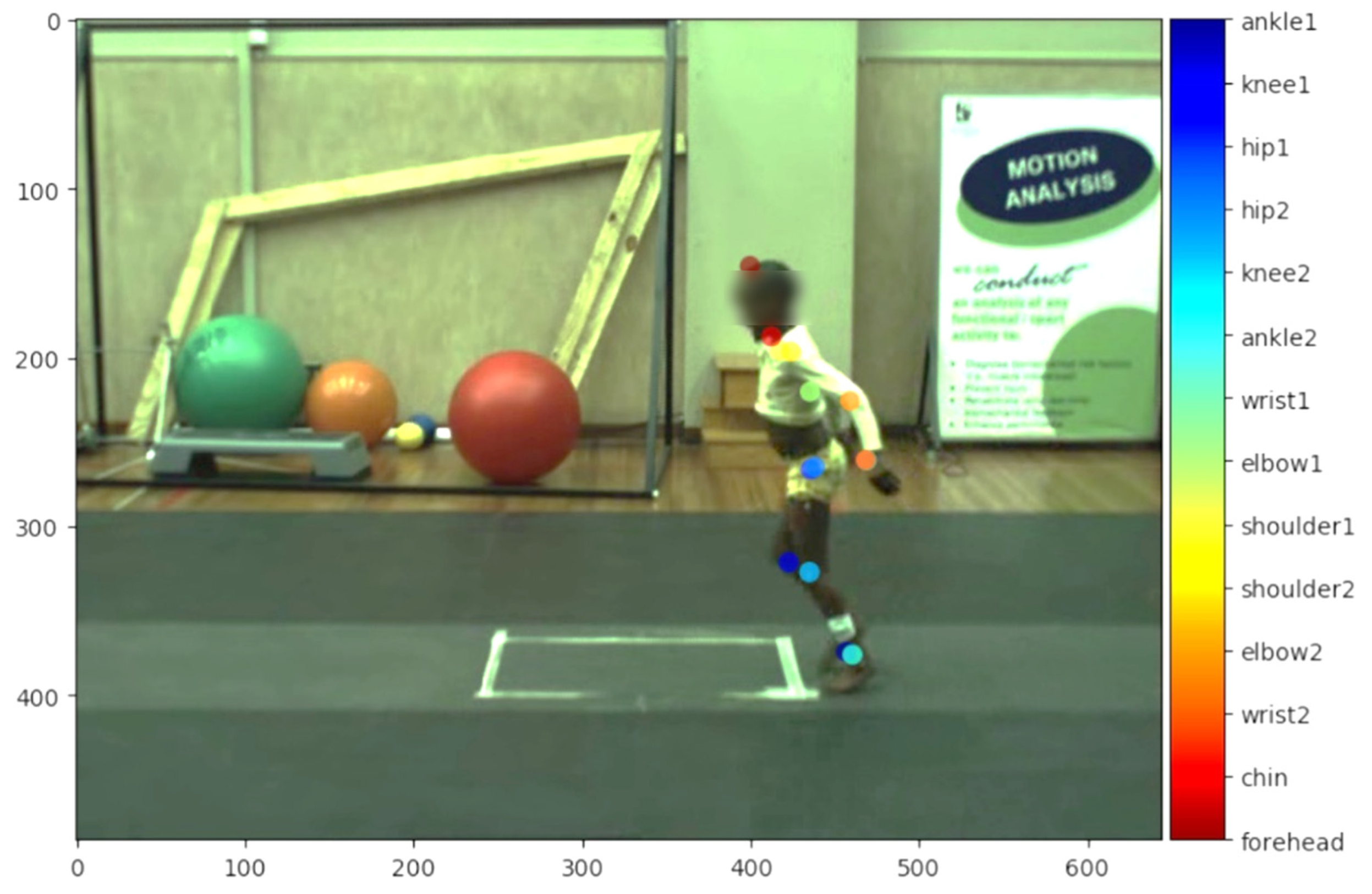
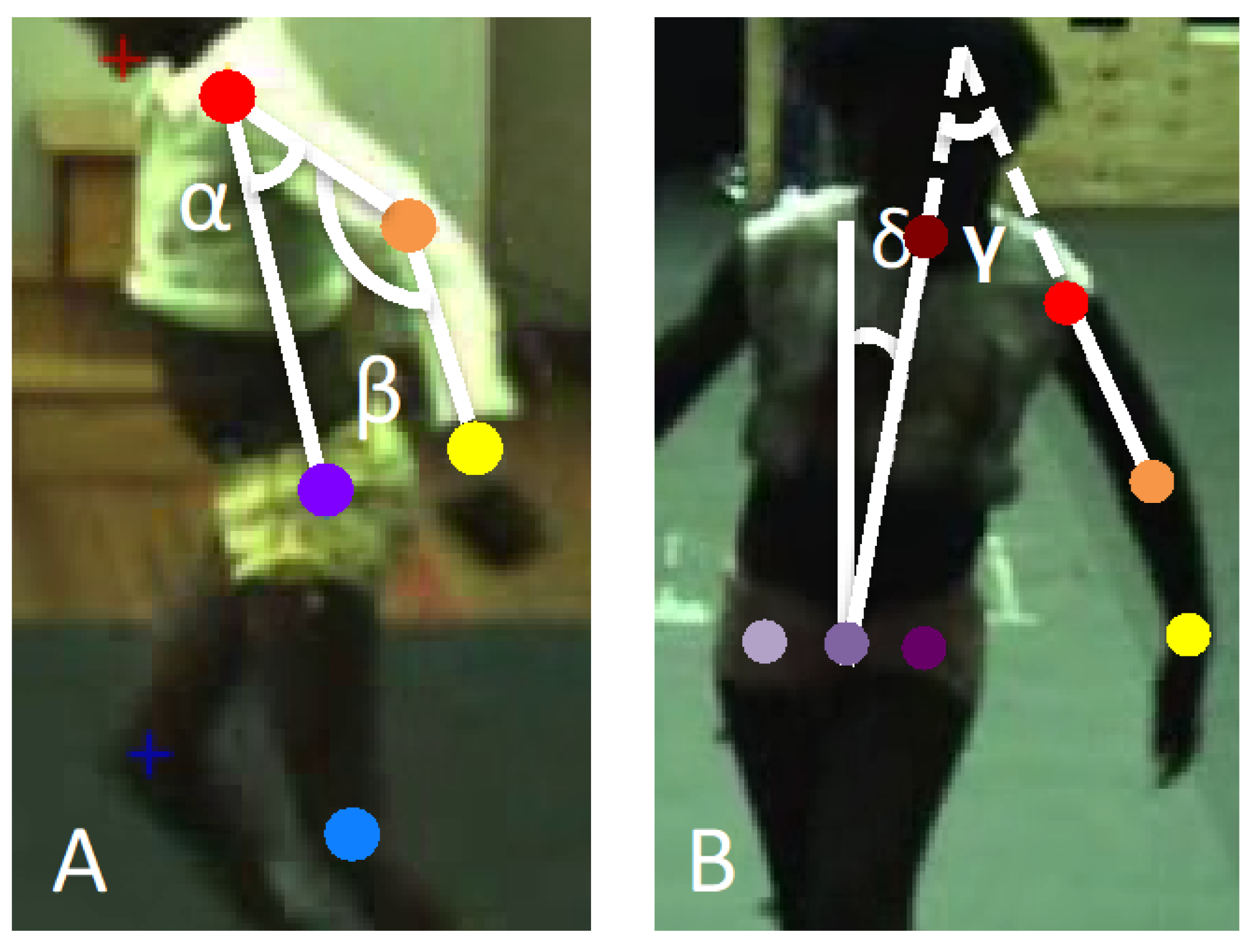
2.3. Assessment Procedures

The movement analyses were conducted in the Neuromechanics Laboratory of the Central Analytics Facilities, Stellenbosch University, Tygerberg, Cape Town, South Africa, which is a setting designed for clinical gait analysis using standard lighting with no interfering daylight changes. All the participants were asked to walk at a self-selected speed on a 10-m runway. Sagittal and frontal video recordings were obtained using static Bonita video cameras (sampling frequency: 50 Hz). Only good-quality color videos were used for the analyses.
Joint poses were tracked with the pre-trained human model of the DeepLabCut toolbox (ResNet101 [13]) in both sagittal (view on the left-hand side; Figure 1) and frontal plane (view on the front side). DeepLabCut is a free and open-source toolbox that can be used for markerless pose estimation in a number of species and behaviors, in different situations and diverse backgrounds. DeepLabCut is based on machine-learning algorithms with deep neural networks programmed in Python 3, and pre-trained models are developed both for humans and other animal species [13]. The following joints and anatomical landmarks were tracked in the sagittal plane: left shoulder, left elbow, left wrist and left hip; and in the frontal plane: chin, right and left hip, left shoulder and left elbow. The tracking of joints and anatomical landmarks was visually inspected. In the case of inaccurate marker tracking (pixel accuracy was above the accuracy threshold of 5 pixels), additional frames were extracted and labeled manually, according to the protocol described by Mathis et al. [12]. First, outliers were extracted to remove labels with a likelihood below 0.9. Second, 20 frames were extracted from various participants and manually labeled. The data sets of the original ResNet101 model and additionally labeled frames were then merged, and the model was retrained until the loss plateaued [12]. No changes were made to the frames regarding occlusions, motion blur or inconsistencies in frame quality.
The trajectories of joint locations and anatomical landmarks were subsequently imported into MATLAB (R2017b, Mathworks Inc., Natick, MA, USA) and included when the likelihood was above 0.9. The positions of the markers were used to calculate joint angles using the cosine rule (Figure 2):
a n g l e   α , β , γ , δ = a c o s a 2 + b 2 c 2 2 a b
The shoulder angle (α) in the sagittal plane (i.e., shoulder flexion/extension) was calculated as the angle between the line from the mid-shoulder point to the hip marker and the line from the mid-shoulder point to the elbow marker. The elbow angle (β) in the sagittal plane (i.e., elbow flexion/extension) was calculated as the angle between the line from the mid-shoulder point to the elbow marker and the line from the elbow to the wrist marker. The shoulder angle (γ) in the frontal plane (i.e., shoulder abduction/adduction) was calculated as the angle between the line from the mid-shoulder point to the elbow marker and the line from the chin to the middle of the two hip markers. The trunk angle (δ) in the frontal plane (i.e., trunk lateral sway) was calculated as the angle between the line from the mid-shoulder point to the middle of the two hip markers and the vertical. In the formula, a and b represent the segments that form the angle, while c represents the segment opposite to the angle. The angles were calculated per frame, and the angle trajectories were subsequently filtered using a Butterworth filter (fc = 10 Hz). Per video, strides were manually extracted (heel strike to heel strike) along with the moment of toe-off. After time normalization (0–100% gait cycle), the average trajectories of the three strides were calculated per participant. Subsequently, the joint angle trajectories and the range of motion (ROM) for shoulder flexion/extension, elbow flexion/extension, shoulder abduction/adduction and trunk sway were determined and compared between children with HIVE and TD children.

2.4. Statistical Analysis
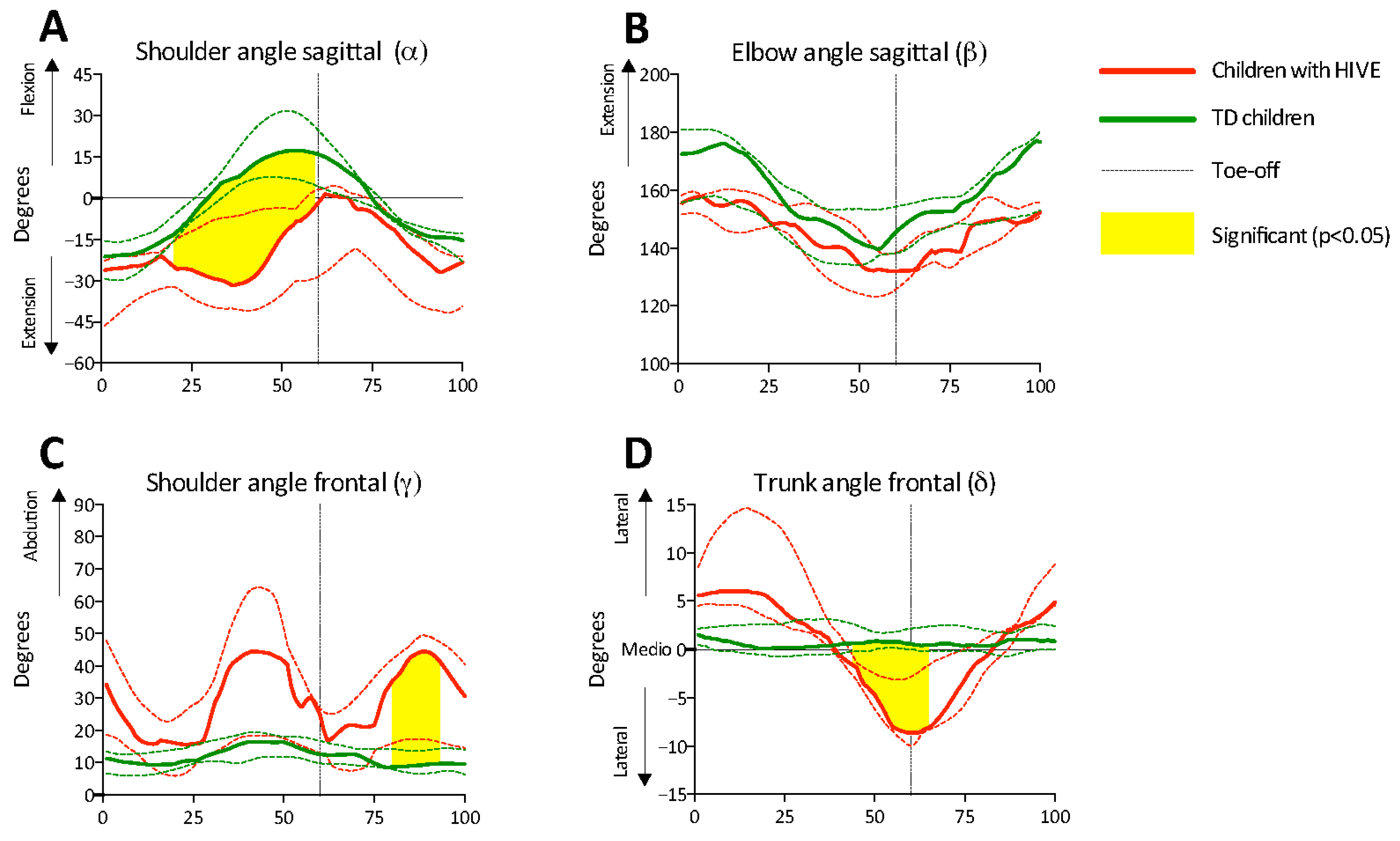
The participants’ characteristics were presented using descriptive statistics and individual data points. Normality was tested using Shapiro–Wilk tests. Given the small sample size and as outcomes were not normally distributed, median values and interquartile ranges (IQR) were determined. Differences in the ROM parameters between the children with HIVE and their TD peers were investigated using non-parametric Mann–Whitney U-tests. The joint angle trajectories of the two cohorts were investigated using the open-source 1-dimensional statistical parametric mapping method (1DSPM) in MATLAB (R2017b, Mathworks Inc., Natick, MA, USA). This is an open-source statistical method that allows analysis of continuous biomechanical data (such as joint angles) across an entire time series rather than at discrete points, enabling the identification of statistically significant differences between conditions or groups throughout the complete movement cycle. Herewith, the non-parametric method was applied to compare the joint angle trajectories (vM0.1, www.spm1d.org: download non-parametric toolbox), and median and IQR of the joint angle trajectories were calculated and presented in figures (0–100% gait cycle). Significance was set at p < 0.05 for all statistical analyses.

3. Results

3.1. Participants’ Background

Five girls with HIVE, classified as GMFCS level II, and five TD girls were included in the study. The participants’ demographics are presented in Table 1. All the children with HIVE were on antiretroviral therapy.

3.2. Accuracy of Pre-Trained Human Network (ResNet101)

In the frontal plane, the pre-trained human network showed accurate tracking of the movement without additional training of the network (pixel accuracy below 5 pixels). In the sagittal plane, the pre-trained human network did not show accurate tracking of the movements. After manually labeling 20 frames and retraining the network (40.000 iterations), the training loss plateaued, resulting in a training error of 2.8 pixels and test error of 3.1 pixels.

3.3. Arm Swing and Trunk Sway

The ROM per joint angle is reported in Table 2 and shows that the ROM in shoulder abduction of children with HIVE was significantly larger than TD children. In addition, the ROM in trunk sway was significantly larger in children with HIVE than TD children. The ROM for shoulder flexion/extension and for elbow flexion/extension in the sagittal plane did not show significant differences between children with HIVE and TD children.
The trajectories of the shoulder and elbow angles obtained in the sagittal plane, as well as the trajectories of the shoulder and trunk angles obtained in the frontal plane, are presented in Figure 3. Significant differences between the children with HIVE and the TD children were observed in the shoulder angle in the sagittal plane between 20% and 59% of the gait cycle (p = 0.004), showing that the shoulder angle of the children with HIVE remained in extension, while the shoulder angle of the TD children moved to flexion during the late stance phase. In the frontal plane, significant differences were observed between the children with HIVE and the TD children in the shoulder angle (80–93% gait cycle; p = 0.040) and the trunk angle (44–65% gait cycle; p = 0.004). Children with HIVE showed significantly more shoulder abduction during the mid-swing phase and lateral trunk sway during late stance and early swing, while TD children showed overall minimal shoulder abduction and trunk sway.

4. Discussion

The aim of this study was to investigate the feasibility of using markerless tracking to assess upper body, including upper limb and trunk, movements in children with and without HIVE, and whether expected differences could be quantified between children with and without HIVE. The findings of this study indicate that it is feasible to obtain 2D joint angles from videos using markerless tracking with DeepLabCut in children with and without HIVE, and to obtain differences between children with and without HIVE in upper limb and trunk movements during gait.
This study explored the potential of markerless movement tracking with DeepLabCut software to use as a tool for clinical gait analysis in LMIC settings. The results of the study showed that DeepLabCut can successfully be used to obtain 2D gait analysis, which can save valuable time for clinical research. This observation confirms suggestions made by Cronin (2021), who described the potential value of pose estimation software packages for human movement analysis [8]. The implementation of markerless movement tracking systems in clinical gait analysis can serve as a useful alternative in settings where expensive gait laboratory instruments are unavailable, particularly in LMICs. In many clinical settings in LMICs, there are no resources available (too expensive) for 3D gait analysis systems, while resources are often limited, and only 2D gait analysis systems are available in high-income countries as well.
It is important to note that the pre-trained human model (ResNet101), without additional manual labeling of frames, did not achieve sufficient accuracy to track markers in the sagittal plane, which could be explained by the fact that only children with and without disabilities were included, instead of adults, suggesting that both children and specifically children with motor disorders were most likely less or not present in the large datasets of annotated images to learn hierarchical visual features that represent body parts from which the anatomical joints are detected. Therefore, for the implementation of DeepLabCut in clinical settings, researchers are advised to manually label 20–50 frames with user-identified/user-defined markers specifically applicable for the laboratory and retrain the ResNet101 model to extract body landmarks from new videos automatically, especially when being used for pathological patient groups that have most likely not been included in the training models of the pretrained human model (ResNet101).
In line with our hypothesis, which was based on a clinical study [10], differences in upper limb and trunk movements between children with HIVE and spastic diplegia and their TD peers were observed. The results showed that the shoulder movements of children with HIVE obtained in both sagittal and frontal plane deviated from TD children, showing more shoulder extension from mid-stance to late stance in the sagittal plane and more shoulder abduction as indicated by a higher overall ROM, as well as more abduction during the swing phase in the frontal plane (Figure 3). Trunk sway was also considerably more pronounced in children with HIVE compared to the TD, showing more lateral sway at initial contact, early stance and late swing. Both the deviated shoulder and trunk movements observed in children with HIVE are consistent with upper body movements that have previously been observed in children with spastic diplegia resulting from CP [23], individuals after stroke [24,25,26] and individuals with Parkinson’s disease, who show deviations in trunk sway only [27]. These altered movements observed during gait in individuals with different neuromuscular conditions have been suggested to contribute to compensating for gait instability by helping to maintain balance, redistribute weight or adjust for deficits in lower limb control [14,15,16,17]. In some cases, excessive trunk sway or shoulder deviations may serve as a strategy to counteract muscle weakness, impaired coordination, or reduced proprioception, ultimately influencing overall gait efficiency and energy expenditure. In addition, several children with HIVE appeared to show two movements towards shoulder abduction during the gait cycle. These two ‘bumps’ (Figure 3), which occurred during late stance (around 40% of gait cycle) and late swing (approximately 85% of gait cycle), could be related to the spastic gait pattern, as also shown in children with spastic CP during gait [28,29]. A double bump pattern in the arm swing during one gait cycle (i.e., stride) is often seen in the sagittal plane in healthy adults when walking very slowly [30]. In patients with neurologic disorders, however, this double bump pattern has been found to be present independent of the gait speed [31,32]. In patients with stroke, it was found that the group with this altered arm swing coordination during gait showed a greater level of upper limb impairment (reduced Fugl-Meyer upper extremity score) and more spasticity in the internal shoulder rotators and elbow extensors, suggesting that neuromechanical control might be an important factor contributing to the altered arm-to-leg coordination pattern [32]. This should be further investigated in a larger sample of children with HIVE and spastic diplegia.
Figure 3. Joint angles in sagittal and frontal planes presented for children with HIVE (red) and TD children (green) (thick lines = median; dashed lines = interquartile ranges (1st and 3rd percentile)) over the gait cycle. Parts A and B are modified and updated versions of those presented in Eken et al. 2020 [19].
Applsci 15 04546 g003
In this study, the DeepLabCut pre-trained human model (ResNet101) was used [12]. In the frontal plane, tracking the predefined joints and anatomical landmarks was suitable, showing sufficient accuracy. One of the limitations, however, was that in the sagittal plane, tracking accuracy was limited using the pre-trained human model, requiring retraining of the network with additional self-labeled frames. This could be explained by the pre-trained human model, which was based on typical adult gait, where, in this study, children with an impaired gait pattern were included as well as TD children. It is therefore important to note that additional training, e.g., on frames of individuals with similar conditions or gait complications, or fine-tuning of pre-trained human models, is necessary before implementing markerless pose estimation for clinical gait analysis for individuals with atypical gaits and compensatory upper body movements. Previous research has shown that manual annotation of key points in human movement is a valid and reliable method in typically developing populations [33,34,35,36,37,38]. Furthermore, a recent study showed the validity of 2D markerless tracking using DeepLabCut compared to gold standard laboratory-based optoelectronic three-dimensional motion capture to measure joint angles in children [39]. However, to the best of our knowledge, no studies have investigated the reliability and validity of 2D manual annotation for human movement in individuals with atypical gaits, which is a limitation of this study. Another limitation of the study was that 2D movement analysis was used, while 3D movement analysis provides essential information required to prescribe suitable treatment in clinical settings [1]. Currently, 3D marker-based movement analysis systems that use stereophotogrammetry methods, such as Vicon, Optotrack and BTS systems, are acknowledged as the gold standard [40]. However, these systems are currently expensive, require a dedicated space and need trained personnel to be successfully employed, which limits accessibility, particularly for LMICs. Three-dimensional markerless movement analysis systems that use accessible cameras can provide solutions for these regions specifically. Future research is needed to explore 3D markerless movement analysis systems and investigate the reliability and validity of these systems against gold standard 3D marker-based systems. However, this comes with its challenges due to different factors, such as constant updates to deep learning models, occlusion of body landmarks and segments and high-resolution cameras, which are needed to approach the accuracy of 3D marker-based systems. In turn, these technical developments may also increase the financial burden of these systems. Another limitation of this study is that only a small, homogeneous sample of girls was included, which limits the generalizability of the results presented in this pilot study. Therefore, future research is recommended to include a larger and more heterogeneous sample of children with and without physical disabilities. This study does however show that it is feasible to use DeepLabCut to assess upper limb and trunk movements during gait in children with HIVE and spastic diplegia and provides novel insights into the arm movements during walking in this population.

5. Conclusions

In conclusion, this pilot study showed that DeepLabCut could serve as a useful alternative for conventional gait analysis, with several advantages ranging from no dedicated and expensive laboratory required, more time efficient, no need for highly skilled experts, and does not affect the individual’s gait. Based on the markerless movement tracking system DeepLabCut, children with HIVE showed deviations in their upper limb movements and trunk sway. These abnormalities in upper limb behavior and trunk sway may be related to strategies to compensate for impaired balance control due to spastic diplegia. However, future research is needed to assess its validity and reliability.

Author Contributions

Conceptualization, M.M.E., P.M., N.G.L. and R.P.L.; methodology, M.M.E. and P.M.; software, M.M.E.; validation, P.M.; resources, N.G.L.; writing—original draft preparation, M.M.E. and P.M.; writing—review and editing, M.M.E., P.M., R.P.L. and N.G.L.; visualization, M.M.E. and P.M.; supervision, R.P.L. and N.G.L.; funding acquisition, M.M.E., P.M. and N.G.L. All authors have read and agreed to the published version of the manuscript.

Funding

This project was supported by the DIOS mobility programme 2019 (UHasselt & VLIR-UOS [GM2019]), the International AIDS Society (Collaborative Initiative for Paediatric HIV Education and Research (CIPHER) Grant Programme), the South African National Research Foundation, the Neuroscience Institute at University of Cape Town and the special research fund (BOF20KV07) of Hasselt University.

Institutional Review Board Statement

The study was conducted in accordance with the Declaration of Helsinki and approved by the Human Research Ethics Committee of the University of Cape Town, South Africa (HREC 447/2012).

Informed Consent Statement

Written informed consent was obtained from all the caregivers of the participants before data collection, and assent was obtained from the participants involved in the study.

Data Availability Statement

The data presented in this study may be made available on request from the author in charge of the resources (NGL), depending on privacy, legal and ethical considerations.

Conflicts of Interest

The authors declare no conflicts of interest. The funders had no role in the design of the study; in the collection, analyses, or interpretation of data; in the writing of the manuscript; or in the decision to publish the results.

References

  1. Benedetti, M.G.; Beghi, E.; De Tanti, A.; Cappozzo, A.; Basaglia, N.; Cutti, A.G.; Cereatti, A.; Stagni, R.; Verdini, F.; Manca, M.; et al. SIAMOC position paper on gait analysis in clinical practice: General requirements, methods and appropriateness. Results of an Italian consensus conference. Gait Posture 2017, 58, 252–260. [Google Scholar] [CrossRef]
  2. Castelli, A.; Paolini, G.; Cereatti, A.; Della Croce, U. A 2D Markerless Gait Analysis Methodology: Validation on Healthy Subjects. Comput. Math. Methods Med. 2015, 2015, 186780. [Google Scholar] [CrossRef]
  3. Fernández-González, P.; Koutsou, A.; Cuesta-Gómez, A.; Carratalá-Tejada, M.; Miangolarra-Page, J.C.; Molina-Rueda, F. Reliability of Kinovea® Software and Agreement with a Three-Dimensional Motion System for Gait Analysis in Healthy Subjects. Sensors 2020, 20, 3154. [Google Scholar] [CrossRef] [PubMed]
  4. Damsted, C.; Nielsen, R.O.; Larsen, L.H. Reliability of video-based quantification of the knee- and hip angle at foot strike during running. Int. J. Sports Phys. Ther. 2015, 10, 147–154. [Google Scholar]
  5. Van Rie, A.; Harrington, P.R.; Dow, A.; Robertson, K. Neurologic and neurodevelopmental manifestations of pediatric HIV/AIDS: A global perspective. Eur. J. Paediatr. Neurol. 2007, 11, 1–9. [Google Scholar] [CrossRef] [PubMed]
  6. UNAIDS. UNAIDS Data. 2019. Available online: https://www.unaids.org/en/resources/documents/2019/2019-UNAIDS-data (accessed on 19 April 2025).
  7. Tindyebwa, D.; Kayita, J.; Musoke, P.; Eley, B.; Nduati, R.; Coovadia, H.; Bobart, R.; Mbori-Ngacha, D.; Kieffer, P.M. Handbook on Paediatric AIDS in Africa, 4th ed.; African Network for the Care of Children Affected by HIV/AIDS—ANECCA Publishers: Kampala, Uganda, 2017. [Google Scholar]
  8. Cronin, N.J.; Avela, J.; Finni, T.; Peltonen, J. Differences in contractile behaviour between the soleus and medial gastrocnemius muscles during human walking. J. Exp. Biol. 2013, 216, 909–914. [Google Scholar] [CrossRef]
  9. Langerak, N.G.; du Toit, J.; Burger, M.; Cotton, M.F.; Springer, P.E.; Laughton, B. Spastic diplegia in children with HIV encephalopathy: First description of gait and physical status. Dev. Med. Child. Neurol. 2014, 56, 686–694. [Google Scholar] [CrossRef] [PubMed]
  10. Mann, T.N.; Donald, K.A.; Laughton, B.; Lamberts, R.P.; Langerak, N.G. HIV encephalopathy with bilateral lower limb spasticity: Upper limb motor function and level of activity and participation. Dev. Med. Child. Neurol. 2017, 59, 412–419. [Google Scholar] [CrossRef]
  11. Kidziński, L.; Yang, B.; Hicks, J.L.; Rajagopal, A.; Delp, S.L.; Schwartz, M.H. Deep neural networks enable quantitative movement analysis using single-camera videos. Nat. Commun. 2020, 11, 4054. [Google Scholar] [CrossRef]
  12. Mathis, A.; Mamidanna, P.; Cury, K.M.; Abe, T.; Murthy, V.N.; Mathis, M.W.; Bethge, M. DeepLabCut: Markerless pose estimation of user-defined body parts with deep learning. Nat. Neurosci. 2018, 21, 1281–1289. [Google Scholar] [CrossRef]
  13. Nath, T.; Mathis, A.; Chen, A.C.; Patel, A.; Bethge, M.; Mathis, M.W. Using DeepLabCut for 3D markerless pose estimation across species and behaviors. Nat. Protoc. 2019, 14, 2152–2176. [Google Scholar] [CrossRef] [PubMed]
  14. Meyns, P.; Desloovere, K.; Van Gestel, L.; Massaad, F.; Smits-Engelsman, B.; Duysens, J. Altered arm posture in children with cerebral palsy is related to instability during walking. Eur. J. Paediatr. Neurol. 2012, 16, 528–535. [Google Scholar] [CrossRef] [PubMed]
  15. Meyns, P.; Kerkum, Y.L.; Brehm, M.A.; Becher, J.G.; Buizer, A.I.; Harlaar, J. Ankle foot orthoses in cerebral palsy: Effects of ankle stiffness on trunk kinematics, gait stability and energy cost of walking. Eur. J. Paediatr. Neurol. 2020, 26, 68–74. [Google Scholar] [CrossRef] [PubMed]
  16. Meyns, P.; Duysens, J.; Desloovere, K. The arm posture in children with unilateral Cerebral Palsy is mainly related to antero-posterior gait instability. Gait Posture 2016, 49, 132–135. [Google Scholar] [CrossRef]
  17. Delabastita, T.; Desloovere, K.; Meyns, P. Restricted Arm Swing Affects Gait Stability and Increased Walking Speed Alters Trunk Movements in Children with Cerebral Palsy. Front. Hum. Neurosc 2016, 10, 354. [Google Scholar] [CrossRef]
  18. Eken, M.M.; Meyns, P.; Lamberts, R.P.; Langerak, N.G. Markerless movement tracking using a machine-learning algorithm to assess arm movements during gait in children with HIV encephalopathy. Gait Posture 2020, 81, 85–86. [Google Scholar] [CrossRef]
  19. Mann, T.N.; Laughton, B.; Donald, K.A.; Langerak, N.G. HIV encephalopathy with bilateral lower limb spasticity: Gross motor function and antiretroviral therapy. Dev. Med. Child. Neurol. 2017, 59, 407–411. [Google Scholar] [CrossRef]
  20. Caldwell, M.; Oxtoby, M.; Simonds, R.; Lindegren, M.; Rogers, M. Revised classification system for human immunodeficiency virus infection in children less than 13 years of age. CDC Morbodity Mortal. Wkly. Rep. 1994, 43, 1–10. [Google Scholar]
  21. Palisano, R.J.; Hanna, S.E.; Rosenbaum, P.L.; Russell, D.J.; Walter, S.D.; Wood, E.P.; Raina, P.S.; Galuppi, B.E. Validation of a Model of Gross Motor Function for Children with Cerebral Palsy. Phys. Ther. 2000, 80, 974–985. [Google Scholar] [CrossRef]
  22. World Medical Association. World Medical Association Declaration of Helsinki: Ethical principles for medical research involving human subjects. JAMA 2013, 310, 2191–2194. [Google Scholar] [CrossRef]
  23. Romkes, J.; Peeters, W.; Oosterom, A.M.; Molenaar, S.; Bakels, I.; Brunner, R. Evaluating upper body movements during gait in healthy children and children with diplegic cerebral palsy. J. Pediatr. Orthop. B 2007, 16, 175–180. [Google Scholar] [CrossRef] [PubMed]
  24. De Vlieger, D.; Defour, A.; Bar-On, L.; Cambier, D.; Swinnen, E.; Van der Looven, R.; Van Bladel, A. Speed-dependent changes in the arm swing during independent walking in individuals after stroke. PLoS ONE 2025, 20, e0315332. [Google Scholar] [CrossRef]
  25. Van Criekinge, T.; Saeys, W.; Hallemans, A.; Velghe, S.; Viskens, P.J.; Vereeck, L.; De Hertogh, W.; Truijen, S. Trunk biomechanics during hemiplegic gait after stroke: A systematic review. Gait Posture 2017, 54, 133–143. [Google Scholar] [CrossRef]
  26. Chaparro-Rico, B.D.M.; Cafolla, D.; Tortola, P.; Galardi, G. Assessing Stiffness, Joint Torque, and ROM for Paretic and Non-Paretic Lower Limbs during the Subacute Phase of Stroke Using Lokomat Tools. Appl. Sci. 2020, 10, 6168. [Google Scholar] [CrossRef]
  27. Wolfsegger, T.; Pichler, R.; Assar, H.; Topakian, R. Quantitative trunk sway analysis under challenging gait conditions in early and untreated Parkinson’s disease. Neurol. Sci. 2022, 43, 1411–1413. [Google Scholar] [CrossRef] [PubMed]
  28. Zhou, J.; Butler, E.E.; Rose, J. Neurologic Correlates of Gait Abnormalities in Cerebral Palsy: Implications for Treatment. Front. Hum. Neurosci. 2017, 11, 103. [Google Scholar] [CrossRef]
  29. Wang, K.K.; Munger, M.E.; Chen, B.P.J.; Novacheck, T.F. Selective dorsal rhizotomy in ambulant children with cerebral palsy. J. Child. Orthop. 2018, 12, 413–427. [Google Scholar] [CrossRef]
  30. Wagenaar, R.C.; Emmerik, R.E.A. Resonant frequencies of arms and legs identify different walking patterns. J. Biomech. 2000, 33, 853–861. [Google Scholar] [CrossRef]
  31. Wagenaar, R.C.; Emmerik, R.E.A. Dynamics of pathological gait. Hum. Mov. Sci. 1994, 13, 441–471. [Google Scholar] [CrossRef]
  32. Van Bladel, A.; De Ridder, R.; Palmans, T.; Van Der Looven, R.; Verheyden, G.; Meyns, P.; Cambier, D. Defining characteristics of independent walking persons after stroke presenting with different arm swing coordination patterns. Hum. Mov. Sci. 2024, 93, 103174. [Google Scholar] [CrossRef]
  33. Puig-Diví, A.; Escalona-Marfil, C.; Padullés-Riu, J.M.; Busquets, A.; Padullés-Chando, X.; Marcos-Ruiz, D. Validity and reliability of the Kinovea program in obtaining angles and distances using coordinates in 4 perspectives. PLoS ONE 2019, 14, e0216448. [Google Scholar] [CrossRef]
  34. Iijima, S.; Shiomi, M.; Hara, T. Verification of Reliability and Validity of Trunk Forward Tilt Angle Measurement During Gait Using 2-Dimensional Motion Analysis. J. Chiropr. Med. 2023, 22, 89–95. [Google Scholar] [CrossRef] [PubMed]
  35. Ugbolue, U.C.; Papi, E.; Kaliarntas, K.T.; Kerr, A.; Earl, L.; Pomeroy, V.M.; Rowe, P.J. The evaluation of an inexpensive, 2D, video based gait assessment system for clinical use. Gait Posture 2013, 38, 483–489. [Google Scholar] [CrossRef]
  36. Fatone, S.; Stine, R. Capturing quality clinical videos for two-dimensional motion analysis. J. Prosthet. Orthot. 2015, 27, 27–32. [Google Scholar] [CrossRef]
  37. Wade, L.; Needham, L.; McGuigan, P.; Bilzon, J. Applications and limitations of current markerless motion capture methods for clinical gait biomechanics. PeerJ 2022, 10, e12995. [Google Scholar] [CrossRef]
  38. Needham, L.; Evans, M.; Cosker, D.P.; Wade, L.; McGuigan, P.M.; Bilzon, J.L.; Colyer, S.L. The accuracy of several pose estimation methods for 3D joint centre localisation. Sci. Rep. 2021, 11, 20673. [Google Scholar] [CrossRef] [PubMed]
  39. van den Bogaart, M.; Jacobs, N.; Hallemans, A.; Meyns, P. Validity of Deep Learning-Based Motion Capture Using DeepLabCut to Assess Proprioception in Children. Appl. Sci. 2025, 15, 3428. [Google Scholar] [CrossRef]
  40. Sipari, D.; Chaparro-Rico, B.D.M.; Cafolla, D. SANE (Easy Gait Analysis System): Towards an AI-Assisted Automatic Gait-Analysis. Int. J. Environ. Res. Public Health 2022, 19, 10032. [Google Scholar] [CrossRef]
Figure 1. Representation of markerless movement tracking using the pre-trained human model in DeepLabCut.
Applsci 15 04546 g001
Figure 2. Representation of the tracked joints and anatomical landmarks (colored dots) for the calculated joint angles (α: shoulder flexion/extension; β: elbow flexion/extension); γ: shoulder abduction/adduction; and δ: lateral trunk sway) in (A) sagittal plane and (B) frontal plane. Part (A) is a modified and updated version of the one presented in Eken et al. 2020 [19].
Applsci 15 04546 g002
Table 1. Demographic characteristics of the participants with HIVE and TD participants.
No.Age
(Years)
BMI
(kg/m2)
BMI CategoryVideos Included (n)
SagittalFrontal
HIVE
18.317.9normal33
210.821.7overweight33
310.515.9normal33
49.418.0normal32
512.822.5overweight33
TD
16.813.9normal33
27.913.6normal33
310.817.8normal32
410.815.0normal33
58.219.5overweight33
Abbreviations: HIVE, human immunodeficiency virus encephalopathy; TD, typically developing; and BMI, body mass index.
Table 2. Range of motion of upper limb movements in sagittal and frontal planes for HIVE and TD children.
HIVETD
MedianIQRMedianIQRp Value
Sagittal plane
Shoulder flexion/extension (°) 31.0 [24.4–51.5] 37.2[25.2–61.8] 0.602
Elbow flexion/extension (°)25.4[20.1–38.7]30.7[24.8–36.1] 0.602
Frontal plane
Shoulder abduction/adduction (°) 41.5 [11.1–49.2]6.5[4.5–10.4] 0.028 *
Trunk sway (°) 14.7 [8.5–24.7]1.9[1.6–2.9] 0.009 *
* Significantly different: p < 0.05.
Disclaimer/Publisher’s Note: The statements, opinions and data contained in all publications are solely those of the individual author(s) and contributor(s) and not of MDPI and/or the editor(s). MDPI and/or the editor(s) disclaim responsibility for any injury to people or property resulting from any ideas, methods, instructions or products referred to in the content.

