Article

Effects of β-Cryptoxanthin on Cisplatin-Treated Human Oral Mucosa-Derived Keratinocytes and Fibroblasts

1
Department of Dental Medicine, Medicine Graduate School of Medical Science, Kyoto Prefectural University, Kyoto 602-8566, Japan
2
Department of Immunology, Medicine Graduate School of Medical Science, Kyoto Prefectural University, Kyoto 602-8566, Japan
*
Authors to whom correspondence should be addressed.
These authors contributed equally to this work.
Appl. Sci. 2025, 15(9), 4803; https://doi.org/10.3390/app15094803 (registering DOI)
Submission received: 11 December 2024 / Revised: 7 March 2025 / Accepted: 23 April 2025 / Published: 26 April 2025

Abstract

:
Cisplatin (CDDP) is an anticancer drug that is frequently used to treat head and neck cancers; however, it may cause oral mucositis. The discontinuation of CDDP may be required for some patients with a severe status, and the control of oral mucositis is extremely important. β-Cryptoxanthin (β-cry), a carotenoid, exerts anti-inflammatory effects. Its inhibition of 5-FU-induced inflammatory responses was recently demonstrated. However, the effects of β-cry on CDDP-induced oral mucositis remain unclear. In the present study, we stimulated human oral mucosa-derived keratinocytes (hOMK) and fibroblasts (hOMF) with CDDP, added β-cry, and examined its effects, with a focus on the production of inflammatory cytokines, matrix metalloproteinase (MMPs), and reactive oxygen species (ROS). CDDP increased the mRNA expression and production of inflammatory cytokines and MMPs both in hOMK and hOMF. However, increases in IL-6 and MMP-9 mRNA expression levels and IL-6 production in CDDP-treated hOMK and hOMF were inhibited by β-cry. Furthermore, the production of ROS and the rate of SA-β-gal-positive cells were increased by CDDP, but were not affected by β-cry. CDDP may induce oral mucositis by increasing the levels of inflammatory cytokines, MMPs, and ROS. β-cry partially inhibited CDDP-induced increases in inflammatory cytokines and MMPs, suggesting its potential to attenuate the symptoms of chemotherapy-related oral mucositis.

1. Introduction

Potent treatment approaches, such as the use of high-dose anticancer drugs and combination chemotherapy, have recently been performed to improve the outcomes of cancer treatment. Cisplatin (cis-diamminedichloro-platinum (II), CDDP) is one of the most frequently used anticancer drugs for squamous cell carcinoma, which accounts for the majority of oral cancers, along with 5-FU. The outcomes of multi-drug therapy with CDDP and a target anticancer drug or radiation are favorable, suggesting its utility [1,2]. However, the side effects of CDDP include nausea, general malaise, nephropathy, pancytopenia, and mucositis [3,4,5]. Oral mucositis is a well-known and important side effect of CDDP [6]. The turnover of mucosal epithelial cells comprising the mucosa is fast, and these cells may be readily affected by DNA damage, which ultimately results in cell death or a decrease in regeneration [7,8]. The inflammatory responses of mucosal epithelial cells to the cytotoxicity of anticancer drugs appear on the surfaces of all alimentary canal mucosae from the oral cavity to the rectum [3]. The mucosal barrier is destroyed, resulting in or increasing susceptibility to various symptoms [9]. For example, oral mucositis caused by anticancer drugs is commonly observed in the oral cavity: in chemotherapy alone, 30 to 40%; in hematopoietic stem cell transplantation-related high-dose chemotherapy, 70 to 90%. Since oral mucositis markedly affects oral function, its prevention is extremely important [10,11]. Various treatments, such as laser irradiation, local anesthetics, gargling with mouthwash, and cryotherapy, have been employed to control oral mucositis [12,13,14,15]. However, symptomatic treatment is currently the primary therapeutic approach, and its effects remain insufficient. Therefore, the establishment of effective prevention/treatment methods is needed.
β-Cryptoxanthin (β-cry) is a pigment component of mandarin oranges, persimmons, and oranges, and is also present in chili pepper and paprika as a carotenoid. It is converted to vitamin A in vivo, which maintains physical functions and exerts inhibitory effects on carcinogenesis, exhibits DNA-repairing activity [16] and antioxidant/anti-inflammatory activities [17,18,19], and also exerts preventive effects against osteoporosis and knee osteoarthritis [20]. We previously reported that β-cry exerted anti-inflammatory effects in periodontal ligament-derived cells [21]. We recently showed that β-cry exerted anti-inflammatory effects against the inflammatory responses of epithelial cells induced by 5-FU, a chemotherapeutic agent [22]. However, it currently remains unclear whether β-cry attenuates oral mucositis, which occurs as a side effect of CDDP, an anticancer drug that is frequently used to treat head and neck cancers. Furthermore, the oral mucosa consists of not only an epithelial layer, but also a connective tissue layer, and oral mucositis is induced in both layers. Moreover, the effects of β-cry on oral mucosal fibroblasts have yet to be clarified. Therefore, we herein examined the effects of CDDP and β-cry individually and in combination on oral mucosa-derived cells (keratinocytes and fibroblasts).

2. Materials and Methods

2.1. Cell Culture

Human normal primary oral mucosal keratinocytes (hOMK) and human normal primary oral mucosal fibroblasts (hOMF) derived from oral mucosa of an Asian 37 year old male were purchased from Cell Research Corporation pte Ltd. (Singapore). hOMK and hOMF were seeded at cell densities of 1, 2, 3 × 10⁴, or 1 × 10⁵ cells/well on 96- and 24-well plates and were then incubated at 37 °C in a humidified atmosphere containing 95% air and 5% CO2 with epithelial culture medium (EpiLife basal medium supplemented with EpiLife Defined Growth Supplement, Thermo Fisher Science, Waltham, MA, USA).
After one day, cells were stimulated with CDDP (1 µg/mL) (KYOWA KIRIN, Tokyo, Japan) and β-cry (1 × 10⁻⁷ M) (CaroteNature, Lupsingen, Switzerland) for 2 or 7 days. β-cry was obtained from CaroteNature (Lupsingen, Switzerland) and was dissolved in dimethyl sulfoxide. The concentration used was selected based on previous studies [21,22].

2.2. Cell Viability Assay

hOMK and hOMF were seeded at a cell density of 1 × 10⁴ cells/well on 96-well plates. After 24 h, cells were stimulated with CDDP (0.01–100 µg/mL). Forty-eight hours later, the WST assay (Cell Count Reagent SF, Nacalai Tesque, Kyoto, Japan) was used to examine cell activity. Specifically, after incubating cells with each concentration of CDDP for 48 h, the culture medium was removed, fresh culture medium containing 10% WST-8 was added, and cells were incubated for a further 2 h. The absorbance of the culture medium was then measured at 550 to 650 nm using a microplate reader (Emax®, Molecular Devices, Sunnyvale, CA, USA).

2.3. Cellular Growth and Morphology

hOMK and hOMF were seeded at cell densities of 3 and 2 × 10⁴ cells/well, respectively, on 24-well plates. After 24 h, cells were stimulated with CDDP (1 µg/mL) and β-cry (1 × 10⁻⁷ M). Seven days later, the fixation of cells in 10% formalin (Nacalai Tesque) was performed and their nuclei were stained with Hoechst 33342 (DOJINDO, Kumamoto, Japan). An inverted fluorescence phase-contrast microscope (BZ-X810, KEYENCE, Osaka, Japan) was used to observe the morphology of cells, and cell numbers were assessed by counting nuclei. The mean cell number in the control group was set as 100%, and cell numbers in each group were shown as a percentage (%) of the control.

2.4. Quantitative RT-PCR

hOMK and hOMF were seeded at cell densities of 3 and 2 × 10⁴ cells/well, respectively, on 24-well plates. After 24 h, these cells were stimulated with CDDP (1 µg/mL) and β-cry (1 × 10⁻⁷ M). Seven days later, RNA was collected from cells.
RNA was isolated and cDNA was synthesized according to a previously described method [23,24]. In brief, following the extraction of total RNA with an RNeasy mini kit (QIAGEN, Venlo, The Netherlands), it was reverse-transcribed by ReverTra Ace qPCR RT Master Mix (Toyobo, Osaka, Japan). A real-time RT-PCR assay was conducted using Real-Time PCR Master Mix (Applied Biosystems, Waltham, MA, USA) on the Step One Plus Real-Time PCR System (Applied Biosystems) with the appropriate probes and primers (Thermo Fisher Science). All values (average ± SD) were normalized to the β-actin mRNA level in each sample and shown as relative values. Supplemental Table S1 lists the primer probes used.

2.5. ELISA

hOMK and hOMF were seeded at a cell density of 3 × 10⁴ cells/well on 24-well plates. After 24 h, these cells were stimulated with CDDP (1 µg/mL) and β-cry (1 × 10⁻⁷ M). Seven days later, the culture supernatant was collected, and an ELISA kit (R&D Systems, Minneapolis, MN, USA) was used to assess the production of cytokines (IL-6 and IL-8) and MMPs (MMP-2 and MMP-9). A microplate reader (Emax®, Molecular Devices, Sunnyvale, CA, USA) was employed to measure absorbance at 450 to 650 nm. Cytokine and MMP levels in each sample were then calculated from standard curves.

2.6. Reactive Oxygen Species (ROS) Assay

ROS assays (Cell MeterTM Fluorimetric Intracellular Total ROS Activity Assay Kit, AAT Bioquest, Sunnyvale, CA, USA) were performed as described by the manufacturer.
hOMK and hOMF were seeded at a cell density of 1 × 10⁵ cells/well on 24-well plates. After 24 h, cells were stimulated with CDDP (1 µg/mL) for 8 h in the presence of AmpliteTM ROS green working solution. The fluorescence value of each sample was measured using a fluorescence microplate reader (Excitation: 490 nm, Emission: 525 nm, Cut-off: 515 nm). The mean value in the non-stimulated (control) group was set as 100%, and the fluorescence value in each group was shown as a percentage of the control.

2.7. SA-β-Gal

hOMK and hOMF were seeded at cell densities of 3 and 2 × 10⁴ cells/well, respectively, on 24-well plates. After 24 h, these cells were stimulated with CDDP (1 µg/mL) and β-cry (1 × 10⁻⁷ M). Seven days later, the SA-β-gal Staining kit (Cell Biolabs, San Diego, CA, USA) was used to stain cells. Nuclear staining was then performed with Hoechst 33342 (DOJINDO). The number of SA-β-gal-positive cells and cell nuclei were counted using the inverted fluorescence phase-contrast microscope (BZ-X810). The percent of SA-β-gal-positive cells (%) in each group was presented by establishing the number of SA-β-gal-positive cells as a numerator and the number of cell nuclei as a denominator.

2.8. Statistical Analysis

Data are expressed as the mean ± standard deviation (S.D.). The significance of differences was analyzed using the Student’s t-test and ANOVA with Tukey–Kramer’s post hoc test. p < 0.05 was considered to be significant.

3. Results

3.1. Cell Growth and Morphology of CDDP- and/or β-Cry-Treated hOMK and hOMF

hOMK and hOMF were treated with CDDP at a concentration of 0.01 to 100 µg/mL for 48 h, and hOMK and hOMF viability was then measured using the WST assay. Cell viability was markedly lower in cells treated with ≥5 µg/mL CDDP than in control cells (Supplemental Figure S1). A previous study reported that the peripheral blood concentration of CDDP after a drip infusion was 0.73–3.04 µg/mL [25]. Therefore, in subsequent experiments, CDDP was used at a concentration of 1 µg/mL, and the experimental period was established as 7 days, during which oral mucositis may occur after the start of chemotherapy.
CDDP enlarged the size of hOMK and hOMF with the accumulation of vacuoles in the cell plasma, while β-cry did not markedly affect their morphology (Figure 1A and Supplementary Figure S2).
The numbers of hOMK and hOMF treated with 1 µg/mL CDDP for 7 days were significantly lower than the number of control cells (hOMK: 49.31% ± 4.3, hOMF: 46.98% ± 9.86) (Figure 1B). In contrast, the numbers of hOMK and hOMF treated with 100 nM β-cry for 7 days were significantly higher than those of control cells (hOMK: 127.34% ± 12.98, hOMF: 115.85% ± 8.87) (Figure 1B). Furthermore, the numbers of both cell types were slightly higher following the CDDP+β-cry treatment than after the CDDP treatment (hOMK: 59.60 ± 10.75, hOMF: 52.26 ± 2.6), although there was no significant increase.

3.2. Effects of β-Cry on Inflammatory Cytokine and MMP mRNA Expression Levels in CDDP-Treated hOMK and hOMF

In CDDP-treated hOMK and hOMF, significant increases were observed in the mRNA expression levels of IL-6 (OMK: 5.08 ± 0.6-fold, OMF: 23.4 ± 6.15-fold), IL-8 (2.75 ± 0.25-fold, OMF: 21.77 ± 4.07-fold), and MMP-9 (1.88 ± 0.63-fold, OMF: 3.9 ± 1.1-fold) from those in control cells. However, no significant differences were observed in mRNA expression levels between β-cry-treated cells and control cells. In CDDP+β-cry-treated cells, significant reductions were observed in the mRNA expression levels of IL-6 (2.09 ± 0.43-fold), IL-8 (1.61 ± 0.52-fold), and MMP-9 (0.99 ± 0.28-fold) in hOMK and those of IL-6 (14.49 ± 2.78-fold) and MMP-9 (2.4 ± 0.46-fold) in hOMF (Figure 2).

3.3. Effects of β-Cry on IL-6, IL-8, and ROS Levels in CDDP-Treated hOMK and hOMF

IL-6, IL-8, MMP-2, and MMP-9 levels were significantly higher in CDDP-treated hOMK (IL-6: 104.51 ± 8.0 pg/mL, IL-8: 80.66 ± 9.11 pg/mL, MMP-2: 26.74 ± 3.73 ng/mL, MMP-9: 0.74 ± 0.09 ng/mL) than in control cells (IL-6: 19.09 ± 2.02 pg/mL, IL-8: 26.72 ± 4.06 pg/mL, MMP-2: 6.22 ± 0.46 ng/mL, MMP-9: 0.53 ng/mL). However, no significant changes were observed in β-cry-treated hOMK (Figure 3A). IL-8 levels were significantly lower in CDDP+β-cry-treated hOMK (IL-8: 62.60 ± 4.53 pg/mL) than in CDDP-treated hOMK (Figure 3A).
IL-6, IL-8, and MMP-2 levels were significantly higher in CDDP-treated hOMF (IL-6: 7416.01 ± 2087.53 pg/mL, IL-8: 4095.12 ± 638.94 pg/mL, MMP-2: 227.94 ± 55.58 ng/mL) than in control cells (IL-6: 938.21 ± 376.34 pg/mL, IL-8: 1405.01 ± 145.01 pg/mL, MMP-2: 45.0 ± 5.46 ng/mL). However, no significant changes were observed in β-cry-treated hOMF (Figure 3B). IL-6 levels were significantly lower in CDDP+β-cry-treated hOMF (IL-6: 3780.41 ± 826.81 pg/mL) than in CDDP-treated hOMF (Figure 3B). MMP-9 levels were below the detection limit in all groups.
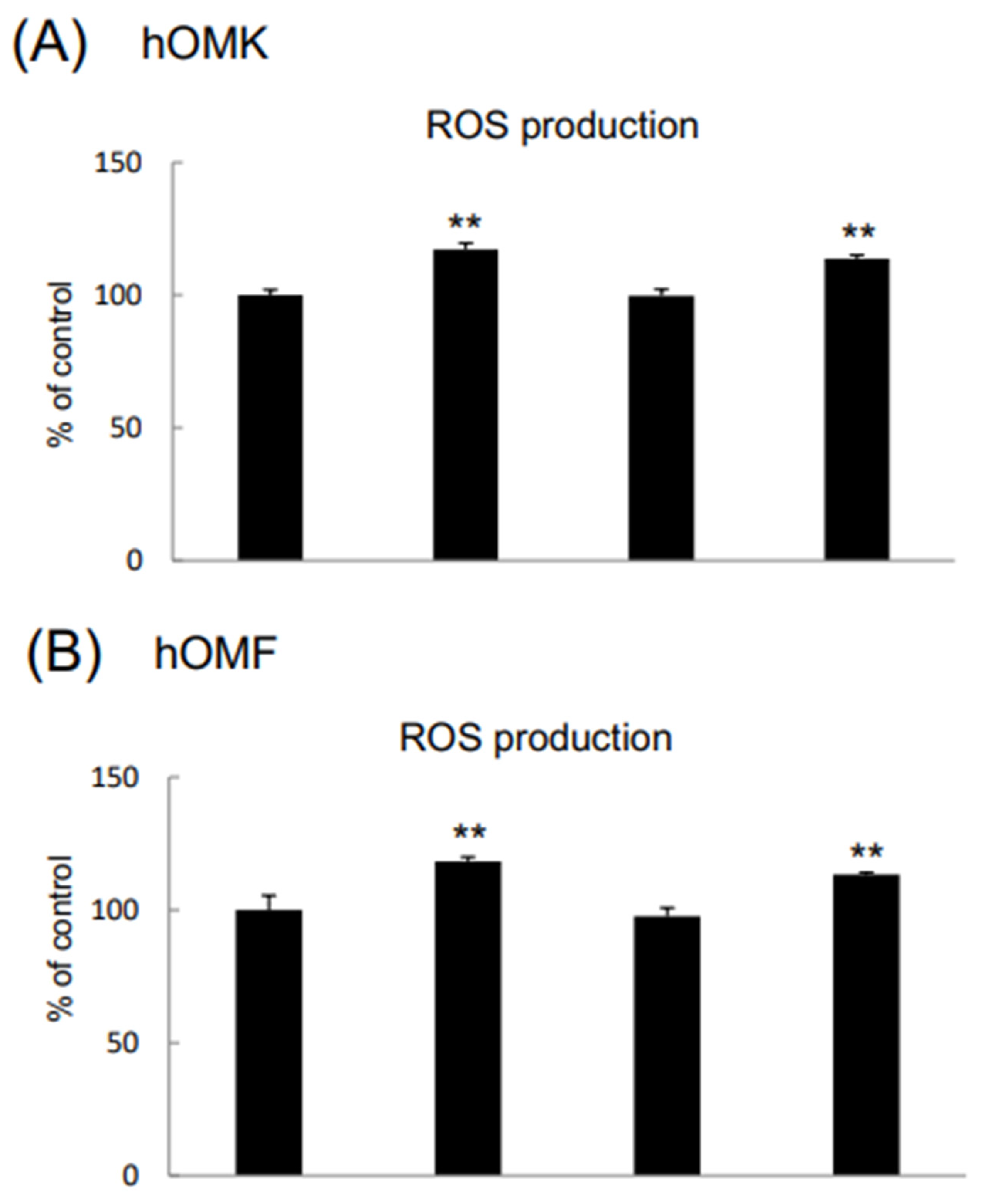
ROS levels were significantly higher in CDDP-treated hOMK (117.11% ± 2.41) and CDDP+β-cry-treated hOMK (113.66% ± 1.47) than in control cells. ROS levels were also significantly higher in CDDP-treated and CDDP+β-cry-treated hOMF (118.33% ± 1.6 and 113.22% ± 0.76, respectively) than in control cells (Figure 4)

3.4. SA-β-Gal-Positive Cells

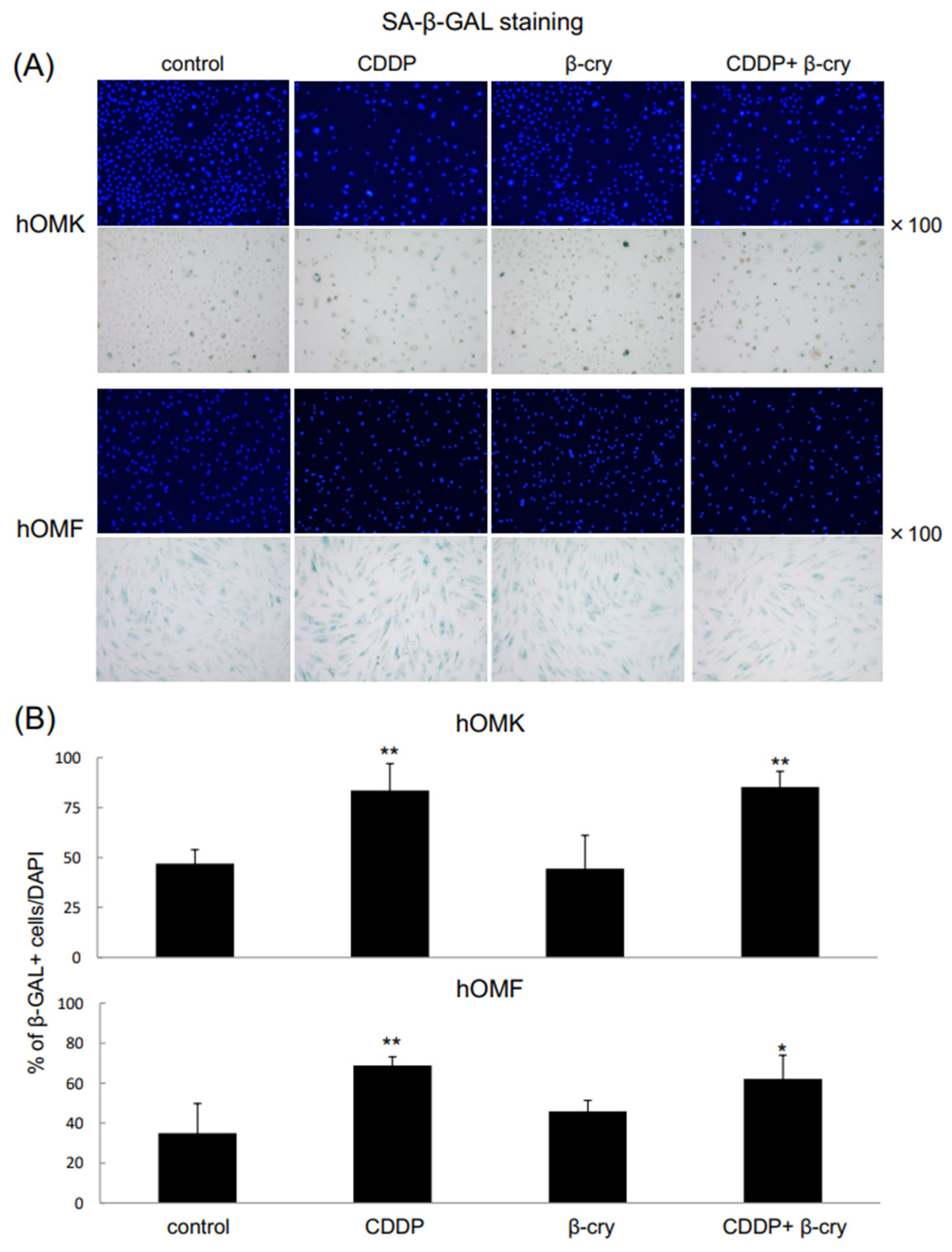
In hOMK and hOMF, the rates of β-gal-positive cells were significantly higher in CDDP-treated cells (hOMK: 83.53% ± 13.52, hOMF: 68.78% ± 4.44) and CDDP+β-cry-treated cells (hOMK: 85.32% ± 7.87, hOMF: 62.02% ± 12.0) than in control cells (hOMK: 46.95% ± 6.96, hOMF: 35.04% ± 14.73). The addition of β-cry did not significantly affect the rate of β-gal-positive cells (Figure 5).

4. Discussion

Oral mucositis, as a side effect of chemotherapy with CDDP or radiochemotherapy, causes pain or swallowing pain in the oral cavity, which reduces the nutritional state of patients, makes conversations difficult, and markedly affects quality of life. In addition, oral mucositis-related susceptibility to infection deteriorates the general condition of some patients or necessitates the reduction in number of doses of chemotherapeutic agents. Oral mucositis also affects the admission period and survival rate [26,27,28].
CDDP inhibits cellular DNA replication or transcription, thereby inducing apoptosis. The present study showed that CDDP inhibited the proliferation of oral mucosa-derived epithelial cells and fibroblasts, whereas β-cry significantly increased the proliferation of both cell types, promoting oral mucosa-derived cell proliferation.
CDDP significantly increased IL-8 production in hOMK and IL-6 production in hOMF. Regarding cytokine expression in skin-derived cells, a previous study reported that the ROS-mediated expression of IL-6 and IL-8 was up-regulated under redox stress conditions [29], and that chemotherapeutic agents and inflammatory cytokines affected the expression of MMPs, potentially contributing to the onset and severity of oral mucositis [30]. Among cytokine-associated MMPs that regulate inflammatory cytokine expression or activity, MMP-2 is responsible for maintaining homeostasis in the immune system, while the production of MMP-9 is increased by inflammation. MMP-9 itself may also promote inflammation [31]. In the present study, MMP-9 production significantly increased in oral mucosa-derived cells. This result suggests that oral mucosa-derived cells produced IL-6, an inflammatory cytokine, and MMP-9 in the presence of CDDP, inducing an inflammatory response in the oral mucosa, which is consistent with the findings reported by Sonis et al. [32]. Pathogenic microorganisms, such as Candida albicans, affect the basement membrane proteins through MMP-2 and MMP-9 [33]. Therefore, MMPs produced by oral mucosal epithelial cells or fibroblasts in response to CDDP in the present study may have directly induced tissue damage. In addition, β-cry was shown to inhibit these cytokines and MMP produced by oral mucosa-derived cells in response to CDDP, exerting anti-inflammatory effects. These results are consistent with previous findings showing β-cry-induced reductions in IL-6/IL-8 production by periodontal ligament cells under mechanical stress or in the presence of periodontal pathogens [22] and the inhibition of IL-8/MMP-2 expression induced by oral mucosa-derived epithelial cells in the presence of 5-FU [23]. In the present study, we examined the mRNA expression and protein production of inflammatory cytokines and MMPs. Although each result did not completely match, similar changes were observed. These results provide support for the effects of CDDP and β-cry on oral mucosal cells.
CDDP-induced inflammation up-regulates the expression of IL-6/IL-8 mediated by the transcription factor nuclear factor-κB (NF-κB) in ovarian cancer cells or the activation of TNF-α through the activation of the MAPK pathway [34]. When NF-κB is activated, senescence-associated secretory phenotype factor is secreted [35]. Further studies are needed to investigate cell signaling in normal oral mucosa-derived cells.
Cellular senescence is induced in normal cells by stress [36]. ROS affects cellular components, such as lipids, protein, and DNA in vivo [37,38]. In senescent cells, ROS production is abnormally enhanced [39,40] and SA-β-gal specifically exists in senescent cells [41]. In addition, senescent cells secrete various factors, such as inflammatory cytokines and MMPs [42,43]. Therefore, cellular senescence is closely associated with not only ROS, but also inflammatory cytokines and MMPs. The present study showed that CDDP increased ROS production in oral mucosa-derived cells and the rate of SA-β-gal-positive cells. However, no significant differences were observed in ROS production or the rate of SA-β-gal-positive cells between CDDP- and CDDP+β-cry-treated cells, which was in contrast to the results obtained on inflammatory cytokines and MMPs. In other words, CDDP promotes biological oxidation and senescence, which may not be inhibited by β-cry. The reasons for this are unknown, but may be related to the smaller changes in ROS and SA-β-gal caused by CDDP rather than those in inflammatory cytokines and MMPs. Previous studies reported that astaxanthin and coenzyme Q10 have similar antioxidant functions to β-cry [44,45].
Keratinocyte growth factor (KGF) may reduce the risk of oral mucositis in adults receiving radiotherapy with CDDP for head and neck cancers or chemotherapy for a mixture of solid and blood cancers [46,47]. Dermal toxicity (exanthema, erythema, edema, and pruritus), oral toxicity (abnormal sensation, discoloration of the tongue, pachyglossia, and changes in the taste), and pain may occur as adverse reactions. KGF receptors are present even in malignant epithelial cells, as demonstrated for other growth factors; therefore, KGF may stimulate the division/growth of malignant cells expressing KGF receptors. On the other hand, the naturally derived ingredient β-cry has been shown to decrease the number of gastric cancer cells and MMP-2/MMP-9 production and also reduce the volume and weight of tumors, inhibiting tumor growth through the induction of apoptosis [48]. Furthermore, β-cry levels were found to be lower in patients with severe periodontal disease in all age groups of all races. An observational study reported a strong correlation between fruit/vegetable/other antioxidant nutrient intakes and quality of life associated with adult oral health [49].
In our previous study on 5-FU, only oral mucosa-derived epithelial cells were used. However, chemotherapy-related oral mucositis is also caused by damage to oral mucosal fibroblasts in not only oral mucosal epithelial cells, but also the inherent mucosal layer. Therefore, the present study investigated fibroblasts in addition to epithelial cells. In the future, this issue needs to be investigated in more detail in vivo.
The present results suggest that CDDP damages epithelial cells and fibroblasts comprising the oral mucosa and causes oral mucositis by inducing cellular senescence and increasing ROS production and inflammatory cytokine/MMP expression/production. Furthermore, β-cry may attenuate CDDP-related oral mucositis and maintain the QOL of patients.

Supplementary Materials

The following supporting information can be downloaded at: https://www.mdpi.com/article/10.3390/app15094803/s1, Figure S1: Assessment of viability of CDDP-treated hOMK and hOMF in the range of 0.01–100 μg/mL; Figure S2: Changes in the morphology of hOMK and hOMF after the treatment with CDDP. Table S1: Primer probes used for quantitative RT-PCR.

Author Contributions

Conceptualization, T.Y. and K.Y.; Methodology, N.W.; Formal analysis, N.W.; Investigation, K.Y., N.W. and F.O.; Resources, O.M. and N.K.; Data curation, F.O.; Writing—original draft, T.Y.; Writing—review & editing, K.Y. and O.M.; Supervision, K.Y., F.O., O.M. and N.K.; Project administration, T.Y. and N.K.; Funding acquisition, T.Y., K.Y. and F.O. All authors have read and agreed to the published version of the manuscript.

Funding

This research was supported by Grants-in-Aid from JSPS (18K09918, 19K24075, 20K10100, 21K10259, 21K09937, and 23K09463).

Institutional Review Board Statement

Not applicable.

Informed Consent Statement

Not applicable.

Data Availability Statement

The original contributions presented in this study are included in the article and Supplementary Material. Further inquiries can be directed to the corresponding author under reasonable request reasons.

Conflicts of Interest

The authors declare no conflicts of interest.

Abbreviations

β-cry; β-cryptoxanthin, CDDP; Cisplatin, hOMK; human oral mucosal keratinocytes, hOMF; human normal oral mucosal fibroblasts, ROS; reactive oxygen species, ELISA; enzyme-linked immunosorbent assay, IL; interleukin, MMP; matrix metalloproteinase, NF-κB; nuclear factor-κB.

References

  1. Cheng, Y.; Li, S.; Gao, L.; Zhi, K.; Ren, W. The molecular basis and therapeutic aspects of Cisplatin resistance in oral squamous cell carcinoma. Front. Oncol. 2021, 11, 761379. [Google Scholar] [CrossRef] [PubMed]
  2. Wang, Z.Q.; Liu, K.; Huo, Z.J.; Li, X.C.; Wang, M.; Liu, P.; Pang, B.; Wang, S.-J. A cell-targeted chemotherapeutic nanomedicine strategy for oral squamous cell carcinoma therapy. J. Nanobiotechnol. 2015, 13, 63. [Google Scholar] [CrossRef] [PubMed]
  3. Menashe, D.S.; Jacobs, S.C. Complications of hypogastric artery cisplatin infusions. J. Surg. Oncol. 1989, 41, 160–164. [Google Scholar] [CrossRef]
  4. Kaneyasu, Y.; Okawa, M.K.; Okawa, T. Clinical evaluation of chemoradiotherapy for advanced cervical cancer. Gan Kagaku Ryoho. 1997, 24, 2084–2091. [Google Scholar]
  5. Kralovánszky, J.; Prajda, N.; Kerpel-Fronius, S.; Gál, F.; Kiss, F. Comparison of intestinal toxic effects of platinum complexes: Cisplatin (CDDP), carboplatin (CBDCA), and iproplatin (CHIP). Cancer Chemother. Pharmacol 1988, 21, 40–44. [Google Scholar] [CrossRef] [PubMed]
  6. Sakamoto, K.; Takeda, S.; Kanekiyo, S.; Nishiyama, M.; Kitahara, M.; Ueno, T.; Yamamoto, S.; Yoshino, S.; Hazama, S.; Okayama, N.; et al. Association of tumor necrosis factor-α polymorphism with chemotherapy-induced oral mucositis in patients with esophageal cancer. Mol. Clin. Oncol. 2017, 6, 125–129. [Google Scholar] [CrossRef]
  7. Topping, R.P.; Wilkinson, J.C.; Scarpinato, K.D. Mismatch repair protein deficiency compromises cisplatin-induced apoptotic signaling. J. Biol. Chem. 2009, 284, 14029–14039. [Google Scholar] [CrossRef]
  8. Browning, R.J.; Reardon, P.J.T.; Parhizkar, M.; Pedley, R.B.; Edirisinghe, M.; Knowles, J.C.; Stride, E. Drug Delivery Strategies for Platinum-Based Chemotherapy. ACS Nano 2017, 11, 8560–8578. [Google Scholar] [CrossRef]
  9. Al-Dasooqi, N.; Sonis, S.T.; Bowen, J.M.; Bateman, E.; Blijlevens, N.; Gibson, R.J.; Logan, R.M.; Nair, R.G.; Stringer, A.M.; Yazbeck, R.; et al. Emerging evidence on the pathobiology of mucositis. Support. Care Cancer 2013, 21, 3233–3241. [Google Scholar] [CrossRef]
  10. Hong, C.H.L.; Gueiros, L.A.; Fulton, J.S.; Cheng, K.K.F.; Kandwal, A.; Galiti, D.; Fall-Dickson, J.M.; Johansen, J.; Ameringer, S.; Kataoka, T.; et al. Systematic review of basic oral care for the management of oral mucositis in cancer patients and clinical practice guidelines. Support. Care Cancer 2019, 27, 3949–3967. [Google Scholar] [CrossRef]
  11. Daugėlaitė, G.; Užkuraitytė, K.; Jagelavičienė, E.; Filipauskas, A. Prevention and treatment of chemotherapy and radiotherapy induced oral mucositis. Medicina 2019, 55, 25. [Google Scholar] [CrossRef]
  12. Chaveli-López, B.; Bagán-Sebastián, J.V. Treatment of oral mucositis due to chemotherapy. Clin. Exp. Dent. 2016, 8, e201–e209. [Google Scholar] [CrossRef]
  13. Saadeh, C.E. Chemotherapy- and radiotherapy-induced oral mucositis: Review of preventive strategies and treatment. Pharmacotherapy 2005, 25, 540–554. [Google Scholar] [CrossRef] [PubMed]
  14. Hayashi, H.; Kobayashi, R.; Suzuki, A.; Yamada, Y.; Ishida, M.; Shakui, T.; Kitagawa, J.; Hayashi, H.; Sugiyama, T.; Takeuchi, H.; et al. Preparation and clinical evaluation of a novel lozenge containing polaprezinc, a zinc-L-carnosine, for prevention of oral mucositis in patients with hematological cancer who received high-dose chemotherapy. Med. Oncol. 2016, 33, 91. [Google Scholar] [CrossRef] [PubMed]
  15. Manzi, N.M.; Silveira, R.C.; Reis, P.E. Prophylaxis for mucositis induced by ambulatory chemotherapy: Systematic review. J. Adv. Nurs. 2016, 72, 735–746. [Google Scholar] [CrossRef]
  16. Lorenzo, Y.; Azqueta, A.; Luna, L.; Bonilla, F.; Domínguez, G.; Collins, A.R. The carotenoid beta-cryptoxanthin stimulates the repair of DNA oxidation damage in addition to acting as an antioxidant in human cells. Carcinogenesis 2009, 30, 308–314. [Google Scholar] [CrossRef] [PubMed]
  17. Liu, X.R.; Wang, Y.Y.; Dan, X.G.; Kumar, A.; Ye, T.Z.; Yu, Y.Y.; Yang, L.G. Anti-inflammatory potential of β-cryptoxanthin against LPS-induced inflammation in mouse Sertoli cells. Reprod. Toxicol. 2016, 60, 148–155. [Google Scholar] [CrossRef]
  18. Quesada-Gómez, J.M.; Santiago-Mora, R.; Durán-Prado, M.; Dorado, G.; Pereira-Caro, G.; Moreno-Rojas, J.M.; Casado-Díaz, A. β-cryptoxanthin inhibits angiogenesis in human umbilical vein endothelial cells through retinoic acid receptor. Mol. Nutr. Food Res. 2018, 62, 201700489. [Google Scholar] [CrossRef]
  19. Burri, B.J.; La Frano, M.R.; Zhu, C. Absorption, metabolism, and functions of β-cryptoxanthin. Nutr. Rev. 2016, 74, 69–82. [Google Scholar] [CrossRef]
  20. Park, G.; Horie, T.; Fukasawa, K.; Ozaki, K.; Onishi, Y.; Kanayama, T.; Iezaki, T.; Kaneda, K.; Sugiura, M.; Hinoi, E. Amelioration of the development of osteoarthritis by daily intake of β-cryptoxanthin. Biol. Pharm. Bull. 2017, 40, 1116–1120. [Google Scholar] [CrossRef]
  21. Nishigaki, M.; Yamamoto, T.; Ichioka, H.; Honjo, K.; Yamamoto, K.; Oseko, F.; Kita, M.; Mazda, O.; Kanamura, N. Beta-cryptoxanthin regulates bone resorption related-cytokine production in human periodontal ligament cells. Arch. Oral. Biol. 2013, 58, 880–886. [Google Scholar] [CrossRef] [PubMed]
  22. Yamanobe, H.; Yamamoto, K.; Kishimoto, S.; Nakai, K.; Oseko, F.; Yamamoto, T.; Mazda, O.; Kanamura, N. Anti-inflammatory effects of β-cryptoxanthin on 5-fluorouracil-induced cytokine expression in human oral mucosal keratinocytes. Molecules 2023, 28, 2935. [Google Scholar] [CrossRef] [PubMed]
  23. Yamamoto, K.; Sato, Y.; Honjo, K.; Ichioka, H.; Oseko, F.; Sowa, Y.; Yamamoto, T.; Kanamura, N.; Kishida, T.; Mazda, O. Generation of directly converted human osteoblasts that are free of exogenous gene and xenogenic protein. J. Cell. Biochem. 2016, 117, 2538–2545. [Google Scholar] [CrossRef]
  24. Yamamoto, K.; Kishida, T.; Nakai, K.; Sato, Y.; Kotani, S.I.; Nishizawa, Y.; Yamamoto, T.; Kanamura, N.; Mazda, O. Direct phenotypic conversion of human fibroblasts into functional osteoblasts triggered by a blockade of the transforming growth factor-β signal. Sci. Rep. 2018, 8, 8463. [Google Scholar] [CrossRef]
  25. Okada, H.; Oguchi, N.; Uchida, J.; Mikami, O.; Matusda, T. Study on platinum concentration in internal iliac venous blood after iliac artery cisplatin infusion for invasive bladder cancer. Hinyokika Kiyo 1999, 45, 145–148. [Google Scholar]
  26. Elting, L.S.; Cooksley, C.; Chambers, M.; Cantor, S.B.; Manzullo, E.; Rubenstein, E.B. The burdens of cancer therapy. Clinical and economic outcomes of chemotherapy-induced mucositis. Cancer 2003, 98, 1531–1539. [Google Scholar] [CrossRef]
  27. Oba, M.K.; Innocentini, L.M.A.R.; Viani, G.; Ricz, H.M.A.; de Carvalho Reis, T.; Ferrari, T.C.; de Macedo, L.D. Evaluation of the correlation between side effects to oral mucosa, salivary glands, and general health status with quality of life during intensity-modulated radiotherapy for head and neck cancer. Support. Care Cancer 2020, 29, 127–134. [Google Scholar] [CrossRef]
  28. Alsheyyab, F.; Al-Momani, D.; Kasht, R.; Kamal, A.; Abusalem, D.; Al-Qasem, W. Impact of severe oral mucositis in pediatric cancer patients on resource utilization and cancer treatment plans. Int. J. Clin. Pharm. 2021, 43, 1322–1326. [Google Scholar] [CrossRef] [PubMed]
  29. Bhartiya, P.; Masur, K.; Shome, D.; Kaushik, N.; Nguyen, L.N.; Kaushik, N.K.; Choi, E.H. Influence of redox stress on crosstalk between fibroblasts and keratinocytes. Biology 2021, 10, 1338. [Google Scholar] [CrossRef]
  30. Cardoso, L.M.; Pansani, T.N.; Hebling, J.; de Souza Costa, C.A.; Basso, F.G. Chemotherapy drugs and inflammatory cytokines enhance matrix metalloproteinases expression by oral mucosa cells. Arch. Oral Biol. 2021, 127, 105159. [Google Scholar] [CrossRef]
  31. Van den Steen, P.E.; Dubois, B.; Nelissen, I.; Rudd, P.M.; Dwek, R.A.; Opdenakker, G. Biochemistry and molecular biology of gelatinase B or matrix metalloproteinase-9 (MMP-9). Crit. Rev. Biochem. Mol. Biol. 2002, 37, 375–5366. [Google Scholar] [CrossRef] [PubMed]
  32. Sonis, S.T. A biological approach to mucositis. J. Support Oncol. 2004, 2, 21–32. [Google Scholar] [PubMed]
  33. Claveau, I.; Mostefaoui, Y.; Rouabhia, M. Basement membrane protein and matrix metalloproteinase deregulation in engineered human oral mucosa following infection with Candida albicans. Matrix Biol. 2004, 23, 477–486. [Google Scholar] [CrossRef] [PubMed]
  34. Vyas, D.; Laput, G.; Vyas, A.K. Chemotherapy-enhanced inflammation may lead to the failure of therapy and metastasis. Onco. Targets Ther. 2014, 7, 1015–1023. [Google Scholar] [CrossRef]
  35. Chien, Y.; Scuoppo, C.; Wang, X.; Fang, X.; Balgley, B.; Bolden, J.E.; Premsrirut, P.; Luo, W.; Chicas, A.; Lee, C.S.; et al. Control of the senescence-associated secretory phenotype by NF-kappaB promotes senescence and enhances chemosensitivity. Genes Dev. 2011, 25, 2125–2136. [Google Scholar] [CrossRef]
  36. Calcinotto, A.; Kohli, J.; Zagato, E.; Pellegrini, L.; Demaria, M.; Alimonti, A. Cellular Senescence: Aging, Cancer, and Injury. Physiol. Rev. 2019, 99, 1047–1078. [Google Scholar] [CrossRef]
  37. Birben, E.; Sahiner, U.; Sackesen, C.; Erzurum, S.; Kalayci, O. Oxidative stress and antioxidant defense. World Allergy Organ. 2012, 5, 9–19. [Google Scholar] [CrossRef]
  38. Juan, C.A.; Lastra, J.M.; Plou, F.J.; Lebeña, E.P. The chemistry of reactive oxygen species (ROS) revisited: Outlining their role in biological macromolecules (DNA, lipids and proteins) and induced pathologies. Int. J. Mol. Sci. 2021, 22, 4642. [Google Scholar] [CrossRef]
  39. Takahashi, A.; Ohtani, N.; Yamakoshi, K.; Iida, S.; Tahara, H.; Nakayama, K.; Nakayama, K.I.; Ide, T.; Saya, H.; Hara, E. Mitogenic signalling and the p16INK4a/Rb pathway cooperate to enforce irreversible cellular senescence. Nat. Cell Biol. 2006, 8, 1291–1297. [Google Scholar] [CrossRef]
  40. Davalli, P.; Mitic, T.; Caporali, A.; Lauriola, A.; D’Arca, D. ROS, Cell Senescence, and Novel Molecular Mechanisms in Aging and Age-Related Diseases. Oxid. Med. Cell. Longev. 2016, 1, 3565–3582. [Google Scholar] [CrossRef]
  41. Debacq-Chainiaux, F.; Erusalimsky, J.D.; Campisi, J.; Toussaint, O. Protocols to detect senescence-associated beta-galactosidase (SA-betagal) activity, a biomarker of senescent cells in culture and in vivo. Nat. Protoc. 2009, 4, 1798–1806. [Google Scholar] [CrossRef]
  42. Rodier, F.; Coppé, J.P.; Patil, C.K.; Hoeijmakers, W.A.; Muñoz, D.P.; Raza, S.R.; Freund, A.; Campeau, E.; Davalos, A.R.; Campisi, J. Persistent DNA damage signalling triggers senescence-associated inflammatory cytokine secretion. Nat. Cell Biol. 2009, 11, 973–979. [Google Scholar] [CrossRef]
  43. Jun, E.-S.; Kim, Y.J.; Kim, H.H.; Park, S.Y. Gold Nanoparticles Using Ecklonia stolonifera Protect Human Dermal Fibroblasts from UVA-Induced Senescence through Inhibiting MMP-1 and MMP-3. Mar. Drugs 2020, 19, 433. [Google Scholar] [CrossRef] [PubMed]
  44. Wu, X.; Liang, S.; Zu, X.; Dong, Z. CoQ10 suppression of oxidative stress and cell senescence increases bone mass in orchiectomized mice. Am. J. Transl. Res. 2020, 12, 4314–4325. [Google Scholar]
  45. Yang, G.; Liu, X.; Jing, X.; Wang, J.; Wang, H.; Chen, F.; Wang, W.; Shao, Y.; Cui, X. Astaxanthin suppresses oxidative stress and calcification in vertebral cartilage endplate via activating Nrf-2/HO-1 signaling pathway. Int. Immunopharmacol. 2023, 119, 110159. [Google Scholar] [CrossRef] [PubMed]
  46. von Bültzingslöwen, I.; Brennan, M.T.; Spijkervet, F.K.; Logan, R.; Stringer, A.; Raber-Durlacher, J.E.; Keefe, D. Growth factors and cytokines in the prevention and treatment of oral and gastrointestinal mucositis. Support. Care Cancer 2006, 14, 519–527. [Google Scholar] [CrossRef] [PubMed]
  47. Riley, P.; Glenny, A.M.; Worthington, H.V.; Littlewood, A.; Mauleffinch, L.M.F.; Clarkson, J.E.; McCabe, M.G. Interventions for preventing oral mucositis in patients with cancer receiving treatment: Cytokines and growth factors. Cochrane Database Syst. Rev. 2017, 11, CD011990. [Google Scholar] [CrossRef]
  48. Gao, M.; Dang, F.; Deng, C. β-cryptoxanthin induced anti-proliferation and apoptosis by G0/G1 arrest and AMPK signal inactivation in gastric cancer. Eur. J. Pharmacol. 2019, 859, 172528. [Google Scholar] [CrossRef]
  49. Ebersole, J.L.; Lambert, J.; Bush, H.; Huja, P.E.; Basu, A. Serum nutrient levels and aging effects on periodontitis. Nutrients 2018, 10, 1986. [Google Scholar] [CrossRef]
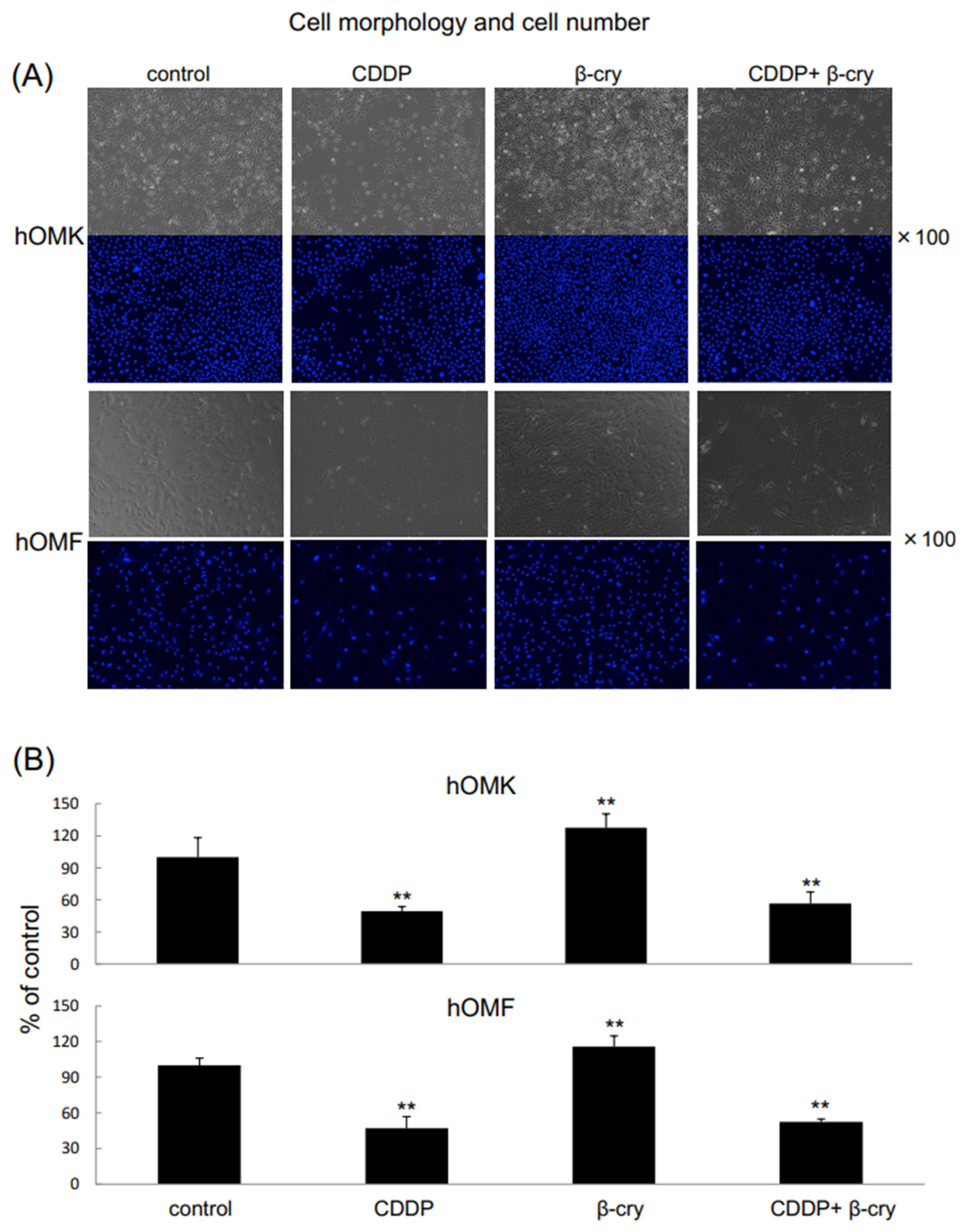
Figure 1. Changes in morphologies and numbers of hOMK and hOMF after the CDDP treatment. (A) Morphologies of hOMK and hOMF in each group. Magnification ×100. (B) The numbers of CDDP-treated hOMK and hOMF were significantly lower than those of control cells, whereas those of β-cry-treated hOMK and hOMF were significantly higher. Values are means ± S.D. n = 6 (sample number in each group) ** p < 0.01 vs. control.
Applsci 15 04803 g001
Figure 2. Effects of CDDP and β-cry on inflammatory cytokine and MMP mRNA expression levels in hOMK and hOMF. IL-6, IL-8, and MMP-9 mRNA expression levels were significantly higher in CDDP-treated hOMK (A) and hOMF (B) than in control cells. IL-6, IL-8, and MMP-9 mRNA expression levels were significantly lower in CDDP+β-cry-treated hOMK than in CDDP-treated hOMK (A). IL-6 and MMP-9 mRNA expression levels were significantly lower in CDDP+β-cry-treated hOMF than in CDDP-treated hOMF (B). Values are means ± S.D. n = 4 * p < 0.05, ** p < 0.01 vs. control, # p < 0.05, ## p < 0.01 between each group.
Applsci 15 04803 g002
Figure 3. Effects of CDDP and β-cry on inflammatory cytokine and MMP levels in hOMK and hOMF. IL-6, IL-8, MMP-2, and MMP-9 levels were significantly higher in CDDP-treated hOMK and in CDDP+β-cry-treated hOMK than in control cells (A). IL-6, IL-8, and MMP-2 levels were significantly higher in CDDP-treated hOMF and CDDP+β-cry-treated hOMF than in control cells (B). IL-6 levels were significantly lower in CDDP+β-cry-treated hOMK and hOMF than in hOMK and CDDP-treated hOMF (A,B). Values are means ± S.D. * p < 0.05, ** p < 0.01 vs. control, ## p < 0.01 between each group.
Applsci 15 04803 g003
Figure 4. Effects of CDDP and β-cry on ROS levels in hOMK and hOMF. ROS levels were significantly higher in CDDP-treated hOMK (A) and hOMF (B) and in CDDP+β-cry-treated hOMK and hOMF than in control cells. Values are means ± S.D. n = 4, ** p < 0.01 vs. control.
Applsci 15 04803 g004
Figure 5. Effects of CDDP and β-cry on cellular senescence in hOMK and hOMF. (A) Images for β-GAL staining and DAPI of hOMK and hOMF in each group. Magnification ×100. (B) The percentages of β-GAL-positive cells were significantly higher in CDDP-treated hOMK and hOMF and in CDDP+β-cry-treated hOMK and hOMF than in control cells. Values are means ± S.D. n = 4 * p < 0.05, ** p < 0.01 vs. control.
Applsci 15 04803 g005
Disclaimer/Publisher’s Note: The statements, opinions and data contained in all publications are solely those of the individual author(s) and contributor(s) and not of MDPI and/or the editor(s). MDPI and/or the editor(s) disclaim responsibility for any injury to people or property resulting from any ideas, methods, instructions or products referred to in the content.