Article

Lactobacillus rhamnosus GG as Biosensor for Oral and Systemic Health Conditions: A Pilot Study

by
Pooja Mali Rai
1,
Andrei Cristian Ionescu
1,2,
Alessio Soggiu
1,
Antonella Panio
1,2,
Sourav Panda
3,
Paolo Savadori
4,5,
Gianluca Martino Tartaglia
1,2,
Massimo Del Fabbro
1,2 and
Funda Goker
1,2,*
1
Department of Biomedical, Surgical and Dental Sciences, Milan University, 20122 Milan, Italy
2
Unit of Maxillo-Facial Surgery and Dentistry, Fondazione IRCCS Ca’ Granda Ospedale Maggiore Policlinico, 20122 Milan, Italy
3
Department of Periodontics, Institute of Dental Sciences, Siksha ‘O’ Anusandhan University, Bhubaneswar 751002, Odisha, India
4
Department of Surgery, Medicine, Dentistry and Morphological Sciences with Transplant Surgery, Oncology and Regenerative Medicine Relevance (CHIMOMO), University of Modena and Reggio Emilia, 42124 Modena, Italy
5
Center for Verification and Research of New Technologies in Dentistry (CRIO), University of Modena and Reggio Emilia, 42124 Modena, Italy
*
Author to whom correspondence should be addressed.
Appl. Sci. 2025, 15(9), 4809; https://doi.org/10.3390/app15094809 (registering DOI)
Submission received: 25 March 2025 / Revised: 18 April 2025 / Accepted: 22 April 2025 / Published: 26 April 2025
(This article belongs to the Special Issue The Oral Microbiome in Periodontal Health and Disease)

Abstract

:
Early disease detection using biosensors is a significant challenge in modern medicine. This study aimed to investigate Lactobacillus rhamnosus GG (LGG) as a bacterial biosensor for biomarkers indicative of oral and systemic health conditions. For this purpose, LGG was cultured and then exposed to phosphate buffer, 10 wt.% sucrose solution, pH = 4.0, lactic acid, and filter-sterile saliva from five subjects. A total of 10 groups consisted of filter-sterile, freshly pooled saliva of subject 1 (SANT), subject 2 (SLAN), subject 3 (SLFU), subject 4 (SLPA), subject 5 (SLPO), phosphate buffer solution (pH = 7.4, BUF), and PBS with resin. Subsequently, the proteomic profiling of the samples was done by high-resolution mass spectrometry, focusing on the expression of bacterial proteins. The samples were evaluated for the biosensing capacity of LGG through its proteomic expression. Statistical comparisons were performed to outline proteomic changes, clustering upregulated and downregulated proteins relevant to stress response, metabolism, and environmental adaptation. The identification of key proteins associated with metabolic regulation, response to oxidative stress, and bacterial adaptation was possible using heatmaps and volcano plots. Each subject’s salivary composition also presented its unique, characteristic proteomic signature. Results showed a massive downregulation of proteins linked with stress under nutrient-rich conditions. In conclusion, the early detection of protein expression modifications related to environmental niche changes has shown that LGG can serve as a promising novel diagnostic tool, potentially overcoming many drawbacks of current physicochemical transducer-based biosensors.

1. Introduction

Currently, the early detection of diseases is one of the primary challenges facing our society and healthcare systems. Early disease detection largely depends on the precise identification of specific disease signals before clinical signs and symptoms emerge, utilizing various diagnostic methods, including the use of biomarkers. Over the past few decades, salivary biomarkers have been studied extensively to signal local and systemic pathologies. They have garnered significant attention from the scientific community, since they provide valuable insights into the diagnosis, prognosis, and therapeutic responsiveness of numerous human diseases [1].
The microbiome plays a vital role in human physiology, and numerous microbes have been residing within the human body, continuously evolving alongside us, thereby forming a symbiotic environment [2]. The gut microbiome has been extensively studied for its role in various health-related issues, including inflammatory diseases, nutrient absorption, immune responses, and metabolic disorders such as obesity, malnutrition, and cancer immunity [3]. Just like the gut microbiome, the oral microbiome contributes to the general health of individuals. In the oral cavity, the unique ecosystem’s complexity and balance depend on specific physical and chemical components, including teeth, gingiva, and tongue, and each foster specific microbial communities depending on varying pH levels, oxygen availability, and nutrient sources [4]. A balanced oral microbiome plays a crucial role in functions such as modulating local immune responses, producing antimicrobial peptides, and inhibiting the growth of pathogenic species, thereby sustaining a healthy oral cavity [5].
The bioengineered microbiomes have attracted researchers, as they represent a promising frontier in biotechnology, aiming to harness and manipulate microbial communities for therapeutic, agricultural, and environmental applications. Bioengineered tools are highly advanced in strains of E. coli and Lactobacillus; however, comparatively, just a few bioengineered strains are available to study the gut or colon anaerobic environment [6]. Escherichia coli Nissle (EcN), in its natural/wild (not engineered) form, has been used to treat several gastrointestinal conditions and irritable bowel syndrome. Additionally, EcN stimulates anti-inflammatory activities in the intestinal epithelium [7,8]. As an alternative to the gut, oral microbiomes have also been studied, as saliva is one of the biofluids rich in biomarkers [9]. Furthermore, saliva collection is simple and non-invasive, which avoids patient stress through easy collection and repetition when needed, without any additional biological cost. Saliva is readily available, and salivary biomarkers, such as matrix metalloproteinases (MMPs), interleukins (ILs), cortisol, and prostaglandins, have already been studied as potential indicators of periodontal disease severity and progression [10,11,12,13].
Molecular medicine is a broad field that describes molecular structures based on their physical, chemical, and biological properties, utilizing bioinformatics and medical techniques. The interactions between proteins, which underpin biological processes, can be understood as dynamic flows of information within the cell and the organism, transmitted through protein pathways and networks [14,15]. Proteomics has significantly contributed to understanding the virulence and structural makeup of bacteria and viruses in disease pathogenesis. Proteomics conventionally began with Western blot and chromatography, and over the past two decades, several modern quantitative techniques have evolved. The study of membrane proteins, surface proteins, and secreted proteins has been investigated using the proteomics technique [16]. Genomics can only aid in understanding the pathophysiology of the disease’s progression, but not its cause [17]. Several techniques are available in protein identification, with various forms of mass spectrometry being the most commonly used [18].
Lactobacillus rhamnosus GG (LGG) is one of the most well-researched probiotic bacterial strains, known for its health benefits driven by its superior ability to adhere and colonize the gastrointestinal tract. LGG can actively prevent bacterial colonization in the gut by producing antimicrobials [19]. Several molecular studies have elucidated the mechanisms of LGG [20]. LGG secretes proteins such as Msp1/p75 and Msp2/p40, which activate the Akt signaling pathway, thereby preventing apoptosis of intestinal epithelial cells and reducing inflammation [21]. Omics research has transformed the understanding of LGG by integrating molecular mechanisms. Proteomics is arguably the most suitable method for studying LGG as a biosensor due to its ability to capture real-time protein expression changes under various environmental conditions. However, there is a lack of scientific research and evidence that evaluates LGG as a biosensor for health detection. Based on this premise, this work aimed to explore the biosensing capacity of LGG through its proteomic expression in diverse oral environments. Specifically, the study focused on how LGG adapts and responds to key factors, including metabolism, oxidative stress, and general health markers. By analyzing proteomic profiles of LGG, its potential as a biosensor for maintaining oral health was evaluated. We hypothesized that the ability of LGG to detect shifts in protein expression under various environmental and systemic conditions might confirm its possible future applications in detecting some health-related conditions.

2. Materials and Methods

This pilot study aimed to evaluate the outcomes of LGG as a bacterial biosensor for biomarkers indicative of oral and systemic health conditions. For this purpose, salivary samples were collected and assessed from five healthy individuals who had no active dental disease and received no antibiotic therapy for at least 3 months before the experiments. Informed consent forms were obtained from all participants. The study adhered to the principles outlined in the Declaration of Helsinki regarding medical protocol and ethics. The protocol used in this study was in accordance with the Declaration of Helsinki of 1975, which was revised in 2013. Informed consent was obtained from all volunteers to participate in the research and publish data for scientific research purposes. The study protocol was approved by the Ethics Committee of the Ca’ Granda Ospedale Maggiore Policlinico Foundation in Milan, Regione Lombardia, Italy, date 05/08/2022, Ethics Committee of Milano Area B Act 3.11/2022-294, N.2143 (Subject: “Transitional study in Salivary Biomarkers”).

2.1. Bacterial Strain and Growth

Becton Dickinson (BD Diagnostics-Difco, Franklin Lakes, NJ, USA) LGG (ATCC 53103) was grown on agar. A single colony was inoculated in brain heart infusion and supplemented with 1 wt. % sucrose at 37 °C at 5% CO2 enrichment for 12 h (early log stage) to produce a pure microorganism suspension. The bacterial cells were then separated by centrifugation (2200× g, 19 °C, 5 min). The bacteria were then rinsed twice with sterile phosphate-buffered saline (PBS) and blended with brain heart infusion agar supplemented with 1 wt% sucrose. After this, the suspension was subjected to a Sonifier type B-150 (Branson, Danbury, CT, USA) with 7-W energy output for 30 s to homogenize the solution. The microbial concentration was approximated at 6.0 × 10⁸ cells/mL after adjusting the suspension to 1.0 on the scale of McFarland.

2.2. Saliva Collection

In this study, unstimulated whole saliva was collected from participants to avoid artificial stimulation that could alter the proteomic profile. Passively pooled saliva collection was conducted in the morning (between 8:00 AM and 10:00 AM) in a dimly lit room to control for diurnal variations. This protocol was followed to ensure consistency in sample composition and reduce variability in proteomic analysis. The whole saliva was collected from five volunteers (coded as SLAN, SANT, SLPO, SLFU, SLPA) following a previously published protocol [22]. Saliva was collected using chilled test tubes and then centrifuged (12,000× g, 4 °C, 15 min). The supernatant was transferred into sterile tubes and filtered (0.2 μm filter, Nunc, Kalstrup, Denmark).

2.3. Sample Preparation and Processing for Proteomic Analysis

Following incubation, LGG was centrifuged to concentrate the cells into a pellet. This process involved removing the supernatant, which comprised the BHI medium, thereby isolating the bacterial cells at the bottom of the centrifuge tube. The bacterial pellet was then washed and resuspended in PBS. To prepare the samples for exposure to different environments and then for proteomic analysis, a structured framework was established to standardize the exposure of the collected specimens. The specimens were methodically transferred into 12-well plates for processing. Each well was carefully loaded with a measured combination of 1 mL of LGG suspension and mixed with 2 mL of either human saliva in 5 wells or alternative solutions, such as PBS, pH 4.0 lactic acid, or 10 wt% sucrose, in other wells. One of the wells was kept solely with 2 mL of LGG as a control. The inoculated samples were further incubated for three h under controlled conditions. The control sample, with only bacterial inoculation, served as a baseline reference, ensuring that any detected proteomic changes could be attributed to various experimental media.

2.3.1. Centrifugation and Sample Processing

After incubation, the samples underwent centrifugation (10,000× g, 4 °C, 2 min). This high-speed centrifugation was designed to isolate cellular components from the surrounding medium and to obtain a well-defined cellular pellet. The supernatant residual medium was discarded, leaving a purified bacterial pellet behind. To preserve the stability and functionality of proteins by preventing protein degradation through the neutralization of proteolytic activity, a protease inhibitor (complete tablet) was introduced during a second round of centrifugation to safeguard the integrity of proteomic components. The sample was centrifuged again under identical conditions, and the inhibitor was discarded. The resulting purified pellet was collected and promptly stored at −80 °C.

2.3.2. RNA Extraction

For quality control on microorganisms in the sample as well as contamination control, full-length 16S rRNA amplification was obtained through 16S Barcoding Kit 24 V14 (SQK-16S114.24, Oxford Nanopore Technologies, Oxford University Science Park, Oxford UK). Purity and quality were determined using the Nanodrop 2000 spectrophotometer (Thermo Scientific, Waltham, MA, USA) at 280 nm wavelength and agarose gel electrophoresis (0.75%, 126 mA, 1 h). Twelve barcoded sequencing libraries were loaded into a MinION FLO MIN114 R10.4.1 flow cell (Oxford) and sequenced onto a MinION Mk1C (Oxford) for 72 h. A 1 kb filter was used prior to each sequencing to bypass the shorter DNA fragments. The fastq files were basecalled using Guppy v.6.4.6 (neural network-based basecalling, super-accuracy basecalling mode, 400 bps, Oxford Nanopore Technologies). EPI2ME pipeline (Oxford) was used to assign genera and species.

2.3.3. Solution Digestion

Before conducting in-solution digestion, 200 μL of 8 M Urea solution containing protease inhibitors (Halt protease and phosphatase single-use inhibitor cocktail, Roche, Basel, Switzerland, code: 78442, lot: WC322135) was added to the pellets following the manufacturer’s instructions. The samples were then subjected to agitation in an ultrasonic bath for 10 min and centrifuged (10,000× g, 4 °C, 10 min). The liquid portion was collected, and the protein concentration was determined using the spectrophotometer (Nanodrop). The samples were prepared by adding a 50 mM ammonium bicarbonate solution in ultrapure water (Ambic) until a final volume of 60 μL was reached. For each sample, a total of 10 μg of protein was digested. To perform that, each sample was added with 3 μL of 100 mM Dithiothreitol solution prepared freshly in ultrapure water (DTT) at 55 °C for 30 min; 6 μL of 150 mM Iodoacetamide solution prepared freshly in ultrapure water and stored in the dark (IAA) for 20 min; 5 μL of trypsin solution 0.1 μg/μL in Ambic 50 mM at 37 °C overnight; and 1 μL of 50% trifluoroacetic acid (TFA). Subsequently, the samples were concentrated using a SpeedVac apparatus until they were completely moisture-free. They were then reconstituted in a solution containing 40 μL of 1% TFA.

2.3.4. Purification Protocol

The purification technique for all samples involved the use of 5 μg ziptip pipette tips, following the protocol outlined below. First, 24 μL of the digest was extracted from each sample to check the pH. Then, it was processed by ZipTip according to the manufacturer’s instructions; the eluate was concentrated using the SpeedVac apparatus, and the eluate was finally resuspended with 10 μL of a 0.1% formic acid solution prepared in ultrapure water.

2.3.5. High-Resolution Mass Spectrometry Analysis (HRMS)

The samples were analyzed at the UNITECH OMICs platform at the University of Milan using a Dionex Ultimate 3000 nano-liquid chromatography system (Sunnyvale, CA, USA). This system was connected to an Orbitrap Fusion Tribrid Mass Spectrometer (Thermo Scientific, Bremen, Germany) equipped with a nano-electrospray ion source.
Peptide mixtures were first concentrated on an Acclaim PepMap 100 C18 column (100 μm × 2 cm) and then separated using a 25 cm × 75 μm EASY-Spray column, packed with Acclaim PepMap RSLC C18 (3 μm particle size, 100 Å pore size), all from Thermo Scientific.
Separation was carried out using two mobile phases: A (0.1% formic acid in ultrapure water) and B (0.1% formic acid in acetonitrile, mixed 20/80 v/v), at a flow rate of 0.3 μL/min. The column temperature was maintained at 35 °C. Each sample (2 μL) was injected in duplicate, with blank runs in between to avoid cross-contamination.
Mass spectra were acquired across an m/z range of 375–1500 Da with a resolution of 120,000. The instrument operated in data-dependent mode, capturing scans in 3-s cycles. Fragmentation was achieved using higher-energy collisional dissociation (HCD) with a collision energy of 35 eV, and polarity switching was enabled.

2.3.6. Data Analysis

The raw data files were processed using Proteome Discoverer 3.1 (Thermo Fisher Scientific, Milan, Italy). Sequest-HT was used as the search engine for the database search, and peptide identification was validated through Percolator with a 1% false discovery rate (FDR) threshold. The search parameters included a precursor mass range of 350 to 5000 Da, a minimum of 6 peaks per spectrum, and a signal-to-noise ratio threshold of 2. Trypsin (full specificity) was set as the digestion enzyme, allowing up to two missed cleavages. Only peptides between 5 and 50 amino acids in length were considered. The precursor mass tolerance was 10 ppm, and the fragment mass tolerance was 0.02 Da. Cysteine residues were considered to have a static modification (carbamidomethylating), while methionine oxidation was set as a variable modification.
The data were analysed against the LGG sequence database (ATCC 700610/UA159). The Spectrum Files RC node was used to perform offline mass recalibration for improved accuracy. Label-free MS1 quantification was carried out using the Minora Feature Detector node, which automatically determined optimal retention time and mass tolerance settings based on data distributions. A feature list was generated using data from both the Feature Mapper and Precursor Ions Quantifier nodes. Peptides that showed intense matches with at least one MS2 spectrum from any sample were quantified by integrating the area under their chromatographic peaks.
The behavior of LGG when exposed to different conditions was analyzed, focusing on proteomic expression. A total of 10 groups consisted of filter-sterile, freshly pooled saliva of subject 1 (SANT), subject 2 (SLAN), subject 3 (SLFU), subject 4 (SLPA), subject 5 (SLPO), phosphate buffer solution (pH = 7.4, BUF), PBS with resin (meaning a PBS solution exposed for 48 h to eluates leaked from experimental dental splints made with TeraHarz, ESADENTAL S.R.L., Lecce, Italy) (BUFR), lactic acid with pH = 4.0 (LAC), 10 wt% sucrose solution (SU10), and control (K), where LGG was kept in its growth medium.
LGG proteomic data were filtered to include only proteins specific to LGG. Human salivary proteins potentially contaminating the samples were excluded from the analysis; then, proteins having at least one positive value in each group were analyzed using FragPipe-Analyst (Proteomics & Integrative Bioinformatics Lab at the University of Michigan (P.I. Alexey Nesvizhskii) and the Monash Proteomics & Metabolomics Facility, Monash University (P.I. Ralf Schittenhelm). A comparison was then performed between each group and the control group to highlight the downregulation and upregulation of selected proteins, which were subsequently investigated. LRHM annotation in all volcano plots refers to a database annotation for the names of all genes encoding LGG proteins.

2.4. Statistical Analysis

Statistical analysis was performed using JMP 17.0 (SAS Institute, Cary, NC, USA). The normality of the data was assessed using the Shapiro–Wilk test, with significance set at p < 0.05. Homogeneity of variances was verified using Levene’s test (p < 0.05). A one-way analysis of variance (ANOVA) was then conducted with a significance level (α) of 0.05. When significant differences were found, Tukey’s Honestly Significant Difference (HSD) test was used for post-hoc comparisons (p < 0.05).

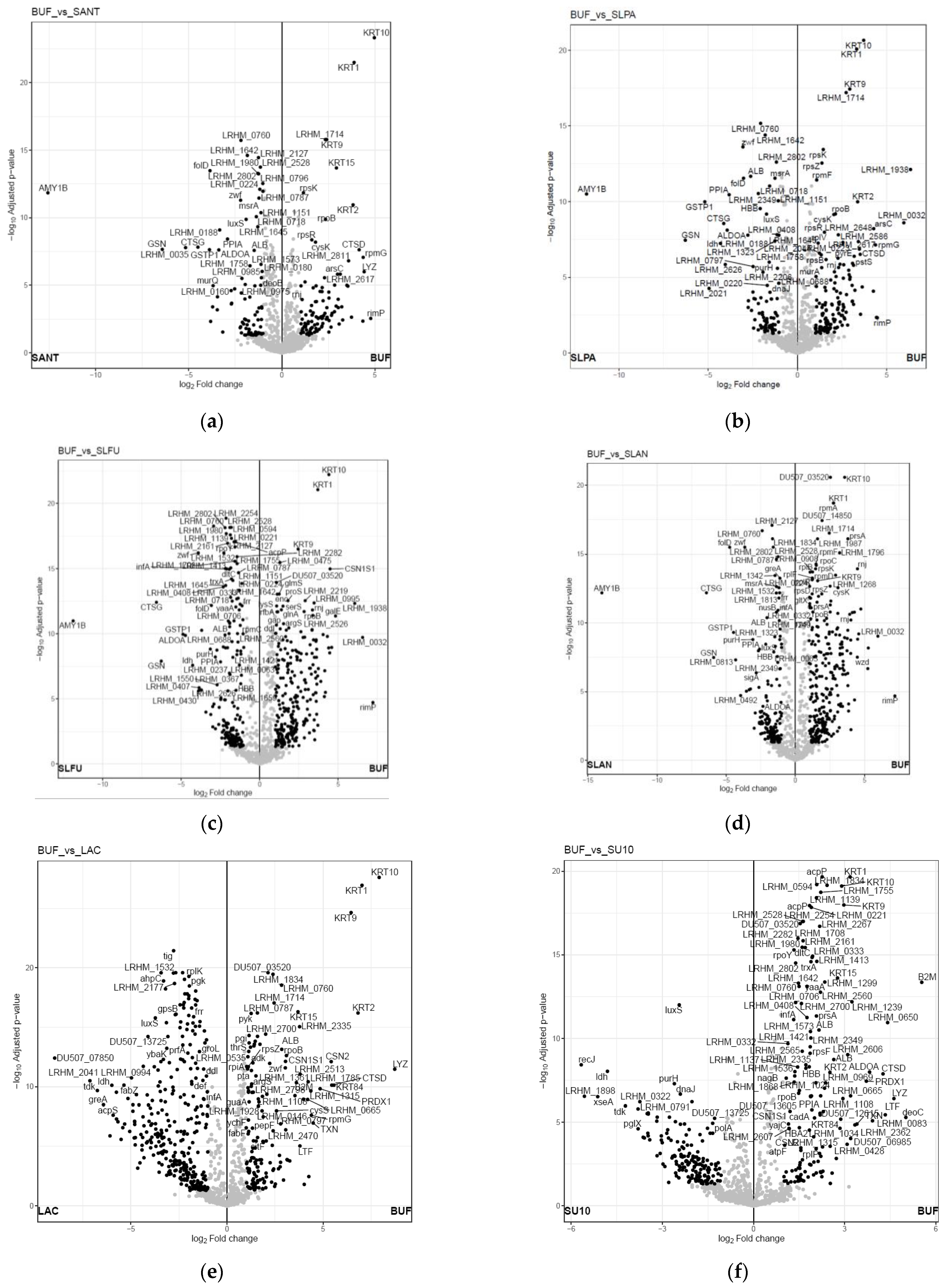
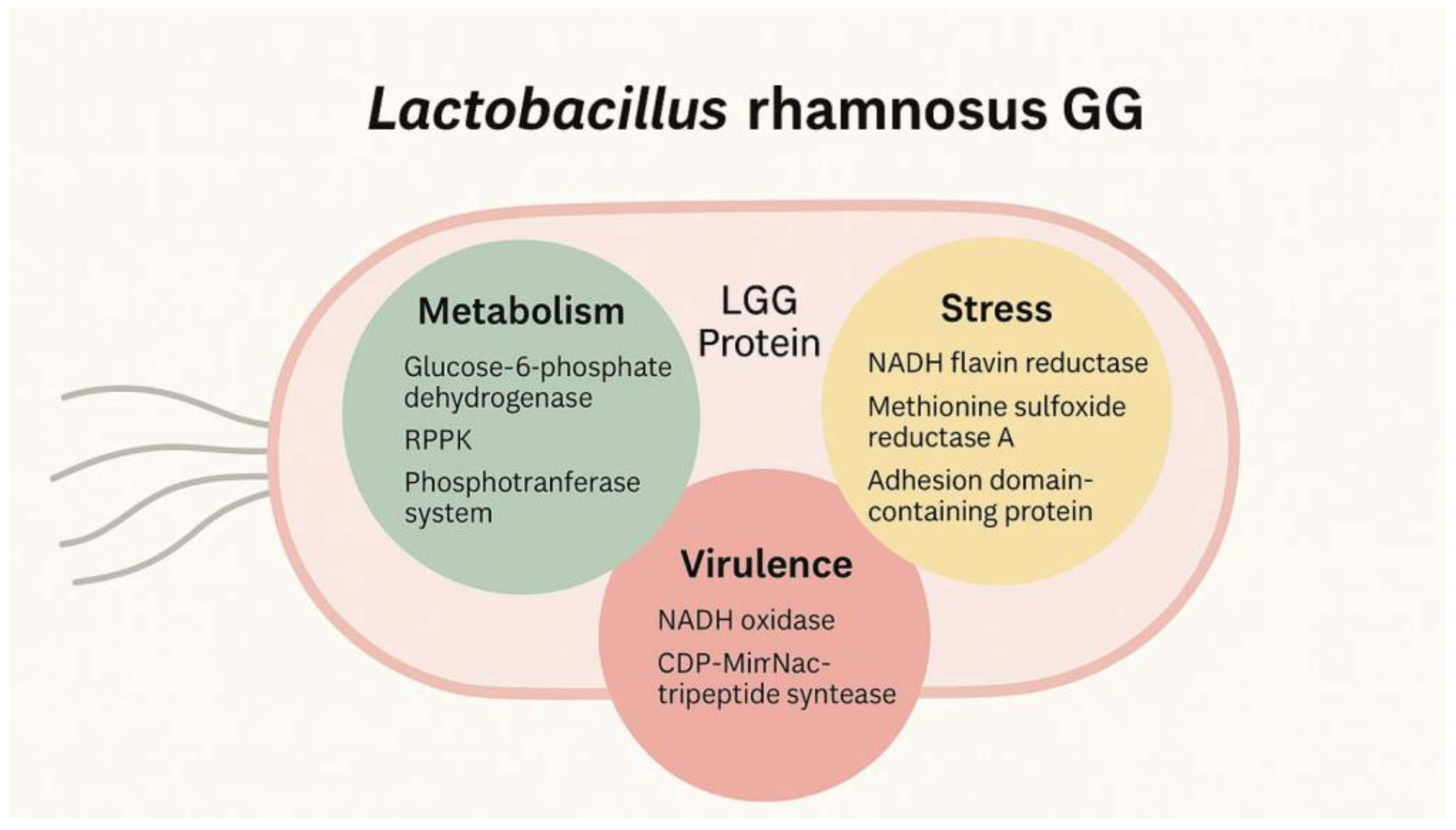
3. Results and Discussion

According to the results of this research, early detection of dysbiosis and changes in the environmental niche showed that LGG can serve as a diagnostic tool. Volunteers for saliva collection were healthy adult subjects (mean age = 35.2) chosen to consider high ethnic (EUROPE/INDIA) origin, dietary, and lifestyle variability, aiming to provide generalizability of the results as much as possible. Unfortunately, no correlation was found between any of these variables and the obtained data. Figure 1 shows an evident clustering of specific samples in the heatmap, such as BUFR, SLPA, and SLFU, suggesting shared proteomic characteristics. In contrast, other samples, like SANT, SLPO, and SLPA, display unique expression patterns. This latter finding was significant, as it suggests the possibility of using such different expressions as markers for unique characteristics of the subjects, possibly demonstrating the biosensing capability of LGG. Specific proteins exhibited distinct abundance trends, with elevated expression in SLAN and SLFU but lower levels in SANT and SLPO, indicating potential biological or functional differences marked by the protein expression. The overall variability in protein expression across the heatmap highlighted patterns of downregulation and upregulation, which seemed to correspond to active biological pathways. This visual representation enabled the identification of outliers, clustering of related proteins, and comparison of expression levels across samples, providing valuable insights into proteomic relationships and sample-specific characteristics, thereby framing the biosensing capability of LGG.
The volcano plot comparing the control sample (BUF) and the SANT subject sample visually highlighted the downregulation and upregulation of proteins (Figure 2a). Proteins such as LRHM_0760 (Phosphoesterase), LRHM_0642, LRHM_1980, and LRHM_2127 were significantly downregulated in SANT, with high statistical significance. In contrast, proteins like LRHM_2811, LRHM_1714, and LRHM_2617 were upregulated, clustering on the right side. The downregulation of specific proteins in SANT could indicate the activation of certain biological pathways or processes in the subject group, potentially linked to stress responses, inflammation, or metabolic changes, which suggest specific variations in the proteomic expression of LGG in response to saliva exposure. Conversely, upregulated proteins might reflect suppressed metabolic activity or pathway inhibition, possibly due to differences in physiological conditions, sample-specific characteristics, or external stimuli. These findings underscore the proteomic shifts between BUF and SANT, highlighting potential biomarkers or pathways of interest for further investigation regarding the use of LGG as a bacterial biosensor for LRHM. The primary function of bacterial GP-PDE is the hydrolysis of glycerophosphodiesters to produce glycerol 3-phosphate (Gro3P), which is required for several bacterial biosynthetic pathways [23]. Glucose and other sugars are readily available in saliva, so the Phosphodiesterase activity is suppressed to conserve energy [24].
In Figure 2, key proteins, such as LRHM_0760, LRHM_1642 (Tagatose-6-phosphate kinase), and LRHM_2802 (Mannitol-specific phosphotransferase enzyme IIA component) were significantly downregulated in SLPA, with high statistical significance. Conversely, proteins such as LRHM_1938 (protein in stress response) and LRHM_1714 (Hydrolase) were upregulated (Figure 2b). Additionally, proteins such as CSTP1 and CTSG were downregulated, while KRT1 showed notable upregulation in the control group, with strong p-values, reflecting their potential biological significance. Overall, the volcano plot identified key proteins differentially expressed between BUF and SLPA, offering insights into their possible roles in the underlying biological processes. Protein LRHM_760 may be involved in nutrient acquisition or metabolic processes. The downregulation of this protein may occur due to direct inhibition by salivary components or a tactical bacterial response to conserve energy and resources under stressful conditions [25]. LRHM_1642 (cell division protein) might be associated with cell wall synthesis or maintenance [26]. The acidic pH and enzymatic activity of human saliva can alter the bacterial cell environment, leading to the downregulation of less critical proteins for immediate survival. This could involve the repression of genes through regulatory proteins that respond to environmental changes [25]. In Figure 2c, notably, the SLFU comparison led to a high number of proteins being displayed in the volcano plot, both upregulated and downregulated. For example, LRHM_2802, LRHM_2254, LRHM_0760, and LRHM_2528 were in regions indicating strong downregulation in SLFU, suggesting that these proteins may play a critical role in SLFU-specific pathways or responses. Conversely, proteins such as LRHM_2282, LRHM_0475, and LRHM_2219 exhibit upregulation in BUF, indicating increased activity under SLFU conditions. These findings highlight the dynamic modification of LRHM proteins in both situations, providing valuable targets for further investigation into their functional significance. Proteins such as LRHM_2802 (Mannitol-specific phosphotransferase enzyme IIA component) were downregulated. This protein participates in signaling or cellular transport. Salivary components may interfere with the signaling pathways that regulate the expression of this protein. For instance, the interaction between salivary mucins and bacterial surface proteins could result in the inhibition of specific genes, leading to a reduction in the synthesis of protein LRHM_2254, an acetyltransferase. Bacterial chemotaxis, metabolism, DNA replication, and other cellular processes are influenced by protein acetylation [27]. The presence of specific proteins and antimicrobial peptides in human saliva can interfere with metabolic processes, resulting in the downregulation of proteins involved in these processes [28]. In Figure 2d, notably, the significantly downregulated protein in SLPO is LRHM_1642 (cell division protein FtsI). This protein is involved in cell division, and saliva contains various enzymes and antimicrobial peptides that can disrupt bacterial cell division, leading to the downregulation of proteins involved in cell multiplication [29]. Proteins such as LRHM_2617, LRHM_2811, and LRHM_1714 were upregulated, indicating reduced functional activity under SLPO conditions compared to BUF. LRHM_2617 (Hydrolase) is the protein that plays a role in DNA replication and repair. In a control buffer environment, the absence of inhibitory factors in BUF allows for the regular expression of proteins involved in essential cellular processes [30]. Downregulated proteins in SLAN, such as LRHM_2802 (Mannitol-specific phosphotransferase enzyme IIA component), are involved in metabolic processes, and changes in the chemical composition of human saliva can affect metabolic pathways, leading to the downregulation of proteins involved in these processes [31]. LRHM_0908 (ABC transporter substrate-binding component) is involved in substrate transportation, and the lack of substrates in the control buffer leads to the upregulation of these proteins, enhancing active substrate binding [32].
In Figure 2e, the volcano plot identified key differentially expressed proteins between BUF and LAC, offering insights into their possible roles in the underlying biological processes. LRHM_2177 (nucleoid-associated protein) plays a key role in the organization, replication, segregation, repair, and expression of bacterial chromosomes [33]. Recent studies have shown that the bacterial chromosome undergoes significant topological changes in response to stress [34]. Exposure to LAC could enhance the expression of nucleoid-associated proteins, facilitating the reorganization of the bacterial genome in response to environmental changes [35]. LRHM_2041 (3R)-Hydroxymyristoyl-ACP Dehydratase protein acts as a catalyst in Type II fatty acid biosynthesis systems, essential for bacterial membrane formation. The upregulation in LAC might be due to increased fatty acid synthesis needed to maintain membrane integrity under stress conditions [36,37]. In Figure 2f, the volcano plot comparing the control sample (BUF) and the subject sample (SU10) describes proteins such as LRHM_0322 (Beta-galactosidase), which are catalysts of lactose hydrolysis and transfer reactions to produce prebiotics such as galacto-oligosaccharides (GOS), which promote gut health by stimulating beneficial bacterial growth. They also have potential food and pharmaceutical applications [38]. The downregulation in SU10 may be due to specific glucose or other factors regulating protein expression [39,40]. LRHM_0791 (Glycosyltransferase) Glycosyltransferases are a subclass of enzymes that catalyze the synthesis of glycosidic linkages by transferring a sugar residue from a substrate [41]. However, the specific function of this protein remains poorly defined. The downregulation in SU10 may be due to the presence of glucose or other factors that regulate protein expression [39].
The dataset was then organized by grouping proteins by their role into three classes: stress, metabolism, and virulence. The analysis was performed on each class. Metabolism did not show any significant differences, indicating that the differences in protein expression are specific to each pathway and are canceled out when considering all metabolic expressions. The stress cluster unsurprisingly showed a clear and highly significant overexpression in the group treated with lactic acid compared to all others. No significant other differences were highlighted. The virulence cluster showed significant upregulation under lactic acid and sucrose conditions. Then, selected proteins were analyzed for their differential expression among the groups to identify potential biomarkers of bacterial expression.
Figure 3a shows significant variation in G6PD expression among the groups. LAC exhibits the lowest expression, suggesting a minimal need for G6PD under baseline conditions. In contrast, SLFU, SLAN, SANT, and SLPA showed notable upregulation, with SLFU demonstrating the highest levels. This variation implies that oxidative stress and increased demand for reducing power (via NADPH) are prominent in SLFU. This result highlights the role of G6PD in responding to oxidative damage and metabolic shifts, particularly in environments with elevated reactive oxygen species (ROS) or high biosynthetic needs. The findings suggest that G6PD may be a marker for the intensity of oxidative stress. Glucose-6-phosphate dehydrogenase (G6PD) is a key enzyme in the pentose phosphate pathway, essential for maintaining cellular redox balance and facilitating biosynthesis. G6PD helps maintain the levels of NADPH, which is crucial in combating oxidative stress [41]. Human saliva contains various components that can induce oxidative stress, and an increase in G6PD expression can indicate a response to this stress [42]. Changes in G6PD expression can reflect the overall health and viability of LGG cells under different conditions, making it a useful biomarker for studying the effects of human saliva on these bacteria [43]. In several studies, the G6PD gene was noted as a potential prognostic biomarker for cancers [44] in cancer metabolism and may be a potential therapeutic target gene for tumor therapy [45]. Another study discusses the role of G6PD in predicting glioma risk and its potential as a biomarker for prognosis and treatment response [46].
In Figure 3b, the expression levels of NADH-flavin reductase are shown. BUFR has the lowest levels, indicating limited activity of flavin-dependent redox reactions in unstressed conditions. On the contrary, LAC shows very high upregulation. SU10 shows intermediate values. SLFU and SLPO exhibit moderate upregulation, indicating a greater reliance on flavin-mediated detoxification and redox balance. This trend suggests that the SLAN, SANT, and sucrose groups experience increased oxidative stress, engaging in metabolic pathways that require increased NADH-flavin reductase activity. The results underscore this enzyme’s role in mitigating oxidative damage and maintaining cellular homeostasis under stressful conditions. NADH-flavin reductase is an enzyme that catalyzes the reduction of flavins, such as flavin mononucleotide (FMN) and flavin adenine dinucleotide (FAD), using NADH as the electron donor [47]. This enzyme is crucial in maintaining redox balance and regulating energy metabolism. When LGG is exposed to different environments, such as lactic acid, buffer, or sucrose, the activity of NADH-flavin reductase can indicate how the bacteria adapt their metabolism to these conditions. It is specifically upregulated in a Lactic acid environment, as described by Corcoran et al. in 2005, LGG is intrinsically resistant to lactic acid, and it needs glucose for survival in the gut environment. The survival of probiotic lactobacilli in acidic environments is enhanced in the presence of metabolizable sugars [48,49]. Changes in the expression or activity of NADH-flavin reductase can reflect the overall health and viability of LGG cells under various environmental stresses, making it a useful biomarker.
In Figure 3c, MsrA (peptide methionine sulfoxide reductase) expression was lowest in sucrose (SU10), moderately upregulated in SLPO, and highest in LAC and SLFU. The observed variance suggests that ROS levels are low and oxidative damage to methionine residues is minimal in BUF but escalates in SLAN, SANT, and SLPA. The pronounced upregulation in SLFU suggests that this situation entails higher oxidative stress, necessitating extensive repair of oxidized methionine residues. These results highlight a critical protective function against ROS-induced protein damage, especially under the stress conditions represented by SLFU and LAC. Peptide methionine sulfoxide reductase is an enzyme that reduces methionine sulfoxide to methionine, repairing the oxidatively damaged proteins. This enzyme is crucial for protecting cells from oxidative stress [50]. This is essential for controlling oxidative stress, particularly in environments like the oral cavity. The protein restores oxidative damage to cells, ensuring they are well functioning. This is crucial for adapting and surviving LGG in various environments, including exposure to human saliva [51]. Changes in the expression or activity of MsrA can demonstrate the general viability and health of LGG cells under various environmental stresses, making it a useful biomarker [52]. In Figure 3d, the PTS System IIA component exhibited distinct expression patterns across LAC, SLFU, SLAN, and SANT. SU10 exhibits the lowest expression level, indicating a reduced need for carbohydrate transport and phosphorylation under normal conditions. SLPA and SLPO exhibit a moderate increase, while SLFU exhibits the highest expression, indicating an increased need for sugar transport and metabolism. This variation implies that SU10 conditions involve either an increased sugar content or increased metabolic activity, requiring efficient carbohydrate uptake. This result also suggests that PTS System IIA is responsive to environmental sugar availability, making it an important regulator of bacterial carbohydrate metabolism. The phosphoenolpyruvate-dependent sugar phosphotransferase system (PTS) is one of the main active carbohydrate transport systems in bacteria. The IIA component is part of the enzyme II complex, which is engaged in the phosphorylation and absorption of sugars. This system is crucial in bacterial metabolism and regulation [53]. The PTS system, including the IIA component, is necessary for sugar transport and phosphorylation. This is particularly important in the oral cavity, where bacteria are exposed to a range of dietary sugars [54]. Changes in the expression or activity of the IIA component can reveal how LGG adapts to different environments, making it a valuable biomarker for studying bacterial responses to human saliva and other conditions. In Figure 3e, RPPK levels were lowest in K (control), and SLAN was markedly upregulated in both BUF and SLPO, with SU10 showing the highest expression. The variation suggested that K and SLAN operate under limited nucleotide synthesis demand, reflecting baseline metabolic activity. SLPO and SU10 implied the increased demand for ribose-phosphate and subsequent nucleotide biosynthesis. This may be driven by infection-related proliferation or stress-induced DNA repair. The results emphasized RPPK’s role in supporting biosynthetic pathways, particularly during cellular adaptation to environmental pressures or stress. Ribose-phosphate pyrophosphokinase (RPPK), also known as phosphoribosyl pyrophosphate synthetase, is an enzyme that catalyzes the conversion of ribose to 5-phosphate, ultimately forming phosphoribosyl pyrophosphate (PRPP). This reaction is crucial for nucleotide biosynthesis, as well as the production of cofactors and certain amino acids [55]. This regulation helps bacteria adapt to various environmental conditions, such as the presence of saliva. RPPK may be elevated in response to increased demand for nucleotide synthesis, such as during the rapid growth of bacteria. This can occur in environments rich in nutrients or when bacteria must replicate and duplicate their DNA, as the graph’s findings indicate that RPPK is upregulated in sucrose [40,56]. Conversely, RPPK may be downregulated when there is less of a requirement to produce nucleotides, such as in nutrient-poor conditions or when bacterial growth is inhibited. This can help conserve energy and resources. In Figure 3f, Cell Shape-Determining Protein (MreB) expression varied notably across BUFR, SLAN, LAC, and SLPA. BUF and K are comparatively at the lowest levels, suggesting cell morphology maintenance operates at a baseline level in unstressed conditions. SANT, LAC, and SLPA exhibited moderate upregulation, while SLPO and SU10 demonstrated the highest expression, indicating increased cellular remodeling and wall synthesis under stress. These results imply that SLPO circumstances severely impair the structural integrity and form of cells, necessitating enhanced MreB activity. The variation highlights MreB’s essential function in maintaining cell structure under environmental and physiological stress. Cell Shape-Determining Protein (MreB) is a protein found in bacteria that is homologous to actin, a cytoskeletal protein in eukaryotes. MreB plays a crucial role in maintaining cell shape, particularly in rod-shaped bacteria, by organizing the cell wall synthesis machinery [57]. MreB may be upregulated in response to conditions that require enhanced cell wall synthesis or repair, such as exposure to stressors that damage the cell wall or in nutrient-rich conditions. This can help maintain cell shape and integrity under adverse conditions.
Currently, little research on bacterial biosensors and proteomics exists. Some studies successfully used proteomics to identify stress response proteins in Escherichia coli-based biosensors. In a study, Alister J. Cumming et al. developed a biosensor capable of detecting cellular stress triggered by inefficient protein secretion from the cytoplasm, as well as protein aggregation in the periplasm [58]. The study showed that the biosensor’s fluorescence fingerprint could be used to identify induction conditions that stay within the cell’s capacity, thereby avoiding stress. These optimized conditions led to improved biomass production and, in some cases, higher yields of soluble recombinant proteins [58].
Similarly, research on Lactobacillus strains has demonstrated their ability to modulate protein expression in response to environmental stressors [57,58]. According to the results of this work, LGG can serve as a biosensor for changes in metabolism, virulence and stress levels (Figure 4). Probiotic effector molecules derived from LGG have been identified, including secreted proteins, surface-anchored proteins, polysaccharides, and lipoteichoic acids, which interact with host physiological processes. To the best of our knowledge, no study has been performed in the oral environment. Compared to these studies, the current research on LGG provides novel insights into its potential as an oral biosensor, particularly in detecting bacterial protein expression and pathways that could be related to human oral and systemic health.
In a study by Allen et al. [59], it was shown that certain genes and proteins involved in oxidative stress resistance play a key role in the redox balance mechanisms of Lactobacillus species, as they are directly linked to the cellular signaling pathways activated by reactive oxygen species (ROS). Among the commonly identified oxidative stress-related genes and proteins in Lactobacillus spp. are thioredoxin reductase, heme-dependent catalase, thioredoxin, DNA protection protein, and NAD(P)/FAD-dependent oxidoreductase. Particularly, thioredoxin and thioredoxin reductase—along with the genes that encode them—form what is known as the thioredoxin antioxidant system, which plays a central role in protecting the cell from oxidative damage. However, it is worth noting that only a limited number of Lactobacillus species naturally possess these oxidative stress resistance genes and proteins [59].
The future of using LGG as a biosensor lies in its integration into carriers, such as wearable dental aligners, to enable extended and continuous monitoring. This approach would be beneficial in conditions such as diabetes, cardiovascular disease, and inflammatory disorders, where treatment effectiveness varies widely among individuals. Future directions should also prioritize the optimization of LGG as biosensor designs to improve sensitivity and specificity. Increasing the range of proteomic analysis to include diverse populations and environmental conditions will further validate the diagnostic capabilities of LGG. Moreover, longitudinal studies can assess the long-term efficacy and reliability of LGG-based biosensors in clinical settings. This discovery opens the door for novel applications in preventative and personalized treatment by bridging the gap between oral microbiota research and health diagnostics. The non-invasive nature of LGG-integrated biosensors holds promise for widespread adoption, revolutionizing the early detection and management of oral and systemic health conditions.
The current study was intended to explore the scientific relevance of the underlying hypothesis. To validate and expand upon these preliminary results, further research involving a more extensive and diverse participant pool is necessary. Future studies will aim to include subjects with various systemic health conditions, such as diabetes, periodontitis, and other chronic illnesses, to better evaluate the clinical applicability of the findings.
This study presents several limitations that warrant consideration. Primarily, the limited sample size reduces the statistical power and restricts the generalizability of the findings. Additionally, the absence of control groups comprising individuals with systemic conditions such as diabetes, periodontitis, and obesity limits the scope of comparative analysis. Despite the high cost-effectiveness of the method employed, this research was designed as a pilot study to screen the full spectrum of proteomic expression. One of the primary objectives was to generate a comprehensive dataset that can assist in identifying single sources of variation, which may serve as targets for future customized diagnostic tests. These future tools, potentially in the form of point-of-care devices, could be developed with an emphasis on affordability and rapid turnaround time.

4. Conclusions

This preliminary research showed that LGG can be a viable biosensing agent with relevant implications for early detection of oral and systemic health and disease diagnosis. By leveraging adaptive proteome responses of LGG, the study seemed to indicate its ability to reflect physiological changes and identify disease markers in diverse oral environments. These findings can offer a foundation for incorporating clinical microbiome research applications and enabling the development of real-time, non-invasive diagnostic tools. Essential outcomes from the study consist of the identification of proteins linked to LGG’s metabolic and stress response pathways. These results demonstrate LGG’s potential in monitoring oral health, detecting early signs of systemic conditions, and considering the utilization of LGG as a technological biosensor in diagnostic platforms, facilitating personalized healthcare approaches. In the future, LGG might help manage conditions such as diabetes, cardiovascular diseases, and inflammatory disorders. However, further studies are needed in the literature to confirm this statement.

Author Contributions

Conceptualization, investigation, data curation, writing—original draft preparation: P.M.R., A.C.I., A.S., A.P., P.S., S.P., G.M.T., M.D.F. and F.G.; conceptualization, methodology, software: P.M.R., A.C.I., A.S., A.P., G.M.T., M.D.F. and F.G., validation and formal analysis: P.M.R., A.C.I., A.S., A.P., P.S., S.P., G.M.T., M.D.F. and F.G., investigation: P.M.R., A.C.I., A.S., A.P., G.M.T., M.D.F. and F.G.; resources, supervision, funding acquisition: P.M.R., A.C.I., A.S., A.P., G.M.T., M.D.F. and F.G.; project administration, supervision: P.M.R., A.C.I., A.S., A.P., G.M.T., M.D.F. and F.G.; visualization, writing—review and editing: P.M.R., A.C.I., A.S., A.P., P.S., S.P., G.M.T., M.D.F. and F.G. All authors have read and agreed to the published version of the manuscript.

Funding

The doctoral scholarship was co-funded with resources in the frame of the “PON REACT-EU financing Program” Action IV.5-Doctorates on green topic by Italian Ministry of Health with number RC24/UOC420.

Institutional Review Board Statement

The study was conducted in accordance with the Declaration of Helsinki. The research is a part of ongoing research on transitional studies on salivary biomarkers, and it was approved by Ethical committee of Milano Area 2 on 5/08/ 2022 with Number: 2143.

Informed Consent Statement

Informed consent was obtained from all subjects involved in the study.

Data Availability Statement

Data are available upon request.

Conflicts of Interest

The authors declare no conflicts of interest.

Abbreviations

LGGLactobacillus rhamnosus GG
EcNEscherichia coli Nissle
MMPmatrix metalloproteinase
ILinterleukin
G6PDGlucose-6-phosphate dehydrogenase
RPPKRibose-phosphate pyrophosphokinase
PRPP5-phosphate to phosphoribosyl pyrophosphate
MreBCell Shape-Determining Protein
Gro3PGlycerol 3-phosphate
FMNFlavin mononucleotide

References

  1. Wang, J.; Schipper, H.M.; Velly, A.M.; Mohit, S.; Gornitsky, M. Salivary biomarkers of oxidative stress: A critical review. Free Radic. Biol. Med. 2015, 85, 95–104. [Google Scholar] [CrossRef] [PubMed]
  2. Willis, J.R.; Gabaldón, T. The human oral microbiome in health and disease: From sequences to ecosystems. Microorganisms 2020, 8, 308. [Google Scholar] [CrossRef] [PubMed]
  3. Turnbaugh, P.J.; Ley, R.E.; Hamady, M.; Fraser-Liggett, C.M.; Knight, R.; Gordon, J.I. The human microbiome project. Nature 2007, 449, 804–810. [Google Scholar] [CrossRef]
  4. Zaura, E.; Keijser, B.J.F.; Huse, S.M.; Crielaard, W. Defining the healthy “core microbiome” of oral microbial communities. BMC Microbiol. 2009, 9, 259. [Google Scholar] [CrossRef] [PubMed]
  5. Belstrøm, D.; Jersie-Christensen, R.R.; Lyon, D.; Damgaard, C.; Jensen, L.J.; Holmstrup, P.; Olsen, J.V. Metaproteomics of saliva identifies human protein markers specific for individuals with periodontitis and dental caries compared to orally healthy controls. PeerJ 2016, 4, e2433. [Google Scholar] [CrossRef]
  6. Börner, R.A.; Kandasamy, V.; Axelsen, A.M.; Nielsen, A.T.; Bosma, E.F. Genome editing of lactic acid bacteria: Opportunities for food, feed, pharma, and biotech. FEMS Microbiol. Lett. 2018, 366, fny291. [Google Scholar] [CrossRef]
  7. Sonnenborn, U. Escherichia coli strain Nissle 1917—From bench to bedside and back: History of a special Escherichia coli strain with probiotic properties. FEMS Microbiol. Lett. 2016, 363, fnw212. [Google Scholar] [CrossRef]
  8. Schultz, M. Clinical use of E. coli Nissle 1917 in inflammatory bowel disease. Inflamm. Bowel Dis. 2008, 14, 1012–1018. [Google Scholar] [CrossRef]
  9. Gleerup, H.S.; Hasselbalch, S.G.; Simonsen, A.H. Biomarkers for Alzheimer’s disease in saliva: A systematic review. Dis. Markers 2019, 2019, 4761054. [Google Scholar] [CrossRef]
  10. Sancesario, G.; Bernardini, S. AD biomarker discovery in CSF and alternative matrices. Clin. Biochem. 2019, 72, 52–57. [Google Scholar] [CrossRef]
  11. Schepici, G.; Silvestro, S.; Trubiani, O.; Bramanti, P.; Mazzon, E. Salivary biomarkers: Future approaches for early diagnosis of neurodegenerative diseases. Brain Sci. 2020, 10, 245. [Google Scholar] [CrossRef] [PubMed]
  12. Malon, R.S.; Sadir, S.; Balakrishnan, M.; Córcoles, E.P. Saliva-based biosensors: Noninvasive monitoring tool for clinical diagnostics. Biomed. Res. Int. 2014, 2014, 962903. [Google Scholar] [CrossRef] [PubMed]
  13. Huynh-Ba, G.; Kuonen, P.; Hofer, D.; Schmid, J.; Lang, N.P.; Salvi, G.E. The effect of periodontal therapy on the survival rate and incidence of complications of multirooted teeth with furcation involvement after an observation period of at least 5 years: A systematic review. J. Clin. Perio 2009, 36, 164–176. [Google Scholar] [CrossRef]
  14. Liotta, L.; Petricoin, E. Molecular profiling of human cancer. Nat. Rev. Genet. 2000, 1, 48–56. [Google Scholar] [CrossRef]
  15. Ideker, T.; Thorsson, V.; Ranish, J.A.; Christmas, R.; Buhler, J.; Eng, J.K.; Bumgarner, R.; Goodlett, D.R.; Aebersold, R.; Hood, L. Integrated genomic and proteomic analyses of a systematically perturbed metabolic network. Science 2001, 292, 929–934. [Google Scholar] [CrossRef]
  16. Adamu, J.Y.; Wawegama, N.K.; Browning, G.F.; Markham, P.F. Membrane proteins of Mycoplasma bovis and their role in pathogenesis. Res. Vet. Sci. 2013, 95, 321–325. [Google Scholar] [CrossRef]
  17. Mora-Montes, H.M. A perspective on the role of proteins and peptides in the virulence and pathogenesis. Curr. Protein Pept. Sci. 2019, 20, 960–961. [Google Scholar] [CrossRef] [PubMed]
  18. Wang, Q.; Yu, Q.; Lin, Q.; Duan, Y. Emerging salivary biomarkers by mass spectrometry. Clin. Chim. Acta 2015, 438, 214–221. [Google Scholar] [CrossRef]
  19. Tuomola, E.M.; Ouwehand, A.C.; Salminen, S.J. The effect of probiotic bacteria on the adhesion of pathogens to human intestinal mucus. FEMS Immunol. Med. Microbiol. 1999, 26, 137–142. [Google Scholar] [CrossRef]
  20. Lebeer, S. Molecular Study of Adaptation and Probiotic Factors in Lactobacillus Rhamnosus GG. Ph.D. Thesis, Faculty of Bio-engineering Sciences, KU Leuven, Leuven, Belgium, 2008. [Google Scholar]
  21. Yan, F.; Cao, H.; Cover, T.L.; Washington, M.K.; Shi, Y.; Liu, L.; Chaturvedi, R.; Peek, R.M.; Wilson, K.T.; Polk, D.B. Colon-specific delivery of a probiotic-derived soluble protein ameliorates intestinal inflammation in mice through an EGFR-dependent mechanism. J. Clin. Investig. 2011, 121, 2242–2253. [Google Scholar] [CrossRef]
  22. Ionescu, A.; Brambilla, E.; Hahnel, S. Does recharging dental restorative materials with fluoride influence biofilm formation? Dent. Mater. 2019, 35, 1450–1463. [Google Scholar] [CrossRef] [PubMed]
  23. Corda, D.; Mosca, M.G.; Ohshima, N.; Grauso, L.; Yanaka, N.; Mariggiò, S. The emerging physiological roles of the glycerophosphodiesterase family. FEBS J. 2014, 281, 998–1016. [Google Scholar] [CrossRef] [PubMed]
  24. Yamamoto, K.; Hiraishi, M.; Haneoka, M.; Fujinaka, H.; Yano, Y. Protease inhibitor concentrations in the saliva of individuals experiencing oral dryness. BMC Oral Health 2021, 21, 661. [Google Scholar] [CrossRef]
  25. Akula, S.; Welinder, C.; Fu, Z.; Olsson, A.K.; Hellman, L. Identification of the Major Protein Components of Human and Cow Saliva. Int. J. Mol. Sci. 2023, 24, 16838. [Google Scholar] [CrossRef]
  26. Harry, E.; Monahan, L.; Thompson, L. Bacterial cell division: The mechanism and its precision. Int. Rev. Cytol. 2005, 253, 27–94. [Google Scholar] [CrossRef]
  27. Ren, J.; Sang, Y.; Lu, J.; Yao, Y. Protein acetylation and its role in bacterial virulence. Trends Microbiol 2017, 25, 768–779. [Google Scholar] [CrossRef] [PubMed]
  28. Proctor, G.B.; Carpenter, G. The function of salivary proteins and the regulation of their secretion by salivary glands. Biomed. Rev. 1998, 9, 3–15. [Google Scholar] [CrossRef]
  29. Proctor, G.B.; Carpenter, G.H. Regulation of salivary gland function by autonomic nerves. Auton. Neurosci. 2007, 133, 3–18. [Google Scholar] [CrossRef]
  30. Ronner, P. DNA Repair and Therapy of Cancer. In Netter’s Essential Biochemistry; Elsevier: Philadelphia, PA, USA, 2018; Volume 1, pp. 10–12. ISBN 978-1-929007-63-9. [Google Scholar]
  31. Schwerdt, G.; Schulz, M.C.; Kopf, M.; Mildenberger, S.; Reime, S.; Gekle, M. Physiological regulation of oral saliva ion composition and flow rate are not coupled in healthy humans-Partial revision of our current knowledge required. Pflug. Arch. 2025, 477, 55–65. [Google Scholar] [CrossRef] [PubMed] [PubMed Central]
  32. Tanaka, K.J.; Song, S.; Mason, K.; Pinkett, H.W. Selective substrate uptake: The role of ATP-binding cassette (ABC) importers in pathogenesis. Biochim. Biophys. Acta Biomembr. 2018, 1860, 868–877. [Google Scholar] [CrossRef]
  33. Browning, D.F.; Grainger, D.C.; Busby, S.J. Effects of nucleoid-associated proteins on bacterial chromosome structure and gene expression. Curr. Opin. Microbiol. 2010, 13, 773–780. [Google Scholar] [CrossRef] [PubMed]
  34. Hołówka, J.; Zakrzewska-Czerwińska, J. Nucleoid-associated proteins: The small organizers that help to cope with stress. Front. Microbiol. 2020, 11, 590. [Google Scholar] [CrossRef] [PubMed]
  35. Kimata, K.; Takahashi, H.; Inada, T.; Postma, P.; Aiba, H. cAMP receptor protein-cAMP plays a crucial role in glucose-lactose diauxie by activating the major glucose transporter gene in Escherichia coli. Proc. Natl. Acad. Sci. USA 1997, 94, 12914–12919. [Google Scholar] [CrossRef] [PubMed]
  36. De Paepe, B.; De Mey, M. Biological Switches: Past and Future Milestones of Transcription Factor-Based Biosensors. ACS Synth. Biol. 2025, 14, 72–86. [Google Scholar] [CrossRef]
  37. Williams, P.; Bainton, N.J.; Swift, S.; Chhabra, S.R.; Winson, M.K.; Stewart, G.S.; Salmond, G.P.; Bycroft, B.W. Small molecule-mediated density-dependent control of gene expression in prokaryotes: Bioluminescence and the biosynthesis of carbapenem antibiotics. FEMS Microbiol. Lett. 1992, 100, 161–167. [Google Scholar] [CrossRef]
  38. Kimber, M.S.; Martin, F.; Lu, Y.; Houston, S.; Vedadi, M.; Dharamsi, A.; Fiebig, K.M.; Schmid, M.; Rock, C.O. The structure of (3R)-hydroxyacyl-acyl carrier protein dehydratase (FabZ) from Pseudomonas aeruginosa. J. Biol. Chem. 2004, 279, 52593–52602. [Google Scholar] [CrossRef]
  39. Foster, J.M.; Davis, P.J.; Raverdy, S.; Sibley, M.H.; Raleigh, E.A.; Kumar, S.; Carlow, C.K. Evolution of bacterial phosphoglycerate mutases: Non-homologous isofunctional enzymes undergoing gene losses, gains and lateral transfers. PLoS ONE 2010, 5, e13576. [Google Scholar] [CrossRef]
  40. Zhou, W.; Tsai, A.; Dattmore, D.A.; Stives, D.P.; Chitrakar, I.; D’alessandro, A.M.; Patil, S.; Hicks, K.A.; French, J.B. Crystal structure of E. coli PRPP synthetase. BMC Struct. Biol. 2019, 19, 1–7. [Google Scholar] [CrossRef]
  41. Schmid, J.; Heider, D.; Wendel, N.J.; Sperl, N.; Sieber, V. Bacterial glycosyltransferases: Challenges and opportunities of a highly diverse enzyme class toward tailoring natural products. Front. Microbiol. 2016, 18, 182. [Google Scholar] [CrossRef]
  42. Wang, D.; Kompaniiets, D.; Hu, Y.; Liu, B. Transcription and its regulation in bacteria. Front. Microbiol. 2023, 14, 1200443. [Google Scholar] [CrossRef]
  43. Khan, S.R.; Banerjee-Bhatnagar, N. Loss of catabolite repression function of HPr, the phosphocarrier protein of the bacterial phosphotransferase system, affects expression of the cry4A toxin gene in Bacillus thuringiensis subsp. israelensis. J. Bacteriol. 2002, 184, 5410–5417. [Google Scholar] [CrossRef]
  44. Zhang, Y.; Bao, C.; Shen, L.; Tian, C.; Zang, X.; Chen, G.; Zhang, S. Microbial cold shock proteins: Overview of their function and mechanism of action. Protein Pept. Lett. 2022, 29, 133–142. [Google Scholar] [CrossRef]
  45. Smetana, J.H.C. Introduction to biomolecular structure and biophysics. In Principles of Protein Structure and Function; Misra, G., Ed.; Springer: Singapore, 2017; pp. 1–33. [Google Scholar]
  46. Surette, M.G.; Bassler, B.L. Quorum sensing in Escherichia coli and Salmonella typhimurium. Proc. Natl. Acad. Sci. USA 1998, 95, 7046–7050. [Google Scholar] [CrossRef] [PubMed]
  47. Hertzberger, R.; Arents, J.; Dekker, H.L.; Pridmore, R.D.; Gysler, C.; Kleerebezem, M.; de Mattos, M.J.T. H2O2 production in species of the Lactobacillus acidophilus group: A central role for a novel NADH-dependent flavin reductase. Appl. Environ. Microbiol. 2014, 80, 2225–2234. [Google Scholar] [CrossRef] [PubMed]
  48. Corcoran, B.M.; Stanton, C.; Fitzgerald, G.F.; Ross, R.P. Survival of probiotic lactobacilli in acidic environments is enhanced in the presence of metabolizable sugars. Appl Environ Microbiol. 2005, 71, 3060–3067. [Google Scholar] [CrossRef] [PubMed] [PubMed Central]
  49. Lee, J.K.; Zhao, H. Identification and characterization of the flavin: NADH reductase (PrnF) involved in a novel two-component arylamine oxygenase. J. Bacteriol. 2007, 189, 8556–8563. [Google Scholar] [CrossRef]
  50. Oien, D.B.; Moskovitz, J. Genetic regulation of longevity and age-associated diseases through the methionine sulfoxide reductase system. Biochim. Biophys. Acta Mol. Basis Dis. 2019, 1865, 1756–1762. [Google Scholar] [CrossRef]
  51. Jalal, N.; Lee, S.F. The MsrAB reducing pathway of Streptococcus gordonii is needed for oxidative stress tolerance, biofilm formation, and oral colonization in mice. PLoS ONE 2020, 15, e0229375. [Google Scholar] [CrossRef]
  52. Weissbach, H.; Etienne, F.; Hoshi, T.; Heinemann, S.H.; Lowther, W.T.; Matthews, B.; John, G.S.; Nathan, C.; Brot, N. Peptide methionine sulfoxide reductase: Structure, mechanism of action, and biological function. Arch. Biochem. Biophys. 2002, 397, 172–178. [Google Scholar] [CrossRef]
  53. Kok, M.; Bron, G.; Erni, B.; Mukhija, S. Effect of enzyme I of the bacterial phosphoenolpyruvate: Sugar phosphotransferase system (PTS) on virulence in a murine model. Microbiology 2003, 149 Pt 9, 2645–2652. [Google Scholar] [CrossRef]
  54. Vadeboncoeur, C.; Pelletier, M. The phosphoenolpyruvate: Sugar phosphotransferase system of oral streptococci and its role in the control of sugar metabolism. FEMS Microbiol. Rev. 1997, 19, 187–207. [Google Scholar] [CrossRef] [PubMed]
  55. Su, T.; Fellers, R.T.; Greer, J.B.; LeDuc, R.D.; Thomas, P.M.; Kelleher, N.L. Proteoform-predictor: Increasing the Phylogenetic Reach of Top-Down Proteomics. J. Proteome Res. 2025, 24, 1861–1870. [Google Scholar] [CrossRef] [PubMed]
  56. Carballido-López, R. The bacterial actin-like cytoskeleton. Microbiol. Mol. Biol. Rev. 2006, 70, 888–909. [Google Scholar] [CrossRef] [PubMed]
  57. Hartmann, F.S.F. Utilizing Genetically Encoded Biosensors for Monitoring Microbial Stress at Different Scales. Ph.D. Thesis, Technical University of Denmark, Department of Biotechnology and Biomedicine, Lyngby, Denmark, 2022. [Google Scholar]
  58. Cumming, A.J.; Khananisho, D.; Balka, M.; Liljestrand, N.; Daley, D.O. Biosensor that detects stress caused by periplasmic proteins. ACS Synth. Biol. 2024, 13, 1477–1491. [Google Scholar] [CrossRef]
  59. Allen, R.G.; Tresini, M. Oxidative stress and gene regulation. Free Radic. Biol. Med. 2000, 28, 463–499. [Google Scholar] [CrossRef]
Figure 1. The heatmap provides an overview of proteomic data across the 10 groups, represented in columns, with each group in triplicate. The rows represent individual protein candidates for markers, while the columns correspond to the samples, with color gradients indicating the relative intensity of protein expression. Note: for an explanation of the abbreviations, please refer to the text in the Data Analysis section.
Applsci 15 04809 g001
Figure 2. In volcano plots, the x-axis represents the log2 fold change, indicating the magnitude of change in protein expression. Positive values on the right indicate upregulation in the control, while negative values on the left indicate downregulation in the experimental subjects. The y-axis represents the −log10 adjusted p-value, where higher values signify higher statistical significance. Proteins with a black dot have significantly different expression, while proteins marked with a grey dot are not significant. (a) The volcano plot comparing control (BUF) and subject (SANT) groups. (b) The volcano plot comparing control (BUF) and subject (SLPA) groups. (c) The volcano plot comparing control (BUF) and subject (SLFU) groups. (d) The volcano plot comparing control (BUF) and subject (SLAN) groups. (e) The volcano plot comparing control (BUF) and (LAC) groups. (f) The volcano plot comparing control (BUF) and (SU10) groups.
Applsci 15 04809 g002
Figure 3. Expression levels of the selected protein in LGG are measured in relative expression units. Whiskers indicate means + 1 standard deviation. Significant group differences are represented as visually separated circles (Tukey’s test, p < 0.05). (a) Glucose-6-phosphate 1-dehydrogenase (G6PD), (b) NADH-flavin reductase, (c) peptide methionine sulfoxide reductase (MsrA), (d) PTS System IIA Component, (e) Ribose-phosphate pyrophosphokinase (RPPK), (f) Adhesin Domain-Containing Protein.
Applsci 15 04809 g003
Figure 4. LGG’s biomarkers in metabolism, virulence, and stress conditions.
Applsci 15 04809 g004
Disclaimer/Publisher’s Note: The statements, opinions and data contained in all publications are solely those of the individual author(s) and contributor(s) and not of MDPI and/or the editor(s). MDPI and/or the editor(s) disclaim responsibility for any injury to people or property resulting from any ideas, methods, instructions or products referred to in the content.

