Article

Comparison of Feature Vector Compositions to Enhance the Performance of NIRS-BCI-Triggered Robotic Hand Orthosis for Post-Stroke Motor Recovery

1
Department of Mechanical Engineering, Faculty of Engineering, Kyushu University, Fukuoka 819-0395, Japan
2
Department of Neurosurgery, Graduate School of Medical Sciences, Kyushu University, Fukuoka 812-8582, Japan
3
Kitakyushu Chuo Hospital, Kitakyushu, Fukuoka 802-0084, Japan
*
Author to whom correspondence should be addressed.
Appl. Sci. 2019, 9(18), 3845; https://doi.org/10.3390/app9183845
Submission received: 21 July 2019 / Revised: 31 August 2019 / Accepted: 6 September 2019 / Published: 13 September 2019

Abstract

:
Recently, brain–computer interfaces, combined with feedback systems and goal-oriented training, have been investigated for their capacity to promote functional recovery after stroke. Accordingly, we developed a brain–computer interface-triggered robotic hand orthosis that assists hand-closing and hand-opening for post-stroke patients without sufficient motor output. In this system, near-infrared spectroscopy is used to monitor the affected motor cortex, and a linear discriminant analysis-based binary classifier estimates hand posture. The estimated posture then wirelessly triggers the robotic hand orthosis. For better performance of the brain–computer interface, we tested feature windows of different lengths and varying feature vector compositions with motor execution data from seven neurologically intact participants. The interaction between a feature window and a delay in the hemodynamic response significantly affected both classification accuracy (Matthew Correlation Coefficient) and detection latency. The ‘preserving channels’ feature vector was able to increase accuracy by 13.14% and decrease latency by 29.48%, relative to averaging. Oxyhemoglobin combined with deoxyhemoglobin improved accuracy by 3.71% and decreased latency by 6.01% relative to oxyhemoglobin alone. Thus, the best classification performance resulted in an accuracy of 0.7154 and a latency of 2.8515 s. The hand rehabilitation system was successfully implemented using this feature vector composition, which yielded better classification performance.

1. Introduction

Clinical demand is growing for a new neurorehabilitation strategy in which post-stroke patients with insufficient or no remaining hand motor function can participate. Currently, these patients cannot benefit from Active Movement Therapy (AMT) [1], one of the widely applied rehabilitation therapies, because the level of remaining motor output in their affected hands is insufficient. Although passive movement therapy is an alternative option, it can only bring minimal motor function recovery [2]. Therefore, roughly one-third of post-stroke patients with motor impairments [3] cannot expect much functional recovery from current rehabilitation strategies.
Incorporating a brain–computer interface (BCI) [4] into neurorehabilitation has been recognized and investigated as a promising solution for post-stroke patients with complete or almost complete paralysis [5]. A BCI, integrated with a motion-assistive device, enables patients to execute motor intention-induced movements that resemble active movements. Thus, patients can participate in effective AMTs and expect motor function recovery. Indeed, several clinical studies have reported that BCI-combined neurorehabilitation improves stroke-impaired motor function [6].
Near-infrared spectroscopy (NIRS) is a new non-invasive neuroimaging technique [7] that relies on the hemodynamic response to local neuronal activities. NIRS measures real-time changes in oxyhemoglobin (HbO) and deoxyhemoglobin (HbR) concentrations ([HbO] and [HbR] respectively) in local cortical areas. The background principle of NIRS is based on neurovascular coupling [8], which is the interaction between local neuronal activity and local changes in Cerebral Blood Flow (CBF); local CBF increases to meet the metabolic demand of local neuronal activity. The absorption or reflection of near-infrared light by hemoglobin depends on the amount of hemoglobin combined with oxygen in that local area [9]. Accordingly, NIRS can monitor the hemodynamic response. This resultant hemodynamic response is slower than its causing neuronal activity and takes place with a delay on the order, of seconds.
A NIRS-Brain–Computer Interface (NIRS-BCI) is a hemodynamic response-based BCI using NIRS as a neuroimaging modality. Hemodynamic responses captured by NIRS are relatively easier to detect and analyze, and more robust against noise than electrical responses captured by electroencephalography (EEG), which is currently the most popular BCI modality. Thus, several studies have successfully introduced NIRS into BCI systems for motor tasks, resulting in classification accuracies ranging from 65% to 95.5% [10].
The drawback of NIRS-BCI is a longer detection latency of motor intention on the order, of seconds, which results from the slow nature of hemodynamic response. Compared with EEG-BCI having a detection latency of a few hundred milliseconds, communication and control using NIRS-BCI can be limited [11]. However, for neurorehabilitation purposes, the longer detection latency of NIRS-BCI might be acceptable. For example, a study showed that real-time NIRS-controlled neurofeedback enhanced motor imagery (MI) performance [12]. Thus, we can infer that a NIRS-BCI system with a longer detection latency evokes a rehabilitative effect, although further assessment is required.
Aside from its classification accuracy and slower but potentially acceptable detection latency, NIRS-BCI has certain advantages over EEG-BCI. NIRS offers comparatively better spatial resolution than EEG. The temporal resolution of NIRS is sufficient for capturing task-induced hemodynamic responses of 0.1 Hz [10]. NIRS is less affected by motion artifacts and eye movements than EEG [7]. Positioning NIRS optodes (optical probe) is easier than positioning EEG electrodes, and NIRS optodes do not require the use of gel between the scalp and optodes, which might be preferable by both therapists and patients. Modulating hemodynamic responses via NIRS biofeedback requires less training time than controlling electrical responses for an EEG-BCI, which can span 4 to 9 repetitions of a 40 min session [11]. These features can make NIRS-BCI more user-friendly and preferable for daily use in rehabilitation.
To meet this demand, we developed a hand rehabilitation system comprising a robotic hand orthosis that is wirelessly triggered by a multichannel-NIRS-BCI. We evaluated system performance with seven neurologically healthy participants. The evaluation focused on how successfully the NIRS-BCI detected motor intention. The delay in hemodynamic response while training a classifier, the length of the feature window, and the method of feature vector composition determined the performance of each NIRS-BCI module. Feature vectors embraced preserving the NIRS channels, using concentrations of the two different types of hemoglobin types ([HbO] and [HbR]), and selecting NIRS channels. Accordingly, we tested and evaluated those conditions to attain the best performance of the NIRS-BCI by comparing them in terms of classification accuracy and detection latency. The evaluation was conducted in a real-time manner, in which moving feature windows were applied to the recorded data. A moving feature window was formed with every sampling timing. This is because our system should detect motor intention in a real-time manner during rehabilitation practice. The hand rehabilitation system presented here is a clinical testing-ready version that has been upgraded from our previously tested prototype, and some of the data has been presented in [13]. In the current study, we have improved upon the prototype system by evaluating the different window lengths and feature vector compositions. This has substantially improved classification performance in terms of accuracy and detection latency.
Thus, we here demonstrate the implementation of our NIRS-BCI-triggered hand rehabilitation system that integrates a NIRS with our previously developed robotic hand orthosis [14]. We have evaluated how the following items have affected the performance (accuracy and detection latency) of the NIRS-BCI module: (1) different hemodynamic delays and lengths of the feature window, and (2) different feature vector compositions. The feature vector compositions that we tested were: (a) ‘preserving channels’ vs. ‘averaging’, (b) using [HbO] together with [HbR] vs. [HbO] or [HbR] separately, and (c) using all channels vs. criterion-selected channels. The purpose of this study was to describe the system implementation and to discuss the results of the evaluation as we develop our hand rehabilitation system.

2. Materials and Methods

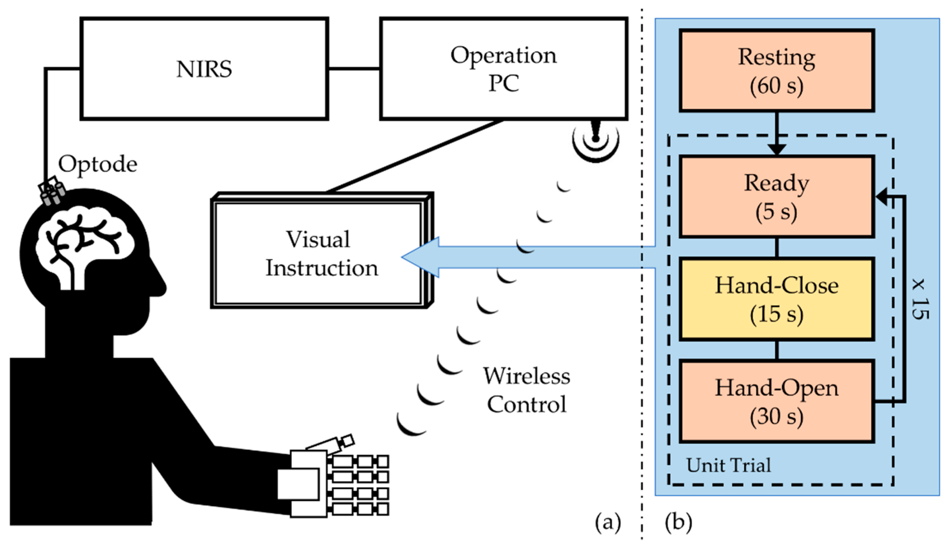
2.1. System and Elements

The hand rehabilitation system consists of a robotic hand orthosis, the NIRS system, and operating personal computer (PC) with a monitor, as shown in Figure 1a. A user, sitting on a comfortable chair in a relaxed manner, performs (or imagines) a hand motion based on the instructions provided on the monitor, located at a 50–60 cm distance from the user’s eyes. The NIRS system measures local cortical activation in real-time and transmits the data to the operating PC. The operating program on the PC processes the data and classifies it into one of two hand motion states: hand-closing and hand-opening. Then, the classification output is wirelessly transmitted to the orthosis. The operation program is coded and implemented with MATLAB®. A system demonstration is available in Video S1 (please see Supplementary Materials).

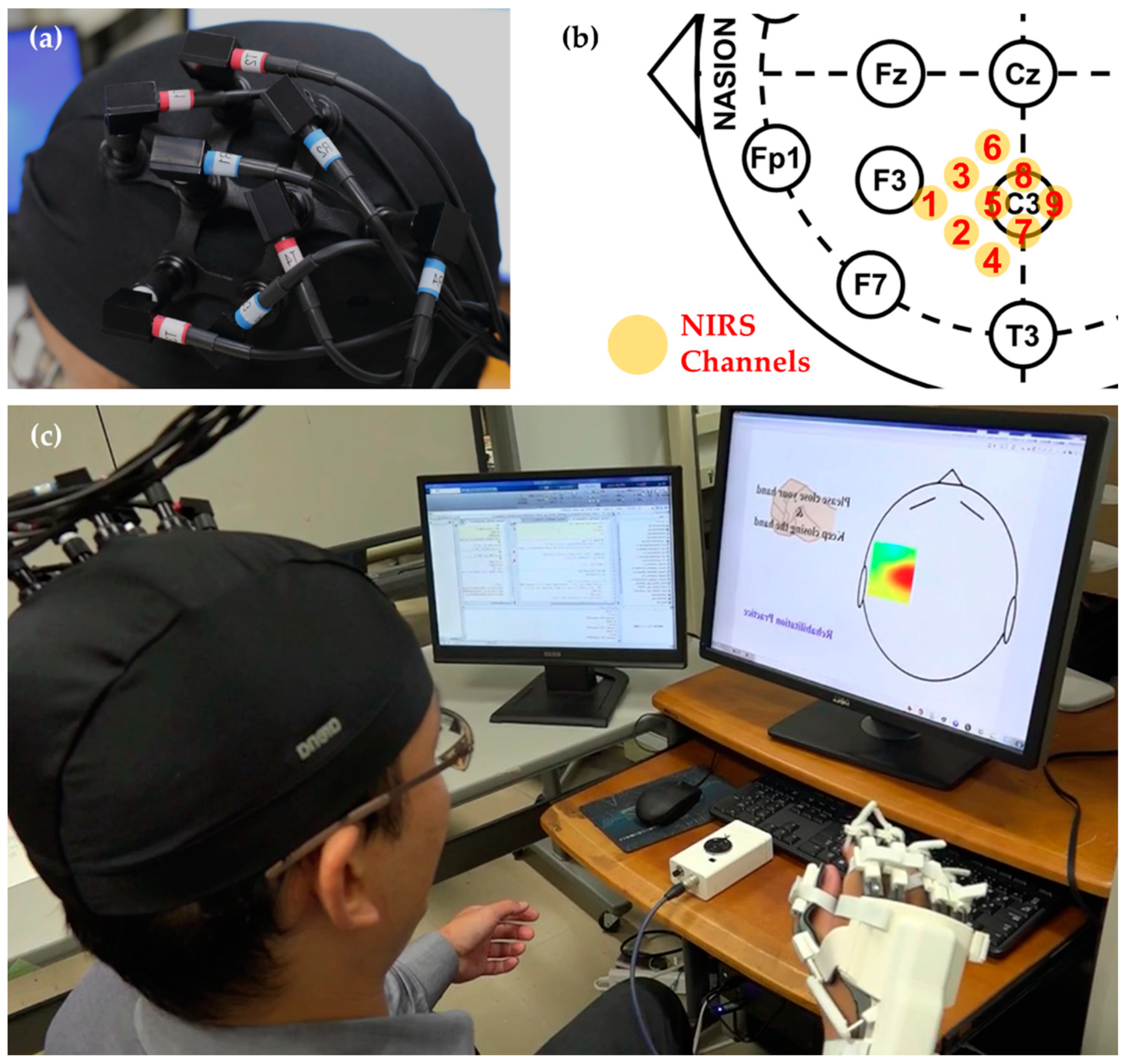
2.1.1. NIRS Measurement

We used the LABNIRS system (Shimadzu Corporation, Kyoto, Japan) to measure variation in [HbO] and [HbR] from local cortical areas. The LABNIRS system applies three wavelengths of near-infrared light (780 nm, 805 nm, and 830 nm) to measure hemoglobin concentrations. The system offers both measured light intensities and hemoglobin concentrations converted simultaneously in real time. We used the [HbO] and [HbR] directly offered by the system.
We used four pairs of NIRS emitting and detecting optodes, resulting in nine measurement channels. The distance between an emitter and a detector was 30 mm, enabling a measurement depth ~20 mm from the scalp. The optodes were positioned on the scalp surface around the motor cortex (C3) according to the international 10–20 system (Figure 2). In particular, emitting optode 4 was on the C3. We combined a Shimadzu FLASH holder kit (Flexible Adjustable Surface Holder) with a swimming cap for more stable fixation of the NIRS optodes and for prevention of the potential effect of ambient light. We set the sampling interval to 120 ms, resulting in the sampling rate of 8.33 Hz. This sampling interval was sufficient, because the hemodynamic responses induced by the motor task occurred at around 0.1 Hz [10].

2.1.2. Robotic Hand Orthosis

For assisting hand motion, we used our robotic hand orthosis [14]. The exoskeleton-type orthosis assists with hand-closing and hand-opening motions, with a force of 10 N and 30 N, respectively. The orthosis consists of the wearable, exoskeletal hand part and an external control box (Figure 2). The exoskeleton is attached to the dorsal side of the hand and covers both the dorsum and the fingertips. A linear actuator on the dorsum actuates all finger components simultaneously in the same direction, which thus enables flexion/extension of all fingers. The control box wirelessly controls the actuator.

2.1.3. Wireless Transmission

A pair of XBee® S1 802.15.4 RF modules offers wireless transmission of control commands from the control box to the wearable portion of the orthosis. There are only two control commands that correspond to the two hand motions (open and close). The default posture is hand-open.

2.2. Experimental Evaluation

2.2.1. Participants

We recruited seven neurologically intact participants for this study (all right-handed males, aged 29.9 ± 5.0 years). No participant had a history of neurological or psychiatric disorders, and none had ever participated in any BCI-related experiment. We obtained informed, written consent from all participants before the experiment. The study was approved by the institutional review boards and ethics committees of Kyushu University Hospital. All procedures were conducted in accordance with the latest version of the Declaration of Helsinki.

2.2.2. Experimental Protocol

All participants completed one session of the motor execution task. In this task, participants closed their right hands during a given period (default posture: open hand). A session consisted of a resting state (60 s) followed by 15 unit trials. A unit trial was a sequence of ready (5 s), hand-closing (15 s), and hand-opening (30 s) periods. Thus, the total length of the session was 810 s (Figure 1b). During the hand-closing period, participants loosely closed their right hands and kept them closed until the next instruction. During resting, ready, and hand-opening periods, they kept their hands opened in a relaxed manner. There was no intended finger hyperextension.

2.2.3. NIRS Channel Selection

We used the Contrast-to-Noise Ratio (CNR) of [Hb] as an indicator of task-induced hemodynamic response. Thus, each channel had two CNR values, one corresponding to [HbO] and the other to [HbR]. The CNR compares the amplitude contrast between hand-closing and hand-opening states. A task-induced response has a CNR value greater than zero for [HbO] and lower for [HbR] as in (1). A larger absolute value means a greater task-induced response. CNRs have been used for selecting NIRS channels containing task-evoked hemodynamic responses [15,16].
CNR = i = 1 15 mean ( [ Hb ] hand close ,   i th ) mean ( [ Hb ] hand open ,   i th ) var ( [ Hb ] hand close ,   i th ) var ( [ Hb ] hand open ,   i th ) ,
The entire hand-closing period (15 s) and the final 15 s of the hand-opening period were used for calculating the CNRs for the respective periods. Here, the CNR values for a channel are the mean CNRs of 15 trials after bandpass filtering.

2.2.4. Preprocessing

For real-time bandpass filtering, we applied a MACD (moving average convergence/divergence) filter (2). The MACD filter is the difference between two EMA (exponential moving average) filters (3) of different parameters. Several NIRS-BCI studies have tested [17] and successfully used MACD for their systems [16,18].
MACD ( x ( t ) ) =   EMA α F ( x ( t ) ) EMA α S ( x ( t ) ) ,
EMA α ( x ( t ) ) = y ( t ) =   α x ( t ) + ( 1 α ) y ( t 1 ) ,
EMAαF is for fast components and EMAαS for slow ones. The applied passband was between 0.01 and 0.2 Hz to remove global ascending trend and the effect of physiological activities, such as respiration, heart-beating, and so on [19]. The corresponding parameters of αF and αS were calculated by (4) [20] (FC = fC/ fS, fC: the cut-off frequency, fS: the sampling frequency).
α = cos ( 2 π F c ) 1 + cos 2 ( 2 π F c ) 4 cos ( 2 π F c ) + 3 ,

2.2.5. Binary Classification

Linear discrimination analysis (LDA) was used to solve the binary classification of the two hand motions. An LDA-classifier finds a line that maximizes the difference between the mean values of two data and minimizes the variance within each individual class belonging to a training data. Then, the classifier applies the line to classify a test datum [21]. LDA has been one of the successfully applied binary classification algorithms for the NIRS-BCI systems [10]. Additionally, our previous work [13] demonstrated that LDA exhibited better classification performance than a support vector machine (SVM). We used MATLAB® Machine Learning Toolbox™ to train and test the LDA-based classifier.

2.2.6. Different Lengths of the Feature Window

The length of the feature window determines the time interval in which the features, mean and slope, are calculated with MATLAB functions, mean and polyfit (set ‘Degree of polynomial fit’ to 1, meaning ‘linear fitting’) respectively. The length of the feature window can affect classification performance. Thus, we investigated how classification results varied depending on the length of the feature window (LFW, in s). We tested nine different feature window lengths (0.6, 1, 3, 5, 7, 9, 11, 13, and 15 s). The longest length, 15 s, corresponds to the length of a hand-closing period (task period). The shorter lengths, except for 0.6 s, were determined by narrowing down the maximum length with a decrement of 2 s. The shortest one, 0.6 s, matches with five data points. Additionally, delay times in the task-induced hemodynamic response (DHR) from 0 to 10 s were introduced, ranging in increments of 1 s. These delays are time gaps between the onset of hand-closing and the visible change in hemoglobin concentrations.

2.2.7. Feature Vector Composition

We assessed the classification performance of 12 feature vector compositions (FCs) (Table 1). The compositions and their dimensions varied depending on the number of NIRS channels and which concentrations ([HbO], [HbR], or both) were incorporated into the feature vector. For calculating the features (mean and slope), we used a 1-s feature window and a zero delay in hemodynamic response (LFW = 1 s, DHR = 0 s). This setup of LFW = 1 s and DHR = 0 s led to the classification accuracy of 0.7586 and the detection latency of 2.4015 s, which were applicable for real-time use.

2.2.8. Classification Evaluation

We performed leave-5-out cross-validation after excluding the resting period. We partitioned the trials into training and test data sets, and the order of the trials in each set was preserved. This was for evaluating the detection latency.
We used two indices: (1) classification accuracy and (2) detection latency. As measures of classification accuracy, we used Balanced Accuracy (BACC) (5) and Matthews Correlation Coefficient (MCC) (6) because our data were class-imbalanced due to the experimental design.
BACC = ( TP TP + FN + TN FP + TN ) × 1 2 ,
MCC = ( TP × TN ) × ( FP × FN ) ( TP + FP ) ( TP + FN ) ( TN + FP ) ( TN + FN ) ,
We rescaled MCC values ([−1, 1]) to [0, 1] so that we could directly compare them with BACC values. The rescaled MCC (MCCRS) was calculated by adding 1 to the MCC and then dividing by 2. Hereafter, we only present the results for MCCRS because both measures showed the same tendency.
A detection latency is the time difference (in s) between the start time of a trial and the classifier-detected start of the motion. We labeled a trial as ‘no-detection’ if its detection latency was longer than the hand-closing period of 15 s. Then, we excluded ‘no-detection’ trials when averaging the detection latencies over all test trials.

3. Results

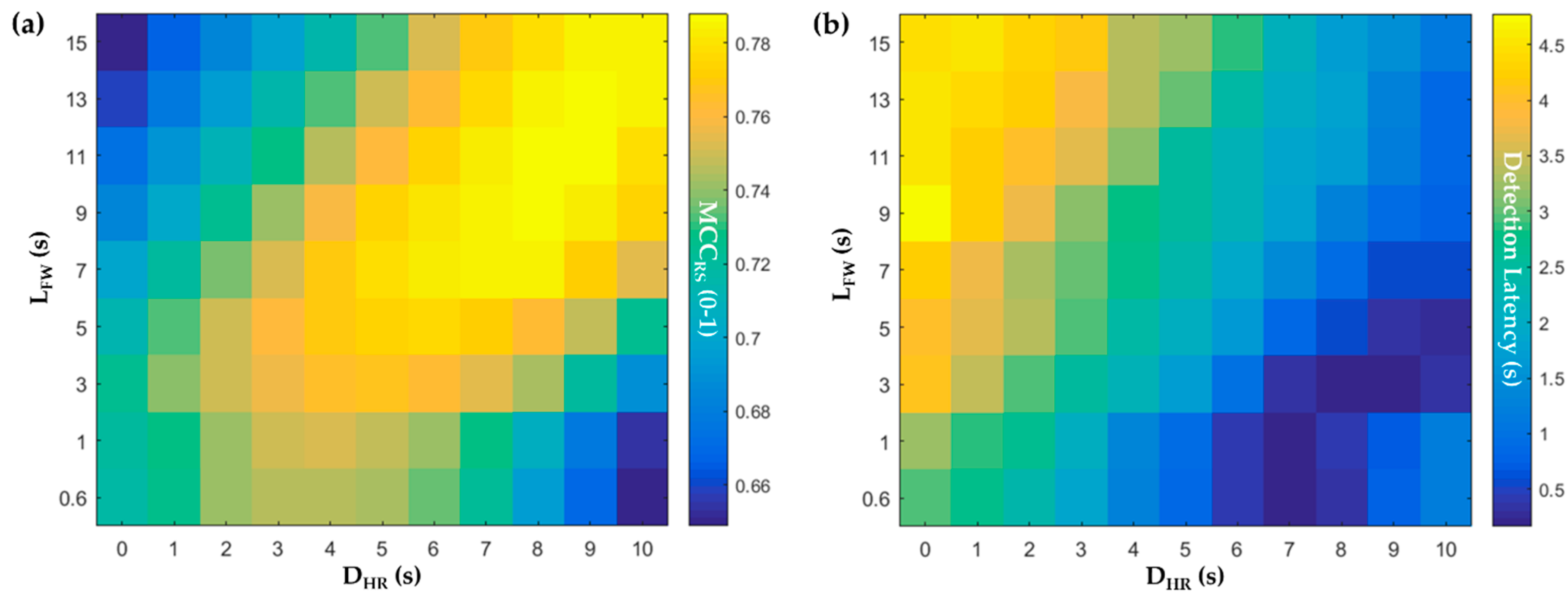
3.1. Classifier Training with Different Delays and Window Lengths

Applying different DHR and LFW affected classification performance. Figure 3 shows the MCCRS and detection latency maps. Depending on the applied DHR and LFW, classification accuracy rose from 0.6489 up to 0.7880 (a 21.42 % increase) in the MCCRS. The DHR and LFW were positively related to each other, as evidenced by the antidiagonal direction of the map. Longer DHR with longer LFW tended to generate better classification accuracy. However, after DHR reached 9 s and LFW reached 13 s, classification accuracy decreased. The combination of long DHR and short LFW, or vice versa resulted in a relatively poor classification accuracy of around 0.65.
Detection latency also depended on the DHR and LFW, ranging between 0.1640 s and 4.7761 s. The detection latency map in Figure 3 was obtained based on the introduced DHR. To obtain the true detection latency value, the corresponding DHR must be added to the value of each pixel.

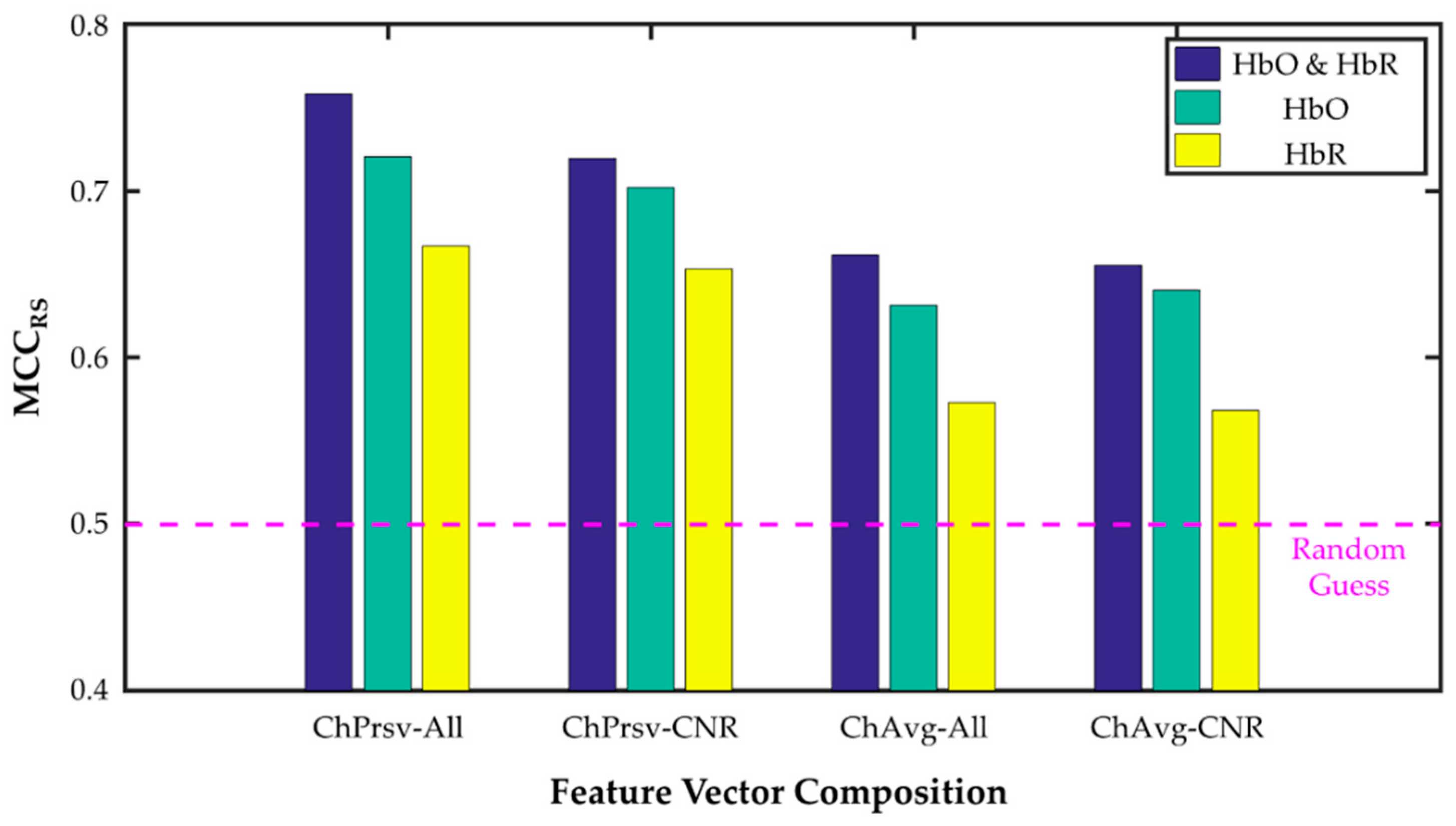
3.2. Channnel-Preserving vs. Channel-Averaging

The compositions with preserved channels (i.e., spatially patterned composition) exhibited a better performance across all channels than those that were averaged. Compared with all-channels-averaging compositions (FC-7, 8, and 9), MCCRS values in all-channels-preserved compositions (FC-1, 2, and 3) were higher by 14.98% (paired t-test, p < 0.00005) and detection latency was shortened by 18.01% (p = 0.11) CNR-channels (Table 2). With the CNR-selected channels, channel-preserved compositions (FC-4, 5, and 6) raised MCCRS values by 11.30% (p < 0.000005) and reduced detection latency by 36.52% (p < 0.005) more than channel-averaging compositions (FC-10, 11, and12).

3.3. [HbO], [HbR], and [HbO & HbR]

Overall, the compositions incorporating both [HbO] and [HbR] (FC-1, 4, 7, and 10) performed better than the other compositions on both indices (Figure 4 and Table 2). The compositions with only [HbR] (FC-3, 6, 9, and 12) generated the lowest performance. Particularly, the compositions using both types of hemoglobin enhanced MCCRS against the compositions using only one type (13.55% (p < 0.0000001) against [HbR], 3.71% (p < 0.0001) against [HbO]). Furthermore, the compositions with both hemoglobins lowered detection latency by 19.24% (p < 0.05) against those with [HbR] only and by 6.01% (p < 0.01) against those with [HbO] only (Figure 5 and Table 2).

3.4. All- and CNR-Selected Channels

The compositions with all channels (FC-1 to 3 and FC-7 to 9) performed better than the ones with CNR-selected channels (FC-4 to 6 and FC-10 to 12). Specifically, the all-channels compositions increased MCCRS by 1.86% (p = 0.11) and curtailed detection latency by 23.05% (p < 0.05) (Table 2). Finally, we carried out one-way ANOVA tests to examine if the 12 different feature vector compositions affected classification accuracy and detection latency, respectively. The results indicate that the compositions significantly improved both classification accuracy (for MCCRS, F = 4.69; p < 0.00005) and detection latency (F = 2.5; p < 0.05).

4. Discussion

The final goal of the rehabilitation system presented in this study is to bring the functional recovery of the impaired hands of post-stroke patients. The essential element of the system is the NIRS-BCI module, and the most important characteristic is how well NIRS-BCI can detect motor intention. We therefore tried several approaches and compared the results to obtain the best performance of the BCI module. To determine the best accuracy, we compared the classification results of differing (1) delays in hemodynamic responses and lengths of the feature window, (2) channel composition, (3) the NIRS signal, and (4) channel selection.
Accuracy and latency depended on DHR and LFW selections, which turned out to be related to each other. This suggests that we must carefully select DHR when LFW depending on the purpose of the NIRS-based system. Although a DHR between 8 s and 9 s and an LFW between 11 s and 13 s guaranteed the maximum classification accuracy, detection latency was distributed between 9 s and 11 s. These combinations are acceptable and could be useful if the system is for offline analysis, where classification accuracy is of importance and detection latency can be ignored. However, these selections are not suitable for real-time use. When a DHR is between 0 s and 2 s and an LFW is around 1 s, some classification accuracy is lost, but detection latency is shortened to between 2 s and 3 s. These selections are preferred for a real-time NIRS system, such as what we have presented here.
Channel-preserved feature vector compositions generated better performance than channel-averaged compositions in terms of classification accuracy and detection latency. Classification after averaging all channels have reportedly produced accuracies of 78% [22] and 65–75% [23]. This approach might be effective and useful for a BCI system with fewer channels that is only concerned with one cortical area. This is indeed the case for our system of only nine channels that are focused on the motor cortex, especially when all channels record similar temporal patterns. However, our comparisons revealed that the channel-preserving composition improved classification accuracy and shortened detection latency. Cui et al. (2010) also reported that the feature space taking account of spatial information (i.e., channel-preserved) increased accuracy by 7.7% and shortened detection latency by 2.4 s. Thus, even though channel-averaging might guarantee an acceptable accuracy and is computationally effective, by reducing the dimensions of feature vectors, we recommend the channel-preserving feature vector for an accurate BCI with short detection latency.
Our results also indicated that CNR is not a perfect criterion for selecting informative NIRS channels for classification. CNR-selected channels could not achieve the levels of both classification accuracy and detection latency that were obtained using all channels (Table 2). These results might indicate that there were some channels whose [HbO] or [HbR] correlated with the imagined hand motions but did not meet the CNR criteria. Thus, it might suggest that additional criteria are necessary. The coefficient of variance or the point-biserial correlation coefficient can be a good candidate to investigate. However, applying CNR can diminish the number of channels that are incorporated into a feature vector, thereby reducing computational time and cost. CNR could be more useful and effective if a system adopts many more NIRS channels, possibly producing more uninformative or noisy channels. This needs to be examined in future studies.
The concentration of HbR helps contribute to classification. The contribution of [HbR] alone is limited in the sense that classification improved when [HbO] was included in the feature vector. The feature compositions that included only [HbR] led to poorer classification performance, which dropped to the level of random guessing (Table 2). Based on our analysis, the worst approach is incorporating only [HbR] into a feature vector after averaging. In contrast, the compositions with both [HbO] and [HbR] improved both classification accuracy and detection latency over those with only [HbO] through all of the tested compositions, across all participants. Most NIRS-BCI systems [10] have not favored [HbR] because of its unreliability, relating to its small dynamic range and its signal-to-noise ratio (SNR). However, [HbR] is conditionally and marginally useful for better classification.
The approach and results of this study with the neurologically intact participants could be translated into clinical cases. We consider that the final user of our system will be patients who suffer from hand motor impairments owing to subcortical stroke. Because of their impairments, the patients must perform motor imagery, not motor execution. However, because of the lesion location being subcortical, the ipsilesional cortical areas are intact and can produce motor imagery-induced activations, which are detectable. The MI-evoked hemodynamic responses by a subcortical stroke patient could be similar to that of a neurologically intact participant. Studies of post-stroke patients that use hemodynamics-related modalities, such NIRS, have revealed that trained patients could produce clear motor imagery-induced cortical activity similar to motor execution [12]. In addition, the clinical studies of the BCI systems based on electrophysiological responses, being neurovascularly coupled with hemodynamic responses, demonstrate the existence of classifiable, MI-evoked responses. In the magnetoencephalography (MEG)- and sensorimotor rhythm (SMR)-BCI studies, post-stroke patients having subcortical lesions could learn how to control a BCI system [24,25]. The successful cases of EEG-BCI-based rehabilitation indirectly support a post-stroke patient’s ability to induce and control a task-related cortical activation [26,27]. Moreover, the pattern of a task-evoked hemodynamic response after a subcortical stroke seems preserved. The fMRI study of patients with cerebral ischemia [28] demonstrated an altered pattern of task-induced activation, i.e., an increase of [HbR] during a task, but this kind of alteration in a patient with a subcortical stroke has not been reported to our knowledge. Even though the altered activation can take place with a subcortical stroke, it implies that there is a task-related activation that exists and that is differentiable from a baseline. Thus, it can be detectable by a machine learning-based classifier, such as LDA.
This study has three main limitations. First, the sample size was small, and evaluation only used one technique (LDA) and two features (mean and slope). The inclusion of non-tested features, such as variance, skewness, and so on, and the testing of different classification techniques, such as SVM, neural networks, hidden Markov model, and naïve Bayesian with sets of tested and non-tested features could bring different results. Second, we must note that the two features here were calculated within a feature window of the same length. Each specific feature might contribute more to classification with a window of its own optimized length. Further research should be conducted for clarifying these problems. Finally, the influence of task-evoked extracerebral activation on our NIRS-BCI performance was not excluded due to the limitation of our NIRS system setup. The hemodynamics in the extracerebral layers of the head, such as scalp, can be affected by task-evoked changes in scalp blood flow and/or volume [29] and by those in the autonomic nervous system activity [30], which could contaminate NIRS signals. Although our single source-detector separation (SDS) of 3 cm could not filter out those extracerebral influences, it is unlikely that the inclusion of those extracerebral influences might compromise our study result. This is because the BCI study with a high-density multi-distance NIRS [23] showed that the use of multi-distance SDS NIRS improved accuracy by 5.2% compared to that of single SDS NIRS. Accordingly, it is inferred that our study results could be improved with a technique against the extracerebral contamination. Our further study will adopt the use of an additional short SDS of 0.5 cm [31], which is a proper option to filter out those extracerebral contamination in real time.

5. Conclusions

We have successfully developed a hand rehabilitation system integrating a reliable, commercial NIRS system with our previously developed robotic hand orthosis, in order to help post-stroke patients participate in effective rehabilitation therapy, such as AMT. Moreover, a series of evaluations was conducted to improve the classification performance of the BCI module, the most fundamental element of the system, in aspects of classification accuracy and detection latency. The evaluation results indicated that a shorter feature window is appropriate for a real-time application because it diminishes detection latency into between 2 s and 3 s, although it can preserve only 91.46% of the maximum classification accuracy that is attainable by a longer window. In addition, the results recommend a feature vector composition that preserves channel information and incorporates both [HbO] and [HbR] for better classification performance. Finally, we have to mention that the testing of our system on patients with subcortical stroke is ongoing, showing a positive translatability of the approach and results of this study into clinical cases.

Supplementary Materials

The following are available online at https://www.mdpi.com/2076-3417/9/18/3845/s1, Video S1: System demonstration with a neurologically intact participant performing motor imagery, Table S1: The values used for Figure 3 and Figure 4.

Author Contributions

Conceptualization, N.M.; J.A.; data curation, J.L.; N.M.; formal analysis, J.L.; funding acquisition, N.M.; J.A.; K.I.; M.H.; investigation, J.L.; N.M.; J.A.; methodology, J.L.; N.M.; J.A.; project administration, N.M.; J.A.; K.I.; M.H.; resources, N.M.; J.A.; K.I.; software, J.L.; N.M.; supervision, J.A.; K.I.; M.H.; validation, J.L.; visualization, J.L.; writing—original draft, J.L.; writing—review and editing, N.M.; J.A.; K.I.; M.H.

Funding

This work was funded in part by the Center for Clinical and Translational Research of Kyushu University (grant number: A122, N.M.) and in part by Japan Agency for Medical Research and Development (AMED) (grant number: 18im0210208h0003, K.I.). The funders had no role in study design, data collection and analysis, decision to publish, or preparation of the manuscript.

Acknowledgments

The authors acknowledge inspiring ideas from Roger Gassert and Olivier Lambercy of the Swiss Federal Institute of Technology in Zurich that led us to combine our robotic hand orthosis with NIRS.

Conflicts of Interest

The authors declare no conflict of interest. The funders had no role in the design of the study; in the collection, analyses, or interpretation of data; in the writing of the manuscript, or in the decision to publish the results.

References

  1. Wolf, S.L.; Winstein, C.J.; Miller, J.P.; Taub, E.; Uswatte, G.; Morris, D.; Giuliani, C.; Light, K.E.; Nichols-Larsen, D.; for the EXCITE Investigators. Effect of Constraint-Induced Movement Therapy on Upper Extremity Function 3 to 9 Months After Stroke: The EXCITE Randomized Clinical Trial. JAMA 2006, 296, 2095. [Google Scholar] [CrossRef] [PubMed]
  2. Lotze, M.; Braun, C.; Birbaumer, N.; Anders, S.; Cohen, L.G. Motor Learning Elicited by Voluntary Drive. Brain 2003, 126, 866–872. [Google Scholar] [CrossRef] [PubMed]
  3. Carod-Artal, F.J.; Egido, J.A. Quality of Life after Stroke: The Importance of a Good Recovery. Cerebrovasc. Dis. 2009, 27, 204–214. [Google Scholar] [CrossRef] [PubMed]
  4. Wolpaw, J.R.; Birbaumer, N.; Heetderks, W.J.; McFarland, D.J.; Peckham, P.H.; Schalk, G.; Donchin, E.; Quatrano, L.A.; Robinson, C.J.; Vaughan, T.M. Brain-Computer Interface Technology: A Review of the First International Meeting. IEEE Trans. Rehabil. Eng. 2000, 8, 164–173. [Google Scholar] [CrossRef] [PubMed]
  5. Silvoni, S.; Ramos-Murguialday, A.; Cavinato, M.; Volpato, C.; Cisotto, G.; Turolla, A.; Piccione, F.; Birbaumer, N. Brain-Computer Interface in Stroke: A Review of Progress. Clin. EEG Neurosci. 2011, 42, 245–252. [Google Scholar] [CrossRef] [PubMed]
  6. Soekadar, S.R.; Birbaumer, N.; Slutzky, M.W.; Cohen, L.G. Brain–Machine Interfaces in Neurorehabilitation of Stroke. Neurobiol. Dis. 2015, 83, 172–179. [Google Scholar] [CrossRef] [PubMed]
  7. Hoshi, Y. Functional Near-Infrared Spectroscopy: Current Status and Future Prospects. J. Biomed. Opt. 2007, 12, 062106. [Google Scholar] [CrossRef]
  8. Iadecola, C. Neurovascular Regulation in the Normal Brain and in Alzheimer’s Disease. Nat. Rev. Neurosci. 2004, 5, 347–360. [Google Scholar] [CrossRef]
  9. Villringer, A.; Dirnagl, U. Coupling of Brain Activity and Cerebral Blood Flow: Basis of Functional Neuroimaging. Cerebrovasc. Brain Metab. Rev. 1995, 6, 240–276. [Google Scholar]
  10. Naseer, N.; Hong, K.-S. FNIRS-Based Brain-Computer Interfaces: A Review. Front. Hum. Neurosci 2015, 9. [Google Scholar] [CrossRef]
  11. Wolpaw, J.R.; Birbaumer, N.; McFarland, D.J.; Pfurtscheller, G.; Vaughan, T.M. Brain–Computer Interfaces for Communication and Control. Clin. Neurophysiol. 2002, 113, 767–791. [Google Scholar] [CrossRef]
  12. Mihara, M.; Hattori, N.; Hatakenaka, M.; Yagura, H.; Kawano, T.; Hino, T.; Miyai, I. Near-Infrared Spectroscopy–Mediated Neurofeedback Enhances Efficacy of Motor Imagery–Based Training in Poststroke Victims: A Pilot Study. Stroke 2013, 44, 1091–1098. [Google Scholar] [CrossRef] [PubMed]
  13. Lee, J.; Mukae, N.; Arata, J.; Iwata, H.; Iramina, K.; Iihara, K.; Hashizume, M. A Multichannel-near-Infrared-Spectroscopy-Triggered Robotic Hand Rehabilitation System for Stroke Patients. In Proceedings of the 2017 IEEE International Conference on Rehabilitation Robotics (ICORR), London, UK, 17–20 July 2017; pp. 158–163. [Google Scholar] [CrossRef]
  14. Arata, J.; Ohmoto, K.; Gassert, R.; Lambercy, O.; Fujimoto, H.; Wada, I. A New Hand Exoskeleton Device for Rehabilitation Using a Three-Layered Sliding Spring Mechanism. In Proceedings of the 2013 IEEE International Conference on Robotics and Automation, Karlsruhe, Germany, 6–10 May 2013; pp. 3902–3907. [Google Scholar] [CrossRef]
  15. Zhang, Y.; Brooks, D.H.; Franceschini, M.A.; Boas, D.A. Eigenvector-Based Spatial Filtering for Reduction of Physiological Interference in Diffuse Optical Imaging. J. Biomed. Opt. 2005, 10, 011014. [Google Scholar] [CrossRef] [PubMed]
  16. Cui, X.; Bray, S.; Reiss, A.L. Speeded Near Infrared Spectroscopy (NIRS) Response Detection. PLoS ONE 2010, 5, e15474. [Google Scholar] [CrossRef] [PubMed]
  17. Durantin, G.; Scannella, S.; Gateau, T.; Delorme, A.; Dehais, F. Moving Average Convergence Divergence Filter Preprocessing for Real-Time Event-Related Peak Activity Onset Detection: Application to FNIRS Signals. In Proceedings of the 2014 IEEE 36th Annual International Conference of the IEEE Engineering in Medicine and Biology Society, Chicago, IL, USA, 26–30 August 2014; pp. 2107–2110. [Google Scholar] [CrossRef]
  18. Gateau, T.; Durantin, G.; Lancelot, F.; Scannella, S.; Dehais, F. Real-Time State Estimation in a Flight Simulator Using FNIRS. PLoS ONE 2015, 10, e0121279. [Google Scholar] [CrossRef] [PubMed]
  19. Tachtsidis, I.; Scholkmann, F. False Positives and False Negatives in Functional Near-Infrared Spectroscopy: Issues, Challenges, and the Way Forward. Neurophoton 2016, 3, 031405. [Google Scholar] [CrossRef] [PubMed]
  20. Lyons, R.G. Understanding Digital Signal Processing; Prentice Hall: Upper Saddle River, NJ, USA, 2011. [Google Scholar]
  21. Theodoridis, S.; Koutroumbas, K. Pattern Recognition, 4th ed.; Elsevier/Acad. Press: Amsterdam, The Netherlands, 2008. [Google Scholar]
  22. Fazli, S.; Mehnert, J.; Steinbrink, J.; Curio, G.; Villringer, A.; Müller, K.-R.; Blankertz, B. Enhanced Performance by a Hybrid NIRS–EEG Brain Computer Interface. NeuroImage 2012, 59, 519–529. [Google Scholar] [CrossRef] [PubMed]
  23. Shin, J.; Kwon, J.; Choi, J.; Im, C.-H. Performance Enhancement of a Brain-Computer Interface Using High-Density Multi-Distance NIRS. Sci. Rep. 2017, 7, 16545. [Google Scholar] [CrossRef]
  24. Buch, E.; Weber, C.; Cohen, L.G.; Braun, C.; Dimyan, M.A.; Ard, T.; Mellinger, J.; Caria, A.; Soekadar, S.; Fourkas, A.; et al. Think to Move: A Neuromagnetic Brain-Computer Interface (BCI) System for Chronic Stroke. Stroke 2008, 39, 910–917. [Google Scholar] [CrossRef]
  25. Soekadar, S.R.; Witkowski, M.; Mellinger, J.; Ramos, A.; Birbaumer, N.; Cohen, L.G. ERD-Based Online Brain–Machine Interfaces (BMI) in the Context of Neurorehabilitation: Optimizing BMI Learning and Performance. IEEE Trans. Neural Syst. Rehabil. Eng. 2011, 19, 542–549. [Google Scholar] [CrossRef]
  26. Ramos-Murguialday, A.; Broetz, D.; Rea, M.; Läer, L.; Yilmaz, Ö.; Brasil, F.L.; Liberati, G.; Curado, M.R.; Garcia-Cossio, E.; Vyziotis, A.; et al. Brain-Machine Interface in Chronic Stroke Rehabilitation: A Controlled Study. Ann. Neurol. 2013, 74, 100–108. [Google Scholar] [CrossRef] [PubMed]
  27. Ono, T.; Shindo, K.; Kawashima, K.; Ota, N.; Ito, M.; Ota, T.; Mukaino, M.; Fujiwara, T.; Kimura, A.; Liu, M.; et al. Brain-Computer Interface with Somatosensory Feedback Improves Functional Recovery from Severe Hemiplegia Due to Chronic Stroke. Front. Neuroeng. 2014, 7. [Google Scholar] [CrossRef] [PubMed]
  28. Murata, Y.; Sakatani, K.; Hoshino, T.; Fujiwara, N.; Kano, T.; Nakamura, S.; Katayama, Y. Effects of Cerebral Ischemia on Evoked Cerebral Blood Oxygenation Responses and BOLD Contrast Functional MRI in Stroke Patients. Stroke 2006, 37, 2514–2520. [Google Scholar] [CrossRef] [PubMed]
  29. Takahashi, T.; Takikawa, Y.; Kawagoe, R.; Shibuya, S.; Iwano, T.; Kitazawa, S. Influence of Skin Blood Flow on Near-Infrared Spectroscopy Signals Measured on the Forehead during a Verbal Fluency Task. NeuroImage 2011, 57, 991–1002. [Google Scholar] [CrossRef] [PubMed]
  30. Giller, C.A.; Giller, A.M.; Cooper, C.R.; Hatab, M.R. Evaluation of the Cerebral Hemodynamic Response to Rhythmic Handgrip. J. Appl. Physiol. 2000, 88, 2205–2213. [Google Scholar] [CrossRef]
  31. Saager, R.B.; Telleri, N.L.; Berger, A.J. Two-Detector Corrected Near Infrared Spectroscopy (C-NIRS) Detects Hemodynamic Activation Responses More Robustly than Single-Detector NIRS. NeuroImage 2011, 55, 1679–1685. [Google Scholar] [CrossRef] [PubMed]
Figure 1. System Configuration and Experimental Design: (a) System Configuration. The system consists of three basic components: a near-infrared spectroscopy (NIRS) system, a robotic hand orthosis, and an operating personal computer (PC). The NIRS system measures changes in local [Hb] in the motor cortex and sends them to the PC. The PC detects hand motor intentions from the NIRS data and wirelessly triggers the robotic orthosis; (b) Experimental Design. One session had a resting state period and a task period comprising 15 repeated unit trials. A unit trial was a sequence of ready, hand-closing, and hand-opening periods. The duration of the whole session was 810 s.
Applsci 09 03845 g001
Figure 2. NIRS Channel Configuration and Experimental Setup: (a) The four pairs of optodes are positioned and fixed with a swimming cap. (b) Channels 5, 7, 8, and 9 (emitted by the emitter 4) are placed around C3. (c) Experimental Setup. A participant wearing the NIRS optodes and robotic hand orthosis performs the task instructed through the monitor.
Applsci 09 03845 g002
Figure 3. Classification Performance according DHR and LFW: (a) The Matthews Correlation Coefficient (MCC)RS map according to DHR (s) and LFW (s); (b) The map of detection latency according to DHR and LFW. The pixel values of the detection latency map were calculated on the basis of assumed delay in hemodynamic response. Thus, for true detection latency values, the corresponding DHR must be added to each pixel value. Note that each pixel is the average from the seven participants. The pixel values for both maps are given in Table S1 (Supplementary Materials).
Applsci 09 03845 g003
Figure 4. MCCRS Comparison according to feature vector compositions. MCCRS (seven participants) results are displayed after being grouped. ChPrsv means ‘channel-preserving, and ChAvg means ‘channel-averaging’. All indicates ‘using all channels’, whereas Contrast-to-Noise Ratio (CNR) means ‘CNR-selected’. The values used for the bar graph are accessible in Table S1 (Supplementary Materials).
Applsci 09 03845 g004
Figure 5. Hemodynamic Responses and Detection Latencies by Feature Vector Compositions (Participant ID: 3): (a) The hemodynamic responses of [HbO] and [HbR] during the hand-closing task. The responses are averages of all channels across all trials after preprocessing by moving average convergence/divergence (MACD); (b) Visual comparison of detection latencies for each of the 12 feature vector compositions.
Applsci 09 03845 g005
Table 1. The Feature Vector Compositions. For channel, ‘all’ means that all channels were included in a feature vector. ‘CNR-selective’ means only the channels meeting CNR-criteria were included. For [Hb], ‘both’ indicates that both [HbO] and [HbR] were used. If ‘Average’ is ‘y (yes)’, [Hb]s were averaged and the features were calculated from the averaged [Hb]. Note that averaging was done with only [Hb]s of the same type, i.e., [HbO] or [HbR].
LabelCompositionDimension
Channel[Hb]Average
FC-1Allboth-36 × 1
FC-2[HbO]-18 × 1
FC-3[HbR]-18 × 1
FC-4CNR-selectiveboth-(NCNR-sel × 2 × 2) × 1
FC-5[HbO]-(NCNR-sel × 2) × 1
FC-6[HbR]-(NCNR-sel × 2) × 1
FC-7Allbothy4 × 1
FC-8[HbO]y2 × 1
FC-9[HbR]y2 × 1
FC-10CNR-selectivebothy4 × 1
FC-11[HbO]y2 × 1
FC-12[HbR]y2 × 1
Table 2. Evaluation Results.
Channel
Use
Channel
Selection
[HbO] & [HbR][HbO][HbR]Mean
MCCRSPreservingAll0.75860.72070.66690.7154
CNR-sel.0.71960.70230.65320.6917
AveragingAll0.66170.63160.57320.6222
CNR-sel.0.65540.64060.56850.6215
Mean0.69880.67380.6154-
Detection
Latency
(s)
PreservingAll2.40152.94643.20662.8515
CNR-sel.2.91843.36373.42573.2359
AveragingAll3.73493.44923.24913.4778
CNR-sel.4.26654.41436.61265.0978
Mean3.33033.54344.1235-

