Article

Development and Evaluation of a Prototype Scratch Apparatus for Wound Assays Adjustable to Different Forces and Substrates

Department of Natural Sciences, Bonn-Rhein-Sieg University of Applied Sciences, von-Liebig-Strasse 20, D-53359 Rheinbach, Germany
*
Authors to whom correspondence should be addressed.
Appl. Sci. 2019, 9(20), 4414; https://doi.org/10.3390/app9204414
Submission received: 27 August 2019 / Revised: 25 September 2019 / Accepted: 15 October 2019 / Published: 18 October 2019
(This article belongs to the Special Issue Novel Methods for Investigating Biomolecular Interactions)

Abstract

:
Scratch assays enable the study of the migration process of an injured adherent cell layer in vitro. An apparatus for the reproducible performance of scratch assays and cell harvesting has been developed that meets the requirements for reproducibility in tests as well as easy handling. The entirely autoclavable setup is divided into a sample translation and a scratching system. The translational system is compatible with standard culture dishes and can be modified to adapt to different cell culture systems, while the scratching system can be adjusted according to angle, normal force, shape, and material to adapt to specific questions and demanding substrates. As a result, a fully functional prototype can be presented. This system enables the creation of reproducible and clear scratch edges with a low scratch border roughness within a monolayer of cells. Moreover, the apparatus allows the collection of the migrated cells after scratching for further molecular biological investigations without the need for a second processing step. For comparison, the mechanical properties of manually performed scratch assays are evaluated.

Graphical Abstract

1. Introduction

As the outer shell of the human body, skin forms the human’s largest organ [1]. When skin is injured, it is able to repair itself while passing through five stages of wound healing: haemostasis, inflammation, proliferation, migration, and maturation [2,3]. As a part of this process, cells migrate into the wounded area [4]. Wound healing assays can be set up in order to enable further investigation of the skin wound-healing process in vitro [5,6,7].
The popularity of these assays can be explained by the simplicity of their procedures. In such procedures, a layer of adherent cells is wounded under specified conditions and the subsequent migration of the contiguous cells into the fresh and cell-free area is observed and analyzed [8,9,10]. Such a defined cell layer can be wounded by scratching, stamping using PDMS or neoprene [11,12,13], thermal wounding [14], electrical wounding [15], or optical wounding [16,17]. Scratching is most commonly performed in well plates using pipette tips [18,19,20]. However, it can also be performed by using a variety of sharp-edged tools like toothpicks [21], metallic micro-indenters [22], or cell scrapers [23].
Furthermore, it is reported that scratches are performed on coverslips, which can be placed into a 6-well plate [24]. In other techniques, inlets within dishes or wells to create a cell free area have been used [25]. The general aim of a scratch assay is to provide straight borders in a reproducible way without damaging cells [26]. However, the reproducibility of manually performed scratch assays strongly depends on the performer and their skills [27].
An attachment for forceps-like scrapers to create scratches of defined direction, length, width, and depth in 6-well plates has been developed to investigate keratinocyte migration [28]. Scratches obtained with this scratch stencil are reported to be inflicted with high precision and in a reproducible fashion [29]. The material chosen by this group, polyoxymethylene, is generally a suitable construction material in concerns of laboratory apparatus construction [30].
In all aforementioned strategies, it is only possible to investigate the migration behavior of cells under different conditions. Subsequent molecular biological investigations using RNA or protein samples from migrated cells were impossible due to the fixation of the cells. Therefore, migration assays were performed separately by using transwell or Boyden chambers. After migration, the migrated cells were collected for further investigations [31,32,33].
Depending on the needs of experimental design and availability, the used cell types for the mentioned investigation methods of migration and wound healing may vary. For instance, to study epithelial repair, epithelial cells from the lung [34] or the retina [35] are commonly used. Another interesting source is represented by mesenchymal stem cells, which can be differentiated towards multiple cell types [36,37,38,39,40,41,42]. Setiawan and colleagues even differentiated human adipose-derived stem cells towards epithelial cells for usage in scratch assays [43]. Fibroblasts from foreskin (HFF), skin (HDF), or nasal polyps (NPDF) have recently been used as well as microglia cells and keratinocytes [25,44,45,46]. In addition to the mentioned cell types, endothelial cells are a favorable source to study migration. Many working groups used either human or bovine endothelial cells in scratch assays and other migration assays [47,48,49,50,51,52,53,54,55,56].
This study covers the development and evaluation of a prototype scratch apparatus for wound assays, focusing on the comparison to conventional procedures. The presented device allows for the collection of migrated cells after scratching for further investigations, without the need of a second processing step. Moreover, the apparatus is adjustable in angle, normal force, shape, and material to adapt to a specific question and demanding substrates. In detail, scratching performance is examined for different configurations to identify parameters leading to an ideal scratch.

2. Experimental Section

2.1. Characterization of Manually and Automatically Performed Scratch Assays

As a model culture, HMEC-1 (ATCC, Manassas, VA, USA) cultivated in 60 mm petri dishes (Sarstedt, Nümbrecht, Germany) was used in all experiments.
For manually performed scratch assays using pipette tips (Sarstedt, Nümbrecht, Germany), rubber spatulas (Deutsch & Naumann, Berlin, Germany) and cell scrapers (Sarstedt, Nümbrecht, Germany), the recommended procedures according to [8,57] were followed. A swift and consistent scratch of 30 mm length was applied to the culture dish surface. Every scratch assay was performed in a sixfold determination.
For the scratching device, normal angles varied between +/−45°, normal forces varied from 0.3 to 1.0 N/scratch, and the directions varied concerning push and pull performance. The control of the force impacting the cell substrate was achieved using calibrated weights mounted to the scratch head. Acting force was acquired using a calibrated load cell (VWR, Radnor, PA, USA) beneath the culture dish. Image processing was performed using OriginPro 2017 (Originlab, Northhampton, MA, USA). The scratching performance is considered as the roughness of the scratch edge obtained, expressed as the mean deviation from its linear regression (Figure 1). In order to identify and classify the parameters leading to an optimal scratch, median values are processed as they are less prone to severe deviations in the overall measurement.

2.2. Evaluation of the Harvesting Capabilities of the Scratch Device

A monolayer of cells was grown (60 mm culture dish, cell density: 200,000 cells/dish, medium: DMEM + 10% FBS), scratched and re-scratched using only one culture dish. To increase the number of scratch edges, 30 mm blades made from standard cell culture scrapers (Sarstedt, Nümbrecht, Germany) with set-in jags were used. After scratching under different tested conditions, the cells were allowed to migrate for 24 h at 37 °C in the incubator. The dishes were mounted in the device and marked to set the correct orientation for re-scratching and microscopic assessment. To evaluate the migration capacity of the cells using a Zeiss Axio Observer D.1 microscope (Oberkochen, Germany) and AxioVision software, the dishes were placed onto a dish- and slide-holder at the set orientation before and after incubation. For facilitated and accurate microscopic determination of the migration behavior, the pre-scratched coordinates within the culture dish were re-approached after scratching.

3. Results and Discussion

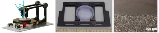
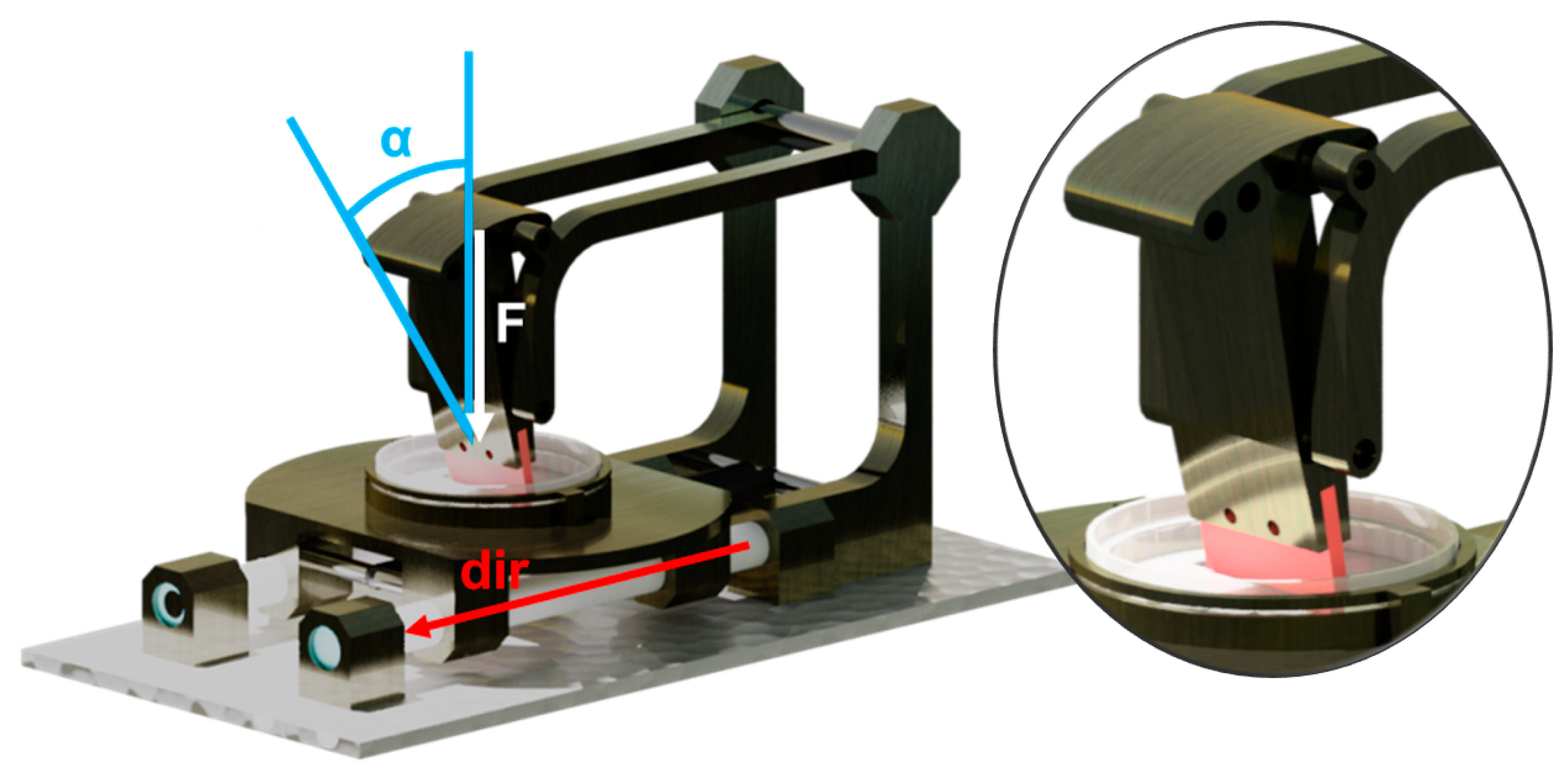
3.1. Conception and Construction of the Apparatus

In order to meet the requirements for both reproducibility and easy handling, a simple mechanical apparatus was designed. The overall requirements for this device, which can be considered a laboratory or even medical product, include an operation at room temperature, contact to cell cultures, cleaning in a conventional laboratory washer, sterilization in an autoclave, and portability [58].
The device itself was designed to be completely disassemblable and to be easily manufactured by a common milling machine and lathe. It consists of two modular parts to fulfil the function of the sample scratching and locomotion, respectively (Figure 2).
The scratching part of the device needs to scratch a cell substrate of undefined thickness from above in different, defined angles with respect to the sample surface [18,57,59,60]. The scratching blade should be disposable or at least replaceable and changeable in geometry for multiple simultaneous scratches. Moreover, the force impacting on the sample should be able to be varied as different cell cultures are known to show diverging adhesion forces.
This has been realized by a T-shaped scratch head that can mount any suitable construction material (e.g., polymers) with a thickness of 2 mm. The blade is fixed in position by two threaded shafts. The scratch head itself can be adjusted by rotating around the lower axis and fixed in five positions. Two extension arms, fixed to a polytetrafluoroethylene (PTFE) bushed axle, enable the head to be lifted up for sample exchange or revision. In order to tune the normal force impacting on the sample surface, additional weights can be attached. The rear mounting brackets also feature an axle that allows mounting a gas spring for an automated sample locomotion. Stainless steel was chosen for the axles and aluminum alloy for all other construction parts.
The sample locomotion part of the device needs to suppress culture dish rotation, tilting, as well as undesired axial and radial movements. Additionally, the actual locomotion should be automated, smooth, directionally stable under force impact, easy to handle and to release, and ensure maximum accuracy.
This is solved by the construction of a linear slide. The sled guide consists of a PTFE sleeved stainless steel rod on which the sled’s brackets can glide. The sample holder plate provides an annular culture dish holder with a bayonet type lock that secures the culture dish.

3.2. Comparison of Manually and Automatically Performed Scratching

In order to evaluate the capabilities and limitations of the scratching performances, the scratch border roughness is interpreted as a function of its respective operating procedure. For manually, as well as automatically performed scratch assays, the impacting forces and the border roughness values are depicted (Figure 3).
It shows that manually and automatically performed roughness values vary depending on their operational mode and especially on the method used. While the manually performed values range between 30 and 170 µm, the mechanically performed scratches enable values of 20 to 70 µm. Concerning the force accuracy, the manually performed assays vary by roughly 0.1 to 0.15 N per scratch and exceed 0.2 N per scratch for the usually favoured pipette tip technique. Mechanically performed scratch assays undergo a variation of less than 0.1 N/scratch, even with the directional mode not being differentiated.
When taking a closer look at the manually performed scratch techniques, the force deviation depends on the tool used. While the rigid and barely deformable pipette tip shows a large force deviation due to the operator guiding it, the rubber spatula and cell scraper perform with little deviation. Both these elastic tools show a more rigid and therefore inconsistent force application in push direction; however, in pull direction deviations can be smoothed out more easily. Concerning the automated scratching, the deviation in force results from the simultaneous consideration of push and pull assays.
At this point, it can be stated that the conventionally used pipette tip provides the scratching border with the least roughness values of all manual techniques investigated.

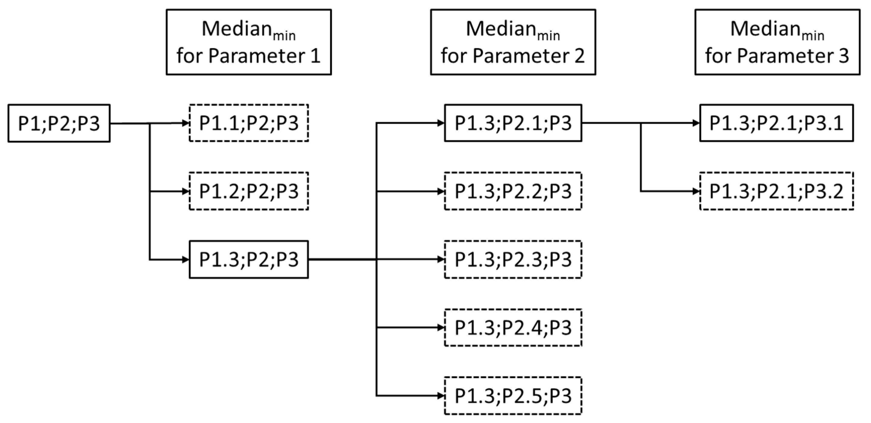
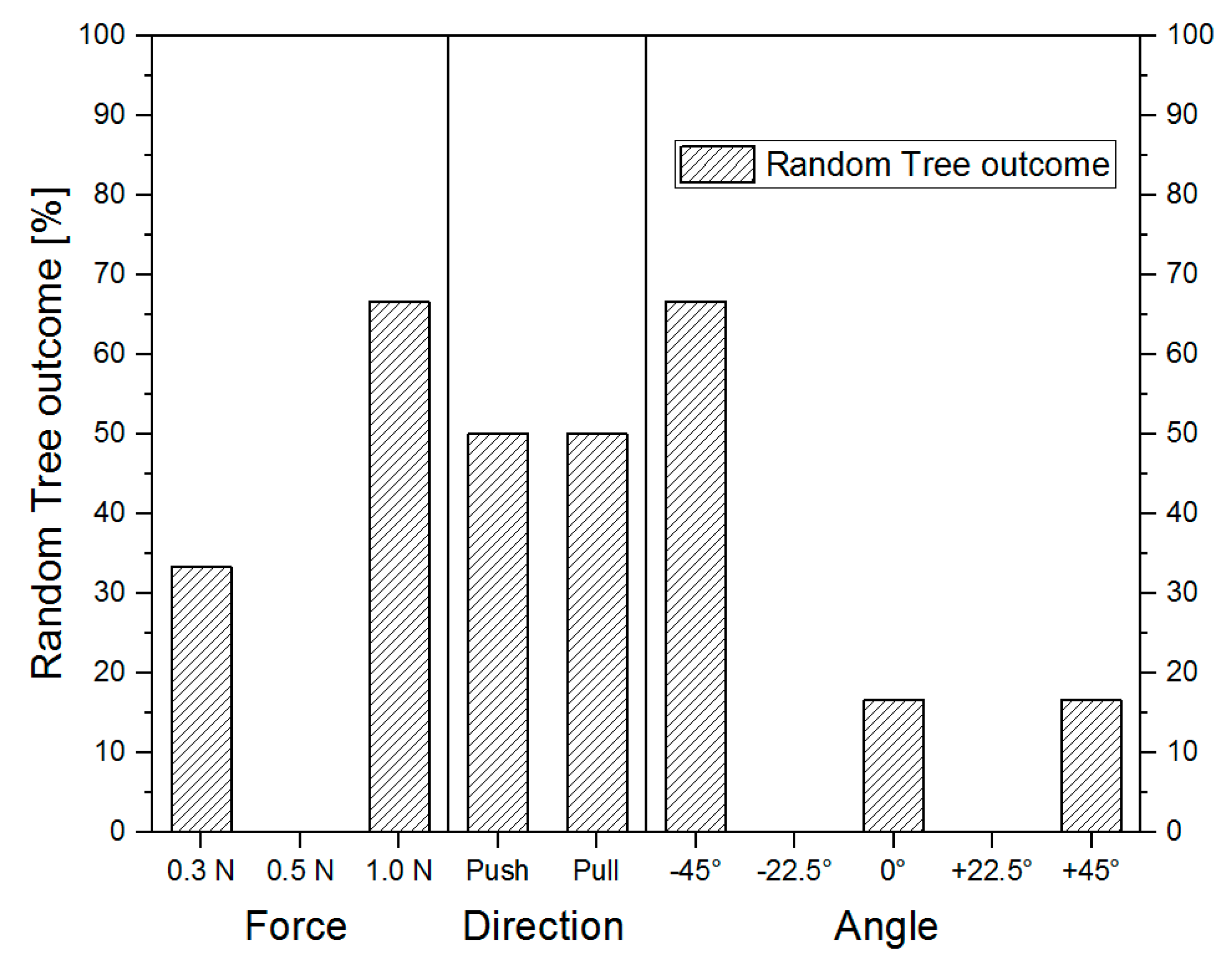
3.3. Comparison of Different Operational Settings Using the Scratch Device

As a low roughness value was desired, a closer differentiation of the influencing parameters on the mechanically produced scratch borders was carried out. A classification of all experimental data into the varied parameters via box plot (Figure 4) revealed that there are distinct settings for direction, force, and angle that lead to lower mean and median values for optimum scratch performance.
As the order of classification plays an important role [61,62], the three classification parameters (angle, direction, and force) have been checked for the lowest roughness median value in all six possible decision tree permutations (Figure 5).
A subsequent evaluation of the step-wise refinement for the ideal parameters (Figure 6) shows a probability for achieving a low scratch border roughness.
When the highest force is applied, a good scratch is deemed the most probable. A chosen angle of −45° also leads to a desirable outcome of the scratch border. Concerning the scratching direction, no preference was observed.
As to the refinement of the scratching parameters, the outcome represents a probability to achieve a good scratch border (meaning a low roughness value). As the outcome of the scratch direction shows a 50% distribution for every direction, the influence can be considered inessential. A different picture can be drawn when the applied force and scratching angle are considered. In four out of six decision trees, the best outcomes have been recorded for the highest force applied. This is in accordance with the manual scratch techniques, which also obtained smoother scratch borders when a reasonably high force was applied. In the range of the results observed, the application of 0.5 N force per scratch had to be avoided.

3.4. Perfomance of the Automated Scratch Assays on the Harvesting Capabilities

As shown before, the pipette tip technique as a quick scratch assay tool has its justification for good reason. However, the advantages of easy handling and a good scratch border are outmatched by the fact that the pipette tip is the only technique investigated that is incapable of scratching (Figure 7A) and re-scratching (=harvest) migrated (Figure 7B) cells. In contrast, by using the scratch device, a clear and linear scratch edge was created (Figure 7C). Cells that migrated into the scratch within 24 h (Figure 7D) were collected by re-scratching the same position (Figure 7E).

4. Conclusions

The need for a simple device for the performance of reproducible scratch assays including harvesting was met by the construction of an apparatus that consisted of a modular setup featuring a sample scratching and a sample locomotion system. The design was kept simple to enable both individual adjustment in force, angle, and direction. The conducted experiments show that scratch borders were generated, which were straighter and more reproducible than manually performed assays. Concerning both manually and automatically performed assays, the lowest scratch border roughness data were obtained when using high forces on the substrate.
In contrast to conventional methods, which include a two-step model of scratch assays in well plates and a subsequent migration assay in transwell chambers or Boyden chambers, the presented scratch device simplifies the procedure into just a single step. In conclusion, this apparatus is suitable for facilitated microscopic monitoring of cell migration and proliferation and enables the harvesting and investigation of these cells only.

Author Contributions

Data curation (apparatus design and construction and experiments regarding the mechanical scratching performance) and writing/original draft preparation, R.G.; data curation (cell culture experiments) and writing/original draft preparation, P.B.; data analysis and writing, P.G.; methodology and resources, K.-P.S.; writing/review and editing, M.S.; conceptualization, methodology, supervision, review, and editing, E.T.

Funding

This work was supported and financed by the Federal Ministry of Education and Research program “FHprofUnt” project “MeMoAthero” (grant number: 03FH012PB2). A PhD scholarship was given to P.B. by the Graduate Institute of the Bonn-Rhein-Sieg University of Applied Sciences.

Acknowledgments

We gratefully thank Debora Turetsky (Northwestern University, Evanston, Illinois, U.S.) for English corrections.

Conflicts of Interest

The authors declare no conflict of interest.

References

  1. Kanitakis, J. Anatomy, histology and immunohistochemistry of normal human skin. Eur. J. Dermatol. 2002, 12, 390–401. [Google Scholar] [PubMed]
  2. Boateng, J.; Catanzano, O. Advanced Therapeutic Dressings for Effective Wound Healing—A Review. J. Pharm. Sci. 2015, 104, 3653–3680. [Google Scholar] [CrossRef] [PubMed]
  3. Sorg, J.M.R.H. Wound Repair and Regeneration. Eur. Surg. Res. 2012, 49, 35–43. [Google Scholar]
  4. Lampugnani, M.G. Cell Migration into a Wounded Area In Vitro. In Adhesion Protein Protocols; Humana Press: Totowa, NJ, USA, 1999; pp. 177–182. [Google Scholar]
  5. Rodriguez, L.G.; Wu, X.; Guan, J.-L. Wound-Healing Assay. In Cell Migration: Developmental Methods and Protocols; Guan, J.-L., Ed.; Humana Press: Totowa, NJ, USA, 2005; pp. 23–29. ISBN 978-1-59259-860-1. [Google Scholar]
  6. Stamm, A.; Reimers, K.; Strauß, S.; Vogt, P.; Scheper, T.; Pepelanova, I. In vitro wound healing assays - State of the art. BioNanoMaterials 2016, 17, 79–87. [Google Scholar] [CrossRef]
  7. Pijuan, J.; Barceló, C.; Moreno, D.F.; Maiques, O.; Sisó, P.; Marti, R.M.; Macià, A.; Panosa, A. In vitro Cell Migration, Invasion, and Adhesion Assays: From Cell Imaging to Data Analysis. Front. Cell Dev. Biol. 2019, 7, 1–16. [Google Scholar] [CrossRef]
  8. Liang, C.; Park, A.Y.; Guan, J. In vitro scratch assay: A convenient and inexpensive method for analysis of cell migration in vitro. Nat. Protoc. 2007, 2, 329–333. [Google Scholar] [CrossRef]
  9. Silva Nunes, J.P.; Martins Dias, A.A. ImageJ macros for the user-friendly analysis of soft-agar and wound-healing assays. Biotechniques 2017, 62, 175–179. [Google Scholar] [CrossRef] [Green Version]
  10. Vargas, A.; Angeli, M.; Pastrello, C.; Mcquaid, R.; Li, H.; Jurisicova, A.; Jurisica, I. Robust quantitative scratch assay. Bioinformatics 2016, 32, 1439–1440. [Google Scholar] [CrossRef]
  11. Lan, R.; Geng, H.; Hwang, Y.; Mishra, P.; Skloss, W.L. A novel wounding device suitable for quantitative biochemical analysis of wound healing and regeneration of cultured epithelium. Wound Repair Regen. 2010, 18, 159–167. [Google Scholar] [CrossRef] [Green Version]
  12. Fong, E.; Tzlil, S.; Tirrell, D.A. Boundary crossing in epithelial wound healing. Proc. Natl. Acad. Sci. USA 2010, 107, 19302–19307. [Google Scholar] [CrossRef] [Green Version]
  13. Nikolić, D.L.; Boettiger, A.N.; Bar-Sagi, D.; Carbeck, J.D.; Shvartsman, S.Y. Role of boundary conditions in an experimental model of epithelial wound healing. Am. J. Physiol. Cell Physiol. 2006, 291, 68–76. [Google Scholar] [CrossRef] [PubMed]
  14. Hettler, A.; Werner, S.; Eick, S.; Laufer, S.; Weise, F. A New In Vitro Model to Study Cellular Responses after Thermomechanical Damage in Monolayer Cultures. PLoS ONE 2013, 8, e82635. [Google Scholar] [CrossRef] [PubMed]
  15. Giaever, I.; Keese, C.R. Micromotion of mammalian cells measured electrically. Proc. Natl. Acad. Sci. USA 1991, 88, 7896–7900. [Google Scholar] [CrossRef] [PubMed]
  16. Zordan, M.D.; Mill, C.P.; Ii, D.J.R.; Leary, J.F. A High Throughput, Interactive Imaging, Bright-Field Wound Healing Assay. Cytom. Part A 2011, 79, 227–232. [Google Scholar] [CrossRef] [PubMed]
  17. Song, Q.; Uygun, B.; Banerjee, I.; Nahmias, Y.; Zhang, Q.; Berthiaume, F.; Latina, M.; Yarmush, M.L. Low power laser irradiation stimulates the proliferation of adult human retinal pigment epithelial cells in culture. Cell. Mol. Bioeng. 2009, 2, 87–103. [Google Scholar] [CrossRef]
  18. Grada, A.; Otero-Vinas, M.; Prieto-Castrillo, F.; Obagi, Z.; Falanga, V. Research Techniques Made Simple: Analysis of Collective Cell Migration Using the Wound Healing Assay. J. Invest. Dermatol. 2017, 137, e11–e16. [Google Scholar] [CrossRef] [Green Version]
  19. Menger, B.; Vogt, P.M.; Allmeling, C.; Radtke, C. AmbLOXe—An Epidermal Lipoxygenase of the Mexican Axolotl in the Context of Amphibian Regeneration and Its Impact on Human Wound Closure In Vitro. Ann. Surg. 2011, 253, 410–418. [Google Scholar] [CrossRef]
  20. Bennett, J.; Cassidy, H.; Slattery, C.; Ryan, M.; McMorrow, T. Tacrolimus Modulates TGF-β Signaling to Induce Epithelial-Mesenchymal Transition in Human Renal Proximal Tubule Epithelial Cells. J. Clin. Med. 2016, 5, 50. [Google Scholar] [CrossRef]
  21. Klettner, A.; Tahmaz, N.; Dithmer, M.; Richert, E.; Roider, J. Effects of aflibercept on primary RPE cells: Toxicity, wound healing, uptake and phagocytosis. Br. J. Ophthalmol. 2014, 98, 1448–1452. [Google Scholar] [CrossRef]
  22. Topman, G.; Sharabani-yosef, O.; Gefen, A. Medical Engineering & Physics A standardized objective method for continuously measuring the kinematics of cultures covering a mechanically damaged site. Med. Eng. Phys. 2012, 34, 225–232. [Google Scholar]
  23. Baudin, B.; Bruneel, A.; Bosselut, N.; Vaubourdolle, M. A protocol for isolation and culture of human umbilical vein endothelial cells. Nat. Protoc. 2007, 2, 481–485. [Google Scholar] [CrossRef] [PubMed]
  24. Molladavoodi, S.; Wulff, D.; Robichaud, M.; Gorbet, M. Corneal epithelial cells exposed to shear stress show altered cytoskeleton and migratory behaviour. PLoS ONE 2017, 12, e0178981. [Google Scholar] [CrossRef] [PubMed]
  25. Tang, Y.; Cui, Y.; Li, Z.; Jiao, Z.; Zhang, Y.; He, Y.; Chen, G.; Cheng, G.; Zhou, Q.; Wang, W.; et al. Radiation-induced miR-208a increases the proliferation and radioresistance by targeting p21 in human lung cancer cells. J. Exp. Clin. Cancer Res. 2016, 35, 7. [Google Scholar] [CrossRef] [PubMed]
  26. Kramer, N.; Walzl, A.; Unger, C.; Rosner, M.; Krupitza, G.; Hengstschläger, M.; Dolznig, H. In vitro cell migration and invasion assays. Mutat. Res. Mutat. Res. 2013, 752, 10–24. [Google Scholar] [CrossRef] [PubMed]
  27. Goetsch, K.P.; Niesler, C.U. Optimization of the scratch assay for in vitro skeletal muscle wound healing analysis. Anal. Biochem. 2011, 411, 158–160. [Google Scholar] [CrossRef]
  28. Büth, H.; Luigi Buttigieg, P.; Ostafe, R.; Rehders, M.; Dannenmann, S.R.; Schaschke, N.; Stark, H.J.; Boukamp, P.; Brix, K. Cathepsin B is essential for regeneration of scratch-wounded normal human epidermal keratinocytes. Eur. J. Cell Biol. 2007, 86, 747–761. [Google Scholar] [CrossRef]
  29. Wiegand, C.; Hipler, U.-C. Methods for the measurement of cell and tissue compatibility including tissue regeneration processes. GMS Krankenhaushygiene Interdiszip. 2008, 3, 1–9. [Google Scholar]
  30. Penick, K.J.; Solchaga, L.A.; Berilla, J.A.; Welter, J.F. Performance of polyoxymethylene plastic (POM) as a component of a tissue engineering bioreactor. J. Biomed. Mater. Res. Part A 2005, 75, 168–174. [Google Scholar] [CrossRef]
  31. Klepeis, V.E.; Weinger, I.; Kaczmarek, E.; Trinkaus-Randall, V. P2Y receptors play a critical role in epithelial cell communication and migration. J. Cell. Biochem. 2004, 93, 1115–1133. [Google Scholar] [CrossRef]
  32. Chmielewski, S.; Olejnik, A.; Sikorski, K.; Pelisek, J.; Błaszczyk, K.; Aoqui, C.; Nowicka, H.; Zernecke, A.; Heemann, U.; Wesoly, J.; et al. STAT1-dependent signal integration between IFNγ and TLR4 in vascular cells reflect pro-atherogenic responses in human atherosclerosis. PLoS ONE 2014, 9, 1–26. [Google Scholar] [CrossRef]
  33. Horckmans, M.; Robaye, B.; Léon-Gómez, E.; Lantz, N.; Unger, P.; Dol-Gleizes, F.; Clouet, S.; Cammarata, D.; Schaeffer, P.; Savi, P.; et al. P2Y4 nucleotide receptor: A novel actor in post-natal cardiac development. Angiogenesis 2012, 15, 349–360. [Google Scholar] [CrossRef] [PubMed]
  34. McClendon, J.; Jansing, N.L.; Redente, E.F.; Gandjeva, A.; Ito, Y.; Colgan, S.P.; Ahmad, A.; Riches, D.W.H.; Chapman, H.A.; Mason, R.J.; et al. Hypoxia-Inducible Factor 1α Signaling Promotes Repair of the Alveolar Epithelium after Acute Lung Injury. Am. J. Pathol. 2017, 187, 1772–1786. [Google Scholar] [CrossRef] [PubMed]
  35. Hirano, Y.; Yasuma, T.; Mizutani, T.; Fowler, B.J.; Tarallo, V.; Yasuma, R.; Kim, Y.; Bastos-Carvalho, A.; Kerur, N.; Gelfand, B.D.; et al. IL-18 is not therapeutic for neovascular age-related macular degeneration. Nat. Med. 2014, 20, 1372–1375. [Google Scholar] [CrossRef] [PubMed] [Green Version]
  36. Kaebisch, C.; Schipper, D.; Babczyk, P.; Tobiasch, E. The role of purinergic receptors in stem cell differentiation. Comput. Struct. Biotechnol. J. 2015, 13, 75–84. [Google Scholar] [CrossRef] [PubMed]
  37. Babczyk, P.; Conzendorf, C.; Klose, J.; Schulze, M.; Harre, K.; Tobiasch, E. Stem Cells on Biomaterials for Synthetic Grafts to Promote Vascular Healing. J. Clin. Med. 2014, 3, 39–87. [Google Scholar] [CrossRef]
  38. Zippel, N.; Limbach, C.A.; Ratajski, N.; Urban, C.; Luparello, C.; Pansky, A.; Kassack, M.U.; Tobiasch, E. Purinergic Receptors Influence the Differentiation of Human Mesenchymal Stem Cells. Stem Cells Dev. 2011, 21, 884–900. [Google Scholar] [CrossRef]
  39. Hielscher, D.; Kaebisch, C.; Braun, B.J.V.; Gray, K.; Tobiasch, E. Stem Cell Sources and Graft Material for Vascular Tissue Engineering. Stem Cell Rev. Reports 2018, 14, 642–667. [Google Scholar] [CrossRef]
  40. Zhang, Y.; Khan, D.; Delling, J.; Tobiasch, E. Mechanisms underlying the osteo- and adipo-differentiation of human mesenchymal stem cells. Sci. World J. 2012, 2012, 793823. [Google Scholar] [CrossRef]
  41. Seifert, A.; Werheid, D.F.; Knapp, S.M.; Tobiasch, E. Role of Hox genes in stem cell differentiation. World J. Stem Cells 2015, 7, 583. [Google Scholar] [CrossRef]
  42. Tobiasch, E. Differentiation Potential of Adult Human Mesenchymal Stem Cells. In Stem Cell Engineering; Springer: Berlin/Heidelberg, Germany, 2011; pp. 61–77. [Google Scholar]
  43. Setiawan, M.; Tan, X.-W.; Goh, T.-W.; Hin-Fai Yam, G.; Mehta, J.S. Inhibiting glycogen synthase kinase-3 and transforming growth factor-β signaling to promote epithelial transition of human adipose mesenchymal stem cells. Biochem. Biophys. Res. Commun. 2017, 490, 1381–1388. [Google Scholar] [CrossRef]
  44. Ramenzoni, L.; Weber, F.; Attin, T.; Schmidlin, P. Cerium Chloride Application Promotes Wound Healing and Cell Proliferation in Human Foreskin Fibroblasts. Materials (Basel) 2017, 10, 573. [Google Scholar] [CrossRef] [PubMed]
  45. Park, H.J.; Salem, M.; Semlali, A.; Leung, K.P.; Rouabhia, M. Antimicrobial peptide KSL-W promotes gingival fibroblast healing properties in vitro. Peptides 2017, 93, 33–43. [Google Scholar] [CrossRef] [PubMed]
  46. Scheiblich, H.; Roloff, F.; Singh, V.; Stangel, M.; Stern, M.; Bicker, G. Nitric oxide/cyclic GMP signaling regulates motility of a microglial cell line and primary microglia in vitro. Brain Res. 2014, 1564, 9–21. [Google Scholar] [CrossRef] [PubMed]
  47. Mavi, M.F.; Ji, J.Y. Endothelial wound recovery is influenced by treatment with shear stress, wound direction, and substrate. Cell. Mol. Bioeng. 2013, 6, 310–325. [Google Scholar] [CrossRef]
  48. Lin, X.; Helmke, B.P. Cell structure controls endothelial cell migration under fluid shear stress. Cell. Mol. Bioeng. 2009, 2, 231–243. [Google Scholar] [CrossRef]
  49. Kang, Z.; Zhu, H.; Jiang, W.; Zhang, S. Protocatechuic acid induces angiogenesis through PI3K-Akt-eNOS-VEGF signalling pathway. Basic Clin. Pharmacol. Toxicol. 2013, 113, 221–227. [Google Scholar] [CrossRef]
  50. De Jong, E.K.; Breukink, M.B.; Schellevis, R.L.; Bakker, B.; Mohr, J.K.; Fauser, S.; Keunen, J.E.E.; Hoyng, C.B.; Den Hollander, A.I.; Boon, C.J.F. Chronic central serous chorioretinopathy is associated with genetic variants implicated in age-related macular degeneration. Ophthalmology 2015, 122, 562–570. [Google Scholar] [CrossRef]
  51. Zhou, Y.; Li, S.; Li, J.; Wang, D.; Li, Q. Effect of microRNA-135a on Cell Proliferation, Migration, Invasion, Apoptosis and Tumor Angiogenesis Through the IGF-1/PI3K/Akt Signaling Pathway in Non-Small Cell Lung Cancer. Cell. Physiol. Biochem. 2017, 42, 1431–1446. [Google Scholar] [CrossRef]
  52. Lin, P.-Y.; Sung, P.-H.; Chung, S.-Y.; Hsu, S.-L.; Chung, W.-J.; Sheu, J.-J.; Hsueh, S.-K.; Chen, K.-H.; Wu, R.-W.; Yip, H.-K. Hyperbaric Oxygen Therapy Enhanced Circulating Levels of Endothelial Progenitor Cells and Angiogenesis Biomarkers, Blood Flow, in Ischemic Areas in Patients with Peripheral Arterial Occlusive Disease. J. Clin. Med. 2018, 7, 548. [Google Scholar] [CrossRef]
  53. Stoeckius, M.; Erat, A.; Fujikawa, T.; Hiromura, M.; Koulova, A.; Otterbein, L.; Bianchi, C.; Tobiasch, E.; Dagon, Y.; Sellke, F.W.; et al. Essential roles of Raf/extracellular signal-regulated kinase/mitogen- activated protein kinase pathway, YY1, and Ca2+ influx in growth arrest of human vascular smooth muscle cells by bilirubin. J. Biol. Chem. 2012, 287, 15418–15426. [Google Scholar] [CrossRef]
  54. Liebert, M.A.; Soares, M.P.; Usheva, A.; Brouard, S.; Berberat, P.O.; Gunther, L.; Tobiasch, E.; Bach, F.H. Heme Oxygenase-1-Derived Carbon Monoxide. J. Biol. Chem. 2002, 277, 17950–17961. [Google Scholar]
  55. Brouard, S.; Berberat, P.O.; Tobiasch, E.; Seldon, M.P.; Bach, F.H.; Soares, M.P. Heme oxygenase-1-derived carbon monoxide requires the activation of transcription factor NF-κB to protect endothelial cells from tumor necrosis factor-α-mediated apoptosis. J. Biol. Chem. 2002, 277, 17950–17961. [Google Scholar] [CrossRef] [PubMed]
  56. Brouard, S.; Otterbein, L.E.; Anrather, J.; Tobiasch, E.; Bach, F.H.; Choi, A.M.; Soares, M.P. Carbon monoxide generated by heme oxygenase 1 suppresses endothelial cell apoptosis [In Process Citation]. J. Exp. Med. 2000, 192, 1015–1026. [Google Scholar] [CrossRef] [PubMed]
  57. Pinto, B.I.; Cruz, N.D.; Lujan, O.R.; Propper, C.R.; Kellar, R.S. In Vitro Scratch Assay to Demonstrate Effects of Arsenic on Skin Cell Migration. J. Vis. Exp. 2019, 144, e58838. [Google Scholar] [CrossRef] [PubMed]
  58. Carr, J.J.; Brown, J.M.; John, M. Introduction to Biomedical Equipment Technology; Prentice Hall: Upper Saddle River, NJ, USA, 2001; ISBN 0130104922. [Google Scholar]
  59. Yue, P.Y.K.; Leung, E.P.Y.; Mak, N.K.; Wong, R.N.S. A Simplified Method for Quantifying Cell Migration/Wound Healing in 96-Well Plates. J. Biomol. Screen. 2010, 15, 427–433. [Google Scholar] [CrossRef] [PubMed] [Green Version]
  60. Jonkman, J.E.N.; Cathcart, J.A.; Xu, F.; Bartolini, M.E.; Amon, J.E.; Stevens, K.M.; Colarusso, P. An introduction to the wound healing assay using live-cell microscopy. Cell Adhes. Migr. 2014, 8, 440–451. [Google Scholar] [CrossRef] [PubMed] [Green Version]
  61. Breiman, L. Random Forests. UC Berkeley 1999, TR567, 1–35. [Google Scholar]
  62. Obulkasim, A.; Meijer, G.A.; van de Wiel, M.A. Stepwise classification of cancer samples using clinical and molecular data. BMC Bioinformatics 2011, 12, 422–434. [Google Scholar] [CrossRef]
Figure 1. Scheme of the data acquisition. In the first step, acting forces are collected in a variety of experimental configurations. Secondly, each scratching setup (F, α, dir) is evaluated microscopically in a sixfold determination. Finally, the obtained pictures are processed concerning the scratch border recognition and subsequent regression for roughness quantification.
Applsci 09 04414 g001
Figure 2. 3D model of the constructed apparatus consisting of two main components: while the sample translation system (front) ensures track accuracy, the actual scratching system (back/top) enables different angles and forces for the scratching blade (depicted in red). The parameters under investigation are highlighted.
Applsci 09 04414 g002
Figure 3. Comparison of manually performed scratch assays versus assays performed with the scratch device (thick lines). Nearly independent from its operation mode, the scratch device achieves scratch borders that show a lower roughness than many conventional methods that allow cell harvesting.
Applsci 09 04414 g003
Figure 4. Roughness values obtained during the scratch performances, unfiltered and classified into their respective configuration (force, direction, and angle), displayed as a box plot (symbols: □ = mean value, x = min/max values). It is possible to visually identify operational modes that have enabled reliable scratch borders with a low border roughness.
Applsci 09 04414 g004
Figure 5. Construction of the decision trees for the classification of the roughness data. The order of the refinement parameters is permuted to obtain all possible outcomes independently from each other.
Applsci 09 04414 g005
Figure 6. With all decision trees simulated, the resulting outcomes can be evaluated. For the best scratching results via scratch device, the scratching direction can be neglected whereas high scratching forces (1.0 N) and a specific angle (−45°) are favored.
Applsci 09 04414 g006
Figure 7. Comparison of conventional scratch assay versus scratch device. Cells were scratched with a 100 µL pipette tip (A) and cells migrated into the scratch within 24 h (B). By using the device, a clear and linear scratch was created (C) and the cells migrated into the scratch within 24 h (D). After the migration, the cells were re-scratched and collected at the same position (E).
Applsci 09 04414 g007

