Article

Adjuvant Templating Improves On-Target/Off-Target Antibody Ratio Better than Linker Addition for M2-Derived Peptide Amphiphile Micelle Vaccines

by
Megan C. Schulte
1,
Adam C. Boll
1,
Natalie L. Conomos
2,
Farnoushsadat Rezaei
1,
Agustin T. Barcellona
1,
Adam G. Schrum
1,3,4,5 and
Bret D. Ulery
1,5,6,*
1
Department of Chemical and Biomedical Engineering, University of Missouri, Columbia, MI 65211, USA
2
Department of Biological Sciences, University of Missouri, Columbia, MI 65211, USA
3
Department of Molecular Microbiology and Immunology, University of Missouri, Columbia, MI 65211, USA
4
Department of Surgery, University of Missouri, Columbia, MI 65211, USA
5
NextGen Precision Health Institute, University of Missouri, Columbia, MI 65211, USA
6
Materials Science & Engineering Institute, University of Missouri, Columbia, MI 65211, USA
*
Author to whom correspondence should be addressed.
Vaccines 2025, 13(4), 422; https://doi.org/10.3390/vaccines13040422
Submission received: 27 February 2025 / Revised: 31 March 2025 / Accepted: 31 March 2025 / Published: 17 April 2025
(This article belongs to the Special Issue Synthetic Vaccines)

Abstract

:
Background: Peptide amphiphile micelles (PAMs) are a promising lipid-based nanotechnology currently in development for a variety of applications ranging from atherosclerosis to cancer therapy. Especially relevant for immune applications, PAMs improve trafficking through lymphatic vessels, enhance uptake by antigen-presenting cells, and inhibit the protease-mediated degradation of cargo. However, the creation of the peptide amphiphiles (PAs) necessary to induce micellization often requires modifying an immunotarget peptide with non-native moieties, which can induce the production of off-target antibodies. Methods: PAs containing different linkers between the antigen and non-native flanking regions were synthesized and physically characterized. BALB/c mice were then subcutaneously immunized on days 0 and 14 with these formulations and ELISAs were conducted on the sera collected from vaccinated mice on day 35 to evaluate antibody responses. Results: We determined that Palm2K-M22–16-(KE)4 PAMs elicited off-target antibody responses and sought to avoid these unintended responses by adding linkers in between the M22–16 antigen and the non-native flanking regions (i.e., Palm2K- and -(KE)4) of the PA. Most significantly, the addition of diproline linkers on either side of the M22–16 antigen conferred a loss of β-sheet structure, whereas changing the method of lipid attachment from Palm2K- to Pam2CS-induced the formation of primarily spherical micelles compared to a mixture of spherical and short cylindrical micelles. Despite these morphological changes, all linker-containing PAMs still induced the production of off-target antibodies. Excitingly, however, the formulation containing a Pam2CS moiety (intended to mimic the adjuvanticity of the TLR2 agonist adjuvant Pam2CSK4) elicited high on-target antibody titers similar to those induced by PAMs co-delivered with Pam2CSK4. Conclusions: While the linkers tested did not completely eliminate the production of off-target antibodies elicited by the PAMs, the inclusion of a Pam2CS moiety both increased the amount of on-target antibodies and improved the ratio of on-target to off-target antibodies in response to the M22–16 vaccine.

1. Introduction

Peptide amphiphile micelles (PAMs) have the potential to be highly effective drug-delivery vehicles and have been researched for a variety of applications including atherosclerosis diagnostics, cancer therapeutics, antimicrobial agents, and vaccines [1,2,3,4,5]. They offer several advantages over soluble peptides, including increasing localized concentration, site-specific trafficking, cell interaction and uptake, and cargo stability [6,7,8,9,10]. With the wide variety of bioactive peptides currently being researched, micelles also offer the ability to decrease the amount of sacrificial/non-bioactive material in the delivery vehicle compared to other nanoparticle platforms like lipid nanoparticles by attaching lipids to the bioactive peptides themselves, rather than packaging them within the nanoparticle core [11].
For vaccines in particular, PAMs have been synthesized using antigens including the J8 peptide (from Group A Streptococcus), OVABT peptide (from the model immunogen ovalbumin), and the M21–24 peptide (the ectodomain of the influenza envelope M2 protein), which have been shown to enhance lymphatic vessel trafficking and uptake by antigen-presenting cells [12,13,14]. Specifically, we have established that smaller PAMs (i.e., spheres or short cylinders) are more highly immunogenic than larger PAMs (i.e., long cylinders, twines, and braids) [13]. In order to drive PAM self-assembly toward these more favorable morphologies, the moieties—such as lipids and non-native amino acid sequences—often have to be attached to the peptide antigen. In particular, attaching dipalmitoyllysine (Palm2K) to the N-terminus of the antigen and a (KE)4 block to the C-terminus of the antigen (i.e., Palm2K-antigen-(KE)4) has been shown to generate immunostimulatory spherical and short, cylindrical micelles [13].
In previous studies, we have found that Palm2K-M21–24-(KE)4 PAMs, in addition to eliciting strong anti-M21–24 IgG titers (when co-delivered with the adjuvant Pam2CSK4), also elicited a substantial production of off-target antibodies specific to the non-native regions of the peptide amphiphile (PA) [14]. These results were quite unexpected as these outcomes had not been previously seen using other antigens, suggesting this was a sequence-specific effect [5,12]. We have also established that, like M21–24, the truncated M22–16 antigen elicited strong on-target IgG titers when co-delivered with the adjuvant Pam2CSK4, both as an unmodified peptide antigen (i.e., M22–16, herein referred to as Orig PMs—peptidyl micelles) and as a peptide amphiphile (PA) (i.e., Palm2K-M22–16-(KE)4—referred to as Orig PAM (peptide amphiphile micelle) in this manuscript) [15]. Since the production of off-target antibodies can distract the immune system from generating a productive response, in this work, we tested whether the Palm2K-M22–16-(KE)4 PAMs (like the Palm2K-M21–24-(KE)4 PAMs) elicited off-target antibodies and whether various strategies could be employed to improve the on-target to off-target antibody ratio [16,17].

2. Materials and Methods

2.1. Peptide Synthesis and Purification

Pam2CSK4 was purchased from Invivogen (San Diego, CA, USA). All other peptides were made using a previously described protocol [15]. Peptides were synthesized on Sieber Amide resin (ChemPep, Wellington, FL, USA) with a Tetras Peptide Synthesizer (Louisville, KY, USA) using an Fmoc solid-phase synthesis strategy. Nα-Fmoc-L-amino acids were coupled to the resin using a ratio of 3 equivalents of amino acid, 3 equivalents hydroxybenzotriazole (HOBt), 6 equivalents N,N-diisopropylethylamine (DIPEA), and 2.7 equivalents hexafluorophosphate benzotriazole tetramethyl uranium (HBTU) in N-methylpyrrolidone (NMP) to 1-molar-equivalent resin reaction sites. Fmoc protecting groups were removed with 6% piperazine and 0.1 M HOBt in dimethylformamide (DMF). To attach dipalmitoyllysine (Palm2K), a non-native Fmoc-Lysine(Fmoc)-OH was attached to the N-terminus of the peptide, then both amines of the lysine were deprotected and coupled with palmitic acid using the coupling protocol described above. To attach an S-[2,3-bis(palmitoyloxy)propyl]-L-cysteine (Pam2C), Fmoc-Cys(Pam2)-OH (CAS 139573-77-6) was coupled to the N-terminal amine of the peptide using the previously described coupling procedure, then the Fmoc group was deprotected with piperazine. Biotinylation was performed by attaching a short polyethylene glycol (PEG) linker Fmoc-NH-PEG2-CH2COOH (CAS 166108-71-0), then D-biotin, to the N-terminus of the peptide, again using the same scheme. Peptides and PAs were cleaved from resin and deprotected using a cocktail of 2.5% each of water, phenol, triisopropylsilane, ethane-1,2-dithiol, and thioanisole in trifluoroacetic acid (TFA), then precipitated in diethyl ether. Peptides and PAs were purified to greater than 90% purity by high-performance liquid chromatography–mass spectroscopy (LC-MS; Beckman Coulter, Brea, CA, USA) using a gradient of water and acetonitrile (with 0.1% TFA) on either a reverse-phase C18 column for peptides or a C4 column for peptide amphiphiles. The percentage of acetonitrile at the elution of the peptides and PAs can be found in Table 1. LC-MS chromatograms and spectra for the purified peptides and PAs are provided in Figure S1.

2.2. Micelle Characterization

Micelle formulations were characterized by a critical micelle concentration (CMC) assay, transmission electron microscopy (TEM), and circular dichroism (CD), as previously described [15]. To determine the CMCs, peptides or PAs were serially diluted from 100 μM to 0.92 nM using a solution of 1 μM 1,6-diphenylhexatriene (DPH) in phosphate-buffered saline (PBS). After incubating in the dark for one hour, the fluorescence of the dilutions was measured at an excitation/emission of 350/428 nm using a Biotek Cytation 5 Spectrophotometer (Santa Clara, CA, USA). The CMC was established as a marked increase in fluorescence, indicating DPH entrapment within the micelle cores. More specifically, this was the intersection of logarithmic regression lines on a plot of fluorescence intensity versus peptide concentration in which one regression line was clearly below the CMC (where the slope was relatively flat) and the other above the CMC (where the slope was approximately ten times that of the first regression line).
To visualize the micelles by TEM, 100 μM peptide or PA solution in PBS was applied to a carbon-coated copper grid and incubated for 5 min, before the grid was wicked dry with filter paper. The grid was then stained with Nano-Tungsten (2% methylamine tungstate) for 5 min before the stain was wicked away with filter paper. Micrographs of the grids were captured using a JEOL JEM-1400 Transmission Electron Microscope (Tokyo, Japan) at 15,000× and 25,000× magnification. The size and aspect ratio of the micelle formulations were assessed using ImageJ Particle Size Analysis (Version 1.53e) of the TEM micrographs.
CD studies were conducted on 250 μM peptide or PA in PBS using a Jasco J-1500 Circular Dichroism Spectrometer (Oklahoma City, OK, USA). Scans were run from 190 nm to 250 nm using a 0.1 nm step size. The data were fit to reference curves of the CD of poly(lysine) and poly(glutamine) to approximate the percent character of α-helix, β-sheet, and random coil. Secondary structure averages (plus/minus one standard deviation) over three to four runs are reported.

2.3. Bone-Marrow-Derived Dendritic Cell (BMDC) Studies

Bone marrow was harvested from the femurs and tibias of 15-week-old BALB/c mice and processed as previously described [15]. Cells collected from the femur and tibia bone marrow were cultured in complete RPMI-1640 supplemented with 10% fetal bovine serum (FBS), 100 U/mL penicillin, 100 μg/mL streptomycin, and 50 μM granulocyte–macrophage colony-stimulating factor (GM-CSF). After 10 days, cells were plated in untreated 24-well plates and cultured with products at the dosages described in Table 2 for 24 h. Media from the treated cells were collected and frozen for further use. Cells were stained with PE-Cy7 anti-mouse CD11c (BioLegend, San Diego, CA, USA), FITC anti-mouse CD40 (BioLegend), and APC anti-mouse MHC-II (BioLegend), then fixed with 4% p-formaldehyde. Cells were analyzed using a BD LSR Fortessa Flow Cytometer (Franklin Lakes, NJ, USA) and gated according to the strategy shown in Figure S2. Cytokine secretion (TNF-α and IL-12/IL-23p40) was determined by enzyme-linked immunosorbent assays (ELISAs) using the cell media following the protocols described in the respective kits (BioLegend).

2.4. Vaccination Schedule

Vaccines (outlined in Table 3) consisted of 20 nmol peptide or peptide amphiphile with or without 2.22 nmol adjuvant in 100 μL of PBS. In vivo studies were conducted in accordance with protocol 32204 approved by the University of Missouri’s Animal Care and Use Committee. BALB/c mice approximately 10 weeks in age (4 females and 3–4 males per group) were subcutaneously injected in the nape of the neck with the vaccines. Immunizations were given on days 0 and 21, with blood collected via cardiac puncture after CO2 asphyxiation on day 35.

2.5. Serum Enzyme-Linked Immunosorbent Assay (ELISA)

Serum was separated from blood samples by centrifuging the blood at 9400× g for 10 min and collecting the supernatant. Serum was frozen at −80 °C until future use. ELISAs were conducted to quantify the antibody content, as previously described [15]. Briefly, 1.69 μM coating antigen (or 4 μg/mL streptavidin) in carbonate buffer was incubated in Maxisorp ELISA plates (Thermo Fisher Scientific, Waltham, MA, USA) at 4 °C overnight. For streptavidin plates, the wells were then washed three times with 0.05% Tween-20 in PBS, after which 1.69 μM of biotinylated antigen in carbonate buffer was incubated in the wells for 1 h. All plates were then blocked for 1 h with assay diluent (10% fetal bovine serum (FBS) in PBS) followed by incubation at 4 °C overnight with murine serum samples that were serially diluted 2-fold in assay diluent 21 times from a 100-fold to a 209,715,200-fold dilution. The next day, plates were incubated with a 1:3000 dilution of goat anti-mouse IgG-HRP (Invitrogen, Waltham, MA, USA) secondary antibody in assay diluent for 1 h, then with TMB (3,3′,5,5′ tetramethylbenzidine, BioLegend) substrate for 30 min. The absorbance of the wells was measured at 650 nm using a Biotek Cytation 5 Spectrophotometer. Samples were normalized across plates by subtracting the absorbance of assay-diluent-only wells from the absorbance of each serum-containing well. Antibody titers were calculated as the lowest serum dilution with an absorbance at least twice the average absorbance of the serum samples from PBS-vaccinated mice at a given serum dilution.

2.6. Statistics

A one-way analysis of variance (ANOVA) followed by Tukey’s Honestly Significant Difference (HSD) test was performed using GraphPad Prism software (Version 7.02). Within a graph, groups that possess different letters have statically significant differences in means (p ≤ 0.05) whereas those that have the same letter have similar means (p > 0.05).

3. Results

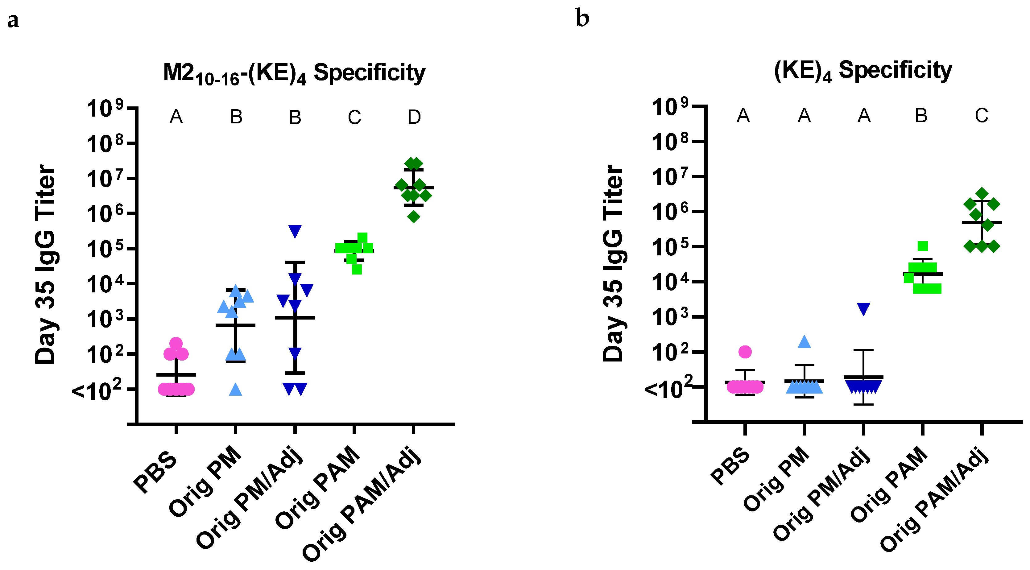
3.1. Palm2K-M22–16-(KE)4 Peptide Amphiphile Micelles Elicited Off-Target Antibody Production

To test whether Palm2K-M22–16-(KE)4 PAMs induced off-target antibodies, mice were vaccinated with Orig PMs (with or without Pam2CSK4 (i.e., Adj)) or Orig PAMs (with or without Adj) on days 0 and 21. Blood was collected on day 35 and used in ELISAs to quantify the antibody response to the vaccine formulations. ELISAs were conducted to evaluate serum antibody specificity to the C-terminus of the Palm2K-M22–16-(KE)4 PA, specifically using coating antigens of M210–16-(KE)4 (Figure 1a) and (KE)4 (Figure 1b). In Figure 1a, M22–16 (i.e., Orig PM) and M22–16/Pam2CSK4 (i.e., Orig PM/Adj) induced moderate anti-M210–16-(KE)4 IgG titers, whereas Orig PAM and Orig PAM/Adj induced higher and the highest titers, respectively. Results were even more stark in Figure 1b, in which there were expectedly no detectable anti-(KE)4 titers in the vaccine groups containing Orig PMs but there were strong titers in both Orig PAM-containing vaccine groups, confirming the presence of off-target antibodies.

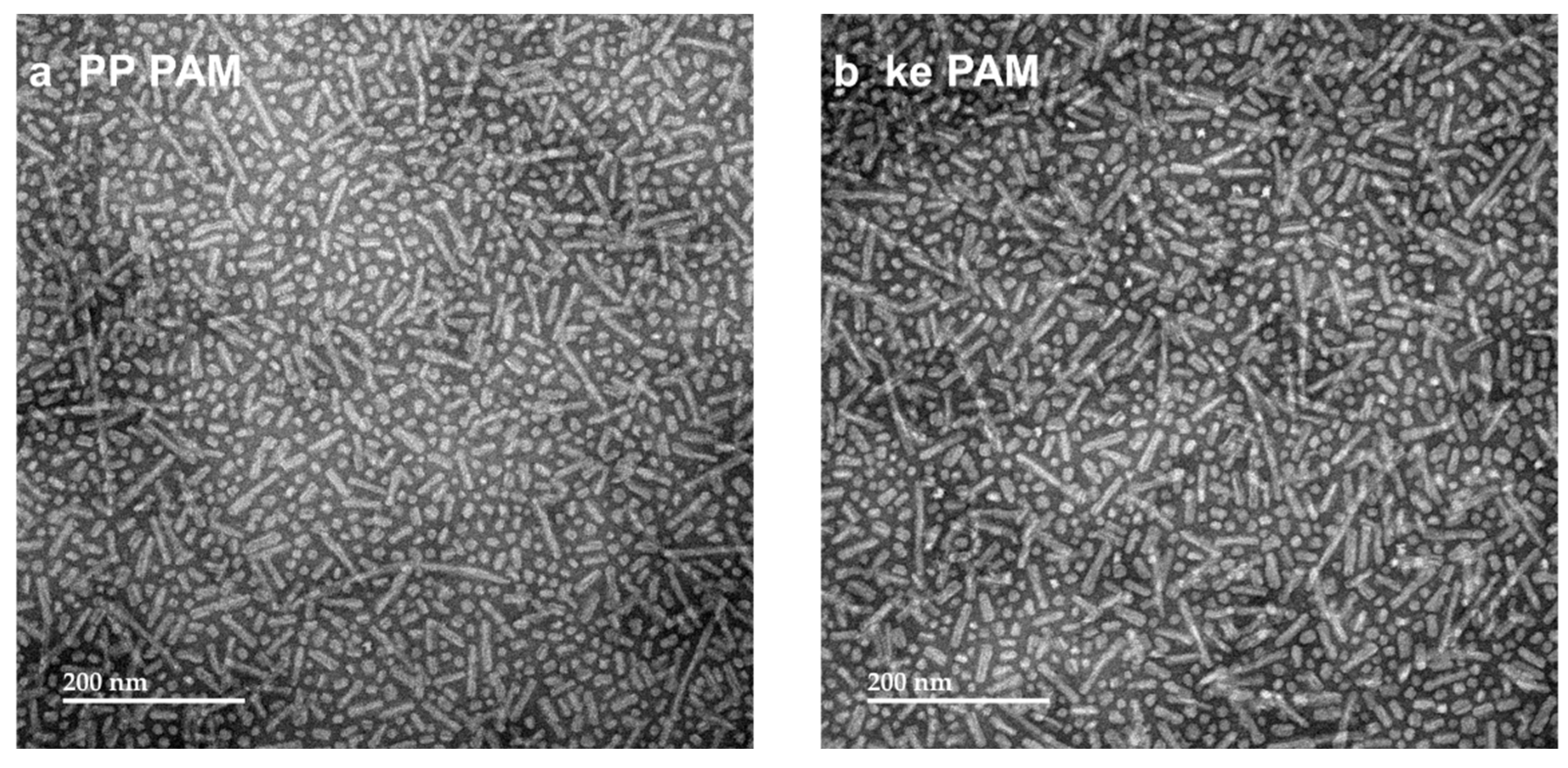
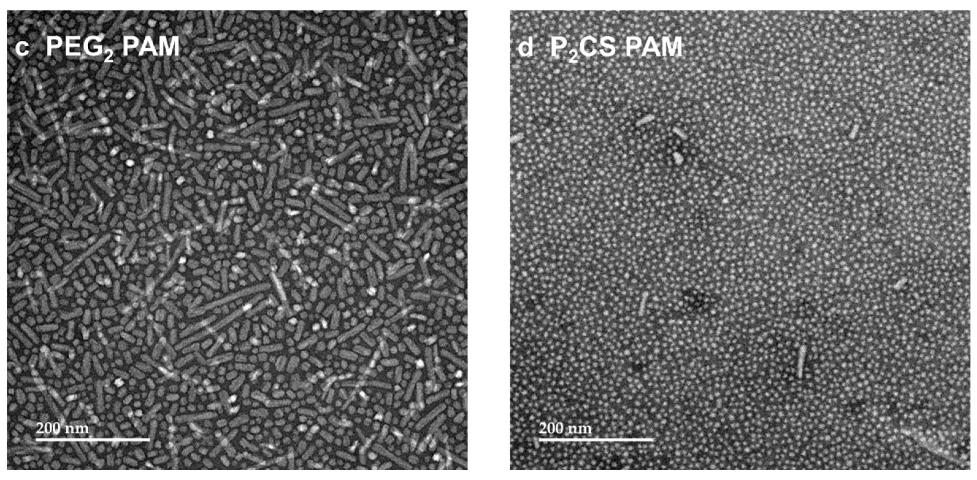
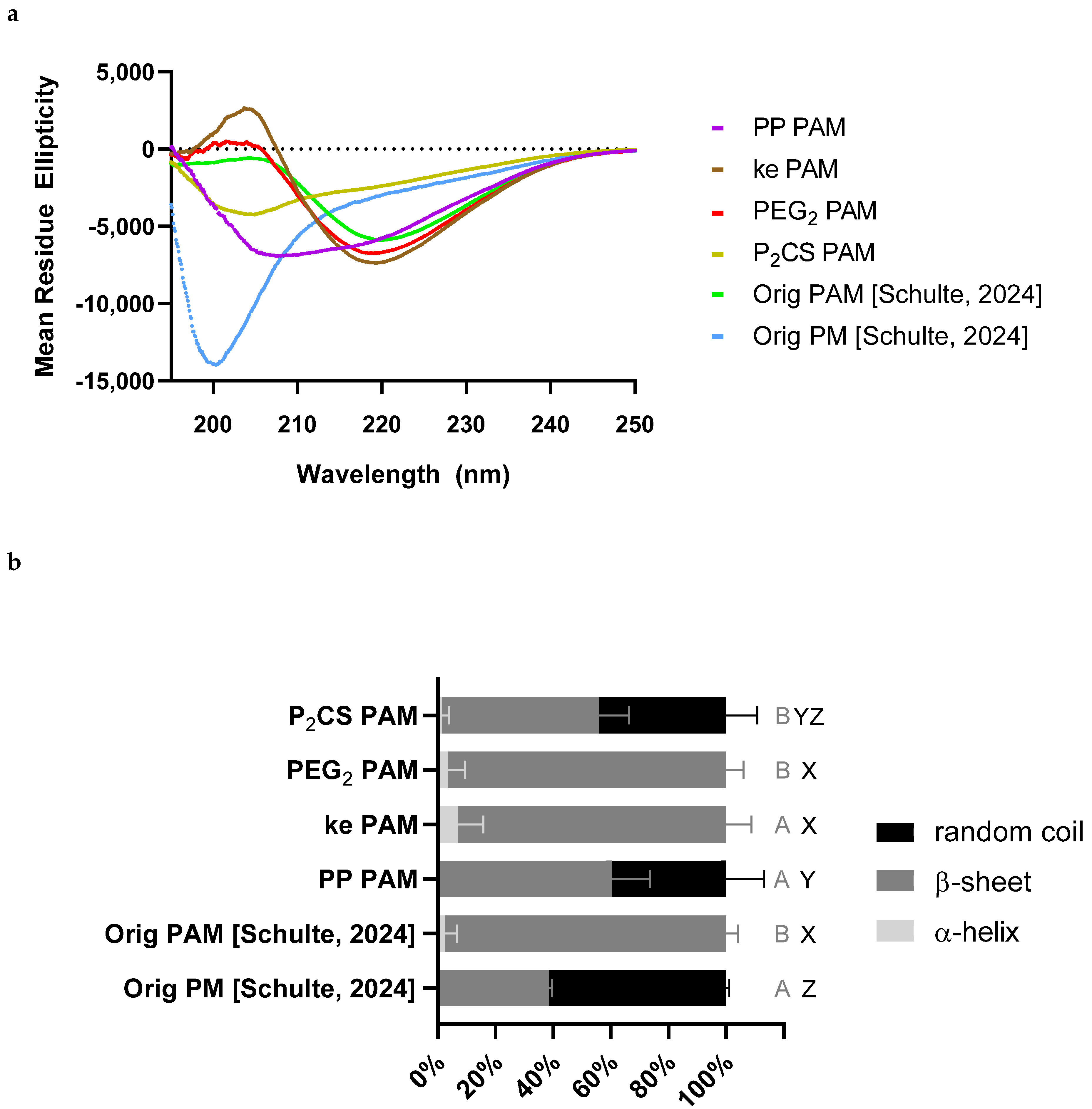
3.2. Proline–Proline and Pam2CS Moieties Induced Slight Changes to the Physical Properties of M22–16-Containing Peptide Amphiphile Micelles

While we have not yet determined whether the off-target antibodies negatively impact PAM vaccine efficacy, we sought to improve the on-target to off-target antibody ratio elicited by PAMs. Based on the data presented in Figure 1 and previous work using the M21–24 antigen, it seemed that antibodies were being produced with partial specificity to both the M2 antigen and the non-native flanking regions (i.e., Palm2K and (KE)4) [14]. Thus, we hypothesized that linkers could be used to alter the PA conformation and/or add distance to prevent antibodies from simultaneously binding to both the antigen and the (KE)4 block.
Several different linkers were tested to investigate how they changed micelle morphology and secondary structure as well as the subsequent antibody response. The specific formulations used are listed in Table 4. Double proline linkers (Figure 2a) on both sides of the antigen were used to disrupt the PA secondary structure because of the kinking that prolines cause due to their secondary α-amine [18,19]. PEG2 linkers (Figure 2b) were used to disrupt the amide backbone of the peptide and add distance and flexibility between the antigen and non-native modifications [20]. The incorporation of D-amino acids into the (KE)4 charge block (instead of the usual L-amino acids) was another approach to disrupt the secondary structure (Figure 2c) [21,22,23,24,25]. Lastly, the method of lipid attachment to the antigen was changed (Figure 2d) by replacing Palm2K (in which the lipids were attached by amide linkages to the α- and ε-amines of the lysine) with Pam2CS (in which both lipids were attached via a glycerol to the thiol of the cysteine). In multiple previous vaccine studies using a variety of antigens, the conjugation of the Pam2C moiety to a peptide antigen has been shown to elicit strong immune responses [26,27,28,29,30,31]. Specifically, the templation of Pam2CS to the N-terminus of the antigen could potentially allow for the lipids of the resulting PA to impart adjuvant activity (as it mimics the structure of the TLR2 agonist Pam2CSK4) in addition to influencing PA off-target antibody production [5,32]. The additional serine between Pam2C and M22–16 was included because it has been shown to be important for TLR2 activation [32].
The new formulations were synthesized and purified by LC-MS (Figure S1). The PAs were characterized by CMC, TEM, and CD. CMC assessments showed that all new PAs formed micelles, with the PP PAMs and P2CS PAMs having slightly higher CMCs compared to Orig PAMs and the other formulations (Table 5). CMC curves are displayed in Figure S3. All formulations were micellized at sufficiently low enough concentrations to ensure sustained micelle presence in vivo [33]. TEM micrographs (Figure 3) showed that, like the original formulation Palm2K-M22–16-(KE)4 (Orig PAM), all of the PAs formed small spherical and/or short cylindrical micelles (with average maximum diameters of 22–24 nm, as seen in Table 6). P2CS PAMs were the only PAMs with noticeably different morphology from Orig PAMs, yielding almost entirely spherical micelles alone (Figure 3d) and an average maximum diameter of 11 ± 4 nm (Table 6).
To further characterize the PAMs, CD was performed on all formulations (Figure 4a) to determine whether any of the linkers produced a substantial change in the secondary structure from the Orig PAMs (i.e., Palm2K-M22–16-(KE)4). The PA’s secondary structure was estimated from the CD spectra using known reference curves (Figure 4b). All micelle formulations showed predominantly β-sheet character and minimal α-helical behavior (10% or less), with the secondary structures of ke PAMs and PEG2 PAMs closely aligning with that of the Orig PAMs. Lipidating the peptide using Pam2Cys (i.e., in P2CS PAMs) and using double-proline spacers between the antigen and flanking regions of the peptide (i.e., in PP PAMs) caused secondary structure changes that reduced the β-sheet presence and increased random coil content. The addition of PEG2 linkers between the antigen and non-native regions (i.e., in PEG2 PAMs and ke PAMs) did not introduce secondary structure changes.

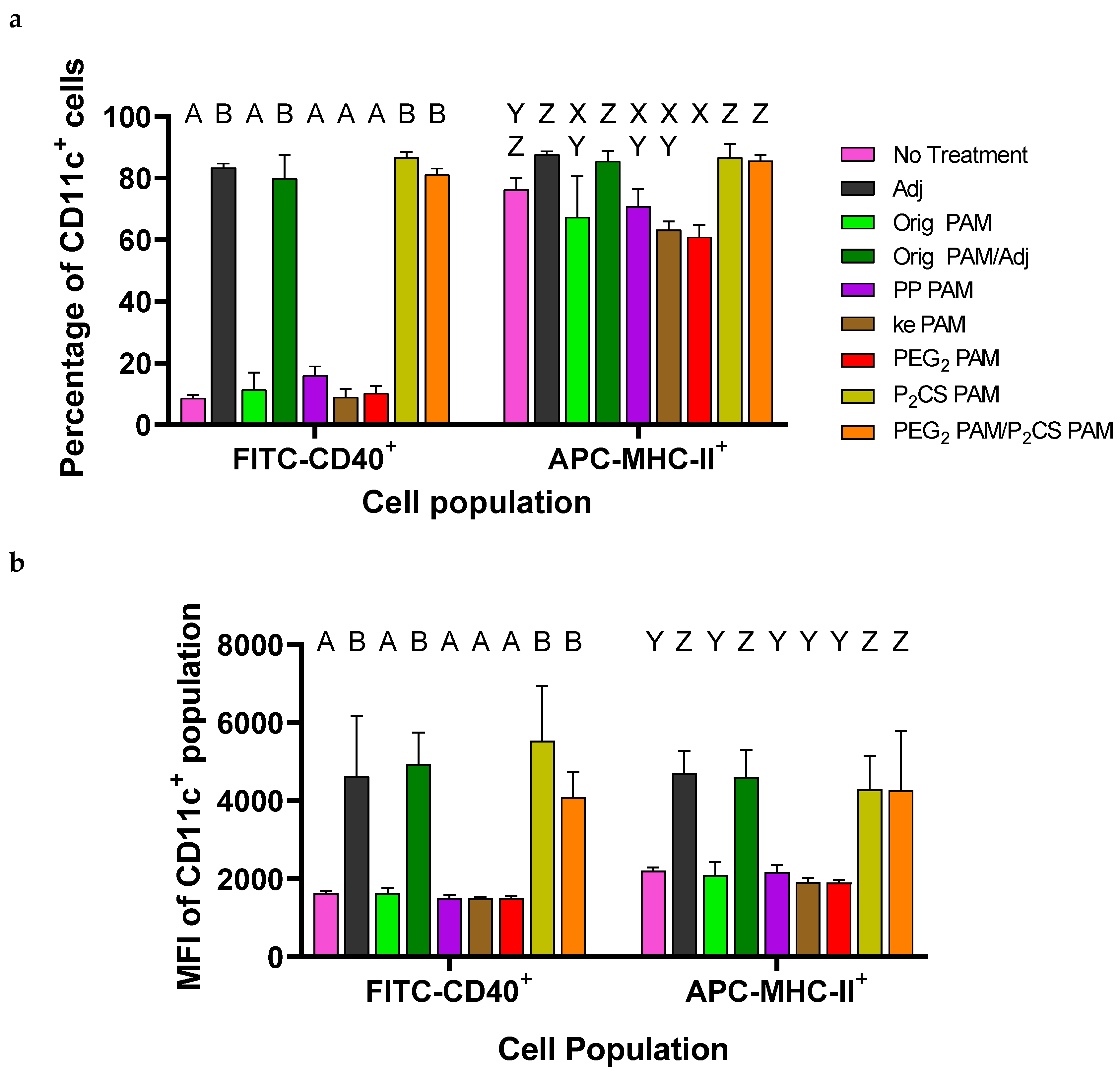
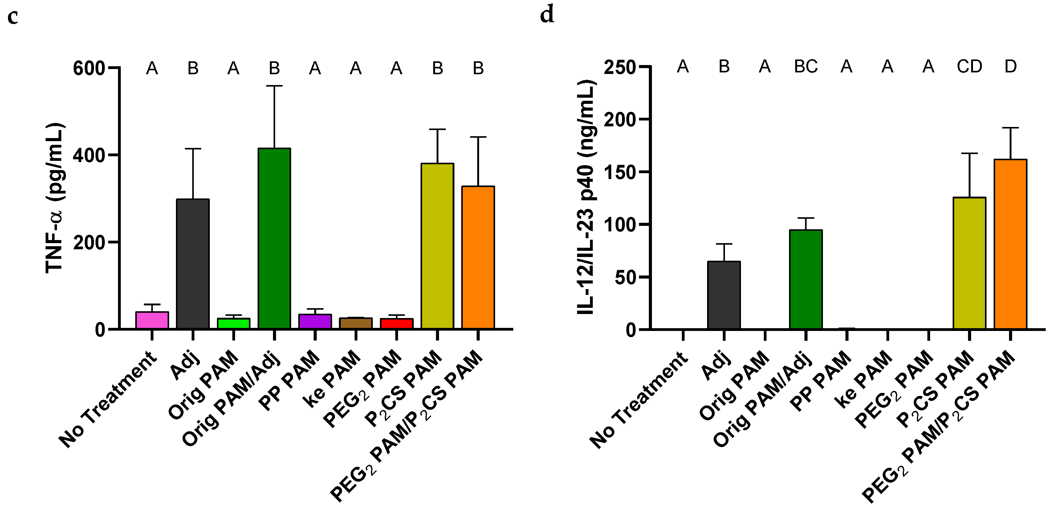
3.3. Bone-Marrow-Derived Dendritic Cells Were Activated by P2CS PAMs

To evaluate whether changes in micelle morphology would influence innate immune responses against the linker-containing PAMs when compared to Orig PAMs, BMDCs were cultured and treated with the various formulations. After 24 h, the media were removed from the cells and saved to evaluate cytokine secretion using ELISAs. Cells were stained with PE-Cy7 anti-mouse CD11c, FITC anti-mouse CD40, and APC anti-mouse MHC-II antibodies and processed for flow cytometry. BMDC activation was detected by upregulated CD40 and/or MHC-II expression on the CD11c+ cells. The percentage of CD11c+ cells expressing elevated levels of CD40 was highest in treatment groups containing Adj (i.e., Pam2CSK4) or P2CS PAMs (Figure 5a). To a much lesser extent, MHC-II+CD11c+ cell percentages were also elevated in adjuvant-containing groups but without statistical significance as the No Treatment group possessed a high percentage of these cells already. Further evidence of BMDC activation by adjuvants was seen by the increased FITC median fluorescence intensity (MFI) in CD40+CD11c+ cells and increased APC MFI in MHC-II+CD11c+ cells (Figure 5b). Flow cytometry data did not indicate significant BMDC activation in the PP PAM, ke PAM, or PEG2 PAM treatment groups in the absence of an adjuvant.
To further determine BMDC activation, sandwich ELISAs were conducted using the media collected from the treated BMDCs. These assays quantified pro-inflammatory cytokine secretion—in particular TNF-α (Figure 5c) and IL-12/IL-23 p40 (Figure 5d)—as these two have been commonly shown to be cell signaling markers of activated BMDCs [34,35,36]. In reasonable agreement with the flow cytometry data, increased secretions of TNF-α and IL-12/IL-23 p40 were seen in cells treated with adjuvant (again, either Pam2CSK4 or P2CS PAMs), while groups treated with PAMs without adjuvant exhibited similar levels of TNF-α and IL-12/IL-23 p40 secretion to the No Treatment group.

3.4. Linker-Containing Peptide Amphiphile Micelles Elicited Off-Target IgG Antibodies but P2CS PAMs Also Elicited Strong On-Target Titers

To evaluate the immunogenicity of the linker-containing PAMs in vivo, mice were administered two doses of PAM vaccines (on days 0 and 21), using the same protocol described above. The vaccine groups can be found in Table 3. In addition to testing the in vivo immunogenicity of each linker-containing PAM individually, an additional group was included to test the adjuvant effect of P2CS PAMs at a dose equivalent to the dose of Pam2CSK4 in the Orig PAM/Adj vaccine group (i.e., 2.22 nmol P2CS PAMs co-delivered with 20 nmol PEG2 PAMs). Blood was collected on day 35 and used in ELISAs to evaluate antibody content. Serum antibody titers specific to M21–24 (the complete ectodomain of the influenza M2 protein) were quantified to measure on-target antibody specificity (Figure 6a). Excitingly, P2CS PAM-vaccinated mice exhibited high on-target IgG titers similar to that in Orig PAMs/Adj-vaccinated mice. The remaining linker-containing PAMs (including the PEG2 PAM/P2CS PAM group) elicited significantly weaker on-target titers against M21–24, on par or below the titers of non-adjuvanted Orig PAMs. To test whether these PAMs were also inducing off-target antibodies, an ELISA testing antibody specificity against (KE)4 was conducted (Figure 6b). This test confirmed the existence of somewhat comparable amounts of off-target antibodies among all linker-containing PA formulations, although anti-(KE)4 titers were statistically significantly higher among P2CS PAM-containing vaccine groups than in the PP PAM and ke PAM groups. Interestingly, when the ratios of on-target to off-target antibody titers were calculated for each animal using anti-M21–24 and anti-(KE)4 titers, these were found to be more favorably skewed toward on-target antibody production for adjuvant-containing vaccine groups, with P2CS PAMs having a slightly higher ratio than Orig PAM/Adj (Figure 6c).

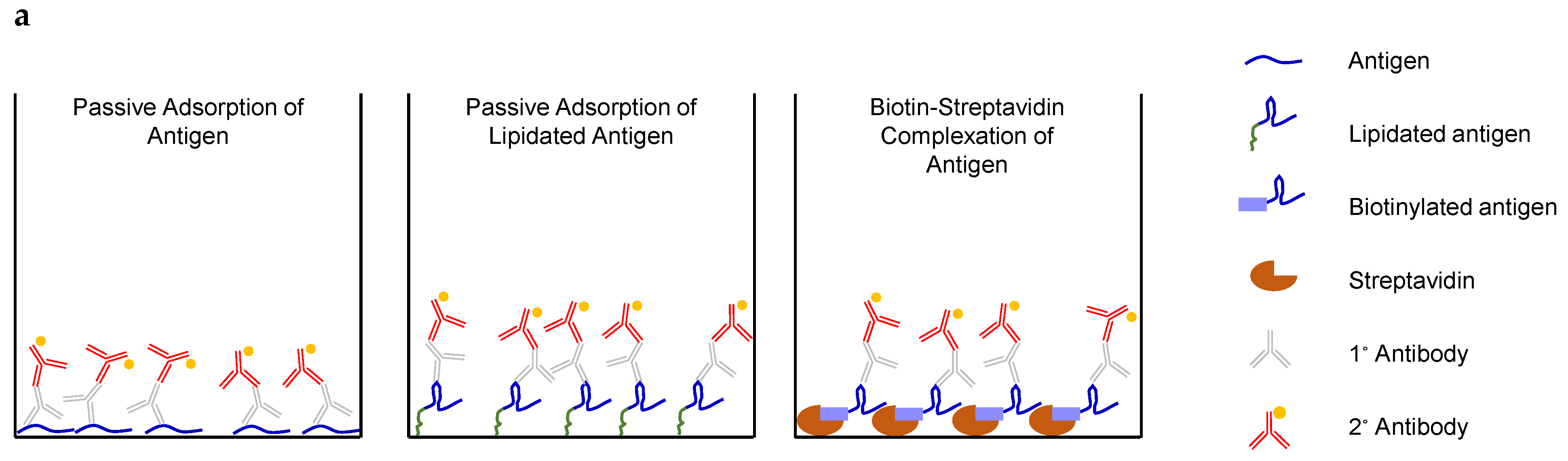
3.5. IgG Titers Were More Dependent on the Sequence than the Attachment Method of the Coating Antigen

While it is evident that significant quantities of off-target antibodies were elicited by the linker-containing PAMs, we sought to determine if there were any additional explanations for the lower-than-expected on-target antibody titers in the PAM groups relative to the Orig PM/Adj (i.e., M22–16/Pam2CSK4) group. We hypothesized that, especially given the commonality of conformational B-cell epitopes [37], the currently used method of attaching the coating antigen to the ELISA plate (i.e., passive adsorption) could have a confounding effect on the antibody titers detected. This would be especially true if the coating antigen did not favorably present the target epitope for antibody binding, something that has been previously shown in the literature [38,39,40,41,42].
To investigate this possible effect, a few immobilization approaches were tested (Figure 7a). As has been used previously, the first strategy was passive adsorption of the unmodified M22–16 antigen, in which the coating antigen associates with the high-binding surface of a Maxisorp plate via hydrophobic and other non-covalent forces. In this case, the truncated M22–16 antigen was used, rather than the previously employed M21–24 coating antigen, to allow for more equivalent comparisons to the other coating approaches. The second strategy involved passive adsorption of the Palm2K-PEG2-M22–16-PEG2-(KE)4 PA to the plate to test whether the lipids—rather than the peptidyl portion of the PA—preferentially bind to the hydrophobic plate surface, thus potentially affecting antigen display. This approach also allowed us to test for the presence of any N-terminal-specific off-target antibodies. The last strategy was streptavidin complexation with biotinylated analogs of either M22–16 peptide or Palm2K-PEG2-M22–16-PEG2-(KE)4 PA. To achieve this, streptavidin was first adsorbed to the plate surface, then the biotinylated “coating” antigen (either Biotin-PEG2-M22–16 or Biotin-PEG2-M22–16-PEG2-(KE)4, the analog to the PA coating antigen) added to initiate complexation. The rationale for biotin–streptavidin complexation was that this method could allow the antigen more conformational freedom due to its single anchoring point compared to the two-dimensional constraints of the unspecific passive adsorption method.
Interestingly, the IgG titers against the biotinylated coating antigens (Figure 7b) largely aligned with those of their analogous unbiotinylated coating antigens (i.e., for most cases, the light and dark blue bars were similar to each other and the light and dark red bars aligned) for each given vaccine group. For the non-PAM vaccine groups (i.e., PBS and Orig PM/Adj), IgG titers were also very consistent across all four coating antigens tested. The titers for each PAM vaccine group, however, were significantly higher for the coating antigen Palm2K-PEG2-M22–16-PEG2-(KE)4 (light red bars) and its biotinylated analog (i.e., Biotin-PEG2-M22–16-PEG2-(KE)4, dark red bars) than for the coating antigen M22–16 (light blue bars) and its biotinylated analog (i.e., Biotin-PEG2-M22–16, dark blue bars). For coating antigens M22–16 and Biotin-PEG2-M22–16, the presence of adjuvant in the vaccine was the biggest predictor of high titers, as seen for the coating antigen M21–24 in Figure 6a. Notably, titers induced by PAM vaccine groups without an adjuvant (i.e., Orig PAM, PP PAM, ke PAM, and PEG2 PAM) were lower than Orig PM/Adj for these two coating antigens. However, for the PA coating antigen and its biotinylated analog, PAM vaccines (with or without adjuvant) elicited comparable or higher titers than Orig PM/Adj.
To further probe this effect, a final ELISA was conducted, employing the passive adsorption method of attachment, using the coating antigen Palm-RDRD-M22–16 (Figure 7c). This PA was chosen to evaluate antibody binding to a lipidated M22–16, while eliminating the non-native peptidyl modifications used in Palm2K-M22–16-(KE)4 and the linker-containing PAs to avoid capturing off-target antibodies. The RDRD repeat was specifically chosen to improve solubility of the PA. IgG titers captured by Palm-RDRD-M22–16 largely aligned with trends seen in Figure 6a (using M21–24 as the coating antigen) and Figure 7b (using M22–16 or Biotin-PEG2-M22–16 as the coating antigen), namely that titers were highest among vaccine groups containing adjuvant (i.e., Pam2CSK4) or the higher concentration of P2CS PAMs.

4. Discussion

In this work, we showed that Palm2K-M22–16-(KE)4 PAMs, like the previously studied Palm2K-M21–24-(KE)4 PAMs, induced off-target antibody production (Figure 1); however, we were able to significantly improve the on-target to off-target antibody ratio by templating a Pam2CS moiety to the N-terminus of the M2 antigen, as shown with P2CS PAMs [14]. That being said, we have yet to determine whether this off-target antibody production is actually detrimental to the efficacy of these PAM vaccines. However, given that in other studies there was an enhancement in IgG titer from unmodified OVABT or J8 peptides to their analogous PAMs but not in our studies using M2e antigens (i.e., M22–16 or M21–24 peptide), it is reasonable to suggest that Palm2K-M22–16 or 1–24-(KE)4 PAMs could induce higher on-target titers in the absence of immune distraction [5,12,15].
As such, we tested different strategies to prevent the production of off-target antibodies by adding linkers between the antigen and non-native moieties of the PA. Given that the general configuration of Palm2K-antigen-(KE)4 was preserved in the linker-containing formulations (which has been previously shown in multiple cases to elicit small micelles), it is not surprising that all of the linker-containing PAs formed small spherical and/or cylindrical micelles, as shown by TEM in Figure 3 [14,43,44]. The slightly different lipid “orientation” from the Pam2CS moiety in the P2CS PAMs compared to the other PAMs (i.e., those lipidated with dipalmitoyllysine) seemed to have the most significant effect on micelle shape as Pam2CS-M22–16-PEG2-(KE)4 was the only PA that formed almost entirely spherical micelles alone. This aligned with the mostly spherical morphology that has been previously observed in Pam2CSK4-only micelles [15,45]. More significant differences between the formulations were seen in the peptide secondary structure (Figure 4). Specifically, P2CS PAMs possessed more disordered random coil structure content than Orig PAMs, likely due to their increased sphericity, as was observed with Orig PMs (i.e., M22–16 peptide) and has also been noted in the literature [3,15,46]. The increased random coil content in the P2CS PAMs could also likely be attributed to the Pam2CS moiety as the CD of Pam2CSK4 (Figure S4) showed a secondary structure composition of 81.6% random coil and 18.4% β-sheet, which agrees with previously published results [45]. In the PP PAMs, the reduced percentage of β-sheet content could be attributed to the addition of the proline–proline linkers as this same effect has been shown when substituting a serine for a proline in Alzheimer’s disease-associated Tau protein [19]. As for the lack of conformational changes in the PEG2-containing PAMs, although polyethylene glycols are known for their ability to increase the flexibility of polymers, PEG2 is only a dimer of ethylene glycol, which is likely too short to induce any significant changes in the peptide secondary structure [20]. Finally, the lack of any structural changes in the ke PAM was unexpected due to the frequent use of D-amino acids in the literature to induce functional or structural changes in peptides [21,22,23,24,25]. Further, D-amino acids have been used specifically for the disruption of β-sheets and affecting antibody recognition, thus it is likely that the location of the substitutions or the number of D-amino acids for the studied product was insufficient to induce structural changes [21,24].
The in vitro bioactivity of the linker-containing PAMs (Figure 5) agreed with previous work in that PAMs alone were not strongly immunogenic but rather, the presence of adjuvant was the primary driver of BMDC activation [5,15]. The use of most PA linkers did not induce BMDC activation besides P2CS PAMs, which were excitingly found to be immunogenic. This result suggested that the P2CS PAMs possessed a functional adjuvant, most likely by maintaining the TLR2/TLR6-agonist behavior found with Pam2CSK4. Activation of these TLRs together triggers a signal cascade that stimulates the secretion of pro-inflammatory cytokines including TNF-α, IL-12, and IL-23, as well as the increased expression of MHC-II and CD40—among other maturation markers—in BMDCs [36,47,48,49]. This aligns well with the signs of activation observed with the P2CS PAM treatment. The adjuvant activity of P2CS PAMs was especially notable given that our group has previously shown that templating the OVABT antigen with just Pam2C (i.e., Pam2C-OVABT-(KE)4) was insufficient to increase the immunogenicity of the PA [5]. The results here lend credence to the suggestion that the serine residue is indeed important for TLR2 activation by Pam2C, as has been shown in the literature [32,50].
The in vivo immunogenicity of the PAMs aligned well with the BMDC results. Distinctly, high on-target IgG titers in P2CS PAM-vaccinated mice provided additional evidence that the templation of Pam2CS to the N-terminus of the M22–16 antigen allowed the PAM to act both as an adjuvant and an antigen (Figure 6a). The similar anti-M21–24 titers between P2CS PAMs and Orig PAMs/Adj, despite the higher dosage of the Pam2CS moiety in the P2CS PAMs (i.e., 20 nmol vs. 2.22 nmol, respectively), suggested however that unmodified Pam2CSK4 was a more potent adjuvant. The lower anti-M21–24 titers in the PEG2 PAM/P2CS PAM vaccine, despite equimolar amounts of Pam2CS moiety to the Orig PAM/Adj vaccine, further confirmed this. While the K4 region of Pam2CSK4 does not play a primary role in TLR2/TLR6 activation by Pam2CSK4, this region does form weak ionic and hydrogen bonds with the TLR so it is possible that the absence of a cationic K4 region resulted in some loss of adjuvant activity in the P2CS PAMs (compared to Orig PAMs/Adj) [32,51]. Another possibility is that steric hindrance due to the presence of the M22–16 antigen is affecting the binding of the P2CS PAMs to the TLRs.
As seen from the anti-(KE)4 ELISA results (Figure 6b), off-target antibody production still occurred in response to vaccination with all of the linker-containing PAMs. Nonetheless, the fact that the P2CS PAMs elicited a relatively comparable anti-(KE)4 titer—although statistically distinct—to the other linker-containing PAMs while displaying a much stronger on-target anti-M21–24 titer is worth noting. When comparing the ratio of on-target to off-target antibodies between all the PAM groups, Orig PAM/Adj, and especially P2CS PAMs, had more favorable ratios skewed toward relatively similar on-target to off-target antibody production. In contrast, all other PAM groups possessed a ratio well below one, indicating off-target antibodies were produced in much higher quantities than on-target antibodies.
In an effort to ascertain whether the observed antibody titers were solely due to off-target antibody production, we found that differences in PAM vaccine titers between the ELISA coating antigens aligned more closely with the presence or absence of the (KE)4 region in the coating antigen than their attachment method (Figure 7b). The consistency in titers between the coating antigens (i.e., M22–16 or Palm2K-PEG2-M22–16-PEG2-(KE)4) and their biotinylated analogs suggests the validity of the streptavidin–biotin coating strategy as an alternative to the passive adsorption coating method. This technique would be quite beneficial for when passive adsorption prevents antibody access to its cognate epitope within the coating peptide/protein. Furthermore, the similarity in titers between the PA coating antigen and its biotinylated analog demonstrated the lack of off-target antibodies specific to the N-terminus due to the fact that the lipidated PA coating antigen did not capture higher titers relative to the M22–16 coating antigen. Further investigation into this behavior demonstrated that a coating antigen of Palm-RDRD-M22–16 produced results that aligned very closely with the M21–24 and M22–16 ELISAs (Figure 7c), despite the presence of a palmitoyl group on the N-terminus of the coating antigen. Thus, any hypothetical conformational constraints on the coating antigen did not appreciably impact antibody titers. This provided additional evidence that titer differences between the vaccine groups were indeed due to the presence of antibodies specific to a non-antigenic component of the PA (i.e., (KE)4). It is also important to note, however, that the Maxisorp ELISA plates used in this study were sufficiently amphiphilic to favorably bind both peptides and peptide amphiphiles—without a strong preference for lipid binding—but plates using other surface treatments not optimized for peptide adsorption could elicit different results [52,53,54].

5. Conclusions

While Palm2K-M22–16-(KE)4 PAMs elicited strong immune responses in vitro and in vivo when co-delivered with an adjuvant, the non-native (KE)4 charge block induced substantial production of off-target antibodies specific to the (KE)4 peptide. PA formulations containing proline–proline linkers flanking the M22–16 antigen or an N-terminal Pam2CS moiety induced some changes to their critical micelle concentration, morphology, and secondary structure but all formulations tested still induced a significant off-target antibody response against the (KE)4 region of the PAs. While the linkers tested did not abrogate off-target antibody production, we found that templation of the M22–16 peptide by a Pam2CS moiety to yielded a PA (i.e., Pam2CS-M22–16-PEG2-(KE)4) that mimics the adjuvant activity of Pam2CSK4, which resulted in desirable BMDC activation and elevated antibody titers with a more favorable on-target to off-target ratio. The covalent attachment of an adjuvant and antigen could be especially beneficial for applications where localized co-delivery of both components is critical, such as with in situ vaccines.

Supplementary Materials

The following supporting information can be downloaded at https://www.mdpi.com/article/10.3390/vaccines13040422/s1, Figure S1: Peptides and peptide amphiphiles were purified to greater than 90% purity using LC-MS; Figure S2: The gating strategy used for identifying activated bone-marrow-derived dendritic cells is illustrated above; Figure S3: Representative graphs of critical micelle concentrations showed that all peptide amphiphiles formed micelles; Figure S4: Circular dichroism of Pam2CSK4 aligns with a primarily random coil secondary structure as indicated by a minimum at slightly below 200 nm; Table S1. Statistical groups for each coating antigen in Figure 7; Table S2. Statistical groups for each vaccine in Figure 7b.

Author Contributions

Conceptualization: M.C.S., A.G.S. and B.D.U.; methodology: M.C.S., A.C.B., N.L.C., F.R. and A.T.B.; data curation: M.C.S.; writing—original draft preparation: M.C.S.; writing—review and editing: M.C.S., A.G.S. and B.D.U.; funding acquisition: B.D.U. All authors have read and agreed to the published version of the manuscript.

Funding

This research was supported by funding provided by the University of Missouri System to the Center for Vector-Borne and Emerging Infectious Diseases (CVBEID).

Institutional Review Board Statement

The animal study protocol was approved by the Animal Care and Use Committee of the University of Missouri (protocol code, 32204; approved 7 December 2021).

Informed Consent Statement

Not applicable.

Data Availability Statement

All data are contained within the article and Supplementary Materials.

Acknowledgments

The authors would like to thank Fabio Gallazzi for his assistance with peptide synthesis and the Molecular Interactions Core (University of Missouri) for usage of their Tetras Peptide Synthesizer, LC-MS, and J-1500 circular dichroism spectrophotometer. We also wish to express our appreciation to the Electron Microscopy Core (University of Missouri) for access to the JEOL JEM-1400 Transmission Electron Microscope.

Conflicts of Interest

The authors declare no conflicts of interest.

References

  1. Poon, C.; Gallo, J.; Joo, J.; Chang, T.; Bañobre-López, M.; Chung, E.J. Hybrid, metal oxide-peptide amphiphile micelles for molecular magnetic resonance imaging of atherosclerosis. J. Nanobiotechnol. 2018, 16, 92. [Google Scholar] [CrossRef] [PubMed]
  2. Chung, E.J.; Cheng, Y.; Morshed, R.; Nord, K.; Han, Y.; Wegscheid, M.L.; Auffinger, B.; Wainwright, D.A.; Lesniak, M.S.; Tirrell, M.V. Fibrin-binding, peptide amphiphile micelles for targeting glioblastoma. Biomaterials 2014, 35, 1249–1256. [Google Scholar] [CrossRef]
  3. Smith, J.D.; Cardwell, L.N.; Porciani, D.; Nolla, A.; Cornelison, B.T.; Schulte, M.C.; Gallazzi, F.; Burke, D.H.; Daniels, M.A.; Ulery, B.D. Therapeutic peptide delivery: Via aptamer-displaying, disulfide-linked peptide amphiphile micelles. Mol. Syst. Des. Eng. 2020, 5, 269–283. [Google Scholar] [CrossRef]
  4. Rodrigues de Almeida, N.; Han, Y.; Perez, J.; Kirkpatrick, S.; Wang, Y.; Sheridan, M.C. Design, Synthesis, and Nanostructure-Dependent Antibacterial Activity of Cationic Peptide Amphiphiles. ACS Appl. Mater. Interfaces 2019, 11, 2790–2801. [Google Scholar] [CrossRef] [PubMed]
  5. Zhang, R.; Kramer, J.S.; Smith, J.D.; Allen, B.N.; Leeper, C.N.; Li, X.; Morton, L.D.; Gallazzi, F.; Ulery, B.D. Vaccine Adjuvant Incorporation Strategy Dictates Peptide Amphiphile Micelle Immunostimulatory Capacity. AAPS J. 2018, 20, 73. [Google Scholar] [CrossRef]
  6. Boato, F.; Thomas, R.M.; Ghasparian, A.; Freund-Renard, A.; Moehle, K.; Robinson, J.A. Synthetic Virus-Like Particles from Self-Assembling Coiled-Coil Lipopeptides and Their Use in Antigen Display to the Immune System. Angew. Chem. Int. Ed. 2007, 46, 9015–9018. [Google Scholar] [CrossRef]
  7. Lee, K.C.; Carlson, P.A.; Goldstein, A.S.; Yager, P.; Gelb, M.H. Protection of a Decapeptide from Proteolytic Cleavage by Lipidation and Self-Assembly into High-Axial-Ratio Microstructures:  A Kinetic and Structural Study. Langmuir 1999, 15, 5500–5508. [Google Scholar] [CrossRef]
  8. Missirlis, D.; Khant, H.; Tirrell, M. Mechanisms of peptide amphiphile internalization by SJSA-1 Cells in Vitro. Biochemistry 2009, 48, 3304–3314. [Google Scholar] [CrossRef]
  9. Henninot, A.; Collins, J.C.; Nuss, J.M. The Current State of Peptide Drug Discovery: Back to the Future? J. Med. Chem. 2018, 61, 1382–1414. [Google Scholar] [CrossRef] [PubMed]
  10. Banerjee, A.; Onyuksel, H. Peptide delivery using phospholipid micelles. Wiley Interdiscip. Rev. Nanomed. Nanobiotechnol. 2012, 4, 562–574. [Google Scholar] [CrossRef]
  11. Hald Albertsen, C.; Kulkarni, J.A.; Witzigmann, D.; Lind, M.; Petersson, K.; Simonsen, J.B. The role of lipid components in lipid nanoparticles for vaccines and gene therapy. Adv. Drug Deliv. Rev. 2022, 188, 114416. [Google Scholar] [CrossRef] [PubMed]
  12. Barrett, J.C.; Ulery, B.D.; Trent, A.; Liang, S.; David, N.A.; Tirrell, M.V. Modular Peptide Amphiphile Micelles Improving an Antibody-Mediated Immune Response to Group A Streptococcus. ACS Biomater. Sci. Eng. 2017, 3, 144–152. [Google Scholar] [CrossRef] [PubMed]
  13. Zhang, R.; Smith, J.D.; Allen, B.N.; Kramer, J.S.; Schauflinger, M.; Ulery, B.D. Peptide Amphiphile Micelle Vaccine Size and Charge Influence the Host Antibody Response. ACS Biomater. Sci. Eng. 2018, 4, 2463–2472. [Google Scholar] [CrossRef]
  14. Schulte, M.C.; Boll, A.C.; Barcellona, A.T.; Lopez, E.A.; Schrum, A.G.; Ulery, B.D. Peptide Antigen Modifications Influence the On-Target and Off-Target Antibody Response for an Influenza Subunit Vaccine. Vaccines 2025, 13, 51. [Google Scholar] [CrossRef] [PubMed]
  15. Schulte, M.C.; Barcellona, A.T.; Wang, X.; Schrum, A.G.; Ulery, B.D. M2e-Derived Peptidyl and Peptide Amphiphile Micelles as Novel Influenza Vaccines. Pharmaceuticals 2024, 17, 1503. [Google Scholar] [CrossRef]
  16. Tai, W.; Chen, J.; Zhao, G.; Geng, Q.; He, L.; Chen, Y.; Zhou, Y.; Li, F.; Du, L. Rational Design of Zika Virus Subunit Vaccine with Enhanced Efficacy. J. Virol. 2019, 93, e02187-18. [Google Scholar] [CrossRef]
  17. Duan, H.; Chen, X.; Boyington, J.C.; Cheng, C.; Zhang, Y.; Jafari, A.J.; Stephens, T.; Tsybovsky, Y.; Kalyuzhniy, O.; Zhao, P.; et al. Glycan Masking Focuses Immune Responses to the HIV-1 CD4-Binding Site and Enhances Elicitation of VRC01-Class Precursor Antibodies. Immunity 2018, 49, 301–311.e5. [Google Scholar] [CrossRef]
  18. Deupi, X.; Olivella, M.; Govaerts, C.; Ballesteros, J.A.; Campillo, M.; Pardo, L. Ser and Thr Residues Modulate the Conformation of Pro-Kinked Transmembrane α-Helices. Biophys. J. 2004, 86, 105–115. [Google Scholar] [CrossRef]
  19. Lang, E.; Otvos, L. A serine → proline change in the Alzheimer’s disease-associated epitope tau 2 results in altered secondary structure, but phosphorylation overcomes the conformational GAP. Biochem. Biophys. Res. Commun. 1992, 188, 162–169. [Google Scholar] [CrossRef]
  20. Abe, K.; Higashi, K.; Watabe, K.; Kobayashi, A.; Limwikrant, W.; Yamamoto, K.; Moribe, K. Effects of the PEG molecular weight of a PEG-lipid and cholesterol on PEG chain flexibility on liposome surfaces. Colloids Surf. A Physicochem. Eng. Asp. 2015, 474, 63–70. [Google Scholar] [CrossRef]
  21. Uray, K.; Kajtár, J.; Vass, E.; Price, M.R.; Hollósi, M.; Hudecz, F. Effect of D-amino acid substitution in a mucin 2 epitope on mucin- specific monoclonal antibody recognition. Arch. Biochem. Biophys. 2000, 378, 25–32. [Google Scholar] [CrossRef] [PubMed]
  22. Di Grazia, A.; Cappiello, F.; Cohen, H.; Casciaro, B.; Luca, V.; Pini, A.; Di, Y.P.; Shai, Y.; Mangoni, M.L. D-Amino acids incorporation in the frog skin-derived peptide esculentin-1a(1-21)NH2 is beneficial for its multiple functions. Amino Acids 2015, 47, 2505–2519. [Google Scholar] [CrossRef] [PubMed]
  23. Hong, S.Y.; Oh, J.E.; Lee, K.H. Effect of D-amino acid substitution on the stability, the secondary structure, and the activity of membrane-active peptide. Biochem. Pharmacol. 1999, 58, 1775–1780. [Google Scholar] [CrossRef]
  24. Lee, D.L.; Powers, J.P.S.; Pflegerl, K.; Vasil, M.L.; Hancock, R.E.W.; Hodges, R.S. Effects of single d-amino acid substitutions on disruption of β-sheet structure and hydrophobicity in cyclic 14-residue antimicrobial peptide analogs related to gramicidin S. J. Pept. Res. 2004, 63, 69–84. [Google Scholar] [CrossRef] [PubMed]
  25. Rothemund, S.; Beyermann, M.; Krause, E.; Krause, G.; Bienert, M.; Hodges, R.S.; Sykes, B.D.; Sönnichsen, F.D. Structure Effects of Double d-Amino Acid Replacements: A Nuclear Magnetic Resonance and Circular Dichroism Study Using Amphipathic Model Helices. Biochemistry 1995, 34, 12954–12962. [Google Scholar] [CrossRef]
  26. Moyle, P.M.; Dai, W.; Zhang, Y.; Batzloff, M.R.; Good, M.F.; Toth, I. Site-Specific Incorporation of Three Toll-Like Receptor 2 Targeting Adjuvants into Semisynthetic, Molecularly Defined Nanoparticles: Application to Group A Streptococcal Vaccines. Bioconjug. Chem. 2014, 25, 965–978. [Google Scholar] [CrossRef]
  27. Moyle, P.M.; Dai, W.; Liu, T.Y.; Hussein, W.M.; Maruthayanar, P.; Wells, J.W.; McMillan, N.A.J.; Skwarczynski, M.; Toth, I. Combined synthetic and recombinant techniques for the development of lipoprotein-based, self-adjuvanting vaccines targeting human papillomavirus type-16 associated tumors. Bioorg. Med. Chem. Lett. 2015, 25, 5570–5575. [Google Scholar] [CrossRef]
  28. Tan, A.C.L.; Eriksson, E.M.Y.; Kedzierska, K.; Deliyannis, G.; Valkenburg, S.A.; Zeng, W.; Jackson, D.C. Polyfunctional CD8 + T cells are associated with the vaccination-induced control of a novel recombinant influenza virus expressing an HCV epitope. Antivir. Res. 2012, 94, 168–178. [Google Scholar] [CrossRef]
  29. Zeng, W.; Ghosh, S.; Lau, Y.F.; Brown, L.E.; Jackson, D.C. Highly Immunogenic and Totally Synthetic Lipopeptides as Self-Adjuvanting Immunocontraceptive Vaccines1. J. Immunol. 2002, 169, 4905–4912. [Google Scholar] [CrossRef]
  30. Ng, W.C.; Gilbertson, B.; Lim, B.; Zeng, W.; Jackson, D.C.; Brown, L.E. Lipopeptide vaccines illustrate the potential role of subtype-crossreactive T cells in the control of highly virulent influenza. Influenza Other Respir. Viruses 2009, 3, 177–182. [Google Scholar] [CrossRef]
  31. Zeng, W.; Tan, A.C.L.; Horrocks, K.; Jackson, D.C. A lipidated form of the extracellular domain of influenza M2 protein as a self-adjuvanting vaccine candidate. Vaccine 2015, 33, 3526–3532. [Google Scholar] [CrossRef] [PubMed]
  32. Kang, J.Y.; Nan, X.; Jin, M.S.; Youn, S.J.; Ryu, Y.H.; Mah, S.; Han, S.H.; Lee, H.; Paik, S.G.; Lee, J.O. Recognition of Lipopeptide Patterns by Toll-like Receptor 2-Toll-like Receptor 6 Heterodimer. Immunity 2009, 31, 873–884. [Google Scholar] [CrossRef] [PubMed]
  33. Diehl, K.H.; Hull, R.; Morton, D.; Pfister, R.; Rabemampianina, Y.; Smith, D.; Vidal, J.M.; Vorstenbosch, C.V.D. A good practice guide to the administration of substances and removal of blood, including routes and volumes. J. Appl. Toxicol. 2001, 21, 15–23. [Google Scholar] [CrossRef]
  34. Petersen, L.K.; Ramer-Tait, A.E.; Broderick, S.R.; Kong, C.S.; Ulery, B.D.; Rajan, K.; Wannemuehler, M.J.; Narasimhan, B. Activation of innate immune responses in a pathogen-mimicking manner by amphiphilic polyanhydride nanoparticle adjuvants. Biomaterials 2011, 32, 6815–6822. [Google Scholar] [CrossRef] [PubMed]
  35. Black, M.; Trent, A.; Kostenko, Y.; Lee, J.S.; Olive, C.; Tirrell, M. Self-assembled peptide amphiphile micelles containing a cytotoxic T-cell epitope promote a protective immune response in vivo. Adv. Mater. 2012, 24, 3845–3849. [Google Scholar] [CrossRef]
  36. Akazawa, T.; Ohashi, T.; Nakajima, H.; Nishizawa, Y.; Kodama, K.; Sugiura, K.; Inaba, T.; Inoue, N. Development of a dendritic cell-targeting lipopeptide as an immunoadjuvant that inhibits tumor growth without inducing local inflammation. Int. J. Cancer 2014, 135, 2847–2856. [Google Scholar] [CrossRef]
  37. Ansari, H.R.; Raghava, G.P. Identification of conformational B-cell Epitopes in an antigen from its primary sequence. Immunome Res. 2010, 6, 6. [Google Scholar] [CrossRef]
  38. Jung, Y.; Kang, H.J.; Lee, J.M.; Jung, S.O.; Yun, W.S.; Chung, S.J.; Chung, B.H. Controlled antibody immobilization onto immunoanalytical platforms by synthetic peptide. Anal. Biochem. 2008, 374, 99–105. [Google Scholar] [CrossRef]
  39. Danczyk, R.; Krieder, B.; North, A.; Webster, T.; HogenEsch, H.; Rundell, A. Comparison of antibody functionality using different immobilization methods. Biotechnol. Bioeng. 2003, 84, 215–223. [Google Scholar] [CrossRef]
  40. Fu, J.; Reinhold, J.; Woodbury, N.W. Peptide-Modified Surfaces for Enzyme Immobilization. PLoS ONE 2011, 6, e18692. [Google Scholar] [CrossRef]
  41. North, S.H.; Wojciechowski, J.; Chu, V.; Taitt, C.R. Surface immobilization chemistry influences peptide-based detection of lipopolysaccharide and lipoteichoic acid. J. Pept. Sci. 2012, 18, 366–372. [Google Scholar] [CrossRef] [PubMed]
  42. Butler, J.E.; Ni, L.; Brown, W.R.; Joshi, K.S.; Chang, J.; Rosenberg, B.; Voss, E.W. The immunochemistry of sandwich elisas—VI. Greater than 90% of monoclonal and 75% of polyclonal anti-fluorescyl capture antibodies (CAbs) are denatured by passive adsorption. Mol. Immunol. 1993, 30, 1165–1175. [Google Scholar] [CrossRef]
  43. Zhang, R.; Morton, L.; Smith, J.; Gallazzi, F.; White, T.; Ulery, B. Instructive Design of Triblock Peptide Amphiphiles for Structurally Complex Micelle Fabrication. ACS Biomater. Sci. Eng. 2018, 4, 2330–2339. [Google Scholar] [CrossRef] [PubMed]
  44. Wang, X.; Zhang, R.; Lindaman, B.D.; Leeper, C.N.; Schrum, A.G.; Ulery, B.D. Vasoactive Intestinal Peptide Amphiphile Micelle Chemical Structure and Hydrophobic Domain Influence Immunomodulatory Potentiation. ACS Appl. Bio Mater. 2022, 5, 1464–1475. [Google Scholar] [CrossRef]
  45. Hamley, I.W.; Kirkham, S.; Dehsorkhi, A.; Castelletto, V.; Reza, M.; Ruokolainen, J. Toll-like receptor agonist lipopeptides self-assemble into distinct nanostructures. Chem. Commun. 2014, 50, 15948–15951. [Google Scholar] [CrossRef] [PubMed]
  46. Dharmayanti, C.; Clulow, A.J.; Gillam, T.A.; Klingler-Hoffmann, M.; Albrecht, H.; Blencowe, A. Position Matters: Pyridine Regioisomers Influence Secondary Structure and Micelle Morphology in Polymer-Homopolypeptide Micelles. Biomacromolecules 2024, 25, 4095–4109. [Google Scholar] [CrossRef]
  47. Azuma, M.; Sawahata, R.; Akao, Y.; Ebihara, T.; Yamazaki, S.; Matsumoto, M.; Hashimoto, M.; Fukase, K.; Fujimoto, Y.; Seya, T. The Peptide Sequence of Diacyl Lipopeptides Determines Dendritic Cell TLR2-Mediated NK Activation. PLoS ONE 2010, 5, e12550. [Google Scholar] [CrossRef]
  48. Takeda, Y.; Azuma, M.; Hatsugai, R.; Fujimoto, Y.; Hashimoto, M.; Fukase, K.; Matsumoto, M.; Seya, T. The second and third amino acids of Pam2 lipopeptides are key for the proliferation of cytotoxic T cells. Innate Immun. 2018, 24, 323–331. [Google Scholar] [CrossRef]
  49. Dearman, R.J.; Cumberbatch, M.; Maxwell, G.; Basketter, D.A.; Kimber, I. Toll-like receptor ligand activation of murine bone marrow-derived dendritic cells. Immunology 2009, 126, 475–484. [Google Scholar] [CrossRef]
  50. Zaman, M.; Toth, I. Immunostimulation by Synthetic Lipopeptide-Based Vaccine Candidates: Structure-Activity Relationships. Front. Immunol. 2013, 4, 318. [Google Scholar] [CrossRef]
  51. Buwitt-Beckmann, U.; Heine, H.; Wiesmüller, K.H.; Jung, G.; Brock, R.; Akira, S.; Ulmer, A.J. Toll-like receptor 6-independent signaling by diacylated lipopeptides. Eur. J. Immunol. 2005, 35, 282–289. [Google Scholar] [CrossRef] [PubMed]
  52. Deutschmann, C.; Roggenbuck, D.; Schierack, P.; Rödiger, S. Autoantibody testing by enzyme-linked immunosorbent assay-a case in which the solid phase decides on success and failure. Heliyon 2020, 6, e03270. [Google Scholar] [CrossRef] [PubMed]
  53. Chamczuk, A.J.; Ursell, M.; O’Connor, P.; Jackowski, G.; Moscarello, M.A. A rapid ELISA-based serum assay for myelin basic protein in multiple sclerosis. J. Immunol. Methods 2002, 262, 21–27. [Google Scholar] [CrossRef] [PubMed]
  54. Pandolfi, R.; Ramos de Almeida, D.; Alves Pinto, M.; Kreutz, L.C.; Frandoloso, R. In house ELISA based on recombinant ORF2 protein underline high prevalence of IgG anti-hepatitis E virus amongst blood donors in south Brazil. PLoS ONE 2017, 12, e0176409. [Google Scholar] [CrossRef]
Figure 1. PAMs elicited high IgG titers specific to M210–16-(KE)4 and (KE)4: (a) Unmodified M22–16 (i.e., Orig PM and Orig PM/Adj) elicited titers specific to M210–16-(KE)4 above baseline, regardless of the inclusion of Pam2CSK4 (Adj). Palm2K-M22–16-(KE)4 (i.e., Orig PAMs), especially when co-delivered with adjuvant, produced titers significantly higher than Orig PM groups. (b) Only Orig PAM-containing groups elicited titers specific to (KE)4, with incorporated adjuvant further boosting this response. Within a graph, groups that possess different letters have statistically significant differences in means (p ≤ 0.05) whereas those that possess the same letter have similar means (p > 0.05). Statistical groups between graphs are unrelated.
Vaccines 13 00422 g001
Figure 2. Linkers were inserted between the non-native moiety and the antigen on either side of the M22–16 peptide. In PP PAMs and PEG2 PAMs, (a) PP and (b) PEG2 were added to both termini of M22–16, respectively. (c) In ke PAMs, the first four residues of the charge block were inverted from L-amino acids ((KE)4) to D-amino acids ((ke)2(KE)2). (d) In P2CS PAMs, lipids were attached to the thiol on the side chain of the cysteine (Pam2CS) via a glycerol molecule instead of the α- and ε-amines of the lysine (as in Palm2K).
Vaccines 13 00422 g002
Figure 3. TEM of M22–16 PAMs with linkers show that all PAs formed small micelles. (a) PP PAMs, (b) ke PAMs, and (c) PEG2 PAMs were all a similar mix of spherical and short cylindrical micelles. (d) P2CS PAMs were primarily spherical micelles.
Vaccines 13 00422 g003aVaccines 13 00422 g003b
Figure 4. Only peptide amphiphiles with the proline–proline linkers or Pam2C templation had different secondary structures from the original PAM formulation [15]. (a) The CD spectra of the linker-containing PAMs show differences in the location of the minima (from Orig PAM) for only PP and P2CS PAMs at approximately 208 and 205 nm, respectively, which indicated a change in secondary structure for those formulations. (b) PEG2 and ke PAMs were nearly entirely β-sheet, like the Orig PAMs. PP and P2CS PAMs exhibited less β-sheet and more random coil character, more similar to Orig PMs. Groups that possess different letters have statistically significant differences in means (p ≤ 0.05) whereas those that possess the same letter have similar means (p > 0.05). Dark gray and black letters signify statistical groups based on β-sheet and random coil percentages, respectively. There were no statistical differences when comparing the α-helix content of any of the formulations.
Vaccines 13 00422 g004
Figure 5. Treatment groups containing Adj or P2CS PAMs effectively activated BMDCs. (a) The percentage of BMDCs expressing elevated levels of CD40 (and to a lesser extent MHC-II) increased in treatment groups containing Adj or P2CS PAMs. (b) The MFI in CD40+CD11c+ and MHC-II+CD11c+ cells was higher when treated with formulations containing either adjuvant. (c) TNF-α secretion was elevated in cells treated with any formulation with Adj or P2CS PAMs. (d) IL-12/IL-23 p40 secretion increased in BMDC treatment groups containing adjuvant with this being further enhanced by the presence of P2CS PAMs. Within a graph, groups that possess different letters have statistically significant differences in means (p ≤ 0.05) whereas those that possess the same letter have similar means (p > 0.05). Statistical groups in different graphs are unrelated.
Vaccines 13 00422 g005aVaccines 13 00422 g005b
Figure 6. Of the linker-containing PAMs, only P2CS PAMs elicited high on-target antibody titers, while all linker-containing PAMs elicited off-target antibodies against the (KE)4 region. (a) Linker-containing PAMs (with the exception of P2CS PAM) generated low on-target (anti-M21–24) antibody titers, similar to Orig PAM alone. P2CS PAM-containing vaccine groups elicited higher titers, with similar titers between P2CS PAM and Orig PAM/Adj. (b) All linker-containing PAMs elicited roughly similar levels of off-target anti-(KE)4 IgG titers, although P2CS PAM-containing groups elicited titers statistically higher than PP PAM and ke PAM. (c) P2CS PAMs and Orig PAM/Adj had the highest ratios of on-target to off-target antibody titers compared to the other formulations. Groups that possess different letters have statistically significant differences in means (p ≤ 0.05) whereas those that possess the same letter have similar means (p > 0.05). Statistical groups between graphs are unrelated.
Vaccines 13 00422 g006
Figure 7. Coating antigens containing C-terminal (KE)4 blocks captured high titers in PAM vaccine groups. (a) Passive adsorption of a peptidyl or lipidated coating antigen directly to the plate surface (left and middle, respectively) or complexation of a biotinylated coating antigen to streptavidin (right) might change recognizability of the coating antigen by primary antibodies. (b) Unbiotinylated and biotinylated coating analogs (i.e., light and dark bars of the same color) captured comparable titers to each other. Orig PM-containing group titers were similar regardless of coating antigen. PAMs produced higher titers (than Orig PM/Adj) specific to the coating antigens containing non-native C-terminal modifications (i.e., (KE)4). Statistical groups were created using one-way ANOVA and Tukey’s HSD test to evaluate differences between vaccine groups for a given coating antigen (Table S1) and differences between coating antigens for a given vaccine group (Table S2). (c) An ELISA using Palm-RDRD-M22–16 as the coating antigen showed high titers in groups containing either Pam2CSK4 or 100% P2CS PAM. Other PAMs without Pam2CSK4 were not statistically above baseline. Groups that possess different letters have statistically significant differences in means (p ≤ 0.05) whereas those that possess the same letter have similar means (p > 0.05).
Vaccines 13 00422 g007aVaccines 13 00422 g007b
Table 1. Percent acetonitrile at peptide and peptide amphiphile elution time.
AbbreviationPeptide/PA% Acetonitrile at Elution
Orig PMM22–1635%
Orig PAMPalm2K-M22–16-(KE)465%
M210–16-(KE)425%
(KE)4 5%
PP PAMPalm2K-PP-M22–16-PP-(KE)465%
ke PAMPalm2K-PEG2-M22–16-(ke)2(KE)265%
PEG2 PAMPalm2K-PEG2-M22–16-PEG2-(KE)470%
P2CS PAMPam2CS-M22–16-PEG2-(KE)465%
M21–2435%
Biotin-PEG2-M22–1630%
Biotin-PEG2-M22–16-PEG2-(KE)430%
Palm-RDRD-M22–1640%
Note: M22–16 is SLLTEVETPIRNEWG and M21–24 is MSLLTEVETPIRNEWGCRCNDSSD; Palm2K is dipalmitoyllysine. Standard one-letter designations are used for L-amino acids. (KE)x or (ke)x is a repeat of L-lysine-L-glutamic acid or D-lysine-D-glutamic acid, respectively, x times; PEG2 is a polyethylene glycol linker made from Fmoc-NH-PEG2-CH2COOH; and Pam2C is S-[2,3-bis(palmitoyloxy)propyl]-L-cysteine.
Table 2. Vaccine dosages for BMDC activation study.
Experimental GroupTreatment
No Treatmentn/a
Adj0.2 μM Pam2CSK4
Orig PAM1.8 μM Palm2K-M22–16-(KE)4
Orig PAM/Adj1.8 μM Palm2K-M22–16-(KE)4 and 0.2 μM Pam2CSK4
PP PAM1.8 μM Palm2K-PP-M22–16-PP-(KE)4
ke PAM1.8 μM Palm2K-PEG2-M22–16-(ke)2(KE)2
PEG2 PAM1.8 μM Palm2K-PEG2-M22–16-PEG2-(KE)4
P2CS PAM1.8 μM Pam2CS-M22–16-PEG2-(KE)4
PEG2 PAM/P2CS PAM1.8 μM Palm2K-PEG2-M22–16-PEG2-(KE)4 and 0.2 μM Pam2CS-M22–16-PEG2-(KE)4
Table 3. In vivo vaccine treatment groups and dosages.
Experimental GroupTreatment
PBSn/a
Orig PM20 nmol M22–16
Orig PM/Adj20 nmol M22–16 and 2.22 nmol Pam2CSK4
Orig PAM20 nmol Palm2K-M22–16-(KE)4
Orig PAM/Adj20 nmol Palm2K-M22–16-(KE)4 and 2.22 nmol Pam2CSK4
PP PAM20 nmol Palm2K-PP-M22–16-PP-(KE)4
ke PAM20 nmol Palm2K-PEG2-M22–16-(ke)2(KE)2
PEG2 PAM20 nmol Palm2K-PEG2-M22–16-PEG2-(KE)4
P2CS PAM20 nmol Pam2CS-M22–16-PEG2-(KE)4
PEG2 PAM/P2CS PAM20 nmol Palm2K-PEG2-M22–16-PEG2-(KE)4 and 2.22 nmol Pam2CS-M22–16-PEG2-(KE)4
Table 4. Linker-containing PAs.
AbbreviationPeptide/PA
Original PAM formulation:
Orig PAMPalm2K-M22–16-(KE)4
New formulations:
PP PAMPalm2K-PP-M22–16-PP-(KE)4
ke PAMPalm2K-PEG2-M22–16-(ke)2(KE)2
PEG2 PAMPalm2K-PEG2-M22–16-PEG2-(KE)4
P2CS PAMPam2CS-M22–16-PEG2-(KE)4
New PAM formulations are listed with bolding and underlining to indicate changes from the original PAM formulation.
Table 5. Critical micelle concentrations of linker-containing peptide amphiphiles.
FormulationAverage CMC (μM) ± 1 Standard Deviation
Orig PM [15]2.70 ± 1.60
Orig PAM [15]0.15 ± 0.06
PP PAM0.47 ± 0.14
ke PAM0.07 ± 0.05
PEG2 PAM0.14 ± 0.06
P2CS PAM0.76 ± 0.20
Table 6. Linker-containing peptide amphiphile micelle sizes.
FormulationMaximum
Diameter (nm)
Minimum
Diameter (nm)
Aspect Ratio
PP PAM24 ± 1713 ± 72.1 ± 1.1
ke PAM22 ± 1812 ± 91.9 ± 1.0
PEG2 PAM22 ± 1912 ± 81.9 ± 0.9
P2CS PAM11 ± 48 ± 21.3 ± 0.4
Disclaimer/Publisher’s Note: The statements, opinions and data contained in all publications are solely those of the individual author(s) and contributor(s) and not of MDPI and/or the editor(s). MDPI and/or the editor(s) disclaim responsibility for any injury to people or property resulting from any ideas, methods, instructions or products referred to in the content.

