
Immediate Effects of Kinesio Taping on Rectus Abdominis Diastasis in Postpartum Women—Preliminary Report

 , , , ,
Abstract
:1. Introduction
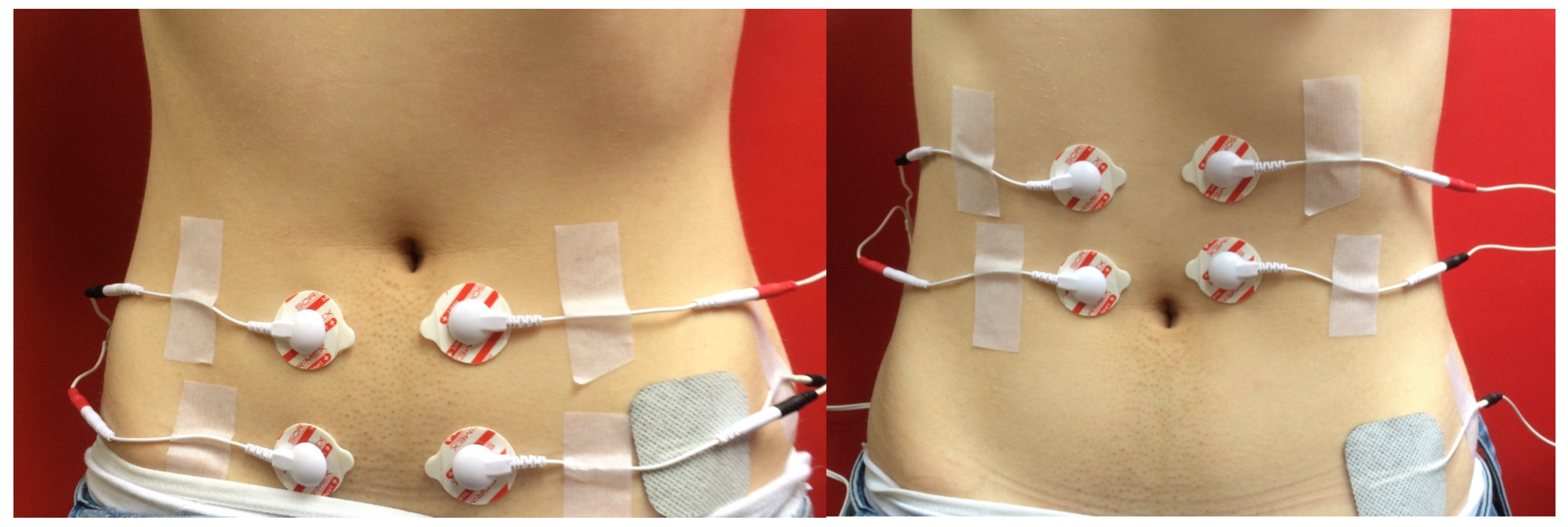
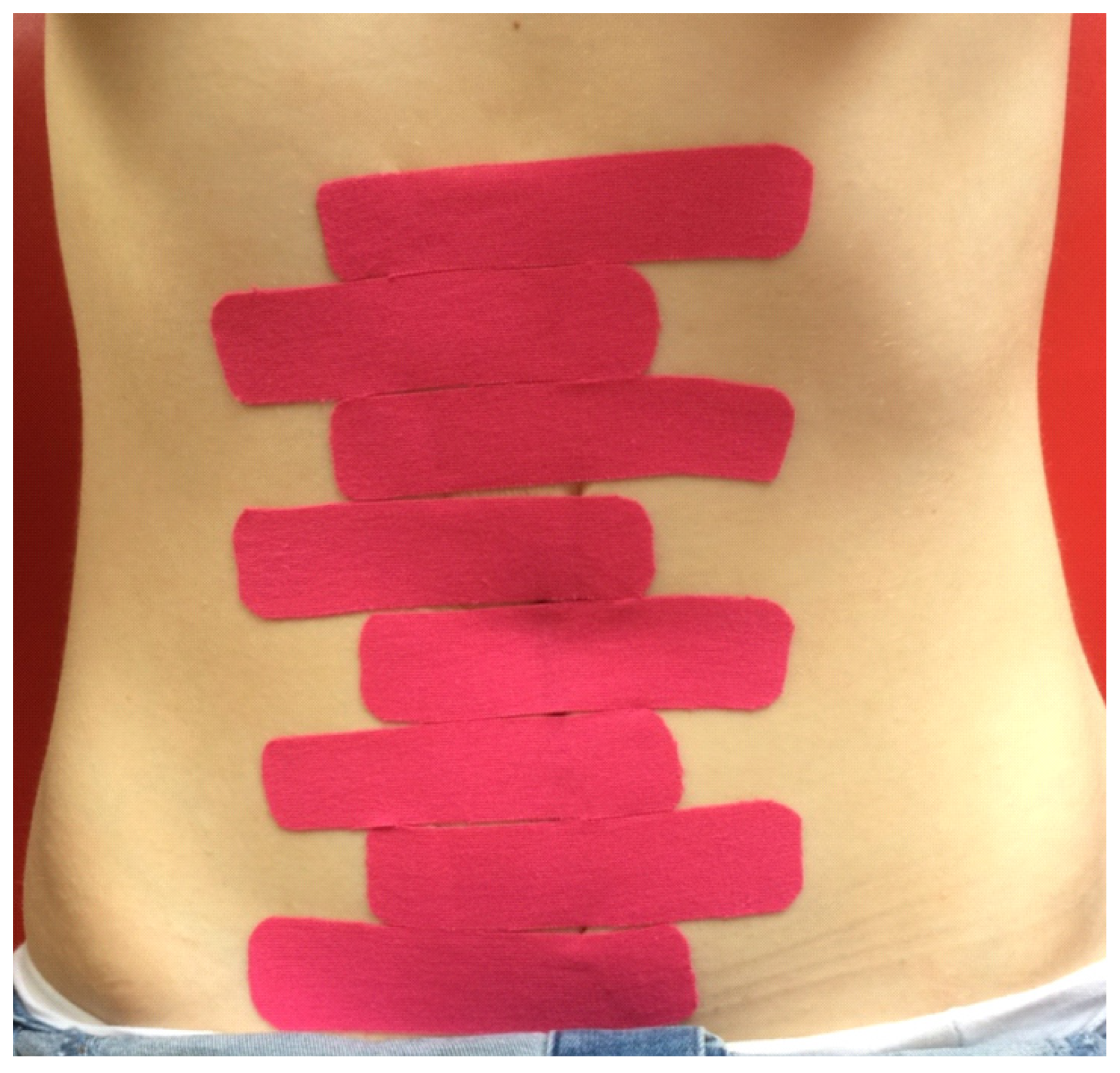
2. Methods
3. Results
4. Discussion
5. Conclusions
Author Contributions
Funding
Institutional Review Board Statement
Informed Consent Statement
Data Availability Statement
Conflicts of Interest
References
- Benjamin, D.R.; van de Water, A.T.M.; Peiris, C.L. Effects of exercise on diastasis of the rectus abdominis muscle in the antenatal and postnatal periods: A systematic review. Physiotherapy 2014, 100, 1–8. [Google Scholar] [CrossRef] [PubMed] [Green Version]
- Benjamin, D.R.; Frawley, H.C.; Shields, N.; van de Water, A.T.M.; Taylor, N.F. Relationship between diastasis of the rectus abdominis muscle (DRAM) and musculoskeletal dysfunctions, pain and quality of life: A systematic review. Physiotherapy 2019, 105, 24–34. [Google Scholar] [CrossRef] [PubMed] [Green Version]
- Bursch, S.G. Interrater reliability of diastasis recti abdominis measurement. Phys. Ther. 1987, 67, 1077–1079. [Google Scholar] [CrossRef] [PubMed]
- Mota, P.; Pascoal, A.G.; Bo, K. Diastasis Recti Abdominis in Pregnancy and Postpartum Period. Risk Factors, Functional Implications and Resolution. Available online: https://www.eurekaselect.com/134908/article (accessed on 11 November 2020).
- Michalska, A.; Rokita, W.; Wolder, D.; Pogorzelska, J.; Kaczmarczyk, K. Diastasis recti abdominis—a review of treatment methods. Ginekol. Pol. 2018, 89, 97–101. [Google Scholar] [CrossRef] [PubMed] [Green Version]
- Boxer, S.; Jones, S. Intra-rater reliability of rectus abdominis diastasis measurement using dial calipers. Aust. J. Physiother. 1997, 43, 109–114. [Google Scholar] [CrossRef] [Green Version]
- Sperstad, J.B.; Tennfjord, M.K.; Hilde, G.; Ellström-Engh, M.; Bø, K. Diastasis recti abdominis during pregnancy and 12 months after childbirth: Prevalence, risk factors and report of lumbopelvic pain. Br. J. Sports Med. 2016, 50, 1092–1096. [Google Scholar] [CrossRef] [PubMed] [Green Version]
- Spitznagle, T.M.; Leong, F.C.; Van Dillen, L.R. Prevalence of diastasis recti abdominis in a urogynecological patient population. Int. Urogynecol. J. Pelvic Floor Dysfunct. 2007, 18, 321–328. [Google Scholar] [CrossRef] [PubMed]
- Mady, M.M. Kinesiotaping Therapy Techniquesfor Treating Postpartum Rectus Diastases: A Comparative Study. IOSR J. Nurs. Health Sci. 2018, 7, 67–74. [Google Scholar] [CrossRef]
- Tuttle, L.J.; Fasching, J.; Keller, A.; Patel, M.; Saville, C.; Schlaff, R.; Walker, A.; Mason, M.; Gombatto, S.P. Noninvasive Treatment of Postpartum Diastasis Recti Abdominis: A Pilot Study. J. Women’s Health Phys. Ther. 2018, 42, 65–75. [Google Scholar] [CrossRef]
- Mostafavifar, M.; Wertz, J.; Borchers, J. A Systematic Review of the Effectiveness of Kinesio Taping for Musculoskeletal Injury. Phys. Sportsmed. 2012, 40, 33–40. [Google Scholar] [CrossRef] [PubMed] [Green Version]
- Gluppe, S.L.; Hilde, G.; Tennfjord, M.K.; Engh, M.E.; Bø, K. Effect of a Postpartum Training Program on the Prevalence of Diastasis Recti Abdominis in Postpartum Primiparous Women: A Randomized Controlled Trial. Phys. Ther. 2018, 98, 260–268. [Google Scholar] [CrossRef] [PubMed] [Green Version]
- Rett, M.; Braga, M.; Bernardes, N.; Andrade, S. Prevalência de diástase dos músculos retoabdominais no puerpério imediato: Comparação entre primíparas e multíparas. Braz. J. Phys. Ther. 2009, 13, 275–280. [Google Scholar] [CrossRef]
- Hsia, M.; Jones, S. Natural resolution of rectus abdominis diastasis. Two single case studies. Aust. J. Physiother. 2000, 46, 301–307. [Google Scholar] [CrossRef] [Green Version]
- Sancho, M.F.; Pascoal, A.G.; Mota, P.; Bø, K. Abdominal exercises affect inter-rectus distance in postpartum women: A two-dimensional ultrasound study. Physiotherapy 2015, 101, 286–291. [Google Scholar] [CrossRef] [PubMed] [Green Version]
- Słomko, W.; Zamojska, P.; Dzierżanowski, M. Physiotherapy in the postpartum problems. J. Educ. Health Sport 2017, 7, 323–333. [Google Scholar] [CrossRef]
- Thabet, A.A.; Alshehri, M.A. Efficacy of deep core stability exercise program in postpartum women with diastasis recti abdominis: A randomised controlled trial. J. Musculoskelet. Neuronal Interact. 2019, 19, 62–68. [Google Scholar] [PubMed]
- Kamel, D.M.; Yousif, A.M. Neuromuscular Electrical Stimulation and Strength Recovery of Postnatal Diastasis Recti Abdominis Muscles. Ann. Rehabil. Med. 2017, 41, 465–474. [Google Scholar] [CrossRef] [PubMed]


| KT Group (n = 13) | Sham KT Group (n = 11) | p-Value * | |||||||||
|---|---|---|---|---|---|---|---|---|---|---|---|
| Mean | Me | Min | Max | SD | Mean | Me | Min | Max | SD | ||
| Age | 27.5 | 27 | 20 | 38 | 5.8 | 27.6 | 28 | 18 | 34 | 4.4 | 0.75 |
| Height [m] | 1.7 | 1.7 | 1.6 | 1.8 | 0.1 | 1.7 | 1.7 | 1.6 | 1.8 | 0.1 | 0.83 |
| Body weight [kg] | 61.4 | 62 | 50 | 70 | 6.4 | 61.9 | 60 | 52 | 60 | 8.7 | 0.87 |
| BMI [kg/m2] | 22.1 | 22 | 19.5 | 25.4 | 1.9 | 22.1 | 21.3 | 19.3 | 25.7 | 2.9 | 0.64 |
| Birth weight of the child [g] | 3461.9 | 3500 | 2825 | 4150 | 419.9 | 3405.5 | 3500 | 2500 | 3950 | 400.6 | 0.95 |
| Length of the last delivery [min.] | 37.2 | 30 | 10 | 90 | 24.7 | 26.8 | 30 | 10 | 50 | 13.1 | 0.60 |
| Time since the last delivery [weeks] | 27.6 | 25 | 7 | 50 | 15.8 | 31.7 | 32 | 6 | 52 | 14.5 | 0.74 |
| Before | After | p-Value * | |||||||||
|---|---|---|---|---|---|---|---|---|---|---|---|
| Mean | Median | Min. | Max. | SD | Mean | Median | Min. | Max. | SD | ||
| KT group (n = 13) | |||||||||||
| At umbilicus | 1.9 | 2.2 | 1.3 | 2.5 | 0.5 | 1.7 | 1.8 | 1.2 | 2.2 | 0.3 | 0.003 |
| Above umbilicus | 1.6 | 1.6 | 0.6 | 2.4 | 0.6 | 1.2 | 1.2 | 0.5 | 2.2 | 0.5 | 0.002 |
| Below umbilicus | 1.1 | 1.3 | 0.02 | 1.8 | 0.6 | 0.8 | 1.0 | 0.0 | 1.4 | 0.5 | 0.008 |
| Sham KT group (n = 11) | |||||||||||
| At umbilicus | 2.1 | 2.2 | 1.0 | 2.5 | 0.4 | 2.1 | 2.2 | 1.3 | 2.5 | 0.4 | 0.55 |
| Above umbilicus | 1.6 | 1.5 | 1.2 | 2.2 | 0.3 | 1.6 | 1.4 | 1.0 | 2.3 | 0.4 | 0.35 |
| Below umbilicus | 1.1 | 1.2 | 0.01 | 2.2 | 0.8 | 1.1 | 1.1 | 0.01 | 2.1 | 0.7 | 0.12 |
| sEMG Activity | Before | After | p-Value * | ||||||||
|---|---|---|---|---|---|---|---|---|---|---|---|
| Mean | Median | Min. | Max. | SD | Mean | Median | Min. | Max. | SD | ||
| Right side above umbilicus | |||||||||||
| Resting | 1.2 | 1.0 | 0.3 | 4.0 | 0.9 | 1.3 | 1.1 | 0.5 | 5.1 | 1.2 | 0.76 |
| Functional | 15.5 | 13.4 | 1.2 | 39 | 13 | 22.3 | 21.2 | 2.1 | 41.4 | 12.8 | 0.65 |
| During isometric contraction | 21.0 | 19.6 | 3.5 | 54.7 | 16.4 | 19.7 | 19.2 | 1.6 | 41.2 | 12.4 | 0.42 |
| Right side below umbilicus | |||||||||||
| Resting | 1.6 | 0.9 | 0.3 | 7.3 | 2.9 | 1.3 | 1.1 | 0.4 | 3.6 | 1.1 | 0.26 |
| Functional | 5.8 | 3.1 | 0.7 | 25.5 | 6.8 | 5.4 | 4.9 | 0.8 | 20.1 | 5.5 | 0.55 |
| During isometric contraction | 7.2 | 2.5 | 0.6 | 23.8 | 8.4 | 6.3 | 2.9 | 0.8 | 31.2 | 7.4 | 0.39 |
| Left side above umbilicus | |||||||||||
| Resting | 0.7 | 0.6 | 0.2 | 1.6 | 0.4 | 0.9 | 0.6 | 0.4 | 2.5 | 0.6 | 0.17 |
| Functional | 21.6 | 21.6 | 1.1 | 58.5 | 16.8 | 21.8 | 24.2 | 7.5 | 41.7 | 10.0 | 0.70 |
| During isometric contraction | 21.4 | 20.5 | 0.8 | 49.4 | 16.1 | 19.3 | 20.3 | 6.6 | 34.2 | 8.9 | 0.97 |
| Left side below umbilicus | |||||||||||
| Resting | 0.9 | 0.7 | 0.3 | 3.2 | 0.8 | 0.8 | 0.6 | 0.3 | 2.0 | 0.4 | 0.96 |
| Functional | 6.6 | 7.0 | 0.7 | 12.6 | 3.6 | 7.4 | 5.9 | 1.5 | 18.6 | 4.8 | 0.55 |
| During isometric contraction | 7.1 | 7.3 | 1.2 | 17.7 | 4.5 | 7.8 | 5.5 | 2.2 | 18.4 | 4.8 | 0.34 |
| sEMG Activity | Before | After | p-Value * | ||||||||
|---|---|---|---|---|---|---|---|---|---|---|---|
| Mean | Median | Min. | Max. | SD | Mean | Median | Min. | Max. | SD | ||
| Right side above umbilicus | |||||||||||
| Resting | 0.7 | 0.7 | 0.4 | 1.0 | 0.2 | 0.8 | 0.5 | 0.3 | 1.7 | 0.5 | 0.79 |
| Functional | 16.7 | 17.0 | 4.4 | 29.1 | 8.5 | 14.6 | 12.0 | 7.0 | 30.1 | 8.1 | 0.25 |
| During isometric contraction | 10.8 | 11.4 | 4.7 | 22.9 | 5.1 | 10.4 | 11.1 | 5.3 | 17.2 | 3.4 | 0.80 |
| Right side below umbilicus | |||||||||||
| Resting | 0.8 | 0.5 | 0.3 | 2.2 | 0.6 | 0.7 | 0.4 | 0.3 | 2.4 | 0.6 | 0.21 |
| Functional | 8.5 | 6.4 | 2.5 | 17.7 | 4.4 | 6.4 | 5.6 | 1.8 | 11.7 | 3.4 | 0.005 |
| During isometric contraction | 7.7 | 6.7 | 2.2 | 17.3 | 4.7 | 5.5 | 5.5 | 1.7 | 10.5 | 2.9 | 0.023 |
| Left side above umbilicus | |||||||||||
| Resting | 0.6 | 0.5 | 0.4 | 1.3 | 0.3 | 1.0 | 0.9 | 0.3 | 2.4 | 0.7 | 0.28 |
| Functional | 19.5 | 14.1 | 2.1 | 36.9 | 10.9 | 16.6 | 14.1 | 3.7 | 35.9 | 8.8 | 0.37 |
| During isometric contraction | 13.8 | 11.0 | 2.4 | 31.7 | 8.1 | 11.6 | 11.2 | 3.7 | 18.3 | 4.2 | 0.21 |
| Left side below umbilicus | |||||||||||
| Resting | 0.7 | 0.5 | 0.3 | 1.6 | 0.4 | 1.2 | 1.0 | 0.3 | 2.3 | 0.7 | 0.051 |
| Functional | 8.3 | 7.1 | 3.3 | 18.0 | 4.4 | 4.2 | 2.6 | 1.4 | 9.1 | 2.9 | 0.006 |
| During isometric contraction | 7.5 | 6.1 | 2.2 | 22.1 | 5.6 | 4.4 | 2.8 | 1.5 | 9.9 | 3.1 | 0.11 |
Publisher’s Note: MDPI stays neutral with regard to jurisdictional claims in published maps and institutional affiliations. |
© 2021 by the authors. Licensee MDPI, Basel, Switzerland. This article is an open access article distributed under the terms and conditions of the Creative Commons Attribution (CC BY) license (https://creativecommons.org/licenses/by/4.0/).
Share and Cite
Ptaszkowska, L.; Gorecka, J.; Paprocka-Borowicz, M.; Walewicz, K.; Jarzab, S.; Majewska-Pulsakowska, M.; Gorka-Dynysiewicz, J.; Jenczura, A.; Ptaszkowski, K. Immediate Effects of Kinesio Taping on Rectus Abdominis Diastasis in Postpartum Women—Preliminary Report. J. Clin. Med. 2021, 10, 5043. https://doi.org/10.3390/jcm10215043
Ptaszkowska L, Gorecka J, Paprocka-Borowicz M, Walewicz K, Jarzab S, Majewska-Pulsakowska M, Gorka-Dynysiewicz J, Jenczura A, Ptaszkowski K. Immediate Effects of Kinesio Taping on Rectus Abdominis Diastasis in Postpartum Women—Preliminary Report. Journal of Clinical Medicine. 2021; 10(21):5043. https://doi.org/10.3390/jcm10215043
Chicago/Turabian StylePtaszkowska, Lucyna, Justyna Gorecka, Malgorzata Paprocka-Borowicz, Karolina Walewicz, Slawomir Jarzab, Marta Majewska-Pulsakowska, Joanna Gorka-Dynysiewicz, Anna Jenczura, and Kuba Ptaszkowski. 2021. "Immediate Effects of Kinesio Taping on Rectus Abdominis Diastasis in Postpartum Women—Preliminary Report" Journal of Clinical Medicine 10, no. 21: 5043. https://doi.org/10.3390/jcm10215043
APA StylePtaszkowska, L., Gorecka, J., Paprocka-Borowicz, M., Walewicz, K., Jarzab, S., Majewska-Pulsakowska, M., Gorka-Dynysiewicz, J., Jenczura, A., & Ptaszkowski, K. (2021). Immediate Effects of Kinesio Taping on Rectus Abdominis Diastasis in Postpartum Women—Preliminary Report. Journal of Clinical Medicine, 10(21), 5043. https://doi.org/10.3390/jcm10215043