Article

One-Stage Immediate Alloplastic Breast Reconstruction in Large and Ptotic Breasts: An Institutional Algorithm

1
Plastic Surgery Unit, University Hospital Trust of Sassari, 07100 Sassari, Italy
2
Plastic, Reconstructive and Aesthetic Surgery Training Program, University of Sassari, 07100 Sassari, Italy
3
Department of Medicine, Surgery and Pharmacy, University of Sassari, 07100 Sassari, Italy
*
Author to whom correspondence should be addressed.
J. Clin. Med. 2023, 12(3), 1170; https://doi.org/10.3390/jcm12031170
Submission received: 31 December 2022 / Revised: 27 January 2023 / Accepted: 30 January 2023 / Published: 1 February 2023

Abstract

:
Immediate implant-based breast reconstruction in patients with large and ptotic breasts may be challenging due to skin redundancy. The use of a reduction mammoplasty pattern for the mastectomy skin excision has proven to be a reliable option for these patients as it allows for a better shape, projection, and symmetrization. This approach has been described in the literature for both one- and two-stage reconstruction with either sub- or pre-pectoral reconstruction with an acellular dermal matrix (ADM) or non-biological mesh. One-stage immediate breast reconstructions have a positive significant impact on patients’ psychosocial well-being and quality of life. The purpose of this paper is to describe an institutional algorithm that allows one to perform one-stage implant-based breast reconstructions in patients with large and ptotic breasts.

1. Introduction

It is a well-known fact that one-stage immediate breast reconstruction has a positive significant impact on patients’ psychosocial well-being and quality of life [1,2,3]. Nevertheless, immediate breast reconstruction in large and ptotic breasts can be challenging due to skin redundancy after skin-sparing mastectomy. The use of a traditional elliptical pattern usually flattens the breasts and leaves noticeable scars medially on the breast to avoid dog-ears. The Wise (Inverted T) [4,5] mammoplasty pattern skin excision, instead, can reduce the redundant skin envelope while lifting and restoring an appropriate shape of the breast. Moreover, this technique maximizes the outcome in terms of symmetrization, as reduction mammoplasty on the opposite site constitutes most of the time required. A major weakness of this technique is that skin flaps are often long and unreliable, with potential skin necrosis at the T junction and subsequent implant exposure [6,7,8]. Coverage of the inferior aspect of the implant with an inferiorly based dermal flap spared from the inferior aspect of the reduction pattern has been first proposed by Bostwick [9] to address the issue. In the original technique, a submuscular pocket was created by raising pectoralis major and serratus muscles, which were sutured to each other and to the dermal flap over a permanent implant; moreover, the nipple–areola complex (NAC) was moved as a full-thickness skin graft. Over the years, several variations have been described in the literature. As pre-pectoral reconstruction with the assistance of matrices was gaining popularity in the last decade [10], their application in breast reconstruction after Wise Pattern Mastectomy (WPM) has been advocated. Matrices can be divided into Acellular Dermal Matrices (ADMs) and synthetic meshes. ADMs are human-, bovine-, or porcine-derived engineered tissue grafts that work as a scaffold for the patient’s own cells and vessels to grow, thereby creating an extra tissue layer [11]. First mentioned as skin replacements in burn patients [12], their use in reconstructive surgery [13] has expanded over the years, as they proved to be reliable in a wide range of applications, from surgical oncology [13,14,15,16] to benign condition treatment [17,18,19]. Synthetic meshes are sheets knitted from permanent or absorbable fibers and have proven to be reliable in different surgical procedures, such as abdominal and pelvic surgery [20,21], where they are used to support organs and/or tissues, or in alloplastic breast reconstruction surgery [14,22], providing additional cover and support to the implant. The combination of matrices with the inferior dermal flap has been reported in the literature. Some authors [23,24] have described the use of matrices to cover the inferolateral aspect of the implant in a submuscular reconstruction; this provides a two-layer coverage of the inferior pole of the implant as the dermal flap is draped over the matrix. Others [25,26], instead, described their use in pre-pectoral reconstruction; matrices can be sutured to the superior border of the dermal flap and placed to cover the superior pole of the implant, or they can be used as a means for total implant coverage under the dermal flap. Different techniques have been also described to preserve the NAC, such as an inferiorly based dermal flap bearing the NAC [27] or a vertical bipedicle McKissock style flap [28]. Furthermore, an alternative reduction pattern for mastectomy skin excision has been proposed by Santanelli et al. [29] for patients requiring the skin excision of an upper quadrant due to the presence of a superficially located tumor or a scar from previous surgeries (e.g., lumpectomy, quadrantectomy) that may increase the risk of Wise skin flap necrosis. In case of unilateral reconstruction with concomitant contralateral reduction mammoplasty, Marongiu et al. [30] proposed the use of an autologous dermal patch obtained from the contralateral breast’s lower pole to complete the pre-pectoral pocket and better cover the implant.
One-stage immediate breast reconstruction has proven to be a highly acceptable treatment for patients requiring mastectomy. It is associated with a high quality of life, short reconstructive journey, and low procedure-related morbidity [1,2,3]. Several techniques have been proposed in the literature for immediate breast reconstruction in patients with macromastia and ptotic breast. Hence, the authors’ aim is to describe an institutional algorithm that allows one to perform one-stage, direct-to-implant breast reconstruction in patients with large and ptotic breasts.

2. Materials and Methods

A prospective application of the present algorithm was performed from October 2021 to the present time at the University Hospital Trust of Sassari, Italy. All the mastectomies were performed by Breast Surgeons of the Breast Unit, while immediate reconstructions were performed by Plastic Surgeons of the Plastic Surgery Unit.
We retrospectively analyzed the medical records of all the patients undergoing skin/nipple sparing Mastectomy with Wise pattern (type IV [31]) or modified Wise pattern (type V [29]), immediate breast reconstruction, and concomitant contralateral reduction mammoplasty, using the Algorithm from October 2021 to September 2022. Demographic, clinical preoperative, surgical, and postoperative information, such as body mass index (BMI), comorbidities, tobacco use, anthropomorphic measurement, type of reconstruction, implant type and volume, complications, and 3-month follow up evaluation, were recorded. Postoperative complications of interest were skin flap necrosis (either partial or full thickness, in relation to whether it was involving the dermal layer with subcutaneous tissue exposition), hematoma, seroma, surgical site infection, nipple necrosis, and implant loss. Early postoperative evaluation at three months of follow-up included both aesthetic and patient satisfaction outcome assessments. All the patients were asked to rate their satisfaction from “poor” to “excellent”. A third-party plastic surgeon of our unit was similarly asked to rate the aesthetic result from ‘‘poor’’ to ‘‘excellent’’.

2.1. Inclusion and Exclusion Criteria
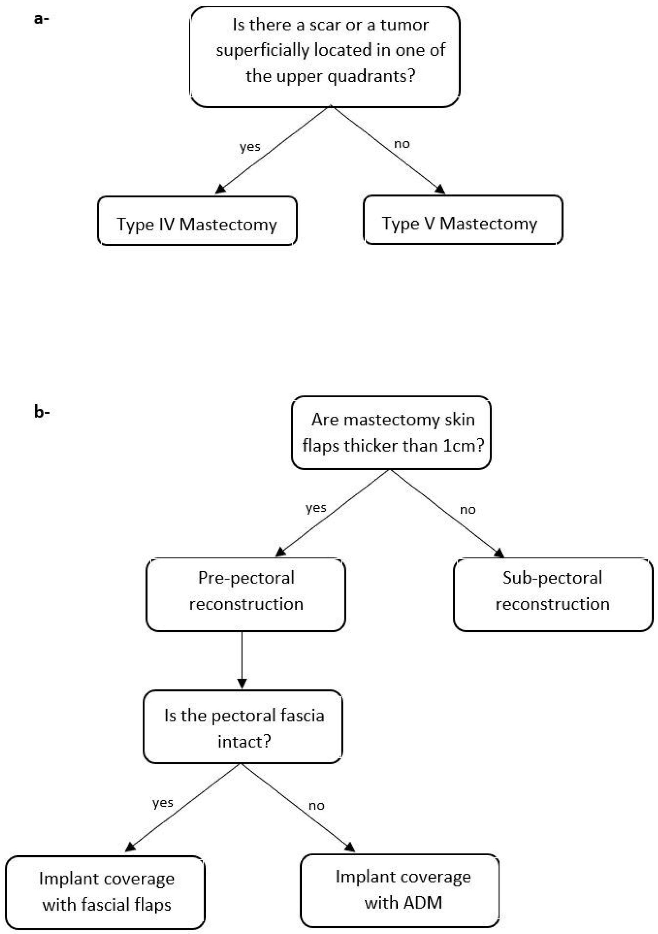
The algorithm is applicable on female patients with large and ptotic beasts, including obese and post-bariatric patients. Inclusion criteria are nipple to sternal notch distance ≥ 26 cm and areola to inframammary fold distance ≥ 8 cm.
Exclusion criteria are history of previous radiotherapy, BMI > 40, heavy smoking habit (more than 20 cigarettes/day), uncontrolled diabetes, vasculitis, collagen diseases, or inflammatory breast cancer.

2.2. Algorithm

The first part of the algorithm (Figure 1a) demands a multidisciplinary preoperative evaluation of the tumor location, the corresponding need of skin resection to pursue oncological radicality, and the presence of a scar from previous surgeries (e.g., lumpectomy, quadrantectomy) in order to select the proper skin excision pattern for mastectomy. In case the breast tumor does not require skin resection, the Wise (Type IV [31]) reduction mammaplasty skin excision pattern is selected (Figure 2a). Alternatively, in case the tumor is superficially located in one of the upper quadrants, the modified Wise pattern (type V) described by Santanelli et al. [29] is chosen (Figure 2b); likewise, if a scar located in an upper quadrant may increase the risk of Wise skin flap necrosis, the latter pattern is preferred. Small variations of the preoperative drawing may be performed in the case of previous scars. In case of unilateral reconstruction, a contralateral Wise pattern reduction mammoplasty is planned.
The reduction mammoplasty pattern and the inferior dermal flap are first de-epithelized. The skin-sparing mastectomy is then performed by the breast surgeon through a different access according to the pattern type (Figure 3). The nipple–areolar complex may or may not be preserved according to the result of the intraoperative retro-areolar frozen biopsy histopathological assessment. Nevertheless, if NAC viability, assessed either clinically or with indocyanine green fluorescence [32], is doubtful, the surgeon may decide to remove it.
The second part of the algorithm (Figure 1b), instead, requires an intraoperative assessment of mastectomy skin flaps thickness, which, in our institution, relies on clinical examination using palpation and a ruler. Furthermore, pectoral fascia integrity is assessed. These evaluations allow the surgeon to identify three main different approaches to breast reconstruction.
Case 1—subpectoral reconstruction (Figure 4a,d): if the mastectomy skin flap’s thickness is ≤1 cm, a submuscular approach is performed. A pocket is first created by raising pectoralis major. The free edge of the muscle is then sutured to the upper border of the dermal flap, starting medially. When most of the pouch is closed, the implant is inserted, and the final suturing is performed. In case of big implants, the lateral aspect may need to be secured with the elevation of a serratus fascial flap, which is sutured to the dermal flap and the pectoralis major muscle.
Case 2—pre-pectoral reconstruction and implant autologous coverage with fascial flaps (Figure 4b,e): if the mastectomy skin flaps are thicker than 1 cm, a pre-pectoral reconstruction can be performed. In case the pectoral fascia is left intact after the mastectomy, the superior and lateral aspects of the implant can be covered by a pectoral and the serratus fascia flaps, respectively, as recently described by the authors of [33]. The pre-pectoral pocket is first created by harvesting a superiorly based fascial flap from the pectoralis major muscle and a laterally based fascial flap from the serratus muscle. The pouch is partially closed by suturing the superior edge of the dermal flap to the pectoral fascia and a permanent silicone implant is then placed into the pocket. Final suturing between the three flaps is performed in order to achieve complete autologous coverage of the implant.
Case 3—pre-pectoral reconstruction and implant coverage with ADM (Figure 4c,f): if the pectoral fascia is not intact after the mastectomy, a pre-pectoral reconstruction with the use of an ADM is preferred. A pre-shaped porcine ADM Braxon® Fast (Decomed S.rl., Venezia, Italy) is used in our institution. The ADM is first wrapped around the implant, and its edges are sutured with interrupted absorbable sutures. The ADM containing the implant is then placed into the pocket and secured to the chest wall with superior, medial, and lateral interrupted absorbable sutures; an additional suture is sewed between the anterior sheet of the ADM and the subcutaneous tissue of the dermal flap and of the mastectomy skin flap in order to avoid any displacement and dead space.

2.3. Ethical Approval
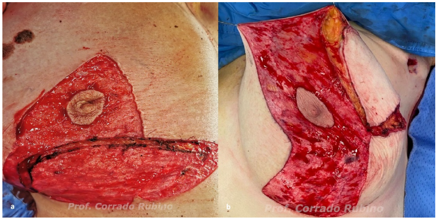
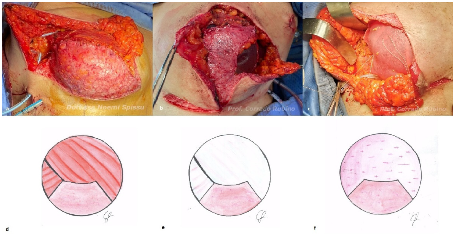
The study was conducted in accordance with the Declaration of Helsinki (as revised in 2013). All the patients provided a written informed consent.

3. Results

From October 2021 to the present time, twelve patients were eligible for the described algorithm, which was used to select the proper skin excision pattern and type of breast reconstruction. Two patients underwent bilateral mastectomy, and ten patients underwent unilateral mastectomy with concomitant contralateral Wise pattern reduction mammoplasty for symmetrization. Mean patients age was 60 years (range 34–80 years) and mean BMI was 29.3 kg/m2 (range 24–37). Type V mastectomy was used in two patients (2 breasts); all the other cases were treated with a type IV mastectomy. Three patients underwent subpectoral reconstruction, six patients (eight breasts) underwent pre-pectoral reconstruction and implant coverage with fascial flaps, and three patients underwent pre-pectoral reconstruction with ADM. Mean implant volume was 390 cc (range 300–560 cc).
Complications included skin and/or NAC necrosis healed by secondary intention (14.3%) and that required surgical revision (28.6%), seroma (7.1%) hematoma (7.1%), and implant removal (7.1%). The most common complication was skin necrosis, which occurred in six breasts. One of these patients developed a seroma after the surgical revision and a subsequent periprosthetic infection, leading to implant removal.
The evaluation at the three-month follow-up (Figure 5) showed good aesthetic and patient satisfaction outcomes. No sign of implant lateral migration has been detected in all patients. Six patients described their satisfaction with the result as “good”, three as “very good”, one as “excellent”, one as “scarce” and, lastly, the patient with implant reconstruction failure described it as “poor”. The aesthetic evaluation was reported as “excellent” in one case, “very good” in three patients, “good” in six patients, and “scarce” in one case. The aesthetic evaluation was not performed on the patients that lost the implant.

4. Discussion

As shown by our early results, the present algorithm is effective in treating patients with large and ptotic breasts as it allows the surgeon to perform both immediate breast reconstruction and concomitant symmetrization in one single procedure. This approach can, at the same time, address the oncological issue, as well as the one related to macromastia and/or ptosis, with a satisfactory aesthetic result and a positive impact on the psychosocial well-being of the patient. Complication rates are comparable to the ones presented in the literature [7,8,34,35]. One single implant loss occurred in our series. Pre-pectoral reconstruction with a 560 cc silicone implant covered with fascial flaps was selected for this case. The implant loss is likely to be related to the volume of the implant, which was probably too big.
Wise pattern mastectomy with an inferior dermal flap and submuscular direct-to-implant reconstruction has been well described in the literature [31,36,37]. We prefer to use this approach when mastectomy skin flap thickness is scarce. In these cases, skin viability is reduced with a high risk of skin necrosis; likewise, it is more likely to observe a visible implant rippling, making the subpectoral approach a better option. Mastectomy skin flaps thinner than 0.8 cm are, as a matter of fact, associated with a higher risk of ischemic complication, as recently described in the literature [38,39,40]. Despite this, we prefer to use a cut-off of 1 cm to ensure better implant coverage and to address the risk of visible rippling. The risk of rippling in pre-pectoral breast reconstruction has been associated [41] to a preoperative pinch test <2 cm. Hence, with mastectomy skin flaps thinner than 1 cm, we prefer a submuscular reconstruction to address both above-mentioned issues. Intraoperative use of indocyanine green fluorescence [32] may represent an additional tool to assess tissue perfusion. In our series, we were only able to use this method with two patients as the device was not available before. When mastectomy skin flaps are thicker than 1 cm, instead, we would rather perform a pre-pectoral reconstruction, as it is known to reduce postoperative pain with better functional outcomes [42,43]. In case the pectoral fascia is left intact after the mastectomy, the use of two fascial flaps [33] harvested from the pectoralis and serratus muscles is used to complete the pre-pectoral pocket. This technique provides complete autologous coverage of the implant; however, most importantly, it secures the superior and the lateral aspects of the implant, avoiding any lateral displacement. Moreover, in comparison to ADM or synthetic meshes [44,45], the use of fascial flaps for pre-pectoral reconstruction has a lower cost per procedure and no risk of phlogistic reaction. Recent findings show that pectoral fascia preservation during mastectomy seems oncologically safe [46]; nevertheless, the literature about the topic is scarce and many centers routinely perform fascial removal. For these reasons, we prefer to leave the choice to the breast surgeon. In case the pectoral fascia is not intact after the mastectomy, pre-pectoral reconstruction with the use of ADM is performed. The use of matrices in pre-pectoral breast reconstruction allows the surgeon to actively position the implant in an ideal location, with better control and definition of the inframammary fold. Moreover, ADMs are meant to modulate the wound-healing process and integrate into the host tissue with neovascularization and cell repopulation, guiding the formation of a soft and thin fibrous capsule around the implant [11]. As recently described in the literature [47], ADM-assisted alloplastic breast reconstruction has a significantly lower incidence of capsular contracture, even though further studies are needed to corroborate these findings.
According to our algorithm, the skin excision pattern is adaptable to a wide range of scenarios. The surgeon can select the proper reduction mammoplasty pattern in relation to the presence of scars from previous surgeries, the tumor location, and the desired final volume of the breast. In selected cases, J scar mammoplasty [48] or another reduction pattern may be used; likewise, different NAC pedicles may be selected according to the surgeon’s experience and preference. The same reduction patterns can also be used with autologous breast reconstruction, as already described in the literature [49,50].
The bilateral approach suggested by our algorithm can indeed achieve a better aesthetic and self-satisfaction outcomes. This approach allows, in one single procedure, to pursue a double goal: breast reconstruction and reduction mammoplasty. The use of a reduction mammoplasty pattern on both breasts can accomplish a better balance between the two sides, thus causing better aesthetic and patient self-satisfaction outcomes.
Patients with large and ptotic breasts undergoing this type of surgery must be selected carefully. Most of the time, these patients are obese, active smokers with different comorbidities; all these aspects are known to be associated with a higher risk of postoperative complications [51,52]. Likewise, the choice of a proper implant size is relevant, as bigger ones are associated with a higher risk of complications [53]. The authors prefer to use implants with a maximum volume of 500 cc. All these aspects demand preoperative counseling with the patient to address patient expectations and elucidate the risk of postoperative complication in order to select the proper surgical plan.
Limitations of this study include the limited cohort size and the short length of the follow-up period. The use of Breast-Q [54] and the Validated Aesthetic Scale [55] are routinely performed in our unit at one year of follow-up; therefore, a longer follow-up will allow us to provide the results obtained from the aesthetic and patient satisfaction outcomes based on validated questionnaires.

5. Conclusions

The present algorithm is a relevant tool for plastic surgeons aiming to treat patients with large and ptotic breasts who are eligible for implant breast reconstruction. It is a comprehensive algorithm addressing both breasts that provides consistent aesthetic results in a one-stage procedure, using different skin reduction patterns and implant coverage methods.

Author Contributions

Conceptualization, C.R.; Methodology, C.R., N.S. and S.R.; Validation, C.R. and E.T., Formal Analysis, S.R.; Investigation, S.R., C.S. and M.R.; Data Curation S.R. and M.R.; Writing—Original Draft Preparation, S.R. and M.T.; Writing—Review and Editing, N.S., M.P., R.N. and G.A.M.S.; Visualization, S.R. and C.S.; Supervision, C.R., G.A.M.S. and N.S.; Project Administration, C.R. All authors have read and agreed to the published version of the manuscript.

Funding

This research received no external funding.

Institutional Review Board Statement

The study was conducted according to the guidelines of the Declaration of Helsinki and approved by the Institutional Review Board of the University Hospital Trust of Sassari.

Informed Consent Statement

Written informed consent was obtained from all subjects involved in the study.

Data Availability Statement

The data used and/or analyzed during the current study are available from the corresponding author upon reasonable request.

Conflicts of Interest

The authors declare no conflict of interest.

References

  1. Heneghan, H.M.; Prichard, R.S.; Lyons, R.; Regan, P.J.; Kelly, J.L.; Malone, C.; McLaughlin, R.; Sweeney, K.J.; Kerin, M.J. Quality of Life after Immediate Breast Reconstruction and Skin-Sparing Mastectomy–A Comparison with Patients Undergoing Breast Conserving Surgery. Eur. J. Surg. Oncol. 2011, 37, 937–943. [Google Scholar] [CrossRef] [PubMed]
  2. Elder, E.E.; Brandberg, Y.; Björklund, T.; Rylander, R.; Lagergren, J.; Jurell, G.; Wickman, M.; Sandelin, K. Quality of Life and Patient Satisfaction in Breast Cancer Patients after Immediate Breast Reconstruction: A Prospective Study. Breast 2005, 14, 201–208. [Google Scholar] [CrossRef] [PubMed]
  3. Dimovska, E.O.F.; Chen, C.; Chou, H.; Lin, Y.; Cheng, M. Outcomes and Quality of Life in Immediate One-stage versus Two-stage Breast Reconstructions without an Acellular Dermal Matrix: 17- Years of Experience. J. Surg. Oncol. 2021, 124, 510–520. [Google Scholar] [CrossRef] [PubMed]
  4. Wise, R.J. A preliminary report on a method of planning the mammaplasty. Plast. Reconstr. Surg. 1956, 17, 367–375. [Google Scholar] [CrossRef]
  5. Toth, B.A.; Lappert, P. Modified Skin Incisions for Mastectomy: The Need for Plastic Surgical Input in Preoperative Planning. Plast. Reconstr. Surg. 1991, 87, 1048–1053. [Google Scholar] [CrossRef]
  6. Di Candia, M.; Lie, K.H.; Forouhi, P.; Malata, C.M. Experience with the Wise Mammaplasty Skin Resection Pattern in Skin-Sparing Mastectomy and Immediate Breast Reconstruction for Large Breast Volumes. Int. J. Surg. 2011, 9, 41–45. [Google Scholar] [CrossRef]
  7. Kilgo, M.S.; Kaufman, G.J.; Shen, A.E.; Korsh, J.; Baranchuk, N.V.; Douglas, B.K.; Brewer, B.W. A Comparison of Elliptical Mastectomy to Inverted-T Pattern Mastectomy in Two-Stage Prosthetic Breast Reconstruction. Plast. Reconstr. Surg. 2015, 136, 426e–433e. [Google Scholar] [CrossRef]
  8. Gunn, J.; Dortch, J.; TerKonda, S.; Schilling, K.; Li, Z.; Diehl, N.; Gibson, T.; Bagaria, S.; Perdikis, G.; McLaughlin, S. Comparing Morbidity Rates between Wise Pattern and Standard Horizontal Elliptical Mastectomy Incisions in Patients Undergoing Immediate Breast Reconstruction. Breast J. 2019, 25, 20–25. [Google Scholar] [CrossRef]
  9. Bostwick, J. Implant Reconstruction with Breast Skin and Volume Reduction Using an Inverted T-Incision. In Plastic and Reconstructive Breast Surgery 2; Quality Medical Publishing: St. Louis, MO, USA, 1990; pp. 1369–1373. [Google Scholar]
  10. Chopra, S.; Al-Ishaq, Z.; Vidya, R. The Journey of Prepectoral Breast Reconstruction through Time. World J. Plast Surg. 2021, 10, 3–13. [Google Scholar] [CrossRef]
  11. du Plessis, M.I.; Cottler, P.S.; Campbell, C.A. Acellular Dermal Matrix Favorably Modulates the Healing Response after Surgery. Plast. Reconstr. Surg. 2022, 150, 290e–299e. [Google Scholar] [CrossRef]
  12. Burke, J.F.; Yannas, I.V.; Quinby, W.C.; Bondoc, C.C.; Jung, W.K. Successful Use of a Physiologically Acceptable Artificial Skin in the Treatment of Extensive Burn Injury. Ann. Surg. 1981, 194, 413–428. [Google Scholar] [CrossRef]
  13. Gierek, M.; Łabuś, W.; Kitala, D.; Lorek, A.; Ochała-Gierek, G.; Zagórska, K.M.; Waniczek, D.; Szyluk, K.; Niemiec, P. Human Acellular Dermal Matrix in Reconstructive Surgery—A Review. Biomedicines 2022, 10, 2870. [Google Scholar] [CrossRef]
  14. Hallberg, H.; Rafnsdottir, S.; Selvaggi, G.; Strandell, A.; Samuelsson, O.; Stadig, I.; Svanberg, T.; Hansson, E.; Lewin, R. Benefits and Risks with Acellular Dermal Matrix (ADM) and Mesh Support in Immediate Breast Reconstruction: A Systematic Review and Meta-Analysis. J. Plast. Surg. Hand Surg. 2018, 52, 130–147. [Google Scholar] [CrossRef]
  15. Margulies, I.G.; Salzberg, C.A. The Use of Acellular Dermal Matrix in Breast Reconstruction: Evolution of Techniques over 2 Decades. Gland Surg. 2019, 8, 3–10. [Google Scholar] [CrossRef]
  16. Marcasciano, M.; Mazzocchi, M.; Kaciulyte, J.; Spissu, N.; Casella, D.; Ribuffo, D.; Dessy, L.A. Skin Cancers and Dermal Substitutes: Is It Safe? Review of the Literature and Presentation of a 2-Stage Surgical Protocol for the Treatment of Non-Melanoma Skin Cancers of the Head in Fragile Patients. Int. Wound J. 2018, 15, 756–768. [Google Scholar] [CrossRef]
  17. Ward, R.M.; Sung, V.W.; Clemons, J.L.; Myers, D.L. Vaginal Paravaginal Repair with an AlloDerm Graft: Long-Term Outcomes. Am. J. Obstet. Gynecol. 2007, 197, 670.e1–670.e5. [Google Scholar] [CrossRef]
  18. Gierek, M.; Łabuś, W.; Słaboń, A.; Ziółkowska, K.; Ochała-Gierek, G.; Kitala, D.; Szyluk, K.; Niemiec, P. Co-Graft of Acellular Dermal Matrix and Split Thickness Skin Graft—A New Reconstructive Surgical Method in the Treatment of Hidradenitis Suppurativa. Bioengineering 2022, 9, 389. [Google Scholar] [CrossRef]
  19. Guo, X.; Mu, D.; Gao, F. Efficacy and Safety of Acellular Dermal Matrix in Diabetic Foot Ulcer Treatment: A Systematic Review and Meta-Analysis. Int. J. Surg. 2017, 40, 1–7. [Google Scholar] [CrossRef]
  20. Neumayer, L.; Giobbie-Hurder, A.; Jonasson, O.; Fitzgibbons, R.; Dunlop, D.; Gibbs, J.; Reda, D.; Henderson, W. Veterans Affairs Cooperative Studies Program 456 Investigators Open Mesh versus Laparoscopic Mesh Repair of Inguinal Hernia. N. Engl. J. Med. 2004, 350, 1819–1827. [Google Scholar] [CrossRef]
  21. Todros, S.; Pavan, P.G.; Natali, A.N. Biomechanical Properties of Synthetic Surgical Meshes for Pelvic Prolapse Repair. J. Mech. Behav. Biomed. Mater. 2015, 55, 271–285. [Google Scholar] [CrossRef]
  22. Logan Ellis, H.; Asaolu, O.; Nebo, V.; Kasem, A. Biological and Synthetic Mesh Use in Breast Reconstructive Surgery: A Literature Review. World J. Surg. Oncol. 2016, 14, 121. [Google Scholar] [CrossRef] [PubMed]
  23. Derderian, C.A.; Karp, N.S.; Choi, M. Wise-Pattern Breast Reconstruction: Modification Using Alloderm and a Vascularized Dermal-Subcutaneous Pedicle. Ann. Plast. Surg. 2009, 62, 528–532. [Google Scholar] [CrossRef] [PubMed]
  24. Rathinaezhil, R.; Ugolini, F.; Osman, H. Early Experience with Implant Based Breast Reconstruction for Early Breast Cancer in Ptotic Breasts with Non Biological Mesh and Lower Pole Dermal Sling. Ann. Surg. Innov. Res. 2015, 9, 3. [Google Scholar] [CrossRef]
  25. Kankam, H.; Hourston, G.; Forouhi, P.; Di Candia, M.; Wishart, G.; Malata, C. Combination of Acellular Dermal Matrix with a De-Epithelialised Dermal Flap during Skin-Reducing Mastectomy and Immediate Breast Reconstruction. Ann. R. Coll. Surg. Engl. 2018, 100, e197–e202. [Google Scholar] [CrossRef] [PubMed]
  26. Maruccia, M.; Elia, R.; Gurrado, A.; Moschetta, M.; Nacchiero, E.; Bolletta, A.; Testini, M.; Giudice, G. Skin-Reducing Mastectomy and Pre-Pectoral Breast Reconstruction in Large Ptotic Breasts. Aesthetic Plast. Surg. 2020, 44, 664–672. [Google Scholar] [CrossRef]
  27. Vrekoussis, T.; Perabo, M.; Himsl, I.; Günthner-Biller, M.; Dian, D. Bilateral Prophylactic Skin-Reducing Nipple-Sparing Mastectomy with Immediate Breast Reconstruction Using Only a Vascularized Dermal–Subcutaneous Pedicle: Technique and Possible Advantages. Arch. Gynecol. Obstet. 2013, 287, 749–753. [Google Scholar] [CrossRef]
  28. Lewin, R.; Jepsen, C.; Hallberg, H.; Hansson, E. Immediate Breast Reconstruction with a Wise Pattern Mastectomy and NAC-Sparing McKissock Vertical Bipedicle Dermal Flap. J. Plast. Reconstr. Aesthet. Surg. 2018, 71, 1432–1439. [Google Scholar] [CrossRef]
  29. Santanelli, F.; Paolini, G.; Campanale, A.; Longo, B.; Amanti, C. The “Type V” Skin-Sparing Mastectomy for Upper QuadrantSkin Resections. Ann. Plast. Surg. 2010, 65, 135–139. [Google Scholar] [CrossRef]
  30. Marongiu, F.; Bertozzi, N.; Sibilio, A.; Curcio, A. The First Use of Autologous Dermal Patch in DTI Pre-Pectoral Breast Reconstruction After Skin Reducing Mastectomy: A New Useful and Cheap Reconstruction Option. Aesthetic Plast. Surg. 2022, 46, 590–592. [Google Scholar] [CrossRef]
  31. Carlson, G.W.; Bostwick, J.; Styblo, T.M.; Moore, B.; Bried, J.T.; Murray, D.R.; Wood, W.C. Skin-Sparing Mastectomy: Oncologic and Reconstructive Considerations. Ann. Surg. 1997, 225, 570–578. [Google Scholar] [CrossRef]
  32. Lauritzen, E.; Damsgaard, T.E. Use of Indocyanine Green Angiography Decreases the Risk of Complications in Autologous- and Implant-Based Breast Reconstruction: A Systematic Review and Meta-Analysis. J. Plast. Reconstr. Aesthet. Surg. 2021, 74, 1703–1717. [Google Scholar] [CrossRef]
  33. Rubino, C.; Rampazzo, S.; Rodio, M.; Spissu, N. Autologous Coverage for Direct-to-Implant Pre-Pectoral Reconstruction in Large and Ptotic Breasts: A New Technique. Plast. Surg. Unit Univ. Hosp. Trust Sassari Sassari Italy 2023. submitted. [Google Scholar]
  34. Newman, M.K. Reconstruction of the Ptotic Breast Using Wise Pattern Skin Deepithelialization. Plast. Reconstr. Surg. Glob. Open 2016, 4, e1077. [Google Scholar] [CrossRef]
  35. Friedman, H.I.; Talebagha, S.; Gilstrap, J.; Mujadzic, M.; Chen, E. Wise Pattern Direct Implant Breast Reconstruction: A Review and Improved Outcomes Using Dermal Matrix. Plast. Reconstr. Surg. Glob. Open 2019, 7, e2439. [Google Scholar] [CrossRef]
  36. Nava, M.B.; Cortinovis, U.; Ottolenghi, J.; Riggio, E.; Pennati, A.; Catanuto, G.; Greco, M.; Rovere, G.Q. della Skin-Reducing Mastectomy. Plast. Reconstr. Surg. 2006, 118, 603–610. [Google Scholar] [CrossRef]
  37. Ross, G.L. One Stage Breast Reconstruction Following Prophylactic Mastectomy for Ptotic Breasts: The Inferior Dermal Flap and Implant. J. Plast. Reconstr. Aesthet. Surg. 2012, 65, 1204–1208. [Google Scholar] [CrossRef]
  38. Pagliara, D.; Montella, R.A.; Garganese, G.; Bove, S.; Costantini, M.; Rinaldi, P.M.; Pino, V.; Grieco, F.; Rubino, C.; Salgarello, M. Improving Decision-Making in Prepectoral Direct-to-Implant Reconstruction After Nipple Sparing Mastectomy: The Key Role of Flap Thickness Ratio. Clin. Breast Cancer 2022, 23, e37–e44. [Google Scholar] [CrossRef]
  39. Salgarello, M.; Pagliara, D.; Barone Adesi, L.; Visconti, G.; Wild, J.B.; Matey, P. Direct to Implant Breast Reconstruction With Prepectoral Micropolyurethane Foam-Coated Implant: Analysis of Patient Satisfaction. Clin. Breast Cancer 2021, 21, e454–e461. [Google Scholar] [CrossRef]
  40. Frey, J.D.; Salibian, A.A.; Choi, M.; Karp, N.S. Mastectomy Flap Thickness and Complications in Nipple-Sparing Mastectomy: Objective Evaluation Using Magnetic Resonance Imaging. Plast. Reconstr. Surg. Glob. Open 2017, 5, e1439. [Google Scholar] [CrossRef]
  41. Vidya, R.; Iqbal, F.M.; Becker, H.; Zhadan, O. Rippling Associated with Pre-Pectoral Implant Based Breast Reconstruction: A New Grading System. World J. Plast. Surg. 2019, 8, 311–315. [Google Scholar] [CrossRef]
  42. Cattelani, L.; Polotto, S.; Arcuri, M.F.; Pedrazzi, G.; Linguadoca, C.; Bonati, E. One-Step Prepectoral Breast Reconstruction With Dermal Matrix–Covered Implant Compared to Submuscular Implantation: Functional and Cost Evaluation. Clin. Breast Cancer 2018, 18, e703–e711. [Google Scholar] [CrossRef] [PubMed]
  43. Ribuffo, D.; Berna, G.; De Vita, R.; Di Benedetto, G.; Cigna, E.; Greco, M.; Valdatta, L.; Onesti, M.G.; Lo Torto, F.; Marcasciano, M.; et al. Dual-Plane Retro-Pectoral Versus Pre-Pectoral DTI Breast Reconstruction: An Italian Multicenter Experience. Aesthetic Plast. Surg. 2021, 45, 51–60. [Google Scholar] [CrossRef] [PubMed]
  44. Wu, P.S.; Winocour, S.; Jacobson, S.R. Red Breast Syndrome: A Review of Available Literature. Aesthetic Plast. Surg. 2015, 39, 227–230. [Google Scholar] [CrossRef] [PubMed]
  45. Ganske, I.; Hoyler, M.; Fox, S.E.; Morris, D.J.; Lin, S.J.; Slavin, S.A. Delayed Hypersensitivity Reaction to Acellular Dermal Matrix in Breast Reconstruction: The Red Breast Syndrome? Ann. Plast. Surg. 2014, 73, S139–S143. [Google Scholar] [CrossRef] [PubMed]
  46. Suijker, J.; Blok, Y.L.; de Vries, R.; van den Tol, M.P.; Krekel, N.M.A. Pectoral Fascia Preservation in Oncological Mastectomy to Reduce Complications and Improve Reconstructions: A Systematic Review. Plast. Reconstr. Surg. Glob. Open 2020, 8, e2700. [Google Scholar] [CrossRef]
  47. Liu, J.; Hou, J.; Li, Z.; Wang, B.; Sun, J. Efficacy of Acellular Dermal Matrix in Capsular Contracture of Implant-Based Breast Reconstruction: A Single-Arm Meta-Analysis. Aesthetic Plast. Surg. 2020, 44, 735–742. [Google Scholar] [CrossRef]
  48. Salgarello, M.; Gasperoni, C.; Gasperoni, P. A Personal Technique: Mammaplasty with J Scar. Ann. Plast. Surg. 2002, 48, 124–130. [Google Scholar]
  49. Laporta, R.; Longo, B.; Sorotos, M.; Pagnoni, M.; Santanelli Di Pompeo, F. One-Stage DIEP Flap Breast Reconstruction: Algorithm for Immediate Contralateral Symmetrization: Symmetrization Algorithm. Microsurgery 2016, 36, 7–19. [Google Scholar] [CrossRef]
  50. Huang, J.-J.; Wu, C.-W.; Leon Lam, W.; Lin, C.-Y.; Nguyen, D.H.; Cheng, M.-H. Simultaneous Contralateral Breast Reduction/Mastopexy With Unilateral Breast Reconstruction Using Free Abdominal Flaps. Ann. Plast. Surg. 2011, 67, 336–342. [Google Scholar] [CrossRef]
  51. Panayi, A.; Agha, R.; Sieber, B.; Orgill, D. Impact of Obesity on Outcomes in Breast Reconstruction: A Systematic Review and Meta-Analysis. J. Reconstr. Microsurg. 2018, 34, 363–375. [Google Scholar] [CrossRef]
  52. Kim, J.Y.S.; Mlodinow, A.S.; Khavanin, N.; Hume, K.M.; Simmons, C.J.; Weiss, M.J.; Murphy, R.X.; Gutowski, K.A. Individualized Risk of Surgical Complications: An Application of the Breast Reconstruction Risk Assessment Score. Plast. Reconstr. Surg. Glob. Open 2015, 3, e405. [Google Scholar] [CrossRef]
  53. Negenborn, V.L.; Dikmans, R.E.G.; Bouman, M.B.; Winters, H.A.H.; Twisk, J.W.R.; Ruhé, P.Q.; Mureau, M.A.M.; Smit, J.M.; Tuinder, S.; Hommes, J.; et al. Predictors of Complications after Direct-to-Implant Breast Reconstruction with an Acellular Dermal Matrix from a Multicentre Randomized Clinical Trial. Br. J. Surg. 2018, 105, 1305–1312. [Google Scholar] [CrossRef] [Green Version]
  54. Pusic, A.L.; Klassen, A.F.; Scott, A.M.; Klok, J.A.; Cordeiro, P.G.; Cano, S.J. Development of a New Patient-Reported Outcome Measure for Breast Surgery: The BREAST-Q. Plast. Reconstr. Surg. 2009, 124, 345–353. [Google Scholar] [CrossRef]
  55. Duraes, E.F.R.; Durand, P.; Morisada, M.; Scomacao, I.; Duraes, L.C.; de Sousa, J.B.; Abedi, N.; Djohan, R.S.; Bernard, S.; Moreira, A.; et al. A Novel Validated Breast Aesthetic Scale. Plast. Reconstr. Surg. 2022, 149, 1297–1308. [Google Scholar] [CrossRef]
Figure 1. Institutional algorithm for immediate implant-based breast reconstruction in large and ptotic breasts: (a) preoperative evaluation; (b) intraoperative evaluation.
Jcm 12 01170 g001
Figure 2. Preoperative markings: (a) Wise skin excision pattern; (b) modified-Wise skin excision pattern; dashed area shows the upper quadrant that has to be removed.
Jcm 12 01170 g002
Figure 3. Different types of access for nipple/skin-sparing mastectomy: (a) type IV mastectomy is performed through a full thickness incision performed along the superior border of the inferior dermal flap; (b) type V mastectomy is performed through a full incision performed along the borders of the upper quadrant that has to be removed.
Jcm 12 01170 g003
Figure 4. Intraoperative picture (above) and schematic drawing (below) of the three different options of implant coverage: (a,d) submuscular reconstruction; (b,e) pre-pectoral reconstruction and implant autologous coverage with fascial flaps; (c,f) pre-pectoral reconstruction with ADM.
Jcm 12 01170 g004
Figure 5. Clinical result at three months of follow-up. The patient was treated with type IV nipple-sparing mastectomy on the right side with submuscular reconstruction (460 cc implants) and concomitant contralateral reduction mammoplasty.
Jcm 12 01170 g005
Disclaimer/Publisher’s Note: The statements, opinions and data contained in all publications are solely those of the individual author(s) and contributor(s) and not of MDPI and/or the editor(s). MDPI and/or the editor(s) disclaim responsibility for any injury to people or property resulting from any ideas, methods, instructions or products referred to in the content.

