
Giant Juvenile Fibroadenoma: Case Report and Review of the Literature
 , , ,
, , , 
Abstract
1. Introduction
2. Materials and Methods
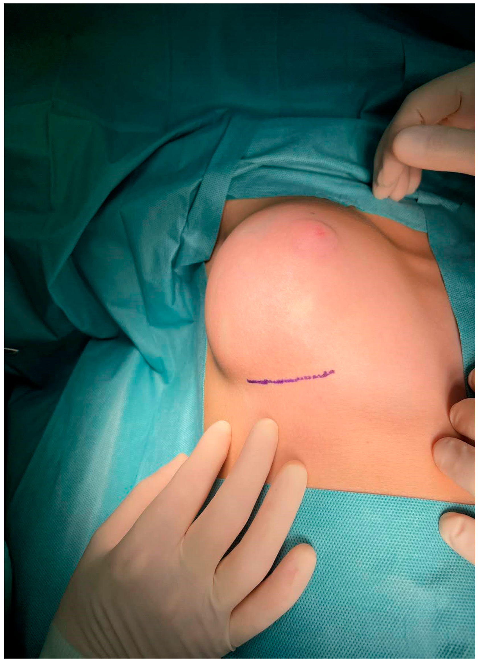
3. Results
4. Discussion
5. Conclusions
Author Contributions
Funding
Institutional Review Board Statement
Informed Consent Statement
Data Availability Statement
Conflicts of Interest
References
- Lee, E.J.; Chang, Y.-W.; Oh, J.H.; Hwang, J.; Hong, S.S.; Kim, H.-J. Breast Lesions in Children and Adolescents: Diagnosis and Management. Korean J. Radiol. 2018, 19, 978–991. [Google Scholar] [CrossRef] [PubMed]
- Jayasinghe, Y.; Simmons, P.S. Fibroadenomas in adolescence. Curr. Opin. Obstet. Gynecol. 2009, 21, 402–406. [Google Scholar] [CrossRef] [PubMed]
- Schnitt, S.J.; Collins, L.C. Pathology of benign breast disorders. In Diseases of the Breast, 4th ed.; Harris, J.R., Lippman, M.E., Morrow, M., Osborne, C.K., Eds.; Wolters Kluwer-Lippincott Williams & Wilkins: Philadelphia, PA, USA, 2010; p. 69. [Google Scholar]
- Adolescent Health. Available online: https://www.who.int/health-topics/adolescent-health#tab=tab_1 (accessed on 26 December 2021).
- Tea, M.-K.M.; Asseryanis, E.; Kroiss, R.; Kubista, E.; Wagner, T. Surgical breast lesions in adolescent females. Pediatr. Surg. Int. 2008, 25, 73–75. [Google Scholar] [CrossRef] [PubMed]
- Dupont, W.D.; Page, D.L.; Parl, F.F.; Vnencak-Jones, C.L.; Plummer, W.D.; Rados, M.S.; Schuyler, P.A. Long-Term Risk of Breast Cancer in Women with Fibroadenoma. N. Engl. J. Med. 1994, 331, 10–15. [Google Scholar] [CrossRef]
- Sosin, M.; Pulcrano, M.; Feldman, E.D.; Patel, K.M.; Nahabedian, M.Y.; Weissler, J.M.; Rodriguez, E.D. Giant juvenile fibroadenoma: A systematic review with diagnostic and treatment recommendations. Gland. Surg. 2015, 4, 312–321. [Google Scholar] [CrossRef]
- Song, B.S.; Kim, E.-K.; Seol, H.; Seo, J.-H.; Lee, J.A.; Kim, D.H.; Lim, J.S. Giant juvenile fibroadenoma of the breast: A case report and brief literature review. Ann. Pediatr. Endocrinol. Metab. 2014, 19, 45–48. [Google Scholar] [CrossRef]
- Makkar, N. Bilateral Giant Juvenile Fibroadenoma of Breast. J. Clin. Diagn. Res. 2017, 11, ED10–ED12. [Google Scholar] [CrossRef]
- Celik, S.U.; Celik, D.B.; Yetiskin, E.; Ergun, E.; Percinel, S.; Demirer, S. Giant juvenile fibroadenoma of the breast: A clinical case. Arch. Argent. de Pediatr. 2017, 115, e428–e431. [Google Scholar] [CrossRef]
- Jategaonkar, P.A. Super-giant Juvenile Breast Fibroadenoma: World’s First Case. J. Coll. Physicians Surg. Pak. 2018, 28, 257–258. [Google Scholar] [CrossRef]
- Juarez, A.M.; Juarez, C.A.S.; Martinez, I.M.A.; Mondragon, E.R. Breast-conserving surgery in giant juvenile fibroadenoma: A case report. AME Surg. J. 2021, 1, 18. [Google Scholar] [CrossRef]
- Kupsik, M.; Yep, B.; Sulo, S.; Memmel, H. Giant juvenile fibroadenoma in a 9-year-old: A case presentation and review of the current literature. Breast Dis. 2017, 37, 95–98. [Google Scholar] [CrossRef] [PubMed]
- Khan, S.; Khan, M.; Rafique, S. Giant Bilateral Juvenile Fibroadenoma of the Breast in Prepubescent Girl. J. Coll. Physicians Surg. Pak. 2015, 25, S95–S96. [Google Scholar] [CrossRef]
- Gaurav, K.; Chandra, G.; Neelam, K.; Kumar, S.; Singla, H.; Yadav, S.K. A pre-pubertal girl with giant juvenile fibroadenoma: A rare case report. Int. J. Surg. Case Rep. 2015, 16, 87–89. [Google Scholar] [CrossRef] [PubMed]
- Rashid, N.F.A.; Firdaus, C.A.M.; Norjazliney, A.J. A case report of juvenile giant fibroadenoma of the breast: How common? Tzu Chi Med J. 2017, 29, 177–179. [Google Scholar] [CrossRef] [PubMed]
- Gkali, C.; Giannos, A.; Primetis, E.; Chalazonitis, A.; Anastasiadi, T.; Feida, E.; Sotiropoulou, M.; Dimitrakakis, C.; Antoniou, A. Giant Juvenile Fibroadenoma in a 12-Year-Old Girl. Ultrasound Q. 2017, 33, 51–54. [Google Scholar] [CrossRef] [PubMed]
- Nishtha; Kaur, N.; Tanveer, N.; Naskar, S. A Diagnostic Enigma—a Rare Presentation of Juvenile Fibroadenoma in a Premenarchal Girl. J. Pediatr. Adolesc. Gynecol. 2021, 34, 558–560. [Google Scholar] [CrossRef] [PubMed]
- Giannos, A.; Stavrou, S.; Gkali, C.; Chra, E.; Marinopoulos, S.; Chalazonitis, A.; Dimitrakakis, C.; Drakakis, P. A prepubertal giant juvenile fibroadenoma in a 12-year-old girl: Case report and brief literature review. Int. J. Surg. Case Rep. 2017, 41, 427–430. [Google Scholar] [CrossRef]
- Bayramoglu, Z.; Yılmaz, R.; Caliskan, E.; Onder, S.; Akkavak, G.; Adaletli, I. Unilateral rapid enlargement of an adolescent breast: Pseudoangiomatous stromal hyperplasia in a giant juvenile fibroadenoma. Breast J. 2018, 24, 648–649. [Google Scholar] [CrossRef]
- Rafeek, N.; Rangasami, R.; Dhanraj, K.; Joseph, S. Multimodality approach in the diagnosis and management of bilateral giant juvenile breast fibroadenoma. BMJ Case Rep. 2016, 2016, bcr2016217588. [Google Scholar] [CrossRef]
- Kitazawa, M.; Futamura, M.; Tokumaru, Y.; Kohyama, K.; Nakakami, A.; Yoshida, K. Breast reconstruction using a tissue expander after enucleation of a giant fibroadenoma: A case report. Int. J. Surg. Case Rep. 2021, 90, 106723. [Google Scholar] [CrossRef]
- Wang, T.; Zhu, L. Mammotome-assisted removal with minimal incision of large juvenile fibroadenoma of breast. Medicine 2020, 99, e19442. [Google Scholar] [CrossRef] [PubMed]
- Heng, S.S.L.; Yahya, M.M.; Sulaiman, W.A.W.; Saad, A.Z.M. A harmless evil: Giant fungating benign breast mass in an adolescent mimicking malignancy - Case report. Int. J. Surg. Case Rep. 2020, 80, 105202. [Google Scholar] [CrossRef] [PubMed]
- Ezer, S.S.; Oguzkurt, P.; Ince, E.; Temiz, A.; Bolat, F.A.; Hicsonmez, A. Surgical Treatment of the Solid Breast Masses in Female Adolescents. J. Pediatr. Adolesc. Gynecol. 2013, 26, 31–35. [Google Scholar] [CrossRef] [PubMed]
- Matz, D.; Kerivan, L.; Reintgen, M.; Akman, K.; Lozicki, A.; Causey, T.; Clynes, C.; Giuliano, R.; Acs, G.; Cox, J.; et al. Breast Preservation in Women With Giant Juvenile Fibroadenoma. Clin. Breast Cancer 2013, 13, 219–222. [Google Scholar] [CrossRef]
- Arowolo, O.; Akinkuolie, A.; Adisa, A.; Obonna, G.; Olasode, B.J. Giant fibroadenoma presenting like fungating breast cancer in a Nigerian teenager. Afr. Heal. Sci. 2013, 13, 162–165. [Google Scholar] [CrossRef]
- Biswas, S.K.; Rahman, A.T.; Paul, A.C.; Mollah, M.; Chowdhury, V. Giant Juvenile Fibroadenoma of the Breast: Report of 2 Cases. Faridpur Med. Coll. J. 2012, 7, 42–45. [Google Scholar] [CrossRef]
- Cheng, P.J.; Vu, L.T.; Cass, D.; Hicks, M.J.; Brandt, M.L.; Kim, E. Endoscopic specimen pouch technique for removal of giant fibroadenomas of the breast. J. Pediatr. Surg. 2012, 47, 803–807. [Google Scholar] [CrossRef]
- Kaur, N.; Saini, S.; Somasekhar, S.; Gupta, A. Bilateral Florid Juvenile Fibroadenomas of the Breast in an Adolescent: A Rare Indication for Subcutaneous Mastectomy. J. Pediatr. Adolesc. Gynecol. 2015, 28, e135–e137. [Google Scholar] [CrossRef]
- Heilmann, T.; Leuschner, I.; Hilpert, F.; Kümper, C.; Strauss, A.; Mundhenke, C.; Jonat, W.; Bauer, M. Diagnosis and management of an unilateral giant fibroadenoma of the breast in pregnancy. Arch. Gynecol. Obstet. 2011, 285, 235–237. [Google Scholar] [CrossRef]
- Izadpanah, A.; Karunanayake, M.; Izadpanah, A.; Sinno, H.; Gilardino, M. An Atypical Growth of a Giant Fibroadenoma after Trauma. J. Pediatr. Adolesc. Gynecol. 2012, 25, e115–e117. [Google Scholar] [CrossRef]
- Marshall, A.P.; Spottswood, S.E.; Grau, A.M.; Jackson, G.P. Juvenile fibroadenoma and granular cell tumor of the breast in an adolescent. J. Pediatr. Surg. 2012, 47, 1930–1933. [Google Scholar] [CrossRef]
- Chepla, K.; Armijo, B.; Ponsky, T.; Soltanian, H. Benefits of immediate dermoglandular preserving reconstruction following giant fibroadenoma excision in two patients. J. Plast. Reconstr. Aesthetic Surg. 2011, 64, e244–e247. [Google Scholar] [CrossRef] [PubMed]
- Ng, W.K.; Mrad, M.A.; Brown, M.H. Juvenile Fibroadenoma of the Breast: Treatment and Literature Review. Can. J. Plast. Surg. 2011, 19, 105–107. [Google Scholar] [CrossRef] [PubMed]
- Nikumbh, D.B.; Desai, S.R.; Madan, P.S.; Patil, N.J.; Wader, J.V. Bilateral Giant Juvenile Fibroadenomas of Breasts:A Case Report. Pathol. Res. Int. 2011, 2011, 1–4. [Google Scholar] [CrossRef] [PubMed]
- Yagnik, V. Juvenile Giant Fibroadenoma. Clin. Pract. 2011, 1, e49. [Google Scholar] [CrossRef] [PubMed]
- Poh, M.M.; Ballard, T.N.; Wendel, J.J. Beckwith-Wiedemann Syndrome and Juvenile Fibroadenoma. Ann. Plast. Surg. 2010, 64, 803–806. [Google Scholar] [CrossRef] [PubMed]
- Biggers, B.D.; Lamont, J.P.; Etufugh, C.N.; Knox, S.K. Inframammary Approach for Removal of Giant Juvenile Fibroadenomas. J. Am. Coll. Surg. 2009, 208, e1–e4. [Google Scholar] [CrossRef]
- Calcaterra, V.; Coscia, D.R.; Sgarella, A.; Burroni, B.; Podetta, M.; Andorno, A.; Ferrari, A.; Larizza, D. Recurrence of giant juvenile breast fibroadenoma in a girl with Turner’s syndrome. J. Pediatr. Endocrinol. Metab. 2009, 22, 281–284. [Google Scholar] [CrossRef]
- Kozomara, D.; Šutalo, N.; Galić, G.; Jurišić, S.; Trninć, Z. Giant unilateral breast juvenile fibroadenoma-the aesthetic outcome of surgical treatment should be considered. Med. Acad. Most. 2016, 28, 72–75. [Google Scholar]
- Gobbi, D.; Dall’Igna, P.; Alaggio, R.; Nitti, D.; Cecchetto, G. Giant fibroadenoma of the breast in adolescents: Report of 2 cases. J. Pediatr. Surg. 2009, 44, e39–e41. [Google Scholar] [CrossRef]
- Mukhopadhyay, M.; Patra, R.; Mondal, S.; Ghosh, A.; Ray, A.K. Bilateral giant juvenile fibroadenoma of breasts. J. Indian Assoc. Pediatr. Surg. 2009, 14, 68–69. [Google Scholar] [CrossRef]
- Wolfram, D.; Behensky, H.; Piza-Katzer, H. Unilateral Gigantomastia Related to Juvenile Fibroadenoma with Idiopathic Thoracic Scoliosis. J. Pediatr. Adolesc. Gynecol. 2009, 22, e25–e27. [Google Scholar] [CrossRef]
- Dolmans, G.H.; Hoogbergen, M.M.; van Rappard, J.H. Giant fibroadenoma of one breast: Immediate bilateral reconstruction. J. Plast. Reconstr. Aesthetic Surg. 2007, 60, 1156–1157. [Google Scholar] [CrossRef] [PubMed]
- Moore, R.L.; Mungara, A.; Shayan, K.; Wallace, A.M. Bilaterally symmetric juvenile fibroadenomas and tubular breast deformity in a prepubescent girl. J. Pediatr. Surg. 2007, 42, 1133–1136. [Google Scholar] [CrossRef] [PubMed]
- Ahuja, A.; Seth, A. Juvenile fibroadenoma of breast. Indian Pediatr. 2005, 42, 72. [Google Scholar] [PubMed]
- Lee, C.; Kim, Y.; Seo, Y.; Pak, S.; Lee, S. Treatment of Multiple Bilateral Juvenile Fibroadenomas in a Teenage Breast by Central Pedicle Breast Reduction, with Vertical and Short Horizontal Scar: Case Report. Aesthetic Plast. Surg. 2004, 28, 228–230. [Google Scholar] [CrossRef]
- Zacharia, T.T.; Lakhar, B.; Ittoop, A.; Menachery, J. Giant Fibroadenoma. Breast J. 2003, 9, 53. [Google Scholar] [CrossRef]
- Hanna, R.M.; Ashebu, S.D. Giant fibroadenoma of the breast in an Arab population. Australas. Radiol. 2002, 46, 252–256. [Google Scholar] [CrossRef]
- Wechselberger, G.; Schoeller, T.; Piza-Katzer, H. Juvenile fibroadenoma of the breast. Surgery 2002, 132, 106–107. [Google Scholar] [CrossRef]
- Celik, M.F.; Dural, A.C.; Unsal, M.G.; Akarsu, C.; Alim, E.R.; Kapan, S.; Kalayci, M.U.; Alis, H. Giant juvenile fibroadenoma. Turk. J. Surg. 2015, 31, 96–98. [Google Scholar] [CrossRef]
- Davis, S.E.; Wallace, A.M. A 19 Year Old with Complete Androgen Insensitivity Syndrome and Juvenile Fibroadenoma of the Breast. Breast J. 2001, 7, 430–433. [Google Scholar] [CrossRef] [PubMed]
- Tantrige, P.M.; Hassanally, D. Recurrent giant juvenile fibroadenomas with hemihypertrophy. Breast Dis. 2011, 33, 41–44. [Google Scholar] [CrossRef] [PubMed]
- Baxi, A.A.M. Multiple Bilateral Giant Juvenile Fibroadenomas of Breast. Eur. J. Surg. 2000, 166, 828–830. [Google Scholar] [CrossRef] [PubMed]
- Kamei, S.T.Y. Natural skin reduction and breast recovery using a tissue expander after enucleation of a giant breast tumour. Scand. J. Plast. Reconstr. Surg. Hand Surg. 2000, 34, 383–385. [Google Scholar] [CrossRef] [PubMed]
- Simmons, R.M.; Cance, W.G.; Iacicca, M.V. A Giant Juvenile Fibroadenoma in a 12-Year-Old Girl: A Case for Breast Conservation. Breast J. 2000, 6, 418–420. [Google Scholar] [CrossRef] [PubMed]
- Silfen, R.; Skoll, P.J.; Hudson, D.A. Florid Juvenile (Cellular) Fibroadenomatosis in the Adolescent: A Case for Subcutaneous Mastectomy? Aesthetic Plast. Surg. 1999, 23, 413–415. [Google Scholar] [CrossRef]
- Tian, J.; Liu, X.; Chen, Q. Giant fibroma of breast in an adolescent female by inverted “T” incision: A case report and literature review. Zhong Nan Da Xue Xue Bao Yi Xue Ban 2020, 45, 204–207. [Google Scholar] [CrossRef]
- Hoffman, S. Giant fibroadenoma of the breast: Immediate reconstruction following excision. Br. J. Plast. Surg. 1978, 31, 170–172. [Google Scholar] [CrossRef]
- Yu, J. Treatment of Giant Juvenile Breast Fibroadenoma by Single Hole Breast Endoscopy: A Case Report. Acta Endocrinol. (Bucharest) 2021, 17, 552–553. [Google Scholar] [CrossRef]
- Zeeshan, S.; Shaikh, K.; Tariq, M.U.; Vohra, L.M. Giant Juvenile Fibroadenoma of the breast in a 13-year-old Pakistani girl with excellent cosmetic outcome after subareolar enucleation–A case report. Int. J. Surg. Case Rep. 2021, 79, 450–454. [Google Scholar] [CrossRef]
- Baral, S.; Gyawali, M.; Thapa, N.; Chhetri, R.K.; Dahal, P. Giant juvenile fibroadenoma in an adolescent female: A case report. Clin. Case Rep. 2020, 8, 3488–3492. [Google Scholar] [CrossRef]
- Islam, S.; Saroop, S.; Bheem, V.; Naraynsingh, V. Largest giant juvenile fibroadenoma of the breast. BMJ Case Rep. 2019, 12, e227277. [Google Scholar] [CrossRef] [PubMed]
- Mashiloane, J.M.C.D. Juvenile fibroadenoma during pregnancy. J. Obstet. Gynaecol. 2000, 20, 86. [Google Scholar] [CrossRef] [PubMed]
- Ugburo, A.O.; Olajide, T.O.; Fadeyibi, I.O.; Mofikoya, B.O.; Lawal, A.O.; Osinowo, A.O. Differential diagnosis and management of giant fibroadenoma: Comparing excision with reduction mammoplasty incision and excision with inframammary incision. J. Plast. Surg. Hand Surg. 2012, 46, 354–358. [Google Scholar] [CrossRef] [PubMed]
- Greydanus, D.E.; Parks, D.S.; Farrell, E.G. Breast Disorders in Children and Adolescents. Pediatr. Clin. N. Am. 1989, 36, 601–638. [Google Scholar] [CrossRef]
- Smith, G.; Burrows, P. Ultrasound diagnosis of fibroadenoma—Is biopsy always necessary? Clin. Radiol. 2008, 63, 511–515. [Google Scholar] [CrossRef]
- Templeman, C.; Hertweck, S.P. Breast disorders in the pediatric and adolescent patient. Obstet. Gynecol. Clin. N. Am. 2000, 27, 19–34. [Google Scholar] [CrossRef]
- Sklair-Levy, M.; Sella, T.; Alweiss, T.; Craciun, I.; Libson, E.; Mally, B. Incidence and Management of Complex Fibroadenomas. Am. J. Roentgenol. 2008, 190, 214–218. [Google Scholar] [CrossRef]
- Emans, S.J.; Laufer, M.R.; DiVasta, A. The breast: Examination and lesions. In Pediatric & Adolescent Gynecology, 7th ed.; Lippincott Williams & Wilkins (LWW): Philadelphia, PA, USA, 2019; p. 781. [Google Scholar]
- Golatta, M.; Harcos, A.; Pavlista, D.; Danes, J.; Klein, R.; Simovich, P.; Gruber, I.; Hahn, M. Ultrasound-guided cryoablation of breast fibroadenoma: A pilot trial. Arch. Gynecol. Obstet. 2014, 291, 1355–1360. [Google Scholar] [CrossRef]
- Kaufman, C.S.; Littrup, P.J.; A Freman-Gibb, L.; Francescatti, D.; Stocks, L.H.; Smith, S.J.; Henry, A.C.; Bailey, L.; Harness, J.K.; Simmons, R. Office-Based cryoablation of breast fibroadenomas: 12-month followup. J. Am. Coll. Surg. 2004, 198, 914–923. [Google Scholar] [CrossRef]
- Littrup, P.J.; Freeman-Gibb, L.; Andea, A.; White, M.; Amerikia, K.C.; Bouwman, D.; Harb, T.; Sakr, W. Cryotherapy for Breast Fibroadenomas. Radiology 2005, 234, 63–72. [Google Scholar] [CrossRef] [PubMed]
- Greenberg, R.; Skornick, Y.; Kaplan, O. Management of breast fibroadenomas. J. Gen. Intern. Med. 1998, 13, 640–645. [Google Scholar] [CrossRef] [PubMed]
- West, K.W.; Rescorla, F.J.; Scherer, L.; Grosfeld, J.L. Diagnosis and treatment of symptomatic breast masses in the pediatric population. J. Pediatr. Surg. 1995, 30, 182–187. [Google Scholar] [CrossRef] [PubMed]
- Chao, T.-C.; Lo, Y.-F.; Chen, S.-C.; Chen, M.-F. Sonographic features of phyllodes tumors of the breast. Ultrasound Obstet. Gynecol. 2002, 20, 64–71. [Google Scholar] [CrossRef] [PubMed]
- ACOG Committee on Adolescent Health Care ACOG Committee Opinion No. 350: Breast Concerns in the Adolescent. Obstet. Gynecol. 2006, 108, 1329–1336. [CrossRef] [PubMed]


| Authors | Country | Size (cm) | Unilateral–Bilateral | Location (Right/Left) | Age (Years) | Menarcheal Status (pre/post) | Treatment | No. of Patients |
|---|---|---|---|---|---|---|---|---|
| Celik et al. 2015 [52] | Turkey | 9.5 × 8 × 6 | unilateral | right | 10 | N/A | total lump excision | 1 |
| Yu et al. 2021 [61] | China | 9.65 × 5.80 × 5.5 | unilateral | left | 11 | N/A | single-hole breast endoscopic resection | 1 |
| Zeeshan et al. 2021 [62] | Pakistan | 13.5 × 11.5 × 6 | unilateral | left | 13 | post | total lump excision | 1 |
| Baral et al. 2020 [63] | Nepal | 9 × 7 | unilateral | right | 18 | post | total lump excision | 1 |
| Islam et al. 2019 [64] | Trinidad and Tobago | 28 × 25 | unilateral | left | 16 | post | total lump excision | 1 |
| Makkar et al. 2017 [9] | India | right lump: 6 × 5/ left lumps: 10 × 6 and 2 × 3 | bilateral | N/A | 14 | pre | bilateral total lump excisions | 1 |
| Celik S. et al. 2017 [10] | India | 17 × 13 × 5.5 | unilateral | left | 14 | post | total lump excision | 1 |
| Jategaonkar et al. 2018 [11] | India | right lump: 63 × 47/ left lump: 51 × 39 | bilateral | N/A | 17 | N/A | bilateral simple mastectomy with synchronous breast reconstructions | 1 |
| Juarez et al. 2021 [12] | Mexico | 16 × 13 | unilateral | left | 14 | N/A | total lump excision | 1 |
| Kupsik et al. 2017 [13] | USA | 11.2 × 8.5 × 6.7 | unilateral | right | 9 | pre | total lump excision | 1 |
| Khan et al. 2015 [14] | Pakistan | right lump: 6 × 5/ left lump: 10 × 15 | bilateral | N/A | 10 | pre | bilateral total lump excisions | 1 |
| Giannos et al. 2017 [19] | Greece | 14 × 12 | unilateral | right | 12 | pre | total lump excision | 1 |
| Gaurav et al. 2015 [15] | India | 20 × 20 | unilateral | left | 10 | pre | total lump excision | 1 |
| Mohd Firdaus et al. 2017 [16] | Malaysia | 12 × 9 × 5 | unilateral | right | 12 | N/A | total lump excision | 1 |
| Gkali et al. 2017 [17] | Greece | 15 × 13 | unilateral | right | 12 | pre | total lump excision | 1 |
| Nishtha et al. 2021 [18] | India | 15.5 × 9 × 10.5 | unilateral | right | 14 | pre | total lump excision | 1 |
| Bayramoglu et al. 2018 [20] | Turkey | 8.3 × 9.9 × 5.5 | unilateral | left | 14 | N/A | partial mastectomy with synchronous breast reconstruction with an implant | 1 |
| Rafeek et al. 2016 [21] | India | right lump: 39 × 22 × 17/ left lump: 40 × 24 × 18 | bilateral | N/A | 13 | post | bilateral total lump excisions with synchronous reduction mammoplasty | 1 |
| Kitazawa et al. 2022 [22] | Japan | 13 × 9 × 4.5 | unilateral | left | 14 | N/A | total lump excision | 1 |
| Wang et al. 2020 [23] | China | 6 × 6 | unilateral | left | 19 | N/A | total lump excision | 1 |
| Heng et al. 2020 [24] | Malaysia | 10.6 × 14.5 × 15.1 | unilateral | right | 19 | post | mastectomy with synchronous breast reconstruction | 1 |
| Tian et al. 2020 [59] | China | 22 × 16 × 11 | unilateral | right | 18 | N/A | total lump excision | 1 |
| Kaur et al. 2015 [30] | India | numerous tumors on both breasts, number not specified, size 1.5–2.5 | bilateral | N/A | 15 | post | bilateral subcutaneous mastectomy | 1 |
| Kozomara et al. 2016 [41] | Boznia Herzegovina | 20 × 20 × 15 | unilateral | left | 16 | N/A | reduction mammoplasty technique of the left breast | 1 |
| Ezer et al. 2013 [25] | Turkey | P1: 25 × 12/ P2: 30 × 16/ P3: 45 × 20/ P4: 60 × 35 | unilateral | not specified | P1: 14, P2: 17, P3: 10, P4: 14 | N/A | total lump excision | 4 |
| Matz et al. 2013 [26] | USA | 8.9 × 9.8 × 8.8 | unilateral | left | 13 | N/A | total lump excision | 1 |
| Arowolo et al. 2013 [27] | Nigeria | 20 × 30 | unilateral | left | 14 | pre | total lump excision with synchronous breast reconstruction | 1 |
| Biswas et al. 2012 [28] | Bangladesh | P1: 15 × 13/ P2: 11 × 11 | unilateral | P1: right, P2: left | P1: 14, P2: 16 | N/A | total lump excision | 2 |
| Sosin et al. 2012 [7] | USA | 12.1 | unilateral | right | 13 | N/A | total lump excision | 1 |
| Cheng et al. 2012 [29] | USA | P1: 5/ P2: 8/ P3: 10.5 | unilateral | P1: left, P2: left, P3: left | P1: 17, P2: 13, P3: 15 | N/A | endoscopic specimen pouch technique | 3 |
| Heilmann et al. 2012 [31] | Germany | 13 × 12.5 × 4.5 | unilateral | right | 17 | post | total lump excision | 1 |
| Izadpanah et al. 2012 [32] | Canada | 7.3 × 9.3 × 8.9 | unilateral | left | 12 | post | total lump excision | 1 |
| Chepla et al. 2011 [34] | USA | P1: 11.6 × 9.8 × 11.3/ P2: 15 × 4 × 4 | unilateral | P1: left/P2: right | P1: 10, P2: 16 | N/A | total lump excision | 2 |
| Ng et al. 2011 [35] | Canada | 19 | unilateral | right | 17 | N/A | total lump excision | 1 |
| Nikumbh et al. 2011 [36] | India | right lump: 15 × 12/ left lump: 17 × 15 | bilateral | N/A | 12 | pre | total excision of bilateral breast lumps conserving the normal breast tissue, nipple, and areola by the plastic surgeon | 1 |
| Tantrige et al. 2011 [54] | UK | 12 | unilateral | left | 13 | N/A | total lump excision | 1 |
| Yagnik et al. 2011 [37] | India | 13 × 11 × 12 | unilateral | right | 15 | N/A | total lump excision | 1 |
| Poh et al. 2010 [38] | USA | not described | bilateral | N/A | 12 | post | Right breast: excision of 6 discreet masses through vertical incision, laterally based pedicle used for nipple–areola complex/Left breast: subcutaneous mastectomy through “inverted T” incision, nipple–areola complex was salvaged as a free nipple graft. | 1 |
| Biggers et al. 2009 [39] | USA | P1: 5.9/ P2: 10.5/ P3: 12/ P4: 17 | unilateral | not specified | P1: 11/ P2: 12/ P3: 14/ P4: 15 | N/A | total lump excision (inframammary approach) | 4 |
| Calcaterra et al. 2009 [40] | Italy | 14 × 16 × 17 | unilateral | right | 12 | N/A | total lump excision (inframammary approach) | 1 |
| Gobbi et al. 2009 [42] | Italy | P1: 8/ P2: 10 | unilateral | P1: left/ P2: left | P1: 12/ P2: 15 | P1: N/A/ P2: post | total lump excision | 2 |
| Mukhopadhyay et al. 2009 [43] | India | right lump: 22 × 20/ left lump: 18 × 16 | bilateral | N/A | 11 | pre | bilateral lump excision conserving normal breast tissue, nipple, and areola | 1 |
| Wolfram et al. 2009 [44] | Austria | 5 | unilateral | right | 15 | N/A | total lump excision | 1 |
| Dolmans et al. 2007 [45] | Netherlands | 9 × 9 | unilateral | left | 18 | N/A | nipple-sparing subcutaneous mastectomy of the left breast | 1 |
| Moore et al. 2007 [46] | USA | right breast fibrofatty mass (1040 g): 17.0 × 15.0 × 7.0/ left breast fibrofatty mass (1111 g): 18.5 × 16 × 7.5 | bilateral | N/A | 9 | pre | bilateral subtotal mastectomies | 1 |
| Ahuja et al. 2005 [47] | India | not described | unilateral | right | 12 | N/A | total lump excision | 1 |
| Lee et al. 2004 [48] | South Korea | multiple nodules, the largest: 17 × 3 × 3, others not described | bilateral | N/A | 11 | post | central pedicle reduction mammoplasty | 1 |
| Zacharia et al. 2003 [49] | India | 8 × 5 | unilateral | right | 13 | N/A | total lump excision | 1 |
| Hanna et al. 2002 [50] | Kuwait | right lump: 6 × 6 × 5/ left lump: 5.5 × 5 × 6 | bilateral | N/A | 15 | N/A | not specified | 1 |
| Wechselberger et al. 2002 [51] | Austria | not described | unilateral | right | 15 | N/A | total lump excision | 1 |
| Davis et al. 2001 [53] | USA | 5 × 3.5 × 2.5 | unilateral | right | 19 | N/A (CAIS) | total lump excision | 1 |
| Baxi et al. 2000 [55] | India | right breast: 17 firm nodules (mean diameter 4, range 1–8)/ left breast: 9 (mean diameter 8, range 1–11) | bilateral | N/A | 16 | post | total lump excision | 1 |
| Kamei et al. 2000 [56] | Japan | P1: 11 × 10 × 5/ P2: 10 × 9 × 8 | unilateral | left | P1: 19/ P2: 17 | N/A | total lump excision | 2 |
| Simmons et al. 2000 [57] | USA | 16 | unilateral | right | 12 | N/A | total lump excision | 1 |
| Silfen et al. 1999 [58] | South Africa | not described | bilateral | N/A | 13 | N/A | bilateral reduction mammoplasty | 1 |
| Hoffman et al. 1978 [60] | US | not described | unilateral | right | 13 | N/A | total lump excision with synchronous reduction mammoplasty | 1 |
| Song et al. 2014 [8] | South Korea | 13 × 8 | unilateral | right | 12 | post | total lump excision | 1 |
| Mashiloane 2000 [65] | South Africa | not described | unilateral | right | 16 | post (during pregnancy) | mastectomy | 1 |
| Ugburo et al. 2012 [66] | Nigeria | not described | 14 patients unilateral, 2 patients bilateral | not specified | mean 14.06 ± 0.42 (range 12–18) | post | total lump excision | 16 |
| Eleftheriades et al. 2022 (our case) | Greece | 10 × 8 × 2.5 | unilateral | right | 11 | pre | total lump excision | 1 |
| Total Number of Cases: | 87 |
Disclaimer/Publisher’s Note: The statements, opinions and data contained in all publications are solely those of the individual author(s) and contributor(s) and not of MDPI and/or the editor(s). MDPI and/or the editor(s) disclaim responsibility for any injury to people or property resulting from any ideas, methods, instructions or products referred to in the content. |
© 2023 by the authors. Licensee MDPI, Basel, Switzerland. This article is an open access article distributed under the terms and conditions of the Creative Commons Attribution (CC BY) license (https://creativecommons.org/licenses/by/4.0/).
Share and Cite
Eleftheriades, A.; Tsarna, E.; Toutoudaki, K.; Paschalidou, E.; Christopoulos, N.; Georgopoulos, I.; Mitropoulou, G.; Christopoulos, P. Giant Juvenile Fibroadenoma: Case Report and Review of the Literature. J. Clin. Med. 2023, 12, 1855. https://doi.org/10.3390/jcm12051855
Eleftheriades A, Tsarna E, Toutoudaki K, Paschalidou E, Christopoulos N, Georgopoulos I, Mitropoulou G, Christopoulos P. Giant Juvenile Fibroadenoma: Case Report and Review of the Literature. Journal of Clinical Medicine. 2023; 12(5):1855. https://doi.org/10.3390/jcm12051855
Chicago/Turabian StyleEleftheriades, Anna, Ermioni Tsarna, Konstantina Toutoudaki, Eleni Paschalidou, Nikolaos Christopoulos, Ioannis Georgopoulos, Georgia Mitropoulou, and Panagiotis Christopoulos. 2023. "Giant Juvenile Fibroadenoma: Case Report and Review of the Literature" Journal of Clinical Medicine 12, no. 5: 1855. https://doi.org/10.3390/jcm12051855
APA StyleEleftheriades, A., Tsarna, E., Toutoudaki, K., Paschalidou, E., Christopoulos, N., Georgopoulos, I., Mitropoulou, G., & Christopoulos, P. (2023). Giant Juvenile Fibroadenoma: Case Report and Review of the Literature. Journal of Clinical Medicine, 12(5), 1855. https://doi.org/10.3390/jcm12051855