Article

A 2-Year Audit on Antibiotic Resistance Patterns from a Urology Department in Greece

by
Ioannis Manolitsis
1,
Georgios Feretzakis
2,3,
Stamatios Katsimperis
1,
Panagiotis Angelopoulos
1,
Evangelos Loupelis
4,
Nikoleta Skarmoutsou
5,
Lazaros Tzelves
1,* and
Andreas Skolarikos
1
1
Second Department of Urology, Sismanogleio General Hospital, 15126 Marousi, Greece
2
School of Science and Technology, Hellenic Open University, 26335 Patras, Greece
3
Department of Quality Control, Research and Continuing Education, Sismanogleio General Hospital, 15126 Marousi, Greece
4
IT Department, Sismanogleio General Hospital, 15126 Marousi, Greece
5
Microbiology Laboratory, Sismanogleio General Hospital, 15126 Marousi, Greece
*
Author to whom correspondence should be addressed.
J. Clin. Med. 2023, 12(9), 3180; https://doi.org/10.3390/jcm12093180
Submission received: 1 March 2023 / Revised: 21 April 2023 / Accepted: 27 April 2023 / Published: 28 April 2023
(This article belongs to the Special Issue Urinary Tract Infection: Prevention, Diagnosis, and Treatment)

Abstract

:
Purpose: The high incidence of urinary tract infections (UTIs), often in nosocomial environments, is a major cause of antimicrobial resistance (AMR). The dissemination of antibiotic-resistant infections results in very high health and economic burdens for patients and healthcare systems, respectively. This study aims to determine and present the antibiotic resistance profiles of the most common pathogens in a urology department in Greece. Methods: During the period 2019–2020, we included 12,215 clinical samples of blood and urine specimens that tested positive for the following pathogens: Escherichia coli, Enterococcus faecium, Enterococcus faecalis, Proteus mirabilis, Klebsiella pneumoniae, or Pseudomonas aeruginosa, as these are the most commonly encountered microbes in a urology department. Results: The analysis revealed a 22.30% mean resistance rate of E. coli strains with a 76.42% resistance to ampicillin and a 54.76% resistance rate to ciprofloxacin in the two-year period. It also showed an approximately 19% resistance rate of P. mirabilis strains and a mean resistance rate of 46.205% of K. pneumoniae strains, with a decreasing trend during the four semesters (p-value < 0.001), which presented an 80% resistance rate to ampicillin/sulbactam and 73.33% to ciprofloxacin. The resistance to carbapenems was reported to be 39.82%. The analysis revealed a 24.17% mean resistance rate of P. aeruginosa with a declining rate over the two-year period (p-value < 0.001). The P. aeruginosa strains were 38% resistant to fluoroquinolones and presented varying resistance against carbapenems (31.58% against doripenem and 19.79% against meropenem). Regarding the Enteroccocal strains, a 46.91% mean resistance was noted for E. faecium with 100% resistance to ampicillin, and a 24.247% mean resistance rate for E. faecalis strains that were 41% resistant to ciprofloxacin. Both types showed 100% sensitivity to linezolid. Conclusions: The dissemination of antibiotic-resistant pathogens poses the need to implement surveillance programs and, consequently, to develop strategies to prevent the emergence of such pathogens in order to optimize patient outcomes.

1. Introduction

Urinary tract infections (UTIs) are one of the most common infectious diseases, accounting for 150–250 million cases globally every year [1]. They occur frequently, in the context of a nosocomial environment, leading to an increasing antimicrobial resistance (AMR) [2]. AMR can emerge through a number of methods. As with fluoroquinolone resistance, which is primarily brought on by changes in DNA-topoisomerase, mutations may occur in the target of the antimicrobial agent. As an alternative, the medication may be inactivated by an enzyme, as in the case of beta-lactamases, or by an aminoglycoside through phosphorylation, acetylation, or adenylation. Two other methods include evading the target, as in the case of vancomycin, and preventing the medicine from reaching its intended targets, as in the example of tetracycline [3,4].
AMR has become a major concern in medicine in the twenty-first century. The dissemination of antibiotic-resistant infections constitutes a worldwide concern with an increasing health and economic burden [5]. The World Health Organization (WHO) is currently considering this issue to be “one of the biggest threats to global health, food security, and development”. The major project undertaken by the WHO to track worldwide AMR and antibiotic usage since 2015 is the worldwide Antimicrobial Resistance and Use Surveillance System (GLASS). In terms of uropathogenic microorganisms, GLASS gathers information on the AMR rates of Escherichia coli and Klebsiella pneumoniae. The report from the year 2021 shows a median AMR against cotrimoxazole of 54.4% (IQR 36.5–69.4) for E. coli and 43.1% (IQR 31.8–57.5) for K. pneumoniae in 12 countries reporting globally [6].
According to data from the European Antimicrobial Resistance Surveillance Network (EARS-Net), Greece has one of the highest burdens of infections caused by antibiotic-resistant bacteria [7]. The majority of this burden was caused by infections with carbapenem-resistant or colistin-resistant bacteria in 2015 [2,7,8]. One of the reasons why Greece ranks high in antibiotic-resistant bacteria is self-medication, as antibiotics are acquired very often from pharmacies without any prescription. In addition, the over-prescription of antibiotics by Greek physicians is a major issue that leads to antibiotic over-consumption and, consequently, to an increasing antimicrobial resistance [9,10].
Antimicrobial stewardship programs aim to optimize the appropriate use of available antimicrobial agents in order to improve therapeutic outcomes for multidrug-resistant (MDR) infections, thereby lowering antimicrobial resistance rates and hospital costs [11]. To implement any preventive measures, it is critical to understand the antibiotic resistance profiles in each hospital ward. The current study aims to determine and present the antibiotic resistance levels of E. coli, P. mirabilis, E. faecium, E. faecalis, P. aeruginosa, and K. pneumoniae isolates over a two-year period, 2019–2020, in one of Greece’s largest urology departments.

2. Materials and Methods

This was a cross-sectional study conducted in Sismanogleio General Hospital, a public tertiary hospital in Athens, Greece, based on data from the Microbiology Department.

2.1. Study Design

The inclusion criteria of this study included clinical isolates from non-identical, hospitalized patients that were not duplicated. On the other hand, we excluded all isolates that originated from a single patient with similar susceptibility patterns. We included E. coli, E. faecium, E. faecalis, P. mirabilis, K. pneumoniae, and P. aeruginosa as they constitute the most common pathogens encountered in a urology department [12]. Resistance to the following antibiotics was measured in P. aeruginosa: aminoglycosides (amikacin, gentamycin, and tobramycin), fluoroquinolones (ciprofloxacin and levofloxacin), cephalosporins (cefepime and ceftazidime), monobactams (aztreonam), carbapenems (doripenem, meropenem, and imipenem), piperacillin/tazobactam (anti-pseudomonal penicillin), and colistin. The following antibiotics were used to assess E. coli resistance: aminoglycosides (amikacin and gentamycin), aminopenicillins (amoxicillin/clavulanic acid and ampicillin), cephalosporins (cefuroxime and ceftazidime), carbapenems (meropenem), trimethoprim/sulfamethoxazole, and piperacillin/tazobactam. The antibiotics used to test K. pneumoniae resistance were aminoglycosides (amikacin, gentamycin, and tobramycin), aminopenicillins (ampicillin/sulbactam), cephalosporins (cefotaxime, ceftazidime, and cefuroxime), fluoroquinolones (levofloxacin and moxifloxacin), tetracycline, and colistin. The resistance for P. mirabilis was measured based on the following antibiotics: aminoglycosides (amikacin and gentamycin), aminopenicillins (amoxicillin/clavulanic acid and ampicillin), cephalosporins (cefotaxime, cefoxitin, ceftazidime, and cefuroxime), carbapenems (imipenem and meropenem), fluoroquinolones (levofloxacin), piperacillin/tazobactam, and trimethoprim/sulfamethoxazole. As for the Enterococci species, the resistance for E. faecium and faecalis was measured based on ampicillin, fluoroquinolones (ciprofloxacin and levofloxacin), aminoglycosides (gentamycin), glycopeptide antibiotics (vancomycin and teicoplanin), lipopeptide antibiotics (daptomycin), oxazolidinones (linezolid), streptogramins (pristinamycin), and tetracycline. Our research focused on the antibiotics mentioned above since there were an adequate number of samples for these in order to extract reliable conclusions and there was also susceptibility testing performed for them in all 4 semesters.

2.2. Source of Isolates

During a 2-year period (January 2019 to December 2020), a total of 12,215 clinical samples from patients hospitalized in the urology department were included in this study and processed by the Microbiology Laboratory according to established protocols [13,14,15]. The samples included blood and urine specimens. Positive urine cultures were classified as having considerable microbial growth, as indicated by standard microbiological criteria [16]. Blood cultures were collected from all febrile patients that were hospitalized in our department. Blood cultures were incubated in the BacT/Alert system (bioMerieux, Marcy-l’Étoile, France). Isolation and identification of pathogens was carried out according to classical microbiological procedures, such as conventional cultivation-based enrichment and plating methods with rapid plating solution [17].

2.3. Antimicrobial Susceptibility Testing

Antimicrobial susceptibility testing was carried out using the MicroScan system (Siemens, Munich, Germany) in accordance with CLSI guidelines [18,19], and the results were confirmed using a gradient MIC (minimum inhibitory concentration) method in accordance with the manufacturer’s guidelines (e.g., the E-test bioMerieux, Solna, Sweden). Colistin MICs were retested using microtiter plates (SensiTestColistin, Liofilchem, Roseto degli Abruzzi, Italy). CLSI criteria were used to determine antibiotic sensitivity and resistance breakpoints [18,19]. As quality control strains for susceptibility tests, Escherichia coli ATCC 25922 and Pseudomonas aeruginosa ATCC 27853 were used. The CLSI-recommended double-disk synergy test (DDST) was used to detect the phenotype of the production of extended-spectrum beta-lactamases (ESBL).
Metallo-beta-lactamases (MBL) and carbapenemases (KPC) were detected phenotypically by (a) the modified Hodge test [19]; (b) the combined disk test, with a meropenem (MER) disk alone, a MER disk plus phenyl boronic acid (PBA), a MER disk plus EDTA, and a MER disk plus PBA and EDTA [20]; and (c) the NG CARBA 5 immunochromato-graphic assay, targeting KPC-, NDM-, VIM-, and IMP-type and OXA-48-like car-bapenemases, following the manufacturer’s guidelines [21]. P. aeruginosa strains were phenotypically tested for MBL using either an imipenem (IPM) disk and IPM plus EDTA combined disk test [22] or an IPM-EDTA double-disk synergy test (DDST) [23].
PCR testing was performed for the detection of the most clinically significant carbapenemase genes (blaNDM, blaVIM, blaKPC, and blaOXA-48), as they encode enzymes that may generate carbapenem and other beta-lactam resistance. The testing was carried out on all strains that phenotypically produced more than one or no carbapenemases, as well as for the OXA producers and all those tested with NG CARBA 5. They were also tested for the presence of the colistin-resistance plasmid-mediated mcr-1 gene [21,24].

2.4. Statistical Analysis

All analyses were performed using SPSS Statistics version 24.0 (IBM, Armonk, NY, USA) [25]. The data were analyzed over a period of two years (2019–2020) and each semester was analyzed in a separate manner. The resistance rates of P. aeruginosa, E. coli, P. mirabilis, E. faecium, E. faecalis, and K. pneumoniae clinical isolates against individual antimicrobial agents were analyzed. The measure of the mean resistance of any microbe is defined as the percentage of samples of a specific microbe that are resistant to any of the antibiotics that were tested in the laboratory. Differences between means regarding the resistance of those six bacteria mentioned above, in the urology department, were determined by univariate analysis of variance and Tukey’s multiple range tests. A significance level of p < 0.05 was used.

3. Results

We analyzed the resistance rates in 5430 tests of E. coli, 1537 tests of E. faecalis, 1315 tests of P. mirabilis, 564 tests of E. faecium, 2302 tests of K. pneumoniae, and 1067 tests of P. aeruginosa, against routinely tested antimicrobial agents. The mean age of our patients was 67.7 years of age, and regarding their sex, 59.2% of them were males and the remaining 40.8% were female patients.
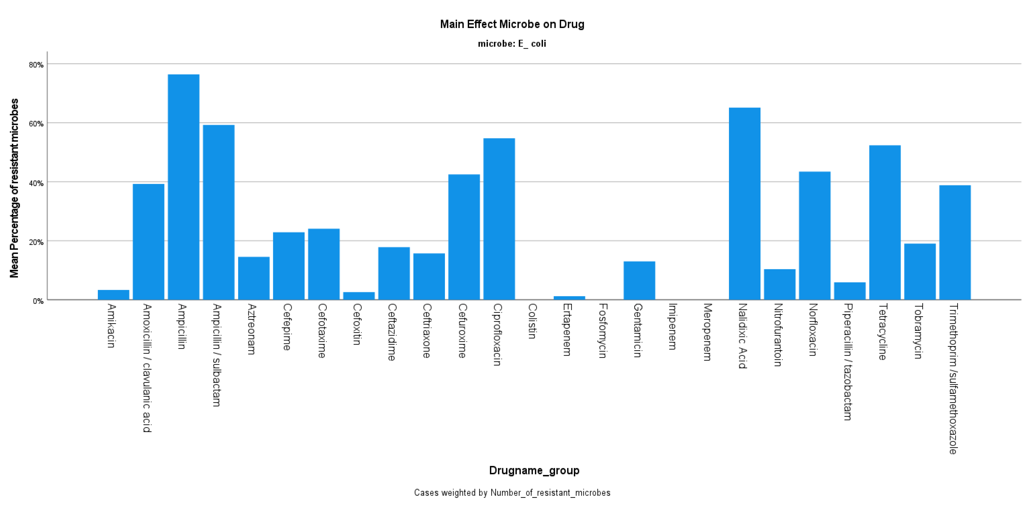
The mean resistance rate of E. coli during the years 2019–2020 was 22.30%, ranging from 19.34 to 26.76% (p-value 0.977) (Figure 1), with the number of isolates being decreased in 2020 compared to 2019 (1739 from 3.691). The E. coli strains were 76.42% resistant to ampicillin, while there was 96.67% sensitivity to amikacin, and 94.09% sensitivity to piperacillin/tazobactam. The resistance rate to ciprofloxacin was reported at 54.76% during the same period (Figure 2).
Regarding P. mirabilis, the 2-year mean resistance rate was 18.94% with no statistical significance between the four semesters (p-value 0.982) (Figure 1), and approximately twice the number of isolates between the two years (807 in 2019 and 508 in 2020). As for the resistance profile of P. mirabilis, a 52.99% resistance to ampicillin was noted, while the sensitivity to other beta-lactams was 92.8% for cefoxitin and 83.05% for cefuroxime. The resistance to fluoroquinolones was reported at 24.53%, while there was 100% sensitivity to carbapenems (Figure 3).
For the same two-year period, the resistance rate of K. pneumoniae was 46.205% on average, decreasing from 55.86% in the first semester of 2019 to 30.93% in the second semester of 2020 (p-value < 0.001) (Figure 1), with the number of isolates also decreasing during the same period. The strains of K. pneumoniae were reported to be mostly resistant to ampicillin/sulbactam at 80% and ciprofloxacin at 73.33%. The resistance to other beta-lactams was 60% for cefuroxime, 61.59% for amoxicillin/clavulanic acid, 45% for piperacillin/tazobactam, and 39.82% for carbapenems. These specific strains were 33.57% resistant to amikacin, while an 86.71% sensitivity rate to colistin was noted (Figure 4).
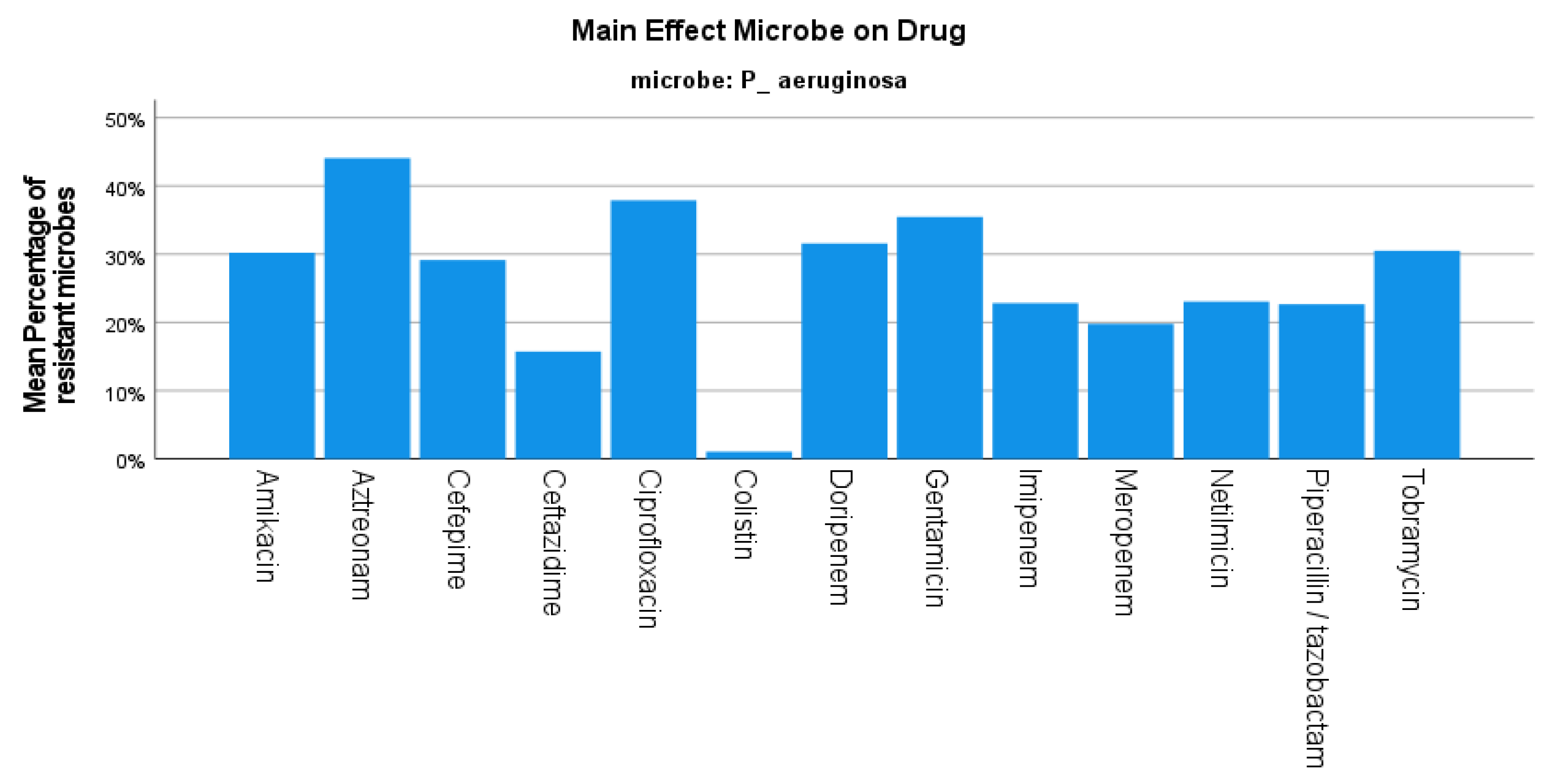
The mean resistance rate of P. aeruginosa during the same period was 24.17%, decreasing from 35.98% in the first semester of 2019 to 17.70% in the second semester of the year 2020 (p-value < 0.001) (Figure 1), while the number of isolates originating from the urology department also decreased in the same period (682 in 2019 and 385 in 2020). A high resistance rate of 38% was reported against fluoroquinolones, while the P. aeruginosa strains were 30% resistant to amikacin and presented varying resistance against carbapenems (31.58% to doripenem and 19.79% to meropenem). In addition, a 77% sensitivity rate to piperacillin/tazobactam was reported in our study (Figure 5).
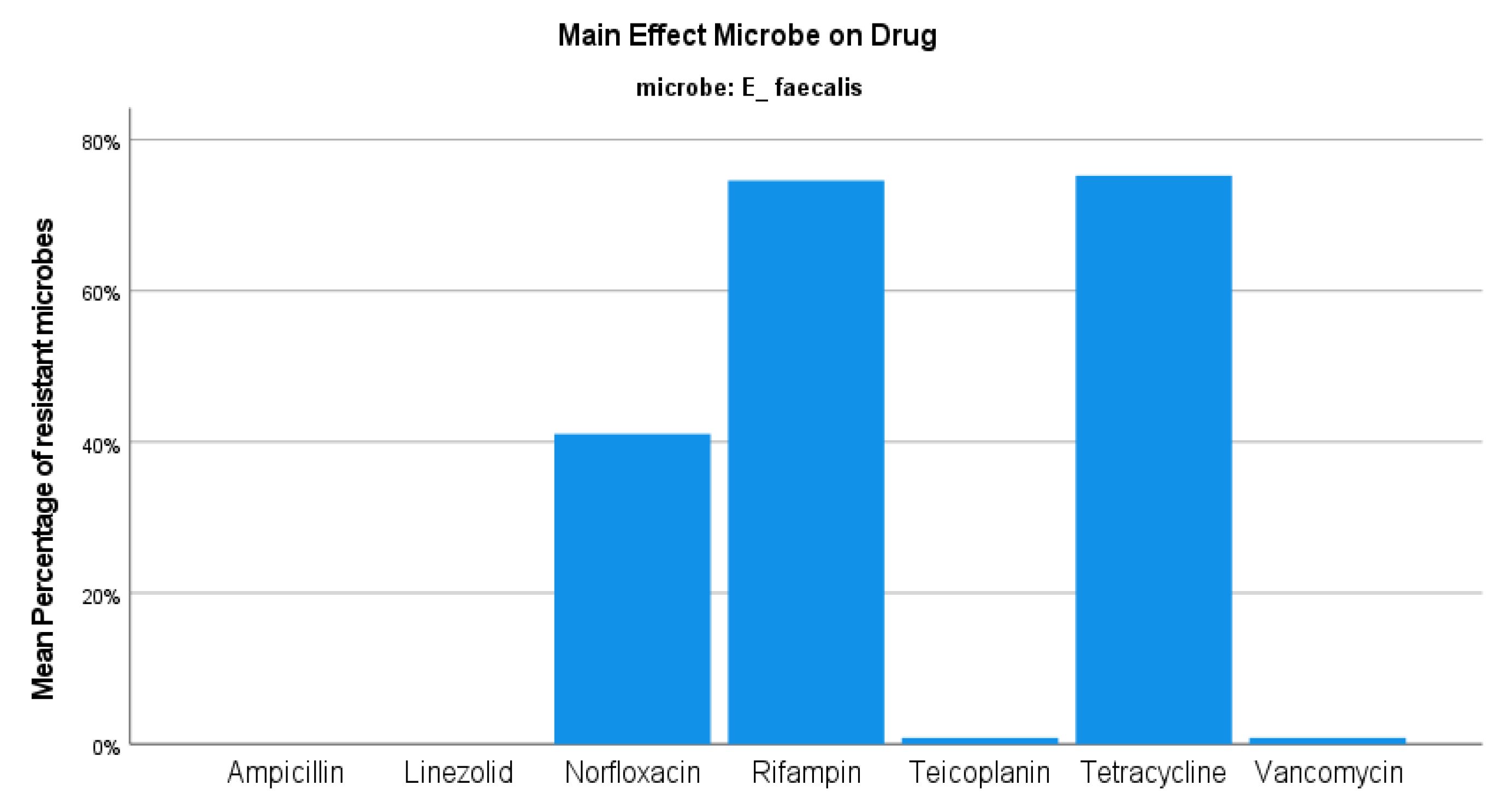
Regarding the E. faecium strains, the resistance rate was set at 46.91%, with no significant variations between semesters (Figure 1). The number of E. faecium isolates also presented small changes in the same period. This type of Enteroccoci presented 100% resistance rate to both ampicillin and fluoroquinolones, while there was almost 80% sensitivity to vancomycin and 100% sensitivity to linezolid (Figure 6). Concerning the E. faecalis isolates, the resistance rate was reported to be 24.247%, ranging from 23.99 to 25.51% during the period 2019–2020 (p-value 1) (Figure 1). In contrast to E. faecium isolates, these specific enterococci presented 100% sensitivity to ampicillin and 41% resistance to fluoroquinolones, while there was also 100% and approximately 99% sensitivity to linezolid and vancomycin, respectively (Figure 7).

4. Discussion

Antibiotic consumption is a major contributor to the spread of AMR, with Cyprus, France, Italy, and Greece being the top four antibiotic prescribers in Europe [5]. According to a study conducted by the European Centre for Disease Prevention and Control (ECDC), more than 50% of E. coli and over 30% of K. pneumoniae isolates were resistant to at least one antimicrobial group under monitoring, and combination resistance to multiple antimicrobial groups was common. Consequently, approximately 33,000 people die each year as a result of an infection caused by bacteria resistant to antibiotics [7]. Furthermore, healthcare-associated infections account for the majority of the burden of these multidrug-resistant infections, while last-line treatments such as carbapenems and colistin are becoming less effective, limiting the available therapeutic options [8].
The antimicrobial resistance profile of uropathogens can hinder the optimal treatment of hospitalized patients. As a result, knowing the local antimicrobial resistance rates is of great importance when recommending empirical treatment [26,27]. This highlights the need to carry out surveillance of the bacterial spectrum and AMR rates during empirical treatment of patients [27].
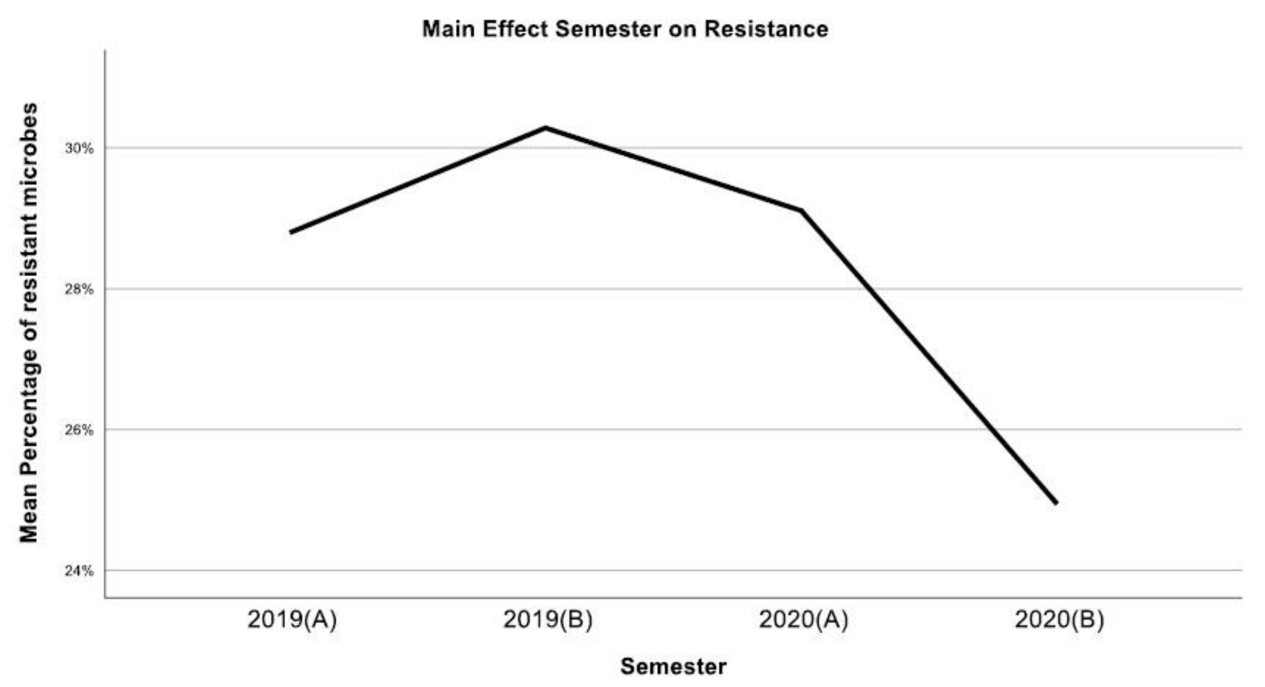
According to our findings, E. coli was the most common pathogen (5430 isolates), followed by K. pneumoniae (2302) and E. faecalis (1537). The overall antimicrobial resistance, after showing a small increase during the first semester of 2019, presented a continuous decrease until the end of 2020 (Figure 8). This could be explained partially by the outbreak of the COVID-19 pandemic in 2020, which led to a global lockdown and, consequently, to significantly less patients hospitalized, in general, worldwide [28]. On the contrary, a study by Mareș et al. [29], conducted in Romania, showed an increased resistance of pathogens to various antimicrobials during the COVID pandemic.
The resistance levels against extended-spectrum cephalosporins are an indicator for the production of extended spectrum beta-lactamases (ESBL), which render the treatment of Escherichia coli and Klebsiella pneumoniae infections extremely challenging [30]. In a Hungarian study by Gajdács et al. [31], the presence of ESBL isolates was reported in 8.85–38.97% and 10.89–36.06% of inpatient E. coli and Klebsiella samples, respectively. In addition, they reported a 15–40% resistance rate of K. pneumoniae to third-generation cephalosporins. Another study that was conducted in Romania by Chibelean et al. also presented a near 40% resistance rate of Klebsiella spp. to third-generation cephalosporins [32]. Our data show the average resistance rate of E. coli and K. pneumoniae to be lower than the global average rate [33], reporting, however, a K. pneumoniae resistance rate of 39% to ceftriaxone and over 62% to cefepime (fourth-generation cephalosporin), while the E. coli strains showcased under 20% resistance rates to third-generation cephalosporins (15.72% to ceftriaxone and 17.85% to ceftazidime). In a study by Ong et al. from the UK, the overall antimicrobial resistance of E. coli strains remained stable over the course of six years. However, there was an increase in the piperacillin/tazobactam resistance rate of 16.56%, while an increasing trend was also noticed in the resistance rate of E. coli to ciprofloxacin [34].
In addition, the overuse of antibiotics in the community, such as fluoroquinolones, contributes greatly to the emergence of ESBL pathogens, as well as the emergence of fluoroquinolone-resistant strains, such as P. aeruginosa, while keeping in mind that a great deal of fluoroquinolone overuse is still due to its use as a prophylaxis in urological procedures [35]. In a study by Benkő et al. that took place in Hungary, the ciprofloxacin resistance rate of P. aeruginosa isolates exceeded 20% [36]. Our study presents nearly 40% ciprofloxacin-resistant P. aeruginosa strains over the two-year period, while the overall resistance rate of ciprofloxacin ranged from 61.86% in the first semester of 2019 to 27.8% in the second semester of 2020 (p-value 0.035).
Regarding the Enterococcal strains, they have caused increasing concern in recent years due to their potency to spread in hospital settings among healthcare workers and patients and have been identified among the main pathogens that cause nosocomial infections. In our study, we identified a higher prevalence of E. faecalis than E. faecium in accordance with other studies [37]. Our data showed high sensitivity rates to both linezolid and vancomycin, thus identifying Vancomycin-resistant Enterococci (VRE) at approximately 20% for E. faecium and under 1% for E. faecalis. In recent years, E. faecium has substantially emerged as a source of hospital-associated infections. This is due to a variety of natural antibiotic resistance mechanisms displayed by this pathogen. Furthermore, this species is capable of acquiring resistance by mutations or by the inclusion of genes contained on mobile genetic elements such as plasmids, transposons, or integrons [38]. Kot et al. [39] showcase data from Poland, presenting multi-drug resistance in all E. faecium isolates, including resistance to ampicillin, imipenem, and ciprofloxacin. In addition, 40% of the isolates were resistant to glycopeptides (VRE) and aminoglycosides (gentamycin). Other European surveillance data report a frequency rate of 6.1% for VRE strains, and an alarming increase from 11.6% in 2016 to 16.8% in 2020 in the mean percentage of vancomycin-resistant E. faecium strains [7,40].
Urinary tract infections (UTI) are also frequently associated with urinary calculi, both as a consequence of a metabolic stone and as a cause of an infection stone due to the creation of struvite crystals by urease-producing bacterial strains. [41]. They affect almost one-third of individuals with stone disease, with the most frequent pathogenic species being Escherichia coli, Klebsiella species, Pseudomonas aeruginosa, Proteus species, and Gram-positive cocci in up to 25% of all cases [42]. These infections can very frequently cause fever, systemic inflammatory reaction, and sepsis. As a result, the selection of appropriate antimicrobial agents in urolithiasis patients is difficult, taking into account their polymicrobial nature, the widespread antibiotic use, and the emerging antimicrobial resistance patterns [43]. A study by De lorenzis et al. concerning urolithiasis patients showed an estimated resistance rate to fluoroquinolones of 39% for E. coli isolates, and over 50% for Pseudomonas isolates. Furthermore, it showed over 50% resistance rates of Klebsiella spp. isolates to cephalosporins, while 21% of E. coli isolates were identified as ESBL [44].
The advent of enormous volumes of medical data, combined with high skills in applied computer science, has resulted in the use of artificial intelligence (AI) approaches in various fields of medical research. AI has been used to diagnose calculi, forecast their composition, and predict surgical results in stone disease. AI has gained traction in microbiology, aided by advances in the genomic analysis of all types of microorganisms, and is now being researched for identifying drug and vaccine candidates, diagnosing pathogenic microorganisms, and predicting antimicrobial resistance and interactions [45,46]. The current microbiology lab capabilities require about 2 h in order for Gram-stain identification, over 24 h for microorganism identification, and up to 2 days in order to deliver an antibiotic sensitivity report. This process is time-consuming, and time is of the upmost importance, as in the case of a septic patient, for example, where an early knowledge of the susceptibilities of pathogens to antibiotics can be lifesaving [46]. A study conducted in Greece performed machine learning techniques in order to predict the antimicrobial profile of patients with stone disease with positive urine or blood cultures. It showed the feasibility of using a model to predict sensitivity to antibiotics early, when either the Gram-stain or the pathogen is known [47].
As for the limitations of this present study, we have to state that our data are based on the time period of 2019–2020 and not the more recent period of 2021–2022, as data are made available every two years according to the practice of the microbiology department of our hospital. We chose to include this dataset in our study as we opted to investigate the AMR profile of our department in a pre-COVID-19 era.

5. Conclusions

The antimicrobial resistance profiles of the most encountered pathogens in our department generally concur with relevant global-level data. The emergence of resistant pathogens has negative impacts, causing increased morbidity in patients with limited therapeutic options and economic consequences by heavily burdening all healthcare systems. This states the ever-increasing need for implementing surveillance programs in order to identify the susceptibility patterns of pathogens and to develop preventive strategies to optimize the outcomes of patients.

Author Contributions

Conceptualization, I.M., G.F., S.K. and L.T.; Methodology, I.M., G.F., P.A. and L.T.; Software, G.F.; Validation, G.F.; Formal analysis, I.M., G.F., E.L. and L.T.; Investigation, N.S.; Data curation, I.M., E.L. and L.T.; Writing—original draft, I.M. and L.T.; Writing—review & editing, G.F., S.K., P.A., L.T. and A.S.; Visualization, L.T.; Supervision, L.T. and A.S.; Project administration, L.T. All authors have read and agreed to the published version of the manuscript.

Funding

This research received no external funding.

Institutional Review Board Statement

The study was conducted in accordance with the Declaration of Helsinki and approved by the Institutional Review Board (or Ethics Committee) of Sismanoglio Hospital, Athens, Greece (protocol code 15178 and date of approval 28 July 2020).

Informed Consent Statement

Informed consent was obtained from all subjects involved in the study.

Data Availability Statement

Data is unavailable due to ethical restrictions.

Conflicts of Interest

The authors declare no conflict of interest.

References

  1. Zowawi, H.M.; Harris, P.N.A.; Roberts, M.J.; Tambyah, P.A.; Schembri, M.A.; Pezzani, M.D.; Williamson, D.A.; Paterson, D.L. The Emerging Threat of Multidrug-Resistant Gram-Negative Bacteria in Urology. Nat. Rev. Urol. 2015, 12, 570–584. [Google Scholar]
  2. Kandil, H.; Cramp, E.; Vaghela, T. Trends in Antibiotic Resistance in Urologic Practice. Eur. Urol. Focus 2016, 2, 363–373. [Google Scholar] [CrossRef]
  3. Castanheira, M.; Simner, P.J.; Bradford, P. Extended-spectrum beta-lactamases: An update on their characteristics, epidemiology and detection. JAC Antimicrob. Resist. 2021, 3, dlab092. [Google Scholar] [CrossRef]
  4. Deininger, S.; Gründler, T.; Deininger, S.H.M.; Lütcke, K.; Lütcke, H.; Agbesi, J.; Ladzaka, W.; Gyamfi, E.; Wichlas, F.; Hofmann, V.; et al. The Antimicrobial Resistance (AMR) Rates of Uropathogens in a Rural Western African Area—A Retrospective Single-Center Study from Kpando, Ghana. Antibiotics 2022, 11, 1808. [Google Scholar] [CrossRef] [PubMed]
  5. Gandra, S.; Barter, D.M.; Laxminarayan, R. Economic Burden of Antibiotic Resistance: How Much Do We Really Know? Clin. Microbiol. Infect. 2014, 20, 973–980. [Google Scholar] [CrossRef] [PubMed]
  6. WHO. Global Antimicrobial Resistance and Use Surveillance System (GLASS) Report 2021; World Health Organization (WHO): Geneva, Switzerland, 2021.
  7. European Centre for Disease Prevention and Control. Antimicrobial Resistance in the EU/EEA (EARS-Net)—Annual Epidemiological Report 2020; ECDC: Stockholm, Sweden, 2022. [Google Scholar]
  8. Potron, A.; Poirel, L.; Nordmann, P. Emerging Broad-Spectrum Resistance in Pseudomonas Aeruginosa and Acinetobacter Baumannii: Mechanisms and Epidemiology. Int. J. Antimicrob. Agents 2015, 45, 568–585. [Google Scholar] [PubMed]
  9. Karakonstantis, S.; Kalemaki, D. Antimicrobial Overuse and Misuse in the Community in Greece and Link to Antimicrobial Resistance Using Methicillin-Resistant S. Aureus as an Example. J. Infect. Public Health 2019, 12, 460–464. [Google Scholar] [CrossRef] [PubMed]
  10. Maltezou, H.C.; Adamis, G.; Tsonou, P.; Moustaka, E.; Katerelos, P.; Gargalianos, P. Consumption of Antibiotics for Community-Acquired Infections by Adults in Greece: A Cross-Sectional Study. Am. J. Infect. Control. 2016, 44, 1741–1743. [Google Scholar] [CrossRef] [PubMed]
  11. Luyt, C.-E.; Bréchot, N.; Trouillet, J.-L.; Chastre, J. Antibiotic Stewardship in the Intensive Care Unit. Crit. Care 2014, 18, 480. [Google Scholar] [CrossRef]
  12. Johansen, T.E.B.; Çek, M.; Naber, K.G.; Stratchounski, L.; Svendsen, M.V.; Tenke, P. Hospital Acquired Urinary Tract Infections in Urology Departments: Pathogens, Susceptibility and Use of Antibiotics. Int. J. Antimicrob. Agents 2006, 28, 91–107. [Google Scholar] [CrossRef]
  13. Balows, A.; Hausler, W.J., Jr.; Herrmann, K.L.; Isenberg, H.D.; Shadomy, H.J. Manual of Clinical Microbiology. Rev. Inst. Med. Trop. São Paulo 1991, 33, 434. [Google Scholar] [CrossRef]
  14. Henry, D.I. Clinical Microbiology Procedures Handbook; ASM Press: Washington, DC, USA, 2004; Volume 3. [Google Scholar]
  15. Murray, P.R.; Baron, E.; Jorgensen, J.H.; Pfaller, M.A.; Yolken, R.H.; Hanna, B.A. Manual of Clinical Microbiology, 8th Edition. Clin. Infect. Dis. 2004, 38, 1199–1200. [Google Scholar] [CrossRef]
  16. Nicolle, L.E.; Gupta, K.; Bradley, S.F.; Colgan, R.; DeMuri, G.P.; Drekonja, D.; Eckert, L.O.; Geerlings, S.E.; Köves, B.; Hooton, T.M.; et al. Clinical Practice Guideline for the Management of Asymptomatic Bacteriuria: 2019 Update by the Infectious Diseases Society of Americaa. Clin. Infect. Dis. 2019, 68, e83–e110. [Google Scholar] [CrossRef] [PubMed]
  17. Jorgensen, J.H.; Pfaller, M.A.; Carroll, K.C.; Funke, G.; Landry, M.L.; Richter, S.S.; Warnock, D.W. Manual of Clinical Microbiology, 11th ed.; ASM Press: Washington, DC, USA, 2015. [Google Scholar]
  18. Clinical and Laboratory Standards Institute. Performance Standards for Antimicrobial Susceptibility Testing, 26th ed.; Clinical and Laboratory Standards Institute: Wayne, PA, USA, 2016. [Google Scholar]
  19. Clinical and Laboratory Standards Institute. Performance Standards for Antimicrobial Susceptibility Testing, 27th ed.; Clinical and Laboratory Standards Institute: Wayne, PA, USA, 2017. [Google Scholar]
  20. Tsakris, A.; Poulou, A.; Pournaras, S.; Voulgari, E.; Vrioni, G.; Themeli-Digalaki, K.; Petropoulou, D.; Sofianou, D. A Simple Phenotypic Method for the Differentiation of Metallo—Lactamases and Class a KPC Carbapenemases in Enterobacteriaceae Clinical Isolates. J. Antimicrob. Chemother. 2010, 65, 1664–1671. [Google Scholar] [CrossRef]
  21. Skarmoutsou, N.; Adamou, D.; Tryfinopoulou, K.; Xirokosta, P.; Mylona, E.; Giakkoupi, P.; Karadimas, K.; Zervogianni, A.; Martsoukou, M. Performance of NG-Test CARBA 5 immunochromatographic assay for the detection of carbapenemases among multidrug-resistant clinical strains in Greece. In Proceedings of the ECCMID 2019, Amsterdam, The Netherlands, 13–16 April 2019. [Google Scholar]
  22. Yong, D.; Lee, K.; Yum, J.H.; Shin, H.B.; Rossolini, G.M.; Chong, Y. Imipenem-EDTA Disk Method for Differentiation of Metallo-β-Lactamase-Producing Clinical Isolates of Pseudomonas spp. and Acinetobacter spp. J. Clin. Microbiol. 2002, 40, 3798–3801. [Google Scholar] [CrossRef]
  23. Lee, K.; Lim, Y.S.; Yong, D.; Yum, J.H.; Chong, Y. Evaluation of the Hodge Test and the Imipenem-EDTA Double-Disk Synergy Test for Differentiating Metallo-β-Lactamase-Producing Isolates of Pseudomonas spp. and Acinetobacter spp. J. Clin. Microbiol. 2003, 41, 4623–4629. [Google Scholar] [CrossRef] [PubMed]
  24. Flountzi, A.; Giakkoupi, P.; Tryfinopoulou, K.; Pappa, O.; Vatopoulos, A.; Martsoukou, M.; Skarmoutsou, N.; Lebessi, E.; Charisiadou, A.E.; Chatzivasileiou, E.; et al. Investigation of Klebsiella pneumoniaeclinical isolates from 2016 onwards for the putative presence of the plasmid-mediated mcr-1 gene for colistin resistance. In Proceedings of the ECCMID 2019, Amsterdam, The Netherlands, 13–16 April 2019. [Google Scholar]
  25. IBM Corp. IBM SPSS Statistics for Windows, Version 24.0; IBM Corp.: Armonk, NY, USA, 2016. [Google Scholar]
  26. Elhanan, G.; Sarhat, M.; Raz, R. Empiric Antibiotic Treatment and the Misuse of Culture Results and Antibiotic Sensitivities in Patients with Community-Acquired Bacteraemia due to Urinary Tract Infection. J. Infect. 1997, 35, 283–288. [Google Scholar] [CrossRef]
  27. Tandogdu, Z.; Cek, M.; Wagenlehner, F.; Naber, K.; Tenke, P.; van Ostrum, E.; Bjerklund Johansen, T. Resistance Patterns of Nosocomial Urinary Tract Infections in Urology Departments: 8-Year Results of the Global Prevalence of Infections in Urology Study. World J. Urol. 2014, 32, 791–801. [Google Scholar] [CrossRef]
  28. Nguyen, J.L.; Benigno, M.; Malhotra, D.; Khan, F.; Angulo, F.J.; Hammond, J.; Swerdlow, D.L.; Reimbaeva, M.; Emir, B.; McLaughlin, J.M. Pandemic-Related Declines in Hospitalization for Non-COVID-19-Related Illness in the United States from January through July 2020. PLoS ONE 2022, 17, e0262347. [Google Scholar] [CrossRef]
  29. Mareș, C.; Petca, R.-C.; Petca, A.; Popescu, R.-I.; Jinga, V. Does the COVID Pandemic Modify the Antibiotic Resistance of Uropathogens in Female Patients? A New Storm? Antibiotics 2022, 11, 376. [Google Scholar] [CrossRef]
  30. Paterson, D.L.; Bonomo, R.A. Extended-Spectrum-Lactamases: A Clinical Update. Clin. Microbiol. Rev. 2005, 18, 657–686. [Google Scholar] [CrossRef] [PubMed]
  31. Gajdács, M.; Ábrók, M.; Lázár, A.; Burián, K. Comparative Epidemiology and Resistance Trends of Common Urinary Pathogens in a Tertiary-Care Hospital: A 10-Year Surveillance Study. Medicina 2019, 55, 356. [Google Scholar] [CrossRef]
  32. Chibelean, C.B.; Petca, R.-C.; Mareș, C.; Popescu, R.-I.; Enikő, B.; Mehedințu, C.; Petca, A. A Clinical Perspective on the Antimicrobial Resistance Spectrum of Uropathogens in a Romanian Male Population. Microorganisms 2020, 8, 848. [Google Scholar] [CrossRef] [PubMed]
  33. Wagenlehner, F.; Tandogdu, Z.; Bartoletti, R.; Cai, T.; Cek, M.; Kulchavenya, E.; Köves, B.; Naber, K.; Perepanova, T.; Tenke, P.; et al. The Global Prevalence of Infections in Urology Study: A Long-Term, Worldwide Surveillance Study on Urological Infections. Pathogens 2016, 5, 10. [Google Scholar] [CrossRef]
  34. Ong, A.; Mahobia, N.; Browning, D.; Schembri, M.; Somani, B.K. Trends in Antibiotic Resistance for over 700,000 Escherichia Coli Positive Urinary Tract Infections over Six Years (2014–2019) from a University Teaching Hospital. Cent. Eur. J. Urol. 2021, 74, 249. [Google Scholar]
  35. World Health Organization. Global Antimicrobial Resistance and Use Surveillance System (GLASS) Report 2022; World Health Organization: Geneva, Switzerland, 2022.
  36. Benkő, R.; Gajdács, M.; Matuz, M.; Bodó, G.; Lázár, A.; Hajdú, E.; Papfalvi, E.; Hannauer, P.; Erdélyi, P.; Pető, Z. Prevalence and Antibiotic Resistance of ESKAPE Pathogens Isolated in the Emergency Department of a Tertiary Care Teaching Hospital in Hungary: A 5-Year Retrospective Survey. Antibiotics 2020, 9, 624. [Google Scholar] [CrossRef]
  37. Boccella, M.; Santella, B.; Pagliano, P.; De Filippis, A.; Casolaro, V.; Galdiero, M.; Borrelli, A.; Capunzo, M.; Boccia, G.; Franci, G. Prevalence and Antimicrobial Resistance of Enterococcus Species: A Retrospective Cohort Study in Italy. Antibiotics 2021, 10, 1552. [Google Scholar] [CrossRef]
  38. Buultjens, A.H.; Lam, M.M.; Ballard, S.; Monk, I.R.; Mahony, A.A.; Grabsch, E.A.; Grayson, M.L.; Pang, S.; Coombs, G.W.; Robinson, J.O.; et al. Evolutionary origins of the emergent ST796 clone of vancomycin resistant Enterococcus faecium. PeerJ 2017, 5, e2916. [Google Scholar] [CrossRef]
  39. Kot, B.; Grużewska, A.; Szweda, P.; Wicha, J.; Parulska, U. Antibiotic Resistance of Uropathogens Isolated from Patients Hospitalized in District Hospital in Central Poland in 2020. Antibiotics 2021, 10, 447. [Google Scholar] [CrossRef]
  40. European Food Safety Authority; European Centre for Disease Prevention and Control. The European Union summary report on antimicrobial resistance in zoonotic and indicator bacteria from humans, animals and food in 2016. EFSA J. 2018, 16, e05182. [Google Scholar]
  41. Zanetti, G.; Paparella, S.; Trinchieri, A.; Prezioso, D.; Rocco, F.; Naber, K.G. Infections and Urolithiasis: Current Clinical Evidence in Prophylaxis and Antibiotic Therapy. Arch. Ital. Urol. Androl. Organo Uff. Soc. Ital. Ecogr. Urol. Nefrol. 2008, 80, 5–12. [Google Scholar]
  42. Chen, D.; Zhang, Y.; Huang, J.; Liang, X.; Zeng, T.; Lan, C.; Duan, X.; Zhao, Z.; Zeng, G.; Tiselius, H.-G.; et al. The Analysis of Microbial Spectrum and Antibiotic Resistance of Uropathogens Isolated from Patients with Urinary Stones. Int. J. Clin. Pract. 2018, 72, e13205. [Google Scholar] [CrossRef] [PubMed]
  43. Perletti, G.; Magri, V.; Cai, T.; Stamatiou, K.; Trinchieri, A.; Montanari, E. Resistance of Uropathogens to Antibacterial Agents: Emerging Threats, Trends and Treatments. Arch. Ital. Urol. Androl. Organo Uff. Soc. Ital. Ecogr. Urol. Nefrol. 2018, 90, 85–96. [Google Scholar] [CrossRef] [PubMed]
  44. De Lorenzis, E.; Alba, A.B.; Cepeda, M.; Galan, J.A.; Geavlete, P.; Giannakopoulos, S.; Saltirov, I.; Sarica, K.; Skolarikos, A.; Stavridis, S.; et al. Bacterial Spectrum and Antibiotic Resistance of Urinary Tract Infections in Patients Treated for Upper Urinary Tract Calculi: A Multicenter Analysis. Eur. J. Clin. Microbiol. Infect. Dis. 2020, 39, 1971–1981. [Google Scholar] [CrossRef] [PubMed]
  45. Yang, B.; Veneziano, D.; Somani, B.K. Artificial Intelligence in the Diagnosis, Treatment and Prevention of Urinary Stones. Curr. Opin. Urol. 2020, 30, 782–787. [Google Scholar] [CrossRef]
  46. Goodswen, S.J.; Barratt, J.L.N.; Kennedy, P.J.; Kaufer, A.; Calarco, L.; Ellis, J.T. Machine Learning and Applications in Microbiology. FEMS Microbiol. Rev. 2021, 45, fuab015. [Google Scholar] [CrossRef] [PubMed]
  47. Tzelves, L.; Lazarou, L.; Feretzakis, G.; Kalles, D.; Mourmouris, P.; Loupelis, E.; Basourakos, S.; Berdempes, M.; Manolitsis, I.; Mitsogiannis, I.; et al. Using Machine Learning Techniques to Predict Antimicrobial Resistance in Stone Disease Patients. World J. Urol. 2022, 40, 1731–1736. [Google Scholar] [CrossRef]
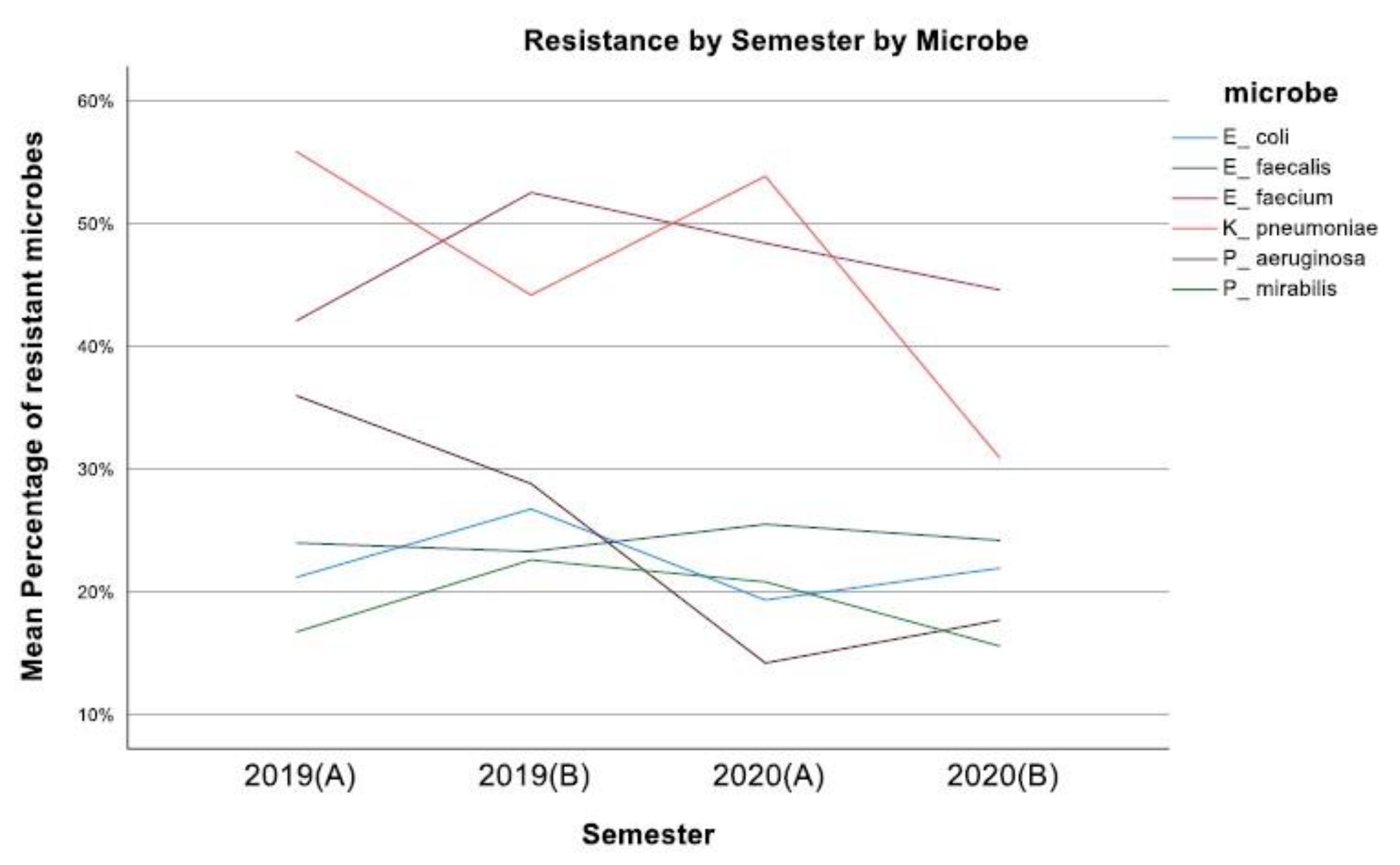
Figure 1. Mean resistance of microbes during the four semesters (2019–2020).
Jcm 12 03180 g001
Figure 2. Resistance of E. coli against tested antibiotics.
Jcm 12 03180 g002
Figure 3. Resistance of P. mirabilis against tested antibiotics.
Jcm 12 03180 g003
Figure 4. Resistance of K. pneumoniae against tested antibiotics.
Jcm 12 03180 g004
Figure 5. Resistance of P. aeruginosa against tested antibiotics.
Jcm 12 03180 g005
Figure 6. Resistance of E. faecium against tested antibiotics.
Jcm 12 03180 g006
Figure 7. Resistance of E. faecalis against tested antibiotics.
Jcm 12 03180 g007
Figure 8. Overall resistance rates over the four semesters.
Jcm 12 03180 g008
Disclaimer/Publisher’s Note: The statements, opinions and data contained in all publications are solely those of the individual author(s) and contributor(s) and not of MDPI and/or the editor(s). MDPI and/or the editor(s) disclaim responsibility for any injury to people or property resulting from any ideas, methods, instructions or products referred to in the content.