Review

Heart Rate Recovery: Up to Date in Heart Failure—A Literature Review

by
Andreea Cozgarea
1,2,3,
Dragoș Cozma
1,2,4,*,
Minodora Teodoru
3,5,*,
Alexandra-Iulia Lazăr-Höcher
1,2,
Liviu Cirin
2,
Adelina-Andreea Faur-Grigori
1,
Mihai-Andrei Lazăr
1,2,4,
Simina Crișan
1,2,4,
Dan Gaiță
1,2,4,
Constantin-Tudor Luca
1,2,4 and
Cristina Văcărescu
1,2,4
1
Institute of Cardiovascular Diseases Timisoara, 300310 Timisoara, Romania
2
Department of Cardiology, “Victor Babeș” University of Medicine and Pharmacy, 300041 Timisoara, Romania
3
County Clinical Emergency Hospital of Sibiu, 550245 Sibiu, Romania
4
Research Center of the Institute of Cardiovascular Diseases Timisoara, 300310 Timisoara, Romania
5
Medical Clinical Department, Faculty of Medicine, “Lucian Blaga” University, 550024 Sibiu, Romania
*
Authors to whom correspondence should be addressed.
J. Clin. Med. 2024, 13(11), 3328; https://doi.org/10.3390/jcm13113328
Submission received: 6 April 2024 / Revised: 15 May 2024 / Accepted: 3 June 2024 / Published: 5 June 2024
(This article belongs to the Special Issue Clinical Management of Patients with Heart Failure)

Abstract

:
The rising prevalence of cardiovascular disease underscores the growing significance of heart failure (HF). Pathophysiological insights into HF highlight the dysregulation of the autonomic nervous system (ANS), characterized by sympathetic overactivity and diminished vagal tone, impacting cardiovascular function. Heart rate recovery (HRR), a metric measuring the heart’s ability to return to its baseline rate post-exertion, plays a crucial role in assessing cardiovascular health. Widely applied across various cardiovascular conditions including HF, coronary artery disease (CAD), and arterial hypertension (HTN), HRR quantifies the difference between peak and recovery heart rates. Given its association with elevated sympathetic tone and exercise, HRR provides valuable insights into the perspective of HF, beyond effort tolerance, reaching toward prognostic and mortality indicators. Incorporating HRR into cardiovascular evaluations enhances our understanding of autonomic regulation in HF, offering potential implications for prognostication and patient management. This review addresses the significance of HRR in HF assessment, analyzing recently conducted studies, and providing a foundation for further research and clinical application.

1. Introduction

Heart rate recovery (HRR) is a parameter that addresses the decrease in the heart rate following a physical exercise, reaching its resting heart rate values. It is typically measured as the difference between the maximum heart rate during effort and the heart rate at a given recovery time [1,2]. It serves as a precious tool for evaluating autonomic nervous system (ANS) imbalance and has been widely used in screening and quantifying cardiovascular risk and all-cause mortality in patients suffering from heart disease [3,4]. This process reflects the dynamic equilibrium and synchronized interaction between the reactivation of the parasympathetic nervous system and the withdrawal of the sympathetic nervous system [5].
Cardiac autonomic dysfunction is frequently linked to cardiovascular disease (CVD) and has been observed in individuals possessing such risk factors [6]. In these populations, autonomic dysfunction is predominantly characterized by diminished parasympathetic drive and increased sympathetic activity. This imbalance amplifies cardiac load, increases ventricular instability, and consequently augments the susceptibility for cardiac arrest, infarction, and sudden death. Therefore, the existence of autonomic dysfunction suggests a more deprived prognosis for individuals with cardiovascular disease and more specifically, with heart failure (HF) [7,8].
Although the recently updated guidelines from the European Society of Cardiology (ESC) explicitly classify acute and chronic HF in the realm of the left ventricular ejection fraction (LVEF), the approach fails to adequately address the diverse pathophysiological mechanisms, etiological factors, and associated comorbidities inherent to HF. Notably, patients diagnosed with HF exhibit substantial clinical similarities irrespective of their LVEF status, underscoring the inadequacy of relying solely on LVEF for complete classification and management strategies [9,10].
In conditions of impaired ventricular contraction, the sympathetic nervous system activation aims to enhance the cardiac output by accelerating the heart rate. Norepinephrine release via adrenergic pathways augments myocardial contractility and triggers activation of the renin–angiotensin–aldosterone system (RAAS), essential for maintaining adequate blood pressure and cardiac output. Chronic elevation in wall stress instigates myocardial hypertrophy and remodeling, alongside vasoconstriction, thus elevating afterload and boosting contraction while impairing myocardial relaxation. This feedback loop intensifies neurohormonal stimulation, exacerbating cardiac output compromise and perpetuating heart failure progression [11,12,13].
On the other hand, increased vagal tone can help protect against ischemia and arrhythmias by reducing heart rate and blood pressure. Modern therapies aim to regulate cardiac dysautonomia by increasing parasympathetic activity through interventions such as vagal nerve stimulation [14].
The purpose of this review is to provide an in-depth analysis of the existing data on HRR in heart failure patients, by identifying underlying correlations ranging from prognosis and mortality to clinical outcomes and echocardiographic evaluation. Additionally, it aims to elucidate the potential of the parameter as a diagnostic metric in detecting and/or characterizing HF patients.

2. Autonomic Influence in Exercise and the Recovery Period

Heart rate during physical exercise is influenced by both central (central command) and peripheral (metabo- and mechanoreflex) mechanisms [15]. The metaboreflex, a physiological reflex triggered during or after exercise-induced muscle ischemia, is activated by stimulation of metabo-receptors. In healthy individuals, this reflex mechanism enhances hemodynamic response, leading to increased cardiac output primarily through flow-mediated mechanisms [16]. Physical exercise causes intricate shifts between the sympathetic and parasympathetic branches of the autonomic nervous system, concluding in an augmentation of heart rate (HR) through vagal withdrawal and adrenergic discharge. This complex physiological response enhances cardiovascular parameters, including cardiac output, HR, cardiac contractility, alveolar ventilation, and venous return to the heart. As exercise intensifies, the peak of sympathetic discharge and catecholamine release induces vasoconstriction in various circulatory systems, with exceptions in muscle, coronary, and cerebral circulations [17]. Despite increased sympathetic activity during exercise, parasympathetic modulation continues to influence HR, ensuring optimal myocardial perfusion during diastolic intervals and contributing to cardioprotective effects [18].
The recovery phase after maximal exercise involves an obvious shift in autonomic tone, typified by sympathetic withdrawal and parasympathetic reactivation. Notably, parasympathetic reactivation promptly manifests within the initial minute following exercise cessation, significantly influencing HR decline. While vagal reactivation predominantly mediates the initial 30 s HR decline, the prominence of sympathetic withdrawal becomes more apparent two minutes post-exercise [6,19]. The available literature data highlight the hemodynamic changes that occur in the recovery phase, when the important HR drop may be caused by a decreased cardiac output, mediated by intrinsic regulation [20]. Autonomic influence marks both HRR and the metaboreflex. Available data reveal that HF patients exhibit an increased metaboreflex, caused by an increased sympathetic activity. This results in excessive arteriolar vasoconstriction and increased systemic vascular resistance. Consequently, ventricular contraction is compromised, leading to a reduced stroke volume. Similar changes were noted in diabetic patients or those with metabolic syndrome [21].
As opposed to heart failure, trained individuals maintain a higher cardiac output during the recovery phase, caused by the redistribution of blood to the central regions, leading to an increased preload [20]. Thus, along with the more augmented vagal activity, it explains the shifts in hemodynamics in a trained individual compared to heart failure states. While chronotropic incompetence (CI) itself serves as a marker of cardiac dysautonomia, conflictual data appeared regarding beta-blockers, that are commonly used to treat heart failure. To address this issue, a lower threshold has been introduced for patients taking beta-blockers, which defines CI as the inability to reach 62% of APMHR [22].
Exercise elicits a sophisticated autonomic response, involving sympathetic dominance and enduring parasympathetic modulation, thereby influencing the dynamics of heart rate and recovery. Considering that heart failure is a state of hypersympathetic activity, exploring the recovery phase contributes to a better understanding of the ongoing processes, allowing the evaluation of therapeutic approaches.

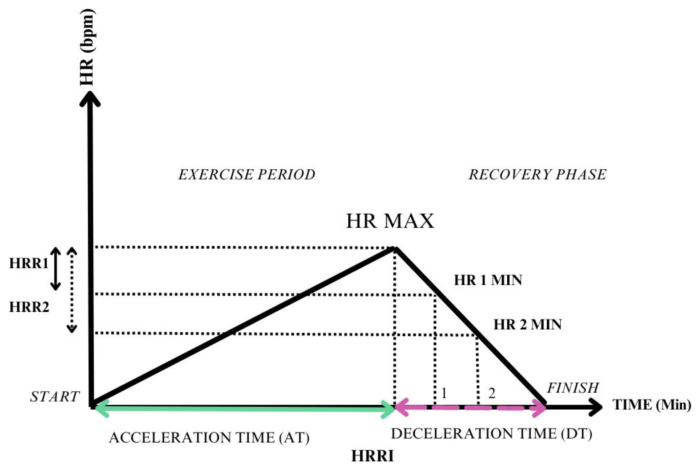
3. Main HRR Parameters

As early as the 1990s, the first data appeared concerning HRR, stating that a HRR < 12 beats per minute in the first minute strongly predicts overall mortality, setting a cornerstone for future studies [23]. Since then, the topic of HRR has been gaining interest among researchers, who developed various means of determining HRR, at different time intervals of the recovery period.
The following paragraph summarizes various HRRs, grouped through measurement methods.

3.1. Difference between Peak HR and HR at a Certain Moment of Recovery

  • HRR1—HRR at 1 minute of recovery subtracted from the peak HR [24,25,26,27,28,29,30].
  • HRR2—HRR at 2 min of recovery subtracted from the peak HR [25,28,29,31,32].
  • HRR150 sec—measured as the difference between the maximum HR and the HR after 150 seconds of recovery [33].
  • HRR3—measured as the difference between the maximum HR and the HR after three minutes of recovery [34].

3.2. A Ratio between Different Phases of the Effort

  • Heart rate recovery index—measured as the ratio between heart rate acceleration time (AT) and heart rate deceleration time (DT) [35].

3.3. A Delay in the Maximum Heart Rate

  • Delay of peak HR: HRR assessment 6 months after heart transplantation reflects the cardiac denervation and the loss of vagal tone, which would normally induce HR drop after exercise [32].
Given the distinct recovery phases and autonomic involvement, evaluating sympathetic nervous system activity during the post-exercise recovery phase could be crucial, given the correlation between cardiovascular issues induced by exercise and raised sympathetic activation. Moreover, while numerous population-based studies have associated HRR with mortality, there remains a gap in research regarding whether the augmentation of HRR following a therapeutic intervention serves as a predictive factor for improved survival during the subsequent follow-up period [36].

4. Clinical Applications of HRR

Ideas regarding HRR have been widely used in various clinical settings besides coronary artery disease. Heart failure, which will be thoroughly discussed further, has intricate connections with dysautonomia, implicit with the consequences of hypersympathetic states. Autonomic nervous system dysfunction is also implied in conditions such as hypertension [3], diabetes mellitus (DM) [37], metabolic syndrome and obesity [38], and obstructive sleep apnea syndrome [39].
HRR evaluation in DM plays a significant role in detecting subclinical LV dysfunction and preventing HF by evaluating diastolic functional reserve among heart failure with preserved ejection fraction (HFpEF) risk factors [40]. In a study by Vukomanovic et al., both type 2 diabetes patients and healthy individuals were enrolled to evaluate autonomic cardiac dysfunction using CPET. The results of the study revealed that patients with type 2 diabetes displayed a blunted HRR, which was associated with altered HRR, yielding significant autonomic nervous system dysfunction [41]. Moreover, another piece of research revealed the importance of dysautonomia by highlighting the associations between diastolic dysfunction and diminished functional capacity in patients with type 2 DM [42].
A recent study involving 2540 patients aimed to examine the correlations between heart rate recovery (HRR) and terminal-pro B type natriuretic peptide (NT-pro-BNP), which are both established as prognostic markers for heart failure. The study found that the NT-pro-BNP value was correlated with HRR2 and HRR3, indicating the importance of HRR in the slow recovery phase. This suggests that NT-pro-BNP is linked to the sympathetic nervous system, which is associated with heart failure processes. The study highlights the significance of cardiac dysautonomia evaluated by HRR and its connection to strong prognostic indicators of heart failure [43].

5. Methodology

The current review aims to encompass the most relevant studies conducted in the last ten years, concerning HRR in HF patients, employing two tables that hold the most relevant aspects of the studies (Table 1 and Table 2).
The methodology used for this review implied searching on different databases, such as PubMed or Google Scholar, using relevant keywords like “heart rate recovery” and “heart failure”, including Mesh terms.
The criteria for inclusion stipulated that the articles be written in English and published between 2014 and 2024, and their type should relate to patients involved in clinical studies. Furthermore, the resulting articles were manually selected considering the relevance, intending to showcase diverse clinical scenarios where HRR was employed to address heart failure.

6. HRR in Heart Failure: Bedside Studies

The resulting population heterogeneity prompted the construction of two tables to effectively organize the resulting data. Table 1 presents patients diagnosed with heart failure with reduced ejection fraction (HFrEF), offering a focused examination of this subgroup within the cohort.
In contrast, Table 2 encompasses a diverse range of study populations and comparison groups, including heart failure with preserved ejection fraction (HFpEF), heart failure with mid-range ejection fraction (HFmrEF), and comparisons between heart failure patients and healthy controls. Furthermore, two specific studies are highlighted within this framework: the study of Carneiro et al. [34], which investigated individuals at risk of developing heart failure (Stage A), and the study of Imamura et al. [32], which delves into patients recovering from advanced heart failure (Stage D), and having undergone heart transplantation.
HRR is a powerful tool for predicting morbi-mortality in various conditions and is considered a marker of cardiac dysautonomia [44].
Table 1 compresses studies conducted on HFrEF patients and reveals several HRR methods of determination, exercise protocols, and controversial aspects such as beta-blockade treatment, alongside the study objectives and results.
Our goal is to assess the various purposes for conducting exercise tests, which range from assessing the impact of reduced quality of life to establishing correlations with mortality in connection with blunted heart rate recovery. We also aim to point out the correlation between reduced heart rate recovery and pro-inflammatory states, as well as the association between exercise tests and conditions such as sarcopenia, CRT responsiveness, and the influence of cardiac rehabilitation.
Delving into HRR determination methods, the most frequently used methods are represented by HRR1 and HRR2. Cozlac et al. used another determinant, namely the heart rate recovery index (HRRI) [35]. This index evaluates both the exercise time, by assessing the heart rate acceleration time (AT), and the time of recovery, by measuring the heart rate deceleration time (DT), being defined as the ratio between AT and DT.
Although the study population is represented by HFrEF patients, and the HRR determinants are equal, cut-off values either differ between studies or have no values available for cut-off, showing a degree of inconsistency. Moreover, additional differences among the studies are evident regarding the exercise test employed, the protocols implemented, and the duration of the recovery period. These studies have employed diverse exercise protocols and test types, spanning from the six-minute walk test (6 MWT) to comprehensive cardio-pulmonary exercise tests (CPET). The recuperation period varies across studies, encompassing active recovery followed by passive recovery, solely passive recovery protocols, or instances where the recovery protocol is not specified.
Similar to a recently published study, the methodology did not exhibit a clear pattern for which exercise type and recovery protocols were used, indicating that further research and studies are necessary to validate distinct protocols for various clinical scenarios [2].
The exercise types varied from the 6 MWT to the treadmill and cycle ergometer. While maximal exercise tests like CPET are considered the benchmark for evaluating functional capacity, the 6 MWT can offer valuable insights into a patient’s daily activity levels and short-term prognosis, particularly in those with heart failure and reduced ejection fraction, whether in a stable chronic condition or following an acute decompensation [45].
In contrast to the 6 MWT, both the cycle ergometer and treadmill offer a more gradual and graded exercise. The bicycle ergometer has several advantages, including a small space requirement, easy quantification of exercise, and suitability for obese patients or those with orthopedic disorders. From a safety standpoint, it is a better choice than other forms of exercise equipment, as it reduces the risk of ventricular arrhythmias and angina pectoris appearance during exercise. The reason for this is that initially, maximal exercise on a treadmill exerts more significant stress on the heart and lungs compared to bicycle ergometers. Research has shown that during exercise tests with a bicycle ergometer, untrained individuals often end the test due to fatigue in the quadriceps femoris muscles, resulting in an average 5–20% lower peak oxygen consumption (peak VO2) compared to treadmill exercise [46].
Table 2. Heart rate recovery (HRR) studies including mixed groups of heart failure (HF) patients and healthy controls.
Study (Year)Patients Enrolled
(n)
HF PopulationPurpose of the StudyExercise Test MethodologyBeta-Blocker TreatmentHRR Evaluation Method and Cut-OffConclusions
Hossri et al.
(2024)
[24]
106HFpEF and HFmrEF with concomitant CADBenefits of CPMR in HF patients with CADTreadmill CPET using an incremental maximal protocol, followed later by a submaximal constant load protocol at 80% of the initial test; recovery period of 6 min87% of the patients. Dosage N/A.HRR1 was evaluated at the first recovery min;
Cut-off N/A
12 weeks of CPMR were associated with improved NYHA class, significant exercise test performance, an increased HRR, and an enhanced QOL.
Irfanullah et al. (2023)
[47]
39HFpEF, HFmrEF, HFrEFThe effects of cycle ergometer training on heart rate recovery and mindfulness in patients with NYHA Class I and II heart failure6 MWT on a 400–700 m distance.All patients received either beta-blockers or CCB. Dosage N/A.HRR1; HRR2 = HRmax-HR at 1; 2 min of recovery;
Cut-off N/A
HRR1 and HRR2 improved after 6 weeks of cycle-ergometer training, as well as the MAAS.
Carneiro et al. (2021)
[34]
2066Participants without HFIncidence of HF and its type (HFpEF and HFrEF) during the follow-up period (16.8 years)Submaximal treadmill exercise test using the Bruce protocol; recovery in a supine position.N/AHRR3 = HRmax-HR at 3 min of recovery;
Cut-off N/A
Slower HRR3 is associated with a higher risk of developing heart failure, particularly HFrEF.
Hajdusek et al. (2017)
[33]
10378 advanced HFrEF and 25 healthy controls, assessed for device implantation or transplant eligibilityEvaluation of HRR and MCR as outcome determinants in HF, during a follow-up of ~3.4 yearsSymptom limited using a bicycle CPET, with a 25 W increase/3 min. 97% of HF patients. Daily low-middle doses (12.5–50 mg Carvedilol; 2.5–200 mg Metoprolol and 2.5–10 mg Bisoprolol)The difference between HRmax and HR at 150 s of recovery;
Cut-off N/A
MCR slope correlates with distinct clinical variables compared to HRR. In heart failure patients, the MCR slope offers significant prognostic value beyond HRR.
Yaylalı et al. (2015)
[31]
41HFrEF and HFmrEFCorrelation between exercise training and HRR improvement, before and after entering a training programSymptom limited bicycle CPET with 10 W/min followed by 3 min active cool-down51.2% of patients.
Dosage N/A
HRR1; HRR2 = HRmax-HR at 1; 2 min of recovery;
N.V. Preestablished HRR1 > 12 bpm; HRR2 > 22 bpm
The training enhanced only HRR2, with IT showing a greater impact on the HRR2 improvement. Both HRR (1 and 2) in those with an abnormal HRR baseline have improved after exercise.
Imamura et al. (2015)
[32]
21Heart transplant patientsImpact of post-heart transplantation parasympathetic reinnervationSymptom limited Cycle-ergometer CPET using a 10 W/min incremental protocol, followed by a 5 min passive recovery in a seated position.19% of patients.
Dosage N/A.
HRR2-the difference between peak HR and HR after 2 min of recovery (measured 2 years after transplant)
Delay of peak HR-the delay from the peak HR after the recovery time initiation (measured 6 months after transplant). Cut-off N/A
Parasympathetic reinnervation coincides with enhanced post-exercise recovery and heart failure-specific quality of life during the 2 years following heart transplantation.
Lindemberg et al. (2014)
[26]
161. 154 completed the test126 patients with HFrEF and 35 healthy individuals.Correlations between HRR1 and 6 MWD6 MWT followed by passive (seated) recovery.70.58% of HF patients.
Carvedilol mean dose 30 ± 29 mg.
HRR1 = HRmax-HR at 1 min of recovery;
N.V. Preestablished HRR1 > 12 bpm
HF patients who received beta-blockers had better exercise tolerance than those without receiving beta-blocker medication, even though they had altered HRR.
Cahalin et al. (2014)
[30]
240200 patients with HFrEF and 40 patients with HFpEFCorrelations between HRR measured post 6 MWT and CPET and predictors of abnormal HRR.6 MWT followed by a passive recovery.
A symptom-limited bicycle CPET lasting 8–10 min. 1 min active cool-down period.
60% of the entire study group.
Dosage N/A
HRR1 = HRmax-HR at 1 min of recovery;
N.V. Preestablished HRR1 > 12 bpm
The 6 MWT and CPET were correlated concerning HRR, HR Reserve, and peak HR. Predictors of abnormal HRR were found to be Peak HR, EOV and E/e’ ratio.
N—Number; HF—heart failure; HFpEF—heart failure with preserved ejection fraction; HFmrEF—heart failure with mid-range ejection fraction; HFrEF—heart failure with reduced ejection fraction; CAD—coronary artery disease; CPET—cardiopulmonary exercise test; CPMR—cardiopulmonary and metabolic rehabilitation; N/A—Not available; HRR—heart rate recovery at a given time; HRmax—maximum Heart Rate; Min—minute; N.V.—Normal Values; S—Second; B.P.M.—beats per minute; NYHA—New York Heart Association; QOL—quality of life; 6 MWT—6 min walk test; 6 MWD—6 min walk distance; CCB—calcium channel blockers; MAAS—mindful attention aware scale; W—watt; MCR—metabolic chronotropic relation; EOV—exercise oscillatory ventilation.
The study’s population distribution is shown for example in the studies of Youn et al. or Cozlac et al. studies. The former implied symptom-limited CPET that was conducted on patients who presented with acute decompensated heart failure (ADHF), after the clinical stabilization of the patients [29]. The latter showed that an increased HRRI was associated with responder status, while a blunted HRRI was linked to non-responders in CRT patients [35].
The presented data from the numerous studies have covered diverse study groups, comprising patients from all HF stages [48]. HRR was studied in numerous population groups, extending from HFrEF and HFmrEF to HFpEF, including cohorts of patients who were at risk of developing HF in the follow-up period, as evaluated by Carneiro et al. The study evaluated the patients enrolled in the Framingham Offspring Study to assess the association between decreased HRR and incidental HF during an average follow-up period of 16.8 years. Considering that nearly 40% of the patients were on antihypertensive treatment, 16% on lipid-lowering treatment, 12% were smokers and 9.3% were suffering from type 2 Diabetes Mellitus (T2DM), they could be considered at risk for developing HF (Stage A heart failure) [48]. Thus, it is noteworthy to mention the correlation between blunted HRR and an increased risk of developing HF, particularly HFrEF [29,34,35].
Conversely, Imamura et al. comprised the opposite spectrum of HF patients, namely those who underwent a heart transplantation, underlining the broad range of HRR applicability [32].
Research has suggested that cardiac rehabilitation programs may have a beneficial effect on HRR. Several studies have indicated that there is a significant improvement in HRR following enrolment in a rehabilitation program [24,27,34]. Not only did rehabilitation result in improved HRR, but CAD patients were shown to have attenuated exercise-induced arrhythmias and less frequent myocardial ischemia responses. Moreover, HRR appears to be correlated with other HF determinants, such as the NYHA class, the BORG scale on exertion perception, and quality of life questionaries [27].
It is common for patients with heart failure to experience mental health concerns like anxiety and depression, which can sometimes be underestimated. Research conducted by Irfanullah et al. has revealed that cardiac rehabilitation not only enhanced HRR but also has a considerable positive effect on mindfulness scale assessment. This is a crucial finding as mindfulness has been shown to promote an increase in vagal stimulation, which is advantageous for heart failure patients and may help to improve their mental and physical well-being [47].
Controversial aspects are contained in the tables, regarding the beta-blocker (BB) treatment and its influences on HRR determinations. β-blocker therapy leads to a decrease in neurohormone levels and an increase in β-receptor sensitivity, ultimately resulting in accentuated inotropy. Additionally, it has been suggested that, among other medications, β-blockers may experience reduced efficacy over time [49].
HRR was not statistically different in patients who were on BBs, compared to those who did not, as stated in several studies [2,29]. This suggests that HRR predominantly reflects vagal tone and remains a viable method for risk stratification across all patients, regardless of β-blocker therapy [2].
In the study of Hajdusek et al., a statistical difference was observed in the baseline characteristics of patients concerning beta-blockers. This was performed by comparing individuals with heart failure with reduced ejection fraction (HFrEF) who were administered beta-blockers (BBs), and healthy controls who did not have any medical reason for receiving BBs. However, HRR was found to reflect the vagal tone and was not associated with BB usage [33].
Yaylali et al. studied HF patients who were enrolled in a cardiac rehabilitation program, pointing out that significantly more patients on BBs had normal baseline HRR1 and HRR2, before starting the rehabilitation program [31].
Lindemberg et al. conducted a study with patients divided into three groups: G1 comprised HF patients treated with BB, G2 consisted of HF patients not treated with BB, and G3 included healthy patients. Even though the patients who received BB had an altered HRR, they showed better exercise tolerance, according to the study’s findings [26].
Various studies that determined HRR are displayed in the tables, but the methods they used were not consistent. Most studies rely on HRR1 evaluation using a cut-off value that was put in place over 30 years ago. However, two studies used statistical methods to evaluate their HRR cut-off; Cozlac et al. referred the HRRI value to the area under the receiver operating characteristic (AuROC) [35]. Likewise, Youn et al. stood out as they use Contal and O’Quigley’s method to establish a cut-off value [29]. In contrast, other studies do not have a clear cut-off value, or they depend on preexisting cut-off values.
Figure 1 shows a graphical representation of the parameters used for assessing HRR. Whereas the most used evaluation methods use the difference between peak HR and HR at a certain moment of recovery (HRR1 and HRR2), a ratio between the exercise phase and the recovery period, such as HRRI, is also displayed. The significant benefit of using HRRI is that it takes into account the entire exercise period and reports a single value that assesses both acceleration and deceleration time.

7. Echocardiographic Correlations

Echocardiography is a significant part of cardiovascular assessment. The following section contains research data that revealed associations between certain echocardiographic determinants and HRR.
LVEF is a cornerstone parameter for evaluating systolic function, yet its exclusive use is inadequate, particularly given the increasing prevalence of HFpEF. Comprehensive assessments now require additional evaluations, including diastolic function assessment, tissue Doppler imaging (TDI), and evaluation of left atrial enlargement [10]. Researchers have revealed that an examination of cardiac hemodynamic parameters, particularly through tissue Doppler echocardiography, indicates a clear correlation between left ventricular diastolic function and HRR. Patients showing decreased left ventricular filling pressures and exhibiting lower E/e’ ratios demonstrate accelerated HRR and a more pronounced chronotropic response, underscoring the intricate relationship between cardiac autonomic function and adaptive responses to exercise [50]. As previously stated in the HF bedside studies, Cahalin et al. revealed that echocardiography can serve as a predictor for HRR values, involving mainly the E/e’ ratio for evaluating the diastolic filling pattern [30].
Whereas conventional echocardiography has a certain contribution, diastolic assessment during stress echocardiography has shown superior results to conventional echocardiography and has been widely evaluated and incorporated in HFpEF diagnostic scores. The most evaluated parameters represent peak tricuspid regurgitation velocity jet and subsequently, pulmonary artery systolic pressure (PASP) and mitral E/E’ to objectify elevated LV filling pressures [51].
The relationship between HRR and left atrial function and size is worth mentioning. Interestingly, blunted HRR has been shown to relate positively to the left atrial volume index, indicating that cardiac dysautonomia mechanisms may contribute to cardiac remodeling processes [51]. As shown in a recent study, abnormal strain rates during the reservoir, conduit, and contraction phases of the left atrium were associated with blunted HRR 120 s in patients with ST-segment elevation myocardial infarction [52].

8. Discussion

This current review discussed the topic of HRR and the main studies that involved HF patients, in which various aspects and correlations of HRR were determined. The main HRR studies that were conducted in the last decade underscore the inconsistency of HRR determinants, underlining that each method has its weaknesses and strengths.
Since the recovery phase is influenced differently by the autonomic nervous system, we consider that it is crucial to comprehend the whole-effort dynamics to better assess heart failure conditions.
Ranging from chronic inflammatory states, metabolic imbalance, and sarcopenia to cardiac outcomes, HF predictability, and mortality, we encompassed the various applicability of HRR in our presented study.
Available data suggest facilitated patient risk quantification with the help of HRR, considering that patients with blunted HRR indices exhibit increased risks of mortality, recurrent HF hospitalizations, and adverse cardiac events [25,29,44]. By identifying high-risk individuals, we presume that clinicians could adapt management strategies accordingly to HRR values.
Another key determinant of cardiac dysautonomia is reflected by heart rate variability (HRV). HRV refers to the variation in the time intervals between successive heartbeats (i.e., the R-R interval) and has been proven to have decreased values in HF [53]. While they both serve as prognostic markers [54,55], they differ in their measurement methods and the information they provide. Firstly, HRV is usually measured on standard resting electrocardiograms (ECG) or during Holter monitoring [56]. HRR on the other hand, is measured after a graded exercise and offers more complex information about both the resting HR and the recovery HR at a given time.
The mutual regulation system observed between HRV and HRR implies a potential connection between the two measures. If so, resting HRV indicators could serve as predictors of HRR before encountering stressors like exercise [57].
However, the diagnostic utility of HRR remains to be further proven, as it would be a crucial aspect to implement HRR among well-established tools, such as natriuretic peptides, in the diagnostic of heart failure.
Exercise stress tests have been helping clinicians evaluate treatment response in advanced HF states, that require CRT. A recently published article underlines the importance of ET in tailoring beta-blockers and ivabradine in CRT fusion pacing. Monitoring dynamic changes of the HR over time can help evaluate the efficacy of treatment regimens and guide adjustments as needed [58]. Therefore, our perspectives may shift towards better HRR comprehension, which may be useful in guiding medical treatment in HF patients. Therapeutic interventions aimed at improving cardiac function, such as medication adjustments, lifestyle improvements, and cardiac rehabilitation programs, may lead to enhancements in autonomic function and subsequently improved HRR.
As previously exposed, exercise training represents a cornerstone of HF management, promoting cardiovascular fitness and improving symptoms, further enhancing the quality of life of HF patients. Among improving cardiovascular fitness, exercise modulates dysautonomia, improves inflammatory states, and prevents muscle loss, which has been proven to be a consequence of the pathophysiology of heart failure [59].
Therapeutic interventions targeting autonomic modulation vary widely, starting with medical treatment, such as beta-blockers, angiotensin-converting enzyme inhibitors (ACEIs), or digoxin. More recently, studies have shown that sodium-glucose cotransporter-2 inhibitors (SGLT2-i) may improve cardiac autonomic dysfunction in type 2DM patients, as published in the SCAN Study, which may lead to new research directions toward HF patients [60]. Besides medical therapies, dysautonomia may be targeted with device-based options, such as CRT, vagal nerve stimulation (VNS), or renal denervation [61].

9. Further Perspectives

Assessing HRR in the treatment of HF patients will offer a simple approach to risk stratification, treatment monitoring, and proactive management.
HRR has been identified as a potential indicator of premature cardiac impairment, such as altered diastolic dysfunction, as demonstrated in a study involving obese adolescents. This underscores its significance as a clinically relevant outcome marker [62]. Although the available determination methods prove some degree of inconsistency with the used parameters, the field is open for novel implementations.
Future studies may show interest in establishing connections between abnormal HRR, HF, and atrial remodeling. Considering the intricate synergy between dysautonomia, electrical and mechanical remodeling, and heart failure progression, the area of research deserves to be unveiled.
Another key aspect concerns inflammation, frequently measured by high-sensitive C reactive protein (hs-CRP) [63] or the neutrophil-to-lymphocyte ratio (NLR) [64], which is known to be linked to the risk of developing cardiovascular disease, such as heart failure [65] and atrial fibrillation [66]. Further studies are necessary to evaluate the risk of quantifying atrial fibrillation in the realm of blunted HRR.
Moreover, delayed HRR has been proven to be independently associated with NLR, indicative of systemic inflammation [63]. This underscores the implications of vagal modulation in inflammation and suggests future perspectives for managing heart failure.

10. Conclusions

In summary, this study underscores the necessity of integrating heart rate recovery (HRR) into cardiovascular assessments, as it is associated with both morbidity and mortality in numerous cardiovascular conditions. The connections between HRR and the autonomic nervous system make HRR an accountable index, for both the assessment of the pathophysiologic dynamic processes and their consequences [67]. However, the domain of heart rate recovery stands as a focal point for future research endeavors, particularly in the context of heart failure, where the potential diagnostic utility of HRR remains to be fully elucidated. As mentioned before, the recently published studies encompassing HF patients lack a standardized evaluation protocol that could be easily applied throughout practices, emphasizing the importance of future research in this area of expertise.
Establishing the diagnostic power of HRR through further investigation holds significant promise, especially considering its ease of evaluation. Such advancements could revolutionize cardiovascular diagnostics, offering clinicians a simple yet potent tool for enhancing risk assessment and patient care.

Author Contributions

Conceptualization, A.C., C.V. and D.C.; methodology, A.C., M.T. and C.-T.L.; software, A.C., M.T., A.-I.L.-H. and M.-A.L.; validation, A.C., D.C., C.V. and D.G.; formal analysis, A.C., A.-I.L.-H. and M.T.; investigation, A.C., D.C., C.V. and M.-A.L.; resources, A.C., L.C., M.T. and A.-A.F.-G.; data curation, A.C., C.V., L.C. and M.T.; writing—original draft preparation, A.C., D.C., C.V. and A.-A.F.-G.; writing—review and editing, A.C., D.C., M.T., C.V. and C.-T.L.; visualization, A.C., D.C., C.V. and S.C.; supervision, A.C., D.C. and S.C.; project administration, A.C., M.T. and C.V.; funding acquisition, A.C., D.G. and D.C. All authors have read and agreed to the published version of the manuscript.

Funding

This research received no external funding. Internal funding: We would like to acknowledge VICTOR BABES UNIVERSITY OF MEDICINE AND PHARMACY TIMISOARA for their support in covering the costs of publication for this research paper.

Institutional Review Board Statement

Not applicable.

Informed Consent Statement

Not applicable.

Data Availability Statement

Data sharing is not applicable.

Conflicts of Interest

The authors declare no conflicts of interest.

References

  1. Dewar, A.; Kass, L.; Stephens, R.C.M.; Tetlow, N.; Desai, T. Heart Rate Recovery Assessed by Cardiopulmonary Exercise Testing in Patients with Cardiovascular Disease: Relationship with Prognosis. Int. J. Environ. Res. Public Health 2023, 20, 4678. [Google Scholar] [CrossRef] [PubMed]
  2. Yu, Y.; Liu, T.; Wu, J.; Zhu, P.; Zhang, M.; Zheng, W.; Gu, Y. Heart rate recovery in hypertensive patients: Relationship with blood pressure control. J. Hum. Hypertens. 2017, 31, 354–360. [Google Scholar] [CrossRef] [PubMed]
  3. Manolis, A.A.; Manolis, T.A.; Manolis, A.S. Neurohumoral Activation in Heart Failure. Int. J. Mol. Sci. 2023, 24, 15472. [Google Scholar] [CrossRef] [PubMed]
  4. Fecchio, R.Y.; Brito, L.; Leicht, A.S.; Forjaz, C.L.M.; Peçanha, T. Reproducibility of post-exercise heart rate recovery indices: A systematic review. Auton. Neurosci. 2019, 221, 102582. [Google Scholar] [CrossRef] [PubMed]
  5. Qiu, S.; Cai, X.; Sun, Z.; Li, L.; Zuegel, M.; Steinacker, J.M.; Schumann, U.; Yin, J.; Jin, X.; Shan, Z.; et al. Heart Rate Recovery and Risk of Cardiovascular Events and All-Cause Mortality: A Meta-Analysis of Prospective Cohort Studies. J. Am. Heart Assoc. 2017, 6, e005505. [Google Scholar] [CrossRef] [PubMed]
  6. Peçanha, T.; Silva-Júnior, N.D.; Forjaz, C.L.D.M. Heart rate recovery: Autonomic determinants, methods of assessment and association with mortality and cardiovascular diseases. Clin. Physiol. Funct. Imaging 2014, 34, 327–339. [Google Scholar] [CrossRef] [PubMed]
  7. Okutucu, S.; Karakulak, U.N.; Aytemir, K.; Oto, A. Heart rate recovery: A practical clinical indicator of abnormal cardiac autonomic function. Expert Rev. Cardiovasc. Ther. 2011, 9, 1417–1430. [Google Scholar] [CrossRef] [PubMed]
  8. Imai, K.; Sato, H.; Hori, M.; Kusuoka, H.; Ozaki, H.; Yokoyama, H.; Takeda, H.; Inoue, M.; Kamada, T. Vagally mediated heart rate recovery after exercise is accelerated in athletes but blunted in patients with chronic heart failure. J. Am. Coll. Cardiol. 1994, 24, 1529–1535. [Google Scholar] [CrossRef] [PubMed]
  9. McDonagh, T.A.; Metra, M.; Adamo, M.; Gardner, R.S.; Baumbach, A.; Böhm, M.; Burri, H.; Butler, J.; Čelutkienė, J.; Chioncel, O.; et al. 2023 Focused Update of the 2021 ESC Guidelines for the diagnosis and treatment of acute and chronic heart failure: Developed by the task force for the diagnosis and treatment of acute and chronic heart failure of the European Society of Cardiology (ESC) With the special contribution of the Heart Failure Association (HFA) of the ESC. Eur. Heart J. 2023, 44, 3627–3639. [Google Scholar] [CrossRef]
  10. Severino, P.; D’amato, A.; Prosperi, S.; Cas, A.D.; Mattioli, A.V.; Cevese, A.; Novo, G.; Prat, M.; Pedrinelli, R.; Raddino, R.; et al. Do the Current Guidelines for Heart Failure Diagnosis and Treatment Fit with Clinical Complexity? J. Clin. Med. 2022, 11, 857. [Google Scholar] [CrossRef]
  11. Gronda, E.; Dusi, V.; D’elia, E.; Iacoviello, M.; Benvenuto, E.; Vanoli, E. Sympathetic activation in heart failure. Eur. Heart J. Suppl. 2022, 24, E4–E11. [Google Scholar] [CrossRef] [PubMed]
  12. Borovac, J.A.; D’Amario, D.; Bozic, J.; Glavas, D. Sympathetic nervous system activation and heart failure: Current state of evidence and the pathophysiology in the light of novel biomarkers. World J. Cardiol. 2020, 12, 373–408. [Google Scholar] [CrossRef] [PubMed]
  13. Zhang, D.Y.; Anderson, A.S. The Sympathetic Nervous System and Heart Failure. Cardiol. Clin. 2014, 32, 33–45. [Google Scholar] [CrossRef] [PubMed]
  14. Kumar, H.U.; Nearing, B.D.; Mittal, S.; Premchand, R.K.; Libbus, I.; DiCarlo, L.A.; Amurthur, B.; KenKnight, B.H.; Anand, I.S.; Verrier, R.L. Autonomic regulation therapy in chronic heart failure with preserved/mildly reduced ejection fraction: ANTHEM-HFpEF study results. Int. J. Cardiol. 2023, 381, 37–44. [Google Scholar] [CrossRef] [PubMed]
  15. Peçanha, T.; de Brito, L.C.; Fecchio, R.Y.; de Sousa, P.N.; Junior, N.D.d.S.; de Abreu, A.P.; da Silva, G.V.; Mion-Junior, D.; Forjaz, C.L.d.M. Metaboreflex activation delays heart rate recovery after aerobic exercise in never-treated hypertensive men. J. Physiol. 2016, 594, 6211–6223. [Google Scholar] [CrossRef] [PubMed]
  16. Roberto, S.; Mulliri, G.; Milia, R.; Solinas, R.; Pinna, V.; Sainas, G.; Piepoli, M.F.; Crisafulli, A. Hemodynamic response to muscle reflex is abnormal in patients with heart failure with preserved ejection fraction. J. Appl. Physiol. 2017, 122, 376–385. [Google Scholar] [CrossRef] [PubMed]
  17. Hellsten, Y.; Nyberg, M. Cardiovascular Adaptations to Exercise Training. In Comprehensive Physiology; Wiley: Hoboken, NY, USA, 2015; pp. 1–32. [Google Scholar] [CrossRef]
  18. Gourine, A.V.; Ackland, G.L. Cardiac vagus and exercise. Physiology 2019, 34, 71–80. [Google Scholar] [CrossRef] [PubMed]
  19. Kannankeril, P.J.; Le, F.K.; Kadish, A.H.; Goldberger, J.J. Parasympathetic Effects on Heart Rate Recovery after Exercise. J. Investig. Med. 2004, 52, 394–401. [Google Scholar] [CrossRef] [PubMed]
  20. Facioli, T.P.; Philbois, S.V.; Gastaldi, A.C.; Almeida, D.S.; Maida, K.D.; Rodrigues, J.A.L.; Sanchez-Delgado, J.C.; Souza, H.C.D. Study of heart rate recovery and cardiovascular autonomic modulation in healthy participants after submaximal exercise. Sci. Rep. 2021, 11, 3620. [Google Scholar] [CrossRef]
  21. Roberto, S.; Milia, R.; Doneddu, A.; Pinna, V.; Palazzolo, G.; Serra, S.; Orrù, A.; Kakhak, S.A.H.; Ghiani, G.; Mulliri, G.; et al. Hemodynamic abnormalities during muscle metaboreflex activation in patients with type 2 diabetes mellitus. J. Appl. Physiol. 2019, 126, 444–453. [Google Scholar] [CrossRef]
  22. Smarz, K.; Tysarowski, M.; Zaborska, B.; Pilichowska-Paszkiet, E.; Sikora-Frac, M.; Budaj, A.; Jaxa-Chamiec, T. Chronotropic incompetence limits aerobic exercise capacity in patients taking beta-blockers: Real-life observation of consecutive patients. Healthcare 2021, 9, 212. [Google Scholar] [CrossRef] [PubMed]
  23. Cole, C.R.; Foody, J.M.; Blackstone, E.H.; Lauer, M.S. Heart Rate Recovery after Submaximal Exercise Testing as a Predictor of Mortality in a Cardiovascularly Healthy Cohort. Ann. Intern. Med. 2000, 132, 552–555. [Google Scholar] [CrossRef] [PubMed]
  24. Hossri, C.A.C.; Araujo, F.; Baldi, B.; Otterstetter, R.; Uemoto, V.; Carvalho, C.; Mastrocola, L.; Albuquerque, A. Association among cardiopulmonary and metabolic rehabilitation, arrhythmias, and myocardial ischemia responses of patients with HFpEF or HFmrEF. Braz. J. Med. Biol. Res. 2024, 57, e13174. [Google Scholar] [CrossRef]
  25. Andrade, G.N.; Rodrigues, T.; Takada, J.; Braga, L.; Umeda, I.; Nascimento, J.; Pereira-Filho, H.; Grupi, C.; Salemi, V.; Jacob-Filho, W.; et al. Prolonged heart rate recovery time after 6-minute walk test is an independent risk factor for cardiac events in heart failure: A prospective cohort study. Physiotherapy 2022, 114, 77–84. [Google Scholar] [CrossRef] [PubMed]
  26. Lindemberg, S.; Chermont, S.; Quintão, M.; Derossi, M.; Guilhon, S.; Bernardez, S.; Marchese, L.; Martins, W.; Nóbrega, A.C.L.; Mesquita, E.T. Heart Rate Recovery in the First Minute at the Six-Minute Walk Test in Patients with Heart Failure. Arq. Bras. Cardiol. 2014, 102, 279–287. [Google Scholar] [CrossRef] [PubMed]
  27. Tanaka, S.; Miyamoto, T.; Mori, Y.; Harada, T.; Tasaki, H. Heart rate recovery is useful for evaluating the recovery of exercise tolerance in patients with heart failure and atrial fibrillation. Heart Vessels 2021, 36, 1551–1557. [Google Scholar] [CrossRef] [PubMed]
  28. da Fonseca, G.W.P.; dos Santos, M.R.; de Souza, F.R.; da Costa, M.J.A.; von Haehling, S.; Takayama, L.; Pereira, R.M.R.; Negrão, C.E.; Anker, S.D.; Alves, M.J.d.N.N. Sympatho-Vagal Imbalance is Associated with Sarcopenia in Male Patients with Heart Failure. Arq. Bras. Cardiol. 2019, 112, 739–746. [Google Scholar] [CrossRef] [PubMed]
  29. Youn, J.C.; Lee, H.S.; Choi, S.-W.; Han, S.-W.; Ryu, K.-H.; Shin, E.-C.; Kang, S.-M. Post-exercise heart rate recovery independently predicts clinical outcome in patients with acute decompensated heart failure. PLoS ONE 2016, 11, e0154534. [Google Scholar] [CrossRef] [PubMed]
  30. Cahalin, L.P.; Arena, R.; Labate, V.; Bandera, F.; Guazzi, M. Predictors of abnormal heart rate recovery in patients with heart failure reduced and preserved ejection fraction. Eur. J. Prev. Cardiol. 2014, 21, 906–914. [Google Scholar] [CrossRef]
  31. Yaylali, Y.T.; Fındıkoglu, G.; Yurtdas, M.; Konukcu, S.; Senol, H. The effects of baseline heart rate recovery normality and exercise training protocol on heart rate recovery in patients with heart failure. Anatol. J. Cardiol. 2015, 15, 727–734. [Google Scholar] [CrossRef]
  32. Imamura, T.; Kinugawa, K.; Okada, I.; Kato, N.; Fujino, T.; Inaba, T.; Maki, H.; Hatano, M.; Kinoshita, O.; Nawata, K.; et al. Parasympathetic Reinnervation Accompanied by Improved Post-Exercise Heart Rate Recovery and Quality of Life in Heart Transplant Recipients. Int. Heart J. 2015, 56, 180–185. [Google Scholar] [CrossRef] [PubMed]
  33. Hajdusek, P.; Kotrc, M.; Kautzner, J.; Melenovsky, V.; Benesova, E.; Jarolim, P.; Benes, J. Heart rate response to exercise in heart failure patients: The prognostic role of metabolic–chronotropic relation and heart rate recovery. Int. J. Cardiol. 2017, 228, 588–593. [Google Scholar] [CrossRef] [PubMed]
  34. Carneiro, H.A.; Song, R.J.; Lee, J.; Schwartz, B.; Vasan, R.S.; Xanthakis, V. Association of Blood Pressure and Heart Rate Responses to Submaximal Exercise with Incident Heart Failure: The Framingham Heart Study. J. Am. Heart Assoc. 2021, 10, e019460. [Google Scholar] [CrossRef] [PubMed]
  35. Cozlac, A.-R.; Petrescu, L.; Crisan, S.; Luca, C.T.; Vacarescu, C.; Streian, C.G.; Lazar, M.-A.; Gurgu, A.; Dragomir, A.; Goanta, E.V.; et al. A Novel and Simple Exercise Test Parameter to Assess Responsiveness to Cardiac Resynchronization Therapy. Diagnostics 2020, 10, 920. [Google Scholar] [CrossRef] [PubMed]
  36. Peçanha, T.; Bartels, R.; Brito, L.C.; Paula-Ribeiro, M.; Oliveira, R.S.; Goldberger, J.J. Methods of assessment of the post-exercise cardiac autonomic recovery: A methodological review. Int. J. Cardiol. 2017, 227, 795–802. [Google Scholar] [CrossRef] [PubMed]
  37. Sydó, N.; Sydó, T.; Merkely, B.; Carta, K.G.; Murphy, J.G.; Lopez-Jimenez, F.; Allison, T.G. Impaired Heart Rate Response to Exercise in Diabetes and Its Long-term Significance. Mayo Clin. Proc. 2016, 91, 157–165. [Google Scholar] [CrossRef] [PubMed]
  38. Alihanoglu, Y.I.; Yildiz, B.S.; Kilic, I.D.; Uludag, B.; Demirci, E.E.; Zungur, M.; Evrengul, H.; Kaftan, A.H. Impaired systolic blood pressure recovery and heart rate recovery after graded exercise in patients with metabolic syndrome. Medicine 2015, 94, e428. [Google Scholar] [CrossRef] [PubMed]
  39. Maeder, M.T.; Ammann, P.; Schoch, O.D.; Rickli, H.; Korte, W.; Hürny, C.; Myers, J.; Münzer, T. Determinants of postexercise heart rate recovery in patients with the obstructive sleep apnea syndrome. Chest 2010, 137, 310–317. [Google Scholar] [CrossRef] [PubMed]
  40. Shim, C.Y. Stress Testing in Heart Failure with Preserved Ejection Fraction. Heart Fail Clin. 2021, 17, 435–445. [Google Scholar] [CrossRef]
  41. Vukomanovic, V.; Suzic-Lazic, J.; Celic, V.; Cuspidi, C.; Grassi, G.; Galderisi, M.; Djukic, V.; Tadic, M. Is there association between left atrial function and functional capacity in patients with uncomplicated type 2 diabetes? Int. J. Cardiovasc. Imaging 2020, 36, 15–22. [Google Scholar] [CrossRef]
  42. Tadic, M.; Suzic-Lazic, J.; Vukomanovic, V.; Cuspidi, C.; Ilic, S.; Celic, V. Functional capacity and left ventricular diastolic function in patients with type 2 diabetes. Acta Diabetol. 2021, 58, 107–113. [Google Scholar] [CrossRef] [PubMed]
  43. Lin, Y.T.; Lin, L.Y.; Chuang, K.J. N terminal prohormone of brain natriuretic peptide is associated with improved heart rate recovery after treadmill exercise test. Int. J. Cardiol. Cardiovasc. Risk Prev. 2023, 18, 200203. [Google Scholar] [CrossRef] [PubMed]
  44. van de Vegte, Y.J.; van der Harst, P.; Verweij, N. Heart rate recovery 10 seconds after cessation of exercise predicts death. J. Am. Heart Assoc. 2018, 7, e008341. [Google Scholar] [CrossRef] [PubMed]
  45. Giannitsi, S.; Bougiakli, M.; Bechlioulis, A.; Kotsia, A.; Michalis, L.K.; Naka, K.K. 6-minute walking test: A useful tool in the management of heart failure patients. Ther. Adv. Cardiovasc. Dis. 2019, 13, 1–10. [Google Scholar] [CrossRef] [PubMed]
  46. Ren, C.; Zhu, J.; Shen, T.; Song, Y.; Tao, L.; Xu, S.; Zhao, W.; Gao, W. Comparison Between Treadmill and Bicycle Ergometer Exercises in Terms of Safety of Cardiopulmonary Exercise Testing in Patients with Coronary Heart Disease. Front. Cardiovasc. Med. 2022, 9, 864637. [Google Scholar] [CrossRef]
  47. Irfanullah, M.W.; Yaqoob, N.; Shah, S.; Tariq, I.; Chaudhary, F.; Ibrahim, U.; Huma, Z.E.; Khan, N. Effects of Cycle Ergometer Training on Heart Rate Recovery and Mind Fullness in NYHA class I, II Heart Failure Patients. Pak. J. Med. Health Sci. 2023, 17, 10–13. [Google Scholar] [CrossRef]
  48. Bozkurt, B.; Coats, A.J.; Tsutsui, H.; Abdelhamid, M.; Adamopoulos, S.; Albert, N.; Anker, S.D.; Atherton, J.; Böhm, M.; Butler, J.; et al. Universal definition and classification of heart failure: A report of the Heart Failure Society of America, Heart Failure Association of the European Society of Cardiology, Japanese Heart Failure Society and Writing Committee of the Universal Definition of Heart Failure: Endorsed by the Canadian Heart Failure Society, Heart Failure Association of India, Cardiac Society of Australia and New Zealand, and Chinese Heart Failure Association. Eur. J. Heart Fail. 2021, 23, 352–380. [Google Scholar] [CrossRef]
  49. Maldonado-Martín, S.; Brubaker, P.H.; Ozemek, C.; Jayo-Montoya, J.A.; Becton, J.T.; Kitzman, D.W. Impact of β-Blockers on Heart Rate and Oxygen Uptake During Exercise and Recovery in Older Patients with Heart Failure With Preserved Ejection Fraction. J. Cardiopulm. Rehabil. Prev. 2020, 40, 174. [Google Scholar] [CrossRef]
  50. Skaluba, S.J.; Litwin, S.E. Doppler-derived left ventricular filling pressures and the regulation of heart rate recovery after exercise in patients with suspected coronary artery disease. Am. J. Cardiol. 2005, 95, 832–837. [Google Scholar] [CrossRef]
  51. Gharacholou, S.M.; Scott, C.G.; Borlaug, B.A.; Kane, G.C.; McCully, R.B.; Oh, J.K.; Pellikka, P.A. Relationship between diastolic function and heart rate recovery after symptom-limited exercise. J. Card. Fail. 2012, 18, 34–40. [Google Scholar] [CrossRef]
  52. Mashayekhi, B.; Mohseni-Badalabadi, R.; Hosseinsabet, A.; Ahmadian, T. Correlation between Heart rate recovery and Left Atrial phasic functions evaluated by 2D speckle-tracking Echocardiography after Acute Myocardial infarction. BMC Cardiovasc. Disord. 2023, 23, 164. [Google Scholar] [CrossRef] [PubMed]
  53. Arshi, B.; Geurts, S.; Tilly, M.J.; Berg, M.v.D.; Kors, J.A.; Rizopoulos, D.; Ikram, M.A.; Kavousi, M. Heart rate variability is associated with left ventricular systolic, diastolic function and incident heart failure in the general population. BMC Med. 2022, 20, 91. [Google Scholar] [CrossRef] [PubMed]
  54. Tsai, C.H.; Ma, H.-P.; Hung, C.-S.; Huang, S.-H.; Chuang, B.-L.; Lin, C.; Lo, M.-T.; Peng, C.-K.; Lin, Y.-H. Usefulness of heart rhythm complexity in heart failure detection and diagnosis. Sci. Rep. 2020, 10, 14916. [Google Scholar] [CrossRef]
  55. Lahiri, M.K.; Kannankeril, P.J.; Goldberger, J.J. Assessment of Autonomic Function in Cardiovascular Disease. Physiological Basis and Prognostic Implications. J. Am. Coll. Cardiol. 2008, 51, 1725–1733. [Google Scholar] [CrossRef]
  56. Julario, R.; Mulia, E.P.B.; Rachmi, D.A.; A’yun, M.Q.; Septianda, I.; Dewi, I.P.; Juwita, R.R.; Dharmadjati, B.B. Evaluation of heart rate variability using 24-hour Holter electrocardiography in hypertensive patients. J. Arrhythm. 2021, 37, 157–164. [Google Scholar] [CrossRef]
  57. Bechke, E.; Kliszczewicz, B.; McLester, C.; Tillman, M.; Esco, M.; Lopez, R. An examination of single day vs. multi-day heart rate variability and its relationship to heart rate recovery following maximal aerobic exercise in females. Sci. Rep. 2020, 10, 14760. [Google Scholar] [CrossRef]
  58. Vacarescu, C.; Luca, C.-T.; Feier, H.; Gaiță, D.; Crișan, S.; Negru, A.-G.; Iurciuc, S.; Goanță, E.-V.; Mornos, C.; Lazăr, M.-A.; et al. Betablockers and Ivabradine Titration According to Exercise Test in LV Only Fusion CRT Pacing. Diagnostics 2022, 12, 1096. [Google Scholar] [CrossRef]
  59. Fiuza-Luces, C.; Santos-Lozano, A.; Joyner, M.; Carrera-Bastos, P.; Picazo, O.; Zugaza, J.L.; Izquierdo, M.; Ruilope, L.M.; Lucia, A. Exercise benefits in cardiovascular disease: Beyond attenuation of traditional risk factors. Nat. Rev. Cardiol. 2018, 15, 731–743. [Google Scholar] [CrossRef]
  60. Sardu, C.; Massetti, M.M.; Rambaldi, P.; Gatta, G.; Cappabianca, S.; Sasso, F.C.; Santamaria, M.; Volpicelli, M.; Ducceschi, V.; Signoriello, G.; et al. SGLT2-inhibitors reduce the cardiac autonomic neuropathy dysfunction and vaso-vagal syncope recurrence in patients with type 2 diabetes mellitus: The SCAN study. Metabolism 2022, 137, 155243. [Google Scholar] [CrossRef] [PubMed]
  61. van Bilsen, M.; Patel, H.C.; Bauersachs, J.; Böhm, M.; Borggrefe, M.; Brutsaert, D.; Coats, A.J.; de Boer, R.A.; de Keulenaer, G.W.; Filippatos, G.S.; et al. The autonomic nervous system as a therapeutic target in heart failure: A scientific position statement from the Translational Research Committee of the Heart Failure Association of the European Society of Cardiology. Eur. J. Heart Fail. 2017, 19, 1361–1378. [Google Scholar] [CrossRef]
  62. Franssen, W.M.A.; Beyens, M.; Al Hatawe, T.; Frederix, I.; Verboven, K.; Dendale, P.; Eijnde, B.O.; Massa, G.; Hansen, D. Cardiac function in adolescents with obesity: Cardiometabolic risk factors and impact on physical fitness. Int. J. Obes. 2019, 43, 1400–1410. [Google Scholar] [CrossRef] [PubMed]
  63. Ackland, G.L.; Minto, G.; Clark, M.; Whittle, J.; Stephens, R.C.; Owen, T.; Prabhu, P.; del Arroyo, A.G. Autonomic regulation of systemic inflammation in humans: A multi-center, blinded observational cohort study. Brain Behav. Immun. 2018, 67, 47–53. [Google Scholar] [CrossRef] [PubMed]
  64. Teodoru, M.; Negrea, M.O.; Cozgarea, A.; Cozma, D.; Boicean, A. Enhancing Pulmonary Embolism Mortality Risk Stratification Using Machine Learning: The Role of the Neutrophil-to-Lymphocyte Ratio. J. Clin. Med. 2024, 13, 1191. [Google Scholar] [CrossRef] [PubMed]
  65. Adamo, L.; Rocha-Resende, C.; Prabhu, S.D.; Mann, D.L. Reappraising the role of inflammation in heart failure. Nat. Rev. Cardiol. 2020, 17, 269–285. [Google Scholar] [CrossRef] [PubMed]
  66. Rachieru, C.; Luca, C.T.; Văcărescu, C.; Petrescu, L.; Cirin, L.; Cozma, D. Future Perspectives to Improve CHA2 DS2 VASc Score: The Role of Left Atrium Remodelling, Inflammation and Genetics in Anticoagulation of Atrial Fibrillation. Clin. Interv. Aging 2023, 18, 1737–1748. [Google Scholar] [CrossRef]
  67. da Fonseca, R.X.; da Cruz, C.J.G.; Soares, E.M.K.V.K.; Garcia, G.L.; Porto, L.G.G.; Molina, G.E. Post-exercise heart rate recovery and its speed are associated with resting-reactivity cardiovagal modulation in healthy women. Sci. Rep. 2024, 14, 5526. [Google Scholar] [CrossRef]
Figure 1. Graphical interpretation of heart rate recovery parameters. HRR1: HR MAX- HR1min (heart rate recovery at 1 min); HRR2: HR MAX-HR2 2 min (heart rate recovery at 2 min); HRRI: AT/DT (heart rate recovery index).
Jcm 13 03328 g001
Table 1. Heart rate recovery (HRR) studies including patients with heart failure with reduced Ejection Fraction (HFrEF).
Study (Year)Patients Enrolled
(n)
HF PopulationPurpose of the StudyExercise Test MethodologyBeta-Blocker TreatmentHRR Evaluation Method and Cut-OffConclusions
Andrade et al. (2022)
[25]
76HFrEFCorrelation of all-cause mortality and HRR1 and HRR2 during a 2-year follow-up6 MWT followed by passive supine recovery.97% of patients. Dosage N/A.HRR1; HRR2 = HRmax-HR at 1; 2 min of recovery;
Preestablished N.V. HRR1 > 12 bpm; HRR2 > 22 bpm
Decreased HRR1 and HRR2 are associated with increased mortality.
Tanaka et al. (2021)
[27]
84HFrEF with AFCorrelation of HRR and exercise capacity of HFrEF and AF patients before and after the rehabilitation programCycle ergometer CPET using a ramp protocol of 10 W/min until exhaustion, with an active 1 min recovery, followed by a 4 min passive recovery.90% of patients. Dosage N/AHRR1 = HRmax-HR at 1 min of recovery. For AF patients, HR was determined by averaging the last ten beats at each point;
Cut-off N/A
Improved HRR is associated with improved exercise capacity in patients with HFrEF and AF after completing the cardiac rehabilitation program.
Cozlac et al.
(2020)
[35]
109HFrEF patients following CRT implantationCorrelation of HRRI and CRT responsivenessCycle ergometer using the Bruce Protocol with a 25 W increase/2 min.82.8% of the patients. Dosage N/A.HRRI = The ratio between HR AT and DT. The cut-off for CRT response predictability was 1.51.HRRI was significantly higher in CRT responders vs. non-responders.
Fonseca et al. (2019)
[28]
116HFrEFAssociation of sarcopenia and autonomic regulationSymptom limited cycle ergometer CPET using a ramp protocol 5–10 W/min. Active recovery for 2 min, followed by 4 min of passive recovery.100% of sarcopenic and 94% of non-sarcopenic patients.
Dosage N/A.
HRR1; HRR2 = HRmax-HR at 1; 2 min of recovery;
Cut-off N/A
Sarcopenia is associated with decreased HRR1 and HRR2 in HF patients.
Youn et al. (2016)
[29]
107Recovered acute decompensated HFrEF (Eligible for discharge)Correlation between HRR and pro-inflammatory states with clinical outcomesTreadmill CPET using a modified Bruce Protocol. Passive recovery in seated position.Total of 33.3% in the CV-events group and 68.8% in the no-CV-events group.
Dosage N/A.
HRR1; HRR2 = HRmax-HR at 1; 2 min of recovery;
Cut off HRR1 < 13, HRR2 < 27
Impaired HRR is associated with an exaggerated pro-inflammatory response and independently predicts clinical outcomes.
N—Number; HF—heart failure; HFrEF—heart failure with reduced ejection fraction; AF—Atrial Fibrillation; 6 MWT—6 min walk test; HRR—heart rate recovery at a given time; HRmax—Maximum Heart Rate; HRRI—heart rate recovery index; CPET—Cardiopulmonary exercise test; Min—minute; S—Second; B.P.M.—Beats per minute; W—Watt; CRT—Cardiac Resynchronization Therapy; AT—Acceleration Time; DT—Deceleration Time; N.V.—Normal Values; N/A—Not available; CV—Cardiovascular.
Disclaimer/Publisher’s Note: The statements, opinions and data contained in all publications are solely those of the individual author(s) and contributor(s) and not of MDPI and/or the editor(s). MDPI and/or the editor(s) disclaim responsibility for any injury to people or property resulting from any ideas, methods, instructions or products referred to in the content.

