
Biologics in T2 Severe Asthma: Unveiling Different Effectiveness by Real-World Indirect Comparison

 , , , and
Abstract
1. Introduction
2. Materials and Methods
2.1. Patients and Study Design
2.2. Baseline Descriptive Clinical, Functional, and Biological Characteristics
2.3. Collection of Variables for “Intra and Inter Biologics” Comparison over Time
2.4. Statistical Analysis
3. Results
3.1. General Characteristics at Baseline of Severe Asthma Biologic-Treated Patients
3.2. T2 Phenotyping Patients
3.3. Analysis of “Intra-Biologic” Parameters over Years
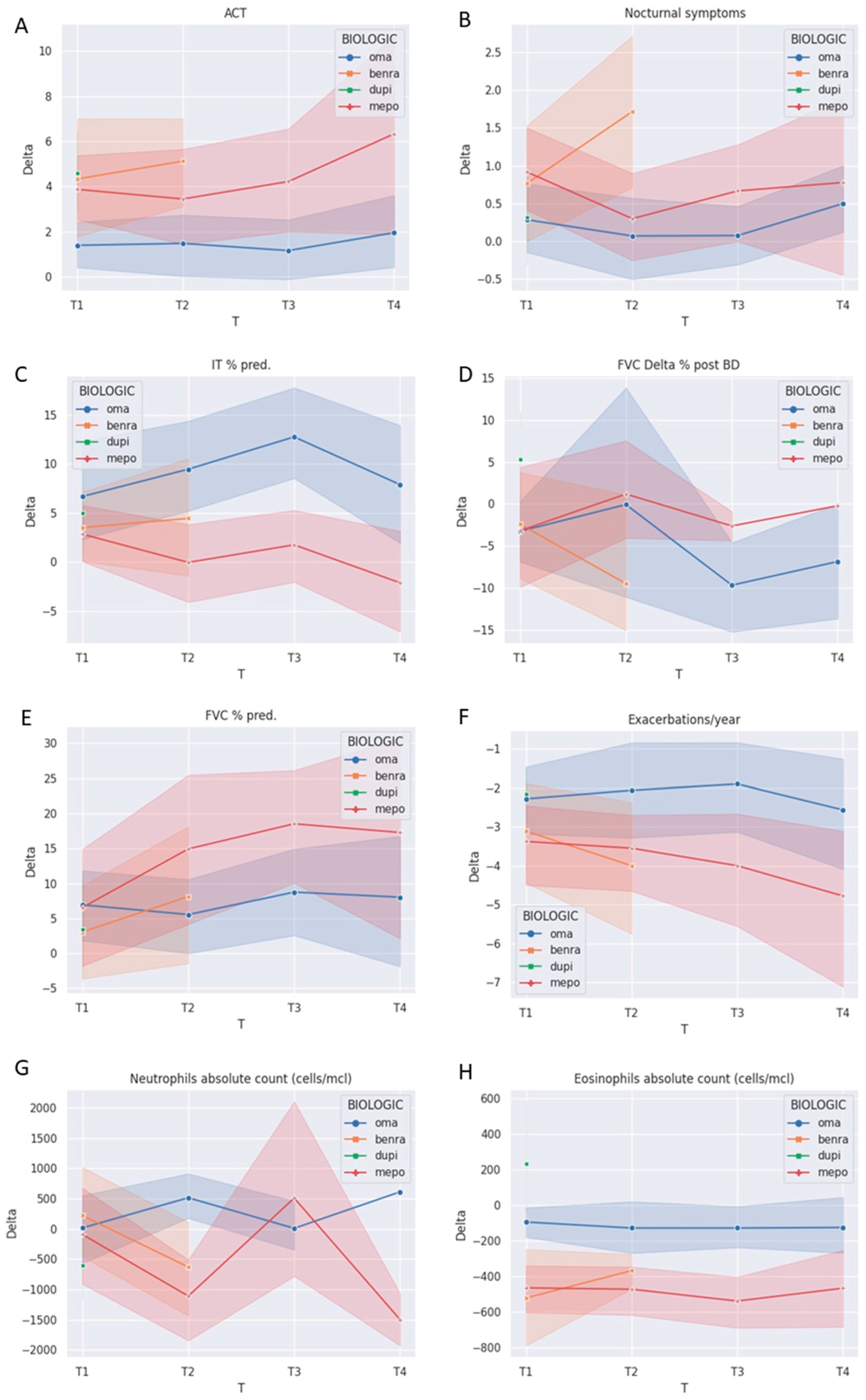
3.4. Analysis of “Inter-Biologic” Parameters over Years
3.5. OCS Chronic Treatment
4. Discussion
5. Conclusions
Supplementary Materials
Author Contributions
Funding
Institutional Review Board Statement
Informed Consent Statement
Data Availability Statement
Acknowledgments
Conflicts of Interest
References
- Chung, K.F.; Wenzel, S.E.; Brozek, J.L.; Bush, A.; Castro, M.; Sterk, P.J.; Adcock, I.M.; Bateman, E.D.; Bel, E.H.; Bleecker, E.R.; et al. International ERS/ATS guidelines on definition, evaluation and treatment of severe asthma. Eur. Respir. J. 2014, 43, 343–373. [Google Scholar] [CrossRef]
- Wenzel, S.E. Asthma phenotypes: The evolution from clinical to molecular approaches. Nat. Med. 2012, 18, 716–725. [Google Scholar] [CrossRef]
- Shah, P.A.; Brightling, C. Biologics for severe asthma—Which, when and why? Respirology 2023, 28, 709–721. [Google Scholar] [CrossRef]
- Panettieri, R., Jr.; Lugogo, N.; Corren, J.; Ambrose, C.S. Tezepelumab for Severe Asthma: One Drug Targeting Multiple Disease Pathways and Patient Types. J. Asthma Allergy 2024, 17, 219–236. [Google Scholar] [CrossRef]
- Ito, A.; Miyoshi, S.; Toyota, H.; Suzuki, Y.; Uehara, Y.; Hattori, S.; Takeshita, Y.; Sakasegawa, H.; Kuramochi, M.; Kobayashi, K.; et al. The overlapping eligibility for biologics in patients with severe asthma and phenotypes. Arerugi 2022, 71, 210–220. (In Japanese) [Google Scholar] [CrossRef]
- Pavord, I.D.; Hanania, N.A.; Corren, J. Controversies in Allergy: Choosing a Biologic for Patients with Severe Asthma. J. Allergy Clin. Immunol. Pract. 2022, 10, 410–419. [Google Scholar] [CrossRef]
- Menzies-Gow, A.; Steenkamp, J.; Singh, S.; Erhardt, W.; Rowell, J.; Rane, P.; Martin, N.; Llanos, J.P.; Quinton, A. Tezepelumab compared with other biologics for the treatment of severe asthma: A systematic review and indirect treatment comparison. J. Med. Econ. 2022, 25, 679–690. [Google Scholar] [CrossRef]
- Al-Shaikhly, T.; Norris, M.R.; Dennis, E.H.; Liu, G.; Craig, T.J. Comparative Impact of Asthma Biologics: A Nationwide US Claim-Based Analysis. J. Allergy Clin. Immunol. Pract. 2024, 12, 1558–1567. [Google Scholar] [CrossRef]
- Ricciardolo, F.L.; Guida, G.; Bertolini, F.; Di Stefano, A.; Carriero, V. Phenotype overlap in the natural history of asthma. Eur. Respir. Rev. 2023, 32, 220201. [Google Scholar] [CrossRef]
- Frøssing, L.; Silberbrandt, A.; Von Bülow, A.; Backer, V.; Porsbjerg, C. The Prevalence of Subtypes of Type 2 Inflammation in an Unselected Population of Patients with Severe Asthma. J. Allergy Clin. Immunol. Pract. 2021, 9, 1267–1275. [Google Scholar] [CrossRef]
- Papadopoulos, N.G.; Barnes, P.; Canonica, G.W.; Gaga, M.; Heaney, L.; Menzies-Gow, A.; Kritikos, V.; Fitzgerald, M. The evolving algorithm of biological selection in severe asthma. Allergy 2020, 75, 1555–1563. [Google Scholar] [CrossRef] [PubMed]
- McDonald, V.M.; Clark, V.L.; Cordova-Rivera, L.; Wark, P.A.B.; Baines, K.J.; Gibson, P.G. Targeting treatable traits in severe asthma: A randomised controlled trial. Eur. Respir. J. 2020, 55, 1901509. [Google Scholar] [CrossRef] [PubMed]
- Guida, G.; Bagnasco, D.; Carriero, V.; Bertolini, F.; Ricciardolo, F.L.M.; Nicola, S.; Brussino, L.; Nappi, E.; Paoletti, G.; Canonica, G.W.; et al. Critical evaluation of asthma biomarkers in clinical practice. Front. Med. 2022, 9, 969243. [Google Scholar] [CrossRef] [PubMed]
- Chen, C.-Y.; Wu, K.-H.; Guo, B.-C.; Lin, W.-Y.; Chang, Y.-J.; Wei, C.-W.; Lin, M.-J.; Wu, H.-P. Personalized Medicine in Severe Asthma: From Biomarkers to Biologics. Int. J. Mol. Sci. 2023, 25, 182. [Google Scholar] [CrossRef] [PubMed]
- Ricciardolo, F.L.M.; Sprio, A.E.; Baroso, A.; Gallo, F.; Riccardi, E.; Bertolini, F.; Carriero, V.; Arrigo, E.; Ciprandi, G. Characterization of T2-Low and T2-High Asthma Phenotypes in Real-Life. Biomedicines 2021, 9, 1684. [Google Scholar] [CrossRef] [PubMed]
- van Dijk, B.C.P.; Svedsater, H.; Heddini, A.; Nelsen, L.; Balradj, J.S.; Alleman, C. Relationship between the Asthma Control Test (ACT) and other outcomes: A targeted literature review. BMC Pulm. Med. 2020, 20, 79. [Google Scholar] [CrossRef]
- Nathan, R.A.; Sorkness, C.A.; Kosinski, M.; Schatz, M.; Li, J.T.; Marcus, P.; Murray, J.J.; Pendergraft, T.B. Development of the asthma control test: A survey for assessing asthma control. J. Allergy Clin. Immunol. 2004, 113, 59–65. [Google Scholar] [CrossRef] [PubMed]
- Graham, B.L.; Steenbruggen, I.; Miller, M.R.; Barjaktarevic, I.Z.; Cooper, B.G.; Hall, G.L.; Hallstrand, T.S.; Kaminsky, D.A.; McCarthy, K.; McCormack, M.C.; et al. Standardization of Spirometry 2019 Update. An Official American Thoracic Society and European Respiratory Society Technical Statement. Am. J. Respir. Crit. Care Med. 2019, 200, e70–e88. [Google Scholar] [CrossRef] [PubMed]
- Carriero, V.; Bertolini, F.; Sprio, A.E.; Bullone, M.; Ciprandi, G.; Ricciardolo, F.L.M. High levels of plasma fibrinogen could predict frequent asthma exacerbations. J. Allergy Clin. Immunol. Pract. 2020, 8, 2392–2395.e7. [Google Scholar] [CrossRef]
- Schatz, M.; Kosinski, M.; Yarlas, A.S.; Hanlon, J.; Watson, M.E.; Jhingran, P. The minimally important difference of the Asthma Control Test. J. Allergy Clin. Immunol. 2009, 124, 719–723.e1. [Google Scholar] [CrossRef]
- Humbert, M.; Beasley, R.; Ayres, J.; Slavin, R.; Hébert, J.; Bousquet, J.; Beeh, K.; Ramos, S.; Canonica, G.W.; Hedgecock, S.; et al. Benefits of omalizumab as add-on therapy in patients with severe persistent asthma who are inadequately controlled despite best available therapy (GINA 2002 step 4 treatment): INNOVATE. Allergy 2005, 60, 309–316. [Google Scholar] [CrossRef] [PubMed]
- Bousquet, J.; Humbert, M.; Gibson, P.G.; Kostikas, K.; Jaumont, X.; Pfister, P.; Nissen, F. Real-World Effectiveness of Omalizumab in Severe Allergic Asthma: A Meta-Analysis of Observational Studies. J. Allergy Clin. Immunol. Pract. 2021, 9, 2702–2714. [Google Scholar] [CrossRef] [PubMed]
- Paganin, F.; Mangiapan, G.; Proust, A.; Prudhomme, A.; Attia, J.; Marchand-Adam, S.; Pellet, F.; Milhe, F.; Melloni, B.; Bernady, A.; et al. Lung function parameters in omalizumab responder patients: An interesting tool? Allergy 2017, 72, 1953–1961. [Google Scholar] [CrossRef] [PubMed]
- Roth, M.; Zhong, J.; Zumkeller, C.; S’ng, C.T.; Goulet, S.; Tamm, M. The role of IgE-receptors in IgE-dependent airway smooth muscle cell remodelling. PLoS ONE 2013, 8, e56015. [Google Scholar] [CrossRef] [PubMed]
- Riccio, A.M.; Negro, R.W.; Micheletto, C.; De Ferrari, L.; Folli, C.; Chiappori, A.; Canonica, G.W. Omalizumab modulates bronchial reticular basement membrane thickness and eosinophil infiltration in severe persistent allergic asthma patients. Int. J. Immunopathol. Pharmacol. 2012, 25, 475–484. [Google Scholar] [CrossRef] [PubMed]
- Pianigiani, T.; Alderighi, L.; Meocci, M.; Messina, M.; Perea, B.; Luzzi, S.; Bergantini, L.; D’alessandro, M.; Refini, R.M.; Bargagli, E.; et al. Exploring the Interaction between Fractional Exhaled Nitric Oxide and Biologic Treatment in Severe Asthma: A Systematic Review. Antioxidants 2023, 12, 400. [Google Scholar] [CrossRef]
- Ortega, H.G.; Liu, M.C.; Pavord, I.D.; Brusselle, G.G.; Fitzgerald, J.M.; Chetta, A.; Humbert, M.; Katz, L.E.; Keene, O.N.; Yancey, S.W.; et al. Mepolizumab treatment in patients with severe eosinophilic asthma. N. Engl. J. Med. 2014, 371, 1198–1207. [Google Scholar] [CrossRef] [PubMed]
- Khurana, S.; Brusselle, G.G.; Bel, E.H.; FitzGerald, J.M.; Masoli, M.; Korn, S.; Kato, M.; Albers, F.C.; Bradford, E.S.; Gilson, M.J.; et al. Long-term Safety and Clinical Benefit of Mepolizumab in Patients with the Most Severe Eosinophilic Asthma: The COSMEX Study. Clin. Ther. 2019, 41, 2041–2056.e5. [Google Scholar] [CrossRef] [PubMed]
- Charles, D.; Shanley, J.; Temple, S.; Rattu, A.; Khaleva, E.; Roberts, G. Real-world efficacy of treatment with benralizumab, dupilumab, mepolizumab and reslizumab for severe asthma: A systematic review and meta-analysis. Clin. Exp. Allergy 2022, 52, 616–627. [Google Scholar] [CrossRef]
- Fyles, F.; Nuttall, A.; Joplin, H.; Burhan, H. Long-Term Real-World Outcomes of Mepolizumab and Benralizumab Among Biologic-Naive Patients with Severe Eosinophilic Asthma: Experience of 3 Years’ Therapy. J. Allergy Clin. Immunol. Pract. 2023, 11, 2715–2723. [Google Scholar] [CrossRef]
- Chupp, G.L.; Bradford, E.S.; Albers, F.C.; Bratton, D.J.; Wang-Jairaj, J.; Nelsen, L.M.; Trevor, J.L.; Magnan, A.; Brinke, A.T. Efficacy of mepolizumab add-on therapy on health-related quality of life and markers of asthma control in severe eosinophilic asthma (MUSCA): A randomised, double-blind, placebo-controlled, parallel-group, multicentre, phase 3b trial. Lancet Respir. Med. 2017, 5, 390–400. [Google Scholar] [CrossRef]
- Li, H.; Zhang, Q.; Wang, J.; Gao, S.; Li, C.; Wang, J.; Zhang, S.; Lin, J. Real-world Effectiveness of Mepolizumab in Severe Eosinophilic Asthma: A Systematic Review and Meta-analysis. Clin. Ther. 2021, 43, e192–e208. [Google Scholar] [CrossRef]
- Flood-Page, P.; Menzies-Gow, A.; Phipps, S.; Ying, S.; Wangoo, A.; Ludwig, M.S.; Barnes, N.; Robinson, D.; Kay, A.B. Anti-IL-5 treatment reduces deposition of ECM proteins in the bronchial subepithelial basement membrane of mild atopic asthmatics. J. Clin. Investig. 2003, 112, 1029–1036. [Google Scholar] [CrossRef] [PubMed]
- Ricciardolo, F.L.M.; Silkoff, P.E. Perspectives on exhaled nitric oxide. J. Breath Res. 2017, 11, 047104. [Google Scholar] [CrossRef]
- Ferguson, G.T.; FitzGerald, J.M.; Bleecker, E.R.; Laviolette, M.; Bernstein, D.; LaForce, C.; Mansfield, L.; Barker, P.; Wu, Y.; Jison, M.; et al. Benralizumab for patients with mild to moderate, persistent asthma (BISE): A randomised, double-blind, placebo-controlled, phase 3 trial. Lancet Respir. Med. 2017, 5, 568–576. [Google Scholar] [CrossRef]
- FitzGerald, J.M.; Bleecker, E.R.; Nair, P.; Korn, S.; Ohta, K.; Lommatzsch, M.; Ferguson, G.T.; Busse, W.W.; Barker, P.; Sproule, S.; et al. Benralizumab, an anti-interleukin-5 receptor α monoclonal antibody, as add-on treatment for patients with severe, uncontrolled, eosinophilic asthma (CALIMA): A randomised, double-blind, placebo-controlled phase 3 trial. Lancet 2016, 388, 2128–2141. [Google Scholar] [CrossRef] [PubMed]
- Menzella, F.; Bargagli, E.; Aliani, M.; Bracciale, P.; Brussino, L.; Caiaffa, M.F.; Caruso, C.; Centanni, S.; D’amato, M.; Del Giacco, S.; et al. ChAracterization of ItaliaN severe uncontrolled Asthmatic patieNts Key features when receiving Benralizumab in a real-life setting: The observational rEtrospective ANANKE study. Respir. Res. 2022, 23, 36. [Google Scholar] [CrossRef]
- Vitale, C.; Maglio, A.; Pelaia, C.; D’amato, M.; Ciampo, L.; Pelaia, G.; Molino, A.; Vatrella, A. Effectiveness of Benralizumab in OCS-Dependent Severe Asthma: The Impact of 2 Years of Therapy in a Real-Life Setting. J. Clin. Med. 2023, 12, 985. [Google Scholar] [CrossRef]
- Guida, G.; Riccio, A.M. Immune induction of airway remodeling. Semin. Immunol. 2019, 46, 101346. [Google Scholar] [CrossRef]
- Gorski, S.A.; Lawrence, M.G.; Hinkelman, A.; Spano, M.M.; Steinke, J.W.; Borish, L.; Teague, W.G.; Braciale, T.J. Expression of IL-5 receptor alpha by murine and human lung neutrophils. PLoS ONE 2019, 14, e0221113. [Google Scholar] [CrossRef]
- Tavernier, J.; Devos, R.; Cornelis, S.; Tuypens, T.; Van der Heyden, J.; Fiers, W.; Plaetinck, G. A human high affinity interleukin-5 receptor (IL5R) is composed of an IL5-specific α chain and a β chain shared with the receptor for GM-CSF. Cell 1991, 66, 1175–1184. [Google Scholar] [CrossRef] [PubMed]
- Castro, M.; Corren, J.; Pavord, I.D.; Maspero, J.; Wenzel, S.; Rabe, K.F.; Busse, W.W.; Ford, L.; Sher, L.; Fitzgerald, J.M.; et al. Dupilumab Efficacy and Safety in Moderate-to-Severe Uncontrolled Asthma. N. Engl. J. Med. 2018, 378, 2486–2496. [Google Scholar] [CrossRef]
- Rabe, K.F.; Nair, P.; Brusselle, G.; Maspero, J.F.; Castro, M.; Sher, L.; Zhu, H.; Hamilton, J.D.; Swanson, B.N.; Khan, A.; et al. Efficacy and Safety of Dupilumab in Glucocorticoid-Dependent Severe Asthma. N. Engl. J. Med. 2018, 378, 2475–2485. [Google Scholar] [CrossRef]
- Pelaia, C.; Heffler, E.; Crimi, C.; Maglio, A.; Vatrella, A.; Pelaia, G.; Canonica, G.W. Interleukins 4 and 13 in Asthma: Key Pathophysiologic Cytokines and Druggable Molecular Targets. Front. Pharmacol. 2022, 13, 851940. [Google Scholar] [CrossRef]
- Louis, R.; Satia, I.; Ojanguren, I.; Schleich, F.; Bonini, M.; Tonia, T.; Rigau, D.; Brinke, A.T.; Buhl, R.; Loukides, S.; et al. European Respiratory Society Guidelines for the Diagnosis of Asthma in Adults. Eur. Respir. J. 2022, 60, 2101585. [Google Scholar] [CrossRef]
- Chan, R.; Lipworth, B.J. Real-life effects of benralizumab on airway oscillometry in severe eosinophilic asthma. BMJ Open Respir. Res. 2023, 10, e001472. [Google Scholar] [CrossRef]
- Papaioannou, A.I.; Mplizou, M.; Porpodis, K.; Fouka, E.; Zervas, E.; Samitas, K.; Markatos, M.; Bakakos, P.; Papiris, S.; Gaga, M.; et al. Long-term efficacy and safety of omalizumab in patients with allergic asthma: A real-life study. Allergy Asthma Proc. 2021, 42, 235–242. [Google Scholar] [CrossRef]
- Gelhorn, H.L.; Balantac, Z.; Ambrose, C.S.; Chung, Y.N.; Stone, B. Patient and physician preferences for attributes of biologic medications for severe asthma. Patient Prefer. Adherence 2019, 13, 1253–1268. [Google Scholar] [CrossRef]


| Demographic Characteristics | |||||
|---|---|---|---|---|---|
| Overall | Omalizumab | Mepolizumab | Benralizumab | Dupilumab | |
| N Patients (%) | 88 (56.05%) | 41 (46%) | 23 (26.1%) | 15 (17%) | 9 (10%) |
| Sex: female n (%)/male n (%) | 38(43.2%) /50 (56.8%) | 16(39.0%) /25(61%) | 10(43.5%) /13(56.5%) | 9(60.0%) /6(40%) | 3(33.3%) /6(66.7%) |
| Age (Years) | 62.58 ± 11.92 | 64.24 ± 16.58 | 65.68 ± 14.82 | 60.87 ± 10.54 | 69.56 ± 16.0 |
| BMI (Kg/m2) | 27.07 ± 5.44 | 28.4 ± 5.758 | 25.41 ± 4.39 | 27.76 ± 5.82 | 28.65 ± 4.46 |
| Never smoker n (%) | 48 (54.5%) | 22 (53.7%) | 13 (56.5%) | 6 (40.0%) | 7 (77.8%) |
| Current smoker n (%) | 2 (2.3%) | 1 (2.4%) | 1 (4.3%) | 0 (0.0%) | 0 (0.0%) |
| Ex smoker n (%) | 38 (43.2%) | 18 (43.9%) | 9 (39.1%) | 9 (60.0%) | 2 (22.2%) |
| P/Y (Current + ex) | 17.8 ± 14.1 | 15.79 ± 11.84 | 12.70 ± 16.12 | 22.00 ± 12.19 | 37.00 ± 21.21 |
| Early onset (year) n (%) | 20 (22.7%) | 13 (31.7%) | 5 (21.7%) | 1 (6.7%) | 1 (11.1%) |
| Age of onset (years) | 33.39 ±16.56 | 36.67 ± 15.89 | 31.96 ± 19.50 | 32.71 ± 19.09 | 39.33 ± 24.41 |
| Comorbidities | |||||
| ASA intolerance n (%) | 16 (18.2%) | 9 (22.0%) | 5 (21.7%) | 1 (6.7%) | 1 (11.1%) |
| Rhinitis n (%) | 68 (77.3%) | 34 (82.9%) §§§ | 21 (91.3%) §§§ | 11 (73.3%) § | 2 (22.2%) |
| Sinusitis (with or without polyps) n (%) | 50 (56.8%) | 25 (61.0%) | 17 (73.9%) | 8 (53.3%) | 6 (66.7%) |
| Nasal polyposis n (%) | 33 (37.5%) | 11 (26.8%) | 10 (43.5%) | 7 (46.7%) | 5 (55.6%) |
| Bronchiectasis n (%) | 8 (9.1%) | 2 (4.9%) | 2 (8.7%) | 4 (26.7%) *§ | 0 (0.0%) |
| GERD n (%) | 20 (22.7%) | 5 (12.2%) | 5 (21.7%) | 6 (40.0%) | 4 (44.4%) * |
| OSAS n (%) | 4 (4.5%) | 1 (2.4%) | 1 (4.3%) | 1 (6.7%) | 1 (11.1%) |
| Obesity n (%) | 24 (27.3%) | 8 (19.5%) | 11 (47.8%) * | 3 (20.0%) | 2 (22.2%) |
| Diabetes n (%) | 7 (8%) | 3 (7.3%) | 1 (4.3%) | 1 (6.7%) | 2 (22.2%) |
| Hypertension n (%) | 24 (27.3%) | 11 (26.8%) | 8 (34.8%) | 4 (26.7%) | 1 (11.1%) |
| MI n (%) | 3 (3.4%) | 1 (2.4%) | 0 (0.0%) | 0 (0.0%) | 2 (22.2%) |
| Heart failure n (%) | 4 (4.5%) | 1 (2.4%) | 0 (0.0%) | 1 (6.7%) | 2 (22.2%) |
| Arrhythmias n (%) | 6 (6.8%) | 4 (9.8%) | 0 (0.0%) | 1 (6.7%) | 1 (11.1%) |
| SAD n (%) | 10 (11.4%) | 6 (14.6%) | 3 (13.0%) | 0 (0.0%) | 1 (11.1%) |
| VCD n (%) | 2 (2.3%) | 0 (0.0%) | 0 (0.0%) | 0 (0.0%) | 2 (22.2%) |
| EGPA n (%) | 0 (0%) | 0 (0.0%) | 0 (0.0%) | 0 (0.0%) | 0 (0.0%) |
| Osteoporosis n (%) | 12 (13.6%) | 8 (19.5%) | 2 (8.7%) | 1 (6.7%) | 1 (11.1%) |
| Past pneumoniae n (%) | 15 (17%) | 6 (14.6%) | 5 (21.7%) | 3 (20.0%) | 1 (11.1%) |
| ABPA n (%) | 2 (2.3%) | 2 (4.9%) | 0 (0.0%) | 0 (0.0%) | 0 (0.0%) |
| Chronic pain n (%) | 4 (4.5%) | 2 (4.9%) | 1 (4.3%) | 0 (0.0%) | 1 (11.1%) |
| Arthropathies n (%) | 6 (6.8%) | 1 (2.4%) | 3 (13.0%) | 2 (13.3%) | 0 (0.0%) |
| Familiarity n (%) | 16 (18.2%) | 7 (17.1%) | 2 (8.7%) | 4 (26.7%) | 3 (33.3%) |
| Atopy n (%) | 64 (72.7%) | 41 (100.0%) | 12 (52.2%) **** | 7 (46.7%) **** | 4 (44.4%) **** |
| Monosesitize n (%) | 12 (13.6%) | 5 (12.2%) | 3 (13.0%) | 2 (13.3%) | 2 (22.2%) |
| Polysensitized n (%) | 52 (59.1%) | 35 (85.4%) | 9 (39.1%) *** | 7 (46.7%) ** | 1 (11.1%) **** |
| Seasonal allergen n (%) | 52 (59.1%) | 33 (80.5%) | 10 (43.5%) ** | 8 (53.3%) * | 1 (11.1%) ****/# |
| Perennial allergen n (%) | 49 (55.7%) | 35 (85.4%) | 6 (26.1%) **** | 4 (26.7%) **** | 4 (44.4%) ** |
| Alternaria n (%) | 7 (7.95%) | 6 (14.6%) | 1 (4.3%) | 0 (0.0%) | 0 (0.0%) |
| Aspergillus n (%) | 16 (18.18%) | 11 (26.8%) | 4 (17.4%) | 0 (0.0%) * | 1 (11.1%) |
| Specific IgE n (%) | 13 (14.8%) | 8 (19.5%) | 4 (17.4%) | 0 (0.0%) | 1 (11.1%) |
| Prick test n (%) | 6 (6.8%) | 6 (14.6%) | 0 (0.0%) | 0 (0.0%) | 0 (0.0%) |
| Treatment/Clinical outcome | |||||
| BDP HFA dose, mcg | 702.30 ± 216.00 | 673.17 ± 244.97 | 650.00 ± 174.93 | 783.33 ± 154.35 *° | 757.89 ± 216.84 |
| LABA n (%) | 88 (100%) | 41 (100%) | 23 (100%) | 15 (100%) | 9 (100%) |
| LAMA n (%) | 33 (37.5%) | 12 (29.3%) | 10 (43.5%) | 6 (40.0%) | 5 (55.6%) |
| Chronic OCS n (%) | 24 (27.3%) | 9 (22%) | 8 (34.8%) | 4 (26.7%) | 3(33.3%) |
| OCS bursts ≥ 3/year n (%) | 44 (50%) | 17 (41.5%) | 15 (65.2%) | 9 (60.0%) | 3 (33.3%) |
| OCS bursts ≥ 3/year and Chronic OCS n (%) | 15 (17.04%) | 4 (9.7%) | 6 (26.08%) | 3 (20%) | 2 (22.2%) |
| OCS dependence n (%) | 53 (60.2%) | 22 (53.7%) | 17 (73.9%) | 10 (66.7%) | 4 (44.4%) |
| Biologic switches n (%) | 16 (18%) | 8 (19.5%) | 5 (21.7%) | 2 (13.3%) | 1 (11.1%) |
| ACT score | 17.65 ± 4.41 | 19.37 ± 2.97 | 16.69 ± 4.84 * | 17.61 ± 4.98 | 15.42 ± 4.55 ** |
| Controlled (ACT ≥ 20) n (%) | 37 (42%) | 26 (63.4%) | 5 (21.7%) ** | 5 (33.3%) | 1 (11.1%) ** |
| Not controlled (ACT ≤ 19) n (%) | 51 (58%) | 15 (36.6%) | 18 (78.3%) ** | 10 (66.7%) | 8 (88.9%) ** |
| Activity limitations | 3.09 ± 1.24 | 3.19 ± 1.08 | 3.12 ± 1.24 | 3.44 ± 1.46 | 2.84 ± 1.21 |
| Nocturnal symptoms | 3.94 ± 1.40 | 4.52 ± 0.87 | 3.85 ± 1.46 | 3.82 ± 1.59 | 3.95 ± 1.58 |
| Exacerbations/year | 3.55 ± 2.94 | 3.15 ± 3.07 | 4.27 ± 2.91 | 4.00 ± 2.83 | 2.58 ± 1.95 ° |
| ER visits n (%) | 33 (37.5%) | 13 (31.7%) | 8 (34.8%) | 7 (46.7%) | 5 (55.6%) |
| Intubation n (%) | 1 (1.1%) | 1 (2.4%) | 0 (0.0%) | 0 (0.0%) | 0 (0.0%) |
| Functional Parameters/Biomarkers | |||||
|---|---|---|---|---|---|
| Overall | Omalizumab | Mepolizumab | Benralizumab | Dupilumab | |
| T0 | T0 | T0 | T0 | T0 | |
| FVC abs. (L) | 2.75 ± 1.01 | 2.76 ± 1.04 | 2.79 ± 1.05 | 2.95 ± 0.99 | 2.37 ± 0.60 # |
| FVC % pred. | 86.88 ± 17.98 | 89.02 ± 18.91 | 83.50 ± 16.67 | 93.56 ± 22.56 | 83.05 ± 19.70 |
| FEV1 abs. (L) | 1.701 ± 0.71 | 1.67 ± 0.63 | 1.72 ± 0.86 | 1.87 ± 0.75 | 1.32 ± 0.51 *# |
| FEV1 % pred. | 65.50 ± 17.73 | 65.08 ± 15.71 | 62.88 ± 19.41 | 72.67 ± 21.08 | 57.21 ± 18.89 # |
| IT abs. | 59.43 ± 12.34 | 59.19 ± 13.47 | 58.59 ± 11.70 | 60.24 ± 14.47 | 53.58 ± 13.11 |
| IT % pred. | 72.74 ± 14.78 | 69.50 ± 13.71 | 74.46 ± 15.65 | 75.29 ± 18.95 | 67.78 ± 15.90 |
| RV abs. (L) | 3.09 ± 1.11 | 3.08 ± 1.04 | 3.18 ± 1.03 | 3.22 ± 1.45 | 3.13 ± 1.14 |
| RV % pred. | 147.70 ± 47.37 | 159.11 ± 46.37 | 148.73 ± 45.27 | 139.72 ± 49.73 | 150.94 ± 44.80 |
| FVC post BD abs. (L) | 3.02 ± 1.16 | 3.03 ± 1.2 | 2.95 ± 1.24 | 3.22 ± 1.11 | 2.57 ± 0.66 # |
| FVC Delta abs. post BD (L) | 0.30 ± 0.24 | 0.33 ± 0.28 | 0.27 ± 0.21 | 0.25 ± 0.22 | 0.23 ± 0.17 |
| FVC Delta % post BD | 11.11 ± 8.3 | 11.92 ± 9.24 | 9.12 ± 7.18 | 7.36 ± 7.23 | 8.05 ± 9.17 |
| FEV1 post abs. (L) | 1.93 ± 0.85 | 1.95 ± 0.76 | 1.82 ± 1.10 | 2.05 ± 0.85 | 1.53 ± 0.6 *# |
| FEV1 Delta abs. post BD (L) | 0.23 ± 0.19 | 0.24 ± 0.17 | 0.23 ± 0.19 | 0.18 ± 0.22 | 0.21 ± 0.18 |
| FEV1 Delta % post BD | 15.01 ± 9.70 | 15.58 ± 8.35 | 15.66 ± 9.72 | 10.49 ± 12.76 | 16.16 ± 9.36 |
| DLCO % | 85.50 ± 20.16 | 87.71 ± 9.94 | 78.42 ± 23.13 | 85.38 ± 26.87 | 76.60 ± 13.94 |
| DLCO/Va % | 100.20 ± 22.13 | 102.86 ± 19.62 | 93.70 ± 20.64 | 101.30 ± 30.07 | 98.00 ± 22.40 |
| FENO (ppb) | 40.34 ± 29.42 | 35.47 ± 27.81 | 52.73 ± 33.00 | 39.96 ± 24.39 | 31.13 ± 22.94 ° |
| Total IgE (UI/mL) | 215.50 ± 180.40 | 323.45 ± 261.19 | 307.65 ± 421.13 | 466.07 ± 461.11 | 327.75 ± 649.39 |
| Leucocytes absolute count (cells/mcl) | 8124.00 ± 2121.00 | 8190.00 ± 1951.47 | 7771.65 ± 1822.44 | 8248.75 ± 1923.05 | 9152.63 ± 2546.82 |
| Neutrophils (%) | 55.00 ± 10.00 | 55.12 ± 9.19 | 52.95 ± 10.77 | 54.54 ± 8.11 | 58.55 ± 11.33 |
| Neutrophils absolute count (cells/mcl) | 4513.00 ± 1676.00 | 4588.46 ± 1583.27 | 4058.47 ± 1566.19 | 4570.62 ± 1274.58 | 5266.84 ± 1930.38 |
| Eosinophils (%) | 5.90 ± 4.45 | 5.44 ± 4.06 | 7.02 ± 4.13 | 7.00 ± 5.57 | 3.59 ± 2.59 °°#* |
| Eosinophils absolute count (cells/mcl) | 436.30 ± 294.70 | 426.79 ± 310.28 | 560.38 ± 312.03 | 563.89 ± 468.83 | 296.84 ± 193.16 °#* |
| Fibrinogen (mg/dL) | 355.00 ± 94.42 | 356.62 ± 95.72 | 363.40 ± 112.49 | 327.29 ± 86.61 | 366.86 ± 79.42 |
| T2 Biomarkers | |||||
|---|---|---|---|---|---|
| (n) | Overall (88) | Omalizumab (41) | Mepolizumab (23) | Benralizumab (15) | Dupilumab (9) |
| 1–2 biomarker n (%) | 39 (44.3%) | 20 (48.8%) | 7 (30.4%) | 5 (33.3%) | 7 (77.7%) °# |
| 3–4 biomarkers n (%) | 49 (55.6%) | 21 (51.2%) | 16 (69.5%) | 10 (66.6%) | 2 (22.2%) °# |
| Omalizumab | Mepolizumab | Benralizumab | Dupilumab | ||||||||||||
|---|---|---|---|---|---|---|---|---|---|---|---|---|---|---|---|
| T0 | T1 | T2 | T3 | T4 | T0 | T1 | T2 | T3 | T4 | T0 | T1 | T2 | T0 | T1 | |
| BDP HFA dose, mcg | 673.17 ± 244.97 | 662.50 ± 258.88 | 590.91 ± 271.99 | 558.06 ± 293.00 | 500.00 ± 258.20 * | 650.00 ± 174.93 | 580.00 ± 261.41 | 600.00 ± 215.21 | 600.00 ± 181.50 | 688.89 ± 247.21 | 783.33 ± 154.35 | 766.67 ± 123.67 | 650.00 ± 232.99 | 757.89 ± 216.84 | 800.00 ± 240.37 |
| ACT score | 19.37 ± 2.97 | 20.70 ± 2.56 * | 21.03 ± 3.48 * | 20.83 ± 3.44 | 21.57 ± 2.92 * | 16.69 ± 4.84 | 20.67 ± 3.41 °° | 20.40 ± 4.67 ° | 21.83 ± 3.00 °°°° | 22.22 ± 3.42 °° | 17.61 ± 4.98 | 21.94 ± 4.28 # | 21.00 ± 3.96 | 15.42 ± 4.55 | 20.00 ± 3.86 §§ |
| Activity limitation | 3.19 ± 1.08 | 4.05 ± 0.90 * | 4.35 ± 1.00 ** | 4.17 ± 1.20 * | 4.50 ± 1.16 * | 3.12 ± 1.24 | 3.92 ± 0.97 ° | 3.95 ± 1.19 ° | 4.39 ± 0.78 °°° | 4.56 ± 0.73 °° | 3.44 ± 1.46 | 4.17 ± 1.25 | 4.38 ± 0.74 # | 2.84 ± 1.21 | 3.95 ± 1.13 § |
| Nocturnal symptoms | 4.52 ± 0.87 | 4.82 ± 0.50 | 4.76 ± 0.97 | 4.83 ± 0.38 | 4.93 ± 0.27 | 3.85 ± 1.46 | 4.83 ± 0.64 ° | 4.30 ± 1.38 | 4.67 ± 0.77 ° | 4.67 ± 1.00 | 3.82 ± 1.59 | 4.61 ± 1.04 | 4.88 ± 0.35 # | 3.95 ± 1.58 | 4.26 ± 1.15 |
| Exacerbations/year | 3.15 ± 3.07 | 1.00 ± 1.47 *** | 1.22 ± 1.70 ** | 1.68 ± 1.72 * | 1.13 ± 1.58 ** | 4.27 ± 2.91 | 1.08 ± 1.35 °°°° | 0.75 ± 0.91 °°°° | 0.61 ± 0.70 °°°° | 0.44 ± 0.73 °°°° | 4.00 ± 2.83 | 0.89 ± 0.90 ## | 1.12 ± 1.25 ## | 2.58 ± 1.95 | 0.42 ± 0.96 §§§ |
| FVC abs. (L) | 2.76 ± 1.04 | 3.04 ± 1.12 | 2.94 ± 1.07 | 2.94 ± 1.07 | 2.78 ± 1.03 | 2.79 ± 1.05 | 3.02 ± 1.19 | 2.88 ± 0.86 | 3.39 ± 1.34 | 2.81 ± 0.72 | 2.95 ± 0.99 | 3.11 ± 1.11 | 3.44 ± 1.22 | 2.37 ± 0.60 | 2.54 ± 0.69 |
| FVC % pred. | 89.02 ± 18.91 | 97.72 ± 16.27 * | 95.55 ± 16.57 | 97.63 ± 17.47 * | 99.08 ± 18.92 * | 83.50 ± 16.67 | 91.61 ± 25.16 | 99.12 ± 27.29 ° | 108.40 ± 16.15 °°°° | 98.71 ± 18.51 | 93.56 ± 22.56 | 96.61 ± 23.09 | 105.00 ± 23.00 | 83.05 ± 19.70 | 85.71 ± 21.93 |
| FEV1 abs. (L) | 1.67 ± 0.63 | 1.87 ± 0.72 | 1.87 ± 0.63 | 1.90 ± 0.71 | 1.81 ± 0.73 | 1.72 ± 0.86 | 2.01 ± 1.04 | 1.82 ± 0.73 | 2.33 ± 1.15 | 1.89 ± 0.57 | 1.87 ± 0.75 | 2.11 ± 1.00 | 2.72 ± 0.95 # | 1.32 ± 0.51 | 1.58 ± 0.65 |
| FEV1 % pred. | 65.08 ± 15.71 | 72.97 ± 17.64 * | 75.30 ± 16.93 * | 77.26 ± 19.49 * | 76.70 ± 21.90 * | 62.88 ± 19.41 | 73.62 ± 28.37 | 76.06 ± 28.78 | 86.73 ± 20.79 °° | 81.43 ± 18.61 ° | 72.67 ± 21.08 | 79.06 ± 28.85 | 98.12 ± 20.57 # | 57.21 ± 18.89 | 64.47 ± 19.23 |
| IT abs. | 59.19 ± 13.47 | 61.69 ± 9.19 | 63.43 ± 9.53 | 63.97 ± 11.01 | 61.21 ± 11.53 | 58.59 ± 11.70 | 61.18 ± 12.54 | 60.10 ± 12.97 | 64.33 ± 13.04 | 65.99 ± 8.27 | 60.24 ± 14.47 | 62.21 ± 14.24 | 72.00 ± 5.63 # | 53.58 ± 13.11 | 59.56 ± 13.12 |
| IT % pred. | 69.50 ± 13.71 | 77.82 ± 11.10 * | 78.85 ± 12.5 * | 79.41 ± 14.3 * | 75.96 ± 13.12 | 74.46 ± 15.65 | 76.77 ± 15.77 | 73.88 ± 16.09 | 80.67 ± 17.22 | 79.43 ± 8.89 | 75.29 ± 18.95 | 79.94 ± 19.59 | 94.14 ± 8.13 ## | 67.78 ± 15.90 | 73.33 ± 15.43 |
| RV abs. (L) | 3.08 ± 1.04 | 2.63 ± 0.78 | 2.57 ± 1.06 | 2.79 ± 0.82 | 2.18 ± 0.85 * | 3.18 ± 1.03 | 2.85 ± 0.97 | 3.08 ± 0.83 | 2.55 ± 0.56 | 2.44 ± 0.73 | 3.22 ± 1.4 | 2.78 ± 1.17 | 3.25 ± 2.08 | 3.13 ± 1.14 | 2.47 ± 0.07 § |
| RV % pred. | 159.11 ± 46.37 | 127.15 ± 38.82 * | 132.64 ± 44.90 | 139.33 ± 42.27 | 113.00 ± 28.21 ** | 148.73 ± 45.27 | 143.13 ± 51.42 | 154.36 ± 36.97 | 127.58 ± 31.77 | 121.20 ± 41.03 | 139.72 ± 49.73 | 142.71 ± 50.81 | 105.43 ± 46.24 | 150.94 ± 44.80 | 125.33 ± 41.85 |
| FVC post BD abs.(L) | 3.03 ± 1.21 | 3.48 ± 1.26 | 3.03 ± 0.63 | 2.99 ± 0.78 | 2.82 ± 1.23 | 2.95 ± 1.24 | 3.13 ± 1.08 | 3.02 ± 0.96 | 3.62 ± 1.29 | 3.21 ± 0.46 | 3.22 ± 1.11 | 3.30 ± 1.08 | 3.35 ± 1.52 | 2.57 ± 0.66 | 2.69 ± 0.73 |
| FVC Delta abs. post BD (L) | 0.33 ± 0.28 | 0.15 ± 0.21 * | 0.22 ± 0.29 | 0.18 ± 0.17 | 0.16 ± 0.21 | 0.27 ± 0.21 | 0.15 ± 0.12 | 0.20 ± 0.22 | 0.17 ± 0.19 | 0.08 ± 0.03 ° | 0.25 ± 0.22 | 0.24 ± 0.33 | 0.16 ± 0.14 | 0.23 ± 0.17 | 0.19 ± 0.16 |
| FVC Delta % post BD | 11.92 ± 9.24 | 6.48 ± 9.08 | 8.95 ± 13.60 | 6.39 ± 7.20 | 4.00 ± 6.22 * | 9.12 ± 7.18 | 4.93 ± 4.25 | 6.62 ± 5.84 | 5.75 ± 6.95 | 2.42 ± 1.25 °° | 7.36 ± 7.23 | 5.87 ± 6.96 | 5.62 ± 3.74 | 8.05 ± 9.17 | 9.91 ± 8.58 |
| FEV1 post abs. (L) | 1.95 ± 0.76 | 2.06 ± 0.84 | 1.84 ± 0.49 | 1.99 ± 0.59 | 2.01 ± 0.79 | 1.82 ± 1.10 | 2.26 ± 0.95 | 2.01 ± 0.88 | 2.58 ± 1.10 | 2.38 ± 0.21 | 2.05 ± 0.85 | 2.20 ± 0.82 | 2.56 ± 1.24 | 1.53 ± 0.63 | 1.60 ± 0.62 |
| FEV1 Delta Post BD abs. (L) | 0.24 ± 0.17 | 0.17 ± 0.12 | 0.15 ± 0.15 | 0.16 ± 0.14 | 0.21 ± 0.17 | 0.23 ± 0.19 | 0.16 ± 0.10 | 0.10 ± 0.46 | 0.17 ± 0.16 | 0.13 ± 0.14 | 0.18 ± 0.22 | 0.17 ± 0.20 | 0.13 ± 0.15 | 0.21 ± 0.18 | 0.17 ± 0.06 |
| FEV1 Delta % post BD | 15.58 ± 8.35 | 10.60 ± 8.24 | 8.38 ± 7.67 * | 9.39 ± 9.98 | 11.55 ± 6.71 | 15.66 ± 9.72 | 7.52 ± 2.91 °° | 1.72 ± 33.20 | 8.37 ± 10.17 | 6.55 ± 7.31 | 10.49 ± 12.76 | 10.49 ± 9.79 | 6.13 ± 5.31 | 16.16 ± 9.36 | 13.75 ± 9.05 |
| DLCO % | 87.71 ± 9.94 | 98.75 ± 21.87 | 105.50 ± 17.68 | 92.60 ± 14.98 | 97.00 ± 22.00 | 78.42 ± 23.13 | 64.00 ± 46.67 | 88.20 ± 15.06 | 85.38 ± 26.87 | 92.67 ± 24.01 | 94.50 ± 17.68 | 76.60 ± 13.94 | 89.00 ± 19.37 | ||
| FENO (ppb) | 35.47 ± 27.81 | 31.54 ± 21.89 | 42.64 ± 34.23 | 35.85 ± 17.31 | 48.11 ± 51.61 | 52.73 ± 33.00 | 56.87 ± 50.08 | 41.99 ± 30.12 | 59.75 ± 50.89 | 39.96 ± 24.39 | 50.84 ± 60.13 | 53.60 ± 40.48 | 31.13 ± 22.94 | 29.48 ± 17.17 | |
| Total IgE (UI/mL) | 323.45 ± 261.19 | 792.33 ± 649.35 * | 606.55 ± 419.06 * | 557.97 ± 409.13 * | 576.06 ± 519.86 | 307.65 ± 421.13 | 236.91 ± 152.03 | 197.13 ± 213.36 | 149.00 ± 49.50 | 641.95 ± 833.04 | 466.07 ± 461.11 | 613.67 ± 793.41 | 327.75 ± 649.39 | 221.05 ± 280.84 | |
| Leukocytes absolute count (cells/mcl) | 8190.00 ± 1951.47 | 7357.42 ± 1952.10 | 7977.14 ± 3673.14 | 7351.15 ± 1679.39 | 7321.50 ± 2063.33 | 7771.65 ± 1822.44 | 7977.39 ± 1870.07 | 7325.56 ± 2491.86 | 7248.82 ± 2038.64 | 7220.00 ± 1464.66 | 8248.75 ± 1923.05 | 7551.76 ± 2973.69 | 6182.86 ± 1190.44 ## | 9152.63 ± 2546.82 | 8011.25 ± 1772.18 |
| Neutrophils % | 55.12 ± 9.19 | 59.48 ± 11.54 | 58.88 ± 8.09 | 54.04 ± 7.84 | 56.07 ± 7.01 | 52.95 ± 10.77 | 57.99 ± 8.98 | 55.28 ± 10.03 | 60.73 ± 11.93 | 56.89 ± 7.43 | 54.54 ± 8.11 | 58.46 ± 10.16 | 49.07 ± 9.79 | 58.55 ± 11.33 | 53.83 ± 5.49 |
| Neutrophils absolute count (cells/mcl) | 4588.46 ± 1583.27 | 4246.47 ± 1556.59 | 5072.31 ± 2982.80 | 4062.31 ± 1195.96 | 4072.22 ± 1414.37 | 4058.47 ± 1566.19 | 4574.55 ± 1522.37 | 4115.00 ± 1994.25 | 4817.65 ± 2309.18 | 4085.71 ± 821.52 | 4570.62 ± 1274.58 | 4490.00 ± 2040.00 | 3115.00 ± 299.25 ## | 5266.84 ± 1930.38 | 4304.50 ± 1610.92 |
| Eosinophils % | 5.44 ± 4.06 | 4.59 ± 3.71 | 5.17 ± 4.76 | 5.35 ± 3.44 | 5.03 ± 2.97 | 7.02 ± 4.13 | 1.43 ± 1.16 °°°° | 1.50 ± 1.25 °°°° | 1.29 ± 1.37 °°°° | 1.04 ± 0.59 °°°° | 7.00 ± 5.57 | 0.53 ± 2.00 ### | 0.00 ± 0.00 #### | 3.59 ± 2.59 | 5.94 ± 6.57 |
| Eosinophils absolute count (cells/mcl) | 426.79 ± 310.28 | 341.68 ± 271.28 | 363.29 ± 271.66 | 396.40 ± 231.73 | 403.12 ± 218.90 | 560.38 ± 312.03 | 108.26 ± 79.98 °°°° | 105.00 ± 81.47 °°°° | 85.88 ± 81.17 °°°° | 74.29 ± 38.23 °°°° | 563.89 ± 468.83 | 47.06 ± 188.94 ### | 0.00 ± 0.00 #### | 296.84 ± 193.16 | 527.06 ± 613.29 |
| Fibrinogen (mg/dl) | 356.62 ± 95.72 | 344.33 ± 100.47 | 330.67 ± 73.95 | 429.40 ± 88.71 | 363.40 ± 112.49 | 369.25 ± 95.12 | 404.22 ± 101.63 | 419.75 ± 129.64 | 327.29 ± 86.61 | 327.67 ± 92.39 | 294.00 ± 75.36 | 366.86 ± 79.42 | 382.50 ± 98.99 | ||
| Omalizumab | Mepolizumab | Benralizumab | Dupilumab | ||||||||
|---|---|---|---|---|---|---|---|---|---|---|---|
| Delta T1 | Delta T2 | Delta T3 | Delta T4 | Delta T1 | Delta T2 | Delta T3 | Delta T4 | Delta T1 | Delta T2 | Delta T1 | |
| BDP HFA dose, mcg | −17.50 ±169.29 | −69.70 ±251.85 | −87.10 ±301.93 | −100.00 ±263.00 | −72.00 ±279.17 | −40.00 ±270.28 | −16.67 ±214.89 | 55.56 ±278.89 | −16.67 ±161.79 | −37.50 ±346.15 | 42.11 ± 254.55 |
| ACT score | 1.40 ± 3.42 | 1.48 ± 3.78 | 1.17 ± 3.75 | 1.96 ± 3.97 | 3.88 ± 3.78 | 3.45 ± 4.66 | 4.22 ± 4.98 * | 6.33 ± 7.07 | 4.33 ± 5.76 * | 5.12 ± 3.09 * | 4.58 ± 4.03 ** |
| Activity limitation | 0.81 ± 1.36 | 1.14 ± 1.56 | 0.77 ± 1.24 | 1.38 ± 1.19 | 0.75 ± 1.07 | 0.75 ± 1.33 | 1.06 ± 1.30 | 1.67 ± 1.41 | 0.72 ± 1.71 | 1.12 ± 1.25 | 1.11 ± 1.05 |
| Nocturnal symptoms | 0.29 ± 1.06 | 0.07 ± 1.07 | 0.08 ± 0.76 | 0.50 ± 0.76 | 0.92 ± 1.35 | 0.30 ± 1.38 | 0.67 ± 1.46 | 0.78 ± 1.86 | 0.76 ± 1.64 | 1.71 ± 1.38 *° | 0.32 ± 1.38 |
| Exacerbations/year | −2.28 ±2.80 | −2.06 ± 3.74 | −1.90 ±3.32 | −2.57 ± 3.54 | −3.38 ±2.63 | −3.55 ±2.31 | −4.00 ±3.22 * | −4.78 ±3.46 | −3.11 ±2.78 | −4.00 ±2.78 | −2.16 ±1.61 * |
| FVC abs. (L) | 0.24 ±0.39 | 0.22 ±0.38 | 0.26 ±0.46 | 0.16 ±0.64 | 0.15 ±0.54 | 0.14 ±0.47 | 0.29 ±0.44 | −0.02 ± 0.31 | 0.16 ±0.47 | 0.26 ±0.49 | 0.15 ±0.39 |
| FVC % pred. | 6.94 ±15.91 | 5.55 ±14.80 | 8.76 ±16.62 | 8.04 ±23.81 | 6.57 ±21.27 | 14.94 ±23.08 | 18.53 ±16.82 | 17.29 ±22.34 | 3.06 ±14.95 | 8.14 ±14.58 | 3.47 ±15.99 |
| FEV1 abs. (L) | 0.19 ±0.42 | 0.23 ±0.35 | 0.28 ±0.39 | 0.19 ±0.47 | 0.21 ±0.45 | 0.14 ±0.40 | 0.31 ±0.45 | 0.04 ±0.29 | 0.24 ±0.46 | 0.41 ±0.47 | 0.23 ±0.46 |
| FEV1 % pred. | 7.17 ±18.52 | 9.93 ±13.72 | 13.60 ±16.68 | 12.23 ±20.58 | 9.50 ±17.37 | 12.94 ±19.21 | 16.00 ±16.27 | 13.86 ±18.40 | 6.39 ±14.97 | 13.12 ±15.53 | 7.35 ±19.54 |
| IT abs. (L) | 2.28 ±10.81 | 4.67 ±9.42 | 5.75 ±10.92 | 4.18 ±10.99 | 2.38 ±4.56 | 1.44 ±6.38 | 2.26 ±4.69 | 2.53 ±5.71 | 2.28 ±5.47 | 3.00 ±7.14 | 5.27 ±10.61 |
| IT % pred. | 6.67 ±13.42 | 9.46 ±11.87 | 12.75 ±11.80 | 7.88 ±12.41 | 2.82 ±6.62 | −0.06 ±8.76 * | 1.73 ± 7.82 ** | −2.14 ±7.67 * | 3.50 ±7.31 | 4.43 ±8.92 | 5.00 ±8.43 |
| RV abs. (L) | −0.37 ±1.33 | −0.29 ±0.68 | −0.86 ±0.88 | −0.94 ±0.47 | −0.77 ±1.55 | 0.34 ±0.76 | −0.37 ±0.47 | −0.12 ±0.62 | −0.29 ±0.24 | −0.52 ±1.00 | |
| RV % pred. | −35.08 ±59.49 | −9.50 ±36.03 | −42.29 ±38.81 | −54.00 ±17.11 | −12.00 ±55.61 | 8.60 ±32.42 | −17.80 ±19.69 | 10.57 ±46.88 * | −17.29 ±16.10 | −21.40 ±35.64 | |
| FVC post BD abs. (L) | 0.16 ±0.25 | 0.22 ±0.41 | 0.24 ±0.60 | 0.32 ±0.86 | 0.28 ±0.55 | 0.14 ±0.47 | −0.03 ±0.53 | 0.11 ±0.33 | 0.19 ±0.37 | −0.04 ±0.47 | |
| FVC Delta abs. post BD (L) | −0.08 ±0.19 | −0.12 ±0.23 | −0.25 ±0.30 | −0.36 ±0.52 | −0.04 ±0.24 | 0.01 ±0.17 | −0.09 ±0.07 | −0.01 ±0.49 | −0.29 ±0.26 | −0.02 ±0.27 | |
| FVC Delta % post BD | −3.25 ±6.19 | −0.07 ±18.79 | −9.68 ±7.88 | −6.86 ±9.04 | −3.19 ±8.45 | 1.18 ±8.32 | −2.62 ±2.29 | −2.42 ±11.01 | −9.49 ±9.19 | 5.33 ±8.18 * | |
| FEV1 post abs. (L) | 0.07 ±0.35 | 0.12 ±0.16 | 0.41 ±0.53 | 0.52 ±0.66 | 0.27 ±0.51 | 0.11 ±0.48 | 0.01 ±0.41 | 0.14 ±0.30 | 0.28 ±0.27 | 0.13 ±0.48 | |
| FEV1 Delta post BD abs. (L) | −0.02 ±0.17 | −0.03 ±0.09 | −0.04 ±0.14 | −0.02 ±0.08 | −0.07 ±0.15 | −0.15 ±0.42 | −0.10 ±0.12 | −0.02 ±0.29 | −0.18 ±0.18 | −0.00 ±0.16 | |
| FEV1 Delta % post BD | −1.33 ±10.02 | 17.00 ±5.66 | 2.00 ±18.38 | −0.67 ±3.06 | −12.50 ±17.68 | −8.00 ±11.31 | |||||
| DLCO % | −1.42 ±8.01 | −4.22 ±8.82 | −7.19 ±10.01 | −5.29 ±5.61 | −6.75 ±9.06 | −17.80 ±36.10 | −4.87 ±6.36 | 0.63 ±15.24 | −8.10 ±8.80 | −0.23 ±10.56 | |
| FENO (ppb) | −1.34 ±23.16 | 4.77 ± 36.86 | −9.32 ±27.86 | 4.11 ±46.58 | 4.36 ±45.94 | −7.94 ±21.08 | 4.61 ±31.11 | 13.68 ±58.70 | −12.65 ±44.47 | −0.61 ±29.36 | |
| Total IgE (UI/mL) | 433.87 ± 469.39 | 244.41 ± 296.61 | 280.61 ± 267.28 | 336.62 ± 362.71 | −150.81 ±335.34 * | −510.51 ±727.59 | 37.50 ±26.16 * | −245.20 ±361.76 | 138.75 ± 425.38 | ||
| Leukocytes absolute count (cells/mcl) | −613.20 ±1861.53 | 637.65 ±3656.95 | −202.00 ±1700.98 | −103.33 ±2191.77 | 235.52 ±2347.21 | −25.72 ±3029.60 | −591.18 ±2212.59 | −637.14 ±1793.19 | −359.33 ±2304.89 | −531.43 ±933.82 | −1332.50 ±2656.25 |
| Neutrophils % | 2.65 ± 6.80 | 5.56 ± 4.21 | 1.35 ± 4.89 | −2.47 ±1.56 | 2.09 ±12.31 | −2.95 ±11.22 * | 4.55 ±9.29 | −6.78 ±8.06 | 5.71 ±10.29 | −3.03 ±11.69 | −4.32 ±9.67 *# |
| Neutrophils absolute count (cells/mcl) | 14.62 ± 1092.97 | 512.86 ± 557.37 | 8.00 ± 505.64 | 376.92 ± 2331.59 | −546.70 ±2450.23 | 507.78 ± 2400.87 | −1500.00 ± 608.11 | 218.00 ± 1553.83 | −631.67 ±1050.17 * | −1056.75 ±2574.46 | |
| Eosinophils % | −1.33 ±3.15 | −1.87 ±3.53 | −2.66 ±4.00 | −2.03 ±3.08 | −5.70 ±4.39 ** | −5.83 ±4.07 * | −6.93 ±3.98 ** | −6.27 ±5.19 | −6.49 ±6.79 * | −5.94 ±2.89 * | 2.49 ±6.97 °°##* |
| Eosinophils absolute count (cells/mcl) | −94.72 ±236.97 | −128.50 ±349.03 | −128.50 ±261.76 | −125.38 ±298.07 | −463.91 ±329.31 **** | −471.50 ±308.17 ** | −537.65 ±312.28 ** | −465.71 ±338.62 * | −520.59 ±560.21 ** | −367.14 ±148.74 * | 231.18 ±674.46 °°°## |
| Fibrinogen (mg/dL) | 0.00 ±0.00 | 16.75 ±25.58 | 54.00 ±119.25 | 22.88 ± 46.77 | −20.00 ±23.61 | −38.00 ±11.31 | 1.64 ± 30.03 | 15.00 ± 41.01 | −38.33 ±44.66 | ||
Disclaimer/Publisher’s Note: The statements, opinions and data contained in all publications are solely those of the individual author(s) and contributor(s) and not of MDPI and/or the editor(s). MDPI and/or the editor(s) disclaim responsibility for any injury to people or property resulting from any ideas, methods, instructions or products referred to in the content. |
© 2024 by the authors. Licensee MDPI, Basel, Switzerland. This article is an open access article distributed under the terms and conditions of the Creative Commons Attribution (CC BY) license (https://creativecommons.org/licenses/by/4.0/).
Share and Cite
Riccardi, E.; Guida, G.; Garino, S.; Bertolini, F.; Carriero, V.; Brusamento, M.; Pizzimenti, S.; Giannoccaro, F.; Falzone, E.; Arrigo, E.; et al. Biologics in T2 Severe Asthma: Unveiling Different Effectiveness by Real-World Indirect Comparison. J. Clin. Med. 2024, 13, 4750. https://doi.org/10.3390/jcm13164750
Riccardi E, Guida G, Garino S, Bertolini F, Carriero V, Brusamento M, Pizzimenti S, Giannoccaro F, Falzone E, Arrigo E, et al. Biologics in T2 Severe Asthma: Unveiling Different Effectiveness by Real-World Indirect Comparison. Journal of Clinical Medicine. 2024; 13(16):4750. https://doi.org/10.3390/jcm13164750
Chicago/Turabian StyleRiccardi, Elisa, Giuseppe Guida, Sonia Garino, Francesca Bertolini, Vitina Carriero, Mattia Brusamento, Stefano Pizzimenti, Fabiana Giannoccaro, Erica Falzone, Elisa Arrigo, and et al. 2024. "Biologics in T2 Severe Asthma: Unveiling Different Effectiveness by Real-World Indirect Comparison" Journal of Clinical Medicine 13, no. 16: 4750. https://doi.org/10.3390/jcm13164750
APA StyleRiccardi, E., Guida, G., Garino, S., Bertolini, F., Carriero, V., Brusamento, M., Pizzimenti, S., Giannoccaro, F., Falzone, E., Arrigo, E., Levra, S., & Ricciardolo, F. L. M. (2024). Biologics in T2 Severe Asthma: Unveiling Different Effectiveness by Real-World Indirect Comparison. Journal of Clinical Medicine, 13(16), 4750. https://doi.org/10.3390/jcm13164750