Review

The Frontal Fibrosing Alopecia Treatment Dilemma

by
Wiktoria Julia Krzesłowska
and
Anna Woźniacka
*
Department of Dermatology and Venereology, Medical University of Lodz, Hallera 1, 90-647 Lodz, Poland
*
Author to whom correspondence should be addressed.
J. Clin. Med. 2024, 13(7), 2137; https://doi.org/10.3390/jcm13072137
Submission received: 4 March 2024 / Revised: 2 April 2024 / Accepted: 2 April 2024 / Published: 7 April 2024
(This article belongs to the Section Dermatology)

Abstract

:
Frontal fibrosing alopecia (FFA) is a type of cicatricial alopecia predominantly observed in postmenopausal women, with the incidence rising since its initial description in 1994. The exact etiopathogenesis of the disease has not been completely elucidated. FFA is characterized by an inflammatory process affecting the hair follicles of the fronto-temporal hairline, leading to its gradual recession. Eyebrows, particularly the lateral parts, may also be affected. Early diagnosis and an implementation of effective therapy to limit the inflammatory process are crucial in halting disease progression. Various treatment possibilities have been reported, including anti-inflammatory and immunosuppressive agents, as well as 5-alpha-reductase inhibitors, retinoids, and antimalarial agents. The use of phototherapy and surgical procedures has also been described. However, most available data have been obtained retrospectively, frequently consisting of descriptions of case reports or small case series, and not from randomized controlled trials. In addition, the etiopathogenesis of FFA remains unclear and its course unpredictable, occasionally being linked with spontaneous stabilization. Hence, no precise guidelines exist regarding treatment modalities. Therefore, the aims of this study were to provide a comprehensive review of the efficacy of existing therapeutic modalities for FFA and to highlight novel therapeutic options.

Graphical Abstract

1. Aim and Methods

The aims of this study were to conduct a comprehensive review of the current state of knowledge regarding the treatment of FFA and to identify potential areas for further evaluation. To achieve these aims, an exhaustive search of references related to FFA published in PubMed was undertaken, using the search term “frontal fibrosing alopecia”. Articles with larger patient cohorts and reviews were included, along with publications featuring smaller patient samples that offer novel treatment approaches for FFA.

2. Introduction

Frontal fibrosing alopecia, first described in 1994 by Kossard et al., is a form of cicatricial alopecia [1,2,3]. While initially classified as a variant of lichen planopilaris (LPP), some consider it a distinct entity, mainly due to differences in clinical presentation [4,5]. Although global data on the prevalence of FFA are limited, its occurrence in the general population is estimated to be 0.015% [6]. The vast majority of FFA patients (97%) are women [4], mostly in the postmenopausal period (mean age of onset: 56–63 years) [4,6,7]; however, its frequency may be underestimated in men, due to a potential overlap with androgenetic alopecia. In addition, the mean age of onset in men (47 years) is lower than in women [4,6,8,9].
Although the etiopathogenesis of FFA remains unclear [10], it is believed that autoimmune, hormonal, environmental, and genetic factors may play a role [4,10,11,12]. It may also coexist with allergic diseases, rosacea, thyroid diseases, or tumors. It has been proposed that hormonal changes during pregnancy, breastfeeding, hysterectomy, and hormone or raloxifene therapy may also play a role, as well as exposure to sunscreen components, moisturizers, or sunlight [10,13,14,15,16,17,18,19].

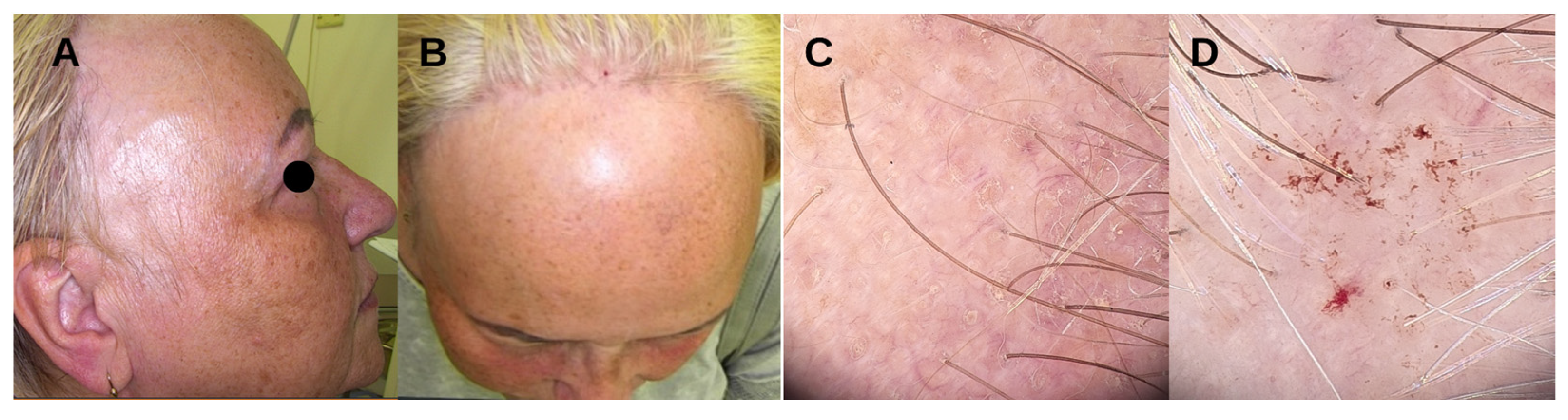
3. Clinical Features and Diagnosis

FFA is characterized by a gradual, usually symmetrical regression of the hairline in the frontal or fronto-temporal region [12,13,17,20]. In 10–25% of cases, the occipital hairline is also affected [4,21]. At the sites of the above-mentioned hairlines, single stems of terminal hair may be observed (the “lonely hair” sign) [22,23]. Total or partial loss of eyebrows occurs in 64–94% of cases, with the lateral parts being particularly affected. In 25% of cases, hair loss also occurs on the axillae, groin, or extremities [7,24,25,26].
Some patients, mainly men and premenopausal women, present facial papules (FP). These non-inflammatory, monomorphic lesions indicate involvement of the vellus follicular units and are similar to the follicular red dots of the glabella (FRD—follicular red dots) also observed in FFA patients [4,27,28,29]. Additional facial manifestations include erythema, pigmented patches, and frontal vein prominence or depression [27]. Inflammation-related symptoms including pruritus, burning sensation, or trichodynia are also reported [4].
Trichoscopy reveals perifollicular erythema, a marker of FFA activity, as well as perifollicular hyperkeratosis and areas with a lack of follicular units [30]. Mild cases often present yellow dots, corresponding to the follicles having regrowth potential, and lack vellus follicular units. Advanced stages are characterized by black dots, broken or twisted hairs, branched blood vessels, and white patches [17]. Long-term use of topical corticosteroids may lead to the appearance of arborizing blood vessels and extravasated hemorrhages [31] (Figure 1). Eyebrow dermatoscopy reveals a “road sign image”, with hairs growing in different directions, together with black, yellow, and gray dots, diffuse erythema, and loss of follicular units. Perifollicular erythema and hyperkeratosis are usually not present [32]. Histological examination reveals abundant peri- and infundibular lymphocytic infiltration, keratinization in the infundibulum and hair follicle orifices, as well as apoptosis and vacuolar degeneration of basal layer keratinocytes. Scalp biopsy remains the gold standard for diagnosing FFA in equivocal cases, including men and premenopausal women [33,34]. However, diagnosis is mostly based on clinical examination. The diagnostic criteria proposed by Tolkachjov and Vañó-Galván et al. (Table 1) may aid in identifying the condition [35,36].

4. Disease Severity

4.1. Five-Point Severity Scale

The severity of FFA can be assessed using a five-point scale proposed by Vañó-Galván et al. (Table 2). The cicatricial area largest measure is defined as the largest measure [cm] of cicatricial skin produced by the recession of the frontal and temporal hairline [4]. Grades I and II indicate mild cases, and grades III, IV, and V severe FFA [4,6].
Additionally, the International FFA Cooperative Group (IFFACG) have proposed the following principles to provide a standardized approach for evaluating hairline recession. The degree of frontal hairline recession is determined by the distance from the superior edge of forehead wrinkling to the area of maximum hair density in the central frontal scalp, recorded in centimeters with one decimal point precision. For the temporal recession, the same procedure is applied bilaterally, measuring the distance from the superior edge of forehead wrinkling to the area of maximum hair density in each temporal area. For those having had previous botulinum injections, the end of photodamaged skin substitutes for the superior edge of forehead wrinkling [37].

4.2. FFASI and FASS

Disease assessment can also be performed using the Frontal Fibrosing Alopecia Severity Index (FFASI) and the Frontal Fibrosing Alopecia Severity Scale (FFASS). However, both have limitations, and their use remains controversial.
In addition to the features typical of FFA, the FFASI also takes into account characteristics such as nail, oral mucosa, or genital involvement. They are typical for lichen planus, which some authors consider a distinct entity from FFA. Moreover, this scale is complex, encompassing two sections, thereby demanding considerable time for routine clinical implementation [38].
The FFASS (Table 3) lacks trichoscopic evaluation and was developed based solely on data from one center, with patients exclusively being Caucasian women. Some authors have indicated that due to the potential linkage of FFA with LPP or traction alopecia among African and Asian individuals, the use of the FFASS in these populations may confuse the clinical assessment and potentially overestimate FFA [39,40].
As a result of these limitations, these scales are not typically applicable in selecting the appropriate form of the treatment and are not widely used in clinical practice; nevertheless, the FASS may be utilized for evaluating the efficacy of the treatment and may serve as an indicator for clinicians while considering treatment modifications.

4.3. Frontal Fibrosing Alopecia Global Staging Score and ALODEX-FFA

In response to the limitations posed by the previous scales, the IFFACG proposed the Frontal Fibrosing Alopecia Global Staging Score (FFAGSS) implemented in Table 4, as well as the Alopecia Density and Extent Score for FFA (ALODEX-FFA). The FFAGSS includes an assessment of the five common FFA characteristics: scalp hair loss (S), eyebrow loss (E), facial papules (P), prominent forehead veins (V), and hyperpigmentation (H). As such, the staging for a particular patient is as follows: S (0–4), E (0–2), P (0–1), V (0–1), H (0–1).
The ALODEX-FFA is determined by summing the hair density loss, scored from 1 to 5 (0 = 0–49%, 1 = 50–74%, 2 = 75–89%, 3 = 90–99%, 4 = 100%), in each of the 1% areas and dividing by 100. Additionally, the authors also propose assessing the severity of perifollicular hyperkeratosis and perifollicular erythema using the SALT 3 criterion: 0 = none, 1 = mild, 3 = severe [37].

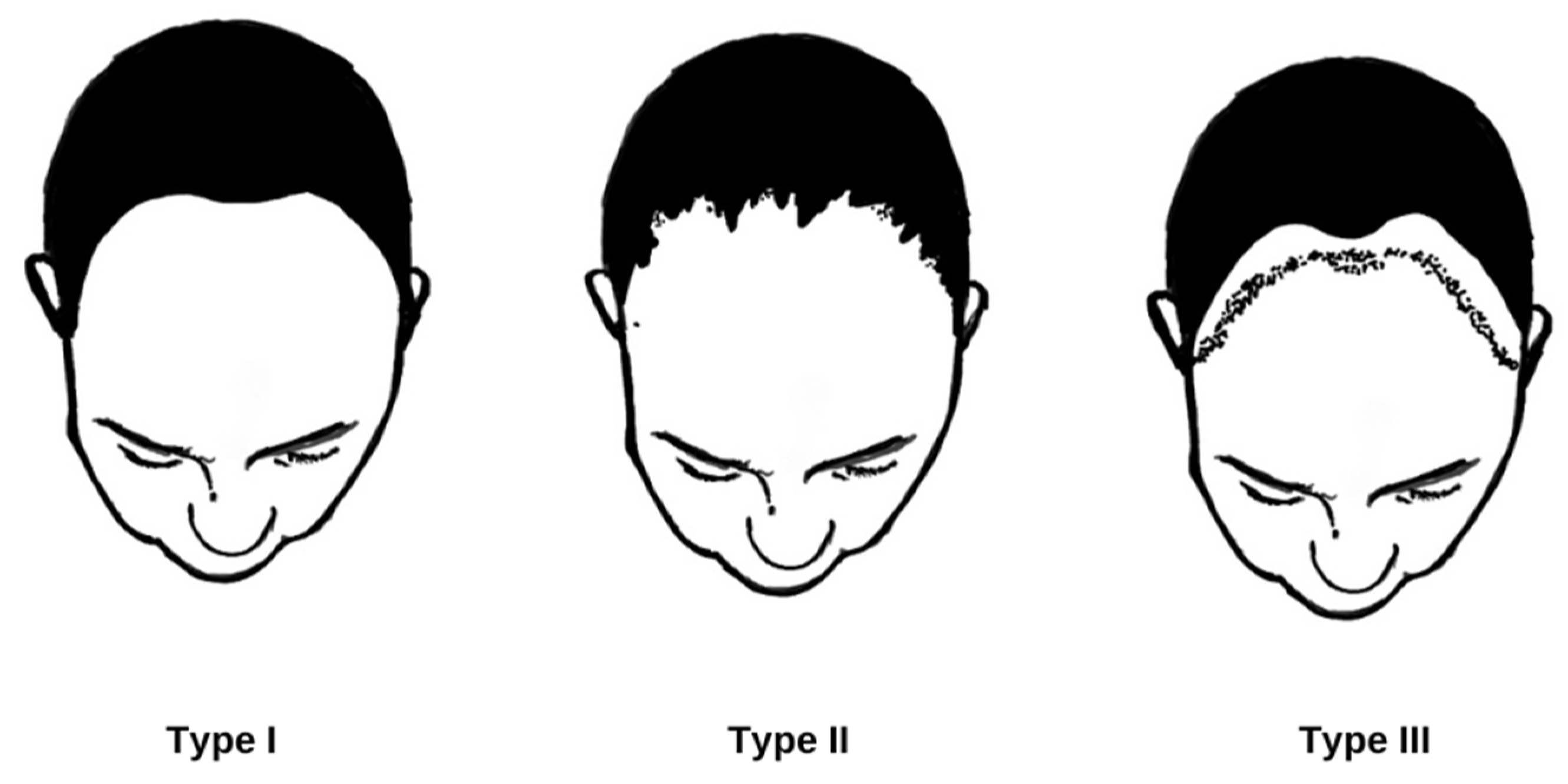
5. Clinical Course and Prognostic Factors
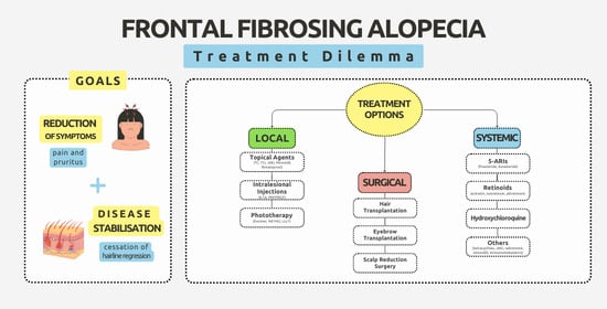
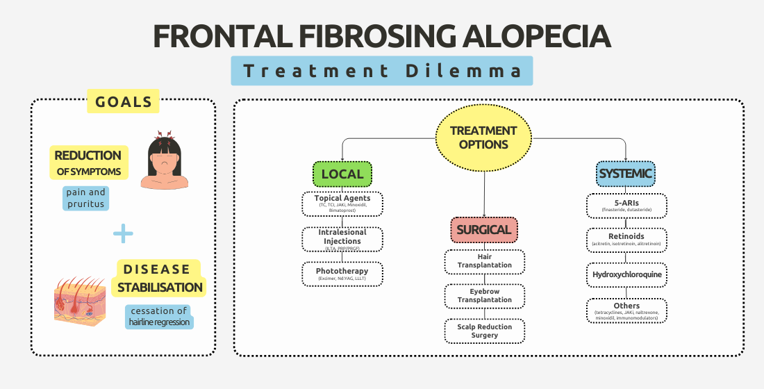
FFA is a chronic disorder with an unpredictable course [41]. It can begin and progress insidiously, remain stable for a long time, progress rapidly, or spontaneously stabilize after several years of progression [4,5,7]. Without treatment, gradual hairline regression at a rate of 0.2 to 2.1 cm per year is observed [4]. It is believed that the hair loss resulting from FFA is irreversible [40]; however, hair regrowth has been reported in affected areas in some cases [4,42]. Older age at onset [42], higher BMI [13], peripheral hair loss, eyelash loss, FP [4], or coexistence of rosacea are associated with a severe disease course [43], while younger age at onset [26] corresponds to a milder course. Regarding the clinical types of FFA (Figure 2), the best prognosis is associated with Type III and the worst with Type II. Type I, the most common, has an intermediate prognosis [41]. Additionally, the presence of follicular fluorescence on ultraviolet-enhanced trichoscopy (UVET), known as the “starry sky sign”, may indicate a potential for hair regrowth [44].

6. Treatment

The main aims of treatment are to ameliorate symptoms and to suppress disease progression [7,24,45]. A positive response is manifested as stabilization, i.e., cessation of hairline regression, or improvement, i.e., hair regrowth; a negative response is considered as progressive hairline recession [4]. Some authors define disease stabilization as the absence of inflammation on biopsy; however, biopsy is an invasive procedure and is not routinely performed [46,47].
FFA treatment commonly consists of combined therapy, involving topical and systemic treatments [45,48]. Selected therapeutic options are presented in Table 5.

6.1. Local Treatment

6.1.1. Anti-Inflammatory Agents

Corticosteroids/Calcineurin Inhibitors

The use of topical corticosteroids (TCs) such as clobetasol or betamethasone in FFA remains controversial [17,45,46,49]. Some studies suggest that while TC use may result in stabilization in the early stages, discontinuation can be associated with relapse [17], and the treatment can also result in scalp skin atrophy, telangiectasia, erythema, or folliculitis [42,46]. In comparison, topical calcineurin inhibitors (TCIs) appear to have higher efficacy [47]. Patients receiving tacrolimus (0.3%) were more likely to achieve stabilization within 3 months compared to those treated with TCs [26]. However, the use of TCs or TCIs as monotherapy is ineffective and results in remission in 1–3% of cases [45], and they should hence be considered only as part of combined therapy.

JAK Inhibitors (JAKis)

Plante et al. report that combined therapy with 2% tofacitinib resulted in clinical improvement after 3 months [50]. Ruxolitinib treatment (1.5% twice daily), in combination with systemic agents, led to a significant reduction in erythema and pruritus after 12–15 weeks in two patients and hair regrowth in another patient [51]. In both studies, the main treatment modification was based on a change in topical medication, while the groups of systemic drugs remained unchanged. This suggests that achieving stabilization in FFA refractory to previous treatments may simply require an alteration in topical therapy, without the need for changes in systemic medications.

Mechlorethamine

Mechlorethamine induces lymphocyte apoptosis and reduces the number of Langerhans cells in the epidermis, these being the main population of antigen-presenting cells in the hair follicle. A pilot study evaluating the use of mechlorethamine (0.016% gel) involved topical application of the drug for 24 weeks in patients with FFA (n = 7) and LPP (n = 5). Partial and complete responses to treatment were achieved in 73% and 18% of patients, respectively; however, the efficacy was assessed using the LPPAI scale (the main tool for assessing LPP activity), the FFA patients were not analyzed as a separate group, and all patients developed contact dermatitis. This may be related to inflammatory activity in LPP/FFA, increasing the risk of primary antigen immunization and the development of acute contact dermatitis.
Due to the limited effectiveness of topical therapies for FFA, the use of mechlorethamine should be implemented only in patients with severe, treatment-resistant disease or as a short-term adjunct to topical treatment in patients receiving systemic therapy. Further research on its use is needed, with a particular emphasis on reducing acute contact dermatitis and improving its effectiveness in disease stabilization [52].

6.1.2. Hair Growth Modulators

Bimatoprost

Bimatoprost is a synthetic analog of prostamide F2a, which induces the anagen phase in the hair follicle and leads to an increased number, thickness, and length of hair [53,54,55]. Bimatoprost (0.03% eye drops) applied twice daily to the eyebrow area leads to eyebrow regrowth within 6–9 months without causing adverse effects [54,55].

Minoxidil

Topical application of minoxidil (5% solution) was found to have a positive response in up to 72% of cases when used as part of combined therapy with intralesional triamcinolone acetonide (ILTA) injections or systemic agents [45,56]; as such, the drug may be an effective complement for combined treatment of FFA [56]. However, treatment can result in facial hypertrichosis, contact dermatitis, irritation, or exacerbation of seborrheic dermatitis [45].

6.1.3. Phototherapy

Neodymium YAG Laser (Nd:YAG Laser)

One study examined the effect of Nd:YAG laser therapy in treating FFA. The laser was applied to the facial and frontotemporal area three times at monthly intervals. Four out of five patients reported improvement in at least one symptom: four experienced reduced pruritus, and two reported reduced pain and burning. Additionally, perifollicular hyperkeratosis and erythema was reduced, and hair loss was stabilized. No significant adverse effects were noted in patients [57].

Excimer Laser

Compared to other types of phototherapy, excimer laser treatment offers lower UV exposure, shorter treatment duration, and the ability to target specific areas of the skin without damaging adjacent healthy tissue [58,59,60]. Navalini et al. examined the effect of excimer laser treatment twice a week on 13 patients with FFA or LPP. A reduction in erythema, pain, or pruritus was noted after a series of 6–16 treatments, as well as hair regrowth. Two patients achieved remission [61]. It should be emphasized that in this study, FFA was classified as a type of LPP, and systemic therapy was not standardized. Further observations are necessary to assess the use of the excimer laser in FFA.

Low-Level Light Therapy (LLLT)

Photobiomodulation, known as low-level light therapy, involves the use of low-power light sources that do not emit ions [62,63,64]. LLLT is believed to act by increased adenosine triphosphate (ATP) synthesis, oxidative stress modification, and transcription factor induction. It is hypothesized that LLLT affects the hair growth cycle by preventing premature catagen and increasing the proliferation rate of active anagen-phase hair follicles [65,66,67]. In a study conducted on 16 women, eyebrow hair regrowth was observed after 10 sessions conducted once a week. Improvement was greater in patients with partial eyebrow loss. Patients did not report side effects during or after the treatment [68].

6.1.4. Intralesional Treatment

Intralesional Triamcinolone Acetonide (ILTA) Injections

ILTA is an FDA-approved medication used for managing diverse inflammatory conditions, including skin disorders such as discoid lupus, lichen planus, or alopecia areata. This drug is consistently recommended in all hair loss treatment guidelines and for FFA, and ILTA is acknowledged as one of the most effective therapeutic options so far [17,19,45,47,56]. As part of combination therapy, ILTA results in a favorable outcome in up to 84% of cases [42]. Administration every 6–8 weeks can limit disease progression or lead to complete stabilization [46], while application every 3–6 months (at 20 mg/mL) can result in hair regrowth. This therapy is also effective for eyebrow loss, especially partial loss [17]. In daily practice, ILTA is used as a first-line treatment, mainly in combination with oral 5-alpha-reductase inhibitors (5-ARIs), hydroxychloroquine, retinoids, or tetracyclines [19,45,46]. Nevertheless, before consenting to ILTA, it is imperative to consider the potential risks, such as scalp stiffness, skin atrophy, and exacerbation of skin fibrosis in the advanced stages of FFA [45].

Platelet-Rich Plasma (PRP) and Plasma Rich in Growth Factors (PRGF)

PRP and PRGF contain growth factors that may positively influence hair growth and maintain the integrity of the hair follicle [69,70,71]. A retrospective study involving 118 patients showed that adding PRGF to the treatment regimen contributed to more frequent achievement of disease stabilization and reduction in inflammation, as well as hair regrowth [71]. In one reported case, the addition of PRP to systemic therapy for five treatments led to a reduction in erythema and perifollicular hyperkeratosis within 1 month [72].
Before implementing PRP or PRGF, it is important to consider that FFA could be associated with other autoimmune diseases, and it is recommended to check for the presence and specificity of antinuclear antibodies or autoimmune markers. Previous research suggests that FFA may be associated with SLE, polymyositis, RA, or autoimmune thyroid diseases.

6.2. Systemic Treatment

6.2.1. 5-ARIs

These medications are often used as a first-line treatment in combination with topical therapy or ILTA [19,45,56]. Their use was found to elicit a favorable treatment response in 69% of cases [45]. In one study involving 355 patients with FFA, finasteride treatment achieved a positive response in 100% of cases: 53% (54/102) achieved stabilization, and 47% (48/102) showed improvement. In addition, dutasteride was found to be effective for disease stabilization (10/18) and clinical improvement (8/18) [4]. Finasteride treatment is typically based on an initial dose of 2.5 mg/day, with an increase to 5 mg/day in case of no improvement within 6 months, while dutasteride is typically used at a dose of 0.5 mg/week—0.5 mg/day [4,73,74]. Although 5-ARIs are generally considered safe [75], they are category X drugs in pregnancy and may cause male fetus feminization. Therefore, finasteride is recommended in women due to its shorter half-life compared to dutasteride. Effective contraception is also recommended [45,75]. Although 5-ARIs are considered the most effective drugs for FFA [4,17,45,56], further research is necessary to clarify their therapeutic mechanism. Many authors suggest that the positive treatment response may result from the high efficacy of the drugs against androgenetic alopecia, which often coexists with FFA [76].

6.2.2. Retinoids

First-generation retinoids, such as isotretinoin (10–40 mg/day) and alitretinoin (30 mg/day), and second-generation ones, such as acitretin (20 mg/day), were effective in FFA [77,78,79,80]. In a study conducted by Rakowska et al., comparing the effectiveness of isotretinoin, acitretin, and finasteride, no disease progression was observed after 12 months of therapy in 76% of patients treated with isotretinoin, 73% of patients treated with acitretin, and 42% of patients treated with finasteride. Furthermore, the FFA did not progress for 24 months after therapy in 72% of patients treated with isotretinoin and 73% of patients treated with acitretin [80]. The use of oral vitamin A derivatives plays an important role in FFA cases presenting with FP. Isotretinoin added to the therapy at a dose of 10–40 mg/day reduces these lesions rapidly, in some cases within 2 weeks. Complete resolution usually occurs within 1–5 months. Retinoids are also effective in reducing pruritus, a common symptom in patients with FP [79,81].

6.2.3. Hydroxychloroquine

Hydroxychloroquine is an antimalarial drug often used as a first-line therapy in FFA. Positive outcomes have been noted in 63% of patients [45]. The usual dose is 200–400 mg/day. The effectiveness of this drug increases with long-term use [82]. A positive response is observed after 6–12 months [45,83,84]. Moreover, hydroxychloroquine and 5-ARIs appear to demonstrate comparable efficacy, as confirmed by a randomized study involving 36 patients [85].
However, the possibility of side effects of chronic hydroxychloroquine use, such as retinopathy, gastrointestinal disorders, myopathy, and headaches, should be considered. Hydroxychloroquine is safe in pregnancy, unlike other oral medications such as 5-ARIs or retinoids [45,83,86].

6.2.4. Tetracyclines

Tetracyclines are a group of antibiotics with documented anti-inflammatory properties. Studies indicate a positive response to therapy in 60% of cases. It is suggested that doxycycline at a dose of 100 mg/day may be effective in FFA patients with concurrent rosacea [19,45].

6.2.5. JAKis

Tofacitinib

Tofacitinib, an inhibitor of JAK1, JAK3, and, to a lesser extent, JAK2, was the initial JAKi developed for treating autoimmune diseases. It was the first JAKi to be used in the systemic treatment of FFA. Treatment at a dose of 10 mg/day, in combination with other systemic drugs, resulted in disease stabilization and hair regrowth in two patients with FFA after 6–9 months [83]. Plante et al. [50] report a case where a lower dose, i.e., 5 mg/day for 4 months, also resulted in a positive outcome.

Baricitinib

Oral use of baricitinib (3.4 mg/day, 4 mg/day, 6.8 mg/day), a JAKi targeting JAK1 and JAK2, has also achieved promising results, especially in FFA unresponsive to previous treatment methods, such as topical and oral tofacitinib [51].
However, systemic JAKi therapy can result in weight gain, nausea, and increased risk of infections, and these should be taken into account before starting. It is also recommended to perform a complete blood count; assess kidney and liver function parameters and lipid levels; and screen for hepatitis B and C viruses, tuberculosis, and HIV [87,88].

6.2.6. Oral Minoxidil

Oral monotherapy with minoxidil has shown efficacy in moderate eyebrow hair loss. A study of treatment in seven patients for 6 months (initial dose: 0.5–1.25 mg/day, increasing to 1.25–2.5 mg/day after 3 months) resulted in the complete regrowth of eyebrows in five patients and partial regrowth in two. No hypertrichosis was observed [89]. Oral minoxidil (1 mg/day) in combination with dutasteride (0.1 mg/day) also resulted in long-term disease stabilization [90].

6.2.7. Naltrexone

Low doses of naltrexone (1–5 mg/day) may exhibit anti-inflammatory properties by antagonizing toll-like receptor 4 [91,92,93,94]. Regarding FFA treatment, naltrexone contributed to a reduction in erythema and trichodynia when used at a dose of 3 mg/day as an additional component of therapy. However, no reduction in pruritus or perifollicular hyperkeratosis was observed. The side effects of treatment include vivid dreams, insomnia, headaches, thirst, and xerostomia, and these should be taken into consideration before treatment [95].

6.2.8. Pioglitazone

Pioglitazone is an agonist of the peroxisome proliferator-activated receptor gamma (PPAR-γ), used in the treatment of type 2 diabetes mellitus. Its effectiveness in FFA is controversial [96,97,98], and it is rarely used as part of combination therapy [45].

6.2.9. Other Anti-Inflammatory and Immunosuppressive Agents

Oral Corticosteroids

Studies indicate a positive response to oral prednisone (0.5–1 mg/kg/day). However, the discontinuation of such therapy leads to disease relapse [49].

Calcineurin Inhibitors

Some authors suggest the use of cyclosporine (3–5 mg/kg/day) as a first-line treatment, although the results of most studies are controversial. Others propose the use of cyclosporine or mycophenolate mofetil in cases of non-response to previous therapies or recurrent FFA [19,45,99].

Methotrexate

To date, four cases of methotrexate monotherapy have been reported in FFA patients. Stabilization of the disease process was observed in only two of them [26,47,100,101,102].

6.3. Surgical Treatment

6.3.1. Hair Transplantation (HT)

HT in FFA therapy remains controversial. Over half of patients experience a decrease in hair density or disease recurrence between 8 and 48 months post-surgery [103]. Assessing the effectiveness of this treatment is challenging due to the limited number of patients reported in the literature and differences in HT techniques. However, there is a consensus that HT in FFA patients should be considered only after disease stabilization lasting at least 1–2 years [56,104]. Furthermore, long-term patient observation post-transplantation is essential, with follow-up visits recommended every 4–6 months to detect early signs of FFA recurrence [56].

6.3.2. Scalp Reduction Surgery

Scalp reduction surgery prior to HT resulted in a satisfactory outcome for one FFA patient. After 2 years of disease stability, the patient underwent scalp reduction surgery followed by HT. Six years later, the patient remained stable, without signs of hairline recession or loss of the transplanted follicular units [105].

6.3.3. Eyebrow Transplantation

Eyebrow transplantation is a widely-accepted procedure resulting in positive long-term results in patients with eyebrow loss due to trauma or excessive plucking [106]. However, in FFA, the procedure remains controversial. Short-term and long-term outcomes are comparable with scalp HT for most patients. Hair follicles in the eyebrows are maintained during the first 12 months post-surgery but demonstrate progressive loss in subsequent years [107].

7. Summary

FFA treatment is challenging. Currently, the only FDA-approved drug for FFA treatment is triamcinolone acetonide, administered via intralesional injections. Clinical observations suggest that combined therapy is more effective than monotherapy [45]. Data from the literature suggest that ILTA, 5-ARIs, retinoids, and hydroxychloroquine are the most effective therapeutic options. First-line medications should be chosen individually, based on the drug safety profile, the FFA phenotype, and the presence of coexisting conditions [19,85].
As topical agents appear to be ineffective in monotherapy, they should be considered as a part of combined treatment. TCIs appear to have superior efficacy over TCs; therefore, the former should be prioritized as the primary option for topical therapy. Despite the promising potential of JAKis, their application has only been described in a few case series.
Intralesional therapies appear to be effective; however, treatment involves discomfort even with local anesthesia. In addition, they may not be as effective in the advanced stages of FFA due to potential exacerbation of scalp fibrosis.
Laser therapy and LLLT have demonstrated efficacy in clinical settings while maintaining a favorable side effect profile, and they thus merit particular attention. They may well represent a noteworthy adjunctive treatment option for patients with FFA.
When opting for systemic drugs, it is crucial to consider not only their efficacy but also the safety profile and the influence of any comorbidities associated with FFA. For example, 5-ARIs are recommended in the presence of concomitant androgenic alopecia; finasteride appears to be a better option than dutasteride within this drug class among female patients due to its shorter half-life. Moreover, patients with FP may benefit from oral retinoids, while patients with rosacea may benefit from tetracyclines. Low-dose naltrexone may be considered as an adjunctive therapy to reduce inflammatory symptoms.
While hair and eyebrow transplantation yields initial hair follicle maintenance, the treatment typically results in rejection. However, the effectiveness of scalp reduction surgery as an intervention to reduce therapeutic failure needs further investigation.

8. Further Perspectives

A crucial initial step towards approaching the frontal fibrosing alopecia treatment dilemma involves gaining a comprehensive understanding of the etiopathogenesis of FFA, which would facilitate identifying targets for potential drugs. This understanding would open the door to randomized clinical trials aimed at assessing the effectiveness and safety profiles of emerging treatment modalities.
Additionally, given the psychological distress that hair loss, which accompanies FFA, inflicts upon patients, investigations into potential hair regrowth and its correlation with disease severity, treatment modalities, and trichoscopy findings should be included. A positive response requires the early implementation of therapy, when the fibrosis has not yet affected all the follicular units; in this case, the stimulation and the reduced inflammation in the follicles may contribute to hair regrowth.

Author Contributions

Conceptualization, W.J.K. and A.W.; validation, A.W.; writing—original draft preparation, W.J.K.; wiring—review and editing, W.J.K. and A.W.; supervision, A.W. All authors have read and agreed to the published version of the manuscript.

Funding

This research received no external funding.

Institutional Review Board Statement

Not applicable.

Informed Consent Statement

Informed consent was obtained from all subjects involved in the study.

Data Availability Statement

Data sharing is not applicable to this review as no new data were generated.

Conflicts of Interest

The authors declare no conflicts of interest.

References

  1. Kossard, S. Postmenopausal frontal fibrosing alopecia. Scarring alopecia in a pattern distribution. Arch. Dermatol. 1994, 130, 770–774. [Google Scholar] [CrossRef]
  2. To, D.; Beecker, J. Frontal Fibrosing Alopecia: Update and Review of Challenges and Successes. J. Cutan. Med. Surg. 2018, 22, 182–189. [Google Scholar] [CrossRef] [PubMed]
  3. Rudnicka, L.; Rakowska, A. The increasing incidence of frontal fibrosing alopecia. In search of triggering factors. J. Eur. Acad. Dermatol. Venereol. 2017, 31, 1579–1580. [Google Scholar] [CrossRef] [PubMed]
  4. Vañó-Galván, S.; Molina-Ruiz, A.M.; Serrano-Falcón, C.; Arias-Santiago, S.; Rodrigues-Barata, A.R.; Garnacho-Saucedo, G.; Martorell-Calatayud, A.; Fernández-Crehuet, P.; Grimalt, R.; Aranegui, B.; et al. Frontal fibrosing alopecia: A multicenter review of 355 patients. J. Am. Acad. Dermatol. 2014, 70, 670–678. [Google Scholar] [CrossRef] [PubMed]
  5. Kossard, S.; Lee, M.S.; Wilkinson, B. Postmenopausal frontal fibrosing alopecia: A frontal variant of lichen planopilaris. J. Am. Acad. Dermatol. 1997, 36, 59–66. [Google Scholar] [CrossRef] [PubMed]
  6. Trager, M.H.; Lavian, J.; Lee, E.Y.; Wilkinson, B. Prevalence estimates for lichen planopilaris and frontal fibrosing alopecia in a New York City health care system. J. Am. Acad. Dermatol. 2021, 84, 1166–1169. [Google Scholar] [CrossRef]
  7. MacDonald, A.; Clark, C.; Holmes, S. Frontal fibrosing alopecia: A review of 60 cases. J. Am. Acad. Dermatol. 2012, 67, 955–961. [Google Scholar] [CrossRef]
  8. Porriño-Bustamante, M.L.; García-Lora, E.; Buendía-Eisman, A.; Arias-Santiago, S. Familial frontal fibrosing alopecia in two male families. Int. J. Dermatol. 2019, 58, e178–e180. [Google Scholar] [CrossRef] [PubMed]
  9. Alegre-Sánchez, A.; Saceda-Corralo, D.; Bernárdez, C.; Molina-Ruiz, A.; Arias-Santiago, S.; Vaño-Galvan, S. Frontal fibrosing alopecia in male patients: A report of 12 cases. J. Eur. Acad. Dermatol. Venereol. 2017, 31, e112–e114. [Google Scholar] [CrossRef] [PubMed]
  10. Tavakolpour, S.; Mahmoudi, H.; Abedini, R.; Hesari, K.K.; Kiani, A.; Daneshpazhooh, M. Frontal fibrosing alopecia: An update on the hypothesis of pathogenesis and treatment. Int. J. Womens Dermatol. 2019, 5, 116–123. [Google Scholar] [CrossRef]
  11. Vañó-Galván, S.; Saceda-Corralo, D.; Blume-Peytavi, U.; Cucchía, J.; Dlova, N.C.; Dias, M.F.R.G.; Grimalt, R.; Guzmán-Sánchez, D.; Harries, M.; Ho, A.; et al. Frequency of the Types of Alopecia at Twenty-Two Specialist Hair Clinics: A Multicenter Study. Skin Appendage Disord. 2019, 5, 309–315. [Google Scholar] [CrossRef] [PubMed]
  12. Tan, K.T.; Messenger, A.G. Frontal fibrosing alopecia: Clinical presentations and prognosis. Br. J. Dermatol. 2009, 160, 75–79. [Google Scholar] [CrossRef] [PubMed]
  13. Moreno-Arrones, O.M.; Saceda-Corralo, D.; Rodrigues-Barata, A.R.; Castellanos-Gonzalez, M.; Fernández-Pugnaire, M.A.; Grimalt, R.; Hermosa-Gelbard, A.; Bernardez, C.; Molina-Ruiz, A.M.; Ormaechea-Perez, N.; et al. Risk factors associated with frontal fibrosing alopecia: A multicentre case-control study. Clin. Exp. Dermatol. 2019, 44, 404–410. [Google Scholar] [CrossRef] [PubMed]
  14. Donati, A.; Lindgren, B.R.; Abreu, G.; Hordinsky, M. Prevalence of frontal fibrosing alopecia among Brazilian dermatologists: A cross-sectional survey. JAAD Int. 2020, 1, 148–150. [Google Scholar] [CrossRef] [PubMed]
  15. Ramos, P.M.; Anzai, A.; Duque-Estrada, B.; Farias, D.C.; Melo, D.F.; Mulinari-Brenner, F.; Pinto, G.M.; Abraham, L.S.; Santos, L.D.N.; Pirmez, R.; et al. Risk factors for frontal fibrosing alopecia: A case-control study in a multiracial population. J. Am. Acad. Dermatol. 2021, 84, 712–718. [Google Scholar] [CrossRef] [PubMed]
  16. Aldoori, N.; Dobson, K.; Holden, C.R.; McDonagh, A.J.; Harries, M.; Messenger, A.G. Frontal fibrosing alopecia: Possible association with leave-on facial skin care products and sunscreens; a questionnaire study. Br. J. Dermatol. 2016, 175, 762–767. [Google Scholar] [CrossRef] [PubMed]
  17. Porriño-Bustamante, M.L.; Fernández-Pugnaire, M.A.; Arias-Santiago, S. Frontal Fibrosing Alopecia: A Review. J. Clin. Med. 2021, 10, 1805. [Google Scholar] [CrossRef] [PubMed]
  18. Banka, N.; Mubki, T.; Bunagan, M.J.; McElwee, K.; Shapiro, J. Frontal fibrosing alopecia: A retrospective clinical review of 62 patients with treatment outcome and long-term follow-up. Int. J. Dermatol. 2014, 53, 1324–1330. [Google Scholar] [CrossRef] [PubMed]
  19. Rudnicka, L.; Kaczorowska, A.; Waśkiel-Burnat, A.; Rakowska, A.; Olszewska, M. Treatment of diseases associated with cicatricial alopecia. Dermatol. Rev./Przegląd Dermatol. 2022, 109, 32–42. [Google Scholar] [CrossRef]
  20. Ross, E.K.; Tan, E.; Shapiro, J. Update on primary cicatricial alopecias. J. Am. Acad. Dermatol. 2005, 53, 1–40. [Google Scholar] [CrossRef] [PubMed]
  21. Elloudi, S.; Gallouj, S.; Meziane, M.; Mernissi, F.Z.; Rimani, M. Alopécie frontale fibrosante: Étude prospective de 20 cas [Frontal fibrosing alopecia: A prospective study of 20 cases]. Ann. Dermatol. Venereol. 2017, 144, 409–414. [Google Scholar] [CrossRef] [PubMed]
  22. Rudnicka, L.; Olszewska, M.; Rakowska, A.; Slowinska, M. Trichoscopy update 2011. J. Dermatol. Case Rep. 2011, 5, 82–88. [Google Scholar] [CrossRef] [PubMed]
  23. Melo, D.F.; de Mattos Barreto, T.; de Souza Albernaz, E.; Haddad, N.C.; Tortelly, V.D. Ten clinical clues for the diagnosis of frontal fibrosing alopecia. Indian J. Dermatol. Venereol. Leprol. 2019, 85, 559–564. [Google Scholar] [PubMed]
  24. Imhof, R.L.; Villalpando, B.; Proffer, S.; Cantwell, H.; Tolkachjov, S.; Torgerson, R. Eyebrow hair loss: Review of causes and treatment options. Clin. Cosmet. Investig. Dermatol. 2020, 13, 935–940. [Google Scholar]
  25. Katoulis, A.C.; Damaskou, V.; Diamanti, K.; Pouliakis, A.; Mortaki, D.; Zacharatou, A.; Bozi, E.; Sgouros, D.; Panayiotides, I.G. Eyebrow involvement in frontal fibrosing alopecia: A clinicopathologic cohort study for the reversibility of hair loss. J. Am. Acad. Dermatol. 2020, 82, 755–757. [Google Scholar] [CrossRef] [PubMed]
  26. Strazzulla, L.C.; Avila, L.; Li, X.; Sicco, K.L.; Shapiro, J. Prognosis, treatment, and disease outcomes in frontal fibrosing alopecia: A retrospective review of 92 cases. J. Am. Acad. Dermatol. 2018, 78, 203–205. [Google Scholar] [CrossRef] [PubMed]
  27. López-Pestaña, A.; Tuneu, A.; Lobo, C.; Ormaechea, N.; Zubizarreta, J.; Vildosola, S.; Del Alcazar, E. Facial lesions in frontal fibrosing alopecia (FFA): Clinicopathological features in a series of 12 cases. J. Am. Acad. Dermatol. 2015, 73, e1–e987. [Google Scholar] [CrossRef]
  28. Ormaechea-Pérez, N.; López-Pestaña, A.; Zubizarreta-Salvador, J.; Jaka-Moreno, A.; Panes-Rodrigues, A.; Tuneu-Valls, A. Frontal Fibrosing Alopecia in Men: Presentations in 12 Cases and a Review of the Literature. Alopecia frontal fibrosante en el varón: Presentación de 12 casos y revisión de la literatura. Actas Dermosifiliogr. 2016, 107, 836–844. [Google Scholar] [CrossRef] [PubMed]
  29. Pirmez, R.; Donati, A.; Valente, N.S.; Sodré, C.; Tosti, A. Glabellar red dots in frontal fibrosing alopecia: A further clinical sign of vellus follicle involvement. Br. J. Dermatol. 2014, 170, 745–746. [Google Scholar] [CrossRef] [PubMed]
  30. Inui, S.; Nakajima, T.; Shono, F.; Itami, S. Dermoscopic findings in frontal fibrosing alopecia: Report of four cases. Int. J. Dermatol. 2008, 47, 796–799. [Google Scholar] [CrossRef] [PubMed]
  31. Saceda-Corralo, D.; Moreno-Arrones, Ó.M.; Fonda-Pascual, P.; Pindado-Ortega, C.; Hermosa-Gelbard, A.; Rodrigues-Barata, A.R.; Vañó-Galván, S. Steroid-induced changes noted on trichoscopy of patients with frontal fibrosing alopecia. J. Am. Acad. Dermatol. 2018, 79, 956–957. [Google Scholar] [CrossRef] [PubMed]
  32. Waśkiel-Burnat, A.; Rakowska, A.; Kurzeja, M.; Czuwara, J.; Sikora, M.; Olszewska, M.; Rudnicka, L. The value of dermoscopy in diagnosing eyebrow loss in patients with alopecia areata and frontal fibrosing alopecia. J. Eur. Acad. Dermatol. Venereol. 2019, 33, 213–219. [Google Scholar] [CrossRef] [PubMed]
  33. Stefanato, C.M. Histopathology of alopecia: A clinicopathological approach to diagnosis. Histopathology 2010, 56, 24–38. [Google Scholar] [CrossRef] [PubMed]
  34. Hurt, M.A.; Weedon, D. Weedon D. Weedon’s Skin Pathology. Dermatol. Pract. Concept 2012, 2, 79–82. [Google Scholar] [CrossRef]
  35. Tolkachjov, S.N.; Chaudhry, H.M.; Imhof, R.L.; Camilleri, M.J.; Torgerson, R.R. Reply to: “Updated diagnostic criteria for frontal fibrosing alopecia”. J. Am. Acad. Dermatol. 2018, 78, e23–e24. [Google Scholar] [CrossRef] [PubMed]
  36. Vañó-Galván, S.; Saceda-Corralo, D.; Moreno-Arrones, Ó.M.; Camacho-Martinez, F.M. Updated diagnostic criteria for frontal fibrosing alopecia. J. Am. Acad. Dermatol. 2018, 78, e21–e22. [Google Scholar] [CrossRef] [PubMed]
  37. Olsen, E.A.; Harries, M.; Tosti, A.; Bergfeld, W.; Blume-Peytavi, U.; Callender, V.; Chasapi, V.; Correia, O.; Cotsarelis, G.; Dhurat, R.; et al. Guidelines for clinical trials of frontal fibrosing alopecia: Consensus recommendations from the International FFA Cooperative Group (IFFACG). Br. J. Dermatol. 2021, 185, 1221–1231. [Google Scholar] [CrossRef] [PubMed]
  38. Holmes, S.; Ryan, T.; Young, D.; Harries, M. British Hair and Nail Society. Frontal Fibrosing Alopecia Severity Index (FFASI): A validated scoring system for assessing frontal fibrosing alopecia. Br. J. Dermatol. 2016, 175, 203–207. [Google Scholar] [CrossRef] [PubMed]
  39. Saceda-Corralo, D.; Moreno-Arrones, Ó.M.; Fonda-Pascual, P.; Pindado-Ortega, C.; Buendía-Castaño, D.; Alegre-Sánchez, A.; Segurado-Miravalles, G.; Rodrigues-Barata, A.R.; Jaén-Olasolo, P.; Vaño-Galván, S. Development and validation of the Frontal Fibrosing Alopecia Severity Score. J. Am. Acad. Dermatol. 2018, 78, 522–529. [Google Scholar] [CrossRef] [PubMed]
  40. Iorizzo, M.; Tosti, A. Frontal Fibrosing Alopecia: An Update on Pathogenesis, Diagnosis, and Treatment. Am. J. Clin. Dermatol. 2019, 20, 379–390. [Google Scholar] [CrossRef] [PubMed]
  41. Moreno-Arrones, O.M.; Saceda-Corralo, D.; Fonda-Pascual, P.; Rodrigues-Barata, A.; Buendia-Castaño, D.; Alegre-Sanchez, A.; Pindado-Ortega, C.; Molins, M.; Perosanz, D.; Segurado-Miravalles, G.; et al. Frontal fibrosing alopecia: Clinical and prognostic classification. J. Eur. Acad. Dermatol. Venereol. 2017, 31, 1739–1745. [Google Scholar] [CrossRef] [PubMed]
  42. Batra, P.; Sukhdeo, K.; Shapiro, J. Hair Loss in Lichen Planopilaris and Frontal Fibrosing Alopecia: Not Always Irreversible. Skin Appendage Disord. 2020, 6, 125–129. [Google Scholar] [CrossRef] [PubMed]
  43. Porriño-Bustamante, M.L.; Fernández-Pugnaire, M.A.; Arias-Santiago, S. A Cross-sectional Study of Rosacea and Risk Factors in Women with Frontal Fibrosing Alopecia. Acta Derm. Venereol. 2019, 99, 12. [Google Scholar] [CrossRef] [PubMed]
  44. Rodrigues-Barata, A.R.; Moreno-Arrones, O.M.; Corralo, D.S.; Galvan, S.V. The “Starry Night Sky Sign” Using Ultraviolet-Light-Enhanced Trichoscopy: A New Sign That May Predict Efficacy of Treatment in Frontal Fibrosing Alopecia. Int. J. Trichol. 2018, 10, 241–243. [Google Scholar] [CrossRef] [PubMed]
  45. Kępińska, K.; Jałowska, M.; Bowszyc-Dmochowska, M. Frontal Fibrosing Alopecia—A review and a practical guide for clinicians. Ann. Agric. Environ. Med. 2022, 29, 169–184. [Google Scholar] [CrossRef] [PubMed]
  46. Kerkemeyer, K.L.S.; Eisman, S.; Bhoyrul, B.; Pinczewski, J.; Sinclair, R.D. Frontal fibrosing alopecia. Clin. Dermatol. 2021, 39, 183–193. [Google Scholar] [CrossRef] [PubMed]
  47. Kelati, A.; Mernissi, F.Z. Frontal Hairline Recession: A Diagnostic Pitfall. Skin Appendage Disord. 2017, 3, 18–19. [Google Scholar] [CrossRef] [PubMed]
  48. Ho, A.; Shapiro, J. Medical therapy for frontal fibrosing alopecia: A review and clinical approach. J. Am. Acad. Dermatol. 2019, 81, 568–580. [Google Scholar] [CrossRef] [PubMed]
  49. Moreno-Ramírez, D.; Ferrándiz, L.; Camacho, F.M. Alopecia frontal fibrosante. Valoración diagnóstica y terapéutica [Diagnostic and therapeutic assessment of frontal fibrosing alopecia]. Actas Dermosifiliogr. 2007, 98, 594–602. [Google Scholar] [CrossRef] [PubMed]
  50. Plante, J.; Eason, C.; Snyder, A.; Elston, D. Tofacitinib in the treatment of lichen planopilaris: A retrospective review. J. Am. Acad. Dermatol. 2020, 83, 1487–1489. [Google Scholar] [CrossRef] [PubMed]
  51. Dunn, C.; Griffith, V.; Coican, A.; Dane, A.; Chow, W.; Aneja, S.; Nathoo, R.; Leavitt, A.; Hawkins, S.D. Janus kinase inhibition for the treatment of refractory frontal fibrosing alopecia: A case series and review of the literature. JAAD Case Rep. 2023, 40, 47–52. [Google Scholar] [CrossRef] [PubMed]
  52. Pincelli, T.P.; Heckman, M.G.; Cochuyt, J.J.; Sluzevich, J.C. Valchlor® in the Treatment of Lichen Planopilaris and Frontal Fibrosing Alopecia: A Single Arm, Open-label, Exploratory Study. Int. J. Trichol. 2020, 12, 220–226. [Google Scholar]
  53. Woodward, D.F.; Liang, Y.; Krauss, A.H. Prostamides (prostaglandin-ethanolamides) and their pharmacology. Br. J. Pharmacol. 2008, 153, 410–419. [Google Scholar] [CrossRef] [PubMed]
  54. Murad, A.; Bergfeld, W.F. Prostaglandin analogue for eyebrow loss in frontal fibrosing alopecia: A case report. J. Eur. Acad. Dermatol. Venereol. 2019, 33, e403–e405. [Google Scholar] [CrossRef] [PubMed]
  55. Murad, A.; Bergfeld, W. Prostaglandin analogue for treatment of eyebrow loss in frontal fibrosing alopecia: Three cases with different outcomes. J. Eur. Acad. Dermatol. Venereol. 2021, 35, e138–e140. [Google Scholar] [CrossRef] [PubMed]
  56. Beyzaee, A.M.; Goldust, M.; Patil, A.; Ghoreishi, B.; Ghahremanloo, T.; Rokni, G.R. Treatment of Frontal Fibrosing Alopecia. Dermatol. Ther. 2023, 3, 1–22. [Google Scholar] [CrossRef]
  57. Subash, J.; Eginli, A.; Bomar, L.; McMichael, A. Frontal fibrosing alopecia treatment with Nd:YAG (1064 nm) nonablative laser. Int. J. Womens Dermatol. 2020, 7, 355–356. [Google Scholar] [CrossRef] [PubMed]
  58. Spencer, J.M.; Hadi, S.M. The excimer lasers. J. Drugs Dermatol. 2004, 3, 522–525. [Google Scholar] [PubMed]
  59. Grema, H.; Raulin, C. Der Excimer-Laser in der Dermatologie und ästhetischen Medizin [The excimer laser in dermatology and esthetic medicine]. Hautarzt 2004, 55, 48–57. [Google Scholar] [CrossRef] [PubMed]
  60. Mehraban, S.; Feily, A. 308nm Excimer Laser in Dermatology. J. Lasers Med. Sci. 2014, 5, 8–12. [Google Scholar] [PubMed]
  61. Navarini, A.A.; Kolios, A.G.; Prinz-Vavricka, B.M.; Haug, S.; Trüeb, R.M. Low-dose excimer 308-nm laser for treatment of lichen planopilaris. Arch. Dermatol. 2011, 147, 1325–1326. [Google Scholar] [CrossRef] [PubMed]
  62. Anders, J.J.; Lanzafame, R.J.; Arany, P.R. Low-level light/laser therapy versus photobiomodulation therapy. Photomed. Laser Surg. 2015, 33, 183–184. [Google Scholar] [CrossRef] [PubMed]
  63. Hamblin, M.R. Mechanisms and applications of the anti-inflammatory effects of photobiomodulation. AIMS Biophys. 2017, 4, 337–361. [Google Scholar] [CrossRef] [PubMed]
  64. de Freitas, L.F.; Hamblin, M.R. Proposed Mechanisms of Photobiomodulation or Low-Level Light Therapy. IEEE J. Sel. Top. Quantum Electron. 2016, 22, 7000417. [Google Scholar] [CrossRef] [PubMed]
  65. Avci, P.; Gupta, G.K.; Clark, J.; Wikonkal, N.; Hamblin, M.R. Low-level laser (light) therapy (LLLT) for treatment of hair loss. Lasers Surg. Med. 2014, 46, 144–151. [Google Scholar] [CrossRef] [PubMed]
  66. Sorbellini, E.; Rucco, M.; Rinaldi, F. Photodynamic and photobiological effects of light-emitting diode (LED) therapy in dermatological disease: An update. Lasers Med. Sci. 2018, 33, 1431–1439. [Google Scholar] [CrossRef] [PubMed]
  67. Bartosińska, J.; Gerkowicz, A.; Niewiedzioł, M.; Szczepanik-Kułak, P.; Kwaśny, M.; Krasowska, D. Application of photodynamic therapy with the use of superluminescent light-emitting diode (sLED) lamp in actinic keratosis. Dermatol. Rev./Przegl. Dermatol. 2019, 106, 372–383. [Google Scholar] [CrossRef]
  68. Gerkowicz, A.; Bartosińska, J.; Wolska-Gawron, K.; Michalska-Jakubus, M.; Kwaśny, M.; Krasowska, D. Application of superluminescent diodes (sLED) in the treatment of scarring alopecia—A pilot study. Photodiagnosis Photodyn. Ther. 2019, 28, 195–200. [Google Scholar] [CrossRef] [PubMed]
  69. Giordano, S.; Romeo, M.; Lankinen, P. Platelet-rich plasma for androgenetic alopecia: Does it work? Evidence from meta analysis. J. Cosmet. Dermatol. 2017, 16, 374–381. [Google Scholar] [CrossRef] [PubMed]
  70. Dina, Y.; Aguh, C. Use of platelet-rich plasma in cicatricial alopecia. Dermatol. Surg. 2019, 45, 979–981. [Google Scholar] [CrossRef] [PubMed]
  71. García, A.; Navarro, M.R.; Ramirez, A.; Pino, A.; Navarro, A.; Moles, I.; Gallego, E.; Anitua, E. Plasma Rich in Growth Factors as an Adjuvant Treatment for the Management of Frontal Fibrosing Alopecia: A Retrospective Observational Clinical Study. J. Cutan. Med. Surg. 2023, 27, 340–349. [Google Scholar] [CrossRef] [PubMed]
  72. Özcan, D.; Tunçer Vural, A.; Özen, Ö. Platelet-rich plasma for treatment resistant frontal fibrosing alopecia: A case report. Dermatol. Ther. 2019, 32, e13072. [Google Scholar] [CrossRef] [PubMed]
  73. Danesh, M.; Murase, J.E. Increasing utility of finasteride for frontal fibrosing alopecia. J. Am. Acad. Dermatol. 2015, 72, e157. [Google Scholar] [CrossRef] [PubMed]
  74. Georgala, S.; Katoulis, A.C.; Befon, A.; Danopoulou, I.; Georgala, C. Treatment of postmenopausal frontal fibrosing alopecia with oral dutasteride. J. Am. Acad. Dermatol. 2009, 61, 157–158. [Google Scholar] [CrossRef] [PubMed]
  75. Rogers, N.E.; Avram, M.R. Medical treatments for male and female pattern hair loss. J. Am. Acad. Dermatol. 2008, 59, 547–568. [Google Scholar] [CrossRef] [PubMed]
  76. Kini, M.A.; Riaz, R.; Jolliffe, V. A Retrospective Analysis of Efficacy and Safety of Intralesional Triamcinolone Injections in the Treatment of Frontal Fibrosing Alopecia Either as Monotherapy or as a Concomitant Therapy. Int. J. Trichol. 2018, 10, 162–168. [Google Scholar]
  77. Pham, C.T.; Hosking, A.M.; Cox, S.; Mesinkovska, N.A. Therapeutic response of facial papules and inflammation in frontal fibrosing alopecia to low-dose oral isotretinoin. JAAD Case Rep. 2020, 6, 453–456. [Google Scholar] [CrossRef] [PubMed]
  78. Lee, J.Y.; Hong, J.S.; Lee, S.H.; Lee, A.Y. Successful treatment of frontal fibrosing alopecia with alitretinoin. Dermatol. Ther. 2019, 32, e13037. [Google Scholar] [CrossRef] [PubMed]
  79. Pirmez, R.; Duque-Estrada, B.; Barreto, T.; Quintella, D.C.; Cuzzi, T. Successful Treatment of Facial Papules in Frontal Fibrosing Alopecia with Oral Isotretinoin. Skin Appendage Disord. 2017, 3, 111–113. [Google Scholar] [CrossRef] [PubMed]
  80. Rakowska, A.; Gradzińska, A.; Olszewska, M.; Rudnicka, L. Efficacy of Isotretinoin and Acitretin in Treatment of Frontal Fibrosing Alopecia: Retrospective Analysis of 54 Cases. J. Drugs Dermatol. 2017, 16, 988–992. [Google Scholar] [PubMed]
  81. Terry-Flores, M.; Garcia-Arpa, M.; Franco-Muñóz, L.; González-Ruiz, L. Facial Response to Isotretinoin. Actas Dermo Sifiliogr. 2018, 109, 831–833. [Google Scholar]
  82. Zbiciak-Nylec, M.; Brzezińska-Wcisło, L.; Salwowska, N. The efficacy of antimalarial drugs in the therapy of selected forms of cicatricial alopecia. Adv. Dermatol. Allergol./Postępy Dermatol. I Alergol. 2021, 38, 302–309. [Google Scholar] [CrossRef] [PubMed]
  83. Gamret, A.C.; Potluri, V.S.; Krishnamurthy, K.; Fertig, R.M. Frontal fibrosing alopecia: Efficacy of treatment modalities. Int. J. Womens Health 2019, 11, 273–285. [Google Scholar] [CrossRef] [PubMed]
  84. Van Beek, M.J.; Piette, W.W. Antimalarials. Dermatol. Clin. 2001, 19, 147–160. [Google Scholar] [CrossRef] [PubMed]
  85. Saber, M.; Farokhshahi, M.; Fatemi Naeini, F.; Mohaghegh, F.; Asilian, A. Clinical effectiveness of finasteride versus hydroxychloroquine in the treatment of frontal fibrosing alopecia: A randomized controlled trial. J. Cosmet. Dermatol. 2024, 23, 576–584. [Google Scholar] [CrossRef] [PubMed]
  86. Khalil, S.; Bardawil, T.; Stephan, C.; Darwiche, N.; Abbas, O.; Kibbi, A.G.; Nemer, G.; Kurban, M. Retinoids: A journey from the molecular structures and mechanisms of action to clinical uses in dermatology and adverse effects. J. Dermatolog. Treat. 2017, 28, 684–696. [Google Scholar] [CrossRef] [PubMed]
  87. Yang, C.C.; Khanna, T.; Sallee, B.; Christiano, A.M.; Bordone, L.A. Tofacitinib for the treatment of lichen planopilaris: A case series. Dermatol. Ther. 2018, 31, e12656. [Google Scholar] [CrossRef] [PubMed]
  88. Damsky, W.; King, B.A. JAK inhibitors in dermatology: The promise of a new drug class. J. Am. Acad. Dermatol. 2017, 76, 736–744. [Google Scholar] [CrossRef] [PubMed]
  89. Pirmez, R.; Spagnol Abraham, L. Eyebrow regrowth in patients with frontal fibrosing alopecia treated with low-dose oral minoxidil. Skin Appendage Disord. 2021, 7, 112–114. [Google Scholar] [CrossRef] [PubMed]
  90. Cranwell, W.C.; Sinclair, R. Familial frontal fibrosing alopecia treated with dutasteride, minoxidil and artificial hair transplantation. Australas. J. Dermatol. 2017, 58, e94–e96. [Google Scholar] [CrossRef] [PubMed]
  91. Jaros, J.; Lio, P. Low dose naltrexone in dermatology. J. Drugs Dermatol. 2019, 18, 235–238. [Google Scholar] [PubMed]
  92. Lee, B.; Elston, D.M. The uses of naltrexone in dermatologic conditions. J. Am. Acad. Dermatol. 2019, 80, 1746–1752. [Google Scholar] [CrossRef] [PubMed]
  93. Ekelem, C.; Juhasz, M.; Khera, P.; Mesinkovska, N.A. Utility of naltrexone treatment for chronic inflammatory dermatologic conditions: A systematic review. JAMA Dermatol. 2019, 155, 229–236. [Google Scholar] [CrossRef]
  94. Sikora, M.; Rakowska, A.; Olszewska, M.; Rudnicka, L. The use of naltrexone in dermatology; current evidence and future directions. Curr. Drug Targets. 2019, 20, 1058–1067. [Google Scholar] [CrossRef] [PubMed]
  95. Hamel, R.K.; Chen, L.; O’Connell, C.; Mann, C. Oral Low-Dose Naltrexone in the Treatment of Frontal Fibrosing Alopecia and Lichen Planopilaris: An Uncontrolled Open-Label Prospective Study. Cureus 2023, 15, 34169. [Google Scholar]
  96. Spring, P.; Spanou, Z.; de Viragh, P.A. Lichen planopilaris treated by the peroxisome proliferator activated receptor-γ agonist pioglitazone: Lack of lasting improvement or cure in the majority of patients. J. Am. Acad. Dermatol. 2013, 69, 830–832. [Google Scholar] [CrossRef] [PubMed]
  97. Mesinkovska, N.A.; Tellez, A.; Dawes, D.; Piliang, M.; Bergfeld, W. The use of oral pioglitazone in the treatment of lichen planopilaris. J. Am. Acad. Dermatol. 2015, 72, 355–356. [Google Scholar] [CrossRef]
  98. Ladizinski, B.; Bazakas, A.; Selim, M.A.; Olsen, E.A. Frontal fibrosing alopecia: A retrospective review of 19 patients seen at Duke University. J. Am. Acad. Dermatol. 2013, 68, 749–755. [Google Scholar] [CrossRef] [PubMed]
  99. Ioannides, D.; Vakirlis, E.; Kemeny, L.; Marinovic, B.; Massone, C.; Murphy, R.; Nast, A.; Ronnevig, J.; Ruzicka, T.; Cooper, S.; et al. European S1 guidelines on the management of lichen planus: A cooperation of the European Dermatology Forum with the European Academy of Dermatology and Venereology. J. Eur. Acad. Dermatol. Venereol. 2020, 34, 1403–1414. [Google Scholar] [CrossRef] [PubMed]
  100. Zhang, M.; Zhang, L.; Rosman, I.S.; Mann, C.M. Frontal fibrosing alopecia demographics: A survey of 29 patients. Cutis 2019, 103, E16–E22. [Google Scholar] [PubMed]
  101. Suchonwanit, P.; Pakornphadungsit, K.; Leerunyakul, K.; Khunkhet, S.; Sriphojanart, T.; Rojhirunsakool, S. Frontal fibrosing alopecia in Asians: A retrospective clinical study. Int. J. Dermatol. 2020, 59, 184–190. [Google Scholar] [CrossRef] [PubMed]
  102. Morandi Stumpf, M.A.; do Rocio Valenga Baroni, E.; Schafranski, M.D. Frontal fibrosing alopecia: Successfully treated with methotrexate or just the natural disease progression? Acta Dermatovenerol. Croat. 2020, 28, 202–203. [Google Scholar]
  103. Lee, J.A.; Levy, D.A.; Patel, K.G.; Brennan, E.A.; Oyer, S.L. Hair Transplantation in Frontal Fibrosing Alopecia and Lichen Planopilaris: A Systematic Review. Laryngoscope 2020, 131, 59–66. [Google Scholar] [CrossRef] [PubMed]
  104. Unger, W.; Unger, R.; Wesley, C. The surgical treatment of cicatricial alopecia. Dermatol. Ther. 2008, 21, 295–311. [Google Scholar] [CrossRef] [PubMed]
  105. Arasu, A.; Meah, N.; Marzola, M.; Sinclair, R. Scalp reduction surgery does not reactivate frontal fibrosing alopecia: A case report. Dermatol. Ther. 2022, 35, e15451. [Google Scholar] [CrossRef] [PubMed]
  106. Klingbeil, K.D.; Fertig, R. Eyebrow and eyelash hair transplantation: A systematic review. J. Clin. Aesthet. Dermatol. 2018, 1, 21–30. [Google Scholar]
  107. Audickaite, A.; Alam, M.; Jimenez, F. Eyebrow Transplantation in Frontal Fibrosing Alopecia: Pitfalls of Short- and Long-Term Results. Dermatol. Surg. 2020, 46, 922–925. [Google Scholar] [CrossRef]
Figure 1. FFA; regression of fronto-temporal lines, pale skin in contrast with normal, photoaged skin, lateral eyebrow loss (A,B). Perifollicular hyperkeratosis, perifollicular erythema, and a lack of follicular units (C). Extravasated hemorrhages due to reduced erythema and perifollicular hyperkeratosis as a result of long-term topical corticosteroid use (D).
Jcm 13 02137 g001
Figure 2. Clinical patterns of FFA presentation. Type I, “linear pattern”: a band of uniform frontal hairline recession. Type II, “diffuse pattern”: a diffuse or zigzag band-like alopecia of the frontal hairline with loss of hair density behind the hairline. Type III, “pseudo fringe-sign pattern” (original authors’ sketch).
Jcm 13 02137 g002
Table 1. Diagnostic criteria proposed by Tolkachjov and Vañó-Galván [35,36]. Diagnosis requires two major criteria, or one major criterion and two minor criteria.
Major CriteriaMinor Criteria
  • Cicatricial alopecia of the frontal, temporal, or frontotemporal scalp and absence of follicular keratotic papules on the body.
  • Diffuse bilateral eyebrow hair loss.
  • Perifollicular erythema, perifollicular hyperkeratosis, or solitary hair shafts on examination in the frontal/frontotemporal area.
  • Histopathological features of cicatricial alopecia in the pattern of FFA/LPP on scalp biopsy.
  • Involvement of additional FFA sites: occipital area, facial hair, sideburns, body hair.
  • Facial papules.
  • Preceding/concurrent symptoms, such as pain or pruritus at sites of involvement.
FFA—frontal fibrosing alopecia. LPP—lichen planopilaris.
Table 2. Five grades of FFA severity based on largest measure of cicatricial area [4].
Grade of SeverityCicatricial Area Largest Measure [cm]
I<1
II1–2.99
III3–4.99
IV5–6.99
V≥7
Table 3. FFASS—Frontal Fibrosing Alopecia Severity Score.
Signs and SymptomsGradePunctuation
Clinical symptoms
Hairline recession [cm]
Frontal
  • >1
  • 1–2.99
  • 3–4.99
  • 5–6.99
  • >=7
grade × 2
Temporal leftgrade × 1
Temporal rightgrade × 1
Eyebrow lossNo0
Partial0.5
Total1
Alopecia scorethe sum of clinical symptoms punctuation
max. 21
Inflammation
Severity
Perifollicular hyperkeratosisNo0
Mild0.1
Severe0.2
Perifollicular erythemaNo0
Mild0.1
Severe0.2
Extent along the frontotemporal line [%]
Perifollicular hyperkeratosisNo0
<25%0.1
>25%0.2
Perifollicular erythemaNo0
<75%0.1
>75%0.2
Associated symptoms
Pruritus
SeverityNo0
Mild0.3
Severe0.6
FrequencyNo0
Occasional0.3
Daily0.6
Pain
SeverityNo0
Mild0.3
Severe0.6
FrequencyNo0
Occasional0.3
Daily0.6
Inflammation scorethe sum of inflammation and associated symptoms punctuation
max. 4
FFASSthe sum of alopecia score and inflammation score
max. 25
Table 4. Frontal Fibrosing Alopecia Global Staging Score [37].
SymptomGradePunctuation
Scalp hair lossNone0
Minimal (<1 cm)1
Mild (1 to <3 cm)2
Moderate (3 to <5 cm)3
Severe (≥5 cm)4
Eyebrow loss None0
Partial1
Total loss in at least one eyebrow2
Facial papulesNone0
Some1
Prominent forehead veinsNone0
Some1
Facial hyperpigmentationNone0
Some1
Table 5. Selected treatment options for FFA. ILTA—intralesional triamcinolone acetonide. PRP/PRGF—Platelet-Rich Plasma/Plasma Rich in Growth Factors.
Local TherapySystemic TherapySurgical Procedures
Topical agents5-alpha-reductase inhibitors
Dutasteride
Finasteride

Antimalarial agents
Hydroxychloroquine

Retinoids
Isotretinoin
Acitretin
Alitretinoin

Tetracyclines
Tetracycline
Doxycycline

JAK inhibitors
Tofacitinib
Baricitinib

Minoxidil
Pioglitazone
Naltrexone

Calcineurin inhibitors
Cyclosporine
Mycophenolate mofetil

Azathioprine

Oral corticosteroids
Hair transplantation
Scalp reduction surgery
Eyebrow transplantation
Corticosteroids
Betamethasone
Clobetasol

Calcineurin Inhibitors
Tacrolimus
Pimecrolimus

JAK inhibitors
Tofacitinib
Ruxolitinib

Mechlorethamine

Hair Growth Modulators
Minoxidil
Bimatoprost
Intralesional injections
ILTA *
PRP/PRGF
Phototherapy
Low-Level Light Therapy
Excimer Laser
Nd:YAG
* It is important to emphasize that the only drug registered by the Food and Drug Administration (FDA) for FFA treatment is triamcinolone acetonide, administered via intralesional injections, and, to date, no systemic drug has been approved.
Disclaimer/Publisher’s Note: The statements, opinions and data contained in all publications are solely those of the individual author(s) and contributor(s) and not of MDPI and/or the editor(s). MDPI and/or the editor(s) disclaim responsibility for any injury to people or property resulting from any ideas, methods, instructions or products referred to in the content.

