Review

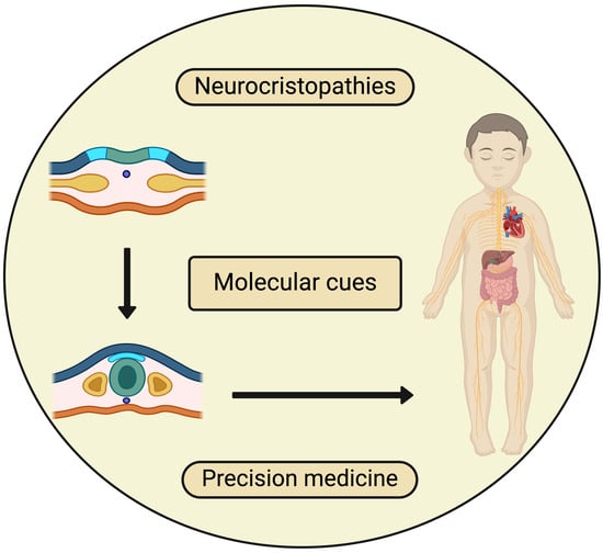
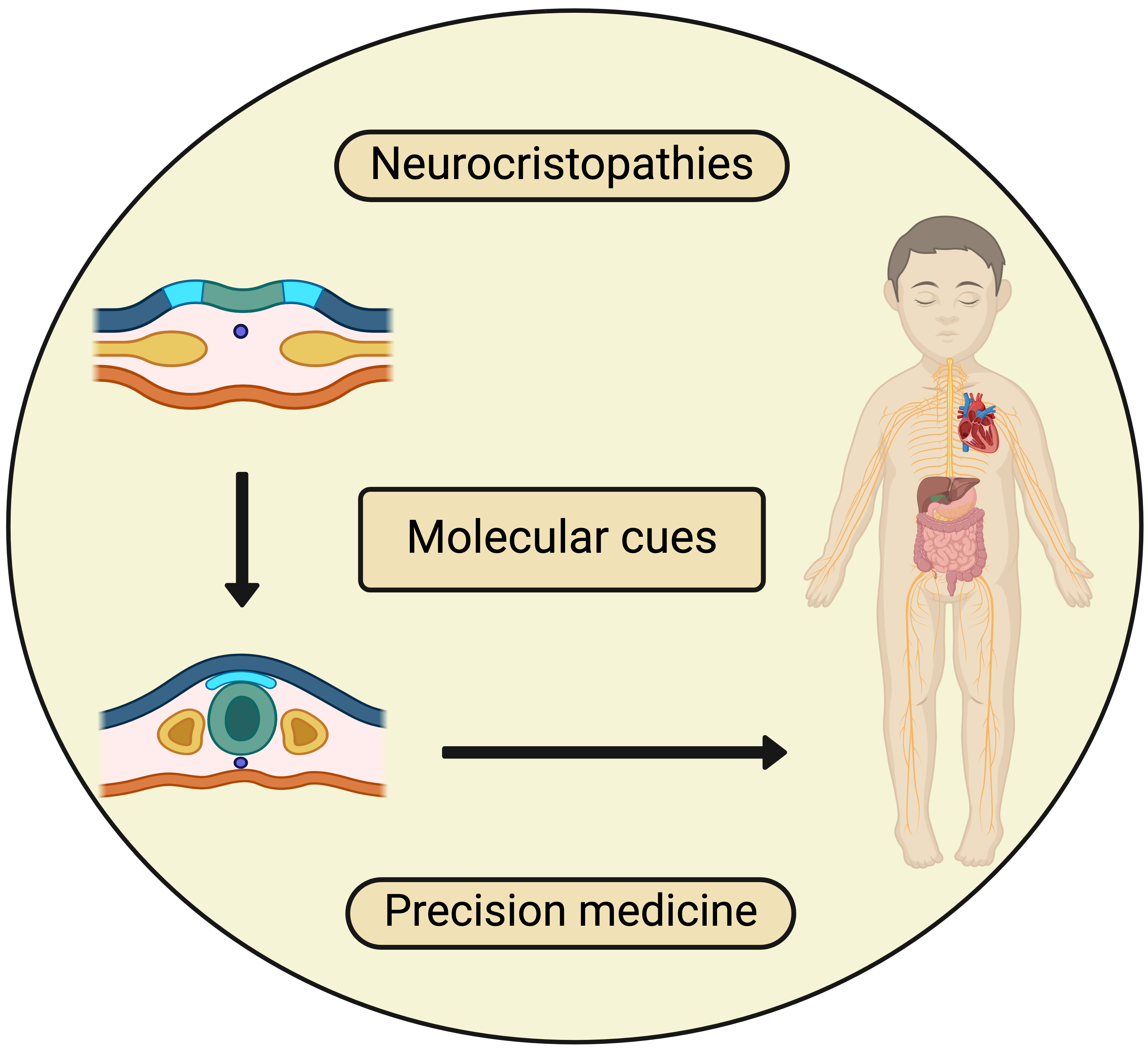
Clinical and Genetic Correlation in Neurocristopathies: Bridging a Precision Medicine Gap

by
Despoina Chatzi
,
Stella Aikaterini Kyriakoudi
,
Iasonas Dermitzakis
,
Maria Eleni Manthou
,
Soultana Meditskou
and
Paschalis Theotokis
*
Department of Histology-Embryology, School of Medicine, Aristotle University of Thessaloniki, 54124 Thessaloniki, Greece
*
Author to whom correspondence should be addressed.
These authors contributed equally to this work.
J. Clin. Med. 2024, 13(8), 2223; https://doi.org/10.3390/jcm13082223
Submission received: 27 February 2024 / Revised: 9 April 2024 / Accepted: 10 April 2024 / Published: 11 April 2024
(This article belongs to the Special Issue Review Special Issue Series: Current Advances in Clinical Neurology)

Abstract

:
Neurocristopathies (NCPs) encompass a spectrum of disorders arising from issues during the formation and migration of neural crest cells (NCCs). NCCs undergo epithelial–mesenchymal transition (EMT) and upon key developmental gene deregulation, fetuses and neonates are prone to exhibit diverse manifestations depending on the affected area. These conditions are generally rare and often have a genetic basis, with many following Mendelian inheritance patterns, thus making them perfect candidates for precision medicine. Examples include cranial NCPs, like Goldenhar syndrome and Axenfeld–Rieger syndrome; cardiac–vagal NCPs, such as DiGeorge syndrome; truncal NCPs, like congenital central hypoventilation syndrome and Waardenburg syndrome; and enteric NCPs, such as Hirschsprung disease. Additionally, NCCs’ migratory and differentiating nature makes their derivatives prone to tumors, with various cancer types categorized based on their NCC origin. Representative examples include schwannomas and pheochromocytomas. This review summarizes current knowledge of diseases arising from defects in NCCs’ specification and highlights the potential of precision medicine to remedy a clinical phenotype by targeting the genotype, particularly important given that those affected are primarily infants and young children.

Graphical Abstract

1. Introduction

In early embryonic development, the neural stage begins with the formation of the primitive neural plate, which subsequently folds to form the neural tube, the precursor to the central nervous system. Specific molecular cues and drivers strictly regulate each developmental process [1,2]. The neural crest arises on each side of the neural plate between the neural and non-neural ectoderm [3,4]. Comprising cephalic, cardiac, truncal, and enteric regions, the neural crest contributes to various tissues and systems, including head and neck structures, the heart outflow tract, pigment cells, peripheral nervous system, and enteric nervous system [5]. Before commencing migration, NCCs undergo epithelial–mesenchymal transition (EMT). During EMT, epithelial cells undergo structural and molecular changes, including the loss of junctions and polarity and cytoskeleton reorganization, ultimately enhancing cell motility and promoting the mesenchymal migratory capacity [6].
The broad spectrum of cells originating from the neural crest can result in various pathologies known as neurocristopathies (NCPs), impacting multiple systems [7]. Additionally, deregulated EMT plays a role in advancing tumor growth and facilitating metastasis [8]. Robert Bolande (1974) pioneered the conceptual framework for NCPs and its initial nomenclature [7]. Bolande grossly categorized NCPs into distinct, tumor-based classes and syndromes. However, NCPs do not conform strictly to such delineations, as evidenced by a significant intersection across developmental stages in tissue and organ formation, where these conditions manifest [9]. Research exploring the developmental signaling pathways governing NCC migration and differentiation has revealed pivotal molecular components that undergo dysregulation in NCPs.
Despite genetic screening being the primary method for detecting and diagnosing genetic disorders, the identification of causative genes remains limited, encompassing only a minority of patients. Therefore, there is a concerted effort to unveil additional genes implicated in typical development perturbations, with intensive investigations employing various animal models underway. Assessing the functionality of these candidate genes in animal models, which closely resemble humans, holds promise for promptly identifying key candidates, ensuring diagnostic accuracy, and facilitating timely intervention. In this review, initially, the pool of genes implicated before and after EMT are highlighted. Secondly, the NCPs listed here are categorized based on the origin of the problem within the neural crest, although there are numerous other disease processes that can involve multiple aspects of neural crest development. Lastly, NCP-related cancers are discussed.

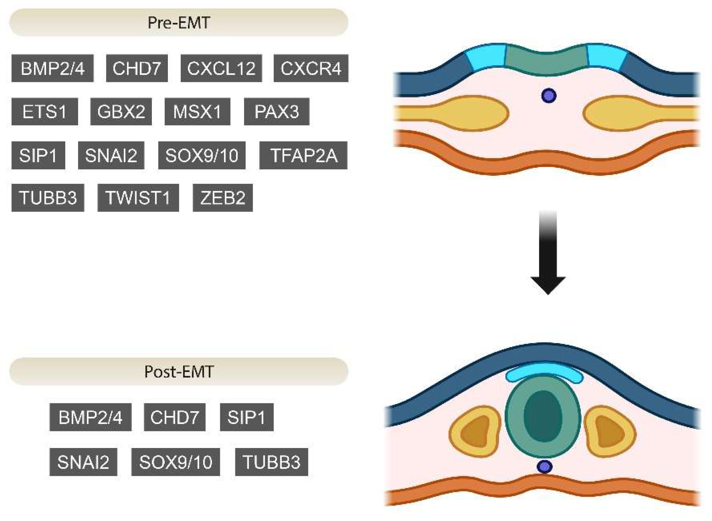
2. Genes Involved in EMT during Neural Crest Development

2.1. Pre-EMT Genes

The induction of the neural crest requires the combined distribution of BMP, FGFs, and WNT inhibitors. When NCCs are specified, they undergo changes in gene regulation that ultimately steer their differentiation into specific cell types. As mentioned earlier, signals from BMP, FGF, WNT, and retinoic acid play a crucial role in inducing neural crest identity [3,10]. Specifically, FGFs initiate this process through the activation of proneural genes in the neural ectoderm, while BMP and WNT signaling molecules are situated at the interference of neural and non-neural ectoderm and their role is to stimulate several transcription factors, commonly referred to as specifiers of the neural plate border. Signals derived from BMP and WNT molecules are predominant in lateral areas of the developing embryo, whereas their suppressors, such as Dkk and Cer [11], conquer the central regions, thus establishing a mediolateral gradient so that the cells destined to become the neural plate border emerge from an area exposed to moderate levels of WNT and BMP activity, finally forming the neural fold [12]. The action of such signaling molecules leads to the stimulation of neural plate border genes that include Msx1, Zic1, Tfap2, Pax3/7, Gbx2, Hairy2, Foxi1/2, Dlx5/6 and Gata2/3 [10,13]. Notably, certain border specifier genes such as Gbx2, Pax3, and Msx1, appear to be influenced by Wnt signaling. The activation of Pax3 might be indirect, as WNT signals also trigger the expression of Tfap2, which then directly binds to regulatory elements of Pax3 [14,15]. Research conducted across various vertebrates has demonstrated that once these transcription factors start being expressed in the neural plate border region, they initiate a sequence of reciprocal regulatory interactions. These interactions result in the consolidation of this regulatory condition and guarantee the sustained expression of these factors [3]. The relatively slow development rate of the lamprey embryo makes it a useful tool in the investigation and apprehension of interactions between these early expressed genes, which can coactivate each other. For example, in zebrafish embryos, tfap2 appears to play a crucial role in the activation of neural plate border specification and is essential for the activation of gata2/3 and foxi genes [16,17]. Ultimately, the demarcation line separating the neural plate from the neural plate border is refined through inhibitory exchanges involving transcription factors of both neural and non-neural origins.
Moving further from the neural plate border, the first genes found to be expressed in chicks’ neural crests are Ets1, FoxD3 and Snai1/2 [13]. During gastrulation, Pax7 is linked to groups of cells that eventually give rise to the neural crest. Although not only neural crest progenitors express this gene, Pax7 is present in the cells that will become the neural crest, so we can conclude that it might restrict them to this fate [18]. Additionally, along with Pax3 and Msx1, it is necessary for the activation of Ets1 [19]. The combined activity of Zic1 and Pax3/7 in chicks controls the expression of FoxD3 in the developing trunk, with WNT signaling playing a crucial role in the expression of Snai2 and FoxD3, while in frog embryos, this combination is involved in the specification of the neural crest [20]. In Xenopus, WNT collaborates with pax3 and zic1 to kickstart differentiation [3].
Apart from FoxD3, Sip1/Zeb2 and Sox9/10 are considered premigratory transcription factors, usually cooperating with Twist and Snail1/2 in order to decrease the expression of Cadherin-6B and other epithelial cadherins and upregulate the transcription of more mesenchymal cadherins. Along with other neural crest markers, TUBB3, which produces an element of microtubules, is also expressed in premigratory cranial NCCs [21]. Chd7, which belongs to the chromodomain helicase-binding family [22], is expressed in Xenopus and mice in premigratory NCCs [23] and is present in neural crest-derived tissues in mice [24].
The expression of key EMT transcriptional regulators, such as Snail2 or Twist1, is present. Wnt/β-catenin signaling has a direct impact on the expression of Snail2, which in turn directly inhibits the expression of E-cadherin and cadherin 6B. Additionally, this signaling pathway triggers the expression of mesenchymal cadherin 7 and cadherin 11 [25]. E-cadherin’s expression gradually decreases in the neural crest. In addition, Twist expression is under the control of the Hif signaling pathway, which also controls the expression of CXCR4, which encodes the receptor for the chemokine CXCL12/stromal cell-derived factor 1 [26].
The genes responsible for specifying neural crest development mutually enhance each other’s activity. For instance, in mice, Sox10 expression is enhanced by FoxD3, while foxD3, sox9, and twist are upregulated under the influence of snail2 in frog embryos. In this model, snail2 is both self-activated and activated by sox10 [3]. Pre-EMT genes implicated concurrently in NCPs and NCCs’ development during embryogenesis are presented in Figure 1.

2.2. Post-EMT Genes and Regulation
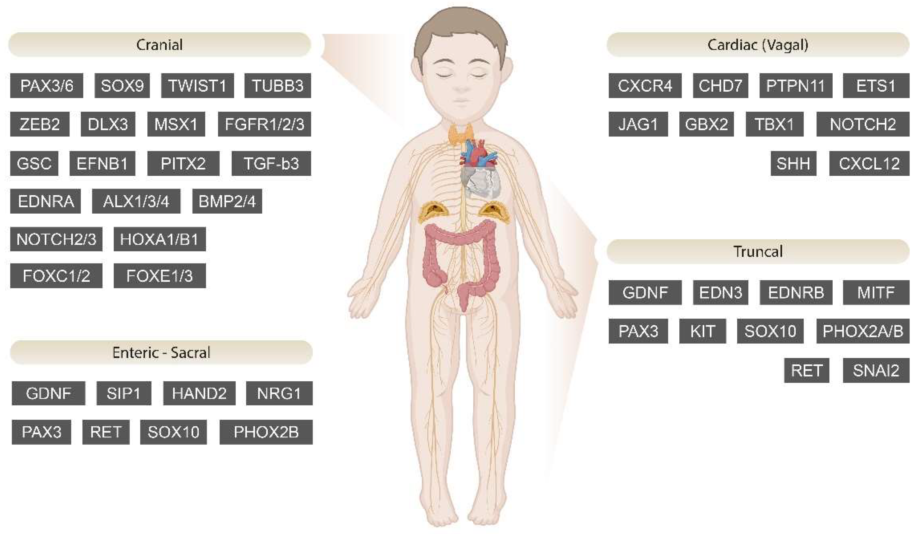
Research conducted across multiple species chickens, frogs, mice, and/or zebrafish), proves that EMT is partially regulated by the Slug/Snail family and Sip1, leading to changes in the cadherin expression of the cells in the mid-neurula stage. A prime example is Sox9, which together with Slug/Snail is enough to initiate the induction of EMT. Foxd3, Slug/Snail, and genes belonging to the SoxE family affect the levels of adhesion molecules that mediate cell-to-cell adhesion [26]. Research carried out in zebrafish provides knowledge of the downregulation of some genes, being related to the ability of NCCs to migrate. In this context, the Wnt target transcription factor Ovo1 inhibits genes related to the secretory pathway, such as Rab3c, Rab12, Rab11fip2, and Sec6, which all encode Rab GTPases [27]. TUBB3, which is also expressed before EMT, is present in early and late migrating NCCs, highlighting its connection to EMT and to the migration of NCCs [21]. Chd7, which belongs to the chromodomain helicase-binding family [22], is expressed in Xenopus and mice migratory NCCs [23] and is present later in neural crest-derived tissues in mice [24].
WNT signaling, also, plays a fundamental role in the delamination of NCCs. Canonical WNT signaling regulates the expression of cyclin D1 and facilitates the transition from the G1 phase to the S phase at the segmental plate mesoderm [28,29]. Additionally, BMP4 enhances the expression of its downstream gene Wnt1, leading to the phosphorylation of Sox9. This phosphorylation event, in turn, triggers an interaction with Snail2, promoting the delamination of NCCs in chick embryos. Furthermore, the BMP4/WNT pathway activates ADAM10, a protease protein, which cleaves the C-terminal fragment-1 of N-cadherin, which interacts with β-catenin [30]. This cleavage process produces CTF2, facilitated by γ-secretase, enhancing the transcriptional activity of β-catenin and cyclin D1. Consequently, this inhibits the G1/S transition, ultimately inducing the delamination of NCCs [31]. However, the connection between BMPs and WNT is more complex. In Xenopus and zebrafish, the ectoderm, from which the neural plate will develop, exhibits a BMP gradient. High BMP levels encourage ectoderm development, while low BMP levels support neural fate. Moderate BMP activity is thought to define the neural plate border and neural crest [32]. When it comes to Wnt, its levels were most elevated pre-EMT, and later, these levels significantly dropped after the NCCs delaminated. This might be in part due to activation of BMPs. There is reciprocal inhibitory communication between WNT and BMP signaling, where BMP hampers the proliferation induced by WNT, whereas WNT obstructs BMP-triggered neuronal differentiation [33].
Histone methylation is associated with the spatial and temporal regulation of gene expression in neural crest precursor cells [4]. The delay between the induction of NCCs during gastrulation and the initial activation of neural crest genes is linked to repressive trimethylation (me3) of lysine 9 (K9) on histone H3 (H3K9me3) in chick embryos [13,34]. This methylation occurs near the transcription start site of crucial neural crest transcription factors, Sox10 and Snail2. The demethylase KDM4A, which removes the trimethylation H3K9me3, enables the expression of Sox10 and Snail2 [35]. When the last two are expressed, their gene bodies are subjected to methylation (H3 lysine 36 (K36) me3 or H3K36me3 and H3K9me3, respectively) [35]. Nsd3 showed increased expression in the neural plate before the initiation of neural crest specifier gene expression, while earlier, Nsd3 mRNA was most prominent at the borders of the rostral neural plate. Later, its expression was heightened at the dorsal neural tube. As Nsd1 and Nsd2 were generally minimally expressed throughout the whole embryo, this points to the fact that Nsd3 is the main NSD methyltransferase in the neural crest. Nsd3 actively and directly regulates the expression of Sox10, and it secondarily alters the levels of Snail2, Sox9, or FoxD3 expression, but it does this in an indirect manner. Consequently, Nsd3 is crucial and necessary for Sox10 induction and NC migration [4]. Post-EMT genes are depicted in Figure 1.

3. NCPs

3.1. Cranial NCPs

3.1.1. Goldenhar Syndrome

Goldenhar syndrome, also known as hemifacial microsomia or oculo-auriculo-vertebral spectrum (OAVS), is a developmental disorder that in the majority of cases is unilateral. It has an impact on the first and the second pharyngeal arch, formed during the fourth week of gestation, possibly because of a deviation from normal angiogenesis [36] together with other external factors that influence vessels, such as drugs, medications and hormones [36,37,38]. Any other deviation from the normal differentiation and ventrolateral migration of the cranial NCCs in order to form the PAs is correlated with OAVS [39,40]. Its phenotype includes implications in the eyes and ears, craniofacial malformations, and various abnormalities in the cardiac, respiratory, gastrointestinal and central nervous system [41,42]. The etiopathogenesis of OAVS is based on a combination of genetic and environmental/developmental factors, although the exact mechanism has not been completely understood yet, with its multisystemic implications remaining without a known origin. As far as the genetic factors are concerned, the MSX gene family has been found to be partially responsible for this syndrome [42]. This gene family takes part both at the specification of neural crest borders and in craniofacial development. In frog embryos, msx1 is needed for the induction of other neural crest border genes including slug, snail and foxd3 [10], while in mice, Msx1 and Msx2 have active roles in the formation of craniofacial, limb, and neural tube features. Knockout mice for Msx1 display mandibular and maxillary underdevelopment, with the frontonasal region and the forebrain being smaller than the usual. Exencephaly was present in some mice, along with variations in the size and shape of optic cup, when the neural tube appeared with defects [43]. In humans, MSX1 mutations are responsible for a phenotype including total dental agenesis and cleft lip and palate [44]. Other characteristics that arise from the abnormal development of the first and second PAs are small ear and body size, a coloboma in the upper eyelid, and an epibulbar dermoid [45]. In some cases, other genes have been found to contribute to OAVS, such as MYT1 and SF3B2 [41,46]. Apart from the genetic factors and the process of angiogenesis that were mentioned before, external stimuli that contribute to the OAVS phenotype include mothers diagnosed with diabetes mellitus [38], celiac disease, and hypothyroidism [47].

3.1.2. Axenfeld–Rieger Syndrome (ARS)

ARS is a developmental autosomal dominant disorder [48] of the anterior segment, which mainly affects the craniofacial region and incorporates ocular malformation with posterior embryotoxon and underdevelopment of the iris, polycoria, which can be combined with corectopia. Abnormalities in other systems like the cardiac, dental, and myoskeletal systems can be observed. The pathophysiology of this syndrome is associated with disrupted neural crest migration in the early stages of embryogenesis. Two major genes that have been proved to be responsible for this syndrome are PITX2 and FOXC1 [49,50]. Both Pitx2 and Foxc1 in mouse embryos are transcription factors that are regulated by Tgfβ, a signaling molecule that aids the neural crest cells in their migration, guiding them towards the periocular mesenchyme, and controls ocular development [51]. Further data confirm the involvement of TGFβ (through a pathway that includes PITX2 and FOXC1) in the propriate formation of ocular structures in humans too, thus proving the contribution of these molecules to ocular abnormalities present in ARS [52]. FOXC1 is also active in premigratory or migrating cardiac neural crest cells as well as in tissues located near the pathways of migration, explaining in this way the nonocular multisystemic impacts of ARS [50]. Other genes that are responsible for some of the aspects of the syndrome in a small amount of individuals include CYP1B1, PRDM5, JAG1, USP9X, CDK13, HCCS, AMELX and BCOR [50,53].

3.1.3. Craniosynostosis

Syndromic craniosynostosis is an autosomal dominant condition characterized by the premature closure of one or more cranial sutures. Its pathogenesis has its roots in the development of the cranial vault during embryogenesis, and it can be divided into six different subtypes, all of which affect the coronary sutures (Saethre–Chotzen syndrome also affects the frontal suture). All of them are the result of mutations in gene families that affect the EMT process and migration of NCCs. In Pfeiffer syndrome, the skull has a cloverleaf shape, accompanied with intense hypoplasia of the midface and proptosis. The genes that are involved in this syndrome are FGFR1 and FGFR2. Generally, in the pre-EMT stage of embryonic development, FGF/FGFR interaction is crucial for the induction of the neural crest and the activation of proneural genes in the neural ectoderm [3,54]. Especially, the FGFR2 receptor is present in large amounts in the cartilage of the cranial base and is responsible for the proper induction, terminal differentiation, and apoptosis of the precursors of osteocytes, meaning that the early differentiation of cells that express those receptors can cause the early closure of cranial sutures. This syndrome is also associated with other implications including intellectual disability and conductive loss of hearing. Other subtypes of craniosynostosis are affected by FGFR genes in a similar way. Namely, Apert syndrome is caused by a mutation in FGFR2 and includes phenotypic characteristics like cleft palate, hearing loss, and hypertelorism, while Crouzon syndrome is a result of FGFR2 and FGFR3 mutation, presenting with mandibular prognathism, tarsal bones’ fusion, and hearing loss. Moreover, Muenke syndrome is a subtype of craniosynostosis induced by FGFR3 mutation and characterized additionally by macrocephaly, carpal and tarsal bones’ fusion, and hypertelorism [55]. Another gene of embryonic origin that participates in this family of craniofacial abnormalities is TWIST. Apart from its role in the pre-EMT phase of neural crest development, when it cooperates with Sip1/Zeb2, Sox9/10 and Snai1/2 in order to decrease the expression of Cadherin-6B and other epithelial cadherins and upregulate the transcription of more mesenchymal cadherins in vertebrate embryos [56], in humans, it is a negative regulator of the FGF/FGFR expression, thus determining the extent and the time of osteocytic differentiation. Mutations in TWIST1 are indicative of Saethre–Chotzen syndrome, having an phenotype that is analogous to Muenke syndrome in addition to small ear size, a lower hairline, and syndactyly [55]. Craniofrontonasal syndrome is the last category of craniosynostosis, and it is characteristic of mutations in EFNB1. The normal allele takes part in the process of bone formation. Specifically, EFNB1 is expressed by osteoblast progenitors that arise from NCCs and is responsible for increasing the thickness of the bones through upregulating the transcription of SP7, a key gene in bone formation. In avian embryos, the distinction between neural and non-neural crest cells and the kickstart of migration is controlled by ephrin-B1 and ephrin-B2. Particularly, crest cells that migrate express ephrin-B2, while, on the other hand, the formation of borders that indicate the migration pathways consist of non-neural crest cells that express ephrin-B1 [57]. In humans, the equivalent receptor ligand, EFNB1, participates in neural crest adhesion and migration routes as well as in normal bone formation. Its mutations are responsible for the disruption of proper osteogenesis, leading to the disrupted development of long bones, of osteoblast/osteoclast balance, and of the growth plate [58]. Thus, such mutations in EFNB1 can be linked to CFNS, characterized by a wide nasal bridge, brachycephaly, a nasal tip that is wide or divided, and even consequences for the bones and joints [55].

3.1.4. Craniofacial–Deafness–Hand Syndrome (CFDS)

CFDS is a rare disorder mainly affecting the craniofacial region of the developing embryo, with traits like intense hearing defects, malformed facial structures including hyperteloroism, small mouth size, missing or underdeveloped wrist and nasal bones, and hand abnormalities [53,59]. The gene responsible for this syndrome is PAX3, which plays a major role at the pre-EMT phase in many species. In Xenopus, pax3 in accordance with zic1 kickstarts the differentiation of the neural crest cells [3]. Mutation in PAX3 in humans can lead to various facial dysmorphisms along with symptoms from other systems, thereby causing CFDS [60].

3.1.5. Tricho-Dento-Osseous Syndrome (TDO)

TDO is an uncommon, penetrative, autosomal dominant disorder, with its basic phenotype enclosing abnormal formation of the hair, dental structures (taurodontism, enamel hypoplasia) and skull, featuring augmented bone density [61,62]. The gene that is involved in this syndrome is DLX3, a member of the DLX family that is vital for the determination of the dorsal–ventral axis and for the formation of the structures coming from Pas during embryogenesis. In mouse embryos, the domains where Dlx3, together with Dlx4, is active, are restricted to the farthest tip of the lower jaw structure, inhabiting the ventral region of the PAs. Their activity and their contribution to the formation of the lower jaw structure are stimulated or sustained by other members of the Dxl family, including Dlx5 and Dlx6, which are located in the first PA [63]. Findings regarding the contribution of DLX3 to human neural crest development and PA formation are similar to those obtained in rodent models. DLX3 starts its expression early in the first and second pharyngeal arches, from which odontoblasts and other craniofacial traits come. Its induction in later stages is crucial for the epithelium, placodes, limbs, hair, and the earliest developmental stage of teeth, contributing also to the formation of enamel [64]. Multiple mutations of this gene lead to abnormal odontoblast differentiation and enamel formation, taurodontism, and wavy hair immediately after birth [65]. These traits are necessary for the diagnosis of TDO, whose first clinical manifestations are usually found post birth and during the first year of life [66].

3.1.6. Peter’s Anomaly

Peter’s anomaly features a dysmorphic central cornea and malformed anterior eye segment that includes the corneal posterior stroma, endothelium, and Descemet’s membrane [53]. Peter’s anomaly can be divided into three subcategories: Peter’s anomaly type I, whose phenotype includes central corneal opacity with iridocorneal adhesions; Peter’s anomaly type II, which features central corneal opacity accompanied by cataracts or corneolenticular adhesions; and Peter’s-plus syndrome, which combines the syndrome with facial abnormalities such as cleft lip/palate and abnormal ear morphogenesis, heart defects, and intellectual disability and developmental delay [67]. The homeobox genes PAX6, PITX2, FOXE3, and FOXC1 are responsible for the emergence of the syndrome, as the dysmorphogenesis of the lens vesicle and its failure to separate from the surface ectoderm is attributed to their mutations [51,52,68]. PAX6 is one of the genes necessary to begin human eye formation and lens differentiation [39], and its homologous gene eyeless in Drosophila can alone induce eye morphogenesis [69]. Moreover, it has been observed in zebrafish embryos that two distinct neural crest streams enter the optic cup, with pax6 being necessary for their proper guidance. As pax6 is responsible for the appropriate guidance of these two neural crest streams, as well as for the expression of guidance molecules that aid this procedure, its loss can lead to serious anterior segment abnormalities [39]. Thus, PAX6 mutations can impede eye formation and contribute to the malformed anterior eye segment present in Peter’s anomaly. Another one of the genes connected to Peter’s anomaly, FoxE3, is placed downstream of Pax6 in mice studies [40]. In humans, FOXE3 is important for eye morphogenesis, with its expression being confined mainly to the lens, particularly in the anterior lens epithelium, albeit with some presence observed in the posterior region. Mutations in FOXE3 can result in various congenital eye abnormalities, leading to the phenotype of Peter’s anomaly [40]. Pitx2 and Foxc1 in mouse embryos are transcription factors and are both regulated by Tgfβ, a signaling molecule that aids the NCCs in their migration, guiding them towards the periocular mesenchyme and that controls ocular development, a fact that explains their involvement in ocular anomalies [51]. Certain mutations of CYP1B1 are also connected to the syndrome [70], while B3GLCT mutations are connected to Peter’s-plus syndrome, establishing it as a glycosylation disorder [71].

3.1.7. Bamforth–Lazarus Syndrome

The observable characteristics of this condition include abnormalities such as thyroid malformation, with resulting hypothyroidism; split palate; micrognathia; and coarse hair texture, sometimes accompanied by either choanal atresia or a divided epiglottis [72]. Patients with BLS exhibit various mutations in the FOXE1 gene, which produces a forkhead transcription factor [53]. In mice, Foxe1 is found in the endoderm of the foregut and in the ectoderm of the craniopharyngeal region crucial for palate development, and it is present in the early stages of thyroid formation. Human FOXE1 expression was observed in the epithelial lining of the oropharynx and in the thymus, but its onset was delayed compared to mice [73]. Hair defects in BLS patients are most likely connected to FOXE1, as it is involved in hair follicle morphogenesis downstream of the SHH/GLI pathway [74]. Split palate and micrognathia can be attributed to the disruption of facial chondrogenesis, which has been observed in zebrafish. In the latter, when foxe1 has been knocked down, shortening of the Meckel’s cartilage and inverted or shortened ceratohyal cartilages are exhibited [75]. When it comes to palate formation, Foxe1-null mice feature cleft palate and thyroid malformation, the same severe features as patients with the orresponding syndrome [76]. Furthermore, during embryogenesis, FOXE1 regulates MSX1 and TGF-b3, which take part in cranial development and are expressed in vertebrate embryos at epithelial–mesenchymal points of contact. Consequently, FOXE1 mutations and their indirect effect on MSX1 and TGF-b3 might explain some of the facial deformities in this syndrome. In mice, Msx1 is important for mandible, maxilla and tooth formation, while Tgf-b3 is crucial for the morphogenesis of neural crest-derived eye structures [73]. In chicken embryos, Msx1 is a pre-EMT neural plate border gene, which appears to be influenced by WNT signaling and is necessary for the activation of the Est1 gene [3]. Moreover, Msx1 is expressed in the distal incisal mesenchyme in mammals, something that indicates its role in tooth formation and incisors in particular [32]. Lastly, while Msx1 has not been found to be expressed in cardiac NCCs, it has been found active in NCCs arriving at the heart to create its outflow tract [77].

3.1.8. Branchio-Oculo-Facial Syndrome (BOFS)

BOFS is an autosomal dominant congenital disorder characterized by dysmorphogenesis of the first and second PA [53]. The most prominent characteristics are craniofacial abnormalities, which include supra-auricular sinuses, malformed auricles, external and middle ear anomalies, microphthalmia, lacrimal duct obstruction, abnormal philtrum, and pseudocleft of the upper lip, sometimes accompanied by intellectual disability and developmental delay [78]. This syndrome’s pathogenesis is connected to the TFAP2A gene, as mice lacking Tfap2a display atypical facial neural crest-derived structures [79]. Its importance in craniofacial development arises from its involvement in cranial closure and the morphogenesis of the facial prominences and the lens vesicle [80,81]. Tfap2a is expressed in both pre-migratory and migratory NCCs [82], while it has been found that TFAP2A is also present in migratory human neural crest [83]. In particular, mice lacking Tfap2a display atypical facial neural crest-derived structures [79]. In zebrafish, genes of the tfap2 family are neural plate border specifiers and play a crucial role in the activation of neural plate border specification [3], while zebrafish embryos injected with anti-tfap2 morpholinos display severely dysmorphic Pas, showing that tfap2a expression is vital for the proper development of craniofacial structures [84]. This gene family is also implicated in cardiac development, as gene mutations can cause patent ductus arteriosus [33].

3.1.9. Cerebral Autosomal Dominant Arteriopathy with Subcortical Infarcts and Leukoencephalopathy (CADASIL)

CADASIL is an autosomal dominant small vessel disease that affects the brain. It is an arteriopathy that causes subcortical infarcts, migraines, and restriction in blood supply to cerebral tissues; it may manifest as cognitive decline, psychiatric symptoms, and leukoencephalopathy due to the degeneration of vascular smooth muscle cells and pericytes [53,85]. This degeneration is mostly dependent on NOTCH3 mutations. While NOTCH3 is expressed in the nervous system of the embryo, in adult life, it is mainly expressed in vascular smooth muscle cells [86]. Due to NOTCH3 mutations, the extracellular domain of the Notch3 receptor, which contains EGF-like repeats, is accumulated between the smooth muscle cells of the brain’s blood vessels, leading to their degeneration and, consequently, causing restriction of blood flow [86,87]. Since the receptor protein produced by the NOTCH3 gene is present not just in VSMCs but also in pericytes, pericytes and the capillary vessels they support can also be affected by CADASIL [88]. The role of NOTCH3 during embryogenesis is mainly in VSMCs differentiation, with Notch3 being an important factor in proliferation and the impediment of VSMCs apoptosis [89,90]. Notch3-null mutant mice are characterized by loss of VSMCs, blood–brain barrier disruptions, and decreased vessel integrity in the central nervous system, characteristics found in patients with CADASIL [91]. During brain morphogenesis, NOTCH signaling is crucial for mural cell (also known as vSMCs and pericytes) differentiation from neural crest-derived stem cells. It has been shown, using hPSC-derived neural crest, that NOTCH signaling is a deciding factor in driving the terminal differentiation of mural cells in the face and the forebrain [92,93]. FOXC1 and PITX2 are two other genes that are implicated in small-vessel disease, with both of them being expressed in NCCs. FOXC1 it contributes to vascular stability, and its aberrant function contributes to cerebral vascular diseases like CADASIL. It also physically interacts with PITX2, which produces a transcription factor and regulates vascular smooth muscle cell proliferation. PITX2 mutations can lead to phenotypes with cerebral vascular dysfunction, similar to the CADASIL phenotypes [94]. Lastly, TIMP3 and vitronectin are two extracellular proteins that might play a role in CADASIL pathogenesis. Reducing TIMP3 and vitronectin levels in mice can prevent cerebral blood flow deficits and white matter lesions, leading to the conclusion that increased levels of TIMP3 and vitronectin contribute together with NOTCH3 mutations to CADASIL’s arteriopathy [95].

3.1.10. Congenital Aniridia

Congenital aniridia is an uncommon autosomal dominant eye condition characterized by the complete or partial absence of the iris. This disorder affects numerous eye components. Typically, there is underdevelopment of the fovea, which often leads to nystagmus and diminished vision. Aniridia-related complications such as glaucoma, keratopathy, and cataracts are severe and progressive, potentially exacerbating visual impairment. The underlying cause of aniridia is a mutation in the PAX6 gene, a deletion that leads to a premature stop codon [96]. PAX6 is a crucial gene in the proper development of the human eye, with experiments in vertebrates confirming its significance. Specifically, in zebrafish embryos, pax6 is necessary for the normal formation of the anterior segment of the eye. In the eye development, two distinct neural crest streams have been found to enter the optic cup; the first reaches the proximal region and is restricted there, while the second population migrates in the distal anterior segment. pax6 is responsible for the appropriate guidance of these two neural crest streams and for the expression of guidance molecules that aid this procedure, with its loss being the cause for serious anterior segment abnormalities leading to the congenital aniridia phenotype with underdeveloped fovea, corectopia, reduced visual sharpness, and nystagmus [97,98]. In humans, the phenotype of aniridia has also been linked to mutations in other genes like FOXC1, PITX2, FOXD3, TRIM44, ELP4, DCDC1, CYP1B1 [96,99,100]. PITX2 takes part in the proper development of structures of the anterior segment of the eye in embryogenesis as well, with its mutations being responsible for abnormalities in the development of the eyes, teeth, and umbilicus; at the same time, mutations in FOXC1 result in eye, heart, and hearing defects.

3.1.11. Frontonasal Dysplasia (FND)

Frontonasal dysplasia consists of a broad spectrum of craniofacial anomalies. It comprises various defects of the craniofacial midline, and its phenotype includes hypertelorism, a broad nasal root, a lack of nasal tip, facial clefts, anterior cranium bifidum, and widow’s peak [101,102]. There are three types of FND. FND1, which is characterized by a distinct concave shape of the nasal tip, is attributed to ALX3 [103]. FND2, which is characterized by craniosynostosis, cranium bifidum, and brain abnormalities, is a result of ALX4 mutations [104]. FND3, which is the most severe phenotype of frontonasal dysplasia, is caused by loss of the ALX1 gene, which results in complete failure of fusion of the frontonasal process and the maxillary arch. The Alx genes produce homeodomain-containing transcription factors and are expressed in broad overlapping domains in cranial NCCs. In mouse embryos, Alx1, Alx2, and Alx3 are expressed in NCCs in the frontonasal process [105], something that highlights their connection to frontonasal dysplasia, while in zebrafish, alx1 is present in migrating NCCs during the first stages of migration [106]. During craniofacial development, Alx3/4 are present in the first PA and contribute to the formation of the lower jaw under the regulation of Dlx5/6 [107]. Independently of Alx3, Alx4 is also expressed in the distal incisor mesenchyme and contributes to the development of incisor morphology [63]. Therefore, the Alx gene family is closely related to the morphogenesis of neural crest-derived tissues, and Alx mutations can lead to defects in the craniofacial midline, facial clefts, and lack of nasal tip. Additionally, mutations in Shh, Bmp4, and Fgf8 can be connected to FND [101]. The coordinated expression of genes such as Shh, Bmp4, and Fgf8 is pivotal in establishing the morphogenesis of the facial primordia. Disruption of this coordination can lead to frontonasal process defects and facial clefting, like in FND. Specifically, the expression domains of Fgf8 and Shh in the frontonasal ectoderm form a signaling center called the frontonasal ectodermal zone, which guides neural crest’s migration [108,109]. Moreover, Fgf8 and Bmp4 are crucial for the patterning of the mandible mesenchyme, whose formation and proliferation are disrupted in FND. Proximal mandible mesenchymal markers are stimulated by Fgf8, which is secreted by the proximal epithelium, while distal mesenchymal markers are induced by Bmp4, secreted by the distal epithelium [63]. Disrupting the complicated expression of Fgf8 and Bmp4 in chicken embryos can lead to lack of development in the midface, like in patients with FND that feature hypertelorism, a broad nasal root, and clefting [110]. EFNB1, which is expressed in cells surrounding the streams of cranial NCCs, might also be related to the pathogenesis of this dysplasia, as female patients with EFNB1 mutations exhibit a more severe phenotype [111]. Another relative gene is Kif3a, with mice lacking neural crest Kif3a expression featuring a wider frontonasal process and facial clefts, similar to the phenotype of human frontonasal dysplasia [112].

3.1.12. Hajdu–Cheney Syndrome

Hajdu–Cheney is a rare autosomal dominant disorder accompanied by arthrodentoosteodysplasia, which causes specific craniofacial defects, cardiovascular defects, osteoporosis, and kidney cysts [53]. Hajdu–Cheney is characterized by increased bone remodeling and decreased bone formation, something that explains skeletal defects related to low bone density. NOTCH2 gain-of-function mutations are linked to this syndrome [113]. NOTCH2 is generally implicated in cardiac neural crest development [114,115], as NOTCH signaling takes part in the differentiation of cardiac NCCs and outflow development. Disruption of NOTCH signaling is linked to cardiac dysmorphogenesis and therefore could be related to the cardiovascular defects in Hadju–Cheney [53]. After the migration of the cardiac NCCs, NOTCH receptors and ligands are expressed in the outflow tract and the developing arteries [116] and play a crucial role in the differentiation of smooth muscle cells [117]. NOTCH signaling partakes in skeletal morphogenesis, chondrogenesis and in osteoblast and osteoclast differentiation [118,119,120], which could impact craniofacial formation, leading to Hajdu–Cheney. Blocking NOTCH signaling or NOTCH2 leads to problematic enamel formation, while it also causes the death of dental epithelial stem cells in mouse incisors [121,122], leading to the dentodysplasia present in Hajdu–Cheney patients.

3.1.13. Moebius Syndrome (MBS)

MBS is a non-progressive neurological condition that stems from the underdevelopment of facial nerves and is accompanied by strabismus, hypertelorism, and partial facial paresis, which presents itself as mask-like facies and strained control of eye movements [123]. Mostly affected are the trigeminal, abducens, and facial nerves. Some patients also feature microstomia, cleft palate, lingual dysplasia, and epicanthic folds [124,125]. MBS1, MBS2, and MBS3 are the genetic loci most prominently linked to the syndrome, but the specific genes and their connection to the neural crest remains to be elucidated [126,127]. Moreover, mutations of another gene, TUBB3, which is expressed in premigratory NCCs and later present in neural crest-derived neurons [21], are linked to MBS [128]. A mouse study has connected this phenotype with elevated SHH signaling in combination with correlated decreased WNT signaling [129]. SHH levels are essential for proper neural crest cell differentiation, particularly in the development of the head and face. When Shh is absent in mutant mice and humans (SHH), it leads to conditions like holoprosencephaly and cyclopia, where the hemispheres fail to separate correctly [130]. Enhanced SHH signaling affects the proliferation and migration of NCCs and impedes the interactions between NCCs and the placodes and is correlated with disorganized trigeminal and facial nerves, which are affected in MBS [129]. In cranial morphogenesis, WNT signaling is important for the delamination of NCCs and later determines the sensory fate of neural progenitors [51]. Disruption, therefore, of WNT signaling renders nerve abnormalities like those in MBS probable. The implication of HOX family genes, which are key genes in the morphogenesis of the cranial neural crest, is also likely, with emphasis on HOXA1 and HOXB1 [127,131,132]. Mutations in PLXND1 and REV3L account for a percentage of MBS patients. Plxnd1-null mice exhibit hypoplasia of the neural fibers that connect areas of the brain as well as disruption of neuronal migration; meanwhile, in Rev3l-mutant mice, brain volume is decreased. Therefore, the clinical features of MBS patients are similar to those in mutant mice models [106]. Lastly, Moebius syndrome is connected to the genes GSH1, CDX2, CRBP1, PBX2, EGR2, and SOX14 [133,134,135].

3.1.14. Pierre Robin Sequence (PRS)

PRS is a rare congenital condition, including the clinical triad of micrognathia, cleft palate, and glossoptosis, which carries the risk of mechanical airway obstruction. It can be divided into two subtypes; the isolated, nonsyndromic and the syndromic, usually accompanied by genetic syndromes like Stickler syndrome, velocardiofacial syndrome, and Treacher Collins syndrome [136]. Among the cases that are reported, the genetic factor that is responsible for PRS seems to be a mutation in the SOX9 gene, which is important during embryogenesis for the formation of chondrocytes deriving from multipotent NCCs [137]. In mouse embryos, Sox9 is expressed in cranial NCCs, which are responsible for the proper formation of the cranial and facial skeleton [3]. Contribution of SOX9 in the differentiation of cranial NCCs into chondrocytes and in the formation of many craniofacial structures could explain the micrognathia and glossoptosis observed in PRS. An additional role of Sox9 is to positively regulate the expression of Col2a1, Col11a1, and Col11a2 in both mice and chicken (SOX9 COL2A1, COL11A1, and COL11A2, respectively) embryos in the process of chondrogenesis, with their mutations leading to PRS as well [138,139]. Another genetic locus found to play a significant role in PRS is BMP2. It is a member of the BMPs family, which is necessary for the proper formation of the palate. Specifically, in mouse embryos, Bmp2 is present in NCCs that give rise to chondroblasts and osteoblasts, giving them the ability to proliferate and differentiate into their terminal fate. As a consequence, in mouse mutants, a lower rate of cell proliferation results in a decrease in osteogenic and chondrogenic precursors arising from NCCs, contributing to the development of a smaller mandible. This, in turn, hinders the descent of the tongue and ultimately leads to the formation of a cleft palate. Additional mutated genes in mice that are considered to contribute to the cleft palate phenotype are Prdm16 and Tak1, found in the TGFβ signaling pathway [140,141], as well as Erk, a member of the BMP, TGFβ, FGF, and EGF pathways [142].

3.1.15. Mowat–Wilson Syndrome

Mowat–Wilson syndrome is a disorder characterized by abnormal facial traits including eye hypertelorism, prominent chin, a mouth-open expression, earlobes located higher than usual, and underdevelopment of the optic nerve [143,144]. It is also accompanied by congenital hearing loss, cardiovascular and genitourinary abnormalities, HSCR, and impaired intellectual sharpness [143]. It is caused by mutations in the gene ZEB2 [145,146], which is expressed in different domains during embryogenesis. In mice, it is important for the proper development of the neuroepithelium [147] the neural plate, and as a result the NCCs, cortex of the brain, and the mesoderm [148]. In humans, the ZEB2 gene plays a central role in coding for the Smad interaction protein 1 (SIP1), which is crucial for the appropriate formation of different components within the eye during development. In particular, SIP1 activates the TGF-β pathway and promotes the proliferation and differentiation of NCCs, assuring normal organogenesis. The importance of ZEB2 in the formation of the craniofacial domain, part of the nervous system and the mesenchyme, leads to the phenotype of this syndrome, with eye abnormalities affecting the formation of the lens, causing congenital cataract. Increased expression of these molecules in the eyeball, NCCs, genital glands, and musculoskeletal system comes in accordance with the eventual harm inflicted upon these cells, directly aligning with the range of abnormalities observed in MWS [144].

3.1.16. SAMS Disorder

SAMS disorder is an autosomal recessive syndrome that causes short stature, auditory canal atresia, mandibular hypoplasia, and skeletal abnormalities, including femoral, humeral and pelvic abnormalities [149]. Mutations in GSC, which encodes Goosecoid homeodomain transcription factor, are responsible for this phenotype [150]. gsc is required for mesodermal organization during Xenopus gastrulation [125], while later in mouse embryos, its expression is detected in the first pharyngeal arch, the mandible, the auditory canal, and the limb buds [151]. Goosecoid is involved in the patterning of the pharyngeal arches, thus placing SAMS in the category of NCPs [149]. This pattern of expression of GSC is in accordance with the mandibular, auditory and limb anomalies found in patients with SAMS disorder. Gsc-knockout mice exhibit craniofacial anomalies, limb defects and skeletal malformations in humeri and femora, also compatible with the phenotype of patients with SAMS disorder [152,153]. Gsc is also connected to EDNRA signaling, as its expression is absent after loss of EDNRA signaling [154], while in embryos where both Ednra1 and Ednra2 are depleted, there is a notable decrease in Gsc expression, specifically within the ventral regions of the mandibular and hyoid arches [155]. When Gsc is not expressed in ET-/- mice embryos, craniofacial abnormalities arise, connecting Gsc with SAMS disorder [154]. Therefore, apart from GSC, EDNRA signaling might also play a part in SAMS pathogenesis.

3.2. Cardiac (Vagal) NCPs

3.2.1. DiGeorge Syndrome

Several cardiocraniofacial defects are present in this syndrome, such as truncus arteriosus, tetralogy of Fallot, and double-outlet right ventricle; hypothyroidism, hypoparathyroidism, and a hypoplastic thymus are also present [117]. TBX1 is a gene whose product is T-Box transcription factor; it has a significant role in cardiac neural crest migration and final differentiation, connected to the pathophysiology of this syndrome [77]. During migration, while it is not significantly expressed by cardiac NCCs themselves, it is vastly present in the surrounding environment and in particular the pharyngeal ectoderm, endoderm and the second heart field; thus, it guides the cardiac NCCs to their final destination [117]. When TBX1 expression is decreased, due to mutations or deactivation, migration of cardiac NCCs is not concluded and their maturation is not finalized due to absent communication between the neural crest and the mesenchyme, which leads to decreased activation of the MAPK pathway and early expression of the BMP pathway [77]. In mutant Tbx1 mice, altered neural crest migration has been observed in the third, fourth, and sixth PAs [156]. Additionally, Tbx1 deletion has been proven to result in aorticopulmonary defects and incomplete morphogenesis of the aortic arch and the major arteries, due to decreased cardiac NCC migration in the cardiac field [77]. The indirect action of Tbx1 is manifested through its effect on the expression of Gbx2 in the pharyngeal ectoderm, which most likely interferes with the migration of cardiac NCCs by potentially reducing the activity of the SLIT/ROBO signaling pathway and perhaps downregulating the Slit2 ligand [157]. Apart from Gbx2, Tbx1 also affects Cxcr4 and Cxcl12, which encode the receptor for the chemokine CXCL12/Stromal cell-derived factor 1 and the CXCL12 chemokine, respectively [158]. In zebrafish, cxcr4 is present in the NCCs heading towards the PAs, while its receptor cxcl12 lies in the endoderm of the PA [107]. At the same time, the chemotaxis orchestrated by Cxcr4 plays a crucial role in the formation of neural crest-derived structures in the placodes [25]. These genes in mice appear to be downstream of Tbx1, and their inactivity results in disrupted neural crest migration in the PAs and major cardiocraniofacial abnormalities [158]. This complex web of interactions explains to some extent the cranial and cardiac defects found in patients with DiGeorge. Disease-related genes implicated in NCCs’ differentiation are depicted in Figure 2.

3.2.2. CHARGE Syndrome

The phenotype of this syndrome, from which its name derives, includes coloboma, heart anomaly, atresia of choanae, intellectual and physical disability, genital hypoplasia, and ear anomalies (with cleft palates also being mentioned) [159]. This syndrome’s cardiac anomalies and outflow tract defects, which stem from the cardiac NCCs genes TBX1 and CHD7, are similar to the ones present in DiGeorge and velocardiofacial syndrome. However, the broader spectrum of organs it encompasses means an earlier impact on development or a bigger expression pattern of the genes involved [117]. CHD7, which belongs to the chromodomain helicase-binding family [22], is expressed in Xenopus and mice in premigratory and migratory NCCs [23], is present in neural crest-derived tissues in mice [24], and is involved in melanocyte differentiation in zebrafish [160]. Xenopus chd7 in particular is crucial for the activation of several post-EMT genes, including sox9, twist and snail2. In humans, the morphogenesis of the trachea and the craniofacial cartilage is severely dependent on CHD7 [53]. Mice with a heterozygous mutation in Chd7 exhibit inconsistent dysmorphogenesis in tissues typically impacted in CHARGE syndrome, such as the eye, inner ear, heart, craniofacial tissues, and genitalia. These manifestations include cardiovascular abnormalities, like heart septal defects and hemorrhages, issues like choanal atresia and cleft palate, and genital abnormalities [161].

3.2.3. Velocardiofacial Syndrome

This syndrome shares a lot of similarities with DiGeorge syndrome, both in phenotype and in the embryological genes that influence its pathogenesis. The same cardiac anomalies—truncus arteriosus, tetralogy of Fallot, and double-outlet right ventricle—are present, but velocardiofacial syndrome is also characterized by facial anomalies such as cleft lip and cleft palate [117]. TBX1 is the gene most associated with this syndrome, as it has been proved that its deletion results in aorticopulmonary defects and incomplete morphogenesis of the aortic arch and the major arteries due to decreased cardiac neural crest presence in the cardiac field [22,77].

3.2.4. Fetal Alcohol Spectrum Disorders

Fetal alcohol syndrome, the most extreme phenotype of fetal alcohol spectrum disorders, presents with cardiac anomalies [117]; dysmorphic facial features such as flat philtrum, low nasal bridge, flattened lip, thin maxilla, and cleft palate; skeletal anomalies; ear malformations [162]; growth deficiency; microcephaly; and developmental delays [117]. The involvement of the neural crest is prevalent, as several of these characteristics arise from reduced cranial and cardial neural crest migration [53]. The pathophysiology of this syndrome derives from the effects of ethanol on the neural crest. Ethanol causes apoptosis in cranial NCCs as it binds to the L1 adhesion molecule present on them [22]. Ethanol also disrupts morphogenesis by inhibiting midline expansion and SHH signaling. Ethanol disrupts the SHH protein while at the same time suppressing SHH expression by activating protein kinase A, which represses SHH [163]. In mouse embryos, it has been concluded that SHH signaling, combined with its effect on BMP signaling and Sox9 expression, is vital for PA formation and cardiogenesis. Disruptions in this pathway lead to this specific phenotype, which consists of cardiac anomalies, maxilla malformations, and cleft palate. When Shh is ablated, loss of NCCs is observed, and cardiac defects similar to tetralogy of Fallot are present [164]. In mice, Shh is expressed in large quantities in PA1, while the Shh signaling cascade also includes Tbx1, which acts downstream of Shh [165]. This involvement of Tbx1 may possibly explain the aorticopulmonary defects and the malformations of the aortic arch that are sometimes present. Shh is also implicated in the development of the enteric nervous system, as it participates in the activation of the Foxf1 and Foxf2 forkhead transcription factors [166], something that might later explain other characteristics of this phenotype, such as chronic intestinal pseudo-obstruction [167].

3.2.5. Alagille Syndrome

Alagille syndrome, also known as arteriohepatic dysplasia, manifests as cardiac outflow tract defects, aortic valve hyperplasticity, tetralogy of Fallot, peripheral pulmonary artery stenosis, pigmentary retinopathy, posterior embryotoxon, and dysplastic kidneys. There are two common mutations that cause this syndrome: mutations in JAG1, which cause Alagille syndrome Type 1, and mutations in NOTCH2, which cause Alagille syndrome Type 2 [117]. NOTCH signaling has been implicated in the differentiation of cardiac NCCs and outflow development; thus, its impairment is connected with cardiac dysmorphogenesis [53], but it has not been connected to cardiac NCCs’ migration [22]. After the migration of the cardiac NCCs, NOTCH receptors and ligands are expressed in the outflow tract and the developing arteries [116] and play a crucial role in the differentiation of smooth muscle cells [117]. JAG1 is the most present mediator of NOTCH signaling [168]. Mutations in JAG1 cause blood vessel formation abnormalities, including aortic valve coarctation, that contribute to the basic phenotype [169].

3.2.6. Noonan–LEOPARD Syndrome

Noonan syndrome is a genetic disorder characterized by cardiac defects, postnatal developmental delay, renal malformations [170], and facial and skeletal abnormalities [117]. These cardiac defects include pulmonary stenosis and septal defects [22], combined sometimes with aortic coarctation and hypertrophic cardiomyopathy [117]. These cardiac anomalies are also present in patients with LEOPARD syndrome, which is a similar condition whose acronym stands for lentigines, ECG conduction abnormalities, ocular hypertelorism, pulmoic stenosis, abnormal genitalia, retardation of growth, and sensorineural deafness. The gene most commonly mutated in these two syndromes is PTPN11 and its protein (namely SHP2), which encodes a tyrosine-phosphatase with a role in the RAS-MAPK (ERK) pathway [171]. Noonan syndrome is typically associated with increased signaling through this pathway, while mutations in LEOPARD typically result in an inactive SHP2 protein. It has been shown in mice that PTPN11 and SHP2 are important, not only for the completion of the role of cardiac neural crest cells and the morphogenesis of the cardiac outflow tract [117] but also for the ossification of neural crest-derived cranial bones. Thus, mutations in this gene lead to the aforementioned facial abnormalities [172]. The RAS-MAPK (ERK) pathway and its increased or decreased activity is the catalyst for these two syndromes. Decreased MAPK signaling results in diminished NCC presence in the heart, while the maturation and specification of cardiac NCCs is obstructed, presenting great vessel malformations and valve defects as a result [77]. Moreover, disruption of the ERK/MAPK signaling pathway in the embryonic stages hinders neural crest development, resulting in abnormalities in cardiac, craniofacial, and central nervous system structures and leading to a phenotype compatible with Noonan syndrome. Other pathogenic genes that are intertwined with this disease and affect RAS-MAPK signaling are SOS1, KRAS, NRAS, RAF1, BRAF, SHOC2, MAP2K1, and CBL [53].

3.2.7. Jacobsen Syndrome

The most common features of this syndrome include cardiac malformations; facial deformities; kidney, genitalia and gastrointestinal tract abnormalities; and hematological conditions, which are present at birth [173]. Loss of ETS1 is largely responsible for at least a percentage of congenital heart defects as it is normally expressed in the cardiac neural crest, while this gene also plays a role in the coexisting immunodeficiency in Jacobsen syndrome [174]. During embryogenesis, Ets1 is expressed in chick NCCs before EMT as they move further away from the neural plate border [3]. At a later stage, Ets1 is responsible for the final stage of migration of cardiac crest cells and the determination of their fate; in its absence, crest cells cannot reach their final destination, which in this case is the conal cushions. In knockout mice, Ets1 deletion resulted in a phenotype with double-outlet right ventricle, confirming the involvement of Ets-1 in cardiac abnormalities [174]. Furthermore, during the initial stages of mouse heart development, Ets-1 is active in both the endocardium and cardiac neural crest-derived structures. When Ets-1 is deleted in mice, it frequently leads to significant membranous ventricular septal defects and a split cardiac apex, while occasionally resulting in a left ventricle that fails to form an apex. The same mechanism, therefore, could be applicable in human cardiac dysmorphogenesis [175]. Moreover, Ets1 is regulated by the MEK/ERK signaling pathway in order to prevent NCCs from forming cartilage [168].

3.3. Truncal NCPs

3.3.1. Waardenburg Syndrome (WS)

WS is an autosomal dominant disorder accompanied by a phenotype that includes sensorineural loss of hearing and atypical pigmentation of the iris of the eye, of the hair, and of the skin. Its diagnosis is based on major and minor criteria. The major include different iris color, hair pigmentation defects, sensorineural deafness, relocation of the inner canthus, and a diagnosed first-degree relative. The minor criteria involve differentiations in the width of the nasal root, synophrys, early acquisition of gray hair, and underdevelopment of nasal alae. This syndrome is divided into four subtypes depending on the phenotype and clinical signs. WS1 requires two major criteria for its diagnosis (or, alternatively, one major together with two minors). It is characterized by a wide nasal root, dystopia of the canthus, and brief retropositional maxilla and philtrum. WS2 involves differences in iris color and sensorineural loss of hearing, while WS type 3 is similar to WS1 but is correlated with abnormalities in the myoskeletal system. WS4 has traits like WS2, with the distinction based on the presence of HSCR, which is present only in WS4 [176,177]. The gene that is responsible for WS1 is PAX3, which, apart from its functions reported in the CFDS, plays a crucial role in the pigmentation process during embryogenesis too. At the time of melanocytic differentiation in mice embryos, Pax3 increases the survival rate of melanocyte precursors that are formed from NCCs and activates the Mitf promoter, another important gene in the pigmentation procedure, in cooperation with Sox10. Mutations in either of the genes that are involved in this process cause white coat color phenotypes in mice [178], thus accounting for the pigmentation defects in humans with mutations in the PAX3 analogue. As far as Sox10 is concerned, quite apart from its significance in conserving survival [179] and multipotency of NCCs at the early stages of embryogenesis in mice [180], it is later crucial for melanocytic specification in zebrafish (sox10) [181] and prevention of premature neuronal differentiation of NCCs in mice [182], thus revealing its role in the WS phenotype. MITF mutations in humans are associated with WS2, and MITF’s role in the formation of osteoclasts, apart from being a key gene in the differentiation of NCCs into melanocytes, could explain the myoskeletal defects that are present in WS2 [183]. EDN3/EDNRB alterations have been linked with WS4 and, in some cases, with WS2. This binding molecule and its receptor are necessary for the maintenance, multiplication, and migration of NCCs that are going to differentiate into melanoblasts in mice [182]; they are also necessary for the hindrance of early differentiation of NCCs into enteric neurons in chick embryos [184]. Consequently, taking into consideration the roles of EDN3/EDNRB during neural crest differentiation, the phenotype of WS4 featuring pigmentation defects and HSCR can be justified. Finally, in some isolated WS cases, mutations in SNAI2—a promoter of the EMT process and a basic suppressor of cadherin interactions in neural crest cells—have been found. Its participation in the formation of embryonic mesoderm and various progenitors from NCCs, including melanocytic precursors [185], can explain the presence of its mutant in minor cases of WS2 [186,187].

3.3.2. Piebaldism

Piebaldism is a disorder that is inherited in an autosomal dominant way, and it is characterized by pigmentation abnormalities. Specifically, its phenotype includes isolated vitiligo, which is distributed in the central scalp and forehead as well as in the middle portion of limbs and in the anterior part of central trunk. It is also associated with loss of hair color in the central forehead (poliosis). The key gene whose mutations are in charge of such pigmentation defects in the majority of collected clinical cases is KIT [188,189], with its role in the terminal differentiation of melanocytes in embryonic development being undoubtedly crucial. Melanocytes are formed by the differentiation of NCCs through a process mediated by KIT signaling. Ιn mice embryos, Kit acts synergistically with Mitf, a gene marker that is expressed in the pigmented retina epithelium, only in NCCs that are going to give rise to melanocyte precursors. This interaction determines the extent of the proliferation of melanocyte precursors. Kit is usually detected, apart from in melanocytes (where it is necessary for melanoblast migration), in cell populations like blood cells and primordial germ cells [178]. Another gene that has been proven to be involved in the pathogenesis of this disorder in some cases is SNAI2. Patients with Piebaldism who lacked mutations in KIT have been found with heterozygous deletion in SNAI2, a result that is in accordance with the depigmentation that is observed in mice mutants for Slug (equivalent to SNAI2 in humans) [190]. In Xenopus embryos, snai2 is directly induced and stabilized by the WNT signaling pathway, an interaction that in zebrafish is necessary for the differentiation of cranial NCCs into melanocytes, suggesting a possible explanation for pigmentation defects resulting from wnt/snai2 mutations [191].

3.3.3. Oculocutaneous Albinism (OCA)

OCA is in the category of rare congenital diseases that affect the production of melanin from melanocytes, products of NCCs. Melanin is responsible for pigmentation. The basic phenotype includes decreased melanin levels in the skin, hair, and iris, along with ocular abnormalities like nystagmus and decreased visual sharpness. Patients with OCA are more sensitive to skin damage caused by sun and have an increased risk of skin malignancies [192], as the pigment that is missing has a protective role against skin damage caused by UVR [193]. It should be noted that people with albinism have a proper population of melanocytes, but they are defective due to a mutation in a gene crucial for melanogenesis. The responsible gene is TYR, which is necessary for melanin formation. Specifically, melanoblasts come from the differentiation of NCCs during embryogenesis. After the process of EMT and the induction of migration, melanoblast precursors express MITF, a transcription factor that activates a variety of other genes involved in pigmentation; these include TYR, which converts L-tyrosine into DOPA within the pathway of melanin production [194]. Another gene involved in the proper differentiation of NCCs into melanoblasts is KIT, which increases the viability of melanoblasts and prevents their programmed cell death [195]. The WNT/β-catenin pathway is also present in melanogenesis as it is responsible for determining melanoblasts’ melanocytic fate after their terminal differentiation [196]. NOTCH signaling is also significant for pigmentation as it takes part in the development and survival of melanocytes. Its absence results in gray hair in mouse experiments, as the melanocytic population directly affects the amount of melanin produced [197]. Disruptions at any of these stages of the pigmentation procedure are responsible for the hypocolorization observed in OCA.

3.3.4. Congenital Central Hypoventilation Syndrome (CCHS)

CCHS, otherwise known as Ondine’s curse, is an uncommon congenital disorder with an autosomal dominant inheritance. It is a life-threatening condition that affects ventilation because of the abnormal development of the autonomic nervous system [198]. The gene that is responsible for CCHS is PHOX2B, and its mutations are separated into two categories; polyalanine repeat expansion mutations (PARMs) and non-PARMs (NPARMs) [199]. In mice embryos, the presence of the transcription factor Phox2b is crucial for the sustenance of sympathetic precursors, and its expression is triggered by BMP signaling. Alk3, the receptor of BMP, controls the regulation of Phox2b, and the absence of Phox2b might explain the absence of sympathetic nervous system precursors in Alk3 knockout embryos [200]. Its contribution to the development of the mouse’s sympathetic nervous system is achieved by leading neural progenitors that arise from the neural crest to stop proliferating and start differentiating into neurons [201]. Phox2b promotes the formation of motor neurons during embryogenesis, while after birth, it is distributed in the medulla (together with medullary cells that are present in the adrenal glands) and the pons [201,202,203]. Along with Phox2a, they are present in the autonomic ganglia and sensory ganglia of the skull [204], with Phox2b also being responsible for the development of the carotid body and other chemoreceptors that regulate the function of the ventilation center by recognizing levels of carbon dioxide [205]. Consequently, loss of Phox2b in mice means that they will not be able to form autonomic ganglia and sensory cranial ganglia properly, with the last of them being responsible for autonomic reflexes [204]. Apart from malformation of autonomic ganglia, knockout mice are characterized by increased apoptosis and lack of TH and DBH, revealing the role of Phox2b in the determination of neurotransmitter identity [204,206]. In humans, the mechanism that causes CCHS seems to be common with that of the mice, leading to a phenotype that involves inadequate ventilation, mainly during their sleep at the NREM stage, because of dysregulation of autonomic nervous system; this is because PHOX2B mutations affect both the autonomic ganglia and the formation of chemoreceptors [202]. In one rare case, a patient was recorded as having CCHS but no mutations in PHOXB2. Mutations were instead in the RET gene, which plays a role in sensitivity to the amount of inhaled carbon dioxide. Isolated mutations in other genes like EDN3, BMP2, MYO1H, LBX1 have also been mentioned but are not usually encountered in the majority of cases of CCHS [207,208,209] pathogenesis.

3.4. Enteric–Sacral NCPs

Hirschsprung Disease (HSCR)

HSCR is a congenital disorder identified by the lack of inherent ganglion cells in the myenteric and submucosal wall plexuses/layers of the lower gastrointestinal tract, causing irregularities in smooth muscle function and leading to absence of gut motility and intestinal blockage [209]. The most common gene that is altered and causes lack of enteric ganglia (and consequently HSCR) is RET. In humans, RET is important during embryogenesis for the multiplication of enteric NCCs before the neural progenitors start to form neurons, as it upregulates the expression of genes. Mutations in its sequence, as a result, cause a reduction in the number of enteric crest cells in the developing gut, leading to a complete deficit in the enteric nervous system, a situation that is also confirmed in mouse embryos. Additionally, in humans, RET cooperates with EDNRB, which results in a transcriptional dependence between them, meaning that EDNRB can also be involved in the pathogenesis of HSCR. GDNF, a ligand that is responsible for the activation of RET, has a similar dependence [210]. Another gene involved in embryonic development that can be correlated with HSCR is BMP4. In chicken embryos, during embryogenesis, BMP4 regulates migration of enteric NCCs and their terminal differentiation into glial cells or neurons, with its inhibition leading to the formation of neurons rather than ganglia [211,212]. In mutants that lack BMP4, the impaired ability to form enteric ganglia leads to hypoganglionosis of the hindgut [213]. An additional gene involved in pathogenesis of this disease is PAX3. In mouse embryos, Pax3, together with Sox10 and c-Ret, plays a vital role in the development of the neural crest in the intestine and the enteric ganglia in particular, the malformation of which can lead to HSCR. Precursor cells that express Pax3 are fated to form enteric ganglia, a morphogenesis event that Pax3 is crucial for [214]. Mutations in Pax3 could, consequently, explain the absence of gut innervation and aganglionosis that are characteristics of HSCR. SOX10 is also a gene of interest for HSCR. In Xenopus embryos, sox10 is necessary for both the survival and the induction of the terminal differentiation of progenitor cells that come from the neural crest, which later give rise to the melanocytes and ganglia of the peripheral nervous system. Mutants that are depleted of this gene are characterized by a loss of expression of marker genes Mitf and c-kit, which are specially expressed in melanocytic lineage, and c-ret, which is expressed in ganglia precursors, indicating the contribution of sox10 to the absence of enteric ganglia in HSCR [215]. A number of other genes have been found to contribute to HSCR; these are PHOX2B, L1CAM, NRG1, SIP1, GFRA1, ECE1, NTN, ARTN, and PSPN [216,217,218,219,220,221,222]. All the aforementioned NCPs and implicated genes are summarized in Table 1.

4. Cancers

4.1. Melanocytic Cancers

Malignant Melanoma

Malignant melanoma is the most common cancer originating from NCCs. It is caused by the uncontrolled proliferation of melanocytes that are responsible for the coloration of the skin [223]. Melanomas metastasize aggressively, possibly due to melanocyte expression of EMT factors, and they have a poor prognosis. Animal models suggest melanoma cell lines follow NCC migration pathways. In about 50% of melanoma cases, activated mutations in BRAF gene are identified, while other cases include variants that play a significant role in the process of embryogenesis and are necessary not only for the proper differentiation of NCCs into melanocytes but also for the EMT phase. Canonical WNT signaling, together with the TGF-β/BMP signaling pathways, is of great significance for the induction of the EMT process during embryogenesis in many species [224,225] because it alters the interactions and adhesion between them. At the same time, these pathways are crucial for the pathogenesis of melanoma, because, in a similar way to EMT, they alter interactions between neoplastic melanocytes. The exact impact of these signaling pathways in the progression of the disease is yet unclear. Research conducted in human metastatic melanoma cell lines indicates that β-catenin upregulates both migration and invasion of neoplastic cells through the EMT procedure that is a milestone of embyogenesis in normal NCCs [225]. However, on the other hand, a survey including zebrafish embryos concluded that the WNT or TGF-β/BMP pathways facilitated the regeneration of melanocytes but strongly inhibited the invasiveness, migration, and proliferation of human melanoma cells [223]. Additionally, KIT is a gene that leads one of the basic pathways for melanoblast migration after their differentiation from NCCs. Although its exact mechanism is not yet fully understood, it acts synergistically with MITF (another gene that orchestrates the normal pigmentation process) and determines the extent of the proliferation of melanocyte precursors [178].
In some melanoma cases, mutations in KIT have been detected more frequently in the acral, mucosal, and chronically sun-damaged skin, a fact that is in accordance with the places that KIT-mutated melanoma first appears [226,227]. As far as MITF is concerned, in mouse embryos, it is a transcription factor that is specific and indicative of melanocyte progenitors. This marker gene is responsible not only for the survival of melanocyte progenitors by regulating anti apoptotic factors (ex. Bcl12) but also for cell multiplication and cell differentiation [228]. Its roles are preserved in neoplastic melanocytes, which maintain reliance on MITF [229]. Melanocytes that exhibit an increase of MITF expression have the capacity to either differentiate or multiplicate. Conversely, reduced MITF activity is associated with invasive behavior [230]. PAX3 is another gene involved both in embryogenesis and melanoma. It belongs to the PAX family of genes, whose expression is predominantly noticeable in embryonic development, but it becomes inactive as most structures derived from NCCs undergo final differentiation in later stages [60]. Although it plays a crucial role in the development and viability of melanoblasts during embryonic stages [231,232], its expression extends to melanocyte stem cells and pluripotent precursor cells derived from the skin in post-embryonic stages. In stem cells, it seems to inhibit terminal differentiation, and, alongside its antiapoptotic capacity, it encourages the commitment of cells originating from neural crest to the melanocyte lineage [60,233]. In melanomas, PAX3 is associated with non-chronic sun-damaged tumors and with tumors that are caused by chronic exposure to the sun, with the latter having the ability to affect molecules included in the PAX3 pathway and lead to its loss of function [234]. Other genes involved in melanoma are NRAS [235] and NF1 [236].

4.2. Schwann Cell Cancers

Schwannoma

Schwannomas are tumors of the peripheral nervous system that are most common in adults and consist of Schwann cells and other local cell types that partake in the creation of the neoplastic microenvironment [237]. Schwannomas are more prone to appear in areas affected by trauma and strain. NF2 and SOX10 are the identified genes related to the genesis of these tumors. The NF2 tumor suppressor gene has been implicated in Schwann cells’ tumorigenic transformation, and its loss of function can lead to schwannomas [237,238,239]. SOX10 is a characteristic neural crest gene that is detected in glial cell progenitors [240] and is a major regulator of peripheral glial development [241]. It drives some forms of segmental schwannoma, particularly schwannomas arising from non-vestibular cranial nerves [242,243]. SOX10 encodes a transcription factor that regulates the differentiation and myelination of Schwann cells [243]. Furthermore, in the embryo, SOX10 is a crucial gene that is responsible for the differentiation of NCCs into neuronal and glial progenitors, while it also plays a substantial role in the formation of enteric ganglia [244]. SOX10’s significance in the assumption of fate and differentiation in neuronal lineages support its involvement in the pathogenesis of schwannomas. Another hypothesis that has been made is that Hedgehog signaling might contribute to schwannomas’ formation, but further testing and research is required.

4.3. Sympathetic Cell Cancers

4.3.1. Neuroblastoma

Neuroblastoma is the most common tumor in infancy and the most common tumor of extracranial location in childhood. It arises from stem cells of the sympathetic nervous system and especially sympathetic ganglia, which are composed of cells of NCC origin [245]. The vast majority of neuroblastomas exhibit mutations in PHOX2B, which is a neural crest gene involved in the formation of the peripheral nervous system [246]. In the embryo, the initiation of sympathetic neuron differentiation is characterized by BMPs production then followed by the induction of multiple transcription factors such as Phox2b. Research in chick embryos unveiled a step-by-step initiation of gene activity, commencing with Phox2b and Ascl1 and succeeded by other transcription factors [247]. The presence of the transcription factor Phox2b is crucial for the sustenance of sympathetic precursors. As Phox2b expression is triggered by BMP signaling, Alk3, the receptor of Bmp, controls the regulation of Phox2b, and the absence of Phox2b might explain the absence of sympathetic nervous system precursors in Alk3 knockout embryos [200]. Its contribution to the development of the mouse’s sympathetic nervous system is achieved by guiding neural progenitors that arise from neural crest in order for them to start differentiating into neurons [180]. Loss-of-function mutations in PHOX2B have been demonstrated to hinder the differentiation of neuroblastoma by impeding the maturation of early sympathetic neurons [248]. As a result, the crucial role of PHOX2B in sympathetic ganglia formation can be used to outline the basics of neuroblastoma pathogenesis. Unrelated to the neural crest, other genes implicated in neuroblastoma are MYCN, ALK, ATRX, and TERT [249].

4.3.2. Pheochromocytomas–Paragangliomas (PPGLs)

PPGLs are uncommon tumors, in their majority benign, whose origins are chromaffin cells that originate from NCCs. They can be categorized either as sporadic types or those that develop on a genetic basis, with inherited paraganglioma being more frequent than the inheritable type of pheochromocytoma [250]. They are otherwise known as neuroendocrine tumors. Common symptoms of these neoplasms include increased blood pressure in combination with headaches, sweating, and a sensation of rapid, irregular, or strong heartbeats [251]. Questions have previously been raised about the contribution of PHOX2B mutations to the etiopathogenesis of these tumors. As has been previously explained, PHOX2B is the master gene in the development of the autonomic nervous system from NCCs during embryogenesis, which controls the expression of DBH and TH from the medulla of adrenal glands in mice [204]. So, mutations in PHOX2B could explain dysregulations in the development of the sympathetic nervous system, resulting in alterations in the level of catecholamines and therefore the phenotype of PPGL featuring hypertension and palpitation. Nowadays, it is estimated that around 40% of PPGLs are positive in PHOX2B mutations [252]. Another gene whose variants are responsible for PPGL is SDHB, which is also associated with the type of the cancer that is more likely to metastasize [253]. Another rare mutation that is linked with PPGL is that in the RET proto-oncogene, which also plays a significant role in the proliferation of enteric neural crest cells and the formation of enteric ganglia (see HSCR) [232]. SDHC, SDHD, and VHL are some other variants that have been identified in rare cases with these neoplasms [248,254].

4.4. Cancers from Multiple Lineages

Familial Medullary Thyroid Carcinomas

Familial medullary thyroid carcinoma is a malignant neural crest-derived cell tumor, which concerns the calcitonin-secreting parafollicular C cells of the thyroid gland. It can be sporadic or related to multiple endocrine neoplasia type 2 syndrome [255]. RET mutations are the predominant cause of pathogenesis of this tumor [256]. RET is a proto-oncogene that produces a tyrosine kinase receptor that is notably present in neural crest-derived cells, including the parafollicular C cells of the thyroid, and is crucial for controlling cell proliferation, migration, and terminal differentiation of neural crest-derived tissues [257]. Medullary thyroid carcinomas have been found to overexpress proto-ret mRNA [258], indicating RET overexpression as one of the main players in the tumor’s pathophysiology. PITX2, which is expressed in NCCs, is also involved in the genesis of the tumor and is implicated in WNT/b-catenin signaling [255]. All the aforementioned cancer-based NCPs and implicated genes are summarized in Table 2.

5. Conclusions and Future Perspectives

Recognizing the uniqueness of each patient, health practitioners have traditionally relied on generalized one-size-fits-all approaches due to limited understanding of individual differences. However, with the rise of “personalized medicine”, these conventional methods are expected to become outdated [259]. Although precision medicine is widely accepted in many fields, its adoption in reproductive medicine is still in its early stages. Despite advancements in in vitro fertilization (IVF) techniques over the past forty years, success rates remain relatively low at 25–30% per cycle. New technologies such as next-generation sequencing (NGS), time-lapse imaging, and various omics approaches, combined with artificial intelligence, offer hope for a deeper understanding of biomarkers and improved clinical outcomes tailored to each patient [260].
Interestingly, studies investigating the developmental signaling pathways regulating neural crest cell migration and differentiation have identified key molecular players that are dysregulated in NCPs. This knowledge assists in developing precision medicine approaches that specifically target the underlying molecular defects in individual patients [261,262]. Emerging technologies such as induced pluripotent stem cells (iPSCs) offer a revolutionary platform for modelling NCPs in the laboratory. Researchers can study disease mechanisms, screen potential therapeutics, and develop personalized treatment approaches by reprogramming patient-specific cells into neural crest derivatives. This innovative use of iPSCs bridges the gap between embryological insights and precision medicine, paving the way for more targeted and effective interventions in NCPs [263].
The second critical application of precision medicine in NCPs is identifying genetic variants associated with these conditions. Through whole-exome or genome sequencing and understanding the precise role of each gene in embryonic development, researchers can pinpoint the specific genetic mutations that predispose individuals to NCPs, enabling diagnosis and targeted interventions as early as possible [264]. By analyzing the genetic landscape of individual patients, healthcare providers can predict disease progression, assess the risk of complications, and design personalized treatment plans. In conclusion, integrating embryological insights with precision medicine in NCPs opens new avenues for diagnosis and treatment.

Author Contributions

Conceptualization, P.T.; writing—original draft preparation, D.C. and S.A.K.; writing—review and editing, I.D., S.M., M.E.M. and P.T.; visualization, I.D. and P.T. All authors have read and agreed to the published version of the manuscript.

Funding

This research received no external funding.

Institutional Review Board Statement

Not applicable.

Informed Consent Statement

Not applicable.

Conflicts of Interest

The authors declare no conflicts of interest.

Abbreviations

ALKALK receptor tyrosine kinase
ALXALX homeobox
AMELXAmelogenin X-linked
ARSAxenfeld–Rieger Syndrome
ARTNArtemin
ATRXATRX chromatin remodeler
B3GLCTBeta 3-glucosyltransferase
BCORBCL6 corepressor
BLSBamborth–Lazarus Syndrome
BMPBone morphogenetic protein
BOFSBranchio-oculo-facial syndrome
BRAFB-Raf proto-oncogene
CADASILCerebral autosomal dominant arteriopathy with subcortical infarcts and leukoencephalopathy
CCHSCongenital central hypoventilation syndrome
CDHCadherin
CDK13Cyclin-dependent kinase 13
CDX2Caudal type homeobox 2
CFDSCraniofacial–deafness–hand syndrome
CFNSCraniofrontonasal syndrome
CHD7Chromodomain helicase DNA-binding protein 7
COLCollagen
CRBP1Retinol-binding protein 1
CTFC terminal fragment
CTFC-Terminal Fragment
Cxcl12C-X-C motif chemokine ligand 12
Cxcr4C-X-C motif chemokine receptor 4
CYP1B1Cytochrome P450 family 1 subfamily B member 1
DBHDopamine beta-hydroxylase
DCDC1Doublecortin domain-containing 1
DkkDickkopf WNT signaling pathway inhibitor
DLXDistal-less homeobox
DOPADihydroxyphenylalanine
ECE1Endothelin-converting enzyme 1
ECGElectrocardiography
EDN3Endothelin 3
EDNRBEndothelin receptor type B
EFNB1Ephrin-B1
EGFEpidermal growth factor
EGR2Early growth response 2
ELP4Elongator acetyltransferase complex subunit 4
EMTEpithelial–mesenchymal transition
ETS1ETS proto-oncogene 1, transcription factor
FGFFibroblast growth factor
FGFRFibroblast growth factor receptor
FNDFrontonasal dysplasia
FOXC1Forkhead box C1
FOXD3Forkhead box D3
FOXFForkhead box F
FOXI1/2Forkhead box I1/2
GATAGATA binding domain
GBX2Gastrulation brain homeobox 2
GFRA1Glial cell-derived neurotrophic factor family receptor alpha 1
GLI1GLI family zinc finger 1
GSCGoosecoid
GSX1GS homeobox 1
HCCSHolocytochrome c synthase
HCSHajdu–Cheney syndrome
HOXHomeobox
HSCRHirschsprung disease
iPSCsInduced pluripotent stem cells
IVFIn vitro fertilization
JAG1Jagged canonical Notch ligand 1
KITKIT proto-oncogene, receptor tyrosine kinase
KRASKRAS proto-oncogene, GTPase
L1CAML1 cell adhesion molecule
LBX1Ladybird homeobox 1
MAP2K1Mitogen-activated protein kinase kinase 1
MAPKMitogen-activated protein kinase 1
MBSMoebius syndrome
MITFMelanocyte-inducing transcription factor
MSXMsh Homeobox
MWSMowat–Wilson syndrome
MYCNMYCN proto-oncogene
MYO1HMyosin IH
MYT1Myelin transcription factor 1
NCNeural crest
NCCsNeural crest cells
NF1Neurofibromin 1
NGSNext-generation sequencing
NOTCHNOTCH receptor
NRASNRAS proto-oncogene
NRASNRAS proto-oncogene, GTPase
NREMNon-rapid eye movement
NRG1Neuregulin 1
NSDNuclear receptor binding SET domain protein
NSD3Nuclear receptor binding SET domain protein 3
NTN1Netrin 1
OAVSOculo-auriculo-vertebral spectrum
OCAOculocutaneous albinism
PAPharyngeal arch
PARMsPolyalanine repeat expansion mutations
PAX3/6/7Paired Box3/6/7
PBX2Pre-B-cell leukemia homeobox 2
PHOX2BPaired-like homeobox 2B
PITX2Paired-like homeodomain 2
PPGLPheochromocytoma–paraganglioma
PRDM5/16PR/SET domain 5/16
PRSPierre Robin sequence
PSPNPersephin
PTPN11Protein tyrosine phosphatase non-receptor type 11
RABMember of the RAS oncogene family
RAF1Raf-1 proto-oncogene, serine/threonine kinase
RETRearranged during transfection
ROBORoundabout guidance receptor
SAMSShort stature, auditory canal atresia, mandibular hypoplasia, and skeletal abnormalities
SDHBSuccinate dehydrogenase complex iron sulfur subunit B
SDHCSuccinate dehydrogenase complex subunit C
SDHDSuccinate dehydrogenase complex subunit D
SF3B2Splicing factor 3b subunit 2
SHHSonic hedgehog
SHOC2SHOC2 leucine-rich repeat scaffold
SIP1Smad interaction protein 1
SLITSlit guidance ligand
SNAI1/2Snail family transcriptional repressor 1/2
SOS1Son of Sevenless Ras/Rac guanine nucleotide exchange factor 1
SOXSRY-box transcription factor
SP7Transcription factor Sp7
Tak1Transforming growth factor beta-activated kinase 1
TBX1T-box transcription factor 1
TDOTricho-dento-osseous syndrome
TERTTelomerase reverse transcriptase
TFAP2Transcription factor AP-2
TGF-βTransforming growth factor beta
THTyrosine hydroxylase
TIMP3Tissue inhibitor 3 of metalloprotease-1
TRIM44Tripartite motif-containing 44
TUBB3Tubulin beta 3 class II
TWISTTwist family bHLH transcription factor
USP9XUbiquitin specific peptidase 9 X-linked
UVAUltraviolet A
VHLVon Hippel–Lindau tumor suppressor
VSMCsVascular smooth muscle cells
WSWaardenburg syndrome
ZEB2Zinc finger E-box binding homeobox 2
ZIC1Zic family member 1

References

  1. Dermitzakis, I.; Manthou, M.E.; Meditskou, S.; Miliaras, D.; Kesidou, E.; Boziki, M.; Petratos, S.; Grigoriadis, N.; Theotokis, P. Developmental Cues and Molecular Drivers in Myelinogenesis: Revisiting Early Life to Re-Evaluate the Integrity of CNS Myelin. Curr. Issues Mol. Biol. 2022, 44, 3208–3237. [Google Scholar] [CrossRef] [PubMed]
  2. Theotokis, P. Exploring Myelin Dynamics in Demyelinating Disorders at the Molecular Level. Curr. Issues Mol. Biol. 2024, 46, 1754–1756. [Google Scholar] [CrossRef] [PubMed]
  3. Simões-Costa, M.; Bronner, M.E. Establishing Neural Crest Identity: A Gene Regulatory Recipe. Development 2015, 142, 242–257. [Google Scholar] [CrossRef]
  4. Jacques-Fricke, B.T.; Gammill, L.S. Neural Crest Specification and Migration Independently Require NSD3-Related Lysine Methyltransferase Activity. Mol. Biol. Cell 2014, 25, 4174–4186. [Google Scholar] [CrossRef] [PubMed]
  5. Bronner, M.E.; LeDouarin, N.M. Development and Evolution of the Neural Crest: An Overview. Dev. Biol. 2012, 366, 2–9. [Google Scholar] [CrossRef] [PubMed]
  6. Taneyhill, L.A. To Adhere or Not to Adhere: The Role of Cadherins in Neural Crest Cell Development. Cell Adhes. Migr. 2008, 2, 223–230. [Google Scholar] [CrossRef]
  7. Bolande, R.P. Neurocristopathy: Its Growth and Development in 20 Years. Pediatr. Pathol. Lab. Med. J. Soc. Pediatr. Pathol. Affil. Int. Paediatr. Pathol. Assoc. 1997, 17, 1–25. [Google Scholar]
  8. Brabletz, T.; Kalluri, R.; Nieto, M.A.; Weinberg, R.A. EMT in Cancer. Nat. Rev. Cancer 2018, 18, 128–134. [Google Scholar] [CrossRef]
  9. Watt, K.E.N.; Trainor, P.A. Neurocristopathies; Elsevier: Amsterdam, The Netherlands, 2014; pp. 361–394. [Google Scholar]
  10. Meulemans, D.; Bronner-Fraser, M. Gene-Regulatory Interactions in Neural Crest Evolution and Development. Dev. Cell 2004, 7, 291–299. [Google Scholar] [CrossRef]
  11. Vega-Lopez, G.A.; Cerrizuela, S.; Aybar, M.J. Trunk Neural Crest Cells: Formation, Migration and Beyond. Int. J. Dev. Biol. 2017, 61, 5–15. [Google Scholar] [CrossRef]
  12. Groves, A.K.; LaBonne, C. Setting Appropriate Boundaries: Fate, Patterning and Competence at the Neural Plate Border. Dev. Biol. 2014, 389, 2–12. [Google Scholar] [CrossRef]
  13. Khudyakov, J.; Bronner-Fraser, M. Comprehensive Spatiotemporal Analysis of Early Chick Neural Crest Network Genes. Dev. Dyn. Off. Publ. Am. Assoc. Anat. 2009, 238, 716–723. [Google Scholar] [CrossRef]
  14. Nikitina, N.; Sauka-Spengler, T.; Bronner-Fraser, M. Dissecting Early Regulatory Relationships in the Lamprey Neural Crest Gene Network. Proc. Natl. Acad. Sci. USA 2008, 105, 20083–20088. [Google Scholar] [CrossRef]
  15. Garnett, A.T.; Square, T.A.; Medeiros, D.M. BMP, Wnt and FGF Signals Are Integrated through Evolutionarily Conserved Enhancers to Achieve Robust Expression of Pax3 and Zic Genes at the Zebrafish Neural Plate Border. Development 2012, 139, 4220–4231. [Google Scholar] [CrossRef]
  16. Bhat, N.; Kwon, H.-J.; Riley, B.B. A Gene Network That Coordinates Preplacodal Competence and Neural Crest Specification in Zebrafish. Dev. Biol. 2013, 373, 107–117. [Google Scholar] [CrossRef]
  17. Matsuo-Takasaki, M.; Matsumura, M.; Sasai, Y. An Essential Role of Xenopus Foxi1a for Ventral Specification of the Cephalic Ectoderm during Gastrulation. Development 2005, 132, 3885–3894. [Google Scholar] [CrossRef]
  18. Murdoch, B.; DelConte, C.; García-Castro, M.I. Pax7 Lineage Contributions to the Mammalian Neural Crest. PLoS ONE 2012, 7, e41089. [Google Scholar] [CrossRef]
  19. Barembaum, M.; Bronner, M.E. Identification and Dissection of a Key Enhancer Mediating Cranial Neural Crest Specific Expression of Transcription Factor, Ets-1. Dev. Biol. 2013, 382, 567–575. [Google Scholar] [CrossRef]
  20. Aybar, M.J.; Nieto, M.A.; Mayor, R. Snail Precedes Slug in the Genetic Cascade Required for the Specification and Migration of the Xenopus Neural Crest. Development 2003, 130, 483–494. [Google Scholar] [CrossRef]
  21. Chacon, J.; Rogers, C.D. Early Expression of Tubulin Beta-III in Avian Cranial Neural Crest Cells. Gene Expr. Patterns 2019, 34, 119067. [Google Scholar] [CrossRef]
  22. Karunamuni, G.H.; Ma, P.; Gu, S.; Rollins, A.M.; Jenkins, M.W.; Watanabe, M. Connecting Teratogen-Induced Congenital Heart Defects to Neural Crest Cells and Their Effect on Cardiac Function. Birth Defects Res. Part C Embryo Today Rev. 2014, 102, 227–250. [Google Scholar] [CrossRef]
  23. Fujita, K.; Ogawa, R.; Kawawaki, S.; Ito, K. Roles of Chromatin Remodelers in Maintenance Mechanisms of Multipotency of Mouse Trunk Neural Crest Cells in the Formation of Neural Crest-Derived Stem Cells. Mech. Dev. 2014, 133, 126–145. [Google Scholar] [CrossRef]
  24. Multiple Mutations in Mouse Chd7 Provide Models for CHARGE Syndrome|Human Molecular Genetics|Oxford Academic. Available online: https://academic.oup.com/hmg/article/14/22/3463/614337?login=false (accessed on 18 February 2024).
  25. Barriga, E.H.; Theveneau, E. In Vivo Neural Crest Cell Migration Is Controlled by “Mixotaxis”. Front. Physiol. 2020, 11, 586432. [Google Scholar] [CrossRef]
  26. Gallik, K.L.; Treffy, R.W.; Nacke, L.M.; Ahsan, K.; Rocha, M.; Green-Saxena, A.; Saxena, A. Neural Crest and Cancer: Divergent Travelers on Similar Paths. Mech. Dev. 2017, 148, 89–99. [Google Scholar] [CrossRef]
  27. Taneyhill, L.A.; Schiffmacher, A.T. Cadherin Dynamics during Neural Crest Cell Ontogeny. Prog. Mol. Biol. Transl. Sci. 2013, 116, 291–315. [Google Scholar]
  28. Ji, Y.; Hao, H.; Reynolds, K.; McMahon, M.; Zhou, C.J. Wnt Signaling in Neural Crest Ontogenesis and Oncogenesis. Cells 2019, 8, 1173. [Google Scholar] [CrossRef]
  29. Kumar, D.; Nitzan, E.; Kalcheim, C. YAP Promotes Neural Crest Emigration through Interactions with BMP and Wnt Activities. Cell Commun. Signal. 2019, 17, 69. [Google Scholar] [CrossRef]
  30. Shoval, I.; Ludwig, A.; Kalcheim, C. Antagonistic Roles of Full-Length N-Cadherin and Its Soluble BMP Cleavage Product in Neural Crest Delamination. Development 2007, 134, 491–501. [Google Scholar] [CrossRef]
  31. Ahmad, M.H.; Ghosh, B.; Rizvi, M.A.; Ali, M.; Kaur, L.; Mondal, A.C. Neural Crest Cells Development and Neuroblastoma Progression: Role of Wnt Signaling. J. Cell. Physiol. 2023, 238, 306–328. [Google Scholar] [CrossRef]
  32. Ille, F.; Atanasoski, S.; Falk, S.; Ittner, L.M.; Märki, D.; Büchmann-Møller, S.; Wurdak, H.; Suter, U.; Taketo, M.M.; Sommer, L. Wnt/BMP Signal Integration Regulates the Balance between Proliferation and Differentiation of Neuroepithelial Cells in the Dorsal Spinal Cord. Dev. Biol. 2007, 304, 394–408. [Google Scholar] [CrossRef]
  33. Hong, L.; Li, N.; Gasque, V.; Mehta, S.; Ye, L.; Wu, Y.; Li, J.; Gewies, A.; Ruland, J.; Hirschi, K.K.; et al. Prdm6 Controls Heart Development by Regulating Neural Crest Cell Differentiation and Migration. JCI Insight 2022, 7, e156046. [Google Scholar] [CrossRef]
  34. Basch, M.L.; Bronner-Fraser, M. Neural Crest Inducing Signals. Neural Crest Induction Differ. 2006, 589, 24–31. [Google Scholar]
  35. Strobl-Mazzulla, P.H.; Sauka-Spengler, T.; Bronner-Fraser, M. Histone Demethylase JmjD2A Regulates Neural Crest Specification. Dev. Cell 2010, 19, 460–468. [Google Scholar] [CrossRef]
  36. Beleza-Meireles, A.; Clayton-Smith, J.; Saraiva, J.M.; Tassabehji, M. Oculo-Auriculo-Vertebral Spectrum: A Review of the Literature and Genetic Update. J. Med. Genet. 2014, 51, 635–645. [Google Scholar] [CrossRef]
  37. Wieczorek, D.; Ludwig, M.; Boehringer, S.; Jongbloet, P.H.; Gillessen-Kaesbach, G.; Horsthemke, B. Reproduction Abnormalities and Twin Pregnancies in Parents of Sporadic Patients with Oculo-Auriculo-Vertebral Spectrum/Goldenhar Syndrome. Hum. Genet. 2007, 121, 369–376. [Google Scholar] [CrossRef]
  38. Barisic, I.; Odak, L.; Loane, M.; Garne, E.; Wellesley, D.; Calzolari, E.; Dolk, H.; Addor, M.-C.; Arriola, L.; Bergman, J.; et al. Prevalence, Prenatal Diagnosis and Clinical Features of Oculo-Auriculo-Vertebral Spectrum: A Registry-Based Study in Europe. Eur. J. Hum. Genet. 2014, 22, 1026–1033. [Google Scholar] [CrossRef]
  39. Singhal, D.; Tripathy, K. Oculo Auriculo Vertebral Spectrum. In StatPearls; StatPearls Publishing: Treasure Island, FL, USA, 2024. [Google Scholar]
  40. Szabo-Rogers, H.L.; Smithers, L.E.; Yakob, W.; Liu, K.J. New Directions in Craniofacial Morphogenesis. Dev. Biol. 2010, 341, 84–94. [Google Scholar] [CrossRef]
  41. Berenguer, M.; Tingaud-Sequeira, A.; Colovati, M.; Melaragno, M.I.; Bragagnolo, S.; Perez, A.B.A.; Arveiler, B.; Lacombe, D.; Rooryck, C. A Novel de Novo Mutation in MYT1, the Unique OAVS Gene Identified so Far. Eur. J. Hum. Genet. EJHG 2017, 25, 1083–1086. [Google Scholar] [CrossRef]
  42. Varma, A.R.; Meshram, R.J.; Varma, A.R.; Dixit, A.S.; Zabak, S.S.; Kulkarni, C.A. Multicorrection Goldenhar Syndrome (Facio-Auriculo-Vertebral Dysplasia): A Rare Follow-up Case of 12-Year-Old Female. Pan Afr. Med. J. 2021, 39, 96. [Google Scholar] [CrossRef]
  43. Foerst-Potts, L.; Sadler, T.W. Disruption of Msx-1 and Msx-2 Reveals Roles for These Genes in Craniofacial, Eye, and Axial Development. Dev. Dyn. 1997, 209, 70–84. [Google Scholar] [CrossRef]
  44. Lopes, M.; Goupille, O.; Cloment, C.S.; Lallemand, Y.; Cumano, A.; Robert, B. Msx Genes Define a Population of Mural Cell Precursors Required for Head Blood Vessel Maturation. Development 2011, 138, 3055–3066. [Google Scholar] [CrossRef]
  45. Stoll, C.; Viville, B.; Treisser, A.; Gasser, B. A family with dominant oculoauriculovertebral spectrum. Am. J. Med. Genet. 1998, 78, 345–349. [Google Scholar] [CrossRef]
  46. Zhang, Y.; Bi, S.; Dai, L.; Zhao, Y.; Liu, Y.; Shi, Z. Clinical Report and Genetic Analysis of a Chinese Neonate with Craniofacial Microsomia Caused by a Splicing Variant of the Splicing Factor 3b Subunit 2 Gene. Mol. Genet. Genomic Med. 2023, 11, e2268. [Google Scholar] [CrossRef]
  47. Rosa, R.F.M.; Graziadio, C.; Lenhardt, R.; Alves, R.P.M.; Paskulin, G.A.; Zen, P.R.G. Central Nervous System Abnormalities in Patients with Oculo-Auriculo-Vertebral Spectrum (Goldenhar Syndrome). Arq. Neuropsiquiatr. 2010, 68, 98–102. [Google Scholar] [CrossRef]
  48. Seifi, M.; Walter, M.A. Axenfeld-Rieger Syndrome. Clin. Genet. 2018, 93, 1123–1130. [Google Scholar] [CrossRef]
  49. Zamora, E.A.; Salini, B. Axenfeld-Rieger Syndrome. In StatPearls; StatPearls Publishing: Treasure Island, FL, USA, 2024. [Google Scholar]
  50. Reis, L.M.; Amor, D.J.; Haddad, R.A.; Nowak, C.B.; Keppler-Noreuil, K.M.; Chisholm, S.A.; Semina, E.V. Alternative Genetic Diagnoses in Axenfeld–Rieger Syndrome Spectrum. Genes 2023, 14, 1948. [Google Scholar] [CrossRef]
  51. Taneyhill, L.A.; Bronner-Fraser, M. Recycling Signals in the Neural Crest. J. Biol. 2006, 4, 10. [Google Scholar] [CrossRef]
  52. Ittner, L.M.; Wurdak, H.; Schwerdtfeger, K.; Kunz, T.; Ille, F.; Leveen, P.; Hjalt, T.A.; Suter, U.; Karlsson, S.; Hafezi, F.; et al. Compound Developmental Eye Disorders Following Inactivation of TGFbeta Signaling in Neural-Crest Stem Cells. J. Biol. 2005, 4, 11. [Google Scholar] [CrossRef]
  53. Vega-Lopez, G.A.; Cerrizuela, S.; Tribulo, C.; Aybar, M.J. Neurocristopathies: New Insights 150 Years after the Neural Crest Discovery. Dev. Biol. 2018, 444, S110–S143. [Google Scholar] [CrossRef]
  54. Sauka-Spengler, T.; Bronner-Fraser, M. A Gene Regulatory Network Orchestrates Neural Crest Formation. Nat. Rev. Mol. Cell Biol. 2008, 9, 557–568. [Google Scholar] [CrossRef]
  55. Katouni, K.; Nikolaou, A.; Mariolis, T.; Protogerou, V.; Chrysikos, D.; Theofilopoulou, S.; Filippou, D. Syndromic Craniosynostosis: A Comprehensive Review. Cureus 2023, 15, e50448. [Google Scholar] [CrossRef]
  56. Piacentino, M.L.; Li, Y.; Bronner, M.E. Epithelial-to-Mesenchymal Transition and Different Migration Strategies as Viewed from the Neural Crest. Curr. Opin. Cell Biol. 2020, 66, 43–50. [Google Scholar] [CrossRef]
  57. Mellott, D.O.; Burke, R.D. Divergent Roles for Eph and Ephrin in Avian Cranial Neural Crest. BMC Dev. Biol. 2008, 8, 56. [Google Scholar] [CrossRef] [PubMed]
  58. Bush, J.O. Chapter Five—Cellular and Molecular Mechanisms of EPH/EPHRIN Signaling in Evolution and Development. In Current Topics in Developmental Biology; Soriano, P.M., Ed.; Cell Signaling Pathways in Development; Academic Press: Cambridge, MA, USA, 2022; Volume 149, pp. 153–201. [Google Scholar]
  59. Gad, A.; Laurino, M.; Maravilla, K.R.; Matsushita, M.; Raskind, W.H. Sensorineural Deafness, Distinctive Facial Features, and Abnormal Cranial Bones: A New Variant of Waardenburg Syndrome? Am. J. Med. Genet. Part A 2008, 146, 1880–1885. [Google Scholar] [CrossRef]
  60. Wang, Q.; Fang, W.-H.; Krupinski, J.; Kumar, S.; Slevin, M.; Kumar, P. Pax Genes in Embryogenesis and Oncogenesis. J. Cell. Mol. Med. 2008, 12, 2281–2294. [Google Scholar] [CrossRef]
  61. Fazel, M.; Afshari, E.; Jarrahi, N. Dental Management of Tricho-Dento-Osseous Syndrome in Adolescent Patients: Literature Review and Case Presentation. Dent. Res. J. 2021, 18, 98. [Google Scholar] [CrossRef]
  62. Jin, H.-S.; Son, J.; Seo, Y.-J.; Choi, S.-R.; Ahn, H.-B.; Go, Y.; Lim, J.; Oh, K.-I.; Ryu, K.-S.; Lee, J.-H. Salt Dependence of DNA Binding Activity of Human Transcription Factor Dlx3. Int. J. Mol. Sci. 2022, 23, 9497. [Google Scholar] [CrossRef]
  63. Miletich, I.; Sharpe, P.T. Neural Crest Contribution to Mammalian Tooth Formation. Birth Defects Res. Part C Embryo Today Rev. 2004, 72, 200–212. [Google Scholar] [CrossRef]
  64. Viale-Bouroncle, S.; Felthaus, O.; Schmalz, G.; Brockhoff, G.; Reichert, T.E.; Morsczeck, C. The Transcription Factor DLX3 Regulates the Osteogenic Differentiation of Human Dental Follicle Precursor Cells. Stem Cells Dev. 2012, 21, 1936–1947. [Google Scholar] [CrossRef]
  65. Jain, P.; Kaul, R.; Saha, S.; Sarkar, S. Tricho-Dento-Osseous Syndrome and Precocious Eruption. J. Clin. Exp. Dent. 2017, 9, e494–e497. [Google Scholar] [CrossRef]
  66. Whitehouse, L.L.E.; Smith, C.E.L.; Poulter, J.A.; Brown, C.J.; Patel, A.; Lamb, T.; Brown, L.R.; O’Sullivan, E.A.; Mitchell, R.E.; Berry, I.R.; et al. Novel DLX3 Variants in Amelogenesis Imperfecta with Attenuated Tricho-dento-osseous Syndrome. Oral Dis. 2019, 25, 182–191. [Google Scholar] [CrossRef] [PubMed]
  67. Bhandari, R.; Ferri, S.; Whittaker, B.; Liu, M.; Lazzaro, D.R. Peters Anomaly: Review of the Literature. Cornea 2011, 30, 939. [Google Scholar] [CrossRef] [PubMed]
  68. Iseri, S.U.; Osborne, R.J.; Farrall, M.; Wyatt, A.W.; Mirza, G.; Nürnberg, G.; Kluck, C.; Herbert, H.; Martin, A.; Hussain, M.S.; et al. Seeing Clearly: The Dominant and Recessive Nature of FOXE3 in Eye Developmental Anomalies. Hum. Mutat. 2009, 30, 1378–1386. [Google Scholar] [CrossRef] [PubMed]
  69. Halder, G.; Callaerts, P.; Gehring, W.J. Induction of Ectopic Eyes by Targeted Expression of the Eyeless Gene in Drosophila. Science 1995, 267, 1788–1792. [Google Scholar] [CrossRef] [PubMed]
  70. Vincent, A. Further support of the role of CYP1B1 in patients with Peters anomaly. Molecular Vision. 2006. Available online: http://www.molvis.org/molvis/v12/a59/ (accessed on 20 February 2024).
  71. Weh, E.; Reis, L.M.; Tyler, R.C.; Bick, D.; Rhead, W.J.; Wallace, S.; McGregor, T.L.; Dills, S.K.; Chao, M.C.; Murray, J.C.; et al. Novel B3GALTL Mutations in Classic Peters plus Syndrome and Lack of Mutations in a Large Cohort of Patients with Similar Phenotypes. Clin. Genet. 2014, 86, 142–148. [Google Scholar] [CrossRef] [PubMed]
  72. Sandal, G.; Pirgon, O.; Ormeci, A.R. Bamforth Syndrome: Is Porencephaly a New Finding? Genet. Couns. 2013, 24, 279–282. [Google Scholar]
  73. Venza, I.; Visalli, M.; Parrillo, L.; De Felice, M.; Teti, D.; Venza, M. MSX1 and TGF-Β3 Are Novel Target Genes Functionally Regulated by FOXE1. Hum. Mol. Genet. 2011, 20, 1016–1025. [Google Scholar] [CrossRef] [PubMed]
  74. Brancaccio, A.; Minichiello, A.; Grachtchouk, M.; Antonini, D.; Sheng, H.; Parlato, R.; Dathan, N.; Dlugosz, A.A.; Missero, C. Requirement of the Forkhead Gene Foxe1, a Target of Sonic Hedgehog Signaling, in Hair Follicle Morphogenesis. Hum. Mol. Genet. 2004, 13, 2595–2606. [Google Scholar] [CrossRef] [PubMed]
  75. Nakada, C.; Iida, A.; Tabata, Y.; Watanabe, S. Forkhead Transcription Factor Foxe1 Regulates Chondrogenesis in Zebrafish. J. Exp. Zoolog. Part B Mol. Dev. Evol. 2009, 312, 827–840. [Google Scholar] [CrossRef]
  76. A Mouse Model for Hereditary Thyroid Dysgenesis and Cleft Palate|Nature Genetics. Available online: https://www.nature.com/articles/ng0898_395 (accessed on 19 February 2024).
  77. De Bono, C.; Liu, Y.; Ferrena, A.; Valentine, A.; Zheng, D.; Morrow, B.E. Single-Cell Transcriptomics Uncovers a Non-Autonomous Tbx1-Dependent Genetic Program Controlling Cardiac Neural Crest Cell Development. Nat. Commun. 2023, 14, 1551. [Google Scholar] [CrossRef]
  78. Raveh, E.; Papsin, B.C.; Forte, V. Branchio-Oculo-Facial Syndrome. Int. J. Pediatr. Otorhinolaryngol. 2000, 53, 149–156. [Google Scholar] [CrossRef] [PubMed]
  79. Craniofacial Defects in AP-2 Null Mutant Mice–Morriss-Kay. BioEssays-Wiley Online Library. 1996. Available online: https://onlinelibrary.wiley.com/doi/abs/10.1002/bies.950181004?casa_token=O6qzygTSCA0AAAAA:pvNHU655PPfieeFglu1hOIrq5v85mVvPjgk9rW2H6Oq297OGf-RI4zX8JBky9CAm0v4QCteZWmgCgm0 (accessed on 20 February 2024).
  80. Schorle, H.; Meier, P.; Buchert, M.; Jaenisch, R.; Mitchell, P.J. Transcription Factor AP-2 Essential for Cranial Closure and Craniofacial Development. Nature 1996, 381, 235–238. [Google Scholar] [CrossRef] [PubMed]
  81. West-Mays, J.A.; Zhang, J.; Nottoli, T.; Hagopian-Donaldson, S.; Libby, D.; Strissel, K.J.; Williams, T. AP-2αTranscription Factor Is Required for Early Morphogenesis of the Lens Vesicle. Dev. Biol. 1999, 206, 46–62. [Google Scholar] [CrossRef] [PubMed]
  82. Li, W.; Cornell, R.A. Redundant Activities of Tfap2a and Tfap2c Are Required for Neural Crest Induction and Development of Other Non-Neural Ectoderm Derivatives in Zebrafish Embryos. Dev. Biol. 2007, 304, 338–354. [Google Scholar] [CrossRef] [PubMed]
  83. Yi, S.; Albino, F.P.; Wood, B.C.; Sauerhammer, T.M.; Rogers, G.F.; Oh, A.K. An Unconventional Presentation of Branchio-Oculo-Facial Syndrome. J. Craniofac. Surg. 2016, 27, 1412. [Google Scholar] [CrossRef] [PubMed]
  84. Balikova, I.; Devriendt, K.; Fryns, J.-P.; Vermeesch, J.R. FOXD1 Duplication Causes Branchial Defects and Interacts with the TFAP2A Gene Implicated in the Branchio-Oculo-Facial Syndrome in Causing Eye Effects in Zebrafish. Mol. Syndromol. 2011, 1, 255–261. [Google Scholar] [CrossRef]
  85. Mizuno, T.; Mizuta, I.; Watanabe-Hosomi, A.; Mukai, M.; Koizumi, T. Clinical and Genetic Aspects of CADASIL. Front. Aging Neurosci. 2020, 12, 91. [Google Scholar] [CrossRef] [PubMed]
  86. Joutel, A.; Andreux, F.; Gaulis, S.; Domenga, V.; Cecillon, M.; Battail, N.; Piga, N.; Chapon, F.; Godfrain, C.; Tournier-Lasserve, E. The Ectodomain of the Notch3 Receptor Accumulates within the Cerebrovasculature of CADASIL Patients. J. Clin. Investig. 2000, 105, 597–605. [Google Scholar] [CrossRef] [PubMed]
  87. The Developmental Biology of Genetic Notch Disorders|Development|The Company of Biologists. Available online: https://journals.biologists.com/dev/article/144/10/1743/47916/The-developmental-biology-of-genetic-Notch (accessed on 20 February 2024).
  88. Pericytes as a New Target for Pathological Processes in CADASIL-Dziewulska. Neuropathology-Wiley Online Library. 2012. Available online: https://onlinelibrary.wiley.com/doi/abs/10.1111/j.1440-1789.2011.01290.x?casa_token=SpjS1YB0kP8AAAAA:XRDYcQMER26N94doc-DjuuCABGaKI5EshSPMkI4he232bPTEUc4Z3HYA-jrogVrUdVCxZOeScOxftI0 (accessed on 20 February 2024).
  89. Angiogenesis in Health and Disease|Nature Medicine. Available online: https://www.nature.com/articles/nm0603-653 (accessed on 20 February 2024).
  90. Baeten, J.T.; Lilly, B. Differential Regulation of NOTCH2 and NOTCH3 Contribute to Their Unique Functions in Vascular Smooth Muscle Cells. J. Biol. Chem. 2015, 290, 16226–16237. [Google Scholar] [CrossRef]
  91. Notch3 Is Necessary for Blood Vessel Integrity in the Central Nervous System|Arteriosclerosis, Thrombosis, and Vascular Biology. Available online: https://www.ahajournals.org/doi/full/10.1161/ATVBAHA.114.304849 (accessed on 20 February 2024).
  92. Notch3 Directs Differentiation of Brain Mural Cells from Human Pluripotent Stem Cell–Derived Neural Crest. Available online: https://www.science.org/doi/10.1126/sciadv.adi1737 (accessed on 20 February 2024).
  93. Ando, K.; Wang, W.; Peng, D.; Chiba, A.; Lagendijk, A.K.; Barske, L.; Crump, J.G.; Stainier, D.Y.R.; Lendahl, U.; Koltowska, K.; et al. Peri-Arterial Specification of Vascular Mural Cells from Naïve Mesenchyme Requires Notch Signaling. Development 2019, 146, dev165589. [Google Scholar] [CrossRef]
  94. French, C.R.; Seshadri, S.; Destefano, A.L.; Fornage, M.; Arnold, C.R.; Gage, P.J.; Skarie, J.M.; Dobyns, W.B.; Millen, K.J.; Liu, T.; et al. Mutation of FOXC1 and PITX2 Induces Cerebral Small-Vessel Disease. J. Clin. Investig. 2014, 124, 4877–4881. [Google Scholar] [CrossRef] [PubMed]
  95. Capone, C.; Cognat, E.; Ghezali, L.; Baron-Menguy, C.; Aubin, D.; Mesnard, L.; Stöhr, H.; Domenga-Denier, V.; Nelson, M.T.; Joutel, A. Reducing Timp3 or Vitronectin Ameliorates Disease Manifestations in CADASIL Mice. Ann. Neurol. 2016, 79, 387–403. [Google Scholar] [CrossRef] [PubMed]
  96. Daruich, A.; Duncan, M.; Robert, M.P.; Lagali, N.; Semina, E.V.; Aberdam, D.; Ferrari, S.; Romano, V.; des Roziers, C.B.; Benkortebi, R.; et al. Congenital Aniridia beyond Black Eyes: From Phenotype and Novel Genetic Mechanisms to Innovative Therapeutic Approaches. Prog. Retin. Eye Res. 2023, 95, 101133. [Google Scholar] [CrossRef] [PubMed]
  97. Landsend, E.C.S.; Lagali, N.; Utheim, T.P. Congenital Aniridia—A Comprehensive Review of Clinical Features and Therapeutic Approaches. Surv. Ophthalmol. 2021, 66, 1031–1050. [Google Scholar] [CrossRef] [PubMed]
  98. Takamiya, M.; Stegmaier, J.; Kobitski, A.Y.; Schott, B.; Weger, B.D.; Margariti, D.; Cereceda Delgado, A.R.; Gourain, V.; Scherr, T.; Yang, L.; et al. Pax6 Organizes the Anterior Eye Segment by Guiding Two Distinct Neural Crest Waves. PLoS Genet. 2020, 16, e1008774. [Google Scholar] [CrossRef]
  99. Samant, M.; Chauhan, B.K.; Lathrop, K.L.; Nischal, K.K. Congenital Aniridia: Etiology, Manifestations and Management. Expert Rev. Ophthalmol. 2016, 11, 135–144. [Google Scholar] [CrossRef] [PubMed]
  100. Tibrewal, S.; Ratna, R.; Gour, A.; Agarkar, S.; Dubey, S.; Ganesh, S.; Kekunnaya, R.; Sangwan, V.; Liu, Y.; Vanita, V. Clinical and Molecular Aspects of Congenital Aniridia—A Review of Current Concepts. Indian J. Ophthalmol. 2022, 70, 2280–2292. [Google Scholar] [CrossRef] [PubMed]
  101. Farlie, P.G.; Baker, N.L.; Yap, P.; Tan, T.Y. Frontonasal Dysplasia: Towards an Understanding of Molecular and Developmental Aetiology. Mol. Syndromol. 2016, 7, 312–321. [Google Scholar] [CrossRef]
  102. Guion-Almeida, M.L.; Richieri-Costa, A.; Saavedra, D.; Cohen, M.M. Frontonasal Dysplasia: Analysis of 21 Cases and Literature Review. Int. J. Oral Maxillofac. Surg. 1996, 25, 91–97. [Google Scholar] [CrossRef]
  103. Twigg, S.R.; Versnel, S.L.; Nürnberg, G.; Lees, M.M.; Bhat, M.; Hammond, P.; Hennekam, R.C.; Hoogeboom, A.J.M.; Hurst, J.A.; Johnson, D.; et al. Frontorhiny, a Distinctive Presentation of Frontonasal Dysplasia Caused by Recessive Mutations in the ALX3 Homeobox Gene. Am. J. Hum. Genet. 2009, 84, 698–705. [Google Scholar] [CrossRef]
  104. Kariminejad, A.; Bozorgmehr, B.; Alizadeh, H.; Ghaderi-Sohi, S.; Toksoy, G.; Uyguner, Z.O.; Kayserili, H. Skull Defects, Alopecia, Hypertelorism, and Notched Alae Nasi Caused by Homozygous ALX4 Gene Mutation. Am. J. Med. Genet. Part A 2014, 164, 1322–1327. [Google Scholar] [CrossRef]
  105. Beverdam, A.; Brouwer, A.; Reijnen, M.; Korving, J.; Meijlink, F. Severe Nasal Clefting and Abnormal Embryonic Apoptosis in Alx3/Alx4 Double Mutant Mice. Development 2001, 128, 3975–3986. [Google Scholar] [CrossRef]
  106. Dee, C.T.; Szymoniuk, C.R.; Mills, P.E.D.; Takahashi, T. Defective Neural Crest Migration Revealed by a Zebrafish Model of Alx1-Related Frontonasal Dysplasia. Hum. Mol. Genet. 2013, 22, 239–251. [Google Scholar] [CrossRef]
  107. Minoux, M.; Rijli, F.M. Molecular Mechanisms of Cranial Neural Crest Cell Migration and Patterning in Craniofacial Development. Development 2010, 137, 2605–2621. [Google Scholar] [CrossRef]
  108. Hu, D.; Marcucio, R.S.; Helms, J.A. A Zone of Frontonasal Ectoderm Regulates Patterning and Growth in the Face. Development 2003, 130, 1749–1758. [Google Scholar] [CrossRef]
  109. Hu, D.; Young, N.M.; Li, X.; Xu, Y.; Hallgrímsson, B.; Marcucio, R.S. A Dynamic Shh Expression Pattern, Regulated by SHH and BMP Signaling, Coordinates Fusion of Primordia in the Amniote Face. Development 2015, 142, 567–574. [Google Scholar] [CrossRef]
  110. Hu, D.; Marcucio, R.S. A SHH-Responsive Signaling Center in the Forebrain Regulates Craniofacial Morphogenesis via the Facial Ectoderm. Development 2009, 136, 107–116. [Google Scholar] [CrossRef]
  111. van den Elzen, M.E.P.; Twigg, S.R.F.; Goos, J.A.C.; Hoogeboom, A.J.M.; van den Ouweland, A.M.W.; Wilkie, A.O.M.; Mathijssen, I.M.J. Phenotypes of Craniofrontonasal Syndrome in Patients with a Pathogenic Mutation in EFNB1. Eur. J. Hum. Genet. 2014, 22, 995–1001. [Google Scholar] [CrossRef]
  112. Brugmann, S.A.; Allen, N.C.; James, A.W.; Mekonnen, Z.; Madan, E.; Helms, J.A. A Primary Cilia-Dependent Etiology for Midline Facial Disorders. Hum. Mol. Genet. 2010, 19, 1577–1592. [Google Scholar] [CrossRef]
  113. Canalis, E.; Zanotti, S. Hajdu-Cheney Syndrome: A Review. Orphanet J. Rare Dis. 2014, 9, 200. [Google Scholar] [CrossRef]
  114. Majewski, J.; Schwartzentruber, J.A.; Caqueret, A.; Patry, L.; Marcadier, J.; Fryns, J.-P.; Boycott, K.M.; Ste-Marie, L.-G.; McKiernan, F.E.; Marik, I.; et al. Mutations in NOTCH2 in Families with Hajdu-Cheney Syndrome. Hum. Mutat. 2011, 32, 1114–1117. [Google Scholar] [CrossRef]
  115. Manderfield, L.J.; High, F.A.; Engleka, K.A.; Liu, F.; Li, L.; Rentschler, S.; Epstein, J.A. Notch Activation of Jagged1 Contributes to the Assembly of the Arterial Wall. Circulation 2012, 125, 314–323. [Google Scholar] [CrossRef]
  116. High, F.A.; Zhang, M.; Proweller, A.; Tu, L.; Parmacek, M.S.; Pear, W.S.; Epstein, J.A. An Essential Role for Notch in Neural Crest during Cardiovascular Development and Smooth Muscle Differentiation. J. Clin. Investig. 2007, 117, 353–363. [Google Scholar] [CrossRef]
  117. Keyte, A.; Hutson, M.R. The Neural Crest in Cardiac Congenital Anomalies. Differentiation 2012, 84, 25–40. [Google Scholar] [CrossRef]
  118. Hilton, M.J.; Tu, X.; Wu, X.; Bai, S.; Zhao, H.; Kobayashi, T.; Kronenberg, H.M.; Teitelbaum, S.L.; Ross, F.P.; Kopan, R.; et al. Notch Signaling Maintains Bone Marrow Mesenchymal Progenitors by Suppressing Osteoblast Differentiation. Nat. Med. 2008, 14, 306–314. [Google Scholar] [CrossRef]
  119. Zanotti, S.; Smerdel-Ramoya, A.; Stadmeyer, L.; Durant, D.; Radtke, F.; Canalis, E. Notch Inhibits Osteoblast Differentiation and Causes Osteopenia. Endocrinology 2008, 149, 3890–3899. [Google Scholar] [CrossRef]
  120. Sekine, C.; Koyanagi, A.; Koyama, N.; Hozumi, K.; Chiba, S.; Yagita, H. Differential Regulation of Osteoclastogenesis by Notch2/Delta-like 1 and Notch1/Jagged1 Axes. Arthritis Res. Ther. 2012, 14, R45. [Google Scholar] [CrossRef]
  121. Jheon, A.H.; Prochazkova, M.; Meng, B.; Wen, T.; Lim, Y.-J.; Naveau, A.; Espinoza, R.; Cox, T.C.; Sone, E.D.; Ganss, B.; et al. Inhibition of Notch Signaling During Mouse Incisor Renewal Leads to Enamel Defects. J. Bone Miner. Res. 2016, 31, 152–162. [Google Scholar] [CrossRef]
  122. Felszeghy, S.; Suomalainen, M.; Thesleff, I. Notch Signalling Is Required for the Survival of Epithelial Stem Cells in the Continuously Growing Mouse Incisor. Differentiation 2010, 80, 241–248. [Google Scholar] [CrossRef] [PubMed]
  123. Kumar, D. Moebius Syndrome. J. Med. Genet. 1990, 27, 122–126. [Google Scholar] [CrossRef]
  124. Terzis, J.K.; Noah, E.M. Möbius and Möbius-like Patients: Etiology, Diagnosis, and Treatment Options. Clin. Plast. Surg. 2002, 29, 497–514. [Google Scholar] [CrossRef]
  125. Verzijl, H.T.F.M.; van der Zwaag, B.; Cruysberg, J.R.M.; Padberg, G.W. Möbius Syndrome Redefined. Neurology 2003, 61, 327–333. [Google Scholar] [CrossRef]
  126. Van Der Zwaag, B.; Verzijl, H.; De Bernabe, D.B.-V.; Schuster, V.; Van Bokhoven, H.; Kremer, H.; Van Reen, M.; Wichers, G.; Brunner, H.; Padberg, G. Mutation Analysis in the Candidate Möbius Syndrome Genes PGT and GATA2 on Chromosome 3 and EGR2 on Chromosome 10. J. Med. Genet. 2002, 39, e30. [Google Scholar] [CrossRef]
  127. Kadakia, S.; Helman, S.N.; Schwedhelm, T.; Saman, M.; Azizzadeh, B. Examining the Genetics of Congenital Facial Paralysis—A Closer Look at Moebius Syndrome. Oral Maxillofac. Surg. 2015, 19, 109–116. [Google Scholar] [CrossRef] [PubMed]
  128. Patel, R.M.; Liu, D.; Gonzaga-Jauregui, C.; Jhangiani, S.; Lu, J.T.; Sutton, V.R.; Fernbach, S.D.; Azamian, M.; White, L.; Edmond, J.C.; et al. An Exome Sequencing Study of Moebius Syndrome Including Atypical Cases Reveals an Individual with CFEOM3A and a TUBB3 Mutation. Mol. Case Stud. 2017, 3, a000984. [Google Scholar] [CrossRef]
  129. Kurosaka, H.; Trainor, P.A.; Leroux-Berger, M.; Iulianella, A. Cranial Nerve Development Requires Co-Ordinated Shh and Canonical Wnt Signaling. PLoS ONE 2015, 10, e0120821. [Google Scholar] [CrossRef]
  130. Chiang, C.; Litingtung, Y.; Lee, E.; Young, K.E.; Corden, J.L.; Westphal, H.; Beachy, P.A. Cyclopia and Defective Axial Patterning in Mice Lacking Sonic Hedgehog Gene Function. Nature 1996, 383, 407–413. [Google Scholar] [CrossRef]
  131. Goddard, J.M.; Rossel, M.; Manley, N.R.; Capecchi, M.R. Mice with Targeted Disruption of Hoxb-1 Fail to Form the Motor Nucleus of the VIIth Nerve. Development 1996, 122, 3217–3228. [Google Scholar] [CrossRef]
  132. Perrone, L.; Daltoé, F.; Araújo, L.; Nunes, F.; Gallottini, M. Others Clinical and Genetics Findings in Möbius Syndrome: Role of Hoxa1 and Hoxb1 Mutations. Int. J. Pathol. Clin. Res. 2019, 5, 085. [Google Scholar]
  133. Kremer, H.; Kuyt, L.P.; van den Helm, B.; van Reen, M.; Leunissen, J.A.M.; Hamel, B.C.J.; Jansen, C.; Mariman, E.C.M.; Frants, R.R.; Padberg, G.W. Localization of a Gene for Möbius Syndrome to Chromosome 3q by Linkage Analysis in a Dutch Family. Hum. Mol. Genet. 1996, 5, 1367–1371. [Google Scholar] [CrossRef] [PubMed]
  134. Uzumcu, A.; Karaman, B.; Toksoy, G.; Uyguner, Z.O.; Candan, S.; Eris, H.; Tatli, B.; Geckinli, B.; Yuksel, A.; Kayserili, H.; et al. Molecular Genetic Screening of MBS1 Locus on Chromosome 13 for Microdeletions and Exclusion of FGF9, GSH1 and CDX2 as Causative Genes in Patients with Moebius Syndrome. Eur. J. Med. Genet. 2009, 52, 315–320. [Google Scholar] [CrossRef]
  135. Webb, B.D.; Shaaban, S.; Gaspar, H.; Cunha, L.F.; Iacovelli, A.J.; Ye, X.; Camminady, A.; Engle, E.C.; Jabs, E.W. Moebius Syndromes, Its Genetics Remain Undefined. 10, 11 Sorting out Its Genetics Has Been Complicated, in Part, by the Not-Infrequent Misdiagnosis of Moebius Syndrome in Children Who Have CFP but Do Not Have Limited Abduction of the Eye. Am. J. Hum. Genet. 2012, 179, 91–171. [Google Scholar]
  136. Baxter, D.; Shanks, A.L. Pierre Robin Syndrome. In StatPearls; StatPearls Publishing: Treasure Island, FL, USA, 2024. [Google Scholar]
  137. Varadarajan, S.; Balaji, T.M.; Raj, A.T.; Gupta, A.A.; Patil, S.; Alhazmi, T.H.; Alaqi, H.A.A.; Al Omar, N.E.M.; Almutaher, S.A.B.A.; Jafer, A.A.; et al. Genetic Mutations Associated with Pierre Robin Syndrome/Sequence: A Systematic Review. Mol. Syndromol. 2021, 12, 69–86. [Google Scholar] [CrossRef]
  138. Yamashita, S.; Kataoka, K.; Yamamoto, H.; Kato, T.; Hara, S.; Yamaguchi, K.; Renard-Guillet, C.; Katou, Y.; Shirahige, K.; Ochi, H.; et al. Comparative Analysis Demonstrates Cell Type-Specific Conservation of SOX9 Targets between Mouse and Chicken. Sci. Rep. 2019, 9, 12560. [Google Scholar] [CrossRef]
  139. Bi, W.; Deng, J.M.; Zhang, Z.; Behringer, R.R.; de Crombrugghe, B. Sox9 Is Required for Cartilage Formation. Nat. Genet. 1999, 22, 85–89. [Google Scholar] [CrossRef] [PubMed]
  140. Bjork, B.C.; Turbe-Doan, A.; Prysak, M.; Herron, B.J.; Beier, D.R. Prdm16 Is Required for Normal Palatogenesis in Mice. Hum. Mol. Genet. 2010, 19, 774–789. [Google Scholar] [CrossRef]
  141. Song, Z.; Liu, C.; Iwata, J.; Gu, S.; Suzuki, A.; Sun, C.; He, W.; Shu, R.; Li, L.; Chai, Y.; et al. Mice with Tak1 Deficiency in Neural Crest Lineage Exhibit Cleft Palate Associated with Abnormal Tongue Development. J. Biol. Chem. 2013, 288, 10440–10450. [Google Scholar] [CrossRef]
  142. Parada, C.; Han, D.; Grimaldi, A.; Sarrión, P.; Park, S.S.; Pelikan, R.; Sanchez-Lara, P.A.; Chai, Y. Disruption of the ERK/MAPK Pathway in Neural Crest Cells as a Potential Cause of Pierre Robin Sequence. Development 2015, 142, 3734–3745. [Google Scholar] [CrossRef]
  143. Adam, M.P.; Conta, J.; Bean, L.J. Mowat-Wilson Syndrome. In GeneReviews®; Adam, M.P., Feldman, J., Mirzaa, G.M., Pagon, R.A., Wallace, S.E., Bean, L.J., Gripp, K.W., Amemiya, A., Eds.; University of Washington: Seattle, WA, USA, 1993. [Google Scholar]
  144. Tronina, A.; Świerczyńska, M.; Filipek, E. First Case Report of Developmental Bilateral Cataract with a Novel Mutation in the ZEB2 Gene Observed in Mowat-Wilson Syndrome. Medicina 2023, 59, 101. [Google Scholar] [CrossRef] [PubMed]
  145. Ivanovski, I.; Djuric, O.; Caraffi, S.G.; Santodirocco, D.; Pollazzon, M.; Rosato, S.; Cordelli, D.M.; Abdalla, E.; Accorsi, P.; Adam, M.P.; et al. Phenotype and Genotype of 87 Patients with Mowat–Wilson Syndrome and Recommendations for Care. Genet. Med. 2018, 20, 965–975. [Google Scholar] [CrossRef] [PubMed]
  146. Charney, R.M.; Prasad, M.S.; Juan-Sing, C.; Patel, L.J.; Hernandez, J.C.; Wu, J.; García-Castro, M.I. Mowat-Wilson Syndrome Factor ZEB2 Controls Early Formation of Human Neural Crest through BMP Signaling Modulation. Stem Cell Rep. 2023, 18, 2254–2267. [Google Scholar] [CrossRef] [PubMed]
  147. Iwafuchi-Doi, M.; Matsuda, K.; Murakami, K.; Niwa, H.; Tesar, P.J.; Aruga, J.; Matsuo, I.; Kondoh, H. Transcriptional Regulatory Networks in Epiblast Cells and during Anterior Neural Plate Development as Modeled in Epiblast Stem Cells. Development 2012, 139, 3926–3937. [Google Scholar] [CrossRef] [PubMed]
  148. Van de Putte, T.; Maruhashi, M.; Francis, A.; Nelles, L.; Kondoh, H.; Huylebroeck, D.; Higashi, Y. Mice Lacking Zfhx1b, the Gene That Codes for Smad-Interacting Protein-1, Reveal a Role for Multiple Neural Crest Cell Defects in the Etiology of Hirschsprung Disease–Mental Retardation Syndrome. Am. J. Hum. Genet. 2003, 72, 465–470. [Google Scholar] [CrossRef] [PubMed]
  149. Parry, D.A.; Logan, C.V.; Stegmann, A.P.; Abdelhamed, Z.A.; Calder, A.; Khan, S.; Bonthron, D.T.; Clowes, V.; Sheridan, E.; Ghali, N.; et al. SAMS, a Syndrome of Short Stature, Auditory-Canal Atresia, Mandibular Hypoplasia, and Skeletal Abnormalities Is a Unique Neurocristopathy Caused by Mutations in Goosecoid. Am. J. Hum. Genet. 2013, 93, 1135–1142. [Google Scholar] [CrossRef] [PubMed]
  150. Twigg, S.R.F.; Wilkie, A.O.M. New Insights into Craniofacial Malformations. Hum. Mol. Genet. 2015, 24, R50–R59. [Google Scholar] [CrossRef] [PubMed]
  151. Gaunt, S.J.; Blum, M.; De Robertis, E.M. Expression of the Mouse Goosecoid Gene during Mid-Embryogenesis May Mark Mesenchymal Cell Lineages in the Developing Head, Limbs and Body Wall. Development 1993, 117, 769–778. [Google Scholar] [CrossRef] [PubMed]
  152. Yamada, G.; Mansouri, A.; Torres, M.; Stuart, E.T.; Blum, M.; Schultz, M.; Robertis, E.M.D.; Gruss, P. Targeted Mutation of the Murine Goosecoid Gene Results in Craniofacial Defects and Neonatal Death. Development 1995, 121, 2917–2922. [Google Scholar] [CrossRef] [PubMed]
  153. Belo, J.A.; Leyns, L.; Yamada, G.; De Robertis, E.M. The Prechordal Midline of the Chondrocranium Is Defective in Goosecoid-1 Mouse Mutants. Mech. Dev. 1998, 72, 15–25. [Google Scholar] [CrossRef] [PubMed]
  154. Clouthier, D.E.; Garcia, E.; Schilling, T.F. Regulation of Facial Morphogenesis by Endothelin Signaling: Insights from Mice and Fish. Am. J. Med. Genet. Part A 2010, 152, 2962–2973. [Google Scholar] [CrossRef]
  155. Nair, S.; Li, W.; Cornell, R.; Schilling, T.F. Requirements for Endothelin Type-A Receptors and Endothelin-1 Signaling in the Facial Ectoderm for the Patterning of Skeletogenic Neural Crest Cells in Zebrafish. Development 2007, 134, 335–345. [Google Scholar] [CrossRef]
  156. Karpinski, B.A.; Maynard, T.M.; Fralish, M.S.; Nuwayhid, S.; Zohn, I.E.; Moody, S.A.; LaMantia, A.-S. Dysphagia and Disrupted Cranial Nerve Development in a Mouse Model of DiGeorge (22q11) Deletion Syndrome. Dis. Model. Mech. 2014, 7, 245–257. [Google Scholar] [CrossRef] [PubMed]
  157. Calmont, A.; Ivins, S.; Van Bueren, K.L.; Papangeli, I.; Kyriakopoulou, V.; Andrews, W.D.; Martin, J.F.; Moon, A.M.; Illingworth, E.A.; Basson, M.A.; et al. Tbx1 Controls Cardiac Neural Crest Cell Migration during Arch Artery Development by Regulating Gbx2 Expression in the Pharyngeal Ectoderm. Development 2009, 136, 3173–3183. [Google Scholar] [CrossRef] [PubMed]
  158. Escot, S.; Blavet, C.; Faure, E.; Zaffran, S.; Duband, J.-L.; Fournier-Thibault, C. Disruption of CXCR4 Signaling in Pharyngeal Neural Crest Cells Causes DiGeorge Syndrome-like Malformations. Development 2016, 143, 582–588. [Google Scholar] [CrossRef] [PubMed]
  159. Pathologic Features of the CHARGE Association: Support for Involvement of the Neural Crest–Siebert. Teratology-Wiley Online Library. 1985. Available online: https://onlinelibrary.wiley.com/doi/abs/10.1002/tera.1420310303?casa_token=bAHTKYxP0ykAAAAA:oWhuTX_PrEEl269-Hr3_V7wDESo74fnX95vSpg1gZ3EfwMHRuV4_IbYcSbMg0qIru6i_P_VrobE5UgA (accessed on 18 February 2024).
  160. Pauli, S.; Bajpai, R.; Borchers, A. CHARGEd with Neural Crest Defects. Am. J. Med. Genet. Part C Semin. Med. Genet. 2017, 175, 478–486. [Google Scholar] [CrossRef] [PubMed]
  161. Randall, V.; McCue, K.; Roberts, C.; Kyriakopoulou, V.; Beddow, S.; Barrett, A.N.; Vitelli, F.; Prescott, K.; Shaw-Smith, C.; Devriendt, K.; et al. Great Vessel Development Requires Biallelic Expression of Chd7 and Tbx1 in Pharyngeal Ectoderm in Mice. J. Clin. Investig. 2009, 119, 3301–3310. [Google Scholar] [CrossRef] [PubMed]
  162. Kvigne, V.L.; Leonardson, G.R.; Neff-Smith, M.; Brock, E.; Borzelleca, J.; Welty, T.K. Characteristics of Children Who Have Full or Incomplete Fetal Alcohol Syndrome. J. Pediatr. 2004, 145, 635–640. [Google Scholar] [CrossRef] [PubMed]
  163. Smith, S.M.; Garic, A.; Flentke, G.R.; Berres, M.E. Neural Crest Development in Fetal Alcohol Syndrome. Birth Defects Res. Part C Embryo Today Rev. 2014, 102, 210–220. [Google Scholar] [CrossRef]
  164. Washington Smoak, I.; Byrd, N.A.; Abu-Issa, R.; Goddeeris, M.M.; Anderson, R.; Morris, J.; Yamamura, K.; Klingensmith, J.; Meyers, E.N. Sonic Hedgehog Is Required for Cardiac Outflow Tract and Neural Crest Cell Development. Dev. Biol. 2005, 283, 357–372. [Google Scholar] [CrossRef]
  165. Dworkin, S.; Boglev, Y.; Owens, H.; Goldie, S.J. The Role of Sonic Hedgehog in Craniofacial Patterning, Morphogenesis and Cranial Neural Crest Survival. J. Dev. Biol. 2016, 4, 24. [Google Scholar] [CrossRef]
  166. Ormestad, M.; Astorga, J.; Landgren, H.; Wang, T.; Johansson, B.R.; Miura, N.; Carlsson, P. Foxf1 and Foxf2 Control Murine Gut Development by Limiting Mesenchymal Wnt Signaling and Promoting Extracellular Matrix Production. Development 2006, 133, 833–843. [Google Scholar] [CrossRef]
  167. Connor, F.L.; Lorenzo, C.D. Chronic Intestinal Pseudo-Obstruction: Assessment and Management. Gastroenterology 2006, 130, S29–S36. [Google Scholar] [CrossRef] [PubMed]
  168. Kirby, M.L.; Hutson, M.R. Factors Controlling Cardiac Neural Crest Cell Migration. Cell Adhes. Migr. 2010, 4, 609–621. [Google Scholar] [CrossRef] [PubMed]
  169. Kamath, B.M.; Spinner, N.B.; Emerick, K.M.; Chudley, A.E.; Booth, C.; Piccoli, D.A.; Krantz, I.D. Vascular Anomalies in Alagille Syndrome. Circulation 2004, 109, 1354–1358. [Google Scholar] [CrossRef] [PubMed]
  170. Roberts, A.E.; Allanson, J.E.; Tartaglia, M.; Gelb, B.D. Noonan Syndrome. Lancet 2013, 381, 333–342. [Google Scholar] [CrossRef] [PubMed]
  171. Tartaglia, M.; Pennacchio, L.A.; Zhao, C.; Yadav, K.K.; Fodale, V.; Sarkozy, A.; Pandit, B.; Oishi, K.; Martinelli, S.; Schackwitz, W.; et al. Gain-of-Function SOS1 Mutations Cause a Distinctive Form of Noonan Syndrome. Nat. Genet. 2007, 39, 75–79. [Google Scholar] [CrossRef] [PubMed]
  172. Nakamura, T.; Gulick, J.; Pratt, R.; Robbins, J. Noonan Syndrome Is Associated with Enhanced pERK Activity, the Repression of Which Can Prevent Craniofacial Malformations. Proc. Natl. Acad. Sci. USA 2009, 106, 15436–15441. [Google Scholar] [CrossRef] [PubMed]
  173. Mattina, T.; Perrotta, C.S.; Grossfeld, P. Jacobsen Syndrome. Orphanet J. Rare Dis. 2009, 4, 9. [Google Scholar] [CrossRef]
  174. Favier, R.; Akshoomoff, N.; Mattson, S.; Grossfeld, P. Jacobsen Syndrome: Advances in Our Knowledge of Phenotype and Genotype. Am. J. Med. Genet. Part C Semin. Med. Genet. 2015, 169, 239–250. [Google Scholar] [CrossRef] [PubMed]
  175. Ye, M.; Coldren, C.; Liang, X.; Mattina, T.; Goldmuntz, E.; Benson, D.W.; Ivy, D.; Perryman, M.B.; Garrett-Sinha, L.A.; Grossfeld, P. Deletion of ETS-1, a Gene in the Jacobsen Syndrome Critical Region, Causes Ventricular Septal Defects and Abnormal Ventricular Morphology in Mice. Hum. Mol. Genet. 2010, 19, 648–656. [Google Scholar] [CrossRef]
  176. Ahmed, J.N.; Mui, R.K.; Masood, S. Waardenburg Syndrome. In StatPearls; StatPearls Publishing: Treasure Island, FL, USA, 2024. [Google Scholar]
  177. Yang, S.-Z.; Hou, L.; Qi, X.; Wang, G.-J.; Huang, S.-S.; Zhang, S.-S.; Huang, B.-Q.; Yang, Y.; Li, B.-C.; Liu, S.; et al. A Gross Deletion of the PAX3 Gene in a Large Chinese Family with Waardenburg Syndrome Type I. World J. Pediatr. 2023, 19, 1203–1207. [Google Scholar] [CrossRef]
  178. Hou, L.; Pavan, W.J. Transcriptional and Signaling Regulation in Neural Crest Stem Cell-Derived Melanocyte Development: Do All Roads Lead to Mitf? Cell Res. 2008, 18, 1163–1176. [Google Scholar] [CrossRef] [PubMed]
  179. Kapur, R.P. Early Death of Neural Crest Cells Is Responsible for Total Enteric Aganglionosis in Sox10(Dom)/Sox10(Dom) Mouse Embryos. Pediatr. Dev. Pathol. Off. J. Soc. Pediatr. Pathol. Paediatr. Pathol. Soc. 1999, 2, 559–569. [Google Scholar] [CrossRef] [PubMed]
  180. Kim, J.; Lo, L.; Dormand, E.; Anderson, D.J. SOX10 Maintains Multipotency and Inhibits Neuronal Differentiation of Neural Crest Stem Cells. Neuron 2003, 38, 17–31. [Google Scholar] [CrossRef] [PubMed]
  181. Elworthy, S.; Lister, J.A.; Carney, T.J.; Raible, D.W.; Kelsh, R.N. Transcriptional Regulation of Mitfa Accounts for the Sox10 Requirement in Zebrafish Melanophore Development. Development 2003, 130, 2809–2818. [Google Scholar] [CrossRef] [PubMed]
  182. Bondurand, N.; Natarajan, D.; Barlow, A.; Thapar, N.; Pachnis, V. Maintenance of Mammalian Enteric Nervous System Progenitors by SOX10 and Endothelin 3 Signalling. Development 2006, 133, 2075–2086. [Google Scholar] [CrossRef] [PubMed]
  183. Steingrímsson, E.; Copeland, N.G.; Jenkins, N.A. Melanocytes and the Microphthalmia Transcription Factor Network. Annu. Rev. Genet. 2004, 38, 365–411. [Google Scholar] [CrossRef]
  184. Nagy, N.; Goldstein, A.M. Endothelin-3 Regulates Neural Crest Cell Proliferation and Differentiation in the Hindgut Enteric Nervous System. Dev. Biol. 2006, 293, 203–217. [Google Scholar] [CrossRef]
  185. Cobaleda, C.; Pérez-Caro, M.; Vicente-Dueñas, C.; Sánchez-García, I. Function of the Zinc-Finger Transcription Factor SNAI2 in Cancer and Development. Annu. Rev. Genet. 2007, 41, 41–61. [Google Scholar] [CrossRef]
  186. Bondurand, N.; Dastot-Le Moal, F.; Stanchina, L.; Collot, N.; Baral, V.; Marlin, S.; Attie-Bitach, T.; Giurgea, I.; Skopinski, L.; Reardon, W.; et al. Deletions at the SOX10 Gene Locus Cause Waardenburg Syndrome Types 2 and 4. Am. J. Hum. Genet. 2007, 81, 1169–1185. [Google Scholar] [CrossRef]
  187. Pingault, V.; Ente, D.; Dastot-Le Moal, F.; Goossens, M.; Marlin, S.; Bondurand, N. Review and Update of Mutations Causing Waardenburg Syndrome. Hum. Mutat. 2010, 31, 391–406. [Google Scholar] [CrossRef]
  188. Shah, M.; Patton, E.; Zedek, D. Piebaldism. In StatPearls; StatPearls Publishing: Treasure Island, FL, USA, 2024. [Google Scholar]
  189. Agarwal, S.; Ojha, A. Piebaldism: A Brief Report and Review of the Literature. Indian Dermatol. Online J. 2012, 3, 144–147. [Google Scholar] [CrossRef]
  190. Perez-Losada, J.; Sanchez-Martin, M.; Perez, M.; Pérez-Mancera, P.; Sánchez-García, I.; Perez-Losada, J.; Sanchez-Martin, M.; Perez-Caro, M.; Perez-Mancera, P.A.; Sanchez-Garcia, I. The Radioresistance Biological Function of the SCF/Kit Signaling Pathway Is Mediated by the Zinc-Finger Transcription Factor Slug. Oncogene 2003, 22, 4205–4211. [Google Scholar] [CrossRef] [PubMed]
  191. Li, J.; Perfetto, M.; Materna, C.; Li, R.; Thi Tran, H.; Vleminckx, K.; Duncan, M.K.; Wei, S. A New Transgenic Reporter Line Reveals Wnt-Dependent Snai2 Re-Expression and Cranial Neural Crest Differentiation in Xenopus. Sci. Rep. 2019, 9, 11191. [Google Scholar] [CrossRef]
  192. Ma, E.Z.; Zhou, A.E.; Hoegler, K.M.; Khachemoune, A. Oculocutaneous Albinism: Epidemiology, Genetics, Skin Manifestation, and Psychosocial Issues. Arch. Dermatol. Res. 2023, 315, 107–116. [Google Scholar] [CrossRef]
  193. Marçon, C.R.; Maia, M. Albinism: Epidemiology, Genetics, Cutaneous Characterization, Psychosocial Factors. An. Bras. Dermatol. 2019, 94, 503–520. [Google Scholar] [CrossRef] [PubMed]
  194. Cichorek, M.; Wachulska, M.; Stasiewicz, A.; Tymińska, A. Skin Melanocytes: Biology and Development. Adv. Dermatol. Allergol. Dermatol. Alergol. 2013, 30, 30–41. [Google Scholar] [CrossRef] [PubMed]
  195. Hirobe, T. How Are Proliferation and Differentiation of Melanocytes Regulated? Pigment Cell Melanoma Res. 2011, 24, 462–478. [Google Scholar] [CrossRef]
  196. Hari, L.; Miescher, I.; Shakhova, O.; Suter, U.; Chin, L.; Taketo, M.; Richardson, W.D.; Kessaris, N.; Sommer, L. Temporal Control of Neural Crest Lineage Generation by Wnt/β-Catenin Signaling. Development 2012, 139, 2107–2117. [Google Scholar] [CrossRef]
  197. Schouwey, K.; Beermann, F. The Notch Pathway: Hair Graying and Pigment Cell Homeostasis. Histol. Histopathol. 2008, 23, 609–619. [Google Scholar] [CrossRef]
  198. Wang, Y.; Wang, L.; Chen, X.; Liu, S.; Han, W.; Yu, X.; Cao, X.; Liu, X.; Wang, J. Congenital Central Hypoventilation Syndrome in Chinese Population: Analysis of Three New Cases and Review of the Literature. Mol. Genet. Genomic Med. 2023, 11, e2267. [Google Scholar] [CrossRef]
  199. Di Lascio, S.; Benfante, R.; Cardani, S.; Fornasari, D. Research Advances on Therapeutic Approaches to Congenital Central Hypoventilation Syndrome (CCHS). Front. Neurosci. 2021, 14, 615666. [Google Scholar] [CrossRef]
  200. Morikawa, Y.; Zehir, A.; Maska, E.; Deng, C.; Schneider, M.D.; Mishina, Y.; Cserjesi, P. BMP Signaling Regulates Sympathetic Nervous System Development through Smad4-Dependent and -Independent Pathways. Development 2009, 136, 3575–3584. [Google Scholar] [CrossRef] [PubMed]
  201. Ditmer, M.; Turkiewicz, S.; Gabryelska, A.; Sochal, M.; Białasiewicz, P. Adolescent Congenital Central Hypoventilation Syndrome: An Easily Overlooked Diagnosis. Int. J. Environ. Res. Public. Health 2021, 18, 13402. [Google Scholar] [CrossRef] [PubMed]
  202. Pattyn, A.; Morin, X.; Cremer, H.; Goridis, C.; Brunet, J.-F. Expression and Interactions of the Two Closely Related Homeobox Genes Phox2a and Phox2b during Neurogenesis. Development 1997, 124, 4065–4075. [Google Scholar] [CrossRef]
  203. Pla, P.; Hirsch, M.-R.; Le Crom, S.; Reiprich, S.; Harley, V.R.; Goridis, C. Identification of Phox2b-Regulated Genes by Expression Profiling of Cranial Motoneuron Precursors. Neural Develop. 2008, 3, 14. [Google Scholar] [CrossRef]
  204. Pattyn, A.; Morin, X.; Cremer, H.; Goridis, C.; Brunet, J.-F. The Homeobox Gene Phox2b Is Essential for the Development of Autonomic Neural Crest Derivatives. Nature 1999, 399, 366–370. [Google Scholar] [CrossRef] [PubMed]
  205. Gaultier, C.; Trang, H.; Dauger, S.; Gallego, J. Pediatric Disorders with Autonomic Dysfunction: What Role for PHOX2B? Pediatr. Res. 2005, 58, 1–6. [Google Scholar] [CrossRef] [PubMed]
  206. Lo, L.; Morin, X.; Brunet, J.-F.; Anderson, D.J. Specification of Neurotransmitter Identity by Phox2 Proteins in Neural Crest Stem Cells. Neuron 1999, 22, 693–705. [Google Scholar] [CrossRef] [PubMed]
  207. Kanai, M.; Numakura, C.; Sasaki, A.; Shirahata, E.; Akaba, K.; Hashimoto, M.; Hasegawa, H.; Shirasawa, S.; Hayasaka, K. Congenital Central Hypoventilation Syndrome: A Novel Mutation of the RET Gene in an Isolated Case. Tohoku J. Exp. Med. 2002, 196, 241–246. [Google Scholar] [CrossRef]
  208. Spielmann, M.; Hernandez-Miranda, L.R.; Ceccherini, I.; Weese-Mayer, D.E.; Kragesteen, B.K.; Harabula, I.; Krawitz, P.; Birchmeier, C.; Leonard, N.; Mundlos, S. Mutations in MYO1H Cause a Recessive Form of Central Hypoventilation with Autonomic Dysfunction. J. Med. Genet. 2017, 54, 754–761. [Google Scholar] [CrossRef]
  209. Wang, Q.; Pu, S.; Xiang, B.; Chen, J. A Novel Structural Variant of RET Causes Familial Hirschsprung’s Disease via Nonsense-Mediated mRNA Decay. Genes Dis. 2024, 11, 7–10. [Google Scholar] [CrossRef] [PubMed]
  210. Chatterjee, S.; Fries, L.E.; Yaacov, O.; Hu, N.; Berk-Rauch, H.E.; Chakravarti, A. RET Enhancer Haplotype-Dependent Remodeling of the Human Fetal Gut Development Program. PLoS Genet. 2023, 19, e1011030. [Google Scholar] [CrossRef] [PubMed]
  211. Chalazonitis, A.; D’Autréaux, F.; Pham, T.D.; Kessler, J.A.; Gershon, M.D. Bone Morphogenetic Proteins Regulate Enteric Gliogenesis by Modulating ErbB3 Signaling. Dev. Biol. 2011, 350, 64–79. [Google Scholar] [CrossRef] [PubMed]
  212. Chalazonitis, A.; Pham, T.D.; Li, Z.; Roman, D.; Guha, U.; Gomes, W.; Kan, L.; Kessler, J.A.; Gershon, M.D. Bone Morphogenetic Protein Regulation of Enteric Neuronal Phenotypic Diversity: Relationship to Timing of Cell Cycle Exit. J. Comp. Neurol. 2008, 509, 474–492. [Google Scholar] [CrossRef] [PubMed]
  213. Kovács, T.; Halasy, V.; Pethő, C.; Szőcs, E.; Soós, Á.; Dóra, D.; de Santa Barbara, P.; Faure, S.; Stavely, R.; Goldstein, A.M.; et al. Essential Role of BMP4 Signaling in the Avian Ceca in Colorectal Enteric Nervous System Development. Int. J. Mol. Sci. 2023, 24, 15664. [Google Scholar] [CrossRef] [PubMed]
  214. Lang, D.; Chen, F.; Milewski, R.; Li, J.; Lu, M.M.; Epstein, J.A. Pax3 Is Required for Enteric Ganglia Formation and Functions with Sox10 to Modulate Expression of c-ret. J. Clin. Investig. 2000, 106, 963–971. [Google Scholar] [CrossRef] [PubMed]
  215. Honoré, S.M.; Aybar, M.J.; Mayor, R. Sox10 Is Required for the Early Development of the Prospective Neural Crest in Xenopus Embryos. Dev. Biol. 2003, 260, 79–96. [Google Scholar] [CrossRef] [PubMed]
  216. Zhao, J.; Zhu, Y.; Xie, X.; Yao, Y.; Zhang, J.; Zhang, R.; Huang, L.; Cheng, J.; Xia, H.; He, J.; et al. Pleiotropic Effect of Common PHOX2B Variants in Hirschsprung Disease and Neuroblastoma. Aging 2019, 11, 1252–1261. [Google Scholar] [CrossRef]
  217. Okamoto, N.; Del Maestro, R.; Valero, R.; Monros, E.; Poo, P.; Kanemura, Y.; Yamasaki, M. Hydrocephalus and Hirschsprung’s Disease with a Mutation of L1CAM. J. Hum. Genet. 2004, 49, 334–337. [Google Scholar] [CrossRef]
  218. Gunadi; Budi, N.Y.P.; Sethi, R.; Fauzi, A.R.; Kalim, A.S.; Indrawan, T.; Iskandar, K.; Makhmudi, A.; Adrianto, I.; San, L.P. NRG1 Variant Effects in Patients with Hirschsprung Disease. BMC Pediatr. 2018, 18, 292. [Google Scholar] [CrossRef]
  219. Epifanova, E.; Babaev, A.; Newman, A.G.; Tarabykin, V. Role of Zeb2/Sip1 in Neuronal Development. Brain Res. 2019, 1705, 24–31. [Google Scholar] [CrossRef] [PubMed]
  220. Porokuokka, L.L.; Virtanen, H.T.; Lindén, J.; Sidorova, Y.; Danilova, T.; Lindahl, M.; Saarma, M.; Andressoo, J.-O. Gfra1 Underexpression Causes Hirschsprung’s Disease and Associated Enterocolitis in Mice. Cell. Mol. Gastroenterol. Hepatol. 2019, 7, 655–678. [Google Scholar] [CrossRef] [PubMed]
  221. PRIME PubMed|Susceptibility of ECE1 Polymorphisms to Hirschsprung’s Disease in Southern Chinese Children. Available online: https://www.unboundmedicine.com/medline/citation/36619519/Susceptibility_of_ECE1_polymorphisms_to_Hirschsprung’s_disease_in_southern_Chinese_children_ (accessed on 21 February 2024).
  222. Fernandez, R.M.; Ruiz-Ferrer, M.; Lopez-Alonso, M.; Antiñolo, G.; Borrego, S. Polymorphisms in the Genes Encoding the 4 RET Ligands, GDNF, NTN, ARTN, PSPN, and Susceptibility to Hirschsprung Disease. J. Pediatr. Surg. 2008, 43, 2042–2047. [Google Scholar] [CrossRef] [PubMed]
  223. Katkat, E.; Demirci, Y.; Heger, G.; Karagulle, D.; Papatheodorou, I.; Brazma, A.; Ozhan, G. Canonical Wnt and TGF-β/BMP Signaling Enhance Melanocyte Regeneration but Suppress Invasiveness, Migration, and Proliferation of Melanoma Cells. Front. Cell Dev. Biol. 2023, 11, 1297910. [Google Scholar] [CrossRef]
  224. Differential Requirements of BMP and Wnt Signalling during Gastrulation and Neurulation Define Two Steps in Neural Crest Induction|Development|The Company of Biologists. Available online: https://journals.biologists.com/dev/article/136/5/771/65570/Differential-requirements-of-BMP-and-Wnt (accessed on 22 February 2024).
  225. García-Castro, M.I.; Marcelle, C.; Bronner-Fraser, M. Ectodermal Wnt Function as a Neural Crest Inducer. Science 2002, 297, 848–851. [Google Scholar] [CrossRef]
  226. Meng, D.; Carvajal, R.D. KIT as an Oncogenic Driver in Melanoma: An Update on Clinical Development. Am. J. Clin. Dermatol. 2019, 20, 315–323. [Google Scholar] [CrossRef] [PubMed]
  227. Gong, H.Z.; Zheng, H.Y.; Li, J. The Clinical Significance of KIT Mutations in Melanoma: A Meta-Analysis. Melanoma Res. 2018, 28, 259–270. [Google Scholar] [CrossRef]
  228. Bhattacharya, D.; Azambuja, A.P.; Simoes-Costa, M. Metabolic Reprogramming Promotes Neural Crest Migration via Yap/Tead Signaling. Dev. Cell 2020, 53, 199–211.e6. [Google Scholar] [CrossRef]
  229. Haq, R.; Fisher, D.E. Biology and Clinical Relevance of the Micropthalmia Family of Transcription Factors in Human Cancer. J. Clin. Oncol. 2011, 29, 3474–3482. [Google Scholar] [CrossRef]
  230. Hartman, M.L.; Czyz, M. MITF in Melanoma: Mechanisms behind Its Expression and Activity. Cell. Mol. Life Sci. CMLS 2015, 72, 1249–1260. [Google Scholar] [CrossRef]
  231. Toma, J.G.; McKenzie, I.A.; Bagli, D.; Miller, F.D. Isolation and Characterization of Multipotent Skin-Derived Precursors from Human Skin. Stem Cells 2005, 23, 727–737. [Google Scholar] [CrossRef]
  232. Lang, D.; Lu, M.M.; Huang, L.; Engleka, K.A.; Zhang, M.; Chu, E.Y.; Lipner, S.; Skoultchi, A.; Millar, S.E.; Epstein, J.A. Pax3 Functions at a Nodal Point in Melanocyte Stem Cell Differentiation. Nature 2005, 433, 884–887. [Google Scholar] [CrossRef]
  233. Muratovska, A.; Zhou, C.; He, S.; Goodyer, P.; Eccles, M.R. Paired-Box Genes Are Frequently Expressed in Cancer and Often Required for Cancer Cell Survival. Oncogene 2003, 22, 7989–7997. [Google Scholar] [CrossRef]
  234. Plummer, R.S.; Shea, C.R.; Nelson, M.; Powell, S.K.; Freeman, D.M.; Dan, C.P.; Lang, D. PAX3 Expression in Primary Melanomas and Nevi. Mod. Pathol. 2008, 21, 525–530. [Google Scholar] [CrossRef]
  235. Muñoz-Couselo, E.; Adelantado, E.Z.; Ortiz, C.; García, J.S.; Perez-Garcia, J. NRAS-Mutant Melanoma: Current Challenges and Future Prospect. OncoTargets Ther. 2017, 10, 3941–3947. [Google Scholar] [CrossRef]
  236. Kiuru, M.; Busam, K.J. The NF1 Gene in Tumor Syndromes and Melanoma. Lab. Investig. J. Tech. Methods Pathol. 2017, 97, 146–157. [Google Scholar] [CrossRef]
  237. Helbing, D.-L.; Schulz, A.; Morrison, H. Pathomechanisms in Schwannoma Development and Progression. Oncogene 2020, 39, 5421–5429. [Google Scholar] [CrossRef]
  238. Li, W.; You, L.; Cooper, J.; Schiavon, G.; Pepe-Caprio, A.; Zhou, L.; Ishii, R.; Giovannini, M.; Hanemann, C.O.; Long, S.B.; et al. Merlin/NF2 Suppresses Tumorigenesis by Inhibiting the E3 Ubiquitin Ligase CRL4DCAF1 in the Nucleus. Cell 2010, 140, 477–490. [Google Scholar] [CrossRef]
  239. Morrison, H.; Sherman, L.S.; Legg, J.; Banine, F.; Isacke, C.; Haipek, C.A.; Gutmann, D.H.; Ponta, H.; Herrlich, P. The NF2 Tumor Suppressor Gene Product, Merlin, Mediates Contact Inhibition of Growth through Interactions with CD44. Genes Dev. 2001, 15, 968–980. [Google Scholar] [CrossRef]
  240. Li, Z.; Ngan, E.S.-W. New Insights Empowered by Single-Cell Sequencing: From Neural Crest to Enteric Nervous System. Comput. Struct. Biotechnol. J. 2022, 20, 2464–2472. [Google Scholar] [CrossRef] [PubMed]
  241. Britsch, S.; Goerich, D.E.; Riethmacher, D.; Peirano, R.I.; Rossner, M.; Nave, K.-A.; Birchmeier, C.; Wegner, M. The Transcription Factor Sox10 Is a Key Regulator of Peripheral Glial Development. Genes Dev. 2001, 15, 66–78. [Google Scholar] [CrossRef] [PubMed]
  242. Terry, M.; Gupta, R.; Ravindranathan, A.; Wu, J.; Chan, E.; Bollen, A.W.; Chang, S.M.; Berger, M.S.; Jacques, L.; Solomon, D.A. Somatic Mosaic SOX10 Indel Mutations Underlie a Form of Segmental Schwannomatosis. Acta Neuropathol. 2023, 146, 857–860. [Google Scholar] [CrossRef] [PubMed]
  243. Williams, E.A.; Ravindranathan, A.; Gupta, R.; Stevers, N.O.; Suwala, A.K.; Hong, C.; Kim, S.; Yuan, J.B.; Wu, J.; Barreto, J.; et al. Novel SOX10 Indel Mutations Drive Schwannomas through Impaired Transactivation of Myelination Gene Programs. Neuro-Oncology 2023, 25, 2221–2236. [Google Scholar] [CrossRef] [PubMed]
  244. Bahmad, H.F.; Thiravialingam, A.; Sriganeshan, K.; Gonzalez, J.; Alvarez, V.; Ocejo, S.; Abreu, A.R.; Avellan, R.; Arzola, A.H.; Hachem, S.; et al. Clinical Significance of SOX10 Expression in Human Pathology. Curr. Issues Mol. Biol. 2023, 45, 10131–10158. [Google Scholar] [CrossRef] [PubMed]
  245. Brodeur, G.M. Neuroblastoma: Biological Insights into a Clinical Enigma. Nat. Rev. Cancer 2003, 3, 203–216. [Google Scholar] [CrossRef] [PubMed]
  246. Raabe, E.H.; Laudenslager, M.; Winter, C.; Wasserman, N.; Cole, K.; LaQuaglia, M.; Maris, D.J.; Mosse, Y.P.; Maris, J.M. Prevalence and Functional Consequence of PHOX2B Mutations in Neuroblastoma. Oncogene 2008, 27, 469–476. [Google Scholar] [CrossRef]
  247. Rohrer, H. Transcriptional Control of Differentiation and Neurogenesis in Autonomic Ganglia. Eur. J. Neurosci. 2011, 34, 1563–1573. [Google Scholar] [CrossRef]
  248. Wang, W.; Zhong, Q.; Teng, L.; Bhatnagar, N.; Sharma, B.; Zhang, X.; Luther, W.; Haynes, L.P.; Burgoyne, R.D.; Vidal, M.; et al. Mutations That Disrupt PHOXB Interaction with the Neuronal Calcium Sensor HPCAL1 Impede Cellular Differentiation in Neuroblastoma. Oncogene 2014, 33, 3316–3324. [Google Scholar] [CrossRef]
  249. Ponzoni, M.; Bachetti, T.; Corrias, M.V.; Brignole, C.; Pastorino, F.; Calarco, E.; Bensa, V.; Giusto, E.; Ceccherini, I.; Perri, P. Recent Advances in the Developmental Origin of Neuroblastoma: An Overview. J. Exp. Clin. Cancer Res. 2022, 41, 92. [Google Scholar] [CrossRef]
  250. Lider Burciulescu, S.M.; Randon, C.; Duprez, F.; Huvenne, W.; Creytens, D.; Claes, K.B.M.; de Putter, R.; T’Sjoen, G.; Badiu, C.; Lapauw, B. Clinical Presentation of Sporadic and Hereditary Pheochromocytoma/Paraganglioma. Endocr. Oncol. 2023, 3, e220040. [Google Scholar] [CrossRef]
  251. Elder, E.E.; Elder, G.; Larsson, C. Pheochromocytoma and Functional Paraganglioma Syndrome: No Longer the 10% Tumor. J. Surg. Oncol. 2005, 89, 193–201. [Google Scholar] [CrossRef] [PubMed]
  252. Lee, J.P.; Hung, Y.P.; O’Dorisio, T.M.; Howe, J.R.; Hornick, J.L.; Bellizzi, A.M. Examination of PHOX2B in Adult Neuroendocrine Neoplasms Reveals Relatively Frequent Expression in Pheochromocytomas and Paragangliomas. Histopathology 2017, 71, 503–510. [Google Scholar] [CrossRef] [PubMed]
  253. Chatzikyriakou, P.; Brempou, D.; Quinn, M.; Fishbein, L.; Noberini, R.; Anastopoulos, I.N.; Tufton, N.; Lim, E.S.; Obholzer, R.; Hubbard, J.G.; et al. A Comprehensive Characterisation of Phaeochromocytoma and Paraganglioma Tumours through Histone Protein Profiling, DNA Methylation and Transcriptomic Analysis Genome Wide. Clin. Epigenetics 2023, 15, 196. [Google Scholar] [CrossRef] [PubMed]
  254. Williams, S.T.; Chatzikyriakou, P.; Carroll, P.V.; McGowan, B.M.; Velusamy, A.; White, G.; Obholzer, R.; Akker, S.; Tufton, N.; Casey, R.T.; et al. SDHC Phaeochromocytoma and Paraganglioma: A UK-Wide Case Series. Clin. Endocrinol. 2022, 96, 499–512. [Google Scholar] [CrossRef] [PubMed]
  255. Maliszewska, A.; Leandro-Garcia, L.J.; Castelblanco, E.; Macià, A.; de Cubas, A.; Goméz-López, G.; Inglada-Pérez, L.; Álvarez-Escolá, C.; De la Vega, L.; Letón, R.; et al. Differential Gene Expression of Medullary Thyroid Carcinoma Reveals Specific Markers Associated with Genetic Conditions. Am. J. Pathol. 2013, 182, 350–362. [Google Scholar] [CrossRef]
  256. Romei, C.; Ciampi, R.; Elisei, R. A Comprehensive Overview of the Role of the RET Proto-Oncogene in Thyroid Carcinoma. Nat. Rev. Endocrinol. 2016, 12, 192–202. [Google Scholar] [CrossRef] [PubMed]
  257. Mulligan, L.M. RET Revisited: Expanding the Oncogenic Portfolio. Nat. Rev. Cancer 2014, 14, 173–186. [Google Scholar] [CrossRef]
  258. Nakamura, T.; Ishizaka, Y.; Nagao, M.; Hara, M.; Ishikawa, T. Expression of the Ret Proto-Oncogene Product in Human Normal and Neoplastic Tissues of Neural Crest Origin. J. Pathol. 1994, 172, 255–260. [Google Scholar] [CrossRef] [PubMed]
  259. Tan, L.; Jiang, T.; Tan, L.; Yu, J.-T. Toward Precision Medicine in Neurological Diseases. Ann. Transl. Med. 2016, 4, 104. [Google Scholar] [CrossRef]
  260. Kumar, K.R.; Cowley, M.J.; Davis, R.L. Next-Generation Sequencing and Emerging Technologies. In Proceedings of the Seminars in Thrombosis and Hemostasis, Madrid, Spain, 29–31 March 2019; Thieme Medical Publishers: Stuttgart, Germany, 2019; Volume 45, pp. 661–673. [Google Scholar]
  261. Dermitzakis, I.; Manthou, M.E.; Meditskou, S.; Tremblay, M.-È.; Petratos, S.; Zoupi, L.; Boziki, M.; Kesidou, E.; Simeonidou, C.; Theotokis, P. Origin and Emergence of Microglia in the CNS—An Interesting (Hi)Story of an Eccentric Cell. Curr. Issues Mol. Biol. 2023, 45, 2609–2628. [Google Scholar] [CrossRef]
  262. Dermitzakis, I.; Theotokis, P.; Evangelidis, P.; Delilampou, E.; Evangelidis, N.; Chatzisavvidou, A.; Avramidou, E.; Manthou, M.E. CNS Border-Associated Macrophages: Ontogeny and Potential Implication in Disease. Curr. Issues Mol. Biol. 2023, 45, 4285–4300. [Google Scholar] [CrossRef] [PubMed]
  263. Okuno, H.; Okano, H. Modeling Human Congenital Disorders with Neural Crest Developmental Defects Using Patient-Derived Induced Pluripotent Stem Cells. Regen. Ther. 2021, 18, 275–280. [Google Scholar] [CrossRef] [PubMed]
  264. Pilon, N. Treatment and Prevention of Neurocristopathies. Trends Mol. Med. 2021, 27, 451–468. [Google Scholar] [CrossRef] [PubMed]
Figure 1. Genes involved concurrently in neurocristopathies and the epithelial–mesenchymal transition (EMT) of neural crest cells (NCCs).
Jcm 13 02223 g001
Figure 2. Region-based genes implicated in the differentiation of NCCs during the late phase of neural crest-derived tissues formation. Cardiac (vagal) genes refer to the formation of the heart and outflow tract, whereas Enteric–sacral genes contribute to the development of ganglion cells in the myenteric and submucosal wall plexuses/layers of the lower gastrointestinal tract.
Jcm 13 02223 g002
Table 1. Neurocristopathies and potential gene targeting.
Affected RegionsAccepted ExamplesGenesReferences
Cranial–Facial RegionsGoldenhar syndromeMSX1, MYT1, SF3B2[41,42,46]
Axenfeld–Rieger syndromeFOXC1, FOXC2, PITX2, CYP1B1, PRDM5, JAG1, USP9X, CDK13, HCCS, AMELX, BCOR[49,50,53]
CraniosynostosisFGFR1, FGFR2, FGFR3, TWIST1, EFNB1[55]
Craniofacial–deafness–hand syndromePAX3[60]
Tricho-dento-osseus syndromeDLX3[65]
Peter’s anomalyPITX2, PAX6, FOXE3, FOXC1, CYP1B1[63,64,68,70]
Bamforth–Lazarus syndromeFOXE1, MSX1,
TGF-b3
[53,73]
Branchio-oculo-facial syndromeTFAP2A[79]
CADASILNOTCH3, FOXC1, PITX2, TIMP3, VTN[86,94,95]
Congenital aniridiaPAX6, FOXC1, PITX2, TRIM44, ELP4, DCDC1, CYP1B1[96,99,100]
Frontonasal dysplasiaALX3, ALX4
ALX1, EFNB1, KIF3A
[101,103,104,111,112]
Hajdu–Cheney syndromeNOTCH2[113]
Moebius syndromePLXND1, REV3L, TUBB3, HOXA1, HOXB1, GSH1, CDX2, CRBP1, PBX2, EGR2, SOX14[21,126,127,129,131,132,133,134,135]
Pierre Robin sequenceSOX9, BMP2[137]
Mowat–Wilson syndromeZEB2[146]
SAMS disorderGSC, EDNRA[150,154]
Heart and Outflow Tract DiGeorge syndromeTBX1, GBX2,
CXCR4, CXCL12
[77,158]
CHARGE syndromeCHD7,
TBX1
[117]
Velocardiofacial syndromeTBX1[77]
Fetal alcohol spectrum disordersSHH[163]
Alagille syndromeJAG1, NOTCH2[117]
Noonan–LEOPARD syndromePTPN11, SOS1, KRAS, NRAS, RAF1, BRAF, SHOC2, MAP2K1, CBL[53,171]
Jacobsen syndromeETS1[174]
Melanocytes and Neuronal GangliaPiebaldismKIT, SNAI2[188,189,190]
Waardenburg syndromePAX3, EDN3, EDNRB, SOX10, MITF, SNAI2[178,181,182,183,186,187]
Oculocutaneous albinismTYR, KIT[194,195]
Congenital central hypoventilation syndrome PHOX2A, PHOX2B, GDNF, RET, EDN3, BMP2, MYO1H, LBX1[199,207,208,209]
Enteric–Sacral Nervous SystemHirschsprung diseaseRET, EDNRB, BMP4, PAX3, L1CAM, NRG1, SOX10, SIP1, PHOX2B, GFRA1, HASH1, HAND2, ECE1, GDNF, NTN, ARTN, PSPN[210,211,214,215,216,217,218,219,220,221,222]
Table 2. Cancer-based neurocristopathies and genetic correlations.
Affected StructuresTypes of CancerGenesReferences
Cranial–Facial Regions---
Heart And Outflow TractFamilial medullary thyroid carcinomasRET, PITX2[255,256]
Melanocytes, Neuronal Ganglia, and NervesMalignant melanomaKIT, PAX3, MITF, BRAF, NRAS, NF1, CDKN2A, CDK4[60,178,226,227,229,233,235,236]
SchwannomaSOX10, NF2[237,238,239]
NeuroblastomaPHOX2B[246,249]
Pheochromocytoma–ParagangliomaPHOX2B, SDHB, SDHC, SDHD, VHL[248,252,254]
Enteric Nervous System--
Sacral--
Disclaimer/Publisher’s Note: The statements, opinions and data contained in all publications are solely those of the individual author(s) and contributor(s) and not of MDPI and/or the editor(s). MDPI and/or the editor(s) disclaim responsibility for any injury to people or property resulting from any ideas, methods, instructions or products referred to in the content.

