
Revision of Surgery for Adolescent Idiopathic Scoliosis: Reasons, Treatments, and Clinical Management with Case Examples


Abstract
1. Introduction
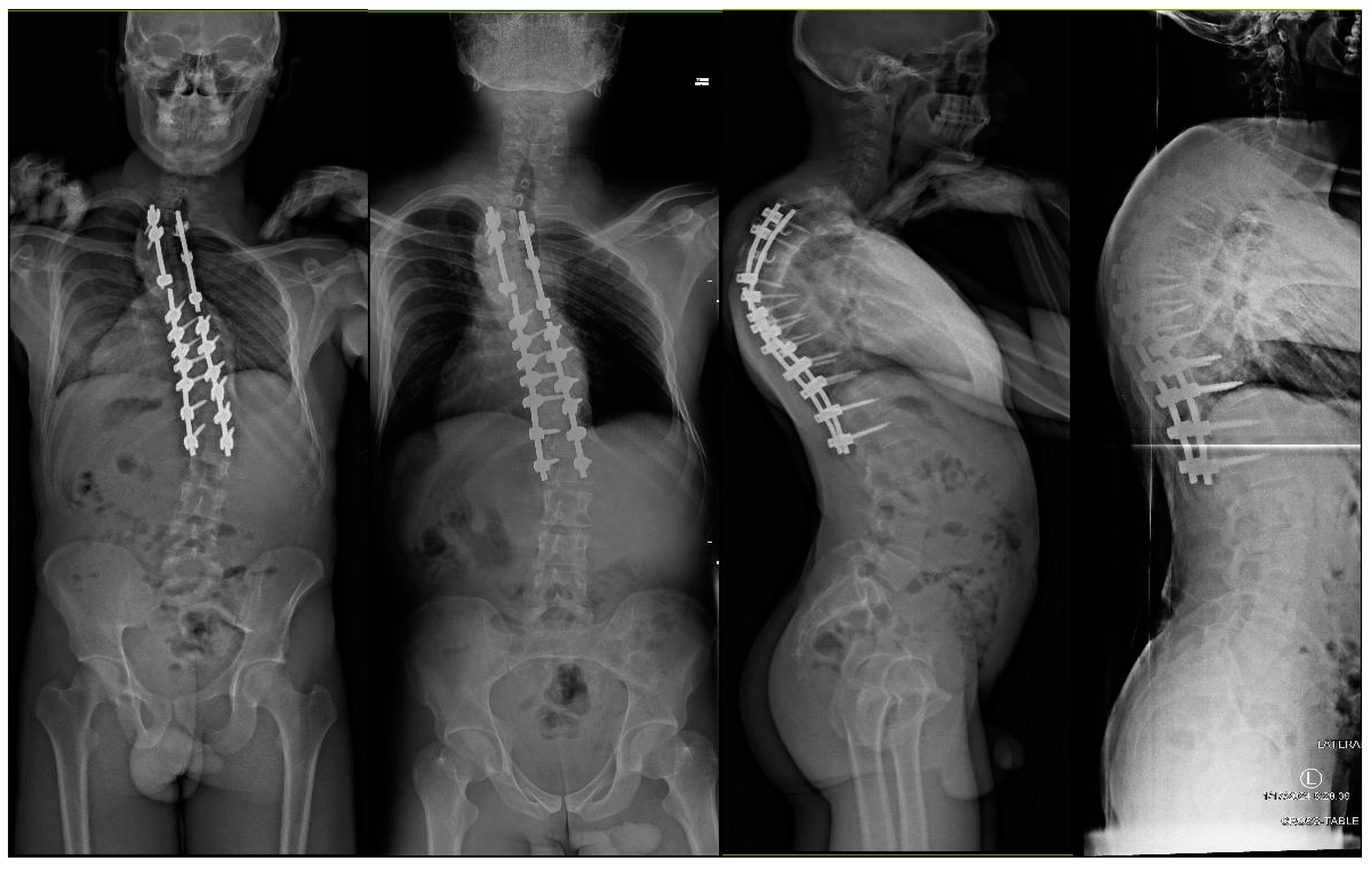
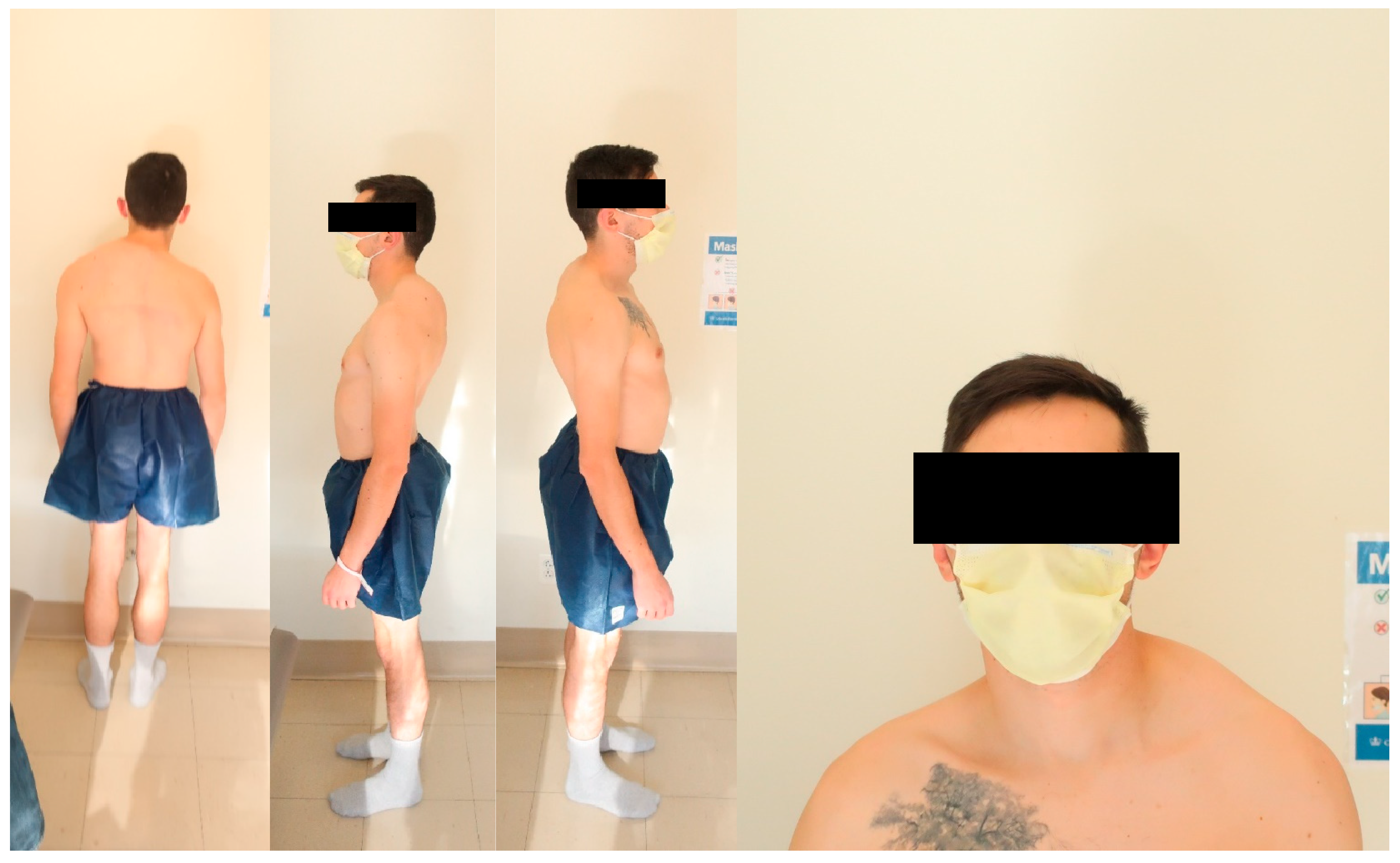
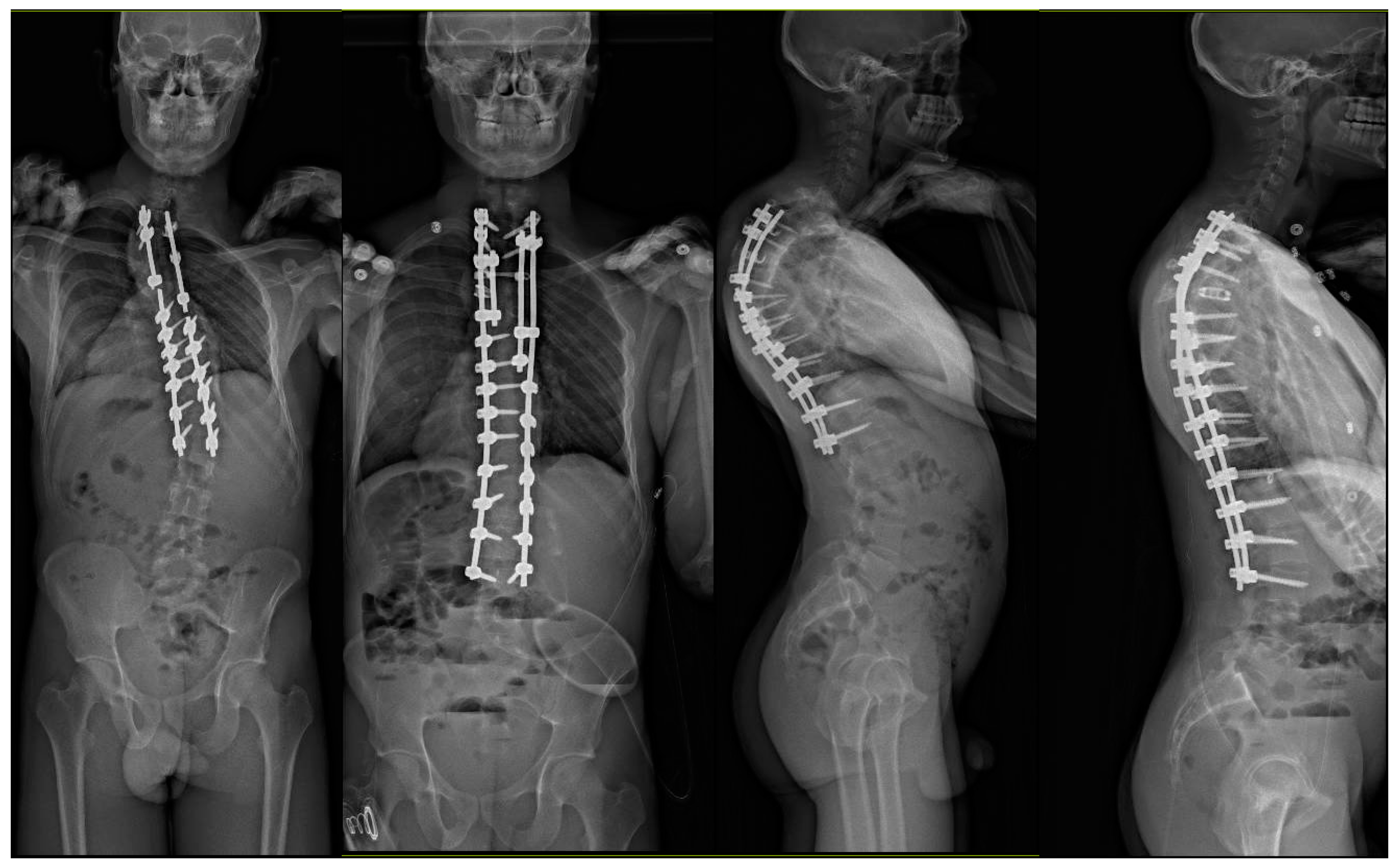
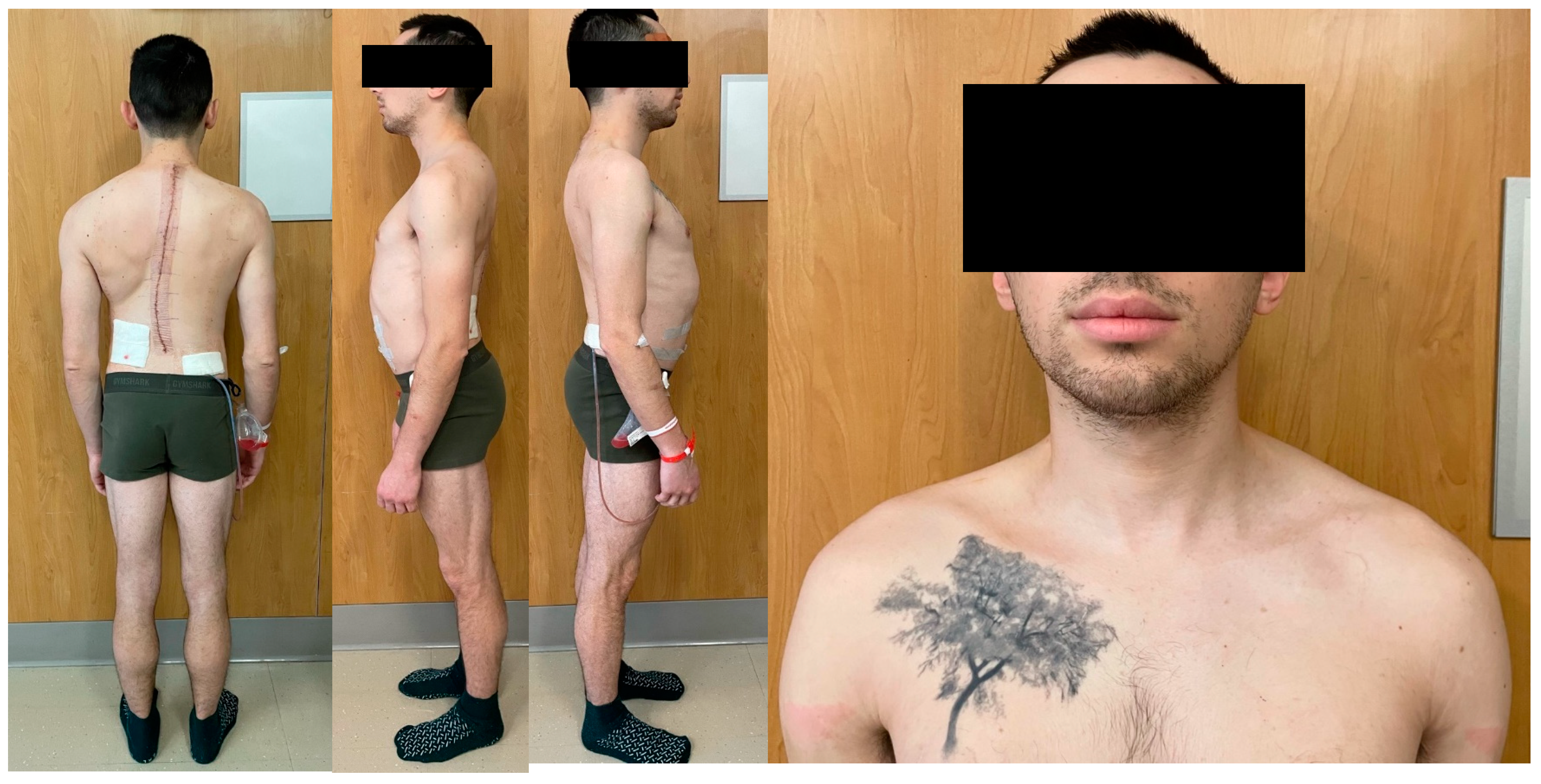
2. Case Discussions
3. Discussions
3.1. Complications and Reasons for Revision
3.2. Revision Surgery and Patient Reported Outcomes
4. Conclusions and Trajectory Moving Forward
Author Contributions
Funding
Institutional Review Board Statement
Informed Consent Statement
Data Availability Statement
Conflicts of Interest
References
- Weinstein, S.L.; Dolan, L.A.; Cheng, J.C.; Danielsson, A.; Morcuende, J.A. Adolescent idiopathic scoliosis. Lancet 2008, 371, 1527–1537. [Google Scholar] [CrossRef] [PubMed]
- Konieczny, M.R.; Senyurt, H.; Krauspe, R. Epidemiology of adolescent idiopathic scoliosis. J. Child. Orthop. 2013, 7, 3–9. [Google Scholar] [CrossRef] [PubMed]
- Lenke, L.G.; Betz, R.R.; Harms, J.; Bridwell, K.H.; Clements, D.H.; Lowe, T.G.; Blanke, K. Adolescent idiopathic scoliosis: A new classification to determine extent of spinal arthrodesis. J. Bone Joint Surg. Am. 2001, 83, 1169–1181. [Google Scholar] [CrossRef]
- Fischer, C.R.; Kim, Y. Selective fusion for adolescent idiopathic scoliosis: A review of current operative strategy. Eur. Spine J. 2011, 20, 1048–1057. [Google Scholar] [CrossRef] [PubMed]
- Lenke, L.G.; Edwards, C.C., 2nd; Bridwell, K.H. The Lenke classification of adolescent idiopathic scoliosis: How it organizes curve patterns as a template to perform selective fusions of the spine. Spine 2003, 28, S199–S207. [Google Scholar] [CrossRef] [PubMed]
- Lenke, L.G.; Betz, R.R.; Bridwell, K.H.; Harms, J.; Clements, D.H.; Lowe, T.G. Spontaneous lumbar curve coronal correction after selective anterior or posterior thoracic fusion in adolescent idiopathic scoliosis. Spine 1999, 24, 1663–1671, discussion 1672. [Google Scholar] [CrossRef] [PubMed]
- Patel, P.N.; Upasani, V.V.; Bastrom, T.P.; Marks, M.C.; Pawelek, J.B.; Betz, R.R.; Lenke, L.G.; Newton, P.O. Spontaneous lumbar curve correction in selective thoracic fusions of idiopathic scoliosis: A comparison of anterior and posterior approaches. Spine 2008, 33, 1068–1073. [Google Scholar] [CrossRef] [PubMed]
- Edwards, C.C., 2nd; Lenke, L.G.; Peelle, M.; Sides, B.; Rinella, A.; Bridwell, K.H. Selective thoracic fusion for adolescent idiopathic scoliosis with C modifier lumbar curves: 2- to 16-year radiographic and clinical results. Spine 2004, 29, 536–546. [Google Scholar] [CrossRef] [PubMed]
- Raitio, A.; Syvanen, J.; Helenius, I. Vertebral Body Tethering: Indications, Surgical Technique, and a Systematic Review of Published Results. J. Clin. Med. 2022, 11, 2576. [Google Scholar] [CrossRef]
- Vigneswaran, H.T.; Grabel, Z.J.; Eberson, C.P.; Palumbo, M.A.; Daniels, A.H. Surgical treatment of adolescent idiopathic scoliosis in the United States from 1997 to 2012: An analysis of 20,346 patients. J. Neurosurg. Pediatr. 2015, 16, 322–328. [Google Scholar] [CrossRef]
- Bartley, C.E.; Yaszay, B.; Bastrom, T.P.; Shah, S.A.; Lonner, B.S.; Asghar, J.; Miyanji, F.; Samdani, A.; Newton, P.O. Perioperative and Delayed Major Complications Following Surgical Treatment of Adolescent Idiopathic Scoliosis. J. Bone Joint Surg. Am. 2017, 99, 1206–1212. [Google Scholar] [CrossRef] [PubMed]
- Lykissas, M.G.; Jain, V.V.; Nathan, S.T.; Pawar, V.; Eismann, E.A.; Sturm, P.F.; Crawford, A.H. Mid- to long-term outcomes in adolescent idiopathic scoliosis after instrumented posterior spinal fusion: A meta-analysis. Spine 2013, 38, E113–E119. [Google Scholar] [CrossRef] [PubMed]
- Reames, D.L.; Smith, J.S.; Fu, K.M.; Polly, D.W., Jr.; Ames, C.P.; Berven, S.H.; Perra, J.H.; Glassman, S.D.; McCarthy, R.E.; Knapp, R.D., Jr.; et al. Complications in the surgical treatment of 19,360 cases of pediatric scoliosis: A review of the Scoliosis Research Society Morbidity and Mortality database. Spine 2011, 36, 1484–1491. [Google Scholar] [CrossRef] [PubMed]
- Menger, R.P.; Kalakoti, P.; Pugely, A.J.; Nanda, A.; Sin, A. Adolescent idiopathic scoliosis: Risk factors for complications and the effect of hospital volume on outcomes. Neurosurg. Focus. 2017, 43, E3. [Google Scholar] [CrossRef] [PubMed]
- Dong, Y.; Wang, S.; Tang, N.; Zhao, H.; Yu, B.; Zhang, J. Revision Surgery After Spinal Fusion in Adolescent Idiopathic Scoliosis. Global Spine J. 2022, 14, 603–609. [Google Scholar] [CrossRef] [PubMed]
- Luhmann, S.J.; Lenke, L.G.; Bridwell, K.H.; Schootman, M. Revision surgery after primary spine fusion for idiopathic scoliosis. Spine 2009, 34, 2191–2197. [Google Scholar] [CrossRef] [PubMed]
- De la Garza Ramos, R.; Goodwin, C.R.; Abu-Bonsrah, N.; Jain, A.; Miller, E.K.; Huang, N.; Kebaish, K.M.; Sponseller, P.D.; Sciubba, D.M. Patient and operative factors associated with complications following adolescent idiopathic scoliosis surgery: An analysis of 36,335 patients from the Nationwide Inpatient Sample. J. Neurosurg. Pediatr. 2016, 25, 730–736. [Google Scholar] [CrossRef] [PubMed]
- Jamnik, A.A.; Datcu, A.M.; Lachmann, E.; Patibandla, S.D.; Thornberg, D.; Jo, C.H.; Morris, W.Z.; Ramo, B.; Johnson, M. Repeat surgical interventions following “definitive” instrumentation and fusion for idiopathic scoliosis: A 30-year update. Spine Deform. 2023, 12, 99–107. [Google Scholar] [CrossRef] [PubMed]
- Delfino, R.; Pizones, J.; Ruiz-Juretschke, C.; Sanchez-Mariscal, F.; Zuniga, L.; Izquierdo, E. Selective Anterior Thoracolumbar Fusion in Adolescent Idiopathic Scoliosis: Long-Term Results After 17-Year Follow-Up. Spine 2017, 42, E788–E794. [Google Scholar] [CrossRef]
- Studer, D.; Awais, A.; Williams, N.; Antoniou, G.; Eardley-Harris, N.; Cundy, P. Selective fusion in adolescent idiopathic scoliosis: A radiographic evaluation of risk factors for imbalance. J. Child. Orthop. 2015, 9, 153–160. [Google Scholar] [CrossRef]
- Suk, S.I.; Lee, S.M.; Chung, E.R.; Kim, J.H.; Kim, S.S. Selective thoracic fusion with segmental pedicle screw fixation in the treatment of thoracic idiopathic scoliosis: More than 5-year follow-up. Spine 2005, 30, 1602–1609. [Google Scholar] [CrossRef]
- Dede, O.; Yazici, M. Revision surgery for pediatric spine deformity: Corrective osteotomies. J. Pediatr. Orthop. 2014, 34 (Suppl. S1), S6–S10. [Google Scholar] [CrossRef]
- Yagi, M.; King, A.B.; Kim, H.J.; Cunningham, M.E.; Boachie-Adjei, O. Outcome of Revision Surgery in Pediatric Spine Deformity Patients. Spine Deform. 2013, 1, 59–67. [Google Scholar] [CrossRef]
- Kim, H.J.; Cunningham, M.E.; Boachie-Adjei, O. Revision spine surgery to manage pediatric deformity. J. Am. Acad. Orthop. Surg. 2010, 18, 739–748. [Google Scholar] [CrossRef]
- Cho, R.H.; Yaszay, B.; Bartley, C.E.; Bastrom, T.P.; Newton, P.O. Which Lenke 1A curves are at the greatest risk for adding-on… and why? Spine 2012, 37, 1384–1390. [Google Scholar] [CrossRef]
- Kwan, K.Y.H.; Wong, C.P.; Koh, H.Y.; Cheung, K.M.C. Selection of Lowest Instrumented Vertebra Using Fulcrum Bending Radiographs Achieved Shorter Fusion Safely Compared With the Last “Substantially” Touching Vertebra in Lenke Type 1A and 2A Curves. Spine 2019, 44, E1419–E1427. [Google Scholar] [CrossRef]
- El Rachkidi, R.; Silvestre, C.; Roussouly, P. Early Revision Surgery for Distal Adding-On Correction in Lenke 1 and 2 Adolescent Idiopathic Scoliosis. Cureus 2022, 14, e30960. [Google Scholar] [CrossRef]
- Diederich, A.; Erwin, J.; Carlson, B.; Bunch, J.; Jackson, R.S.; Burton, D. Indications and timing of revision spine surgery in adults after adolescent surgery for idiopathic scoliosis. Spine Deform. 2022, 10, 1385–1392. [Google Scholar] [CrossRef]
- Zhang, H.; Fan, Y.; Ni, S.; Pi, G. The preliminary outcomes of vertebral body tethering in treating adolescent idiopathic scoliosis: A systematic review. Spine Deform. 2022, 10, 1233–1243. [Google Scholar] [CrossRef]
- Newton, P.O.; Kluck, D.G.; Saito, W.; Yaszay, B.; Bartley, C.E.; Bastrom, T.P. Anterior Spinal Growth Tethering for Skeletally Immature Patients with Scoliosis: A Retrospective Look Two to Four Years Postoperatively. J. Bone Joint Surg. Am. 2018, 100, 1691–1697. [Google Scholar] [CrossRef]
- Aghdasi, B.; Bachmann, K.R.; Clark, D.; Koldenhoven, R.; Sultan, M.; George, J.; Singla, A.; Abel, M.F. Patient-reported Outcomes Following Surgical Intervention for Adolescent Idiopathic Scoliosis: A Systematic Review and Meta-Analysis. Clin. Spine Surg. 2020, 33, 24–34. [Google Scholar] [CrossRef] [PubMed]
- Mariconda, M.; Andolfi, C.; Cerbasi, S.; Servodidio, V. Effect of surgical correction of adolescent idiopathic scoliosis on the quality of life: A prospective study with a minimum 5-year follow-up. Eur. Spine J. 2016, 25, 3331–3340. [Google Scholar] [CrossRef] [PubMed]
- Simony, A.; Hansen, E.J.; Carreon, L.Y.; Christensen, S.B.; Andersen, M.O. Health-related quality-of-life in adolescent idiopathic scoliosis patients 25 years after treatment. Scoliosis 2015, 10, 22. [Google Scholar] [CrossRef] [PubMed]
- Bastrom, T.P.; Yaszay, B.; Shah, S.A.; Miyanji, F.; Lonner, B.S.; Kelly, M.P.; Samdani, A.; Asghar, J.; Newton, P.O. Major Complications at Two Years After Surgery Impact SRS Scores for Adolescent Idiopathic Scoliosis Patients. Spine Deform. 2019, 7, 93–99. [Google Scholar] [CrossRef]
- Jamnik, A.A.; Grigoriou, E.; Kadado, A.; Jo, C.H.; Boes, N.; Thornberg, D.; Tran, D.P.; Ramo, B. Radiographic and clinical outcomes after definitive spine fusion for skeletally immature patients with idiopathic scoliosis. Spine Deform. 2023, 12, 149–157. [Google Scholar] [CrossRef]


Disclaimer/Publisher’s Note: The statements, opinions and data contained in all publications are solely those of the individual author(s) and contributor(s) and not of MDPI and/or the editor(s). MDPI and/or the editor(s) disclaim responsibility for any injury to people or property resulting from any ideas, methods, instructions or products referred to in the content. |
© 2024 by the authors. Licensee MDPI, Basel, Switzerland. This article is an open access article distributed under the terms and conditions of the Creative Commons Attribution (CC BY) license (https://creativecommons.org/licenses/by/4.0/).
Share and Cite
Lenke, L.G.; Lee, V.; Hassan, F.M. Revision of Surgery for Adolescent Idiopathic Scoliosis: Reasons, Treatments, and Clinical Management with Case Examples. J. Clin. Med. 2024, 13, 2233. https://doi.org/10.3390/jcm13082233
Lenke LG, Lee V, Hassan FM. Revision of Surgery for Adolescent Idiopathic Scoliosis: Reasons, Treatments, and Clinical Management with Case Examples. Journal of Clinical Medicine. 2024; 13(8):2233. https://doi.org/10.3390/jcm13082233
Chicago/Turabian StyleLenke, Lawrence G., Veronica Lee, and Fthimnir M. Hassan. 2024. "Revision of Surgery for Adolescent Idiopathic Scoliosis: Reasons, Treatments, and Clinical Management with Case Examples" Journal of Clinical Medicine 13, no. 8: 2233. https://doi.org/10.3390/jcm13082233
APA StyleLenke, L. G., Lee, V., & Hassan, F. M. (2024). Revision of Surgery for Adolescent Idiopathic Scoliosis: Reasons, Treatments, and Clinical Management with Case Examples. Journal of Clinical Medicine, 13(8), 2233. https://doi.org/10.3390/jcm13082233