Article

Concurrent Resistance and Cardiorespiratory Training in Patients with Hypertrophic Cardiomyopathy: A Pilot Study

by
Adrián Bayonas-Ruiz
1,2,
Francisca M. Muñoz-Franco
3,
María Sabater-Molina
4,5,6,
Ignacio Martínez-González-Moro
2,7,
Juan Ramon Gimeno-Blanes
5,8,9 and
Bárbara Bonacasa
1,2,*
1
Department of Physiology, Human Physiology Area, Sports Sciences Faculty, University of Murcia, C. Argentina, 19, 30720 San Javier, Murcia, Spain
2
Research Group of Physical Exercise and Human Performance, University of Murcia, 30720 San Javier, Murcia, Spain
3
Cardiac Unit, General University Hospital Caravaca, 30400 Caravaca de la Cruz, Murcia, Spain
4
Cardiogenetic Laboratory, Instituto Murciano de Investigación Biosanitaria (IMIB), 30120 El Palmar, Murcia, Spain
5
European Reference Network for Rare and Low Prevalence Complex Diseases of the Heart (ERN-Guard Heart), 1105 Amsterdam, The Netherlands
6
Department of Legal and Forensic Medicine, Faculty of Medicine, Health Sciences Campus, Avda. Buenavista n°32, 30120 El Palmar, Murcia, Spain
7
Department of Physiotherapy, Faculty of Medicine, Health Sciences Campus, Avda. Buenavista n°32, 30120 El Palmar, Murcia, Spain
8
Inherited Cardiac Disease Unit (CSUR), Hospital Universitario Virgen de la Arrixaca, 30120 El Palmar, Murcia, Spain
9
Department of Internal Medicine (Cardiology), University of Murcia, 30720 San Javier, Murcia, Spain
*
Author to whom correspondence should be addressed.
J. Clin. Med. 2024, 13(8), 2324; https://doi.org/10.3390/jcm13082324
Submission received: 7 March 2024 / Revised: 14 April 2024 / Accepted: 14 April 2024 / Published: 17 April 2024
(This article belongs to the Special Issue Diagnostic Advances in Cardiomyopathies)

Abstract

:
Background: Exercise training in patients with HCM has evidenced benefits on functional capacity, cardiac function, and a reversion of adverse cardiac remodeling. The objective of this study was to assess the effect of a concurrent resistance and cardiorespiratory training program on functional capacity, biochemical parameters, and echocardiographic variables in a pilot group. Methods: Two HCM patients were evaluated before and after 12 weeks of individualized concurrent training with two sessions/week. Pre- and post-training data were compared for each patient. Evaluations included a cardiopulmonary exercise test (CPET), body composition, echocardiography, electrocardiography, and blood analysis. Results: Training promoted an increase in functional capacity (+4 mL·kg−1·min−1), ventilatory thresholds, and other CPET-derived variables associated with a better prognosis and long-term survival. Muscular mass was augmented (0.8 and 1.2 kg), along with a mean increase of 62% in upper and lower body strength. Echocardiographic features demonstrated the maintenance of cardiac function with signs of positive left ventricular remodeling and an improvement in diastolic function. Blood analyses, including cardiac troponins and NT-proBNP, displayed uneven changes in each patient, but the values fell into normal ranges in both cases. Conclusions: The available data suggest a positive effect of concurrent resistance and cardiorespiratory training on patients’ functional capacity and cardiac function that may improve their functional class, quality of life, and long-term prognosis. The replication of this protocol in a larger cohort of patients is warranted to confirm these preliminary results.

1. Introduction

Hypertrophic cardiomyopathy (HCM) is the most prevalent cardiomyopathy (1:500) and is characterized by the presence of ventricular hypertrophy with a maximal wall thickness (MWT) of ≥15 mm in the absence of other primary causes [1,2]. Physical exercise in patients with HCM has emerged as an adjunctive treatment strategy, and its inclusion has been advocated in numerous theoretical proposals and reviews on potential benefits over the last decade [3,4]. Among these, researchers in this field are contemplating the possibility of improving functional capacity and effort tolerance, reversing cardiac remodeling, enhancing diastolic function, and improving end-systolic and end-diastolic volumes [5].
Regarding the impact of physical exercise, a recent meta-analysis conducted by our group demonstrated an improvement in VO2max of 4.33 mL/kg/min (95% CI: 0.20; 8.45), to a greater extent compared to other pharmacological and/or invasive treatment options, and an enhancement in New York Heart Association (NYHA) functional class [6]. Furthermore, none of the interventions resulted in serious adverse effects.
However, the practical implementation of physical exercise in HCM patients remains limited: starting from 2015, only five trials assessing the effects and safety of training have been published to date [7,8,9,10,11]. Despite most of them demonstrating global improvements with the program, significant heterogeneity in the methodology of the published studies limits the reproducibility of their findings. In particular, this regards the type of exercise, the definition of exercise load, thresholds and repetitions, the length of the training program, and methodology of the evaluation of the response to exercise. Most protocols were based on cardiorespiratory training and did not include strength exercises.
In light of the aspects discussed regarding the exercise methodology and its benefits, there is a need to optimize training protocols in HCM patients. Similarly, a more detailed description of the exercise is essential to facilitate its replication and use as an adjunct strategy in patient management. Advances in exercise physiology will be instrumental in optimizing variables for cardiorespiratory training (intensity based on VO2max and peak HR determined in CPET, duration, recovery, and frequency) and strength training (exercise selection, relative intensity -%RM-, effort grade/character, number of repetitions, daily and weekly volume, and rest between sets). This body of knowledge also encompasses the definition of “concurrent training”, which combines both resistance and cardiorespiratory training in a single program and/or training session [12].
Considering the latter, the objective of this study was to assess the effect of a concurrent resistance and cardiorespiratory training program on functional capacity, biochemical parameters, and echocardiographic variables in a pilot group of two HCM patients. In parallel, it is intended to evaluate the feasibility of the training protocol to replicate it in a larger cohort of patients. We hypothesized that a 12-week concurrent training regime would promote increases in functional capacity and strength without a detrimental effect on cardiac function.

2. Materials and Methods

2.1. Participants

The subjects of this study were two men aged 63 and 60 years with hypertrophic cardiomyopathy and a low level of physical activity, limited to their daily activities. Both patients underwent genetic testing of a gene panel associated with HCM, and this was negative. Patient 1 had obstructive HCM diagnosed 5 years before enrolling in the study, with a maximal left-ventricular outflow tract gradient of 90 mmHg after Valsalva maneuver. His medication included bisoprolol (2.5 mg/day), budesonide (320/9 mcg each 12 h when needed) for chronic obstructive pulmonary disease (COPD) potentially derived from a history of smoking habit, and levothyroxine (75 mcg/day). He had no previous major adverse cardiac events (MACE), but had experienced 2 episodes of anaphylaxis of an unknown cause. Patient 2 was diagnosed with non-obstructive HCM 9 years before enrolling the study. He had subclinical hypothyroidism, hypertension, and dislipemia. His medication included bisoprolol (2.5 mg/day), indapamide (1 tablet/day), and simvastatin (20 mg/day). He had one MACE (syncope) 20 years before enrolling the study, prior to his diagnosis of HCM. None of them had a smoking habit at the moment of the study, nor were they implantable cardioverter defibrillator carriers. Both patients had a low SCD risk according to the current guidelines and ESC risk calculator [13]. Both were informed about the possibility of participating in the study during their annual review with their cardiologist and gave their consent after being informed of the protocol, possible risks, and expected benefits. Further details of their baseline characteristics can be found in the pre-training values in the results section.

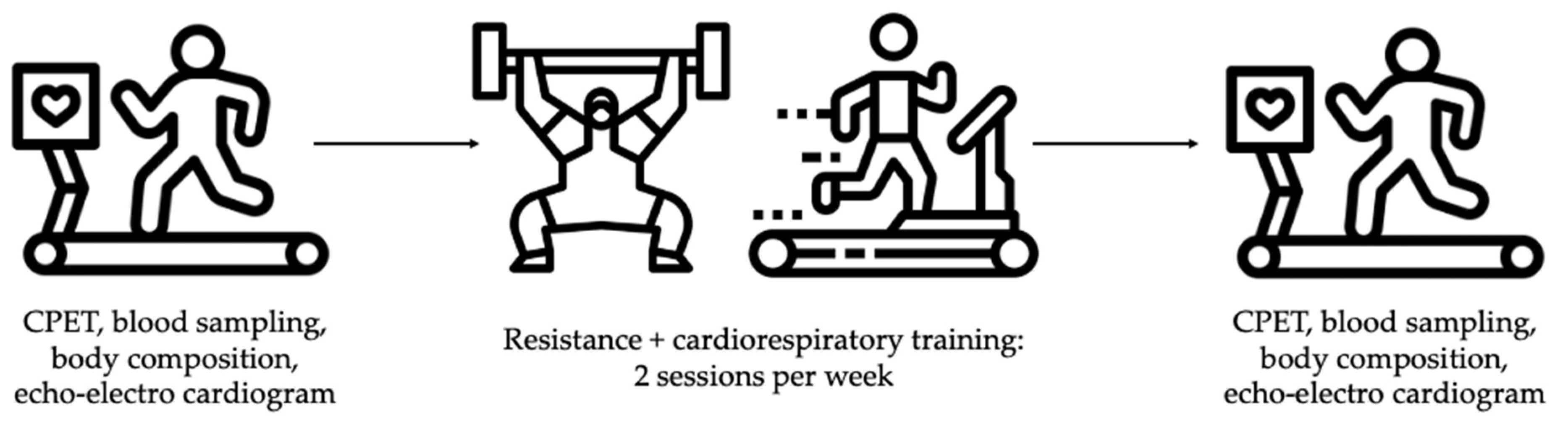
2.2. Experimental Design

The study was performed for a total duration of 14 weeks for each patient: a first week of assessments, followed by 12 weeks of concurrent resistance and cardiorespiratory training, and, finally, another week of assessments. During the first week, in addition to the initial evaluations in the hospital, a familiarization session with the material and exercises was carried out in the gym. After training, the patients were provided with a guide to continue this exercise autonomously following the same guidelines used during the intervention. A graphical diagram of the experimental design is shown in Figure 1 (icons contained in the figure were obtained from www.flaticon.es, accessed on 1 March 2024).

2.3. Testing Procedures

All assessments were conducted in the same facilities, utilizing identical materials and performed at consistent timings for both participants. During their initial visit, the patients were scheduled in the hospital at 15:00, with a recommendation to have their previous meal at least 3 h before, avoiding substantial food intake. Firstly, an echocardiogram was performed using a Phillips Epiq device (Amsterdam, The Netherlands). Subsequently, a blood sample of 8 mL was drawn from the left arm, followed by anthropometric measurements of the hip and waist using a standard tape measure, and a body composition analysis using electrical impedance: the patients were placed standing barefoot on an InBody 120 (Seoul, Republic of Korea) impedance scale, holding this position with raised arms a dual-entry handle that completed the impedance analysis with both foot entries. The patients were then positioned in supine decubitus for the placement of electrocardiogram electrodes (Cardioline ClickECG, Milan, Italy). In this position, readings of the resting electrocardiogram and blood pressure in the right arm (Cardioline) were obtained. An echocardiogram including standard 2D views, Doppler flow velocity, and tissue Doppler analysis was performed with a Philips iE33 Ultrasound System 795052 model (Amsterdam, The Netherlands). Following this, the patients were positioned on the treadmill (Runner-Run 7411, Cavezzo, Italy) and underwent functional assessment following the modified Bruce protocol [14]. Continuous monitoring during the test included gas consumption (Cortex Metalyzer 3B; Cortex Biophysik GmbH, Leipzig, Germany) and exercise electrocardiogram. The patients were encouraged to continue the test to maximum exertion. CPET was considered maximal when a respiratory exchange ratio (RER) of >1.10 was reached. Gas consumption data were used to determine the ventilatory thresholds and associated heart rate for each, and subsequently used to plan and monitor their cardiorespiratory training. Following the CPET, a second blood sample was extracted and both samples were, therefore, aliquoted and stored at −40 °C. All the assessments were repeated one week after finishing the training period.
Additionally, the determination of maximum strength for each resistance training exercise was carried out through the estimation of the 1 Repetition Maximum (1RM). This involved recording the weights and repetitions performed, and the patients were queried about their subjective effort perception according to Borg’s scale [15], indicating how many more repetitions they could have completed in each set. Epley’s formula [16], proven to be the most accurate for the exercises included in this study and beginner practitioners, was employed for the 1RM estimation [17]. Before effective sets (those considered as part of the training), the patients performed unloaded mobilization of the segments, replicating the movement involved for each exercise and an initial warm-up set of 10 repetitions with a low load (e.g., a load with which the patients could easily perform many repetitions). Calculations were conducted for all sets, and the highest estimation from the familiarization session and the first week of training was considered as the initial 1RM, while the highest estimation from weeks 11–12 of training was considered as the final 1RM. Since this was a pilot study involving two patients, statistical analyses were limited to direct comparisons of the pre- to post-training values.

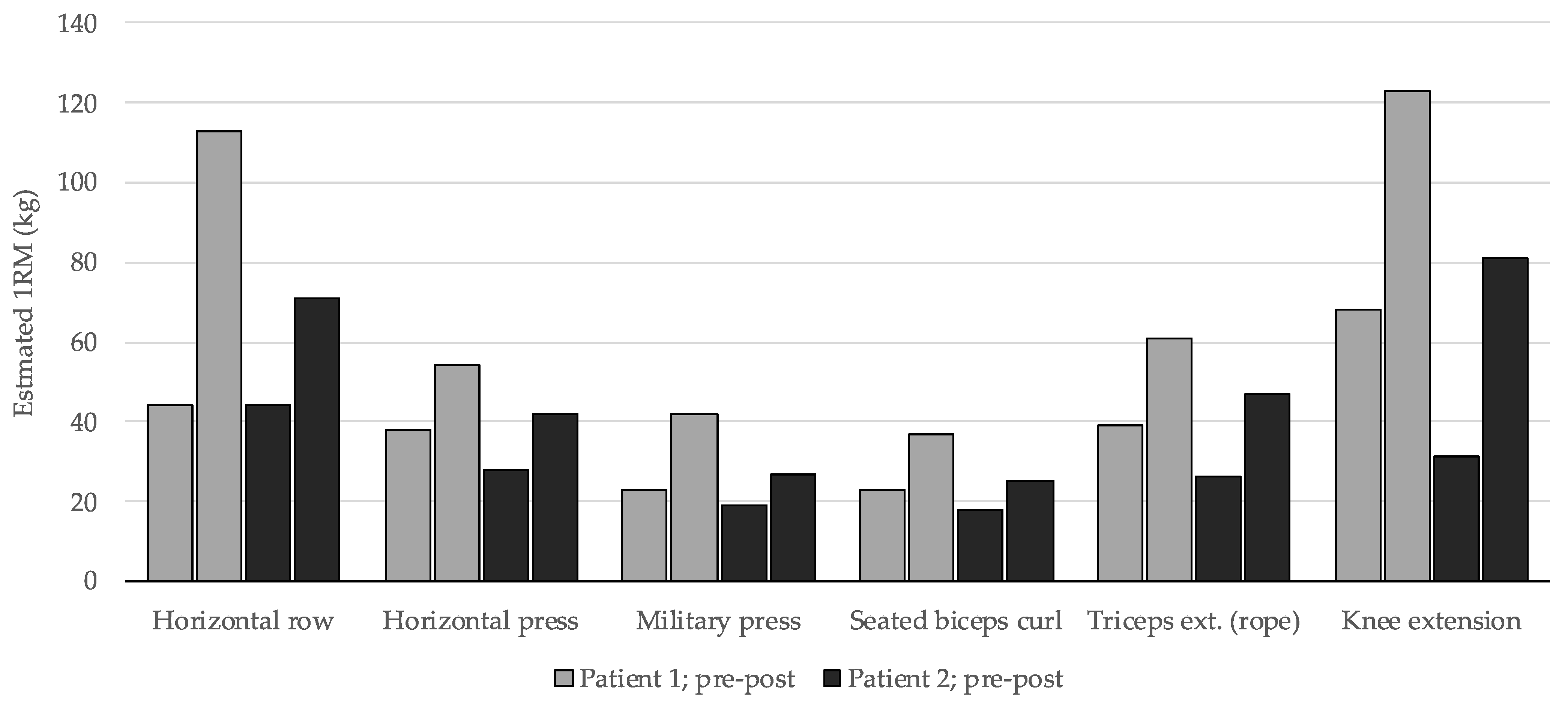
2.4. Training Protocol

The training regimen comprised two weekly sessions over a span of 12 weeks, with a minimum 48 h separation between them. Each session incorporated a resistance training initial part followed by cardiorespiratory training, adhering to an order optimized for minimizing acute [18] and long-term [19] interference between the adaptations of both training modalities. Exercise selection excluded those involving trunk flexion or supine positions to mitigate the likelihood of orthostatic episodes. Six exercises, each targeting one muscle group—back, chest, shoulder, biceps, triceps, and legs—were conducted in each session, utilizing guided machines, pulleys, or free weights. The complete selection is detailed in Table 1.
For each exercise, a warm-up set (60–70% of the weight targeted for set 1) and three effective sets were performed across the 12 weeks, resulting in a total of 18 sets/day and 36 sets/week. A 2 min rest interval was established between sets [20]. Intensity was maintained within the 55–70% range of 1RM throughout the training period. The participants were instructed to choose a weight for each set, allowing them to complete between 16 and 24 repetitions, which corresponds to the specified relative intensities [21]. However, they were advised to execute approximately half of the potential repetitions with the selected loads, maintaining a moderate effort level (e.g., selecting a load they could complete ~20 repetitions with but performing only 8–10). This effort character entails a lower associated fatigue while allowing for greater adaptations in dynamic maximum strength compared to protocols employing high effort levels leading to muscular failure or near failure [22,23].
On the other hand, cardiorespiratory training was individually monitored and guided in all sessions using a heart rate monitor (Polar H10, Polar Electro, Kempele, Finland) simultaneously connected to the treadmill and a control watch for the researchers. The first session of each week focused on a pace slightly above the aerobic threshold (VT1), involving treadmill session from 25 to 50 min with no incline, adjusting the speed to achieve and maintain a heart rate of 5–10 bpm above that associated with the VT1 based on their CPET. The second session introduced fartlek-style interval training, featuring 2–6 min intervals 5–10 bpm below the second ventilatory threshold (VT2) intensity, interspersed with 1–2 min active rests at the VT1 pace, as previously proposed for this type of training [24]. The main segment duration progressively increased from 14 to 25 min in these sessions, with a 10-min warm-up and a 5-min cool-down at a comfortable, self-selected pace. Intensity adjustments were made by modifying the treadmill speed and incline, as in the CPET with the modified Bruce protocol. Full details of every cardiorespiratory training session can be found in Table A1.

3. Results

Both patients were overweighted at baseline and gained weight during the intervention, although a slight reduction in body fat was observed (−0.6% both), along with an average increase of 1 kg in muscular mass (Table 2). Patient 1 reduced his waist circumference by 3 cm, and patient 2 did so by 1 cm. Patient 1 also had a higher baseline waist-to-hip ratio (1.10), which reduced to a greater extent (−0.06) compared to patient 2 (−0.01).
Changes in supine and resting blood pressure varied between the two patients, as did differences in resting respiratory volumes and frequency, which were very subtle. The first consistent changes were observed in variables related to VT1, where, in both cases, a reduction in the heart rate required to achieve the same oxygen consumption was noted (−9 and −11 beats to reach 15 mL/kg/min pre- and post-training in both cases).
At VT2, both patients experienced an increase in oxygen consumption of nearly 0.5 L/min (0.36 and 0.43), translating to improvements of 3 and 6 mL/kg/min, respectively. Both patients also increased the heart rate associated with VT2 after training, thereby expanding the heart rate range between the first and second ventilatory thresholds. While ventilation showed minimal changes at VT1, increases of 13.5 and 20.0 L/min were observed at VT2, respectively.
Finally, VO2max increased by 4 mL/kg/min in both cases: +17% and +12% in predicted VO2max (Table 3), placing both patients above 100% in this parameter. Increased ventilation was also observed, similar to VT2, with values 19.0 and 14.3 L/min higher compared to the first CPET, resulting in an elevated respiratory frequency (+8 and +5 breaths/min). The maximum heart rate achieved increased notably in Patient 1 (+10 beats) and remained similar in Patient 2 (+1 beat). Regarding the recovery of heart rate after the first minute of active recovery, although changes were uneven between the two patients in the pre- and post-training comparisons, in both cases, the heart rate dropped by >18 beats after the first minute following CPET.
In the strength-related training outcomes, an enhancement in estimated 1RM was evident across all exercises and muscle groups in both patients. The increase in this variable ranged from 11% to 161%, with an average of a 62.2% improvement after the 12-week period (patient 1: 73%, patient 2: 56%). Figure 2 illustrates that the estimated 1RM for patient 1 was initially higher in five out of six exercises, yet their percentage increase was greater. Nevertheless, the strength improvement in patient 2 was also noteworthy. In both cases, the most substantial strength gains were observed in the back and leg exercises.
The electrocardiographic findings showed a decrease in the duration of the P wave, PQ interval, and QRS complex in Patient 1, while these remained unchanged in Patient 2 (Table 4). Both the QT and QTc interval were similar in the pre–post comparisons in both patients.
With regard to the resting echocardiographic findings (Table 5), the maximum wall thickness remained unchanged in both patients after training. However, the absolute and indexed left ventricular mass were augmented in both cases. The end-systolic diameter decreased by 0.2 and 0.3 cm, respectively, while the end-diastolic diameter behaved unevenly. The end-diastolic volumes also showed disparities between the patients, while the end-systolic volume was slightly augmented. The left ventricular ejection fraction slightly diminished in both patients, but stayed at around 70%. In patient 2, reductions in left atrial diameter and volume were observed. Patient 2 also exhibited decreases in Vmax E, lateral E, medial E, and medial and lateral E/E′ ratios, while patient 1 only in medial and lateral E/E′.
The blood analysis (Table 6) showed an increase in serum glucose, although changes must be interpreted cautiously, since blood sampling was performed in a non-fasted state. The serum minerals remained mostly unchanged or with slight variations. Cardiac markers such as cardiac troponin T and NT-proBNP showed uneven changes: both markers decreased in one patient’s higher baseline values, while slightly increased in the other. Finally, changes in hormones were more consistent between the patients, with a decrease in serum testosterone and increments in basal cortisol and TSH. No adverse events nor complications of any type occurred during training. Both patients attended 100% of the sessions.

4. Discussion

One of the primary objectives of this intervention was to assess the feasibility of the proposed concurrent training protocol, with the aim of replicating it in a larger cohort of patients. Both the CPET and all training sessions were devoid of adverse events and complications, thereby establishing the feasibility of the training protocol.
Regarding previous investigations, in that by Saberi et al. [8], patients underwent 16 weeks of cardiorespiratory training with 2–3 sessions per week at an intensity of 60–70% of heart rate reserve (HRR). The duration progressed from 20 to 60 min, adding 5–10 min in each session. On the other hand, Klempfner et al. [9] also prescribed exercise based on HRR, starting with a 10 min warm-up at 40–50% of the HRR and then completing a 60 min session with an intensity increasing from 50–60% of the HRR to 65–85%. The authors stated that two sessions were conducted weekly until completing an average of 41 h of training, which suggests an approximate duration of ~20 weeks, although they did not explicitly state this. Wassestrum and colleagues [7] also programmed training sessions based on HRR, but the time between pre- and post-training assessments was very uneven and up to more than three years, which may not precisely identify the changes elicited by the training protocol (Table 7).
Regarding cardiorespiratory training, the most recent trial [10] included significant improvements in intensity control compared to previous ones, precisely defining training zones based on the maximum heart rate and heart rate associated with VT2 in a previous CPET. This intervention was also the first to incorporate high-intensity intervals at 90–95% of peak heart rate. During their study, the authors did not record any serious adverse events. However, to date, only one intervention has considered strength training in its protocol [11], in addition to cardiorespiratory training. In this intervention, participants engaged in three sessions per week for 18 months, cycling at 60–80% of their VO2max and subsequently performing upper limb pushing and pulling exercises, lower limb extensions, trunk flexion, and three unspecified stretching exercises. The intensity in these strength exercises was set at 65% of each subject’s 1RM, although they did not specify how this was calculated or provide progression details for the loads used or the number of repetitions performed.
Previous studies have demonstrated that the optimal distribution of concurrent training to maximize benefits and minimize interference in acute and long-term adaptations is to conduct strength training at the beginning of the session, followed by cardiorespiratory exercise [19,25]. Moreover, it has been observed that the benefits of concurrent training in strength and cardiorespiratory aspects surpass those observed with either modality performed exclusively, especially in sedentary, beginners, and intermediate individuals [12]. Finally, recent advancements in exercise physiology have underscored the role of skeletal muscle as an endocrine tissue in cardiovascular risk prevention [26]. Skeletal muscle releases numerous myokines, particularly during exercise-induced contractions, with potential positive impacts on HCM progression, such as reducing pro-inflammatory status, mitigating vascular stenosis and atherosclerotic processes, or promoting revascularization of ischemic tissue [27,28].
When interpreting changes in body composition, caution should be warranted, as the subjects’ diet was not controlled during the study. Nevertheless, the observed average gain of 1 kg in muscle mass coupled with a 0.6% reduction in body fat percentage provide encouraging data. A previous study examined the effect of cardiorespiratory training on body composition, wherein no changes were observed in the amount of lean mass or body fat among the subjects [10]. Putting together these findings with those observed in our study, it appears that incorporating resistance training alongside the traditionally employed cardiovascular training could prove to be an effective strategy for enhancing body composition. Furthermore, the decrease in the waist-to-hip ratio also suggests an improvement in body composition, which is beneficial for overall health [11]. It is essential to note that both patients were overweight (with Patient 1 classified as obese), a factor previously linked in studies with impaired functional capacity and potential negative effects on symptomatic status and quality of life [29,30]. Future studies should incorporate dietary control and/or interventions aimed at weight loss.
In the realm of exercise testing, numerous derived parameters have shown a significant association with the prognosis of disease progression, mortality, and the occurrence of medium- to long-term events [31]. These parameters encompass variables related to functional capacity (VO2max, % of predicted VO2max, and VO2 at VT2), ventilatory efficiency (V′E/V′CO2), and heart rate (peak HR and post-exercise heart rate recovery).
Firstly, improvements in VO2max have been linked to a reduced risk of mortality and events [32,33,34,35,36]. The benefits observed in our intervention are promising, with a 4 mL/kg/min increase in VO2max in both cases. The latter aligns with findings from a previous meta-analysis conducted by our group, encompassing the prior exercise interventions [6]. Additionally, Finocchiaro et al. reported a 48% reduction in the composite end-point risk for every 5 mL/kg/min increase in VO2max [37]. Notably, the composite endpoints included not only standard events, but also functional deterioration leading to septal reduction therapy. Thus, the functional capacity improvement achieved with the proposed training not only decreases the risk of mortality and severe events, but also the need for invasive therapies.
This absolute increase in VO2max concurrently implies an improvement in the % of the predicted VO2max, a value associated with a better prognosis and increased survival [34,35,36,37,38]. The respective 17% and 12% increases placed both patients above 100% of their predicted values.
Another parameter of interest due to its relationship with long-term prognosis improvement is oxygen consumption at the anaerobic threshold [31]. In our patients, notable increases in this variable were also observed (3 and 6 mL/kg/min). Studies have reported a roughly 20% decrease in the risk of events and sudden death with improvements in this parameter [32,34,35], emphasizing the benefits of the proposed training program.
On the other hand, ventilatory efficiency, as assessed by the V′E/V′CO2 ratio at the moment of VO2max, has been correlated with a poorer prognosis [32], especially when exceeding a ratio of 31 [34] and 34 [37]. In our study, both patients had lower values before and after training, although a slight increase was observed in both cases. However, the recorded ratios are not alarming, as they remain below the aforementioned reference thresholds.
Finally, the recorded maximum heart rate has been linked to a 23% decrease in the risk of composite endpoint for every 10 beats increase [37]. In this context, Patient 1 increased his maximum heart rate by 10 beats after training, representing another positive indicator. In addition to the maximum attained, the recovery of heart rate following the completion of CPET is also employed to assess cardiac function. In this regard, Masri et al. demonstrated a 52% reduction in the risk of reaching a composite endpoint in patients who exhibited a reduction of at least 18 bpm in the first minute post-exercise [38]. It is crucial to note that the CPET protocol they utilized concluded without a recovery phase, whereas ours included one. Nevertheless, both of our patients recovered more than 18 bpm in the first minute after the training period. In the case of Patient 2, during the initial CPET, only a 10 bpm recovery was observed, underscoring the beneficial impact of the training in this aspect as well. Furthermore, the increment (right-shift) observed in the heart rate associated with VT2 and left-shift in that of VT1 increase training possibilities through broadening the inter-threshold range of viable exercise intensities.
Regarding strength adaptations, the long-term implications concerning the prognosis and progression of hypertrophic cardiomyopathy remain unknown, as our study represents the first to incorporate such a strength training protocol. Notably, enhancements in strength and the augmentation of musculoskeletal tissue are linked to health benefits, including a reduction in cardiovascular risk [26,27]. Consequently, it is expected that the strength improvements achieved through the proposed training regimen will exert a positive impact on the health and quality of life of the patients.
In relation to the electrocardiographic findings, the decrease in the P wave duration and PQ interval observed in Patient 1 may be explained by the presence of COPD. Other than that, the training protocol seemed to have no effect on the electrocardiographic parameters. However, further observation is needed, especially in patients with comorbidities affecting the lungs or a smoking history.
Echocardiographic features demonstrated signs of left ventricular remodeling, despite there only being two patients. As the left ventricular diameter is one of the more reliable measurements, it increased by 0.6 and 0.3 cm with minor variations in systolic function. These findings are consistent with those of the most recent protocol, where both moderate- and high-intensity cardiorespiratory training showed a significant 0.3 cm increase in this parameter [10]. Measurements of left ventricular volumes in patients with hypertrophic cardiomyopathy from echocardiography are subject to errors related to the assumption of the Simpson method in asymmetric hypertrophy. As expected, there was no change in wall thickness. Changes in strain parameters and left atrial dimensions might seem too inconsistent and should be taken cautiously. Although these changes have to be considered cautiously given the preliminary nature of the results, available data suggest better outcomes with the proposed program compared to that referred by previous studies, where no changes in echocardiographic findings were observed [7,8,11]. More interestingly, with the limitation of a pilot study, the exercise program seemed to have a positive impact on diastolic function, with a consistent reduction in the E/e’ ratios.
Cardiac troponin T, which is a biomarker of myocardial damage, remained within normal range, which is a reassuring finding of the short-term safety of the training, while NT-proBNP showed a mild increase in one of the patients and a more relevant decrease the other participant. An increase in this biomarker might be expected with exercise in patients with ventricular dysfunction. However, the observed decrease in patient 2 could suggest that the exercise did not impose a significant overload on the ventricle, or it could even indicate an improvement in ventricular function or the heart’s ability to handle physical stress over time, possibly as a result of adapted and controlled physical training. Variations of <100 pg/mL can be considered as day-to-day changes in stable cardiomyopathy patients. An evaluation of a larger number of patients would establish the real impact of training on NT-proBNP values, since previous studies have reported similar values at post-training [8,11].
Concerning hormones, the acute increase in circulating testosterone levels is noteworthy following exercise [39], whereas chronic exposure to training does not imply an elevation in resting concentration [40]. What does persist over a longer period (24–48h) is the mRNA and protein content in androgen receptors, predisposing the organism to greater anabolic adaptation in response to exercise [41]. These findings align with the observed muscle mass increase in the subjects of this pilot study: the slight decrease in baseline testosterone could be attributed to other factors such as age, and may not be a consequence of exercise. In this context, the circulating levels of steroid hormones are contingent upon the individual rate of biosynthesis, the presence of binding proteins in circulation, and modulation through the rate of the hormonal metabolic clearance process [42]. As for changes in basal growth hormones, it is imperative to observe this response in a larger number of patients, as disparate results were found in the two subjects of this pilot study: while patient 1 experienced an increase in concentration, patient 2 exhibited a slight reduction. Nevertheless, the elevation of serum GH is evident within the first 10–20 min of exercise and persists up to 60 min post-exercise, but not chronically [43,44]. Regarding cortisol, despite a slight increase in the baseline values in both patients, both pre- and post-exercise levels were within the expected range. Moreover, it is acknowledged that cortisol levels are significantly influenced by factors such as stress levels, prior meals, or circadian rhythm [45]. Therefore, although we will continue to monitor the results in a larger cohort, the subtle changes in this hormone do not seem to be affected by the proposed training and/or clinically relevant. Finally, studies linking exercise with TSH concentration have revealed an inverse relationship between the degree of physical activity and hormone levels [46] or, at least, no changes after 12 weeks of training [47]. Consequently, the increase in the TSH concentration in patient 2 along with normal levels of free T4 may have been linked to the presence of subclinical hypothyroidism [47].

5. Study Limitations

However, this study is not without limitations that must be addressed. The first of these concerns the small number of included patients: while the results are promising, it is still necessary to replicate this protocol in a larger cohort to validate them. Additionally, there are considerations regarding duration and follow-up. Regarding the duration, it would be interesting to observe the long-term effects of incorporating the training as part of the patients’ habitual lifestyle (for months and years). Concerning follow-up, it would be beneficial to include an assessment after a longer post-training period to determine if improvements in functional capacity, strength, and cardiac function are sustained and for how long.

6. Conclusions

Training in patients with HCM has proven effective in improving functional capacity and variables derived from CPET and other cardiac parameters associated with a better prognosis. The concurrent resistance and cardiopulmonary training protocol proposed in this pilot study appeared to achieve similar benefits in functional capacity as observed in previous studies, albeit in a shorter timeframe and with fewer sessions. Moreover, it may further improve other parameters related to functional class and medium-to-long-term prognosis, such as body composition, diastolic function, and reversal of adverse cardiac remodeling. The positive results obtained encourage the replication of the study in a larger patient cohort to verify the adaptations achieved.

Author Contributions

Conceptualization, J.R.G.-B. and B.B.; methodology, A.B.-R., M.S.-M., I.M.-G.-M., J.R.G.-B. and B.B.; validation, M.S.-M., J.R.G.-B. and B.B.; formal analysis, A.B.-R. and B.B.; investigation, A.B.-R., F.M.M.-F. and I.M.-G.-M.; resources, M.S.-M., I.M.-G.-M. and J.R.G.-B.; data curation, A.B.-R.; writing—original draft preparation, A.B.-R., F.M.M.-F., M.S.-M. and B.B.; writing—review and editing, I.M.-G.-M., J.R.G.-B. and B.B.; visualization, J.R.G.-B.; supervision, B.B.; project administration, M.S.-M., J.R.G.-B. and B.B. All authors have read and agreed to the published version of the manuscript.

Funding

This research received no external funding. Adrián Bayonas-Ruiz is supported by a research grant from the Spanish Ministry of Universities (FPU22/04352).

Institutional Review Board Statement

The study was conducted in accordance with the Declaration of Helsinki and approved by the Ethics Committee of Virgen de la Arrixaca Universitary Hospital (protocol code 2021-10-4-HCUVA; date of approval: 4 January 2022).

Informed Consent Statement

Informed consent was obtained from all subjects involved.

Data Availability Statement

Data are available upon reasonable request to corresponding author.

Conflicts of Interest

The authors declare no conflicts of interest.

Appendix A

Table A1. Cardiorespiratory training sessions.
WeekSession 1Session 2
Week 125 min at VT1 + 5/10 bpm7 × (2 min pace/1 min rec)
Week 225 min at VT1 + 5/10 bpm6 × (3 min pace/1 min rec)
Week 330 min at VT1 + 5/10 bpm5 × (4 min pace/2 min rec)
Week 430 min at VT1 + 5/10 bpm4 × (5 min pace/2 min rec)
Week 535 min at VT1 + 5/10 bpm7 × (3 min pace/1 min rec)
Week 635 min at VT1 + 5/10 bpm5 × (4.5 min pace/1.5 min rec)
Week 740 min at VT1 + 5/10 bpm4 × (5 min pace/2 min rec)
Week 840 min at VT1 + 5/10 bpm6 × (4 min pace/1.5 min rec)
Week 945 min at VT1 + 5/10 bpm4 × (6 min pace/2 min rec)
Week 1045 min at VT1 + 5/10 bpm5 × (5 min pace/2 min rec)
Week 1150 min at VT1 + 5/10 bpm5 × (5 min pace/2 min rec)
Week 1250 min at VT1 + 5/10 bpm5 × (4 min pace/1.5 min rec)
Bpm: beats per minute, VT1: first ventilatory threshold. “Pace” refers to the most intense bouts of the training session, aiming at an intensity 5/10 bpm below the heart rate associated to the second ventilatory threshold as extracted from the CPET. “Rec”: recovery; refers to the recovery lapse interspersed with the pace bouts. These were performed at the same intensity (speed and incline) of session 1.

References

  1. Gersh, B.J.; Maron, B.J.; Bonow, R.O.; Dearani, J.A.; Fifer, M.A.; Link, M.S.; Naidu, S.S.; Nishimura, R.A.; Ommen, S.R.; Rakowski, H.; et al. ACCF/AHA Guideline 2011 ACCF/AHA Guideline for the Diagnosis and Treatment of Hypertrophic Cardiomyopathy: Executive Summary: A Report of the American College of Cardiology Foundation / American Heart Association Task Force on Practice Guidelines. Circulation 2011, 124, 2761–2796. [Google Scholar] [CrossRef] [PubMed]
  2. Wigle, E.; Sasson, Z.; Henderson, M.; Ruddy, T.; Fulop, J.; Rakowski, H.; Williams, W. Hypertrophic Cardiomyopathy. The Importance of the Site and the Extent of Hypertrophy. A Review. Prog. Cardiovasc. Dis. 1985, 28, 1–83. [Google Scholar] [CrossRef] [PubMed]
  3. Hindieh, W.; Adler, A.; Weissler-snir, A.; Fourey, D.; Harris, S.; Rakowski, H. Exercise in Patients with Hypertrophic Cardiomyopathy: A Review of Current Evidence, National Guideline Recommendations and a Proposal for a New Direction to Fitness. J. Sci. Med. Sport 2017, 20, 333–338. [Google Scholar] [CrossRef] [PubMed]
  4. Cavigli, L.; Olivotto, I.; Fattirolli, F.; Mochi, N.; Favilli, S.; Mondillo, S.; Bonifazi, M.; D’Ascenzi, F. Prescribing, Dosing and Titrating Exercise in Patients with Hypertrophic Cardiomyopathy for Prevention of Comorbidities: Ready for Prime Time. Eur. J. Prev. Cardiol. 2020, 28, 1093–1099. [Google Scholar] [CrossRef] [PubMed]
  5. Snir, A.W.; Connelly, K.A.; Goodman, J.M.; Dorian, D.; Dorian, P. Exercise in Hypertrophic Cardiomyopathy: Restrict or Rethink. Am. J. Physiol. Heart Circ. Physiol. 2021, 320, 2101–2111. [Google Scholar] [CrossRef] [PubMed]
  6. Bayonas-Ruiz, A.; Muñoz-Franco, F.M.; Sabater-Molina, M.; Oliva-Sandoval, M.J.; Gimeno, J.R.; Bonacasa, B. Current Therapies for Hypertrophic Cardiomyopathy: A Systematic Review and Meta-Analysis of the Literature. ESC Heart Fail. 2022, 10, 8–23. [Google Scholar] [CrossRef] [PubMed]
  7. Wasserstrum, Y.; Barbarova, I.; Lotan, D.; Kuperstein, R.; Shechter, M.; Freimark, D.; Segal, G.; Klempfner, R.; Arad, M. Efficacy and Safety of Exercise Rehabilitation in Patients with Hypertrophic Cardiomyopathy. J. Cardiol. 2019, 74, 466–472. [Google Scholar] [CrossRef] [PubMed]
  8. Saberi, S.; Agarwal, P.P.; Attili, A.; Concannon, M.; Dries, A.M.; Shmargad, Y.; Salisbury, H.; Kumar, S.; Herrera, J.; Myers, J.; et al. Effect of Moderate-Intensity Exercise Training on Peak Oxygen Consumption in Patients with Hypertrophic Cardiomyopathy A Randomized Clinical Trial. J. Am. Med. Assoc. 2017, 317, 1349–1357. [Google Scholar] [CrossRef] [PubMed]
  9. Klempfner, R.; Kamerman, T.; Schwammenthal, E.; Nahshon, A.; Hay, I.; Goldenberg, I.; Dov, F.; Arad, M. Efficacy of Exercise Training in Symptomatic Patients with Hypertrophic Cardiomyopathy: Results of a Structured Exercise Training Program in a Cardiac Rehabilitation Center. Eur. J. Prev. Cardiol. 2015, 22, 13–19. [Google Scholar] [CrossRef]
  10. MacNamara, J.P.; Dias, K.A.; Hearon, C.M.; Ivey, E.; Delgado, V.A.; Saland, S.; Samels, M.; Hieda, M.; Turer, A.T.; Link, M.S.; et al. Randomized Controlled Trial of Moderate-and High-Intensity Exercise Training in Patients with Hypertrophic Cardiomyopathy: Effects on Fitness and Cardiovascular Response to Exercise. J. Am. Heart Assoc. 2023, 12, e031399. [Google Scholar] [CrossRef]
  11. Limongelli, G.; Monda, E.; D’Aponte, A.; Caiazza, M.; Rubino, M.; Esposito, A.; Palmiero, G.; Moscarella, E.; Messina, G.; Calabro, P.; et al. Combined Effect of Mediterranean Diet and Aerobic Exercise on Weight Loss and Clinical Status in Obese Symptomatic Patients with Hypertrophic Cardiomyopathy. Heart Fail. Clin. 2021, 17, 303–313. [Google Scholar] [CrossRef]
  12. Coffey, V.G.; Hawley, J.A. Concurrent Exercise Training: Do Opposites Distract? J. Physiol. 2017, 595, 2883–2896. [Google Scholar] [CrossRef]
  13. Mahony, C.O.; Jichi, F.; Pavlou, M.; Monserrat, L.; Anastasakis, A.; Rapezzi, C.; Biagini, E.; Gimeno, J.R.; Limongelli, G.; Mckenna, W.J.; et al. A Novel Clinical Risk Prediction Model for Sudden Cardiac Death in Hypertrophic Cardiomyopathy. Eur. Heart J. 2014, 35, 2010–2020. [Google Scholar] [CrossRef]
  14. Bruce, R.A.; Blackmon, J.R.; Jones, J.W.; Strait, G. Exercising Testing in Adult Normal Subjects and Cardiac Patients. Pediatrics 1963, 32, 742–756. [Google Scholar] [CrossRef]
  15. Borg, G. Psychophysical Bases of Perceived Exertion. Med. Sci. Sports Exerc. 1982, 14, 377–381. [Google Scholar] [CrossRef]
  16. Epley, B. Poundage Chart: Boyd Epley Workout; Enterprises, B., Ed.; Body Enterprises: Lincoln, NE, USA, 1985. [Google Scholar]
  17. Wood, T.M.; Maddalozzo, G.F.; Harter, R.A. Accuracy of Seven Equations for Predicting 1-RM Performance of Apparently Healthy, Sedentary Older Adults. Meas. Phys. Educ. Exerc. Sci. 2002, 6, 67–94. [Google Scholar] [CrossRef]
  18. Nájera-Ferrer, P.; Pérez-Caballero, C.; González-Badillo, J.J.; Pareja-Blanco, F. Effects of Exercise Sequence and Velocity Loss Threshold During Resistance Training on Following Endurance and Strength Performance During Concurrent Training. Int. J. Sports Physiol. Perform. 2021, 16, 811–817. [Google Scholar] [CrossRef]
  19. García-Pallarés, J.; Izquierdo, M. Strategies to Optimize Concurrent Training of Strength and Aerobic Fitness for Rowing and Canoeing. Sport. Med. 2011, 41, 329–343. [Google Scholar] [CrossRef]
  20. Grgic, J.; Schoenfeld, B.J.; Skrepnik, M.; Davies, T.B.; Mikulic, P. Effects of Rest Interval Duration in Resistance Training on Measures of Muscular Strength: A Systematic Review. Sport. Med. 2017, 48, 137–151. [Google Scholar] [CrossRef]
  21. Nuzzo, J.L.; Pinto, M.D.; Nosaka, K.; Steele, J. Maximal Number of Repetitions at Percentages of the One Repetition Maximum: A Meta-Regression and Moderator Analysis of Sex, Age, Training Status, and Exercise. Sport. Med. 2023, 54, 303–321. [Google Scholar] [CrossRef]
  22. Rodríguez-Rosell, D.; Yáñez-García, J.M.; Torres-Torrelo, J.; Mora-Custodio, R.; Marques, M.C.; González-Badillo, J.J. Effort Index as a Novel Variable for Monitoring the Level of Effort During Resistance Exercises. J. Strength Cond. Res. 2018, 32, 2139–2153. [Google Scholar] [CrossRef]
  23. Sánchez-Moreno, M.; Cornejo-Daza, P.J.; González-Badillo, J.J.; Pareja-Blanco, F. Effects of Velocity Loss During Body Mass Prone-Grip Pull-up Training on Strength and Endurance Performance. J. Strength Cond. Res. 2020, 34, 911–917. [Google Scholar] [CrossRef]
  24. Morán-Navarro, R.; García-Pallarés, J. Methodological Approach to the Cardiorespiratory Endurance Training. J. Sport Health Res. 2014, 4, 119–136. [Google Scholar]
  25. Wilson, J.; Marin, P.; Rhea, M.; Wilson, S.; Loenekke, J.; Anderson, J. Concurrent Training: A Meta-Analysis Examining Interference of Aerobic and Resistance Exercises. J. Strength Cond. Res. 2012, 26, 2293–2307. [Google Scholar] [CrossRef]
  26. Murphy, R.M.; Watt, M.J.; Febbraio, M.A. Metabolic Communication during Exercise. Nat. Metab. 2020, 2, 805–816. [Google Scholar] [CrossRef]
  27. Fiuza-Luces, C.; Santos-Lozano, A.; Joyner, M.; Carrera-Bastos, P.; Picazo, O.; Zugaza, J.; Izquierdo, M.; Ruilope, L.; Lucía, A. Exercise Benefits in Cardiovascular Disease: Beyond Attenuation of Traditional Risk Factors. Nat. Rev. Cardiol. 2018, 15, 731–743. [Google Scholar] [CrossRef]
  28. Hoffmann, C.; Weigert, C. Skeletal Muscle as an Endocrine Organ: The Role of Myokines in Exercise Adaptations. Cold Spring Harb. Perspect. Med. 2017, 7, a029793. [Google Scholar] [CrossRef]
  29. Fumagalli, C.; Maurizi, N.; Day, S.M.; Ashley, E.A.; Michels, M.; Colan, S.D.; Jacoby, D.; Marchionni, N.; Vincent-Tompkins, J.; Ho, C.Y.; et al. Association of Obesity with Adverse Long-Term Outcomes in Hypertrophic Cardiomyopathy. JAMA Cardiol. 2020, 5, 65–72. [Google Scholar] [CrossRef]
  30. Zhou, Y.; Yu, M.; Cui, J.; Liu, S.; Yuan, J.; Qiao, S. Impact of Body Mass Index on Left Atrial Dimension in HOCM Patients. Open Med. 2021, 16, 207–216. [Google Scholar] [CrossRef]
  31. Coats, C.J.; Maron, M.S.; Abraham, T.P.; Olivotto, I.; Lee, M.M.Y.; Arad, M.; Cardim, N.; Ma, C.S.; Choudhury, L.; Düngen, H.D.; et al. Exercise Capacity in Patients with Obstructive Hypertrophic Cardiomyopathy: SEQUOIA-HCM Baseline Characteristics and Study Design. JACC Heart Fail. 2024, 12, 199–215. [Google Scholar] [CrossRef]
  32. Coats, C.J.; Rantell, K.; Bartnik, A.; Patel, A.; Mist, B.; McKenna, W.J.; Elliott, P.M. Cardiopulmonary Exercise Testing and Prognosis in Hypertrophic Cardiomyopathy. Circ. Heart Fail. 2015, 8, 1022–1031. [Google Scholar] [CrossRef]
  33. Cui, H.; Schaff, H.V.; Olson, T.P.; Geske, J.B.; Dearani, J.A.; Nishimura, R.A.; Sun, D.; Ommen, S.R. Cardiopulmonary Exercise Test in Patients with Obstructive Hypertrophic Cardiomyopathy. J. Thorac. Cardiovasc. Surg. 2022, 167, 701–710. [Google Scholar] [CrossRef]
  34. Magrì, D.; Limongelli, G.; Re, F.; Agostoni, P.; Zachara, E.; Correale, M.; Mastromarino, V.; Santolamazza, C.; Casenghi, M.; Pacileo, G.; et al. Cardiopulmonary Exercise Test and Sudden Cardiac Death Risk in Hypertrophic Cardiomyopathy. Heart 2016, 102, 602–609. [Google Scholar] [CrossRef]
  35. Magri, D.; Re, F.; Agostoni, P.; Zachara, E.; Correale, M.; Mastromarino, V.; Santolamazza, C.; Casenghi, M.; Pacileo, G.; Valente, F.; et al. Heart Failure Progression in Hypertrophic Cardiomyopathy-Possible Insights from Cardiopulmonary Exercise Testing. Circ. J. 2016, 80, 2204–2211. [Google Scholar] [CrossRef]
  36. Magri, D.; Agostoni, P.; Sinagra, G.; Re, F.; Correale, M.; Limongelli, G.; Zachara, E.; Mastromarino, V.; Santolamazza, C.; Casenghi, M.; et al. Clinical and Prognostic Impact of Chronotropic Incompetence in Patients with Hypertrophic Cardiomyopathy. Int. J. Cardiol. 2018, 15, 125–131. [Google Scholar] [CrossRef]
  37. Finocchiaro, G.; Haddad, F.; Knowles, J.; Caleshu, C.; Pavlovic, A.; Homburger, J.; Shmargad, Y.; Sinagra, G.; Magavem, E.; Wong, M.; et al. Cardiopulmonary Responses and Prognosis in Hypertrophic Cardiomyopathy: A Potential Role for Comprehensive Noninvasive Hemodynamic Assessment. JACC. Heart Fail. 2015, 3, 408–418. [Google Scholar] [CrossRef]
  38. Masri, A.; Pierson, L.M.; Smedira, N.G.; Agarwal, S.; Lytle, B.W.; Naji, P.; Thamilarasan, M.; Lever, H.M.; Cho, L.S.; Desai, M.Y. Predictors of Long-Term Outcomes in Patients with Hypertrophic Cardiomyopathy Undergoing Cardiopulmonary Stress Testing and Echocardiography. Am. Heart J. 2015, 169, 684–692.e1. [Google Scholar] [CrossRef]
  39. Hackney, A. Stress and the Neuroendocrine System: The Role of Exercise as a Stressor and Modifier of Stress. Expert Rev. Endocrinol. Metab. 2006, 1, 783–792. [Google Scholar] [CrossRef]
  40. Hooper, D.R.; Kraemer, W.J.; Focht, B.C.; Volek, J.S.; DuPont, W.H.; Caldwell, L.K.; Maresh, C.M. Endocrinological Roles for Testosterone in Resistance Exercise Responses and Adaptations. Sport. Med. 2017, 47, 1709–1720. [Google Scholar] [CrossRef]
  41. Ratamess, N.A.; Kraemer, W.J.; Volek, J.S.; Maresh, C.M.; Vanheest, J.L.; Sharman, M.J.; Rubin, M.R.; French, D.N.; Vescovi, J.D.; Silvestre, R.; et al. Androgen Receptor Content Following Heavy Resistance Exercise in Men. J. Steroid Biochem. Mol. Biol. 2005, 93, 35–42. [Google Scholar] [CrossRef]
  42. Novac, N.; Heinzel, T. Nuclear Receptors: Overview and Classification. Curr. Drug Targets Inflamm. Allergy 2004, 3, 335–346. [Google Scholar] [CrossRef] [PubMed]
  43. Fink, J.; Kikuchi, N.; Nakazato, K. Effects of Rest Intervals and Training Loads on Metabolic Stress and Muscle Hypertrophy. Clin. Physiol. Funct. Imaging 2018, 38, 261–268. [Google Scholar] [CrossRef] [PubMed]
  44. Gharahdaghi, N.; Phillips, B.E.; Szewczyk, N.J.; Smith, K.; Wilkinson, D.J.; Atherton, P.J. Links Between Testosterone, Oestrogen, and the Growth Hormone/Insulin-Like Growth Factor Axis and Resistance Exercise Muscle Adaptations. Front. Physiol. 2021, 11, 621226. [Google Scholar] [CrossRef] [PubMed]
  45. Hill, E.; Zack, E.; Battaglini, C.; Viru, M.; Viru, A.; Hackney, A. Exercise and Circulating Cortisol Levels: The Intensity Threshold Effect. J. Endocrinol. Investig. 2008, 31, 587–591. [Google Scholar] [CrossRef] [PubMed]
  46. Klasson, C.L.; Sadhir, S.; Pontzer, H. Daily Physical Activity Is Negatively Associated with Thyroid Hormone Levels, Inflammation, and Immune System Markers among Men and Women in the NHANES Dataset. PLoS ONE 2022, 17, e0270221. [Google Scholar] [CrossRef]
  47. Ahn, N.; Kim, H.S.; Kim, K. Exercise Training–Induced Changes in Metabolic Syndrome Parameters, Carotid Wall Thickness, and Thyroid Function in Middle-Aged Women with Subclinical Hypothyroidism. Pflugers Arch. Eur. J. Physiol. 2019, 471, 479–489. [Google Scholar] [CrossRef]
Figure 1. Experimental design.
Jcm 13 02324 g001
Figure 2. Pre-training (first column) and post-training (second column) estimated 1RM in the exercises of training session 1.
Jcm 13 02324 g002
Table 1. Exercises included in resistance training.
Muscular GroupSession 1Session 2
BackSeated horizontal rowProne-grip pull-downs
ChestSeated horizontal pressSeated machine chest-flys
ShouldersSeated military pressLateral raises w/dumbbell
BicepsSeated dumbbell curlsStanding dumbbell curls
TricepsHigh pulley ext. (w/rope)High pulley ext. (w/bar)
LegsSeating knee extensionsSeated horizontal leg press
Table 2. Anthropometric and body composition characteristics.
Patient 1 Patient 2
PrePostChangePrePostChange
Height (cm)170170-161161-
Weight (kg)95.797.11.467.869.92.1
BMI (kg/m2)33.133.60.526.2270.8
Muscular mass (kg)36.337.10.825.626.81.2
Body fat (%)32.832.2−0.631.931.3−0.6
Fat mass (kg)31.431.2−0.221.621.90.3
Waist circumference (cm)110107−38786−1
Hip circumference (cm)10010339393-
Waist-to-hip ratio1.101.04−0.060.940.93−0.01
BMI: body mass index.
Table 3. Cardiopulmonary exercise test.
Patient 1Patient 2
PrePostChangePrePostChange
Supine heart rate6363-6860−8
Supine systolic BP13016232181142−39
Supine diastolic BP899239087−3
Resting heart rate7065−57465−9
Resting systolic BP1241512714816315
Resting diastolic BP88946849511
(a) Resting
VO2 (L/min)0.480.41−0.070.380.390.01
VO2/kg (mL/kg/min)54−166-
V′E (L/min)16.713.9−2.813.211.3−1.9
V′E/V′CO232.533.10.632.528.5−4.0
Breathing frequency1917−213174
(b) Ventilatory threshold 1
VO2 (L/min)1.451.510.060.991.010.02
VO2/kg (mL/kg/min)1515-1515-
VO2/kg (% max)60.051.7−8.346.941.7−5.2
Heart rate (bpm)10192−99786−11
Heart rate (% max)72.161.3−10.869.861.4−8.4
V′E (L/min)35.135.30.226.424.6−1.8
V′E/V′CO226.328.42.129.029.60.6
Breathing frequency2223120244
(c) Ventilatory threshold 2
VO2 (L/min)2.362.730.371.692.120.43
VO2/kg (mL/kg/min)2528325316
VO2/kg (% max)10096.5−3.578.186.18.0
Heart rate (bpm)14014661191289
Heart rate (% max)10097.3−2.785.691.45.8
V′E (L/min)68.982.413.544.164.120.0
V′E/V′CO224.125.31.226.2281.8
Breathing frequency29345223210
(d) Maximum VO2
VO2max (L/min)2.362.780.422.182.410.23
VO2max (mL/kg/min)2529432364
VO2max (% pred.)951121710912112
Heart rate (bpm)140150101391401
Heart rate recovery 1′ (bpm)3620−1610199
Respiratory exchange ratio1.141.160.021.141.130.01
V′E (L/min)68.487.419.071.085.314.3
V′E/V′CO224.225.41.228.629.30.7
Breathing frequency2937832375
Max. slope (%)1414-1414-
Max. speed (km/h)5.35.60.35.45.60.2
Test duration (min)8′21″8′56″0′35″8′31″9′27″0′56″
Heart rate recovery after 1′ shows the difference between max HR and HR 1′ after the completion of CPET, during the walking recovery phase. BP: blood pressure.
Table 4. Electrocardiographic findings at rest.
Patient 1 Patient 2
PrePostChangePrePostChange
P wave (ms)15698−58116114−2
PQ interval (ms)174154−20120120-
QRS complex (ms)11090−209898-
QT interval (ms)396396-43844810
QTc interval (ms)402401−1453450−3
Table 5. Echocardiography.
Patient 1Patient 2
PrePostChangePrePostChange
MWT (mm)1717-1515-
LVEDD (cm)4.45.0−0.64.85.10.3
LVESD (cm)3.02.8−0.23.53.2−0.3
LVEDV 4C (mL)134.1114.3−19.868.386.518.2
LVESV 4C (mL)29.532.93.418.527.48.9
Indexed LVEDV (mL/m2)63.755.0−8.739.849.79.9
Indexed LVESV (mL/m2)14.015.81.810.815.85.0
LV mass (g)220.5276.556.0171.3197.127.8
Indexed LV mass (g/m2)104.7133.028.399.9113.313.4
FS (%)31.543.612.126.536.610.1
PWT (mm)10.010.40.48.110.12.0
LVEF 4C (%)78.071.2−6.872.968.3−4.6
TAPSE (cm)2.102.480.382.102.420.32
LA SGL (%)31.027.9−3.123.817.6−6.2
LA diameter (cm)4.84.8-4.54.0−0.5
LA volume 4C (mL)88.989.00.1101.989.0−12.9
Indexed LA volume (mL/m2)40.542.82.348.451.22.8
Vmax E (cm/s)97.087.3−9.783.463.5−19.9
Vmax A (cm/s)105.3103.8−1.538.335.92.4
E/A0.920.84−0.082.201.770.43
Vmax medial E′ (cm/s)4.56.31.86.76.8−0.1
Vmax lateral E′ (cm/s)6.59.12.67.35.9−1.4
Medial E/E′21.313.9−7.412.59.4−3.1
Lateral E/E′15.09.6−5.411.45.9−5.5
FS (%): percentage of fractional shortening, LA: left atrial, LV: left ventricular, LVEDD: left ventricular end-diastolic diameter, LVEDV: left ventricular end-diastolic volume, LVESD: left ventricular end-systolic diameter, LVESV: left ventricular end-systolic volume, MWT: maximum wall thickness, PWT: posterior wall thickness, and TAPSE: tricuspid annular plane systolic excursion.
Table 6. Blood analysis.
Patient 1 Patient 2
PrePostChangePrePostChange
Glucose (mg/dL)83907859611
Urea (mg/dL)3337438446
Sodium (mEq/L)139139-138138-
Potassium (mEq/L)4.74.1−0.64.03.8−0.2
Chlorine (mEq/L)10110329897−1
Cardiac troponin T (pg/mL)1714−3682
NT-proBNP (pg/mL)14424399514282−232
Interleukin 63.22.8−0.41.51.5-
Testosterone (ng/mL)3.112.12−0.995.425.03−0.39
Basal cortisol (mcg/dL)6.97.30.49.211.62.4
Basal GH (ng/mL)0.031.711.680.500.16−0.34
TSH (uIU/mL)2.953.780.834.437.292.86
GH: growth hormone, NT-proBNP: N-terminal pro B-type natriuretic peptide, TSH: thyroid-stimulant hormone.
Table 7. Methodological aspects of previous investigations including exercise protocols in HCM patients.
Author, YearDuration, SessionsCardiorespiratory TrainingResistance TrainingAdditional Measures
Klempfner, 2015 [9]2 sessions/week (41 h in total)
no more details on duration/schedule
50 to 85% of HRR and
13 to 15 points of RPE,
progression not explained
Not includedHolter, physical examination and echocardiography
Saberi, 2017 [8]16 weeks
4–7 sessions/week
60 to 70% of HRR and
12 to 14 points of RPE,
progression explained
Not includedElectrocardiogram, echocardiography, blood tests, CMR, genetic testing
Wasserstrum, 2019 [7]3 to 4 months
2 sessions/week
60 to 70% of HRR and
13 points of RPE,
progression not explained
Not includedElectrocardiogram, echocardiography
Limongelli, 2021 [11]18 months
3 sessions/week
60 to 80% of VO2max,
progression not explained
Exercises poorly defined.
Intensity at 65% of 1RM but neither 1RM estimation nor volume, rest, etc. explained
Holter, physical exam, CMR, blood tests, echo-electro cardiography
MacNamara, 2023 [10]5 months
3–5 sessions/week
Two groups with different %HR based on peak HR and MSS from CPET;
progression well-explained
Not includedEchocardiography, body composition, blood tests
Progression refers to weekly changes in volume, intensity (value and changes/constant intensity) and duration of each bout of exercise. CMR: cardiac magnetic resonance, CPET: cardiopulmonary exercise test, HRR: heart rate reserve, MSS: maximum steady state, RPE: rate of perceived exertion, and 1RM: 1-repetition maximum.
Disclaimer/Publisher’s Note: The statements, opinions and data contained in all publications are solely those of the individual author(s) and contributor(s) and not of MDPI and/or the editor(s). MDPI and/or the editor(s) disclaim responsibility for any injury to people or property resulting from any ideas, methods, instructions or products referred to in the content.

