Review

Genome-Wide Association Screens for Anterior Cruciate Ligament Tears

by
Vincenzo Candela
1,2,
Umile Giuseppe Longo
1,2,*,
Alessandra Berton
1,2,
Giuseppe Salvatore
1,2,
Francisco Forriol
3,
Alessandro de Sire
4,5 and
Vincenzo Denaro
1,2
1
Fondazione Policlinico Universitario Campus Bio-Medico, Via Alvaro del Portillo 200, 00128 Rome, Italy
2
Research Unit of Orthopaedic and Trauma Surgery, Department of Medicine and Surgery, Università Campus Bio-Medico di Roma, Via Alvaro del Portillo 21, 00128 Rome, Italy
3
Orthopaedic Surgery Department, Hospital Universitario Puerta de Hierro, 28222 Majadahonda, Madrid, Spain
4
Physical and Rehabilitative Medicine Division, Department of Experimental and Clinical Medicine, University of Catanzaro “Magna Graecia”, 88100 Catanzaro, Italy
5
Research Center on Musculoskeletal Health, MusculoSkeletalHealth@UMG, University of Catanzaro “Magna Graecia”, 88100 Catanzaro, Italy
*
Author to whom correspondence should be addressed.
J. Clin. Med. 2024, 13(8), 2330; https://doi.org/10.3390/jcm13082330
Submission received: 6 February 2024 / Revised: 29 March 2024 / Accepted: 12 April 2024 / Published: 17 April 2024
(This article belongs to the Section Orthopedics)

Abstract

:
Background: The etiopathogenesis of ACL rupture is not clarified. The aim of this study is to identify genomic regions and genetic variants relevant to anterior cruciate ligament injury susceptibility that could be involved in non-contact anterior cruciate ligament ruptures. Methods: A systematic review of the literature was performed according to the Preferred Reporting Items for Systematic Reviews and Meta-Analyses (PRISMA) guidelines with a PRISMA checklist and algorithm. A search of PubMed, MEDLINE, CINAHL, Cochrane, EMBASE, and Google Scholar databases was conducted using combinations of the terms “anterior cruciate ligament”, “ACL”, “rupture”, “genetics”, “single nucleotide polymorphisms”, and “SNP” since the inception of the databases until 2021. Results: Twenty-three studies were included. A total of 7724 patients were analyzed. In total, 3477 patients had ACL ruptures and 4247 patients were controls. Genetic variants in genes encoding for collagens, elastin, fibrillin, matrix metalloproteinases, proteoglycans, angiogenesis-associated signaling cascade proteins, growth differentiation factors, tissue inhibitors of metalloproteases, interleukins, and fibrinogen were analyzed. Conclusion: Findings regarding the association between genes encoding for collagen (COL3A1, COL1A1, and COL12A1), aggrecan (ACAN), decorin (DCN), matrix metalloproteinase (MMP3), interleukin 6 (IL-6), vascular endothelial growth factor A (VEGFA), biglycan (BGN), fibrinogen (FGB), and ACL injuries were found to be inconclusive. Additional evidence is required in order to establish substantial conclusions regarding the association between genetic variants and ACL rupture.

1. Introduction

The anterior cruciate ligament (ACL) is a stable static structure made up of a large number of collagen fibers. The ACL is composed of the anteromedial (AM) bundle and the posterolateral (PL) bundle. When the knee is flexed, the AM bundle is tightened and the PL bundle is untightened; on the other hand, when the knee is extended, the PL bundle is tightened. The PL bundle also limits internal and external rotation [1].
Collagen is the main component of the ACL ligament. The remaining components are proteoglycans, elastin, and other proteins and glycoproteins [1].
ACL rupture is a serious injury in sports and produces pain, joint effusion, and reduced functional performance. On the other side, in the long term, ACL rupture leads to knee osteoarthritis [2].
The etiopathogenesis of ACL rupture is not clarified [3]. ACL rupture could be caused by non-contact events, direct contact events, or indirect contact events.
Non-contact ACL rupture refers to a type of injury to the anterior cruciate ligament (ACL) of the knee that occurs without direct external force applied to the knee joint. Instead, it typically transpires during sudden pivoting, cutting, or landing movements, often associated with sports activities such as basketball, soccer, or skiing. This type of injury occurs when the knee undergoes rapid deceleration or changes direction abruptly, placing excessive stress on the ACL, leading to its tearing or rupture. Non-contact ACL ruptures are significant due to their prevalence among athletes and their potential for prolonged recovery periods and associated complications, including instability and the increased risk of future knee injuries [4,5,6,7]. Several studies have indicated correlations between ACL rupture and different genetic variations, thereby potentially indicating that genetic predisposition plays a significant role in non-contact ACL rupture [4,5,6,7].
Genetic factors can exert influence on anterior cruciate ligament (ACL) injuries through various mechanisms. Firstly, certain genetic variations may predispose individuals to altered the structural integrity or strength of ligaments, including the ACL, rendering them more susceptible to injury even under minimal stress. Additionally, genetic factors can impact neuromuscular control, proprioception, and biomechanics, affecting how individuals move and land during physical activities. Variations in collagen composition, which influences ligament strength and elasticity, may also be genetically determined, contributing to ACL injury risk. Furthermore, genetic predispositions may interact with environmental factors, such as training intensity or biomechanical stresses, exacerbating the susceptibility to ACL injuries [4,5,6,7].
Regarding genetic factors, single-nucleotide polymorphisms (SNPs) of genes encoding for collagens, proteoglycans, matrix metalloproteinases, angiogenesis-associated signaling cascades and growth differentiation hormone factors, elastin and fibrillin, and interleukins play a role in causing non-contact ACL ruptures.
1.
Genes encoding for collagen
Collagen, being the primary structural protein in ligaments, provides tensile strength and structural integrity to the ACL. Variations in genes responsible for encoding collagen can lead to alterations in the structure, composition, and mechanical properties of the ligament, thereby impacting its susceptibility to injury [8].
Specifically, genetic variations in genes encoding for collagen types I, III, V, and XII have been implicated in ACL injuries [8]. Collagen type I is predominant in ligaments and contributes to their mechanical strength. Mutations or alterations in the COL1A1 gene, which encodes for collagen type I, can affect the overall strength and integrity of the ACL, potentially predisposing individuals to ligament tears [8].
Collagen type III, encoded by the COL3A1 gene, is involved in fibrillogenesis and the formation of collagen fibers. Genetic variations in COL3A1 may disrupt the normal assembly and organization of collagen fibrils within the ACL, leading to structural weaknesses and increased vulnerability to injury.
Collagen type V, encoded by the COL5A1 gene, plays a role in regulating the diameter of collagen fibrils and forming heterotypic fibrils with collagen type I. Variations in COL5A1 may disrupt the proper interaction between collagen types I and V, affecting the mechanical properties of the ACL and potentially increasing the risk of injury.
Additionally, collagen type XII, encoded by the COL12A1 gene, is involved in regulating the organization and mechanical properties of collagen fibril bundles. Genetic alterations in COL12A1 may impair the structural organization of collagen within the ACL, compromising its strength and stability [9].
2.
Genes encoding for proteoglycans
Proteoglycans are essential components of the extracellular matrix (ECM) within ligaments, including the ACL, contributing to its structural integrity, hydration, and biomechanical properties. Several genes encoding for proteoglycans, such as decorin (DCN), biglycan (BGN), and fibromodulin (FMOD), play crucial roles in modulating the ECM composition and organization within the ACL [10]. These proteoglycans interact with collagen fibrils and other matrix components, regulating fibrillogenesis, the diameter of fibrils, and tissue architecture. Decorin, encoded by the DCN gene, is a small proteoglycan known for its ability to control the diameter of collagen fibrils during fibrillogenesis. Mutations or alterations in the DCN gene may disrupt the normal organization of collagen fibrils within the ACL, compromising its structural integrity and predisposing individuals to ligament injuries.
Similarly, biglycan (encoded by the BGN gene) and fibromodulin (encoded by the FMOD gene) are proteoglycans that modulate collagen assembly and organization within the ECM. Variations in these genes can affect the interactions between proteoglycans and collagen fibrils, predisposition plays a significant role in non-contact ACL rupture. Furthermore, proteoglycans are involved in maintaining tissue hydration and lubrication, which are crucial for the proper function and resilience of ligaments like the ACL. Disruptions in proteoglycan synthesis or function due to genetic variations may compromise the hydration and lubrication of the ACL, rendering it more susceptible to mechanical stress and injury [11].
3.
Genes encoding for matrix metalloproteinases
MMPs are a family of enzymes responsible for degrading components of the ECM.
In the context of ACL injuries, MMPs are implicated in the remodeling and degradation of ECM components within the ligament. Specifically, MMPs can degrade collagen fibers, weakening the structural integrity of the ACL and rendering it more susceptible to injury. Additionally, MMPs may disrupt the balance between ECM synthesis and degradation, leading to abnormal tissue remodeling and impaired healing responses following injury. Genetic variations in genes encoding for MMPs, such as MMP1, MMP3, MMP10, and MMP12, can modulate the activity and expression levels of these enzymes [12]. Dysregulated MMP activity due to genetic variations can disrupt the structural integrity and biomechanical properties of the ACL, increasing the risk of injury and impairing the healing response following damage. Overall, genes encoding for matrix metalloproteinases play a critical role in modulating ECM remodeling processes within the ACL and can influence its susceptibility to injury [12,13,14].
4.
Genes encoding for angiogenesis-associated signaling cascades and growth differentiation hormone factors
The angiogenesis-associated signaling cascade encompasses various genes and pathways involved in the formation of new blood vessels, a process known as angiogenesis. Angiogenesis plays a crucial role in tissue repair and regeneration, including in the ACL. Genetic variations in genes encoding for angiogenic factors, such as vascular endothelial growth factor A (VEGFA), can impact the angiogenic response within the ACL following injury. Dysregulated angiogenesis due to genetic variations may impair the vascularization and healing processes of the ACL, leading to delayed or inadequate repair and increased susceptibility to reinjury [15,16,17].
Similarly, growth differentiation hormone factors, such as the growth differentiation factor 5 (GDF5), are involved in regulating cell growth, differentiation, and tissue repair. Genetic variations in genes encoding for GDF factors can affect their expression levels and signaling pathways, influencing the remodeling and repair processes within the ACL. Dysregulated GDF signaling due to genetic variations may disrupt the normal tissue homeostasis and repair mechanisms of the ACL, rendering it more prone to injury and impaired healing responses [15,16,17].
5.
Genes encoding for elastin and fibrillin
Elastin is a protein that contributes to the elasticity and flexibility of connective tissues, including ligaments like the ACL. Genetic variations in genes encoding for elastin, such as the ELN gene, can impact the structural integrity and biomechanical properties of the ACL. Alterations in elastin synthesis or structure due to genetic variations may compromise the elasticity and resilience of the ACL, increasing its vulnerability to injury [18].
Fibrillin is a glycoprotein that forms microfibrils in the extracellular matrix, providing structural support and stability to tissues. Genetic variations in genes encoding for fibrillin, such as the FBN1 and FBN2 genes, can affect the assembly and organization of fibrillin microfibrils within the ACL. Disruptions in fibrillin synthesis or function due to genetic variations may weaken the structural integrity of the ACL, making it more prone to mechanical stress and injury [18].
6.
Genes encoding for interleukins
Interleukins are a group of cytokines that play critical roles in modulating immune responses, inflammation, and tissue repair processes. Several interleukins, such as IL-1, IL-6, and IL-10, have been implicated in the pathophysiology of ACL injuries.
IL-1 is known for its pro-inflammatory properties and can promote the production of matrix metalloproteinases (MMPs) and other inflammatory mediators that contribute to tissue degradation and remodeling [19]. Genetic variations in genes encoding for IL-1, such as the IL1A, IL1B, and IL1RN genes, can influence the expression levels and activity of IL-1, thereby affecting the inflammatory response within the ACL following injury. IL-6 is another cytokine involved in inflammation and tissue repair processes. It can stimulate the production of collagen and other extracellular matrix components, promoting tissue healing and regeneration. Genetic variations in genes encoding for IL-6, such as the IL6 gene, may impact the levels of IL-6 production and signaling, influencing the repair processes within the ACL.
IL-10, on the other hand, is an anti-inflammatory cytokine that can suppress the production of pro-inflammatory mediators and modulate immune responses. Genetic variations in genes encoding for IL-10, such as the IL10 gene, can affect the anti-inflammatory properties of IL-10, potentially influencing the resolution of inflammation and tissue healing processes within the ACL [19].
7.
Genes encoding for b-fibrinogen
Beta-fibrinogen is a component of the blood clotting cascade and plays a crucial role in the formation of fibrin clots, which are essential for wound healing and tissue repair processes [20]. Genetic variations in genes encoding for b-fibrinogen, such as the FGB gene, can affect the structure, function, and levels of fibrinogen circulating in the blood. Elevated levels of fibrinogen have been associated with increased blood viscosity, impaired microcirculation, and enhanced inflammatory responses. These effects can contribute to the development of a pro-thrombotic state and endothelial dysfunction, which may increase the risk of vascular complications and tissue damage, including within the ACL [20].
Genome-wide association studies (GWASs) could be helpful in carefully relating genetic information to patients’ clinical situation. GWASs offer an examination of different allele frequencies of a genome-wide set of selected polymorphic genetic variants (usually single-nucleotide polymorphisms; SNPs) in different groups of individuals to see if any variant is associated with a specific phenotypic trait. Such studies are particularly useful in finding genetic variants that contribute to individual susceptibility to common complex diseases. This approach enables high-powered whole-genome association and grants consistently high coverage across different populations, maximizing genome power and discovery rates.
Therefore, the aim of this systematic review was to identify genomic regions and genetic variants relevant to ACL injury susceptibility that could be involved in non-contact ACL ruptures.

2. Materials and Methods

A systematic review of the literature was performed according to the Preferred Reporting Items for Systematic Reviews and Meta-Analyses (PRISMA) guidelines with a PRISMA checklist and algorithm (Figure 1). A search of PubMed, MEDLINE, CINAHL, Cochrane, EMBASE, and Google Scholar databases was conducted using combinations of the terms “anterior cruciate ligament”, “ACL”, “rupture”, “genetics”, “single nucleotide polymorphisms”, and “SNP”. The search was conducted separately by three independent reviewers (U.G.L., V.C., and F.F.). Initially, all articles were checked for relevance. To avoid selection bias and errors, the three investigators independently assessed each publication’s abstract. The last search was performed on 25 September 2021. Articles in English and case–control studies were included. Articles that reported the association between genetic variants and ACL rupture in both the ACL and control groups were included. Literature reviews; case reports; studies on animals; studied on cadavers or in vitro; biomechanical reports; technical notes; letters to editors; and instructional courses were excluded. All investigators independently extracted the following data: the design of the study; the number of patients; the genes involved; and the association with ACL injuries.
Three independent reviewers (U.G.L., V.C., and A.B.) conducted a risk-of-bias assessment of the studies using the Cochrane Centre’s ‘case–control tool’ [21]. Any discrepancies were resolved through consensus analysis. This tool comprised six questions, four of which addressed bias (refer to Table 1). Selection bias was evaluated based on the recruitment source for cases and controls, with an ideal comparison involving population-based controls. Confounding was assessed considering age and sex, with optimal scenarios involving matched or adjusted groups. Furthermore, studies were evaluated for information bias, aiming for consistency in DNA extraction and genotype determination methods. The risk of bias was categorized into three levels: low, high, and unclear. A study was deemed ‘high-risk’ if at least one bias question received a ‘no’ response and ‘low-risk’ if all other questions received a ‘yes’ response. Studies were labeled ‘unclear risk’ if all questions received a ‘doubtful’ response or a combination of ‘doubtful’ and ‘yes’.

3. Results

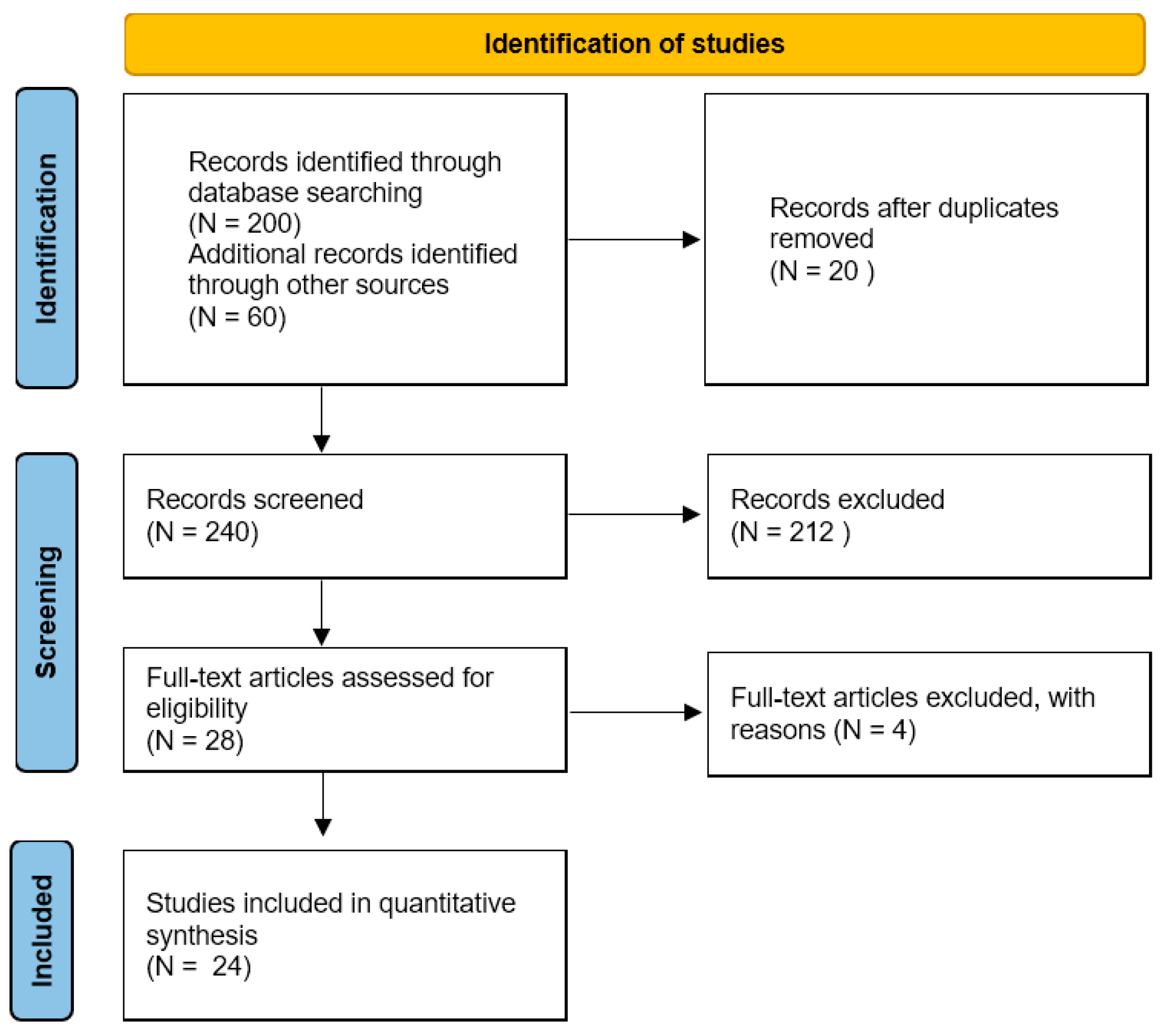
3.1. Study Selection

A total of 260 studies were found when the search results from all databases were combined. Twenty studies were duplicates so they were removed. After screening the title and abstract, a total of 212 investigations were eliminated. After reading the complete text, four studies were eliminated (two because they did not look at the link between genetic variants and ACL ruptures and two because they were not written in full). In the end, 24 publications were included in this systematic review. Figure 1 depicts a flowchart of this procedure.

3.2. Study Characteristics

A summary of the 24 included studies is shown in Table 2. All the included studies were case–control studies. Twelve studies examined genetic variants in or near genes encoding collagens; four examined matrix metalloproteinases; three examined proteoglycans; one study examined a variant near growth–differentiation; four genes examined the angiogenesis-associated signaling cascade; and, finally, elastin and fibrillin, interleukins, and b-fibrinogen were analyzed in one study each.
A total of 7724 patients were analyzed; 3477 patients had ACL ruptures and 4247 patients were controls.
a.
Collagen
The gene most frequently examined was COL1A1. Conflicting findings emerged regarding the association between the TT and GG genotypes of the COL1A1 rs1800012 variant and ACL rupture. Likewise, conflicting evidence was noted regarding the association between the AA genotype of the COL3A1 rs1800255 variant and ACL rupture. Zhao et al. [20] suggest that males with the rs970547 A allele and the rs970547 AA genotype of COL12A1 may be at high risk for developing an ACL rupture. Poor evidence was obtained, suggesting that no association between the COL5A1 rs12722 and COL5A1 rs13946 variants was identified. Furthermore, the evidence was deemed insufficient to establish an association between COL1A1 rs1107946 and COL6A1 rs35796750 variants and ACL rupture.
b.
Proteoglycans
Conflicting findings have emerged regarding the association between the ACAN rs1516797, BGN rs1042103, and BGN rs1126499 variants and ACL rupture. Mannion et al. [11] found that ACAN rs1516797 was independently associated with the risk of developing ACL injury in all analyzed participants. Cięszczyk et al. [10] showed that the ACAN rs1516797 G/T genotype was under-represented in the control group compared to the male ACL injury group. Moreover, they found that the BGN rs1042103 A allele was significantly under-represented in the male control group compared to the male ACL injury group. Willard et al. [22] found that allele combinations across BGN (rs1126499 and rs1042103), COL5A1 (rs12722), and DCN (rs516115), as well as eight miRNA recognition sequences, are involved in increasing susceptibility to ACL rupture.
Evidence indicating an association between the GG (protective) genotype of the DCN rs516115 variant and ACL rupture was inadequate. Similarly, evidence establishing an association between the DCN rs13312816, ACAN rs2351491, ACAN rs1042631, FMOD rs7543148, FMOD rs10800912, and LUM rs2268578 variants and ACL rupture was insufficient.
c.
Matrix Metalloproteinases
Lulinska-Kuklik et al. found that the MMP3 rs591058C and rs679620 G alleles [23] were associated with ACL rupture. Inadequate evidence was found to support the absence of any association between the MMP1 rs1799750, MMP3 rs679620, MMP3-1612, MMP10 rs486055, and MMP12 rs2276109 variants and ACL rupture.
d.
Angiogenesis-Associated Signaling Cascades and Growth Differentiation Hormone Factors
Conflicting findings have emerged regarding the association between VEFGA rs699947 and ACL rupture. Shukla et al. [24] demonstrated that the VEGF A allele (rs699947) and I allele (rs35569394) were significantly over-represented in the ACL injury group. Rahim et al. [16] and Cięszczyk et al. [10] did not find the same association for rs699947.
Evidence indicating an association between the GA genotype (deleterious) of the VEGFA rs1570360 variant and ACL rupture was inadequate. Additionally, evidence indicating the association between the VEFGA rs2010963, KDR 1870377, KDR rs2071559, NGFB rs6678788, HIF1A rs11549465, and GDF5 rs143383 variants and ACL rupture was insufficient.
e.
Elastin and Fibrillin
Evidence indicating no association between the ELN rs2071307 variant and ACL rupture or no association between FBN2 rs331079 variant and ACL rupture was insufficient.
f.
Interleukins
Lulinska-Kuklik et al. found that rs1800795 IL6 gene polymorphism [19] was associated with ACL rupture.
g.
B-fibrinogen
rs1800787, rs1800788, rs1800790, and rs2227389 genotypes in the b-fib promoter region were found to be associated with ACL injury.
Table 2. Characteristics of the included studies.
StudyYearDesignPatients with ACL
Ruptures (n)
Controls
(n)
GeneProductVariantAssociation with ACL Ruptures (Differences in Patients with ACL Ruptures vs. Control)
Ficek et al. [25]2013Case–
control
91143COL1A1Collagenrs1800012 and
rs1107946
No significant differences
Ficek et al. [26]2014Case–
control
91143COL12A1Collagenrs970547No significant differences
Khoschnau et a [27]2008Case–
control
233325COL1A1Collagenrs1800012No significant differences
Khoury et al. [18]2015Case–
control
141219ELNElastinrs2071307No significant differences
-----FBN2Fibrillin-2rs331079No significant differences
Malila et al. [28]2011Case–
control
86100MMP3Matrix metalloproteinase–1612No significant differences
Mannion et al. [11]2014Case–
control
227234ACANProteoglycansrs2351491,
rs1042631, and
rs1516797
rs2351491 and rs1042631:
no significant differences
ACAN rs1516797: was significantly under-represented in the controls group (p = 0.024)
----BGN rs1126499 and
rs1042103
No significant differences
----DCN rs13312816 and
rs516115
rs13312816: no significant differences;
rs516115: significant association
----FMOD rs7543148 and
rs10800912
No significant differences
----LUM rs2268578No significant differences
O’Connell et al. [29]2015Case–
control
242 (South African population)
91 (Polish population)
235 (South African population)
91 (Polish population)
COL3A1 and COL6A1Collagenrs1800255 and rs35796750rs1800255; significant association;
rs35796750: no significant differences
Posthumus et al. [30]2009Case–
control
117130COL1A1Collagenrs1800012rs1800012: significant association
Posthumus et al. [31]2009Case–
control
129216COL5A1Collagenrs12722 and
rs13946
No significant differences
Posthumus et al. [32]2010Case–
control
129216COL12A1Collagenrs970547 and rs240736 No significant differences
Posthumus et al. [14] 2012Case–
control
129216MMP1,
MMP3, MMP10, and
MMP12
Matrix metalloproteinasers1799750, rs679620,
rs486055, and
rs2276109
No significant differences
Rahim et al. [16]2014Case–
control
227227VEGFA, KDR, NGFB, and HIF1A Angiogenesis-associated signaling
cascade genes
rs699947,
rs1570360,
rs2010963,
rs1870377,
rs2071559,
rs6678788, and
rs11549465
No significant differences
Raleigh et al. [17]2013Case–
control
126216GDF5Growth differentiation factorrs143383No significant differences
Stepien-Słodkowska et al. [33]2013Case–
control
138183COL1A1Collagenrs1800012Significant association
Stepien-Słodkowska
et al. [34]
2015Case–
control
138183COL3A1Collagenrs1800255Significant association
Stepien-Słodkowska
et al. [35]
2015Case–
control
138183COL3A1Collagenrs13946 and
rs12722
No significant differences
Cięszczyk et al. [10]2017Case–
control
143229ACANProteoglycansrs1516797Significant association T/T (p > 0.041)
BGN rs1042103 and
rs1126499
BGN rs1042103 A allele: significant association in the male
DCN rs516115No significant differences
VEGFAAngiogenesis-associated signaling
cascade genes
rs699947No significant differences
Lulinska-Kuklik et al. [23]2018Case–
control
229192MMP3Matrix metalloproteinasesrs591058C/T and rs679620 G/ASignificant association: MMP3 rs591058C and
rs679620 G alleles were significantly over-represented in cases compared to controls
(p = 0.021)
MMP8Matrix metalloproteinasesrs11225395C/TNo significant differences
TIMP2Tissue inhibitors of metalloproteasesrs4789932 G/ANo significant differences
Lulinska et al. [13]2020Case–
control
228202MMP1Matrix metalloproteinasesrs1799750,
→G
No significant differences
MMP10Matrix metalloproteinasesrs486055 C > TNo significant differences
MMP12Matrix metalloproteinasesrs2276109 T > CNo significant differences
Lulinska-Kuklik et al. [19]2019Case–
control
229194IL1BInterleukinsrs16944 and rs1143627No significant differences
IL6 Interleukinsrs1800795Significant association
IL6R Interleukinsrs2228145No significant differences
Shukla et al. [24]2020Case–
control
9076VEGFAAngiogenesis-associated signaling
cascade genes
rs699947 and rs35569394Significant association: the A allele (rs699947) and I allele (rs35569394) were significantly over-represented in the ACL group
Shukla et al. [36]2020Case–
control
9076COLIA1Angiogenesis-associated signaling
cascade genes
COLIA1 Sp1 + 1245 G > TNo significant differences
Willard et al. [22]2017Case–
control
227234BGNProteoglycansrs1126499 and
rs1042103
Significant association
COL5A1Collagenrs12722Significant association
DCNProteoglycansrs516115Significant association
Zhao et al. [20]2020Case–
control
101110COL1A1Collagenrs1800012No significant differences
COL5A1Collagenrs12722 and
rs13946
No significant differences
COL12A1Collagenrs970547 and rs240736Significant association
FGBB-fibrinogenrs1800787, rs1800788, rs1800789, rs1800790, rs1800791,
and rs2227389
rs1800789 and rs1800791: no significant differences;
rs1800787, rs1800788, rs1800790, and rs2227389: significant association;
rs1800787, rs1800788, rs1800790, and rs2227389 genotypes in
the b-fib promoter region: association with ACL injuries

3.3. Risk of Bias

Risk of bias is shown in Table 3. In the risk-of-bias assessment, nine studies were determined to have a high risk of bias. Additionally, 15 studies were categorized as unclear in terms of bias risk.

4. Discussion

ACL ruptures are very common in athletes and their instability could lead to an increased risk of future knee injuries, thus making a rehabilitative prevention plan necessary [37,38,39,40].
In this systematic review, we investigated the genetic factors that could predispose athletes to ACL ruptures through simple familial predisposition studies and complex gene association studies. Brothers and sisters (siblings) of patients with a history of ACL ruptures seem to have susceptibility to developing an anterior cruciate ligament rupture. Various authors, by analyzing patients with ACL ruptures who underwent ACL reconstructive surgery from 2010 to 2013, found that having a sibling with a history of ACL ruptures and being female are important risk factors for developing ACL ruptures in the contralateral knee [40]. Flynn et al. [5] reported that having a familial history of ACL ruptures in first-, second-, and third-degree relatives can double the risk of developing ACL ruptures compared to patients without a relative with a history of ACL ruptures. Those data reflect the genetic background of ACL rupture. However, nowadays, the relationship between genetic factors and ACL ruptures or other musculoskeletal injuries is not clearly understood.
The existing literature on genetic variations associated with ACL rupture is summarized in this comprehensive review that includes 24 studies. In the 24 articles that we analyzed, conflicting evidence on the effect of different gene variants on ACL rupture was found.
The most important genes that we analyzed are as follows: COL3A1 [29,34], COL1A1 [30,33], COL12A1 [20], ACAN [10,11], DCN [11,22], MMP3 [23], IL6 [19], VEGFA [24], BGN [22], and FGB [20].
The analyzed studies exhibited either an unclear or high risk of bias and confounding factors. In the risk-of-bias assessment, nine studies were determined to have a high risk of bias. Additionally, 15 studies were categorized as unclear in terms of bias risk. This underscores the importance of evaluating the quality of research and the implications for drawing conclusive findings.
Moreover, we observed heterogeneity in the genetic variants under investigation, outcome definitions, and genetic contrasts, precluding the feasibility of conducting a formal meta-analysis. Consequently, we conducted a best-evidence synthesis of the current literature.
Variants of the analyzed genes were found to play a role in the synthesis, strength, and homeostasis of the ligament. COL1A1 encodes collagen type I, which imparts mechanical strength to various tissues, including ligaments. COL3A1 encodes collagen type III and participates in the fibrillogenesis of collagen type I. The type I collagen molecule is a heterotrimer made up of two a1 chains and one a2 chains, and it is the most abundant protein in ligaments, accounting for 70–80% of their dry weight. The collagen Iα1 (COLIA1) and the collagen Iα2 (COLIA2) genes are the two primary genes that control collagen synthesis.
A polymorphism in the promoter region of COLIA1 intron 1, which is a putative binding site for the transcription factor Sp1, has been linked to several illnesses [41]. The substitution of thymine (T) for guanine (G) in COLIA1 intron 1 is known as “s”, whereas “SS” indicates homozygosity for GG.
Conflicting findings have emerged regarding the association between the TT and GG genotypes of the COL1A1 rs1800012 variant and ACL rupture. Likewise, conflicting evidence has been noted regarding the association between the AA genotype of the COL3A1 rs1800255 variant and ACL rupture. COL5A1 encodes collagen type V, which interacts with collagen type I in the formation of heterotypic fibrils and modulates the diameter of these fibrils. Collagen type XII, encoded by the COL12A1 gene, constitutes the largest member among fibril-associated collagens, regulating the organization and mechanical properties of collagen fibril bundles. Decorin, encoded by the DCN gene, is a member of the small proteoglycan family and plays a role in restricting the diameter of collagen fibrils during fibrillogenesis.
MMPs have a role in the breakdown of the extracellular matrix in both normal physiological processes (like embryonic development, reproduction, and tissue remodeling), as well as pathological events (like arthritis and metastasis).
Interleukins (ILs) play essential roles in the activation and differentiation of immune cells, as well as in proliferation, maturation, migration, and adhesion. They also have pro-inflammatory and anti-inflammatory properties.
The protein encoded by the fibrinogen beta chain (FGB) gene is the beta component of fibrinogen, a blood-borne glycoprotein comprising three pairs of nonidentical polypeptide chains. It is involved in hemostasis and antimicrobial host defense.
VEGFA, which encodes the vascular endothelial growth factor A, serves as a regulator of angiogenesis.
The precise implications of the above-mentioned genetic variants remain uncertain.
Our included studies presented several limitations.
We did not impose a minimum sample size threshold to ensure the inclusion of all available studies, given that genetic investigations typically necessitate larger participant cohorts than those featured in most of our included studies. However, the sample sizes became notably small when stratifying groups based on sex or the mechanism of injury, thereby precluding the reporting of any stratified analyses.
Moreover, several genes and variants were examined in the same population group. While both of these genetic variants may have an association with the risk of developing an ACL rupture, it is conceivable that only one of them truly contributes to the risk, while the association of the other genetic variant is potentially confounding.
The idea of using genetic tests for screening athletes predisposed to sports-related injuries is an appealing one. Genetic testing could help to avoid injury, especially in particular sports. Genetic information could also be used by clinicians and athletes to make more informed decisions regarding LCA injury prevention, diagnosis, and management.
Nevertheless, additional data are essential in order to establish a conclusive association between genetic variants and ACL rupture. Augmented sample sizes and more comprehensive genetic investigations are imperative in order to attain a thorough understanding of these potential associations.

5. Conclusions

Taken together, the findings of this systematic review showed that the association between the COL3A1, COL1A1, COL12A1, ACAN, DCN, MMP3, IL6, VEGFA, BGN, and FGB genes and ACL injuries should be considered inconclusive. More evidence is needed to draw significant conclusions regarding the association between genetic variants and ACL rupture.

Author Contributions

Conceptualization, V.C. and U.G.L.; methodology, A.B. and A.d.S.; software, G.S.; validation, F.F. and V.D.; formal analysis, V.C. and A.B.; investigation, G.S.; resources, F.F.; data curation, G.S.; writing—original draft preparation, V.C., A.B., G.S. and A.d.S.; writing—review and editing, U.G.L., F.F. and V.D.; visualization, U.G.L.; supervision, V.D.; project administration, V.C. All authors have read and agreed to the published version of the manuscript.

Funding

This research received no external funding.

Institutional Review Board Statement

Not applicable.

Informed Consent Statement

Not applicable.

Data Availability Statement

The datasets used and/or analyzed during the current study are available from the corresponding author on reasonable request.

Acknowledgments

SIOT (Società Italiana Ortopedia e Traumatologia).

Conflicts of Interest

The authors declare no conflicts of interest.

References

  1. Giuliani, J.R.; Kilcoyne, K.G.; Rue, J.P. Anterior cruciate ligament anatomy: A review of the anteromedial and posterolateral bundles. J. Knee Surg. 2009, 22, 148–154. [Google Scholar] [CrossRef] [PubMed]
  2. Adhitya, I.P.G.S.; Kurniawati, I.; Sawa, R.; Wijaya, T.F.; Dewi, N.P.A.C. The Risk Factors and Preventive Strategies of Poor Knee Functions and Osteoarthritis after Anterior Cruciate Ligament Reconstruction: A Narrative Review. Phys. Ther. Res. 2023, 26, 78–88. [Google Scholar] [CrossRef] [PubMed]
  3. Longo, U.G.; Nagai, K.; Salvatore, G.; Cella, E.; Candela, V.; Cappelli, F.; Ciccozzi, M.; Denaro, V. Epidemiology of Anterior Cruciate Ligament Reconstruction Surgery in Italy: A 15-Year Nationwide Registry Study. J. Clin. Med. 2021, 10, 223. [Google Scholar] [CrossRef] [PubMed]
  4. Longo, U.G.; Buchmann, S.; Franceschetti, E.; Maffulli, N.; Denaro, V. A systematic review of single-bundle versus double-bundle anterior cruciate ligament reconstruction. Br. Med. Bull. 2012, 103, 147–168. [Google Scholar] [CrossRef] [PubMed]
  5. Flynn, R.K.; Pedersen, C.L.; Birmingham, T.B.; Kirkley, A.; Jackowski, D.; Fowler, P.J. The familial predisposition toward tearing the anterior cruciate ligament: A case control study. Am. J. Sports Med. 2005, 33, 23–28. [Google Scholar] [CrossRef] [PubMed]
  6. Longo, U.G.; King, J.B.; Denaro, V.; Maffulli, N. Double-bundle arthroscopic reconstruction of the anterior cruciate ligament: Does the evidence add up? J. Bone Jt. Surg. 2008, 90, 995–999. [Google Scholar] [CrossRef] [PubMed]
  7. Longo, U.G.; Salvatore, G.; Ruzzini, L.; Risi Ambrogioni, L.; de Girolamo, L.; Vigano, M.; Facchini, F.; Cella, E.; Candela, V.; Ciccozzi, M.; et al. Trends of anterior cruciate ligament reconstruction in children and young adolescents in Italy show a constant increase in the last 15 years. Knee Surg. Sports Traumatol. Arthrosc. Off. J. ESSKA 2021, 29, 1728–1733. [Google Scholar] [CrossRef] [PubMed]
  8. September, A.V.; Schwellnus, M.P.; Collins, M. Tendon and ligament injuries: The genetic component. Br. J. Sports Med. 2007, 41, 241–246; discussion 246. [Google Scholar] [CrossRef]
  9. Kaynak, M.; Nijman, F.; van Meurs, J.; Reijman, M.; Meuffels, D.E. Genetic Variants and Anterior Cruciate Ligament Rupture: A Systematic Review. Sports Med. 2017, 47, 1637–1650. [Google Scholar] [CrossRef]
  10. Cieszczyk, P.; Willard, K.; Gronek, P.; Zmijewski, P.; Trybek, G.; Gronek, J.; Weber-Rajek, M.; Stastny, P.; Petr, M.; Lulinska-Kuklik, E.; et al. Are genes encoding proteoglycans really associated with the risk of anterior cruciate ligament rupture? Biol. Sport 2017, 34, 97–103. [Google Scholar] [CrossRef]
  11. Mannion, S.; Mtintsilana, A.; Posthumus, M.; van der Merwe, W.; Hobbs, H.; Collins, M.; September, A.V. Genes encoding proteoglycans are associated with the risk of anterior cruciate ligament ruptures. Br. J. Sports Med. 2014, 48, 1640–1646. [Google Scholar] [CrossRef] [PubMed]
  12. Kumar, L.; Bisen, M.; Khan, A.; Kumar, P.; Patel, S.K.S. Role of Matrix Metalloproteinases in Musculoskeletal Diseases. Biomedicines 2022, 10, 2477. [Google Scholar] [CrossRef]
  13. Lulinska, E.; Gibbon, A.; Kaczmarczyk, M.; Maciejewska-Skrendo, A.; Ficek, K.; Leonska-Duniec, A.; Wilk, M.; Leznicka, K.; Michalowska-Sawczyn, M.; Huminska-Lisowska, K.; et al. Matrix Metalloproteinase Genes (MMP1, MMP10, MMP12) on Chromosome 11q22 and the Risk of Non-Contact Anterior Cruciate Ligament Ruptures. Genes. 2020, 11, 766. [Google Scholar] [CrossRef] [PubMed]
  14. Posthumus, M.; Collins, M.; van der Merwe, L.; O’Cuinneagain, D.; van der Merwe, W.; Ribbans, W.J.; Schwellnus, M.P.; Raleigh, S.M. Matrix metalloproteinase genes on chromosome 11q22 and the risk of anterior cruciate ligament (ACL) rupture. Scand. J. Med. Sci. Sports 2012, 22, 523–533. [Google Scholar] [CrossRef] [PubMed]
  15. Halper, J. Advances in the use of growth factors for treatment of disorders of soft tissues. Adv. Exp. Med. Biol. 2014, 802, 59–76. [Google Scholar] [CrossRef]
  16. Rahim, M.; Gibbon, A.; Hobbs, H.; van der Merwe, W.; Posthumus, M.; Collins, M.; September, A.V. The association of genes involved in the angiogenesis-associated signaling pathway with risk of anterior cruciate ligament rupture. J. Orthop. Res. Off. Publ. Orthop. Res. Soc. 2014, 32, 1612–1618. [Google Scholar] [CrossRef]
  17. Raleigh, S.M.; Posthumus, M.; O’Cuinneagain, D.; van der Merwe, W.; Collins, M. The GDF5 gene and anterior cruciate ligament rupture. Int. J. Sports Med. 2013, 34, 364–367. [Google Scholar] [CrossRef]
  18. Khoury, L.E.; Posthumus, M.; Collins, M.; van der Merwe, W.; Handley, C.; Cook, J.; Raleigh, S.M. ELN and FBN2 gene variants as risk factors for two sports-related musculoskeletal injuries. Int. J. Sports Med. 2015, 36, 333–337. [Google Scholar] [CrossRef] [PubMed]
  19. Lulinska-Kuklik, E.; Maculewicz, E.; Moska, W.; Ficek, K.; Kaczmarczyk, M.; Michalowska-Sawczyn, M.; Huminska-Lisowska, K.; Buryta, M.; Chycki, J.; Cieszczyk, P.; et al. Are IL1B, IL6 and IL6R Gene Variants Associated with Anterior Cruciate Ligament Rupture Susceptibility? J. Sports Sci. Med. 2019, 18, 137–145. [Google Scholar]
  20. Zhao, D.; Zhang, Q.; Lu, Q.; Hong, C.; Luo, T.; Duan, Q.; Shu, S.; Lv, J.; Zhao, W. Correlations Between the Genetic Variations in the COL1A1, COL5A1, COL12A1, and beta-fibrinogen Genes and Anterior Cruciate Ligament Injury in Chinese Patients(a). J. Athl. Train. 2020, 55, 515–521. [Google Scholar] [CrossRef]
  21. Altman, D.G.; Schulz, K.F.; Moher, D.; Egger, M.; Davidoff, F.; Elbourne, D.; Gøtzsche, P.C.; Lang, T.; Trials, C.G.C.S.o.R. The revised CONSORT statement for reporting randomized trials: Explanation and elaboration. Ann. Intern. Med. 2001, 134, 663–694. [Google Scholar] [CrossRef] [PubMed]
  22. Willard, K.; Mannion, S.; Saunders, C.J.; Collins, M.; September, A.V. The interaction of polymorphisms in extracellular matrix genes and underlying miRNA motifs that modulate susceptibility to anterior cruciate ligament rupture. J. Sci. Med. Sport 2018, 21, 22–28. [Google Scholar] [CrossRef] [PubMed]
  23. Lulinska-Kuklik, E.; Rahim, M.; Moska, W.; Maculewicz, E.; Kaczmarczyk, M.; Maciejewska-Skrendo, A.; Ficek, K.; Cieszczyk, P.; September, A.V.; Sawczuk, M. Are MMP3, MMP8 and TIMP2 gene variants associated with anterior cruciate ligament rupture susceptibility? J. Sci. Med. Sport 2019, 22, 753–757. [Google Scholar] [CrossRef] [PubMed]
  24. Shukla, M.; Gupta, R.; Pandey, V.; Rochette, J.; Dhandapany, P.S.; Tiwari, P.K.; Amrathlal, R.S. VEGFA Promoter Polymorphisms rs699947 and rs35569394 Are Associated With the Risk of Anterior Cruciate Ligament Ruptures Among Indian Athletes: A Cross-sectional Study. Orthop. J. Sports Med. 2020, 8, 2325967120964472. [Google Scholar] [CrossRef] [PubMed]
  25. Ficek, K.; Cieszczyk, P.; Kaczmarczyk, M.; Maciejewska-Karlowska, A.; Sawczuk, M.; Cholewinski, J.; Leonska-Duniec, A.; Stepien-Slodkowska, M.; Zarebska, A.; Stepto, N.K.; et al. Gene variants within the COL1A1 gene are associated with reduced anterior cruciate ligament injury in professional soccer players. J. Sci. Med. Sport 2013, 16, 396–400. [Google Scholar] [CrossRef] [PubMed]
  26. Ficek, K.; Stepien-Slodkowska, M.; Kaczmarczyk, M.; Maciejewska-Karlowska, A.; Sawczuk, M.; Cholewinski, J.; Leonska-Duniec, A.; Zarebska, A.; Cieszczyk, P.; Zmijewski, P. Does the A9285G Polymorphism in Collagen Type XII alpha1 Gene Associate with the Risk of Anterior Cruciate Ligament Ruptures? Balk. J. Med. Genet. BJMG 2014, 17, 41–46. [Google Scholar] [CrossRef]
  27. Khoschnau, S.; Melhus, H.; Jacobson, A.; Rahme, H.; Bengtsson, H.; Ribom, E.; Grundberg, E.; Mallmin, H.; Michaelsson, K. Type I collagen alpha1 Sp1 polymorphism and the risk of cruciate ligament ruptures or shoulder dislocations. Am. J. Sports Med. 2008, 36, 2432–2436. [Google Scholar] [CrossRef] [PubMed]
  28. Malila, S.; Yuktanandana, P.; Saowaprut, S.; Jiamjarasrangsi, W.; Honsawek, S. Association between matrix metalloproteinase-3 polymorphism and anterior cruciate ligament ruptures. Genet. Mol. Res. GMR 2011, 10, 4158–4165. [Google Scholar] [CrossRef]
  29. O’Connell, K.; Knight, H.; Ficek, K.; Leonska-Duniec, A.; Maciejewska-Karlowska, A.; Sawczuk, M.; Stepien-Slodkowska, M.; O’Cuinneagain, D.; van der Merwe, W.; Posthumus, M.; et al. Interactions between collagen gene variants and risk of anterior cruciate ligament rupture. Eur. J. Sport Sci. 2015, 15, 341–350. [Google Scholar] [CrossRef]
  30. Posthumus, M.; September, A.V.; Keegan, M.; O’Cuinneagain, D.; Van der Merwe, W.; Schwellnus, M.P.; Collins, M. Genetic risk factors for anterior cruciate ligament ruptures: COL1A1 gene variant. Br. J. Sports Med. 2009, 43, 352–356. [Google Scholar] [CrossRef]
  31. Posthumus, M.; September, A.V.; O’Cuinneagain, D.; van der Merwe, W.; Schwellnus, M.P.; Collins, M. The COL5A1 gene is associated with increased risk of anterior cruciate ligament ruptures in female participants. Am. J. Sports Med. 2009, 37, 2234–2240. [Google Scholar] [CrossRef] [PubMed]
  32. Posthumus, M.; September, A.V.; O’Cuinneagain, D.; van der Merwe, W.; Schwellnus, M.P.; Collins, M. The association between the COL12A1 gene and anterior cruciate ligament ruptures. Br. J. Sports Med. 2010, 44, 1160–1165. [Google Scholar] [CrossRef] [PubMed]
  33. Stepien-Slodkowska, M.; Ficek, K.; Eider, J.; Leonska-Duniec, A.; Maciejewska-Karlowska, A.; Sawczuk, M.; Zarebska, A.; Jastrzebski, Z.; Grenda, A.; Kotarska, K.; et al. The +1245g/t polymorphisms in the collagen type I alpha 1 (col1a1) gene in polish skiers with anterior cruciate ligament injury. Biol. Sport 2013, 30, 57–60. [Google Scholar] [CrossRef] [PubMed]
  34. Stepien-Slodkowska, M.; Ficek, K.; Maciejewska-Karlowska, A.; Sawczuk, M.; Zietek, P.; Krol, P.; Zmijewski, P.; Pokrywka, A.; Cieszczyk, P. Overrepresentation of the COL3A1 AA genotype in Polish skiers with anterior cruciate ligament injury. Biol. Sport 2015, 32, 143–147. [Google Scholar] [CrossRef]
  35. Stepien-Slodkowska, M.; Ficek, K.; Kaczmarczyk, M.; Maciejewska-Karlowska, A.; Sawczuk, M.; Leonska-Duniec, A.; Stepinski, M.; Zietek, P.; Krol, P.; Chudecka, M.; et al. The Variants Within the COL5A1 Gene are Associated with Reduced Risk of Anterior Cruciate Ligament Injury in Skiers. J. Hum. Kinet. 2015, 45, 103–111. [Google Scholar] [CrossRef] [PubMed]
  36. Shukla, M.; Gupta, R.; Pandey, V.; Tiwari, P.K.; Amrathlal, R.S. COLIA1 + 1245 G > T Sp1 Binding Site Polymorphism is Not Associated with ACL Injury Risks Among Indian Athletes. Indian J. Orthop. 2020, 54, 647–654. [Google Scholar] [CrossRef] [PubMed]
  37. de Sire, A.; Demeco, A.; Marotta, N.; Moggio, L.; Palumbo, A.; Iona, T.; Ammendolia, A. Anterior Cruciate Ligament Injury Prevention Exercises: Could a Neuromuscular Warm-Up Improve Muscle Pre-Activation before a Soccer Game? A Proof-of-Principle Study on Professional Football Players. Appl. Sci. 2021, 11, 4958. [Google Scholar] [CrossRef]
  38. de Sire, A.; Marotta, N.; Demeco, A.; Moggio, L.; Paola, P.; Marotta, M.; Iona, T.; Invernizzi, M.; Leigheb, M.; Ammendolia, A. Electromyographic Assessment of Anterior Cruciate Ligament Injury Risk in Male Tennis Players: Which Role for Visual Input? A Proof-of-Concept Study. Diagnostics 2021, 11, 997. [Google Scholar] [CrossRef]
  39. Marotta, N.; de Sire, A.; Gimigliano, A.; Demeco, A.; Moggio, L.; Vescio, A.; Iona, T.; Ammendolia, A. Impact of COVID-19 lockdown on the epidemiology of soccer muscle injuries in Italian Serie A professional football players. J. Sports Med. Phys. Fit. 2022, 62, 356–360. [Google Scholar] [CrossRef]
  40. Demeco, A.; de Sire, A.; Marotta, N.; Spanò, R.; Lippi, L.; Palumbo, A.; Iona, T.; Gramigna, V.; Palermi, S.; Leigheb, M.; et al. Match Analysis, Physical Training, Risk of Injury and Rehabilitation in Padel: Overview of the Literature. Int. J. Environ. Res. Public. Health 2022, 19, 4153. [Google Scholar] [CrossRef]
  41. Bernad, M.; Martinez, M.E.; Escalona, M.; Gonzalez, M.L.; Gonzalez, C.; Garces, M.V.; Del Campo, M.T.; Martin Mola, E.; Madero, R.; Carreno, L. Polymorphism in the type I collagen (COLIA1) gene and risk of fractures in postmenopausal women. Bone 2002, 30, 223–228. [Google Scholar] [CrossRef] [PubMed]
Figure 1. PRISMA flowchart.
Jcm 13 02330 g001
Table 1. Risk of bias questions.
CriterionQuestion
CaseHave the cases been clearly and sufficiently defined?
ControlHave the controls been clearly and adequately defined?
Selection biasHas selection bias been adequately addressed and excluded?
Defined exposureHas the exposure been clearly defined, and is the method employed to assess this exposure deemed appropriate?
DeterminationWas blinding to exposure status maintained prior to determining the presence of the disease?
ConfoundingHave the primary confounding factors been identified and adequately accounted for in both the study design and analysis?
Table 3. Risk of bias.
CaseControlSelection BiasDefined EposureDetermination
Exposure
ConfoudingOverall
Ficek et al. [25]++?+?+?
Ficek et al. [26]++?+?+?
Khoschnau et al. [27]+?+++--
Khoury et al. [18]++?+?--
Malila et al. [28]++?+?--
Mannion et al. [11]++?+?--
O’Connell et al. [29]+??++--
Posthumus et al. [30]++?+?--
Posthumus et al. [31]++?+?+?
Posthumus et al. [32]++?+?+?
Posthumus et al. [14]++?+?+?
Rahim et al. [16]+??+?--
Raleigh et al. [17]++?+?+?
Stepien-Słodkowska et al. [33]++?+???
Stepien-Słodkowska
et al. [34]
++?+???
Stepien-Słodkowska
et al. [35]
++?+???
Cięszczyk et al. [10]++?+???
Lulinska-Kuklik et al. [23]++?+?+?
Lulinska et al. [13]++?+?+?
Lulinska-Kuklik et al. [19]++?+?+?
Shukla et al. [24]++?+?--
Shukla et al. [36]++?+???
Willard et al. [22]++?+?+?
Zhao et al. [20]++?+?--
Disclaimer/Publisher’s Note: The statements, opinions and data contained in all publications are solely those of the individual author(s) and contributor(s) and not of MDPI and/or the editor(s). MDPI and/or the editor(s) disclaim responsibility for any injury to people or property resulting from any ideas, methods, instructions or products referred to in the content.

